Organisation and expression of plant B chromosomes
261
1^q- Organisation and Expression of Plant B Chromosomes Tamzin Donald, B.Sc.(Hons) A thesis submitted for the degree of Doctor of Philosophy by 1n The University of Adelaide Department of Genetics (Faculty of Science) 16th June, 1999
-
Upload
khangminh22 -
Category
Documents
-
view
2 -
download
0
Transcript of Organisation and expression of plant B chromosomes

1^q-
Organisation and
Expression of Plant B
Chromosomes
Tamzin Donald, B.Sc.(Hons)
A thesis submitted for the degree of
Doctor of Philosophy
by
1n
The University of Adelaide
Department of Genetics
(Faculty of Science)
16th June, 1999

TABLE OF'CONTENTS
ABSTRACT
DECLARATION
ACKNOWLEDGEMENTS
LIST OF ABBREVIATIONS
x
xtu
xiv
xvi
CHAPTER 1: Introduction
1.1 InrRonucrloN
7.2 B Cs OSOIVIES
1.3 Eppecrs
1.3.1 Effects on A chromosome behaviour
1.3.1a Chiasma frequency
I .3.I b Pairing in hybrids
1.3.2 Physiological effects
1.4 B CHnOmOSOME MIINTEN¡NCB
7.4.1 Pre-meiotic drive
7.4.2 Meiotic drive
7.4.3 Post-meioticdrive
1.4.4 NasonÌavítripennis
1.4.5 B chromosomes offering an adaptive advantage
t
2
3
3
4
5
6
7
7
8
9
10
11
1.5 Funrue R LATToN SruolBs
ll
t2

1.6 SsoueNces oN rue B cHROI\{OSO]\,IES
1.6.1 B chromosome specific sequences
l.6.Ia Nasonia vitripennis
1.6.1b Braclryconte dicltrontosontatica
I .6.1c Zea mays
1.6.1d Organisation of repeat sequences on B clrromosone
1.6.2 Ribosomal RNA genes
1.6.2a Structure of rRNA genes
1.6.2b Ribosomal RNA genes o¡r B chrotnosomes
1.6.3 Sequences involved in B chromosome Transmission
1.7 ORrcrN on CHROÌ\'IOSOMES
13
t4
t4
15
T1
l8
18
19
20
2t
1.7.1
1.7.2
B chromosomes originating from the same species
B chromosomes originating from a different species
22
22
24
26
30
1.8 Bn¿ CH''COAIE D TICA
1.9 SunrunRY rND Arvrs op Pno.lncr
CHAPTER 2: Materials and Methods
2.1 N,Inranrals
2.1.1 B. dichrontosomatícø plant material, DNA samples and
genomic library
2.7.2 Chemical reagents
2.7.3 Stains
2.7.4 BnzS,mes
2.1.5 RadioactiveisotoPes
2.7.6 Nucleic acids
33
JJ
JJ
34
34
35
35
ltt

2.t.7
2.1.8
2.1.9
2.7.70
Antibiotics and indicators
Cloning vectors
Bacterial strains
Kits and miscellaneous materials
Solutions and Buffers
Media
35
35
36
40
2.2 SolurroNs. B I]FFERS AND MEDIA 41
4t
42
2.2.1
2.2.2
2.3 MorHons
2.3.1 Plant grorvth conditions
2.3.2 General techniques
2.3.3 General techniques
2.3.3a Restriction enzyme digests
2.3.3b Electrophoretic separation of DNA
2.3.3c Recovery of DNA from agarose gels
2.3.3d Precipitation of DNA
Method l: Precipitatiott blt elltunol
Method 2: Precipitation by isopropartol
2.3.2e Purification of DNA using phenol/chlorofom extraction
2.3.2f Detemrination of DNA and RNA concentration
2.3.4 Preparation of DNA samples
2.3.4a Isolation of genomic DNA fi'om plants
2.3.4b Small-scale plasruid preparation
Method I: Alkaline lysis plasnticl ntittipreps
Method 2 Boiling lysis
2.3.5 Cloning strategies
2.3.5a Preparation of insert DNA and plasnrid vector
2.3.5b Cloning of products generated by PCR
2.3.5c Ligations
43
43
43
43
43
44
44
45
45
45
45
45
46
46
46
46
46
46
46
47
41
lv

2.3.5d Preparation and transformation of competenl E. coli
2.3.6 Preparation of RNA samples
2.3.7 Phage manipulation
2.3.1a Preparation of plating cells for phage
2.3.7b Plating and picking phage
2.3.8 Transfer of DNA and RNA to membranes
2.3.8a Southern blotting
2.3.8b Colony hybridisation
2.3.9 Radiolabelling of DNA probes
2.3.9a Oligolabelling using random oligonucleotides
2.3.9b Oligolabelling using PCR
2.3.9c RNA labelling
2.3.9d Removal of unincotporated nucleotides
2.3.10 Prehybridisation, hybridisation and rvashing
2.3.10a Prehybridiation conditions
2.3. 1 0b Probe denaturation and liybridisation
2.3.10c Washing of filters
2.3.10d Autoradiography, phosphorinlagery and stripping of filters
2.3.11 Design and Synthesis of DNA Oligonucleotides
2.3.11a Primers for sequencing
2.3.11b Prinrers for PCR
2.3.12 Sequencing of double-stranded DNA templates
2.3.IZa Preparation of template DNA fot'manual sequencing
2.3.Izb Preparation of ternplate DNA for automated sequencing
2.3.12c Manual sequencing reactions
2.3.l2dAutonated sequencing reactions
2.3.12e Preparation of sequencing gels
2.3.lzf Denaturing gel electrophoresis
2.3.I2g Sequence analysis software
2.3.13 Polymerase chain reaction (PCR)
47
48
48
48
48
49
49
49
49
49
50
50
50
51
51
51
51
51
52
52
52
53
53
53
54
54
54
54
55
55

2.3.14 Reverse transcription (RT)
2.3.15 Amplified fragment length polymorphisms (AFLP)
2.3.15a Preparation of AFLP DNA samples
2.3.15b Preparation of adaptors for AFLP
2.3.I5c Ligation of adaptors to DNA
2.3 .I 5 d AFLP prearnplifi cation
2.3.I 5 e AFLP arnplifi cation
2.3.I5f Rearnplification of AFLP fragments
2.3.16 Random amplification of polymorphic DNA
2.3.17 In sítu h1'bridisation
2.3.11a Prepalation of probes
2.3.17b Preparation of slides
2.3.17c Fixing of slides
2.3.17 d RNase treatment
2.3.17 e Denaturatiott
2.3.11f Hybridisation
2.3.17 g Sigrral detection
55
5o
56
56
56
57
57
58
58
59
59
59
59
60
60
60
60
CHAPTER 3: Sequence Analysis of Ribosomal DNA Internal
Transcribed Spacet's I and,2
3.1
3.2
Introduction
Analysis of the ITS1 and ITS2 on the A and B cht'omosomes
3.2.I ITS sequences amplified frorn the A chrotnosomes
3.2.2 ITS sequences arnplified frorn the B chromosomes
Analysis of the ITS2 in 0B and *B genomic DNA
Analysis of other species of Brøclt¡tcome
Conclusion
63
67
67
72
75
87
88
3.3
3.4
3.5
vl

CHAPTER 4: Transcriptional Analysis of Ribosomal RNA Internal
Transcribed Spacer 2
4.1
4.2
Introduction
Transcriptional analysis of the ITS2 on the B chromosomes
in leaf tissue
4.2.1 Detection of low levels of B chromosoûle rRNA transcription
Transcriptional analysis of the ITS2 orl the B chromosomes
in floral tissue
Summary and discussion
4.4.1 Intergenic spacer variation in the regulation of
IRNA gene expression
4.4.2 DNA rrretliylation in the regulatior.r of rRNA gene expression
4.4.3 Chromatin modification in the regulation of gene expressiou
95
95
98
4.3
4.4
103
103
106
r01
109
CHAPTER 5: Cloning of the Ribosomal RNA Repeat Unit
5.1
5.2
5.3
172
113
121
Introduction
Cloning of the B. díchrontosomatíca ribosomal RNA repeat unit
Attal¡,5i5 of the B. dichrontosomalica ribosomal DNA clones
5.3.1 Hybridisation of BdrD, BdrE and BdrC to 0B gerror.t.tic
DNA samples
5.3.2 Comparison of BdrD hybridisation to 0B and *B genomic
DNA samples
Mapping BdrD and BdrE
Sequence anall'sis of BdrD
Sequence analysis of BdrE
Sequence analy'sis of BdrC
Attempts to clone the remainder of the ribosomal DNA repeat unit
PCR analysis of the ITSl and 5.8S regions on the B chromosomes
5.4
5.5
5.6
5.7
5.8
5.9
121
t24
724
132
139
140
t40
t41
vll

5.10 Analysis of the intergenic spacer on the A chromosomes
5.10.1 Sequence analysis of the complete A cluomosome IGS
within BdrD
5.11 Analysis of the IGS region in 0B and +B genomic DNA samples
5.1 1 .I Analysis of the IGS in 0B and +B DNA restricted with þe I
5.11.2 Analysis of the IGS in 0B and +B DNA restricted withXbal
5.12 PCR analysis of IGS on A and B chromosomes
5.12.1 Attempted identification of the B chromosorne IGS polymorphism
5.12.2 Furlher PCR analysis of the B chromosorne IGS
5.13 ltt sittt hybridisation analysis of the IGS on the A and
B chromosomes
5.14 Conclusion
t47
t47
153
153
156
159
159
165
165
769
CHAPTBR 6: Isolation of Non-Repetitive Sequences from the
B chromosomes
6.1
6.2
Introduction
Arnplified fragment length polymorphisms
6.2.1 PCR analysis of putative B chrornosonte AFLP products
6.2.2 Screening of 38 genomic library rvith 2aflp3
6.2.3 Sumrnary of AFLP analysis
Random amplified polymorphic DNA sequences
6.3.1 A.nalysis of putative B chromosome specific RAPDs
Conclusion
771
172
r86
189
189
190
r93
206
6.3
6.4
CHAPTER 7: Concluding Discussion
Organisation and expression of plant B chromosomes
7 .l J Ribosomal DNA sequences on the B chromosomes of
B. diclt rontosontalicct
2097.1
vul
209

7.2
21r
2tr
272
7 .l.2 Isolation of non-repetitive sequences fi'on the B chromosomes
7 .1.3 Concluding discussion
Final comment
APPENDICES
RBFERENCES
2L4
2t7
tx

ABSTRACT
Supemumerary B chromosomes are found in all major taxonontic groups of
organisms. Although the B chromosornes significantly increase the nuclear DNA amoutlt,
¡o active genes liave yet been unequivocally localised on these chromosontes by genetic or
molecular lllearls. However, the presence of B chrotnosotnes is often associated with
cha¡ges in chrornosome behaviour, such as meiotic pairing and recombiuation, and it is
therefore thought that they play a significant biological role. The mechanisms which are
responsible for tliese effects remain to be detemined.
BrctclDtcotne dichrontosomatica is an Australiau native ephemeral plant of the arid
regio¡s of South Eastem Australia. This species contail-ts only two pairs of chrornosonres iu
tlie non-¡al contplemeut (A chrornosomes) and 0 - 4 B chrolrosorres in some populatiorls.
Sequences that hybridise to ribosomal RNA gene probes are present llear the end of the
s|oú anl and, to a lesser extent, nearthe centronlere of the B chromosol-nes of .8.
rl i clt ro ttt os otn ctl ic a.
Ip Chapter 3, intemal transcribed spacer 2 (ITS2) rvas amplified by PCR from total
B. rlichrontosomatica DNA using primers rvithiu the conserved regions encoditlg the 5.8S
a¡cl 25S stable rRNA species. Comparison of the A chl'omosone lTS2 sequettces, PCR
aruplified fi'om several individual plants rvithout B chromosolres, with corresponding
sequences delived from microdissected B chromosoìres revealed two consistent
differences betrveen the rDNA of A and B chromosomes. One of these differences
Ì\prodr-rced at't SfcI restriction site that was present only in the tfS2\the B cluomosonre \ cr
\\¡DNA. Arnplification by PCR of ITS2 fi'om total genonlic DNA frorn plants with and
u,ithout B clrromosomes showed an additive relationship between the amount of PCR
product containing the,!fc I site and the number of B cluomosolnes present. Quantitative
X

analysis indicated that tl-re proportion of total nuclear rDNA ptesent on a single B
clr,romosorne varied between 3 and 4o/o in plants.
In parallel experinrents it was found that the rDNA of different chromosome races of
B. lineariloba and B. breviscapis also contained fhe Sfc I restliction site but it rvas absent
fronr B. curvicarpct, B. dentata, B. erÌogotta, B. purvula, B. nrultifida, B. segmetttosa and
B. ciliaris. Furlher sequence analysis of the ITS2 amplified from B. litteariloba (2n:I6)
identified an A at base 175, sirnilar to that in the B chromosome ITS2, as well as an
additional nine bases that differed from both A and B chromosolue derived ITS2
seqLlellces.
In Chapter 4, expeliments using reverse transcriptase-PCR of the ITS2 regiou rvithin
the 40S precursor IRNA transcript suggested that the B chrontosome rDNA was llot
tralrscribed in leaf tissue. Similarly, PCR of reverse-tr-anscribed total leaf and flou'er RNA,
using primers specific for the B chromosome ITS2, rvas unable to detect a transct'ipt from
the B chrorlrosome rDNA.
In Chapter 5, molecular cloning of parl of an A chronlosome rDNA repeat unit
eltabled the complete sequence of the A cl'lrourosome intergenic spacer (IGS) region to be
detentrined and revealed a putative RNA polymelase I transcription initiation site. in
additioli, three rrajor regions of repeats, envisaged to have an it-npotlant role in the
legulation of rRNA transcription, were identified. Soutl.iem hybridisation with IGS-specific
plobes identified aXba I restriction fragment polymorphism, present in genomic DNA
containing B chromosomes and absent in genonic DNA u,ithout B chronrosomes. This
fragment is hypothesised to represent a B-specihc IGS. Compalison of the PCR products
amplified fiom genorr-ric DNA with and without B clrrornosomes and fiom microdissected
B chromosome DNA, with primers specific to the IGS region, identified several putative B
XI

chLomosome specific polymorphisms but no clear reason for IRNA gene inactivity on B
chromosolrtes emerged from the sequeuce data.
In situ hybridisation with lGS-specific probes was not able to confimr the presence of
polyrnorphisrns in the region on the B chronrosomes, but did conoborate the previous
hybridisation of the lieterologous cotton rDNA probe to the distal and proxirnal clusters of
rDNA on the B cll'omosome, indicating that the.B. clichronrcsontatica B clrromosome
contains majgr and minor loci for this sequence.
In Chapter 6, arlplified fragment length polymorphism (AFLP) and random
amplihcation of polyn'rorphic DNA (RAPD) analyses rvere utilised as approaches to obtain
low copy sequences specific to the B chromosonles or sequences commolt to the A and B
chronrosonles. AFLP analysis isolated two sequetrces (2aflp3 and 7aflp3) present in all B
clironrosorne-containing samples exanrined. In addition,2aflp3 was arttplified fronr
microdissected B chromosome DNA and froni one sample without B chroutosonles,
suggesting that it was a sequence present on the B chromosoues and occasionally on the A
chror-r-losomes. 7aflp3 was not able to be arnplified fror-n microdissected B chromosome
DNA and u,as postulated to liave been isolated because of an A clrromosome
polynrorphism.
RAPD analysis isolated a polymorpiric sequence, BdER. Inconclusive evidence
strggested that BdER was present on the A and B chronrosorles and that it rvas not
tubiquitous to either chl'omosome type within the population. Sequencing of BdER revealed
significant nucleotide identity u,ith retroviral and mitochondrial sequences, suggestitrg
invasion of a subpopulation of A and possibly B chrourosotnes with mobile eleurents or
organell e-derived DNA.
xrl


ACKNOWLEDGEMENTS
Firstly, I wish to tliank my supervisor Associate Professor Jeretny Tirnmis for his
advice, never ending encouragement, support and patience.
I arn also indebted to Dr Carolyn Leach for sharing her Brachyconte andB
clrorlosome experlise and for her enthusiasm for my work. Thanks also to Dr Andreas
Hor¡ben for his help and patience with ir si/¡¿s and for Chapter'6.
To everyone in the JT lab, both past and present, it lias been a fantastic lab to work
in. In particular I rvould like to thank Tricia Frauks for all her assistance and Eun-lee
Jeong, for being an inspiration and for always having a smile on her face and sorrethirlg
nice to say. Special thanks also to my buddy Sharon Orford, for helping me out with any
probienr scientific or nolr-scientifrc, big or snrall, for laughing at my jokes!, for all the
chats, conferences, muffins, dinners aud doozers!!. Thaltks Sllazzy. To the other cunent
ntembers of the lab - Darvn Verlin, Sarah Hanner and Nigel Percy - thanks for putting upr
with r.ne and helping to make the lab such an enjolr¿þ1" place to wolk.
Thanks also to all the other ntembers of the depar-tmeut rvho have contributed to this
thesis. ln particular: Paul Moir for being able to hx anything, for help with con'rputittg and
jLrst everything really!! Doug Pottrell for being an all round uice guy, fol his endless
assistance with anything and everything and for his golf lessons; Sharon Kolze fol making
research so uruch easier on a day to day basis by being fi-iendly, efficient and always
willing to help; Daniella Marshall for tolerating me and the endless hours I have spent in
her offìce; and the regular rnorning tea gang for scintillating conversatious about science,
life, and footy.
Sincere thanks to my friend and fellorv PhD student, Sue O'Cottuor, for all the
encouragement, lunches, coffees and fun times, and for sharing the last foul years with me
xlv
Special thanks to all rny family and fi'iends. In particular thanks to Mum and Dad for
their encouragement and support throughout this project. Dad thanks for always asking
how I was getting on, even when my responses weren't always rvorth it! To Lu and Scott
for looking after me when I needed it most. Sue, Ian, Will and Sarah Leitch for their
friendship, supporl and for making me feel so welcome, especially during the 'Dan'in
days'. To Prince John Harvey, for being a great friend with in-rpeccable taste in wine!!
You're groovy baby!
Last but definitely not least, thanks to Gus Leitch for SO ntany things, especially his
lot,e, encoltragement and tolerauce over the last six weeks. Ti Anlo.
Finally, I would like to dedicate this thesis to Sophie Jackson and Bec Mooney for
their undying support, friendship and for all the fun tintes rve hat'e shared. Thanks chicks.

A
LIST OF ABBREVIATIONS
adenine
AFLP arnplified fragrnent length polymorphi sm
¡a-"f1dAte alpha-labelled deoxyadenosine triphosphate
BIBs B chromosome/B chromosomes
OB genomic DNA containing no B chromosotnes
+B genonric DNA containing B chromosome(s)
1B genon-ric DNA containing one B chromosome
2B genomic DNA containing two B chrontosomes
3B genonic DNA containing three B chronrosomes
4B gerromic DNA contaiuing four B chrontosomes
bp, kb base pairs, kilobase pairs
cytosrne
degrees Celsius
deoxyribonucleic acid completnentaty to ribotrucleic acid
Da, kDa Dalton(s), kiloDaltons
DAPI 4', 6-Di amidino-2-Phenylindole
cIATP 2'-deoxyadenosine-5'-triphosphate
C
OC
cDNA
DEPC diethyl pyrocarbonate
xvl

dCTP
dGTP
dH,o
DMSO
DNA
DNase I
dTTP
DTT
EDTA
EtBr
gDNA
(-20)
ùÞ
2'-deoxycytosine-5'-triphosphate
2'-deoxyguano sine- 5'-tripho sphate
distilled water
dirnethylsulphoxide
deoxyribonucleic acid
deoxyribonuclease I
2'-deoxythyrlidine-5'-triphosphate
dithiothreitol
ethylened i aminetetraacetic acid
ethidium bromide
force of gravity
genomic DNA
forward sequencing prinrer
G gualÌlne
g, l't19, l-rg, llg gram(s), miIli gram(s), micro gram(s), n ano gram(s)
¡y-"tlOAtf gamma-labelled deoxyadenosine triphosphate
IGS rntergenrc spacer
IPTG i sopropyl- B-D-thio galactoside
ITSl internal transcribed spacer I
ITS2 internal transcribed spacer 2
bacteriophage LambdaÀ
xvll

L, ml, pl
M
mA
mCi
min, hr
mRNA
litre(s), millilitre(s), microlitre(s)
moles per litre
rnilliAmperes
microCuries
minute(s), hour(s)
messenger ribonuclei c acid
cln, l¡lrì, Fl'ìl, nm centimetre, millimetre, micrometre, nanometre
NOR nucleolar organising region
nt nucleotide(s)
"C degrees Celcius
oDrun optical density at 260 nni
ODonn optical derrsity at 600 nm
PCR polyrrerase cliain reactjon
PEG polyethylene glycol
pfu plaque fonning units
PSR patemal sex ratio
RAPD random arnplification of polyn.rorpliic DNA
registered
rDNA ribosomal deoxyribonucleic acid
RNA ribonucleic acid
@
RNaseA ribonuclease A
xvnl

lpm revolutions per minute
ribosomal RNA
reverse sequencmg pnmer
sodium dodecyl sulphate
saline sodium citrate
thymidine
Tris-acetate-EDTA
Thennus aquaticus
Tris-EDTA
NNN' N' -Tetrarn ethyl ethyl endi ami ne
Tlis flrydroxymethyl] amino methane
unit(s) of enzyme
ultraviolet
Volts
5' -bromo-4-chloro-3 -indoyl- B -D- gal actopyranoside
percent volume per volume
percent weiglit per volume
T
rRNA
RSP
SDS
SSC
TAE
Taq
TEMED
Tris
X-gal
% (vlv)
% (wlv)
TE
U
UV
V
xlx

R
Introduction
a

1.1 INrnoDTICTION.
Genetic variation is a feature of all populations of sexually reproducing organi
However, the chromosomes, which act as the vehicles for the transmission of genetic
material, are generally constant in form and number in all individuals of a species. This
co¡stancy results from the strict requirement for the fidelity of infonnation transfer
betrveen generations of cells. In cases where numerical variations occur due to spindle or
centrontere malfunction, aneuploid or polyploid individuals may be pr-oduced. Aneuploidy
is fi-eque¡tly associated with deleterious consequences to the individual, but it is estirnated
that up to 50% of flowering plants are balanced polyploids (Jones, 1995). Another fom of
numerical variation involves chrolrosomes, additional to the basic cotnplement, which are
¡ot duplicates of any of tlie standard cluomosomes. Several different llanles have been
give¡ to this type of additional clrromosome including supemumerary, dispensable, extra,
accessory and B chromosome. The tenn B chromosome, first r-rsed by Randolph (1928)'
will be used in this thesis to distinguish between the supentutrerary chrourosomes being
discussed aud the standard or A chromosomes'
1.2 B Cnn OSOMES
C¡romosor¡es that behave as B cluomosomes (Bs) rvere first described by'Wilson
(1907). B cluomosomes have since been found in all major taxonomic groups of organisms
and are one of the many causes of the numerical clrromosome variation which exists in
plant, animal and fungal species (Jones and Rees, 1982;Mtao et al.,I99I; Coveft, 1998). It
has been estimated that Bs are likely to be present in 1 5o/o of all living species
(Beukeboorn, i994), and the rnost recent published record of B occurence iu plants stated
that Bs have been found in1372 species of flowering plants, leading to an estimate of their
occrrrrence in 10-15o/o of all flowering plant species (Jones, 1995),
2

B cluomosomes are generally characterised in the following ways (Jones and Rees,
1 982):
1) they ale dispensable chromosomes found in sotne, but not all individuals of some
populations, and therefore are not necessary for survival, growth or t'eproduction of the
individual organism.
2) fhey do not pair or recombine at meiosis witli any of the A cliromosome set.
3) they are rnorphologically distinguishable frorn the A chromosomes, often being
smaller.
4) they show irregular, non-Mendelian modes of inheritauce.
1. 3 Ernpcrs.
Although Bs are dispensable, they can affect a variety of cellular and physiological
processes suggesting they are not genetically inerl (Jones and Rees, 1982). In plants, tlie
preseuce of Bs is often detrimental; reducing fertility, gertlinatiotr, vegetative grou'th and
floweri¡g. These effects have been demonstrated in several species including Secale
cereale (N4i.intzing 1943,1963; Kishikawa,1965), Zea mays (Randolph, 1941; Carlson,
1986), Allitnn shoenoprctsarr (Bougourd and Parker, I979a) and Aegilops spelÍoicles
(Meridelsort andZol'tary,I972). The most prominent phenotypic effect associated with the
presence of Bs is in maize (Zea nnys) where plants with frve ormore Bs often display
longitudi¡al white leaf stripes and/or naffow leaves (Staub, 1987). In gener al the effects on
phenotype increase with increasing numbers of Bs'
on ome Beh
The most enigmatic effect that B chromosomes have is the control their presellce
exerts on the behaviour of the A chromosome sets during lnelosls
3

I .3.1 a Chi Freouencv:
The presence of Bs alters the distribution and frequency of chiasmata in the A
clrromosomes (John and Hewitt , 1965a, 1 965b; Cameron and Rees, 1967 ; Jones and Rees,
1967;Ayo¡adu and Rees, 1968; Jones and Rees, 1982; Clough,1992).In some species
tlrere is an increase in chiasma frequency (e.g. Zea mays; Ayonadu and Rees, 1968) and in
others a decrease (e.g. Loliutn perennei Cameron and Rees, 1967) associated with an
increasing number of Bs'
It is possible that the effect on the number and position of chiasnlata provides a clue
to the persistence of these other-wise parasitic chromosomes (Jones and Rees, 1982, Bell
and Burt, 1990; Jones, 1995). Firstly, in causing an increase in chiasma frequency, Bs
increase the variability within species and may increase genetic variability in populations
giving certaill illdividuals a selective advantage. Secortdly, Bs are par-asitic and may evoke
a response i¡ their host organisms. One l'espollse could be to increase crossing-over rvithin
the A chronrosor-r-res, producing a greater variety of offspling and hence a better chance of
developing individuals that block the accumulation of tlte parasitic Bs. Thildly, as Bs are
purely selfish entities, a mechanism that reduces their tate of meiotic loss, by promoting
the fonration of bivalents between them, might also ilrfluellce chiasma fr:equency and/ol'
distributio¡ in A chrornosomes. Finally, the effects of Bs oll crossiug-over in A
chromosomes may not have a specific function, but the presence of additional
chro¡rosol-ue(s) may result in a general response in the orgauism. As each additional B
increases the total lnass of DNA in the uucleus, it is conceivable that some general
acljustme¡ts need to be made to cope with the extta chrouratin and these may irlclude
changes in chiasna fi'equency and/or distribution on the A chromosomes.
4

In summary, the means by which Bs influence chiasma distribution and ñ'equency in
the A chromosome set is not known, but the variation they illicit within and between
different species highlights the heterogeneous nature of B clrlornosome systems.
1.3.1b Pairins in Hvbrids
In Aegilops a¡d Loliunt species, Bs have a striking effect ou homoeologous
chronrosome pairing at meiosis. The Bs of some Aegílops species have a genetic activity
tlrat can restrict homoeologous pairing in hexaploid rvheat in a similar lnanner to the Ph I
gel-ìe present o¡ chrornosol'ne 58 (Riley and Chapntan, 1958; Sears and Okamoto, 1958). In
lrybrids of A. ntuticl or A. speltoides with polyploid wheat, the presence of B
clrronroso¡res, in the absence of the wheat Phl gerte, drastically reduces the extent of
ho¡roeologous chromosome pairing when contpared with hybrids rvithout both the Pl¡1
gene a¡d B clrronrosomes. It therefore appears that the Bs are able to cot-npensate for the
{¡.tr¡:l¡:¡¡r 1,1 tiieffects of the PIt ge¡eon the absent 5B chromosorre (Dover and Riley, 1972;Ol'fta' 199i)'
Iri tetraploid hybrids between Lolitmt tenntlenlunt attd Lolitttn perettne, pairing occurs
betu,eeu both homologous and homoeologous A cirromosomes, resulting in multivalents at
nreiotic metaphase L However, when B chrornosomes from L. perenne are present in the
hybrids the clrromosor¡e pairing behaviour changes draniatically, rvith bivalellts fomred
betu,een A chrontosomes at metaphase I that are cottrprised strictly of horlrologous
chromosontes (Jenkins, 1 986).
It is possible that the restriction to homologor.ts pairing involves a mechanism lvhich
transfonns multivalents into iromologous bivalents and it could be said that this is similar
to wlrat occurs in systems u,here there is no pairing between A and B chromosomes,
clespite the fact that they are thought to share a high petcentage of common DNA
sequences (Chilton and McCarthy,1973; Tinimis et a\.,1975 John e/ cil.,1991; Tsujimoto
arrd Niwa, I992;Blu¡den et a1.,1993; Cuardro and Jouve, 1994;Lopez-Leon et al',1994;
5

Jamilena et a\.,1994; McQuade et a1.,1994; Bougourd el al., 1995; Stark e/ al., 1996).It ts
therefore conceivable that there is a universal system associated with B ch¡omosomes that
restricts pairing to hornologues, inhibiting any possible pairing between Bs and the A
chromosomes which would inevitably result in the fomation of infertile gametes and
subsequent the elimination of the B cluomosomes.
1.3.2 Phvsiolosical
Other effects associated with Bs are at the cellular level and include increases in cell
size and the duration of the cell cycle, as well as changes in the protein and RNA content of
cells and nuclei (Jones and Rees, 1982). In rye, for exan.rple, the presence of Bs results ill a
general reduction in physiological activity throughout the plant including reductior.rs in
seed and pollen fertility, vegetative growth and vigour as well as decreases in the levels of
total RNA and proteins per cell. These deleterious effects irrcrease in proportion to the
¡unrber of Bs present and interestingly ale mucli stronger rvhen all odd number of Bs are
present compared with an even number of Bs (for revierv see Bougotttd and Jones, \991).
The 'odds-evetìs' effect was also recorded earlier in several other species (revierved in
Jones and Rees, 1982) but the reason for this difference in effect between odd and evell
nurnbers of Bs is not known. However, Bs do pair rvith each other durirrg meiosis and, in
the presence of an odd lluntber of Bs, univalents resltlt. Sinrilarly, in souratic nuclei
homologous chrotnosornes 'associate' rvith each other and this could also be the case
betrveen pairs of Bs, whereas in organisms with an odd number of Bs, single, unassociated
Bs will be present. It is possible that the severity of effects associated with odd uumbers of
Bs, co¡npared with even numbers, is the result of itnbalance and/or hernizygous gene
explessiort resulting frorn the 'unpaired' Bs influencing the general functioning of the cell'
6

The means by which Bs cause the range of effects discussed are not known. One
model for the effects seen is that the increases in nuclear DNA, associated with Bs, affects
ge¡eral cell functioning tlrrough increases in total cell mass and cell cycle time, and
alterations in protein and RNA content (Jones and Rees, 1982). This hypothesis is
supported by the increase in severity of effects associated with increasing B chromosome
number. There are likely to be a number of factors, including environmental conditions,
iutrinsically involved in the rarlge of effects seen and it is probable that a variety of
rnechanisms exist betweetr the B clrromosones ill different organisms'
1.4 B CnROI\{OSOME MAINTENANCE
There are several mechanisnrs by wliich Bs can beconte established and nlaintailled rvithirl
populations, despite their often detrimental effects. Olre such mechanìstrl is abllonnal
beliaviour at cell divisions in specific tissues and cells. Accumulatiotr (drive) of Bs has
beelt obseled in mitotic tissue and during meiosis in ntales and/or fenrales. Fufthenlole,
cjrive rvithi¡ one sex is sometimes associated with the re\/erse (drag) itl the other. The
inheritance of a B chromosome is classified as Mendelian or non-Mendelian on the basis of
u,hether its transmission rate is equal to the Mendelian expectatiou of 0.5 (Jones and Rees,
1982). Transntissìon of Bs at fi'equencies signifìcaritly greater than expected on a
Meudeliall basis results in an increase in tlie average number of Bs in the progeny
conpared rvith the parental generation. This accuurulation of B chlomosomes is achieved
through the processes of non-disjunction and/or directed segregation of tlie Bs in nuclear
divisions before, during, or just after meiosis.
1.4.1 Pre-Meiotic Drive
B chromosomes are generally found to be n-ritotically stable, as indicated by a
constant nunrber of Bs throughout all tissues in the organism. However, iu some systems
1
such as Aegilops speltoides and Crepis capillcu'is, non-disjunction occurs in mitotic
divisiolrs prior to meiosis and the two daughter chromatids move to the same pole at
anaphase. This can result in the accumulation of B chromosomes in one cell lineage and
absence from another (Jones and Rees, 1982). In Aegilops speltoides, B chromosoûtes are
absent from the roots, but maintained in the remaining tissues (Mendelson and Zohary,
I972), an affect most likely to be due to non-disjunction and chromosome loss in mitotic
divisions early in the developing embryo or seedling. Sirnilarly ín Crepis capillaris the
floral components contain a higher average number of Bs than the roots or leaves,
preferentially increasing the average number of Bs in the garnetes (Rutishauser and
Röthlisberger 1966; Parker et a\.,1989). As a result of mitotic non-disjunction, the
inlreritance of B chromosorte in C. capillaris is greatly enhanced over Mendelian
expectations with average transmission of 0.90 througli pollen and 0.83 through eggs
(Parker et al., 1989).
1.4.2 i\{eiotic Drive
During meiosis the B chrontosomes do not pair or recombine with the As, but they
may pair with each other when more than one B cluomosonte is preseut. A clear case of B
clrronrosonre accumulation due to rneiotic drive is found in the grasshopper MyrnteleoteÍ[ir
ntctculatus (Hewitt, 1973). The karyotypiug of progeny from single pair crosses revealed B
cluomosome transmission rates as high as 0.9 in the fernale in some populations (Hewitt,
1973). Using techniques which allowed the observation of cluomosome orientation in the
primary oocyte at the onset of anaphase, it was found that the spindle is asymmetrical, with
the Bs preferentially distributed towards the egg pole as the primary oocyte entered
anaphase of the first rneiotic division. B cluomoson.tes were consequently included in the
secondary oocyte at an increased frequency over Mendelian expectations and likewise in
the gametes and progeny (Hewitt,I976).In males, however, there is rneiotic drag with
8

transrnission rates as low as 0.3. A similar situation was also found in another grasshopper
species, Omocestus burri, with B chromosomes being included in the secondary oocyte at
an increased frequency over Mendelian expectations, but not accurnulating in the male
germ line (Santos et a1.,1993).
In Lillium callosunt there is also segregation distortion during fernale meiosis. In the
egg mother cell, a univalent B lies outside the metaphase plate and is localised at the
micropylar pole of the egg mother cell in about 80% of meiosis (Kayano, 1957)'
1.4.3 Post- eiotic Drive
The most con1rtorl mode of B cl-romosome drive is post-meiotic during the mitotic
divisiolls i¡volved in development of the gametophyte. Post-meiotic non-disjunction has
been obsen¿ed in maize (2. mays) where non-disjunction at the second pollen grain mitosis
ilt the ntale gametopliyte results in increased nullbers of B chrol¡osonles in the mature
gamete (Rornan, 1948a; Carlson, 1986). In addition Roman (1948b) found evidence
indicating that spemr nuclei containing B chromosomes are then more likely to be involved
in fertilisation of the egg. In approximately 60 Lo 70o/o of cases, B-containing pollen was
show¡ to preferentially fertilise the egg, iucreasing the B chrornosoure fi'equency within
tlre populatiorr (Roman, 1948b; Carlson, 1986). Similarly in A. ntutica (Olita, 1996) and A.
speltoicles (Mendelson arñ Zohary, 1972) B chromosome non-disjurrction at first pollen
mitosis is followed by their accumulation in the generative nucleus. Crosses in L
speltoides revealed that, in the male, the accumulated Bs were disproporlionately
frequerrtly)trallsmitted to progeny and reciprocal crosses confirmed that no parallel
accunrulation occuned in the female track (Mendelson andZohary,1972).
B chromosome maintenance in Secale cereale L. (rye) is unusual in that directed
¡on-disjunction takes place through both the male and female tracks. The B chromosomes
undergo rron-disjunction at the first mitosis in both the microspore and megaspore and, in
9

the case of the microspore, then tend to migrate to the pole at which the sperm nucleus will
form. The ¡on-disjunction is constant and occurs at a high frequency of 90-95% in all
populations studied (Romera et a|.,1989).
7.4.4 N as on ia vitripenn is
Perhaps the most selfish mechanism by which B chromosolnes are preferentially
transrnitted between generations, is seen in the parasitic wasp Nasonia vitripemr¡s. Ilr this
species, males develop fi'om unfeftilised eggs, whereas females develop frorn fertilised
eggs. T¡e male of the species can carry a B chromosorle, called the patetnal sex ratio
(pSR) chromosome, and fernales that mate with males carrying the PSR chromosome
produce all male progeny. After fertilisation by a sperm canying the PSR cht'omosome the
patemal A chromosome set condenses and is lost from the zygote. Tlie PSR cluomosome is
itself nraintained and subsequently passed on to the next generation (Eikbush et a|.,1992).
B chronrosomes which undergo the various forms of accumulation discussed can be
considered purely selfish, nraintained despite the often negative effects on their host's
fitness. Interestingly the number of B chromosomes present in any one organism does uot
generally rise above a cefiain number and in many plant species, individuals with a larger
number of Bs show reduced viability and fertility.ln A. speltoides, for example, plants
rvith more than three Bs have reduced vigour and fertility, while plants with zero to three B
chronrosomes show normal growth and ferlility (Mendelsou andZohary,1912). There is
also strong evidence that there is selection against organisms with high numbers of Bs in
C. capillaris (Parker et a\,1991), rye (Jones, 1975; Matthews and Jones, 1982) and maize
(Carlson, 1986). The strong drive mechanisms associated with Bs therefore appear to be
balanced by selection operating against individuals with too many Bs, imposing an upper
limit on the numbers tolerated by mernbers of a given species.
10

B chromosomes generally occur only in outbreeding plant populations (Jones, 1995)
and this is also likely to contribute to the survival of the polymorphism. The existence of
Bs is incompatible with inbreeding because selfing would not facilitate the spread and
accumulation of B chromosomes in populations.
1.4.5 B Chromosomes Offerin s an Adantive vantase
In the plant speci es Loliunt perern'te and Alliunt shoenoprasurn, the B chromosomes
lack drive and there are convincing reports that Bs increase the sun¿ival capacity of these
plants under certain conditions (Rees and Hutchinson, 1973; Hutchinson, I975; Holmes
and Bougourd, 1989; 1991). In a population of A. schoenoprasan¡ found in South Wales
(UK), the maintenance of the B ch¡omosome is not dependent on accumulation
mechanisms and shows significant loss during transmission itl controlled crosses
(Bougourcl and Parker,1979; Bougourd and Plowman, 1996). In addition, in the adult plant
of A. schoenopr(ßunt the presence of B chromosolres has potentially detrimental effects
i¡cludi¡g decreases in plant height, leaf diameter and fertility. Horvever, in the early stages
of the plants life cycle, B clrromosomes provide a selective advantage to individuals,
indicated by a significant difference in the frequency of B-containing individuals between
seed and seedling populations (Hohries and Bougourd, 1989). Experimental population
studies ¡evealed that the presence of B chlomosonles brings for-ward the onset of
ge¡¡ination and enhances the ability of seeds to gerninate ultder drought conditions. The
preseltce of B chromosomes is therefore benefìcial in environments where plants canying
tlrenr are able to take advantage of minir¡al lain and germinate promptly.InA.
schoenoprctsunt lhe Bs increase the likeliliood of secure establisliment before inundation by
flooding whe¡ ungerminated seeds and non-secure seedlings are washed away (Bougourd
et ct\.,1995). The strong selection of plants witli B chromosomes at this stage is therefore
sufficient to maintain thern in the population despite the absence of drive and the presellce
l1

of clear deleterious effects in the adult plant (Holmes and Bougourd, 1989; 1991)
1.5 Funrnnn Popur-nrroN Srrnms
The exarnination of populations of individuals containing B cluonrosomes, such as
those of L schoenoprasutn discussed above, lias highlighted a number of irnporlant aspects
related to the maintenance, distribution, and frequency of B chromosomes.
Tlie distribution and frequency of Bs in natural populations have been closely
associated with environmental variables including soil type, altitude and climate (reviewed
by Jones and Rees, 1982). In Brachycome diclromosontatica, for example, the fi'equency
of +B pla¡ts increases relative to 0B plants after successive dry seasous (Srnith-White and
Carter, 1981).
As rvell as studying natural populations, experinrental populations have been
examined to deterntine envirorunental factors influenciug B chromosome frequency. In
populations of rye it was found that the mean frequency of plants rvith B chromosomes
decreases as the density (ie. the number of plants rvitliin a certain area) increases
(Hutclrinson, 1975), Conversely, in sirnilar experiurents rvith Loliunt Perenne it was found
that increasir-rg plant density resulted in increased fi-equencies of +B plants. It was therefore
concluded that mortality caused by high plarit density directly affects the frequency of
plants with B chromosomes within populations and the preser'ìce of Bs calr be
advantageous or disadvantageous, with regard to population dettsity, depending upon the
species under consideration (Hutchinson, 1975).
B chromosomes have been reported in German, Swiss, French and British
popnlatior-rs of C. capillaris, indicating that they are a widespread component of the genetic
system of this species over a wide range of environments (Parker et a1.,1991). However,
slìrveys of B distribution within forty C. capillaris populations fi'om different habitats
12

withi¡ Britain, revealed that Bs were not a consistent feature of all the populations, or of
individuals within the populations (Parker et a\.,1991). There were generally found to be
r}-t. fewer B cluomosomes in populations in th\rth of Britain compared with the\outh and f\
B chromosomes reached maximum frequencies at middle altitudes suggesting that in
Crepís, Bs are excluded from populations growing under suboptirnal conditions (Parker er
al.,1991). Sirnilarly in Myrmeleotettix maculatus, B chromosomes are found more often in
populatioris in the south and east of tlie lIK, zones which are climatically more favourable
to grasshoppers (Hewi ft, 197 3).
From the population studies which have been carried out, it can be concluded that
u¡der certain conditions, at certain times of the year or in certain seasolls, the selective
pressures acting on *B alld -B individuals vary, such that the maiutenance of Bs does not
require specihc intrinsic accumulation mechanisms. B chromosoures in sotne species are
fou¡d over a wide geographic range, whereas in others they are restricted to a few
pop¡lations. In tlie latter case, the distribution of B chromosomes may be attributable to the
existeltce of varying selective pressures in different environntents as discussed above, or
altematively to geographic or reproductive isolation preventing the spread of B
c¡rorrosomes. In the grassliopper E. plorans, for exantple, a broad inland region of the
Segura River basin (Norlhern Africa) which lacks grasshoppers with Bs is not due to
selection but to geograpliical barriers, whicli separate *B and -B populations, impeding the
B's advance and preserving the non-B chromosome region (Cabrero et a|.,1997).
1.6 Sno CES ON THE B MOSOMES
Although extensive studies have been made examining tl-re effects and naintenance
of B chromosomes, little is known about the specific genes or sequences which reside ou B
chromosorltes and l-row the genetic material of the B is lelated to their chromosomal
13

effects. The wide range of effects associated with the presence of B chromosoûtes suggests
that they are not genetically inerl though rnajor gene activity is rare or even absent in B
clrromosomes (Jones and Rees, 1982).
1.6.1 B chromosome Specific Sequences
Ma¡y of tlie molecular studies of B chlornosorìes have focussed on isolating B-
specific sequences in the hope that they might be involved in the unique mechanisms and
effects associated with Bs. These investigations have yielded B-specific repeat sequences
it't N. t,itripenris (Nur et a1.,1988;Eikbush e/ a|.,1992; Beukeboom and Werren,7993),
rye (Sandery et a\.,1990; Blunden et a1.,1993), B. dichrontosontalica (Jolrr et al., l99I;
Houben et u1.,1997),rnaize (Alfenito and Birchler, 1993), tl're greatet'glider (Petauroicles
t,olrtns; McQuade et a\.,1994), Drosophila subsilvestris (Gutknecl'ft et a|.,1995), and frog
(Leiopelmu hochsteuet'i; Sharbel el a\.,1998). It renains to be detemlined what proporliotl
of a B clrro¡rosome is repetitive DNA; whether repetitive DNA is a general characteristic
of all B chromosomes; whether these repeats are fundamental to the lnaintenance of B
chror¡osor-ues fronr generation to generatiotr; or whether these sequellces in any way
influence the phenotypes that Bs confer on tlie individuals carrying then.
7 .6.7 a Nason i a vitripettn i s
Tþe patemal sex ratio (PSR) cluomosor.ne (Section 1.4.4) contains tluee families of
PSR-specific, short, tandemly repeated sequences called psr 2, psr 18 and psr 22 (Nur et
c/., i988; Eikbusli et al.,1992).In addition a PSR-specific dispersed repeat with identity to
long tenrrinal repeat (LTR)-containing retrotransposons has also been isolated (McAllister,
1 995).
Experiments aimed at detennining the organisation of repeats and location of
functional domains which cause the paternal chromosome destruction have shown that
rvhen key regions of PSR are deleted the chromosome becomes non-functional. All
t4

complete deletions of thepsr 22 repeat family are associated with functional loss
suggesting that the functional domain of PSR maps closely topsr 22. Altemalively nonnal
functioning may be dependent on the non-specific abundance of repeats on the
clrromosome (Beukeboom and'Werren, 1993). Eikbush et al., (1992) proposed two
alternative mechanisms for PSR action; (1) the PSR rnay contain one or a few unique genes
that code for a product (i.e. DNA binding protease or methylase) which prevents proper
processing of patemal chromosomes; or (2) sequences on PSR may act as binding sites for
a product required for paternal chromosome condensatiott and/or replication.
trvo highly conserued, palindromic segments suggests tliat the PSR-specific sequellces are
the fullctional domains of the PSR chromosome (Beukeboom and Wet.ren, 1993).
Moreover, studies ofjunctions betrveen psr 2 and psr' 18 showed that all transitions
betrveen the repeats occurred near one or other of the palindrome regions, flanked by
ulriform blocks of each repeat type. Tliese sequences could enhance recolnbination between
repeat units and therefore may have been involved in tlie expansion of sequences (Reed el
a\.,1994), rather than being directly involved in the functioning of the PSR chrolnosome.
L6.7b B rctcltvconte dì chrom osontatica
A B. dichrontosontatica B-specific sequence, nanted 8d49, is a rnember of a family
of lrigli copy, tandemly repeated sequences with striking sirnilarity (John et a|.,1991).
Quantitative analysis gave an estimate of 1.8 x 10⁵ copies of Bd49-like sequences per B
clrronrosome, coresponding to approximalely I0%o of the B cluomosome complement
l5

(Jolrn et al., i 99 i ). Therefore, a significant proportion of the B chromosome of
B. dichromosomatica consists of a single farnily of tandemly repeated, B-specific
sequences. Junctions of Bd49-like sequence with neighbouring sequences were examined
to determine whether particular regions of Bd49 were comûtonly involved at these sites
(Franks et a1.,1996). These investigations yielded a result similar to that found in wasp,/y'.
vitripennis (Eikbush et al., 1992), in that three junctions were adjacent to an A/T-rich
region in Bd49 arid a fourth was close to a25 base pair imperfect dyadic sequence (Frariks
et ctl.,1996), suggesting common mechanisms fol the expansion of arrays of repeat units in
the rvidely different B chromosomes of plants and insects.
Fluorescence in situ hybridisation localised Bd49 to the centromeric legion of the B
clrromosonte (Leach et ctl., i995) suggesting a possible role for this sequence iu centromere
function. A sealch of GenBank DNA databases showed no plalt seqlletlces lvith signifìcant
lrorlrology to Bd49 (John et al., I991) and no functional role has been assigned to this
sequence.
In addition to the standard B chromosonle so far discussed, B. clichrontosontcttica
also contains dot-like micro B chromosomes (Section 1.8; Carter and Smith-White, I972).
Menrbers of a tandem repeat family called Bdnl29 have been isolated froni this micl'o B
chrontosome and corresponding sequences were also found in tl-re larger standard B
chronrosome, in the Bs of other species within the genus, and at lower levels in the A
clrromosomes of ,8. dichromosontatica (Houben et al.,1997). The existence of this
sequence in high copy number on both types of B chromosomes in.B. dicltrontosonntica,
in addition to Bs in other species of Brachycome, suggests a possible role for this seqr.rence
in tlre furictioning and/or evolution of the B chromosomes of Bracltyconte.
16

1 .6.1c Zea ntavs
B specific repeat sequences in maize were isolated using differential screening
rnethods and were subsequently localised to the centromere of the tnaize B chromosome
using in situhybridisation (Alfenito and Birchler, 1993). B chromosomes of maize undergo
post-meiotic accumulation, via specific non-disjunction at the second pollen mitosis
(Roman, 1948a,1948b; Beckett, l99I; Rushe et a\.,1997). Translocation experiments have
shown that the non-disjunction is at least partly colrtrolled by the centromere of the B
chromosome (Callson, 1986), suggesting that the B-specific repeat sequences rnay be
f¡nctionally i¡volved in non-disjunction (Alfenito and Bilchler,1993' Rushe et a|.,1997).
Random amplified polymorphic DNA sequences (RAPDs) are DNA fragments
amplified from genor¡ic DNA by PCR using 1O-base oligonucleotides as primers (Section
6.3). In n'taize, RAPD analysis enabled sequences specifrc to the Bs to be isolated by
co¡¡parisori of arnplifìcation products from 0B and +B templates. One B-specific RAPD
isolated (pBGBMl8.2) showed significant nucleic acid similarity with amatze retroviral
element called PREM-1 (Stark et a\.,1996). The sequence did not hybridise to genomic
DNA rvithout B chromosomes and the levels of hybr-idisation to genomic DNA with Bs did
not increase lineally in relation to the number of Bs, suggesting variation in copy nutrlber
of the sequeltce betrveen individual Bs (Stark et a\.,1996). PREM-1 elements are long
ten¡inal repeats of a family of putative retroelements, prirnarily transcribed during pollen
developrnent (Stark et a\.,1996). In maize, Bs are inherited tluough directed non-
disjuriction at the second pollen grain mitosis and it may be relevant that the activity of
pREM-1 (and possibly the B-specif,rc sequence pBGBM18.2) coincides with the stage at
rvhich nor-r-disjunction of the B chromosonìes occurs.
t7

1 .6.1d Orqanisation of Repeat Sequences on B Chromosomes
It is interesting that often the B centromere, which behaves differently to the A
centromere, contains B-specific sequences (Donald et a\.,1995;Alfenito and Birchler,
1993). In general, short tandemly repeated sequences are located at heterochromatic,
centromeric and telomeric regions of chromosolnes (Lapitan, 1991;Charlesworth et al.,
Igg4). High copy number, tandemly repeated sequences frorn the B chromosomes of rye
(Tsujimoto and Niwa, 1992; Cuadrado and Jouve, 1994; Wilkes et al',1995), ntaize
(Alfenito and Birchler, 1993), wasp (Beukeboom and Weren, 1993), grasshopper (López-
Leórt et a\.,1994) and the greater glider (McQuade et a1.,1994) have also been assigned to
the telomeres or centromeres of their respective B chromosomes.
Generally DNA arnplif,rcation occurs durir-rg periods of getrome instability (reviewed
in Wintersberger, 1994). The presence of B specific repeats in a range of species suggests
t¡at the lepetitive DNA may have been arnplified during the stabilisation and evolution of
the B cþronrosome, and the sequerìces may well be responsible for the special
characteristics required for survival of the cluomosomes. This hypothesis is supporled by
evidence fi'om D. subsilvestr,ls which has dot-like chromosomes as well as Bs (Gutknecht
et a\.,1995). The repeat sequence, pSsP216, is linited to the centromere of dot-like
chronrosontes, which behave like normal A chromosolnes during meiosis, whereas the B
chrontosomes are cornposed errtirely of amplified pSsP216 and segregate randornly
(Gutknecht et a\.,1995). It is therefore possible that the nucleotide sequence of pSsP216
alrd its tandemly repeated organisation carry infonnatiolt necessary for establishing a B
chromosoure-like structur e.
1.6.2 Ribosomal RNA Genes
Another sequence commonly found on B chromosomes is that of the ribosomal RNA
(rRNA) genes (reviewed by Green, 1990, and Jones, 1995; López-Leon et a1.,1991, 1995)
18

altd these genes are probably the only ones of known function associated with these
chromosomes.
I.6.2a Structure of rRNA Genes
Ribosomal RNA is a major constituent of the ribosome. The genes which code for
the rRNAs are organised into units consisting of the regions encoding the 18S, 5.8S and
25S ribosomal RNAs, separated by internal transcribed spacer one (ITS1) and two (ITS2),
and an extended intergenic spacer region (IGS)(Sollner-Webb and Tower, 1986; see Figure
3.1). The rRNA geue repeat units are arranged in tandem (often temted rDNA) and are
located at one or a few A clrromosome loci which are often cytologically visible as
secondary constrictions at mitotic metaphase. The site for rDNA transcription, rRNA
processing and ribosorne biogenesis is the nucleolus and loci which contain active rRNA
genes are referred to as nucleolar organising regions (NORs; Givens and Phillips, 1976).
The lrucleolus is therefore the cytologically visible consequence of the expression of the
rRNA genes.
The 18S-5.85-25S rRNA is transcribed by RNA polyrrierase I, the transcliption
regulatory sequences for which are located in the intergenic spacer between the 3' end of
the 25S and the 5' end of the 18S of adjacent repeat uuits. Transcription of rRNA produces
a transient 40S precursor nrolecule wliich is processed into the mature 185, 5.8S and 25S
ribosonral subunit RNA species (Sollner-Webb and Tower, i986). The nurnber of rRNA
repeat units present on the A chromosomes varies between different species, and is often
considerably greater than that required to sustain ribosoure requirements and there is good
evidence that rnany of them are not active (Kavanagh and Timrnis, 1986; 1988;Rogers and
Bendich, 1987a,1987b). Greater activity of one NOR compared with another in the sarne
cell is a phenomenon known as nucleolar dominance and is observed in rnany species
containing multiple loci of rRNA genes. Nucleolar dorninance in interspecific hybrids or
19

allopolyploid species is widespread, but the epigenetic mechanisms leading to the
fon¡ation of a nucleolus around the rRNA genes of only one parent are poorly understood
(see Chapter 4).
1.6.2b Ribo al RNA Genes on B SOITìCS
In situ hybridisation detects rDNA sequences present in rnultiple clustered repeats
w¡ether they are transcribed or not, while silver molecules bind to proteins associated with
r:ecently synthesised rRNA (Hubbell, 1985). Visualisation of silver staining is therefore
widely accepted to be indicative of the presence of active rRNA genes. These techniques
have demonstrated that the B clrromosomes of many species have either active nucleolus
organisi¡g capacity (Greerr, 1990; Maluszynska and Schweizer,7989; Friebe, 1989;
Mabuchi, l99l) or contain sequences complementary to ribosomal RNA gene probes
u,hich are apparentlyno longertranscribed (Reed, 1993). hi addition, it is claimed that
sonte rRNA genes are inactive on the B but maintain the ability to be expressed. In E.
plorans activity of B-rRNA is restored after fusion to an A chromosome (Cabrero et al.,
1987), suggesting that their inactivity on the B may be under similal control t¡echanisms to
those involved in nucleolar dominance'
Fluorescen ce in situ hybridisation with labelled rDNA revealed the preseuce of an
rDNA cluster on both the A and B chromosomes of .8. dichromosontatica (Donald et al.,
1995). The patterns revealed by Southern hybridisation with an rÐNA plobe appeared
identical in 0B and 2B genomic DNA after digestion with a variety of restriction enzylnes,
indicatirrg that the B chromosome rDNA clusters are indistinguishable from the A
chromosome in both restriction fragment length and DNA rlethylation status (Donald el
a1.,1995). One interpretation of this result is tliat the ribosomal DNA cluster on this B
clrromosome is potentially an active NOR. Another possibility is that the technique was too
20

i¡sensitive to detect B-specific variants above a large background of A chrornosome
rDNA
It is not yet clear whether the rDNA clusters on B chrotnosomes serve a particular
function, but it is possible that having a B chromosorne with an active NOR may be
selectively advantageous for the +B individual, due to an increase in its ability to meet
requirements for ribosomes. However, it is considered that only a small proportion of the
rRNA genes on the A chromosomes are transcriptionally active (reviewed in Rogers and
Be¡dich, 1987a) and therefore it is unlikely that having a B with potentially active IDNA
would be advantageous. In E. plorarzs, changes in the pattern of NOR activity on the A
chromosomes have been associated with the presence of a B chromosome, suggesting that
the B chromosolle rnay exert some trans-regulatory effect on the A chromosoÌnes. In the
presence of a B chromosome with an active NOR the amouut of NOR activity on the As,
assessed by the frequency of chromosome associations with active NORs, decreased
suggesting that a complex mechanism is involved in tlie regulation of NOR expression
(Cabrero et a\.,1987).
Although silver staining is thought to represent nucleolus orgarlising capacity, the
preserlce of transcribed rÐNA on B chromosomes has not been confimred by direct
ntolecular evidence in any species.
uences I ch mlssl
The sequences directly involved in the processes controlling non-disjunction and
suppression of meiotic loss of the Bs have not been isolated. However, there is strong
evidence that B chromosomes contain genetic infonlation which controls their own
transmission. Analysis of progeny from crosses between rye genotypes exhibiting different
B chromosome transmission rates suggested that non-disjunction in rye is controlled by the
B chromoso¡re itself, perhaps by a major gene (Puerlas et a1.,1993; Puertas et al',1998).
2l

Evidence to support this suggestion was found when the B chromosoüles of rye were
transfened to other species, such as Triticunt aeslivunt and Secale vavilovii, and the same
rate of non-disjunction still occurred (Puertas et a\.,1993).
Translocation experiments in maizehave consistently shown that control of B non-
disjunction at the second pollen mitosis is localised to th¡ee regions on the B including the
centromere, plus a fourth region which is involved in enhancing the frequency of non-
disjunction (Carlson, 19S6). Furthennore, B cluomosolles that are univalent at rneiosis
ofte¡ lag at anapìrase I, or they may divide "rnitotically" and subsequently at anaphase II
the si¡gle clrromatids may lag. Either way they will be excluded from the daughter cell, an
event tenned meiotic loss (Carlson and Roseman, 1992). Translocation experiments
revealed that, il'r rnaize,tlie suppression of B chromosome meiotic loss is genetically
cotitrolled a¡d the fu¡ctional regions were localised to at least one proximal region and one
distal region on the B clrromosome (Carlson and Rosematt, 1992)'
Horvever, control of B chromoso[ìe transntission cannot be entirely attributed to the
B chromosomes in all species, and variation in B chronrosonre transmission rates has also
been associated with differences in background genotyp es i¡ M. ntaculatus (Shaw and
Hewitt, 1985; Shaw et a|.,1985), Pseudococcus affinis (Nur and Btett, 1985; 1987; 1988)'
rye (Romera el ct\.,1991, Puerlas et a1.,1993; Puerlas et a1.,1998), and A' speltoides
(Cebria et al.,lgg4).It therefore appears that although B chromosome transmission is
often controlled by the Bs themselves, it is also subject to genetic contlol by the host.
1.7 OrucIN on B Cnnouosouns
romos nati ame S
The origin of B chromosomes is currently unknown, although many possibilities
have been put forward. The most likely source of B chromosomes is widely thought to be
22

the A chromosomes of the same species. The majority of the B chromosomes which have
been examined contain sequences in common with the A chromosomes, supporting the
origin of the Bs frorn the As. However, the common occulrence of highly repetitious
sequences that are specific to the Bs does not offer obvious support for this theory of B
origin but it is possible tliat these sequences are present on the A chromosomes in low or
uriique copies and are amplified during B chromosome development.
In rye, in situ hybridisation revealed that B chromosomes mostly contain sequences
common to the A chromosome, except for a B-specific region at the telomere of the long
arm of the B chromosome. Thus it is believed that at least part of the rye B chromosome
nray lrave come from the A chromosornes (Tsujimoto and Niwa, 1992; Blunden et al.,
1993). Similar experiments in C. capillaris indicated that the B in this species also consists
predominantly of DNA sequences that are also present on tl're standard chromosomes
(Jarnilena et al.,1994a), suggesting that the B chromosomes also arose fi'om the A
chromosomes.
RAPD-PCR (Section 1.6.1c) and genomic itt situ hybridisation (GISH) have
supporled the origin of the B chromosome from the A chromosonte in maize (Stark eÍ al.,
1996). RAPD fi'agments amplifìed from genomic DNA with Bs and not from genornic
DNA r¡,ithont Bs generally represented sequences on the A altd B chromoso[ìes, likel¡,16
be derived fi'om dispersed repetitive sequences on the Bs which have not diverged from
their A counterparts. In addition RAPD-PCR enabled isolation of a B-specific sequence,
rvlriclr shows significant nucleic acid similarity with amaize retroviral element called
PREM-I (Stark et a1.,1996; Section 1.6.1c). The presence of a PREM-I related sequence
on the Bs supports the theory that the B cluolnosomes originated from within the maize
genome (Stark et a1.,1996) but the retroviral element could also have invaded the B after
the chromosornes initial formation.
23

Total genomic DNA from 0B maizeplants hybridised to almost the entire length of
the B except for a small region at one end of the ch¡omosome. This region is thought to be
B-specific or to contain highly heterocluornatic sequences which may be difhcult for the
probe to access (Stark et a1.,1996). Similarly, in E. plorans, fluorescentin situ
hybridisation revealed that the B chromosome was almost completely composed of rDNA
together with a 180 base pair sequence common to the standard and B chromosomes. The
order of these sequences on the B, determined by double fluorescence itt situ hybridisatiou,
coincided with that on tþe X chromosome and not the As, suggesting that the B originated
from the X of the same species (López-Leon et a1.,1994)'
¡i the greater glider, hybridisation of microtnanipulated and PCR-arnplifìed B
chromosome sequences occurred to both B and non-B chromosolne containing genomic
DNA, suggesting that a high proportion of the sequences on the Bs rvere also preseut on
tlre autosol.ues in this species (McQuade el a\.,1994). Furthemore, in siÍtt hyblidisation of
B chromosome sequences to tlie autosomes and X chromosome suggested tliat the
clrromosonres shared centromeric or pericentromeric sequellces (McQuade et al., ),994).
The likely origin of these B chromosomes was therefore fi-om rvithin the standard
chromosonres of the same species.
1.7.2 B chrom omes Orisinatins from a Different Snecies
Tliere is also evidence to suggest that the B chromosomes in some species are nlore
closely related to the A chromosomes of a different species than to their "host" genonte.
Therefore they utay have originated prior to divergence of a collltlloll ancestor iuto
different species, or through inter-specif,rc hybridisation followed by back crossing with
l¡aintenance of a relic cluomosome. The presence of B-specific repeat sequences in a
number of species (Section 1.6.1) makes this a possible explanation for the origin of B
chromosomes
24
Irt B. dichromosomatica the presence of the B-specific repeat Bd49 at high copy
number in the genomes of other Brachyconre species including B. ciliaris and,B. eriogona
suggests that the one of these species may have been the source of the B chromosome
(John et cil., 1991;Leach et a1.,1995). However the presence of this sequence in low copy
nunrber on at least one.B. dichromosontatica A chromosome allows for the possibility that
the B chromosome in fact originated from within the species with which it is currently
associated (Leach et a\.,1995) by aprocesses of amplification of low-copy sequences.
To test the hypotliesis that the origin of B chromosomes involved inter-specific
hybridisation in Co¡-r; L., Sapre and Despande (1987) constructed hybrids between two
allopatlic species, Coix gigantea and Coix aquaÍicr¡. When one or two cluomosomes from
fhe gigantea species became incorporated as additional chromosomes into the ac¡uatica
genorrìe, they showed B chromosome-like behaviour. Through screening Coix hybríd
derivatives, it was clearly demonstrated that chromosomes which were either homologous,
lior-uoeologous and/or non-honologous could flow betrveeu species and subsequently
establish themselves as B chromosomes. Sequence and liybridisation analysis of the PSR
retrotransposon NATE in the wasp N. vitripennis (Section 1.4.4) also supporled B
chromosonte origin through interspecific hyblidisation, suggesting that the PSR
clrronrosome originated from the closely related genus Trichomalopsis (McAllister, 1995;
McAllister and Wetrert, 1997).
Overall strong evidence is available for both an inter- and intra-genomic origin of B
chromosontes, and the varying structures and effects of B chromosomes in different
organisms suggests that it is likely that the evolution of Bs has been a heterogeneous
process.
25

I.8 Bn tcnvcoun otcnnomosom¿rtc,q
Brachyconte dichrontosomatica is an outbreeding, ephemeral native to Australia and
belongs to the Compositae family (tribe Asteraceae', Figure l.1A). It has two pairs of
chromosomes (2n:4;Figure 1.18; Carter and Srnith-White, 1972). The genus
BraclD,conre is divided into two subgenera, aud contains approximately 70 species.
B. dichrontosontatica is in the subgenus Eubrachyconte, antd was formerly known as Race
A of .8. lineariloba (Carter, 1978a). Tlie species is not interrally homogeneous and is made
up of four cytodemes A,, 4,, A, and Ao (Smith-White and Carter, 1970) which may also be
grouped as var. olba (A,r) and var. dicluomosomatica (4,, A, and Ao) based on their
geographical distribution (Carter, 1978b). The cytodenles are distinguished from one
another by chronrosome interchanges, differences in chromatin condensation patterns at
rrreiotic hrst propliase, and suppression or loss of the nucleolar organiser (Watanabe el al.,
1975). Tlie following thesis will focus on cytodeme A, of -8. dichrontosontatica.
The number of subnretacentric Bs naturally existing within B. dichrotnosontatica
cytodeure A, varies from 0 to 3 (Figure l.1B), but 4B plants have resulted from controlled
crosses in greenhouse conditions. The B chromosomes in cytodeme A, are
characteristically early condensing, contain a near temrinal secondary constriction on the
short amr and are mitotically stable as indicated by a constant number of Bs in root, shoot,
and floral tissue (Carter, 1978a). The B cluomosomes in this species have no
exophenotypically obvious effects, but it has been shown that they affect chiasrna
frequency when present in increasing numbers (Clough, 1992).
Depending on the number of Bs present, meiotic behaviour in the first anaphase of
nreiosis in pollen mother cells can be irregular. 'When oue B is present, a small proportion
of the univalent Bs are lost. When two Bs are present, meiosis is usually regular. The two
Bs pair and divide normally, with each of the four microspores per pollen mother cells,
26

Fieure 1.1:
A. B. dicht otitosontatica an ephemeral arutual, Australian native daisy.
B. Metaplrase spreads of B. clichromosontaticc root tips.
The lcaryotypes of the individual spreads are indicated and the B chrornosomes are
an-owed.
27

A
¿
B
2n=4 2n=4138

receiving one B. Wren tluee Bs are present, it is thought that two of the Bs pair and that
the remaining univalent is regularly lost, supported by a lack of plants with rnore than three
Bs in the population (Smith-White and Carter, 1981).
First pollen grain division, which results in tlie fonnation of the generative and
vegetative nuclei, involves a high percentage of non-disjunction and directed segregation
of B cluomosomes. A microspore with one B yields a generative nuclei with two B
chromosoutes. This nucleus then undergoes a second division to yield the male gamete
¡uclei (Srnith-White and Cafter, 1981). In embryo sacs there is no non-disjunction or
preferential segregation of the B chromosomes, indicated in megaspores with one B
yielding egg nuclei with one B.
In addition to the large, standard B chromosomes (4 ¡rm in length) described above,
clot-like lricro B chromosomes which are less that I prl in leugth, are present in
B. clichrontosontatica (Carter and Smith-White, 1972).In contrast to the larger Bs, the
micro Bs are mitotically uustable, varying in number in somatic tissues of an individual
plant. Fufthenuore, the micro Bs do not pail with each other or with the As or the standard
B clrrornosomes (Carter and Smith-White, 1972) and are visibly very condensed at all
stages
of tlie cell cycle and able to form double micro B chronrosones or chain-like formations at
mitotic nretaphase (Houben et al.,I997b)
The micro B shares a number of DNA sequences with the A chromosomes, including
telomere, rDNA sequences and other sequences colnlnon to the A and B chromosomes
(Hotrben et al.,l997b).In addition the micro Bs contain a highly repeated sequence
(Bdm29) present also in lower copy nurnber on the larger B cluomosome, in other Bs
within tlre genus and at very low levels on the A chromosornes (Houben et a1.,7997b;
Section 1.6.1b). As both the micro Bs and the larger B chrornosomes contain Bdm29 and
29

rDNA sequences it is possible that both types were derived from a comrnon ancestral
chromosome. Alternatively the micro B may represent a forerunner of the larger B into
rvlriclr it evolved by the amplification and addition of more DNA (Houben et al.,1997b).
The work presented in this project concentrates on the standard B cluomosomes of
B. dichrontosotnatica cytodeme A,
1.9 SrmuARY AND Arnrs oF PRo.TECT
In diploid organisms, the gain or loss of cluornosome regions, or complete
chromosomes generally has severe, often lethal effects. The existence of additional DNA at
high frequency in diploid organisms and its lraintenance through generations, makes B
chromosonres unique and of significant interest. Increased knorvledge of the genotlic
quality of B chromosomes and the relationship of theil DNA sequences to those on the A
chromosonles will contribute in determining how B chrontosorltes are tolerated and
maintained, and nray shed light on the possible origin of tliese little-understood
chromosones.
B. dichrontosotttcttica has many advantages as material for the study of B
chrornosot'rres. In addition to the strong background of infon.nation and the variety of plant
nraterial available, they have a lorv A chromosome number ntakirrg thell allenable to both
cytological and molecular analysis.
The specific aims of this project were to use molecular techniques to determine
whetlrer DNA sequences present on the B chromosomes iu B. dichrontosontatica are
transcribed. This goal was approached by characterising much of the rDNA repeat unit
from the A chromosome and comparing the structure and sequence of this region with
hornologous sequences on the B chromoson'ìes. Random arnplification of polymorphic
DNA (RAPD) PCR and amplifìed fragment length polylorpliisn (AFLP) were also used
30

in an attempt to isolate non-repetitive sequences specific to the B chrornosomes of
B. diclrotnosomatica
31

ha r2:t
Materials and Methods
32

2.1 Mnrnnrar-s
Brachyconte dichromosontatica seeds (2n:4 +1-3B) were provided by Associate
Professor Jererny Tirnmis and Dr. Carolyn Leach from stocks which have been collected
frorn wild populations in three areas near Port Augusta in South Australia each year since
1989. In addition, some lineages frorn crosses between suitable chromosome types have
been maintained in glasshouse conditions and the plants with 4 B chromosomes were
obtained frorn these stocks. Seed from Braclryconte lineariloba (race C) rvere provided by
Dr. Carolyn Leach.
B. dichrontosontrttica genomic DNA samples used for AFLP and RAPD analysis
were supplied by Dr. Andreas Houben. Genomic DNA san'rples from B. lineariloba raceE
arrd B, B. bret,iscctpis, B. tetraplerocarpu, B. atn,icrttpa, B. clenlata, B. eriogotta, B'
pctrt,ula, B. muttiJida, B. segntentosa and B. ciliaris were supplied by Dr. Carolyn Leach.
The ,8. clichrontosotnatica 3B genon'ric DNA library was constructed ilr ¡'GEM-IIrM
(Promega) by Dr. Tricia Franks.
2.1.2 Chem al Reasents
General laboratory chemicals were of analytical research grade and were purchased
froln a variety of manufacturers including Aldrich Chenical Co, BDH Chemicals Australia
Pty Ltd, Oxoid Pty Ltd and Sigma Chemical Company. Specialist reagents used in this
study are listed below:
"Amberlite" tnonobed resin
BSA
p-mercaptoethartol
Caesium chloride
Chen-rical s for oli gonucleotide
synthesis
BDH Chemicals, Australia
Boehringer Mannheim, Gennany
BDH Cliemicals, Australia
BDH Chernicals, Australia
Beckman Instruments, Palo Alto, CA
33

DAPI
DEPC
Dextran sulphate
Fluorescein streptavidin
Guanidine thiocyanate
PEG SOOO
p-phenylenediamine
Spermidine
TEMED
TRIzol@ Reagent
Urea
2.1.3 Stains
Bron'rophenol blue
Ethidium bromide
Fuschin (basic)
Calf Intestinal Pltosphatase
Cellulase "Onozuka Rl 0"
DNase I (grade 1)
DNase I (RNase free)
Lysozyrne
M-MLV reverse transcriptase
Pectolyase Y-23
RNaseA
RNasin@
T4 DNA ligase
To polynucleotide kinase
Zaq DNA polyrnerase
Sigma Chemical Co, St Louis, MO
Sigma Chemical Co, St Louis, MO
Progen Industries Ltd, Australia
Vector Laboratories, Inc. Burlingarne, CA
Sigma Chemical Co, St Louis, MO
Sigrna Cliemical Co, St Louis, MO
Sigrna Chemical Co, St Louis, MO
Sigma Chemical Co, St Louis, MO
BDH Chemicals, Australia
Gibco BRL, Gaithersburg, MD
BDH Chernicals, Australia
Sigma Chernical Co, St Louis, MO
Signia Chemical Co, St Louis, MO
BDH Chemicals, Australia
Boehringer Manuhein, Gemrany
Serva, Geulany
Boehringer Mannheim, Gemrany
B oehringer Mannheim, G emranY
Sigma Chenical Co, St Louis, MO
Promega, Madison, WI
Sigma Chemical Co, St Louis, MO
Signa Chemical Co, St Louis, MO
Promega, Madison, WI
New England Biolabs, Beverly, MA
B oeluinger Mannheim, G ermany
Bresatec, Adelaide, Australia
2.1.4 Enzvmes
Restriction endonucleases (and 10 x restriction buffers) were obtained from Promega,
New England Biolabs alld Boelrringer Mannheinr. The reuraining euzyues used in this
str.rdy were obtailted from the following rnanufacturers
34

2.1.5 Radioactive Isotopes
¡cr-"e1dAtt(3000 Cilrnmole; ethanolic)
¡o-"P1dATe(3000 Ci/mmole; Redivue)
¡y-"r1oatn(4000 Ci/mmole; aqueous)
DNA ladder, 100 bp
DNA ladder, 100 bp
Lambda DNA restricted with Hn¡d III
Salmon sperït DNA
Unlabelled deoxyribonucleotides
Bresatec, Adelaide, Australia
Radiochemical Centre, Amersham, UK
Bresatec, Adel aide, Australia
Promega, Madison,'WI
Bresatec, Adelaide, Australia
Progen Industries Ltd, Australia
Sigma Chenrical Co, St Louis, MO
Boehrin ger-Mamheirn, Gernr any
2.1.6 Nucleic Acids
A number of plasrnid clones and oligonucleotides were used in this study and are
presented in Table 2.1 and Table 2.4 respeclively. In Table 2.4b recognition sites for the
restriction enzymes Eco RI (GAATTC) and Xba I (TCTAGA) within the primers are
runderlined. In Table 2.4c selective nucleotides incotporated into AFLP primers are
underlined. Prinrers used for RAPD analysis were provided by Dr. Anclreas Houben.
Molecular weight rnarkers and other nucleic acids were purchased from the following
suppliers:
2.7.7 Ãntibiotics and Indicators
Arrpicillin
IPTG
Kanarnycin
Tetracycline
X-gal
Boehringer-Mannheim, Getmany
Boehringer-Mamheim, Gennany
Sigma Chemical Co, St Louis, MO
Sigma Chemical Co, St Louis, MO
Progen Industries Ltd, Australia
2.1.8 Cloning Vectors
Tl-re plasmid and bacteriophage vectors used in this study are presented in Table 2.2.
35

2.t.9 B Strains
Tlie bacterial strains used in this study are presented in Table 2.3
Table 2.1: Plasmid clones
Plasmid Insert Vector Location Reference
pCUl8
pGltRi
A 10 kb fi'agn, ent of cucumber
Cucumis selivus rDNA, inclucling
25S, 1 85 genes and transcribed
spacers
Complete rDNA repeat unit ofcotton G. hirsuluntL.
pACYCl84 Chapter 5 Kavanagh and
Tinrmis, 1986
pBluescript"SK(-) Chapter 3, 5 Orford, 1996
le 2.2: nd bac
Vector Source Use Refe rence
Plasnlids
pGEM"-T
pGEM"-T Easy
Phagcmids
pBluescript^SK(-)
Pl¡age
?'ZAP'17
ExAssist" helperfilamentous phage
Promega, Madison, rùy'I
Pronrega, Madisorl, WI
Stratagene GmbH, Heidelburg,Gemrany (excision fromÀzAP^II)
¡.ZAP^II digested with EcoRI and
dephosphorylaetd; Stlatagene
GnbH, Heidelburg, Gemrany
Stratagene GmbH, Heidelburg,Genrrany
Cloning of PCR products
Cloning of PCR products
General cloning andsubcloning vector
Cloning of rDNA from 8.dichrontosotnatica.
Rescue of phagemids fromì,ZAP"II vector
Robles and Doels,t994
Robles and Doers,r994
Short e/ r¡l., 1988
Short ef a/., 1988
Hay and Shofi,t992
36

Table 2.3: Iteri
E. coli Strain Source Use Genot¡'pe and Reference
DH-5cr.F' Laboratory stock
KW25t
SOLR'^'
Host for cloning intopGEM"-T and general
cloning
Host for excision ofphagemid fronr2.ZAP"II vector
Host fol plopagationof ).ZAP-ll and
genetal cloniug
recAl, endAl, gyrA96, thi-l,hsdRll (r*-, m*t), supE44, relAl,deoR, L(lacZYA-argF)U169 [F', F
80dl acZLMl5l; Hanahan, I 983
supE44, galK2, galT22, ntetBT,
hsdR2, ntcrBl , ntcrA,
[crgA8 1 :Tnl 0], recDl)l 4
el 4' (mcrA), L(ntcr CB - h s d S M R -
nur)17 1, sbcC, recB, recJ,
n n u C':T n 5 (kan'), u vr C, I a c,
g¡,r'A96, relA l, thi- l, end4 l, )'R, lF'proAB,laclqZ\lt4l SlSu-; Hay and
Sholt,1992
recA l, endA l, g),rA96, thi- l,hsrlR I 7, supE44, relA I , lac,lF'proAB,loclqZ L,M I 5, Tn/O(tet')l;Bullock et a1.,1981
Promega, Madison, WI Host for propagationof ì,GEM-IIrÀ' B.
dichromosontatica 3Bgenomic library
XL I -Blue
Stratagene GmbH,Heidelburg, Genrrany
Stratagene GnibH,Heidelburg, Germany
ti nucleoti la mid v
Prirne r Sequence (5'-+3') Use Location
T7
T3
SP6
RSP
(-20)
ATT AAC CCT CAC TAA AG
AAT ACG ACT CAC TAT AG
TAT TTA CGT GAC ACT ATA G
AAC AGC TAT G.AC CAT G
GTA AAA CGA CGG CCA GT
PCR prinrer fol amplification ofinserts jn pBluescript" clones
PCR primel for amplifìcatio¡r ofinserts in pBluescript-, pGEM'-Tclones
PCR primer for amplification ofpGEM"-T inselts
Sequencir.rg of pBluescript^ inserts
Sequencing of pBluescript" inserts
Chapter 4, 5
Chapter 4, 5
Chapter 6
Clrapters 3,4, 5,6
Chapters 3,4,5,6
37

Prinrer Sequence (5'+3') Use Location
Prinlers for Amplification Across ITS Reqions
XFISS 6CTCTAGACGAAGCACAACTCG
ER58S CTGAA TTCGAGA'IATCCGTTCCC
XF58S CTTCTACACCC-IGGGCGTC/\CG
ER25S CGGAATTCTCACCTGGGGTCGC
BITS2 GCGACACACGACACGATTCT
Primers for S s IGS
Amplification of ITSl from the 3' end Chapter 3,4ofl8S
Anrplifrcation of ITSI from the 5' end Chapter 3,4of 58S
Amplification of ITS2 from the 3'end Chapter 3,4of 58S
Amplification of ITS2 fronr the 5' end Cliapter 3, 4
of 25S
Amplification fi'onr Sfc I site of ITS2torvards the 3'end of the 58S.
Chapter 4
Pd26rl
Pd2613
Pd26fl
Pd26f3
Pd26n
GCTCGTTTCCGAGCAAÀAG
GTCTCACGACACCGCAAAAG
TGAACGCGTGCCCCTGCCTCC
CGCCACCACTCCGATGGGC
CTCCGGAGGGACTACCAAGC
Sequeucing of IGS within BdrD(2.5)
Sequencing of IGS u,ithin BdrD(2.5)
Sequencing of IGS u,ithin BdrD(2.5)
Sequencing of IGS rvithin BdrD(2.5)
Sequencing of IGS rvithin BdlD(2.5)
Amplification of section of IGS from3' end of25S
Amplification of section of IGS fromrvithin IGS towards 25S
Amplification of section of IGS fi'om5' end of 18S
Amplification of section of IGS fromrvithin IGS torvards 18S
Chapter'5
Chapter 5
Chapter 5
Chapter'5
Chapter 5
Chaptel5
Chapter 5
Chapter 5
Chapter'5
Prinrers for Analvsis of IGS
PIGSI TTCAGCCCTTTCTCGCTCCGATCC
PIGS2 TTCCAACTCCAGCGTTGCGTTTCG
P 185 GCATATGTCTACTGGCAGG
PIGS3 CACCCCAAAACCAACGCTCC
38
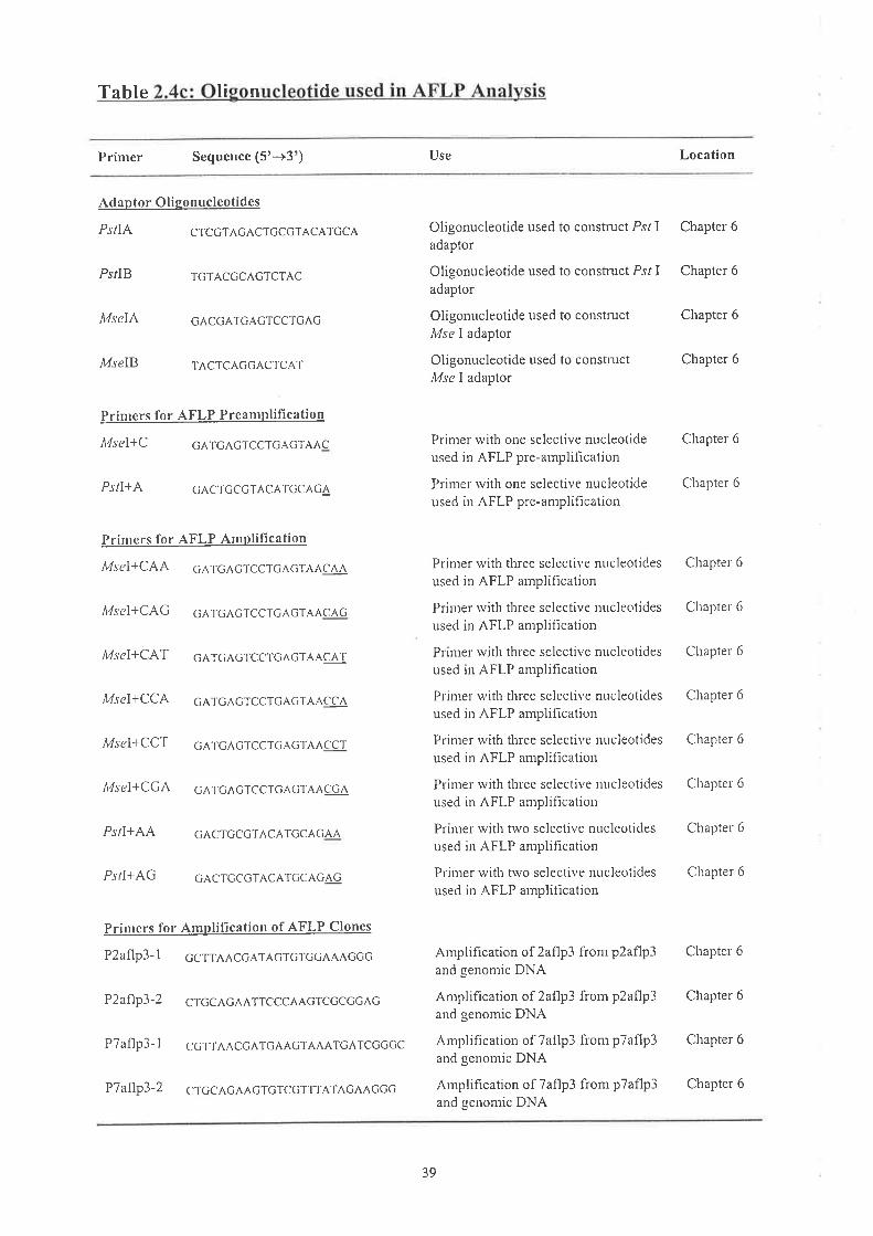
Table Ii n d
Primer Sequence (5'-+3') Use Location
Adaptor Oligonucleotides
PSTIA CTcGTAGACTCCGTACATGCA
Ps/IB TGTACGCAGTCTAC
MseIÃ GACCATCAGTCCTGAG
MselB TACTCAGGACTCAT
Prinrers for AFLP Preanrplifi cation
It4sel+C cATCAGTCcTGAGTAAC
PslI+A GACTGCGTACA'IGC,AGA
p ri nr crs fo r AFLI .z\¡¡pu fica.lupn
MseT+CAA cATGAGTCCTG^GTÂACÂA
MSCT+CAG GATGACTCCTGAGTAAC^G
A4sel+CAT cATCAGTCCTGAcTAAci\T
MseI+CCA GATGAGTCCTCAGTAACCA
Msel+CCT cATGAGTCCTGAGT^^ccr
A4seI+CGA GATGAGTCCTcAGTAACGA
Ps/I+AA G^crccGTAcArccAGAA
PslI+AG GACTCCGTAcATGCAGAG
Primers for AnrplifÏcation of AFLP Clones
P2aflp3-l CCTTAACGATAGTGTGGAAAGGc
P2af1p3-2 CTcCAGAATTCCCÀAGTCGCGcAC
P7aflp3-1 CGTTAAcGA,TGAAcTAAATGATCGGGc
Oligonucleotide used to construct Psl Iadaptor
Oligonucleotide used to construct Psl Iadaptor
Oligonucleotide used to constructMse I adaptor
Oligonucleotide used to conshuctMse I adapTor
Chapter 6
Chapter 6
Chapter 6
Chapter 6
Prir¡er with one selective nucleotideused in AFLP pre-amplification
Prinrer rvith one selective nucleotideused in AFLP ple-amplification
Prinrer rvith three selective nucleotides
used in AFLP amplifìcation
Prinrer rvith tlrree selective Irt¡cleotides
used in AFLP arnplification
Primel' with three selective nucleotides
used in AFLP amplification
Prinrel u,ith three selective nucleotides
used in AFLP anrplifìcation
Prinrer with three selective nucleotides
used in AFLP amplification
Primer with thlee selective nucleotides
used in AFLP anrplifìcation
Pri¡lerrvith two selective ltucleotidesused in AFLP amplification
Primer with trvo selective nucleotides
used in AFLP amplification
Chapter 6
Chapter 6
Chapter 6
Chapter'6
Chapter'6
Chapter'6
Chapter 6
Chapter 6
Chapter 6
Chapter'6
Amplification of 2aflp3 from p2aflp3and genomic DNA
Amplification of 2aflp3 from p2aflp3and genomic DNA
Amplification of 7aflp3 from pTaflp3and genomic DNA
Amplihcation of 7aflp3 from p7aflp3and genomic DNA
Chapter'6
Chapter 6
Chapter 6
P7af1p3-2 CTGCAGAAcTcTCGTTTATAGAAccG
39
Chapter 6

2.1.10 Kits and M ellaneous Materials
Kits and other materials used in this study were obtained from the following
manufacturers:
ABI Prism Dye Terminator Applied Biosystems-Perkin Elmer.
Ready reaction Mix.
Anti-DlG-rhodamine Fab fragments Boehringer-Mannheim, Getmany
Bio-Gel P60 (50-100 mesh)
Bio-Gel P60 (100-200 mesh)
B iotinylated anti-streptavidin
Bi oprobe@ Nick TranslationDNA Labelling System
DIG-Nick Translation Mix
DNA UltraFast Cleavage and
Deprotection Kit
Ex assist'^'/S OLR'^' System
GIGAprirne DNA Labelling Kit
Glass beads, 150-212 ¡lrn
Hybond'^'-N* nylon membrane
Packagene@ Lambda DNA
Packaging Systenr
pGEM@-T Vector System
pGEM@-T Easy Vector Systern
Rhodamine anti-sheep IgG
Sepharose CL-68
Sequenase Version 2.0 DNA
Sequencing Kit
Spin-X@ Centrifuge
Tube Filter, 0.45 ¡rm
Ultrafree@-Mc 0.45 ¡rm filter unit
X-ray film
Bio-Rad Laboratories, Richmond, CA
Bio-Rad Laboratories, Richmond, CA
Vector Laboratories
Sigma Chemical Co, St Louis, MO
B oehringer-Mannheim, Germany
Beckman Instruments, Palo Alto, CA
Stratagene, La Jolla, CA
Bresatec, Adelaide, Australi a
Signra Chemical Co, St Louis, MO
Arnersham,IIK
Promega, Madison,'WI
Promega, Madison, WI
Promega, Madison, V/I
Jackson ImmunoResearch Laboratories Inc
Phannacia, Uppsala, Sweden
United States Biochernical, Cleveland, OH
Corning Incotporated, Coming, NY
Millipore, Bedford, MA
Fuji RX Medical X-ray Fihn, Fuji PliotoFilm Co, Ltd
40

2.2 Sor,urloNs. Bu¡rnns a¡u MBnr¿,
2.2.1 Solutions and Buffers
All solutions and buffers \ilere prepared using millipore water and autoclaved where
possible, otherwise were sterilised by passage tluough a 0.2 pm filter. Solutions for RNA
work were treated with 0.1% (v/v) DEPC as per Sambrook et al. (1989) prior to
autoclaving. All restriction endonucleases and most other enzymes were provided with
incubation buffers. Solutions and all other buffers routinely used in this study were as
follows
Acr.¡lanl ide stock 38% (w/v) acrylamide and2o/o (w/v) bis-acrylamide, deionised with rnonobed resin
Denaturing solution 1.5 M NaCl, 0.5 M NaOH
I x Denhardt's
DNase I Reaction Buffer
A loadi
Feuleen Stain
Hvbridisation buffer
Lvsine solution
Neutralisine solution
Phen
0.02% (rv/v) Ficoll@,0.02o/o (w/v) PVP,
0.02% (rv/v) gelatin
0.1 M NaAc, 5 mM MgSO,, pH 5.0
0.25% (w/v) bromophenolblue, 40% (w/v)sucrose
0A% (wlv) Fuschin (basic), 0.15 M HCl,100 mM NaSrO
5 x SSPE, 5 x Denhardt's, 0.5olo (w/v) SDS,
100 rng/rnl denatured, sonicated sahnonsperm DNA
10 mM glucose, 25 mM Tris-HCl pH 8.0,
10 mM EDTA, 4 mg/ml lysozyme
1.5 M NaCl, 1 M Tris-HCl pH 7.2, I mMEDTA
50% (v/v) phenol, 48% (vlv) chlorofonn,2% (vlv) isoarnylalcohol, buffered with an
equal volume of Tris-HCl pH 8.0,0.2o/o(v/v) B-mercaptoethanol
100 mM NaCl, 10 mM MgSOr, 50 mMTris-HCl pH 7.5, 2% (wlv) gelatin
SM buffer
rofonn
41

))) edia
All liquid and solid media were prepared using millipore water and sterilised
imntediately by autoclaving. The compositions of various media wele as follows:
IxSSC
1 x SSPE
10 x STE
Stop buffer
1 xTAE
Luria broth (L-broth)
Luria agar (L-aear)
NZY agar
NZY top aqar/aearose
TB aear
0.15 M NaCl, 0.015 M Nar-citrate, adjustedto pH 7.2withNaOH
0.18 M NaCl, i0 mM NaHrPOo, 1 mMEDTA pH 7 .2
100 mM Tris-HCl pH 8.0, 1 M NaCl, 10
rnM EDTA
i0 mM Tris-HCl p}l7.5,1 mM EDTA pH8.0,0.2o/o (w/v) SDS
40 mM Tris-HCl, 20 mM Na-acetate, 2 tnMEDTA, adjusted to pH 7.8 with glacialacetic acid
0.9 M Tris-borate, 0.002 M EDTA pH 8.0
10 mM Tris-HCl pH 8.0, 1 mM EDTA pH
8.0
l% (wlv) tryptone, l% (w/v) NaCl, 0.5%(w/v) yeast extract, adjusted to pH 7.2 u,ithi M NaOH
Luria broth witli the addition of 1% (w/v)bacteriological agar No. 1
1% (w/v) NZ amine (casein hydrolysate),0.5% (w/v) yeast extracf,0.5o/o (w/v) NaCl,1% (wlv) bacteriological agar No. I
1% (w/v) NZ amine (casein hydrolysate),0.5% (w/v) yeast extracT,0.5o/o (w/v) NaCl,10 nrM MgSOo, 0.7% (wlv) bacteriologicalagar No. 1 or agarose
1% (w/v) tryptone, 0.5% (w/v) NaCl, 1%
(w/v) bacteriological agal No. 1
l% (wlv) tryptone, 0.5% (w/v) NaCl,0.01 M MgSOo, 0.7%(wlv) bacteriologicalagar No. 1 or agarose
TB top aqarlagarose
42

2.3 MnrHoDS
2.3.1 Plant Growth C ditions
Brachycorne dichrontsontatica seeds were soaked in HrO ovemight. Using a
dissecting microscope the embryo was separated intact from the hard coating and the
sunounding integurnent.
Dissected embryos were stored on absorbent sponge soaked in water to germinate.
After gennination seedlings were transferred to potting compost in seedling trays, covered
with a srnall vial, and placed in a controlled ternperature growth cabinet. In a further 4 - 6
weeks seedlings were large enough for the collection of root tips and rvere transfer¡ed to
larger pots in the glasshouse.
2.3.2 Kar)'otypins techniques
Root tips rvere cut frorn plants and pìaced in ice cold water ovemight, then fixed in
3:1 ethanol:glacial acetic acid.
Fixed root tips were washed in HrO for five mins and treated with HCI at 60'C for 10
mins. They were then rinsed in HrO and stained witli Feulgen Stain for at least 20 minutes.
In order to prepare mitotic chromosornes, the root tips were placed between a slide and a
coverslip i¡ a drop of 45"/o acetic acid and squashed. The different cytodemes (4,, 42, A:
arrd Ar) of B. ctichrontosomaticawere distinguished on tlie basis of their chromosome
rnorplrology as descr-ibed by Watanabe et al. (I975).
2.3.3 General Techniques
2.3.3a ctlon Disests
All reactions involving restriction endonucleases were performed under conditions as
r-ecommended by the manufacturers. In general, five units of enzyme were added per pg of
DNA to a maximuln enzyme volume of 20% of the total reaction volume and the reaction
43
was allowed to proceed for 2-3lu. RNaseA was added to a final concentration of 0.5 pg/pl
to all restrictions. In genomic DNA digestions spermidine was also inclùded to a final
concentration of 3 niM. Samples of plant genomic DNA were lestricted with 10 units of
enzyme per pg of DNA for at least 16 hr.
DNA on
DNA was size fractionated by electrophoresis on0.7o/o (w/v) agarose gels in
submarine gel tanks with 1 x TAE as the running buffer'. Arnplified DNA samples were
a¡alysed by electrophoresis on 1.2-1 .5% (wlv) agarose gels. Products resulting from ^Sjfc
I
restriction of ITS2-PCR products were analysed by electrophoresis on 2o/o (tvlv) agarose
gels. Sarnples were loaded with l x loading buffer and electrophoresed at 60-80 V until the
brornophenol blue marker dye had migrated a sufficient distance to allow adequate
separation of tlie DNA fragnrents. Following electrophol'esis, the DNA was stairied in 5
¡rg/nil of ethidium bromide and viewed under UV light. Genomic DNA gels to be Southerr
blotted were electrophoresed at 30 V for at least 16 hr. Where applicable, sizes of DNA
fragments were estimated using the Fragrap computer program (Duggleby et al., 1 981)'
2.3.3c R of DNA from Gels
DNA was eluted from agarose gels using tlie freeze-squeeze method adapted frorn
Thuring et al. (1975). The frozen gel slice was squeezed through a26G syringe needle into
the upper compartment of an Ultrafree@-MC or Spin-X@ Centrifuge Tube 0.45 ¡rm filter and
rvashed through with 200 pl of 1 x TAE buffer. Tlie DNA was allowed to elute from the
gel at room temperature for 15 min. Centrifugation for 2-3 min at 7000 x g yielded a
solution of DNA in the lower tube compartment and tlie DNA was recovered by
precipitation with ethanol.
44

2.3.3d Precipitation of DNA
Method 1 : Precipitation by Etltanol
DNA was precipitated by the addition of one tenth of the volume of 3M Na-acetate
pH 5.2 followed by 2-2.5 volumes of cold redistilled ethanol. The DNA was left to
precipitate at -20'C for a period of 2-16 hr, depending on its molecular weight. The DNA
was recovered by centrifugation at 1 2000 x g for at least 1 5 min at 4oC. The pellet was
waslred witli cold 70% (vlv) ethanol, dried under vacuum and resuspended in a minimal
volumeoflxTE
Method 2 : Precipitatiott by Isopropanol
In solne cases, DNA was precipitated by addition of one tentli of the volume of 3M
Na-acetate pH 5.2 followed by an equal volurne of cold isopropanol. The DNA was left to
precipitate at roort teniperature for 5 min before microcentriftrgation at 12000 x g for at
least 1 5 min. The resulting DNA pellet was washed with cold l0o/o (v/v) ethanol, dried
under vacuunl and resuspended in a minimal volume of 1 x TE.
f DNA
DNA was purified from proteins by the addition of an equal volume of
phenol/chlorofon¡ and vigorous mixing. The emulsion was centrifuged at 12000 x g for 2
min and the aqueous layer transferred to a new tube before the extraction was repeated.
Contaminating phenol was removed by the addition of an equal volume of chlorofom,
mixing and centrifugation as above, and tlie DNA solution transfened to a new tube.
2.3.3f DeTennination of DNA and RNA Concentration
The concentration of DNA and RNA in solution was detennined by UV
spectrophotometry at a wavelengtli of 260 nm or, in the case of freeze-squeezed restriction
45

fragments, by gel electrophoresis of a sample in conjunction with the rnolecular size
markers lambda bacteriophage DNA restricted with lli¡zd III.
2.3.4 Prenaration of DNA Samnles
lation of PI
Total genomic DNA frorn the leaves of Brachycome dicfuontosontatica was
isolated by the rnethod of Wienand and Feix (1980).
2.3.4b Small-scale Plasmid Preparation
A4ethod I : Alkaline L¡tsis Plasrnid Mittipreps
Plasmid DNA for general applications was prepared using the scaled-up version of
the alkaline lysis method (for 10 ml of ovemight bacterial culture) as per Sambrook e/
a/. ( I 989).
Method 2 : Boiling L)tsis
Plasmid DNA for manual sequencing was prepared using the boiling lysis method of
Holmes and Quigley (1981), scaled down for 1.5 ntl of a 10 ml ovenright bacterial culture.
2.3.5 Clonins Stratesies
2.3.5a on of Tnseñ DNA and Plasm id Vector
DNA fragments for ligation were geuerated in one of two ways. DNA was restricted
to completion with the appropriate lestriction enzyme and then recovered fi'om agarose
gels using the fi'eeze-s qleeze method or purifred by phenol/chlorofom extraction and
precipitation with ethanol.
Vector DNA for use in ligations was digested with the appropriate restriction
enzyrne, purified by extraction with phenol/chloroform and precipitated with ethanol.
Completely digested plasmid DNA, analysed on an agarose gel, was treated with calf
intestinal phosphatase (CIP) essentially as per Sambrook et al. (1989) in order to prevent
46

self-ligation. The 50 ¡rl reaction was incubated aT37"C for 30 min, after which 10 pl 10 x
STE, 5 p,lI0% (v/v) SDS and 40 ¡rl dHrO were added and the mixture incubated at 68"C
for 15 min to inactivate the enzyme. A phenol/chlorofonn extraction was canied out and
the DNA precipitated by the addition of ethanol. The final concentration of the vector was
50 ng/¡rl.
2.3.5b Clonine of Products Generated bv PCR
ITS I and ITS2 PCR products were purified by centrifugation through a Sephalose
CL-6B chromatography column (Murphy and Kavanagh, 1988) priol to restriction r¡,ith
EcoRI and Xba I, purification by phenol/chloroform extraction, precipitation with ethanol
ar.rd cloning into pBluescript@SK(-). Other PCR products rvere cloned directly into pGEM@-
T or pGEM*-T Easy following centrifugation through a Sepharose CL-6B chromatography
column (Murphy and Kavanagh, 1988).
2.3.5cLigatlorts
Ligations were performed using 50-100 ng of vector DNA and an equirnolar amottl'tt
of insefi DNA, catalysed by 1 Weiss unit of To DNA ligase in a l0 pl reaction. All
ligations were placed in a 7 L insulated u,aterbath at 16"C and left oven.right in a 4"C
coldroom, except for ligations involving pGEM@-Tvectors, which were camied out as per
m anufacturer' s instructions.
2.3.5dPreparation and Transfonnation of E. coli
Preparation of DH5uF' and XLI-Blue competent cells for translonnation was
perforrned using the calcium chloride method as per Sambrook et al. (1989).
Transfolrlation of competent E. coli cells with ligation mixtures was achieved using heat
shock at 42oC as per Sarnbrook et al. (1989). The cells were pelleted at 7000 x g for 30 sec,
resuspended in 400 ¡rl L-brotli and plated onto L-agar plates supplemented witl-r 100 ¡rg/ml
4l

ampicillin. Colonies containing recombinant plasrnids were selected by plating onto L-agar
mediurn containing 100 ¡rg/ml ampicillin with an overlayer of L-agal plus 100 pg/ml
ampicillin, 200 ¡rg/rnl X-gal and 160 ¡rg/rnl IPTG
2.3.6 Prenaration of RNA Samnles
Total RNA was prepared from the leaf tissue of -8. dichrontosontatica and B.
lineariloba, using the guanidine thiocyanate-CsCl metliod (Chirgwin et a|.,I979) or
TRIzol@ Reagent. Total RNA was prepared frorn the floral tissue of B. dichrontosotnatica
and B. lineariloba using TRIzol@ Reagent, as per manufacturer's insttuctions.
2.3.7 Phase N{anipulation
2.3.7 a Preparatio n of Platins Cel ls for Phase
Bacterial cells for plating of phage were prepared as per Stratagene instructions for
the Predigested IZAP@II/,Ðcozu Cloning Kit and diluted to ODooo:0.5 rvith 10 mM
MgSOo before use.
2.3.7b Platine and Pickine Phaee
Phage and host cells were mixed and incubated at 37'C for 15-20 rnin prior to plating
on the appropriate nredium. Pliage for the screening of libraries were plated on 150 mm
plates using 600 pl of plating cells and 8 ml top agarose, melted to 48"C. Phage for
secondary and tertiary screening were plated on 85 mm plates using 200 ¡rl of plating cells
and 3 ml top agar or agarose, melted to 48"C. Plates were incubated at 37"C for betrveen 8
and 16 hr.
Positive plaques were aligned to the agar plates. Plaques were plugged into 0.5 nil
SM buffer and 20 ¡,rl chloroform using the large end of a sterile pasteur pipette artd the
phage allowed to elute at room temperature for 2 hr or at 4"C overnight. Phage were plated
48
at low density on an 85 rnm plate for secondary screening alrd the procedure repeated for a
terliary round of purification.
Following tertiary screening, recombinant pBluescript@SK(-) was rescued from
IZAP@II using the ExAssist'^'/SOLF[^' System, following manufacturer's instructions
2.3.8 of DNA to M branes
2.3.8a Southern Blottine
Following electrophoresis, DNA was transferred and alkali-fixed onto a Hybond'^'-
N* nylon metnbrane, following the instructions of the mauufacturer.
2.3.8b Colon]¡ H)rbridisation
Colony hybridisation was achieved using the metliod described by Grunstein and
Hogness (1975). Recombinant bacterial colonies were patch plated onto an L-agar plate
supple¡rented rvith 100 pg/ml arnpicillin and replica plated onto a Hybond-N'^'+ menbl'alle
which overlayed another such plate. Follorving growtli of bacterial colonies ovemight at
3Joc,the ¡ylorr filter rvas placed colony side up ou a pad of tluee pieces of Whatrnan@
3MM paper soaked in lysing solution for 20 min, rvith blotting onto paper towel every' 5
lni¡. The filter was transferred to a fresh pad of 3MM paper soaked in denaturing solution
plus 1% (wlv) SDS for a total of 20 min witli blotting every 7 tnin. This procedure rvas
repeated with 3MM soaked in neutralising solution before the filter was partially air-dried.
Bacterial debris ,was removed frorn the filter using absorbent cotton wool and washing in 2
x SSC. The DNA was alkali-fixed onto the filters as per Amersham instructions.
2.3.9 Radiolabell ins of DNA probes
2.3.9a Olieo ins Usine Random Oli leotides
DNA fragrnents requiring radioactive labelling were purified by size fractionation on
an agarose gel and freeze-squeezing as previously described. Labelling of 50-200 ng of
49

double-stranded DNA was performed by pritner extension of random oligonucleotides
(Feinberg and Vogelstein, 1983) using the GlGAprirne DNA labelling kit and
¡cr-3'?t¡dAtr.
2.3.91r Olieolabe llins IIsins PCR
Reamplified AFLP products were labelled by PCR. Approximately 5 ¡rl of PCR
product was incubated with 3 ¡rCi of [cr-32P1dATP using reaction conditions outlined in
Section 2.3.15e for 15 cycles. The probe was denatured by incubation at94"C for 5 mins
before cooling on ice and addition to a 0.5 ml hyblidisation volune.
2.3.9c RNA labelline
Prior to labelling, 2 pg of total RNA was incubated at 100'C fol 3 mins then cooled
on ice. The total RNA r¡,as end-labelled in 1 x kinase buffel in the presence of 50 ¡tCi of [y-
"PldATP and 15 units of T4 polynucleotide kinase, in a total volunte of 40 ¡-rl. The
reaction was incubated at 37"C for 60 mins then the probe denatured at 90oC for I min
before being added to a 0.5 ml hybridisation volunie'
2.3.9d Removal of Un ted Nucleotides
Utrincorporated nucleotides were rernoved from oligolabelled probes by spun column
chromatography through Bio-Gel P60 resin columns ntade from 100 ¡rl G50-100 beads and
400 ¡-rl G100-200 beads. Columns were packed and equilibrated with l00 ¡-rl stop buffer by
ce¡trifugation at i70 x g for 2 min. Labelled DNA fi'agments were eluted by adding the
labelling reaction plus stop buffer (to a total volume of 100 ptl) to the top of the column and
recentrifugation at 170 x g for 2 rnin.
50

2.3.10 Prehybridisation. Hybridisation and Washine
2.3. 1 0a Prehvbridisation Conditions
All filters were prehybridised, hybridised and washed under the same conditions
except that plaque lifts were separated by nylon grids and prehybridised for at least 16 hr.
Filters were prehybridised in a solution of 5 x SSPE, 5 x Denhardt's, 0.5olo (w/v) SDS and
100 ¡rg/ml denatured sonicated salmon sperrn DNA at 65"C for a minimum of 2 hr. The
volume of prehybridisation solution was approximately 0.1 ml/cm2 of filter.
2.3.10b Probe Denaturation and Hvbridisation
Following addition of 300 ¡rg sonicated salmon sperm DNA, the radiolabelled probe
was denatured by the addition of 35 ¡rl I M NaOH and incubation at room temperature for
l0 min. The solution was neutralised by adding 35 ¡rl I M HCI and the denatured probe
stored on ice. Filters were hybridised by labelled probe at a concentration of approximately
1 x 100 cpm per ml of hybridisation solution (to a rnaximum volume of 6 ml) for a
minimunr of l6 hr at 65"C.
2.3.10c Washine of Filters
Filters were washed in a solution of 2x SSC,0.l% SDS at 65"C for 30 min, I x SSC,
0.1% SDS at 65"C for 30 nrin and 0.5 x SSC, 0.1% SDS at 65"C for a furlher 30 min.
Southem blots subject to a high stringency wash were incubated in 0.1 x SSC, 0.1% SDS at
65"C for 2 hr.
toradi of
Filters were covered in plastic wrap to prevent drying out and exposed to X-ray film
at -70"C for an appropriate period of time, backed by an intensifying screen (DuPont Hi-
Plus). Filters probed with AFLP and RAPDs were often exposed to Fuji Imaging Plates
(Fuji Photo Film, Co., Ltd) for periods of tirne ranging fi'orn 30 mins to ovemight and
5l

alialysed using the Mac.BAS Version 2.X computer program (Fuji Plioto Film, Co., Ltd).
Quantification of the radioactive signals emitted flom ITS2 PCR products before and after
restriction with ^!fc I, was achieved using MacBAS Version 2.X computer program (Fuji
Photo Film, Co., Ltd)
Southern blots and plaque lifts were stripped of probe by incubating for 10 min in 0.1
x SSC, 1% SDS at 90"C. Stripped filters were rinsed ín2 x SSC and stored in plastic wrap
at 4"C for rehybridisation.
2.3.11 Desisn and Svnthesis of DNA Olieonucleotides
Oligonucleotide primers were synthesised on 30 nM columns using an Oligol00
DNA Synthesiser (Beckman) and purifred by the rnethod of Sawadogo and Van Dyke
(1991) using a DNA Ultrafast Cleavage and Deprotection kit. Altematively
oligonucleotides were synthesised commercially (Bresatec). All oligonucleotides were
adjusted to a concelrtration of 100 ng/¡rl for PCR or 10 ¡-tM for automated sequencing.
Pri¡rers used for manual sequencing were fufther diluted to 1 pM. Annealing temperatures
were calculated according to tlie formula TA : 2(A+T)+4(G+C)-5 "C
2.3.1 1 a Primers for Sequencine
Oligonucleotide priruers for sequencing were designed to unambiguous previously
sequenced regions, priming towards a region of unknown sequence. They were generally
synthesised as 2O¡ners at spacings of approximately 200 to 400 bp and contained a GC
proportion of at least 50%.
2.3.1lb ers for PCR
Oligonucleotide primers for PCR were designed to contain the same proportion of
purine and pyrinridine bases as that found in the target sequence. Efficient priming at high
temperatures was maximised by the inclusion of two G or C residues at the 3' end of the
52

primers. In addition, primer pairs were checked at their 3' ends for the possibility of
prinrer-dimer formation and intramolecular bonding using the Pcfold (Zuker, unpublished)
and Molecule (Thornpson, unpublished) computer programs.
2.3.12 Sequencing of Double-stranded DNA Templates
2.3.12a Preparation of Template DNA for Manual Sequencine
A 3 prg sample of the plasmid DNA to be sequenced was made up to 18 ¡rl with dHrO
and 2 ¡rl of 10 rng/rnl RNaseA added before incubation at 37"C for 1 5 min. Following
addition of 5 ¡rl 1 M NaOH, 1 mM EDTA the solution was incubated for a further 15 min
at 37'C. The denatured DNA was purified by centrifugation through a Sepharose CL-68
colum¡r (Murpliy and Kavanagh, 1988), yielding 25 ¡rl of single-stranded ternplate DNA
rvhich rvas then stored on ice.
aratl DN uencr
Plasniid DNA prepared using the alkaline lysis method was resuspended in 250 ¡ll I
x TE and incubated at roorn temperature for 10 rnins with l ptl of 10 mgÁnl RNaseA. DNA
u,as precipitated by the addition of D5 ¡"rl 30% PEG8o0/1.5 M NaCl and the tnixture
incubated on ice for 30 mins. The DNA rvas pelleted by centlifugation at 12,000 x g at 4oC
for 30 mirrs and washed with 95% ethanol before being drained, desiccated briefly and
resuspended in 300 ¡rl of 1 x TE.
The DNA was extracted sequentially with phenol, phenol/chlorofonn and chloroform
and precipitated with one half the volume of 7.5 M NaAc and 2 volumes of 100% ethanol.
The pellet was washed with T0o/o ethanol, vacuum dried and resuspended in 20 pl of I x
TE.
53

2.3. 12c Manual Sequencine Reactions
The dideoxy chain termination reaction (Sanger et a\.,I977) was performed using the
Sequenase Version 2.0 kit and [a32P]dATP, as described by the manufacturer of the kit.
Wl'rere possible, sequence was confirmed by sequencing of both DNA strands.
2.3.1 2d Autor¡ated Sequencins Reactions
Automated sequencing reactions were performed usirig dye-tenniuator cycle
seque¡cing reactions (Applied Biosystems-Perkin Elmer), according to the manufacturer's
i¡structions. All automated sequencing (ABI Prisnr Model 377) was done at the Institute of
Medical and Veterinary Science, Adelaide, South Australia'
2 12eP u
A 4% (w/v) acrylarride, 8 M urea, 1 x TBE gel solution rvas nrade up to 100 ml with
water. Following addition of 78 rng arnmonium persulphate and 60 prl TEMED, the gel rvas
mixed and poured betweelr glass plates.
2.3. 1 2f Denaturine Gel Electrophoresis
Manual sequencing reactions were run on a Model 32 sequettcing gel electrophoresis
systenr (BRL).Using I x TBE as the ruming buffer, the gel was pre-electrophoresed at 50
¡rA for l5 min. Following denaturation for 2 rlin at 90oC, 3 prl of each DNA sequencing
reaction was loaded into the gel wells and electrophoresis contiuued until the tracking dye
had reached the bottom of the gel plates. The gel was run between 30 mA and 50 mA but
the voltage not allowed to exceed 1700 V. The loading process was repeated for medium-
and short-length runs. Following electrophoresis, the sequencing gel was fixed in20o/o
(v/v) methanol and 10% (v/v) glacial acetic acid for 30 min to renrove the urea. Finally, the
gel was dried at 65'C for at least 3 hr and autoradiographed at room temperature.
54

2.3.12s Sequence Analysis Software
The compilation and comparison of multiple nucleotide sequences was performed
using the facilities provided by the Australian National Genomic Information Service
(ANGIS). Multiple sequence aligrunents were done using the Eclustalw program
(Thompson et al., 1994). FASTA was used to search the nucleotide databases (Altschul el
a\.,1990; Gish and States, 1993). The repeat structure of the IGS sequences v/as
detenlined using the DotPlot program (Maizel and Lenk, 1981)
2.3.13 Polymerase Chain Reaction (PCR)
Reaction mixtures (25 pl) fo¡ PCR contained 0.4 mM each of dATP, dCTP, dGTP
and dTTP, 1 x PCR reaction buffer, 3 mM MgCl,, 100 ng of each primer, 1.5 U of Taq
DNA polyrrerase and 100 ng of template DNA. DNA was amplified in a programnrable
temperature controller (ARN Electronics) or a PTC-200 DNA engine (MJ Research, Inc.)
generally programmed as follows: initial template denaturation was at 94'C for 2 min
followed by 25 PCR cycles of de¡raturation at94'C for 1 min, primer annealing at 55"C for
I min and extension at 72'C for 1 rnin. If the product was to be cloned, an additional cycle
was caried out with an extension time of 7 nin.
Annealing temperature varied depending orr the primer pairs but was generally 45-
55"C. Amplifications using BITS2 required annealing temperature of 70"C. The extension
tinre also varied between different amplification reactions, according to the length of the
fragment to be amplified (based on a synthesis rate of I kb per rnin)
2.3.14 Reverse Transcription (RT)
Prior to reverse transcription, 10 ¡rg of total RNA was treated witli DNase I (RNase
free) to remove any contaminating DNA. Specifically, precipitated RNA was incubated in
55

1 x DNase I reaction buffer and20 U of DNase I (RNase-free) for 30 mins at25"C, and the
reaction terminated by incubating at 95'C for 5 mins
Reaction mixtures (30 pl) for reverse transcription contained 0.5 rnM each of dATP,
dCTP, dGTP and dTTP, I x reaction mix , 3.3 pM of primer ER25S, 13.3 U/pl M-MLV
reverse transcriptase, RNasint to 1 U/pl and approximately 10 pg of DNAse-treated total
RNA. The reaction was incubated at42"C for 60 mins, and terninated at 75'C for 10 mins
For RT-PCR 3 pl of the reverse transcription reaction was used and the reactions set up as
for PCR from DNA templates
2.3.15 Amplified F[aement Lensth Polffi
2.3.15a Preparation of AFLP DNA Samples
Initially 2 ¡tgof genomic DNA samples u,as digested rvith 30 U of both Pst I and
MseI in I x restriction-ligation buffer (100 nM Tris HAc, 100 nrM MgAc, 500 mM KAc,
50 mM DTT, pH 7.5)
aratiorr of Ad
Oligorrucleotides MseIA (50 pM) and Mse IB (50 ¡rM) were combined in a total
volume of 20 ¡rl, and Psl IA (5 pM) and Psl IB (5 pM) were combined in a total volume of
40 ¡-tl. To instigate annealing the oligonucleotide combinations were heated at 90'C for 3
rnins and tlien allowed to amreal at room temperature for at least 30 rrins
2.3.15c Lieation o Adantors to DNA
Adaptors were ligated with 1 ¡rg of restricted DNA in 1 x restriction-ligation buffer
with 0.2 nrM ATP, I ¡tl of Mse-adaptor mix, I pl of Psl-adaptor mix and catalysed by 1
Weiss unit of To DNA ligase in 60 ¡rl reaction mix fol3 hours at37"C, then overnight at
4oC. The DNA was subsequently precipitated witli ethanol and resuspended in 60 pl of 0.1
xTE
56

2.3. 1 5d AFLP Preamplification
Initial AFLP reactions were carried out with Psr I and MseI adaptor-specific primers
with one selective riucleotide (Ps/ I+A; MseI+C). Pre-amplification reaction rnixtures (25
¡-rl) contained 0.2 mM each of dATP, dCTP, dGTP and dTTP, 1 x PCR reaction buffer, 1.5
mM MgCl,, 50 ng of each primer, 1.5 U of Zaq DNA polymerase and 4 pl of template
DNA. DNA was arnplified in a programmable ternperature controller (ARN Electronics)
programmed as follows: initial template denaturation was at 94'C for 2 min followed by 20
cycles of denaturation at 94"C for 30 secs, primer annealing at 56"C for I min and
extension at72'C for I min. Following amplification 100 pl of dHrO was added to the
reaction tubes and this rvas used as the template DNA for the arnplification step following
2.3.1 5e AFLP Anrplification
AFLP antplificatiorrs ernployed tu,o oligonucleotide prin-rers one corresponding to
the Psl I ends with trvo selective nucleotides (Ps¡ I+AA or Ps/ I+AG) and one
corresponding to rhe Mse I ends with tluee selective nucleotides (Mse I+CAI\,
Mse I+CAG, Mse I+CAT, Mse I+CCA, Mse I+CCT, or Mse I+CGA). Initially 50 ng of the
Psr I primer was radioactively labelled with 10 pCi of ¡y-"PldATP in 1 x kinase buffer
and tlie reaction catalysed by l0 U of polynucìeotide kinase at 37'C for 60 mins
AFLP reactions (20 frl) contained 0.2 mM each of dATP, dCTP, dGTP and dTTP,
1 x Taq reaction buffer, I .5 mM MgCl,, 30 ng of Mse l*three nucleotides primer, 25 ng of
Psl I*two nucleotides primer, 5 ng of Psr Irtwo nucleotides primer labelled with [y-
"P1dATP, I U of Ia4 DNA polymerase and 2 pl of tenrplate DNA. DNA was amplified in
a programmable temperature controller (ARN Electronics) programmed as follows: initial
tenrplate denaturation was at 94'C for 2 min followed by one cycle of denaturation at 94'C
for 30 secs, primer amealing at 65oC for 30 secs and extension at72'C for 1 min. This
cycle is followed by 9 cycles over which the arurealing temperature is decreased by 1"C per
57

cycle, followed by 25 cycles of denaturation at 94"C for 30 secs, pritler annealing at 56"C
for 30 secs and extension at72oC for 1 mtn.
AFLP reactions were run on 60/o acrylamide gels prepared and run as for sequencing
gels described in Section 2.3.I2. Following electroplioresis, the gel was fixed and dried as
for sequencing gels, and autoradiographed ovemight at room temperature. Bands of interest
were rehydrated with 1 x TE, excised from the gel and the DNA eluted by placing the gel
piece in 10 ¡rl of I x TE.
2.3. I 5f Rearnplifi cation of AFLP Frasn.ìents
AFLP fragments eluted from the gel were reamplified using the same PCR
conditions as for the amplification step outlined above, with I ¡-rl of eluted DNA and
unlabelled Psl I primer
2.3.16 Random Amplifica tion of Polvmornhic DNA
RAPD reactions were perfonned according to the nrethod of Willianls el al., (1990)
witlr some n-rodifications. Reaction volurnes were 25 ¡rl and contained of 2 units of Toq
polymerase,2.5 ¡:,1of l0 x reaction buffer (1 M Tris HCI pH 8.3, 1 M KCl, 1 M MgCl,, I
rrrg/nrl gelatine), 0.8 mM each of dATP, dCTP, dGTP and dTTP, 1 ¡rl of plinter and
approxirnately 50 ng of genonic DNA.
DNA was amplified in a PTC-200 DNA Engine (MJ Research, Inc.) generally
programrlled as follows: initial template denaturation was at94'C for 2 min follorved by 45
PCR cycles of denaturation at94'C for 30 secs, primer annealing at 30"C for 30 secs and
extension af 72'C for 2 mins. An additional cycle was included with an extension time of 7
mins. RAPDs of interest were extracted from the gel by freeze-squeezing and cloned
directly in pGEM@-T Easy
58

2.3.17,Ir sirr hybridisation :
aration of
The probe BdrD(0.6) was labelled with biotin-dUTP using the Bioprobe Nick
Translation DNA Labelling Systern as per manufacturer's instructions. BdrD(2.5) was
iabelled with DIG-dUTP using the DIG-Nick Translation Mix according the
manufacturer' s instructions.
For each cluomosonre sample to be hybridised, approximately 50 ng of the labelled
probe was combined with i 0 pg of sonicated salmon spenrl DNA and made up to a volume
of 20 ¡rl with H,0. The DNA mix rvas then ethanol precipitated at -70oC for 30 mins.
2.3.17b Preparation of slides
Root tips rvere collected and fixed as for karyotyping techniques. Fixed root típs
were then ',vashed in HrO at room tenrperature for 20 mins and the cell rvalls di.gested for
20 mirrutes at 37"C, using an enzylne rnixture of 2.5o/o (w/v) pectolyase Y-23 plus 2.5%
(w/v) Cellulase "Onozuka R10" in 75 mM KCI and 7.5 rnM EDTA, pH 4.0 (Kakeda et ctl.,
1991). After u,ashing rvith HrO, single root tips were placed on a microscope slide in a
drop of 45o/o acetic acid. A coverslip was then placed over the root ureristem and tapped
with a dissecting needle. Preparations wel'e vierved usir-rg pliase-contrast microscopy
(Olympus)
2.3.17c Fixing of Slides
Slides of appropriate quality were frozen in liquid nitrogen and then the coverslip
renroved. Slides were air dried and dehydrated by washing in 7 0o/o, 95o/o and 100% ethanol
eaclr for 30 rninutes at room temperature. Slides were stoled at 4"C and used within one
week of preparatiorr
59
2.3.17 d RNase Treatment
Slides were treated with RNase A (100 ¡rghnl ín2 x SSC) in a 2 x SSC saturated
atmosphere at 37"C for 40 rnins, washed in 2 x SSC pH 7.0 two times at room temperature
for f,rve mins and dehydrated in70%o,95o/o and 100% ethanol before air drying.
2.3.17e Denaturation
Tlre clrror.nosone DNA was denatured for 2 minutes in denaturation mix (70'/o
deionized formamide, 2 x SSC pH 7.0) at70oC, then re-dehydrated in70% then 100%
etlranol for 5 rnins each at -20'C and air dried
2.3.t7f H disation
The precipitated, dried DNA probe was resuspended in 6 ¡rl of fonnarnide per slide to
be hybridised, shaking atroom tenrperatule for i5 urins, followed by the addition of an
equal volurle of 4 x SSC,20 x dextran sulphate and shaking for a further 15 mins. The
probe was then denatured at 95'C for 10 mins. Following chilling on ice, l2 ¡rl of probe
rvas added to each slide and hybridised ovemight at3J"C,tna2 x SSC saturated
atnrosphere
Slides rvere then washed in 50o/o deionised fonnarnide in 2 x SSC at 42'C (2 x 10
nrins), and 2 x SSC pH 7 .0 al42'C (3 x 5 mins)
2.3. 1 7e Signal Detection
Slides were pre-incubated by the addition of 100 pl of blocking solution containing
5% BSA, 0.2o/oTween20,4 x SSC for 30 mins. Detection of hyblidised biotin and DIG-
labelled probes was achieved by incubating slides with fluorescein streptavidin (5 pg/ml)
and anti-DlG-rhodamine Fab fragments (20 ¡rglml) respectively in 5%BS A,0.2o/o Tween
20, 4 x SSC for 60 mins at room temperature. For signal arnplification the slides were
waslred in 0.2o/o Tween 20,4 x SSC (3 x 5 rnins), and treated with Biotinylated anti-
60
streptavidin (1 pglml) and Rhodamine anti-sheep IgG (0.015 mg/ml) in lo/o BSA, 4 x SSC
for 30 minutes then washed in 0.05% Tween 20,4 x SSC (2 x 5 rnins). The FITC signals
were reamplified with fluorescein streptavidin (5 pglml) and the slides were washed in 4 x
SSC, 0.1% Tween 20 (3 x 5 mins).
Chromosomes were counter stained by the addition of DAPI in antifade (10 rng/ml p-
pl'renylenediamine, 90% (vlv) glycerol), and a coverslip applied. Preparations were
examined by epi-fluorescence microscopy using an excitation wavelength of 450 - 490 nm.
Photographs were taken using a CCD camera (Olympus) and manipulated using Adobe
PhotoshoprM.
61

TER a
Sequence Analysis of Ribosomal DI\A
Internal Transcribed Spacers 1 and 2
62

3.1 IxTnODUCTIoN
Previously, in Brachycome dichrontosomatica, fluorescent in sirø hybridisation
revealed the presence of rRNA genes at a major cluster near the satellited end of the short
arm of the B clrromosomes and, to a lesser extent, proximally on the same ann (Donald el
al., 1995; Houben et a1.,1998). The apparent regular occulrence of rRNA genes on B
chronrosomes in many species (Section 1.6.2b; reviewed by Green, 1990; Jones, 1995),
along with the fact that the tenninal region of the B in.B. dichrontosontatica was observed
to be associated with a nucleolus at mitotic prophase in some cells and fì'equently so
associated at diplotene of meiosis (Donald et a\.,1995), suggested tliat the rRNA genes of
tlre B. dichrontosontalicct B clrromosome rrìay be transcriptionally active (Donald el al.,
1 99s)
Individual rRNA gene repeat units consist of three regions which encode the mature
183,5.85 and 25S rRNAs. These regions are separated by interual transcribed spacer 1
(ITSI) and intenral transcribed spacer2 (ITS2). Included iu each repeat unit, upstream of
the 18S region, is an intergenic spacer (IGS) region tllat contains the RNA polymerase I
pronroter (Figure 3.14, Section 1.6.2a)
The regions of the geues that encode the stable rRNA species of tlie ribosome are
among the most consen'ed genomic sequences in evolution, whereas the transcribed and
untranscribed spacers show high levels of sequence variation between different organisms,
between closely related species (Kavanagh and Timmis, 1988; Amheim, 1993; Polanco
and de la Vega, 1994; Hershkovitz and Zimmer, 1996) and even within individuals of the
sanre species (Kavanagh and Timmis, 1986; Ganley and Scott, i998). Intragenomic rDNA
diversity is however generally low and tliis low diversity reflects concefted evolution
rvithin and between ribosomal loci. Concerted evolution is the non-independent evolution
of repetitive DNA sequences resulting in a sequence similarity of repeating units that is
63

Figure 3.1:
A. Organisation of the ribosomal RNA gene repeat unit (adapted from Kavanagh and
Timmis, 19SS). The l85, 5.8S and 25S coding regions are indicated, separated by intemal
transcribed spacer I (ITSI) and internal transcribed spacer 2 (ITS2), and an extended
intergenic spacer region (IGS). The arrow indicates the direction and approxirnate start
point of rRNA transcription.
B. Detail of the ITS2 region with arrows showing position and orientation of primers
XF583, BITS2 and ER25S. The ^!fc I site specific to B chromosome ITS2 sequences is
indicated as well as the size of the PCR (262 bp) and,Ifc I restriction products (164 bp and
98 bp),
64
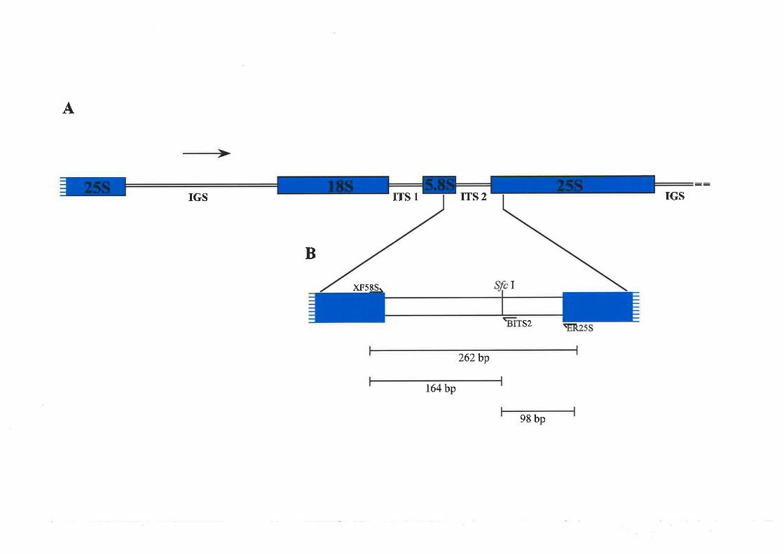
A
IGS ITS 1 IGS
B
s2
262bp
t64bp
98 bp
ITS 2
I)(F58

greater \^,ithin than among species, implying a homogenising mechanism u4iicli results in
the evolution of such sequences in concert (Dover, 1982).
Due to concefted evolution there are no substantial differences ilr sequence between
the several hundred to several thousand copies of the rDNA spacers within each species.
However, comparisons between species show differences indicating that they are not under
strorrg selection but free to diverge. There are also suggestions that concefted evolution can
act betweerr non-hornologons chromosomes when there are loci for a rnultigene family on
more than one pair of chromosomes within a species. In humans, for example, identity in
rDNA seqllerìces is maintained in five NOR regions positioned on non-homologous
chromosor-ne pairs (.Iorgenson el a|.,1992; Dover, 1993).
Alignnrent of the ITS sequences from different angiospenn families indicated that
plarrt sequences within ITSl and ITS2 regions have diverged very rapidly at the nucleotide
level conrpared with regions encoding the nrature IRNA species (Baldwin et a|.,1995)
The ITS regions have consequently been used extensively in phylogerietic studies of the
most recent evolutionary events including inter- and intla-species conrparisons (Baldwin et
o1.,1995; HershkovitzandZirnmer,T996). The use of the rDNA ITS regions in
phylogenetic studies has the advantage that u,ithin a species they are highly homogenised
thror,rgh concerted evolution but between species they shorv enough variation to enable
l'esolntion of species relationships. Therefore, despite the possibility of a homogenisir.rg
mechanism, it was thought feasible that the intemal transcribed spacer regions on the B
chromosol-ne rDNA could differ in sequence frorr those on the A chromosomes
Differences between the A and B chromosome rDNA could be attributed either to the B
chronrosome differing in sequence prior to its introduction into the B. clichrontosontatica
complement, or the independent divergence of ITS sequences on A and B cll'omosomes
over time associated with a different selection on these regions and a separate or absent
66

concerted evolution mechanism for the B chromosome rDNA clustels. Described in tliis
chapter is the sequence analysis undefiaken in order to identify any variation between the
intenral transcribed spacer regions on the A and B chromosomes of B. dichrontosontatica
3.2 AxaIYSIS OF.THE ITS 1 ANI ITS2 ON THE A ¿.xI B Crrnon,rosoMns.
A PCR approach was used to amplify ITSI and ITS2 from both A and B chromosome
rDNA, with primels designed using the nrelon ribosomal RNA sequence (Figure 3.2;
Kavanagh and Timmis, 1988). Forward primers XF185 and XF58S (Table 2.4b) were
designed specifically to the 3' ends of the 18S- and 5.8S-encoding regiorrs respectively
(Figure 3.18,3.2). Tliereverseprimers ER585 and ER25S (Table 2.4b) rvere designed to
anrplify frour the 5' ends of the 5.8S- and 25S-encoding regions respectively (Figure 3.18,
3.2). IntemalXba I (XF585, XF185) and Eco RI (ER25S, ER58S) restriction sites rvere
included in the primers (Table 2.4b) fo facilitate cJoning of the PCR products
3.2.1 ITS Sequences Amplifìed from the A Chromosomes
Using the genomic DNA from.B. dicltromosontaticct 0B plants as a tenrplate, XF18S
and ER58S (Figure 3.18,3.2) amplified a fragnrent of approximately 340 bp in size,
including primers, representing ITSI (result uot shorvrr). The positive control template was
a clorre of tlie entire cotton ribosomal repeat unit (pGhRl; Orford, 1996), from u,hich a
fragment of approximately 400 bp was amplified (result not shown). The
B. clichrontosontalica products obtained were restricted with ,Ðco RI and Xba I and cloned
into pBluescript@SK(-). Clones were selected by hybridisation with the original ITSl PCR
product (Figure 3.34, A'). Sequencing of positive ITSI clories (Figure 3.34') confirmed
that the PCR products were significantly similar to the ITSI of a closely related species of
the Asteraceae, Diplostephium rupestre, which showed 93% identity at the nucleotide level
(GenBank and EMBL, 1995).
6l

AÀCTGCGGA.A,
a caa a ct gtt
aacgcgttta
t cccct gccc
GGÀTCATTGt
cgcgttaggg
aacaaaaccc
cggcctcggc
acccgcgaac
t ggctgcct c
acttgaaatg
t agt cgt at t
gcgtttaaaa
ct ccccttcc
aatt cgcctg
actaaCÀÀCG
AAGTCCÀCTG AACCTTATCÀ TTIÀGAGGAA GGÀGÀAGTCG TAAC.AAGGTT TCCGTÀGGTGXFl8S
cAatgcctaa acatcaaacg
gcggggggaa gcatgctctt
cAcgcaggtc gcgccaagga
gtgcggqgga tggagcattc
ÀCTCTCGGCÀ ACGGÀTATCT CGGCTCTCGC ÀTCGATGAÀG .AÀCGT.AGCGA ÀATGCGÀTÀCERs8S
TTGGTGTGAÀ TTGCÀGGATC CCGCGÀÀCCA CCGÀGTCTTT GAÀCGCÀÀGT TGCGCCCGGÀ
GCCTTCTGGC CGAGGGCÀCG TCTGCCTGGG CGTCÀCFCaI cgctgccccc accacacaacXFs8S
tctccccatg cggggtcAtt gtgaaggcag ggacacacac tggcctcccg tacAcaccgt
cgtgcggatg gcttaaattt gagtcctcga tgctcgtcgt cgcgacacta cggtggttga
ttcaactcAg tgacAcgtct cgacctcgac gtcgacttca cggactcctt cacgaccctt
cgaacgccgc cccttaaaag gacgacgctc tcgacgCGAC CCCAGGTCAG GCGGGACTÀC
ER25S
18S
ITSl
5.8S
ITS2
25S
CCGTGÀGÀGT TTÀÀGCATÀT CAÀTAÀGCGG ÀGGÀJUU\GÀ.A ÀCTTÀCÀAGG ATTCCCCTAG
Fieure 3.2:
Nucleotide sequence of melon 185-25S region adapted from Kavanagh and Tirnmis (1988)
The intemal transcribed spacerregions 1 (ITSI) and 2 (ITS2) are shown in lowercase and
indicated on the right of the figure. The regions to r¡,hich the XF185, ER58S, XF58S and ER25S
¡rrinrers were designed are underlined. The 5' - 3' orientation of each pdmer is indicated by the
direction of the aüows
68

Figure 3.3:
A. Agarose gel electrophoresis of putative ITSI clones derived from 0B genomic DNA
(pBdrTslAl-e).
Each lane contains approximately 2 ¡rg of plasmid DNA restricted with Eco R[ and Xba I.
The numbers on the left of the gel represent the molecular sizes (kb) of lambda
bacteliophage DNA restricted witli lli¡rd IIL The identity of the clone in each lane is
indicated.
A'. Hybridisation of a Southem blot of the gel in A with the original ITSI PCR product
B. Agarose gel electlophoresis of putative ITS2 clones derived fiom 0B genomic DNA
(pBdlTS2Al-12).
Each lane contains approxirlately 2¡rg of recombinant plasmid DNA restricted with Eco RI
at'td Xbu l . The nurnber-s on the left of the gel represent the molecular sizes (kb) of lambda
bacterio¡rhage DNA restricted with //ind III. Tlie identity of the clone in each lane is
indicated.
B'. H1,þ¡idisation of a Southeni blot of the gel in B with the origirral ITS2 PCR product
amplified fi'om genomic DNA. The asterisks indicate the clones from which the inserts
were sequenced (Figure 3.4)
69

C. Agarose gel electrophoresis of PCR products resulting from arnplification across the
inseft of iridividual putative ITS2 clones derived from DOP-PCR-arnplified rnicrodissected
B chromosome DNA (pBdITS2Bl-10).
Each lane was loaded with l0 ¡rl of a 25 ¡tlreacÍion volume. The nutnbers on the left of the
gel represent the molecular sizes (kb) of lambda bacteriophage DNA restricted with
HindIII and the second lane contains molecular size markers (100bp DNA ladder)
consisting of fragmerrts that range in size from 100 bp to 1000 bp in 100 bp incretnents,
plus an additional 1500 bp fragment. Tlie identity of the template clone in each lane is
indicated.
C'. Hybridisation of a Soutliem blot of the gel in C with the ITS2 PCR product from
DOP-PCR antplified microdissected B chromosome DNA. The asterisks indicate the
clones from rvhich the inserts were sequenced (Figure 3.4)
70

A Ar
Br
cr
23.t
-
123456789
* dr ,k*
rt9.46.5
4.3
0.56
-
B
23.1-
2.32.0
9.46.5
,a.L6.54.3
2.32.0
1234567891011 12
4.3
-
0.56
-
J0
2.,,
tf
1234s6789101112
t2345678910C *
0.56 _
II¡ t
*n¡|rlr ¡ts s

Primers XF58S and ER25S (Figure 3.18,3.2) arnplified a product of approximately
280 bp from the cotton rDNA clone pGhRl and a 260bp product frorn DNA prepared
from a range of B. dicltromosontatica plants without B chromosomes (result not shown).
The putative ITS2 PCR products from the different 0B B. dicltromosonmtica plants were
cloned and recombinant clones selected by hybridisation with the original ITS2 PCR
product (Figure 3.38, B'). Sequence of the positive clone pBdITS2A2 (Figure 3.38, B';
lane 2) confinned that the product obtained was the ITS2 of B. díclrontosomatica. The
rnost closely related sequences in tlie database was found within another member of tlie
Asteraceae, Colotis dentex, and sliowed 91olo nucleotide identity across the amplified
r-egion (GenBank and EMBL, 1996)
Alignment of tlie nucleotide sequences obtained fronr clones derived from genomic
DNA of fourdistinct 0B plants (Figure 3.38, B';lanes 2,5,8 and 10), and tlierefore
representative of the A chromosome ITS2 seqLrences, revealed a low level of sequence
lreterogeneity between the individual 0B clones. Of the total2IS base pairs hve bases
differed irr one or Ír1ore individual clones (Figure 3.4), representing 2.3o/ovariation. The
sequence discrepancies probably represent genuine polymorphisms in the A clrromosome
ITS2, or altematively some or all may be due to PCR enors
3.2.2 ITS Sequences Amp lified from the B Chromosomes
The availability of B chromosomes only within an A chromosome background makes
it difficult to specifically analyse sequences on the B chromosolres, especially when they
are similar in sequence to counterparts on the A chromosolnes, as is the case for rDNA.
There was therefore a requirernent for a pure B chromosome DNA source and this
ir-rstigated the use of microdissection to isolate individual B chromosomes which were
snbsequently amplified according to Pich et al. (1994), using the degenerate
12

Figure 3.4:
Alignment of ITS2 nucleotide sequences from BdITS2A clones (42, 45, A8 and 410)
\ /ith BdITS2B clones (B 1, 82, 85, 86 and 87) and a single B. Iineariloba clone (BI).
Consistent differences between the ITS2 of A and B chromosome derived clones are
boxed. The Sfc I site (crecAG) present in the BdITS2B and.B. lineariloba sequences is
highlighted and polymorphisms specific to individual clones are indicated.
73
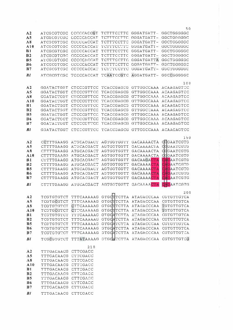
A2A5A8A10B1B2B5B6B7
BI
A2A5A8410B1B2B5B6B7
A2A5A8A'10B1B2B5B6B7
ÀTCGCGTCGC TCCCCACCAT
ATCGCGTCGCÀTCGCGTCGCl\ILbLtTIL(¡LATCGCGTCGCAILtTLl¡ILl¡LATCGCGTCGCA I LtrLb I LtrLA I LtrLtr l LtrLATCGCGTCGC
CCCCCÀCCGTCCCCCACCATCCCCCACCATCCCCCÀCCATCCCCCÀCCÄTCCCCCACCATr-r-r---Àr-r'À.PI,I-ULLALLA ICCCCCACCAT
TCTTTCCTTCTCTTTCCTTCTCTTTCCTTCTCTTTCCTTCTCTTTCCTTCTCTTTCCTTCTCTTTCCTTCTCTTTCCTTCTCTTTCCTTC
TCÀATCCGTC
trtr(:AIl:Al I-t¡LTbAIbAI I-GGGATGÄTT-l:l:l¡AItTAI I-l:btrAItrAI I-GGGATGÀTT-LrtiþAIl¡Al IAGGGÄTGATT-ljlJlJAIlJAII-
ÀGGATGÀTT-
50GGCTGGGGGCtrbL I trl¡bbl:l-GGCTGGGGGCLrLTL I (¡ljl:ljlrl-
GGCTGGGGGCGGCTGGGGGCtrtrL I bb(:trbLbl:L I l¡trl¡l¡trLGGCTGGGGGC
l:l:LUtrl:l:l:l:L
100ÀCÀAGAGTCC,4CAÀGAGTCC,ACAAGAGTCCALAA|JAb I I-LACÀAGÀGTCCACAÀGÀGTCCACAAGAGTCCÀCAAGAGTCCACÀAG.AGTCC
150GAATCGTGGAÀTCGTGtrAAILl:Il:GAÀTCGTG
ATCGTGATCGTGAILtrltrATCGTGAILtrItr
ÀTCGTG
200CGTGTTGTCACGTGTTGTCATGTGTTGTCATGTGTTGTCACGTGTTGTCÀLtrItrl ItrILACGTGTTGTCALl:ItrI -LtrlLACGTGTTGTCÀ
LtrI(:I I(¡IL(:
BI GGÀTÀCTGGT CTCCCGTTCC TCÀCCGAGCG GTTGGCCÀÀÀ ACAAGÀGTCC
GGATACTGGT(:bA I AL I trtr IGGATÄCTGGTGGATACTGGTLrl:A I AL I trtr IGGATÄCTGGT|¡LJAIALIl:l:Il:trA I AL I trtr IGGAT.ACTGGT
L-LL I Il¡Al\tJt:CTTTTGAAGGLt I r _tlJAAlJlJ
CTTTTGAAGGCTTTTGÀAGGCTTTTGAÀGGLI I I IbAAtrtrCTTTTGAAGGCTTTTGA.4GG
CTCCCGTTCCCTCCCGTTCCCTCCCGTTCCCTCCCGTTCCCTCCCGTTCCCTCCCGTTCCCTCCCGTTCCCTCCCGTTCCCTCCCGTTCC
ÄTGCÀCGACTÀTGCACGACT.ATGCACGÀCTA I IJLAL|JAL I
ATGCACGÃCTA I I:LALIJAL IATGCACG,ACTA I IJLALIJAL IATGCÀCGACT
TCACCGAGCGI LALLtrAl:LtrTCACCGAGCGTCÀCCGÀGCGl LALLl¡AtrLtrTCACCGÃGCGI LALLLTAbLItI LALLtrAl¡LtrTCACCGÀGCG
Al:l(:(:Itr(jI IAtrIblJItrtrI IAtrIbl:ll:l:I IAGTGGTGGTTAtrIl:l:Il:l:I IÀGTGGTGGTTAtrItr|:Itrl:I -t
ÀGTGGTGGTTAGTGGTGGTT
GTTGGCCÀÀÀGTTGGCCÄÀAGTTGGCC,qAAGTTGGCCÀÀAGTTGGCCAAAGTTGGCCAAÀl: I I trtrLLAAAGTTGGCCAÀÀGTTGGCCAAA
GÀCAAAÀCTAGACÀÀAÀCTÀGACÀAÀACTAGACAAAACTA
GACÀÀÀABI CTTTTGAÀGG ÀTGCACGACT AGTGGTGGTT
^2A5A8A'10B1B2B5B6B7
BI
A2A5A8A'10B1B2B5B6B7
BI
TCGTGTGTCTTCGTGCGTCTTCGTGTGTCTTCGTGCGTCTTCGTGTGTCTlLl:ltrIl:ILITCGTGTGTCTlUl:ItrItrlL-LTCGTGTGTCT
TCGCGTGTCT
TTTGÀCAACGTTTGÀC.AACGTTTGACÀÀCGTTTGACAACGTTTGACÀÀCGTTTGACAACGTTTGÀCÀACGTTTGACAÀCGTTTGACÄACG
TTTCÀAAAÄGTTTCÀÀÀÀÀGCTTCÀAAÀAGCTTCAAA,4AGTTTCAAAÄAGTTTCÀ.AAÀAGTTTCAAÀAÀGTTTCAAAAÀGTTTCAAÀÀÀG
TTTATAAÄAG
(J1lJ
l:ltrGTGtrltrGTGtrltr(JrbtrltrGTG
GTG
TCTTÀTCTTÀTCTTÀTCTTATCTTATCTTÀTCTTATCTTÀTCTTÄ
TCTTA
ATÀGÄCCCÀÀÀTAGÀCCCÀAÀTAGACCCÃAATÀGÀCCCAAÀTAGÀCCCÀAATÀGACCCAA.ATAGACCCAÀÀTÀGÀCCCÀAATAGÀCCCAA
ATAGÀCCCÀA
zLo
CTTCGACCCTTCGÀCCCTTCGACCCTTCGÀCCCTTCGACCl- I I LtiALLr-TTr-riÀ-r-CTTCGÀCCCTTCGÀCC
TTTGÀCAACG CTTCGACC

oligonucleotide prirner (DOP) MW-6 technique (Telenius et al.,1992). Attempts to
amplify the ITSI directly fi'om microdissected B chromosome DNA using primers XF185
and ER58S were unsuccessful suggesting that this region was either not complete or
uruepresented in the DOP-PCR-amplified DNA, possibly due to its large size
Altematively it may have been unrepresented in the template because of poor arnplification
by the DOP prirners. However, a similar PCR approach, using the ITS2 specific primers
XF58S and ER25S, successfully amplified a product fi'om the DoP-PcR-rnicrodissected B
chromosome DNA which was the same size as that obtained fi'om 0B DNA. Products
resulting fronr several individual PCRs u'ere cloned, as described previously, aud
recorrbinant cloues selected by liybridisation with the ITS2 PCR ploduct arnplified fron.t
DOP-PCR amplifìed nricrodissected B clrromosome DNA (Figure 3.3C, C')
A coniparison of the A chromosome ITS2 sequences rvith five individual insefts fron'r
nricrodissected, DOP-PCR-amplified B chronrosorne clones (Figure 3.3C, C') revealed an
aclditional three sequence variants, tlo of rvhich were consistently different between the A
and B chromosol.ne sequences (Figure 3.4). At base 142, an A was present in each B
chromosonle sequence rvhereas a G was present in all A chromosol'l'le ITS2 clones. In
addition, at base 175 an A was present in B chromosor-ue-derived ITS2 clones compared
rvith a T in those fi-om the A clrronrosomes (Figule 3.4). Fortuitously the substitution at
base 142 introduced a restriction site for the enzyme {fc I (CTACAG) in the B
chromoson-re ITS2 sequence which was not present on the conesponding A chromosome
sequences (Figure 3.4)
3.3 Axalvsls oF THE ITS2 IN OB AND +B GBxonuC DNA
To detennine whether the.!fc I polymorphism rvas a regular difference between the A
and B chromosome rDNA clusters, and not a cloning or PCR-based artifact, PCR of the
75

ITS2 using XF585 and ER25S primers was perfonned using genomic DNA from two
different 0B as well as individual IB,2B and 3B plants. The positive controls in this
experiment were provided by the inclusion of template DNA from a B chromosome-
derived ITS2 clone of known sequence (pBdITS2B1, Figure 3.4) and microdissected-B
chromosome DNA, wliich were amplified in parallel. The negative conûols were an A
chromosome-derived clone of known sequence (pBdITS2A2, Figure 3.4) and a reaction
lacking template included to check for contamination of the components of the PCR
reactiotr. A260 bp product, representing the ITS2 region, was amplified from each
genomic DNA santple and from the microdissected-B cluomosome templates, as well as
fi'onl lTS2 clones of A and B chrourosome derivation (Figule 3.5). Tl're absence of aPCR
product ili the control reaction (Figure 3.5) confimled that the expelirrental reaction was
specific and contained no contaminant DNA. The PCR products obtained rvere restricted
rvith ,fc I and visualised on 1.8% agarose gels alongside their unrestricted counterpafis
(Figure 3.5).
A proporlion of the PCR products amplified from 1B, 28 and 3B plants was digested
rvith.!fc I (Figure 3.5) and the amount of 164 and 98 bp restriction products appeared to
increase additively rvitli tlie number of B chrornosomes present in tlie sample. The 260 bp
atlplicon from 0B sarnples showed no cleavage upon digestion with ^!fc I (Figure 3.5),
supportirrg the hypothesis that ITS2 regions containin g an Sfc I site were found exclusively
on the B chromosome rDNA cluster
Upon restriction with .!fc I, botli the pBdITS2B 1 and microdissected B-derived PCR
products were reduced in their entirety to the two expected fragments, whereas the
pBdlTS2A2-derived PCR product showed no restriction (Figure 3.5). The irnplication of
the positive controls was that the ^!fc I enzynre was working to maxirnum efficierrcy
because the PCR product derived from the B cluomosome clone pBdITS2B 1 (known to
/o

Figure 3.5:
Agarose gel electrophoresis of ITS2-PCR products resulting fi'orn arnplification of
B. clichromosomatica genomic DNA using primers XF585 and ER25S
Each lane is loaded with a 10 pl sample from a reaction volume of 25 ¡tl, except for the
first lane which contains molecular size marker (100 bp ladder with visible fragments
ranging from 100bp to 700bp in 100bp increments). Lanes marked with "-" and "*"
contain PCR product before and after digestion with ^!fc I respectively. The sizes of PCR
and rcstriction fragnrents are nalked on the right hand side of the gel. The
B. clichrontosontatica chromosome constitutions, microdissected B-DNA (BDNA) and
ITS2 clones frorn which DNA ter.nplates were derived are indicated
77

500 bp-
+ 260bp
+ t64bp
<- e8 bp
-o-¡L¡röööü Õfó-
J

contain Tbe Sfc I site, Figure 3.4) was cornpletely reduced to its two component fragments.
Furlhermore, the observation that all ITS2 regions cloned frorn the DOP-PCR products
obtained fi'om nricrodissected B chromosomes contained the .!fc I site (Figure 3.4),
togetlier with the demonstration of complete digestion of sequences amplified directly frorn
microdissected DNA (Figure 3.5), suggested that all the B cluomosome rDNA repeats have
an A at position 142 of the ITS2 region whereas those on the A cluomosomes all have a G
at this position. However, the possibility that DOP-PCR preferentially arnplified the A,o,-
substituted DNA and the presence of G,o, on sonÌe ITS2 sequences of the B cluomosone
could not be ruled out.
In parallel witli the above experiments, the ITS2 region was amplified frorn a closely
related species of Bruclrycome, B. lineru'ilobct race C (Section 3.4). The resulting PCR
product, of approximately the sarne size as that arnplifìed fron B. clicltrontosontatica,was
cloned and sequenced. Alignnent of the B. lineurilobalTS2 seqlreuce (Figure 3.4, B/) rvith
those of the A alld B chromosomes ofB. clichrontosontcttica revealed ahigh level of
siniilarity (Figure 3.4). The B. lineuriloba ITS2 also had an A at base 142 rvithin a ^fc I site
sin-rilar to the B. clichrontosomatica B chronrosome rDNA. B. linearilobu race C has a
chrorlosor-ne contplement of 2n:76 and does not have any B chronosomes (Carler and
Smith-White, 7972). The presence of an A at base 142 it^t B. lineariloba race C suggested
that the ^!fc I site would be found in all the ITS2 sequences in this species because of
concerted evolution (Section 3.1). The ITS2 regions fi'om 0B and 4B B. dicltrontosontatica
and .8. linectriloba tenrplate DNA were amplified in the presence of [cr-32P]dATP, the
products were digested with .!fc I and the fragments resolved by electrophoresis on a 6o/o
polyacrylamide gel (Figure 3.6). The ITS2 product of ,8. linecu'iloba was restricted in its
entirety in to two products of 164 and 98 bp (Figure 3.6, Bl), confinning that B. lineariloba
contained only ITS2 sequences containing the,!fc I site. In addition, a proportion of the
19

Figure 3.6:
Polyacrylarnide gel electrophoresis of ITS2-PCR products resulting frorn amplification of
genomic DNA with primers XF58S and ER25S in the presence of [o-32P]dATP.
Each lane is loaded with a I pl sample from a reaction volume of 25 pl. Lanes marked with
"-" and "*" contain PCR product before and after digestion with ^!fc I respectively. The
sizes of PCR and restriction fragments are marked on the right hand side of the gel. The
chronlosome constitutions and species of origin of the plants from which the leaf DNA
ternplates were derived are indicated (Bd : B. dichrontosontatica, Bl : ^8. lineariloba)'
80

Bd
OB 4B
l- +l- +¡BI
+
<- 260bp
+ t64bp
+ 98bp

ITS2 product amplified from DNA from B. dicltrontosontaticct plants with 48
clrrornosornes was restricted with .ffc I and there was no evidence of any restriction of the
product fi'om 0B DNA template (Figure 3.6). B. lineariloba template nucleic acids were
therefore incorporated in the following experimerrts to nronitor the efficiency of restriction
enzyÌne digestion.
To further test whether the A,o, base was representative of the B chromosorne rDNA
and not of the A chromosome rDNA, ¡cr-"P1dATP was incorporated during PCR
anrplification from total DNA of 08, 18,28,3B and 4B B. clicltrontosontatica plants and
frorn.B. lineariloba. The products were digested with ^!fc I, as before, and fragments
resolved on 60/o polyacrylamide gels (Figure 3.7). The radioactively labelled products
arrrplified fi'onr B. linearilobu and a proporlion of products from 1B, 28, 38 and 48
genomic DNA rvere cleaved by Sfc I into the trvo expected fragluertts (Figure 3.7). Again
tlrere u,as no digestion u,ith ^!fc I of the PCR products originating froru any 0B genomic
DNA tenrplate (Figure 3.7).
Despite previously being totally restricted rvith ,fc I (Figure 3.6), the B. linecu'iloba
ITS2 product in Figure 3.7 did not show complete digestion. This suboptirnal enzyrne
efficiency rvas not considered likely to be present in other samples rvhere the amount of
DNA rvas clearly less (Figure3.7). The prominent band with mobility between the 164 and
98 bp products was caused by the presence of non-denatured PCR products,
The proportion of the ITS2 PCR product that was digested with ^!fc I consistently
corelated with the number of Bs present (Figure 3.7). Pliospholimage analysis of tlie
proporlion of the original ITS2 product that was restricted by ^!fc I (Appendix 1),
confinned this direct correlation aud indicated that 3-4o/o of the ribosornal genes in a 1B
plarrt contain the ^!fc I site. A.n average of 6%o of the ITS2 product from a 28 DNA template
was restricted with SfcI,9-I0o/o from 38 and 13%o fi'om a 48 sarnple (Figure 3.8). The
82

Figure 3.7:
Polyacrylarnide gel electrophoresis of ITS2-PCR products resulting fi'om amplification of
genornic DNA with primers XF58S and ER25S in the presence of ¡a-32P1dATP
Each lane is loaded with a I ¡rl sample from a reaction volume of 25 ¡-rl. Lanes rnarked with
"-" and "*" contain PCR product before and after digestion with ,lfc I respectively. The
sizes of PCR and restriction fragments are marked on the right hand side of the gel. The
chlomosome constitutions and species of origin of the plants from which the leaf DNA
tenrplates rvere derived are indicated (Bd : B. dicltontosonruticct, Bl :.8. lineariloba).
83

+ll-.ll-.ll- + .ll-

Figure 3.8:
The relationship between the amount of the ITS2-PCR product which restricted with .ffc i
and the number of B chromosomes present in template DNA sarnples.
85
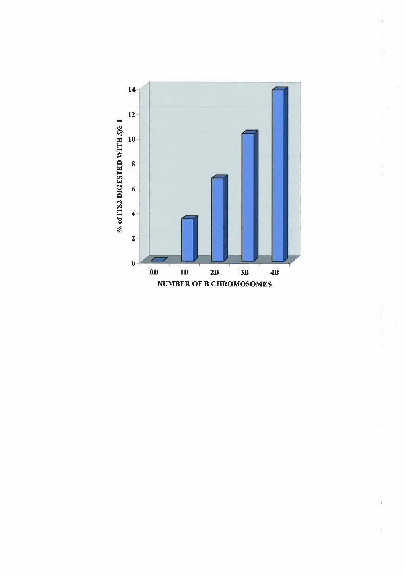
l4
t2
.()àE10IiÈ
Ê8FV)t-lg6âNrn
l4v
2
0
OB 1B 2B 38 4B
NUMBER OF' B CHROMOSOMES

variation noted in different estimates (Appendix 1) rnight reflect real variation in the rRNA
gene number ou the A and B cluomosomes in different genotypes, as well as technical
errors. The level of enzyme efficiency was monitored tlrroughout these experiments by
inclusion of the B. lineariloåa, which contains only dfc I containing ITS2s. It was
collcluded from these experiments that the iTS2 in the B chromosome rDNA cluster of
B. clichrontosontalica is homogeneous for an A at base 142 wliich differs from the G
present in the A chromosome ITS2. The difference creates a Sfclrestriction site specific
for the B cluourosome rDNA which enables the ITS2 of B and A chromosomes to be
readily distinguished. Quantitative estimates indicate that 3-4o/o of the total rDNA
popuJation is colttributed by a single B chronrosome.
3.4 AxalYsrs oF orHBR Spncms oF BRACHyIzME.
Tlre origin of the B chromosonres in B. clichrontosontuticct is unkuown, but it is
possible they origirtated through a hybridisation event betu,een B. dicltromosontrtticct and
arrotlrer Brachyçz¡re species (Section I.7.2;Jol-ntet al.,1991;Leachet ct\.,1995).
B. lineoriloóa rvas originally divided into five races (A-E) which differed in
chl'olllosol-ue uumber. Races A and D were later identified as a different species
(8. dichrontosontatica and B. breviscapis respectively; Carter, 1978b). The presence of a
B-specific restriction site in the irS2 region of the B chromosomes of
B. clichrontosontatica, which is also present in the A chromosomes of B. lineariloba race
C, suggested that a screen for this site in the A chromosome ITS2 regions of other species
may provide infonnation about the geriomic origin of the B chromosorìes.
Amplifìcation of the A chromosome ITS2 region fi'om a range of different
BraclT,com e species, followed by restriction with ^!fc I and visualisation on agarose gels
(Figtrre 3.9) indicated that, in addition To B. lineat'iloba race C (2n:I6),tlieITS2 region of
8'7

twootherchromosomeraces of B. linearilobnraceEandB(2n:10 and2n:I2) asweÌlas
B. breviscaprs (formerly B. lineariloba race D, 2n:8) also contained the ^ffc I restrictiorr
site. However, the site was absent from the A chromosome rDNA of B. tetrapterocarpct,
B. curvicat'pa, B. clentata, B. eriogorn, B. parvula, B. multifida, B. segmentosa and B.
cilictris (Figure 3.9).
Previous comparisons of the karyotypes of races E (2n:10), B (2n:12), C (2n:16) and
B. breviscaprs (fomrerly B. lineariloba race D, 2n:8) suggested tliat these races have a
basic set of four chromosome pairs in common and the higher clrromosorue types are
related to the fomrerraceD by successive chromosome additions (Kyhos et a\.,1977:
Watanabe and Snrith-White, 1987). It was therelore not unexpected that the ITS2
sequellces of races B, D and E are sirlilar to race C (Section 3.3) rvith respect to digestion
n,itlr S/c I. The relationship of the A chronrosonres of B. clichrontosontaticct (fonnerly race
A of B. lineariloba) with B. brevisccrpis and theB. lineurilobu races is not known but is
thought to be lrore distantly related (Watanabe and Smith-White, i987)
3.5 CoxcLUSIoN
Sequence cotnparisons of the ITS2 region fronr the A and B chronrosor-ues of
B. clichrontosotnrtlicct and tlie A clrromosomes of B. Iineuriloba provided sorne clues to the
possible origin and evolution of the B chromosome IDNA. Superfìcially the presence of
tlre A,o, and A,r, nucleotides in B. lineat'iloba ITS2 suggests an origin of the B
chromosonle rDNA in the B. lineariloåa species. However, full sequence comparison of
the ITS2 derived from the A chrolnosomes of B. lineariloba (race C) and
B. tlicltrontosontctticct witli the B chromosomes of B. clichrontosontatica indicated that the
entire B chronrosorre ITS2 sequence was more similar to tliose of the A clrromosome
rDNA cluster of its "host" genome than that of B. lineat'iloba. The ITS2 of B. lineat'iloba
88

Figure 3.9:
Agarose gels showing ITS2-PCR products resulting from amplification of geuomic DNA
from a range of Brachycorne species using primers XF58S and ER25S.
Each lane is loaded with a 10 ¡rl sample fi'om a reaction volume of 25 ¡ll, except for the
first lane u,hich contains molecular size marker (100 bp ladder) with visible fragnrents
ranging fì'om 100 bp to 700 bp in 100 bp increments, Laues marked with "-" and "*"
contain PCR product before and after digestion with ^!fc I respectively. The size of PCR
and restriction products are l.narked on the right hand side of the gel.
The tenrplate DNA was derived fi'om the following Bracltycorrre species:
A. B. linectriloba Race C (2n:16)
B. B. lineurilobu Race E (2n:10)
C. B. lineuriloba Race B (2rr72)
D. B. bret,iscctpis (fonlerly Race D, 2n:8)
E. B. tetraplerocarpu
F. B. cut't'iclrpú
G. B. clentctta
H. B. eriogotta
l. B. pun,ula
J. B. rttultif da
K. B. segntenlosl
L. B. ciliaris
N. no DNA template (-ve control).
89

ABCDE F'
+l + + + +
GH I J KL
+
500 bp-
500 bp-
+l + l- + lN
< 262bp
<- I64bp
+ esbp
+ 262bp
+ + +
tlÞC|JS * #staüf ü* r#
-

90

race C, in addition to the,!fc I site and A/T substitution at position 175, also differed fron
any of tlre B. dicltrontosontatica ITS2 B or A ch¡omosome sequences at nine additional
sites (Figure 3.4). Alignment of angiosperm ITS2 sequences identified six regions that
were conserr¡ed in all sampled angiosperms and six altemating variable regions
(Herslikovilz and Zirnmer, 1996). With one exception, the polyniorphisrns observed
between B. dichrontosontatica and.B. lineariloba were confined to the variable regions as
rvould be expected for variation within a genus (Hershkovitz and Zimmer, 1996).
It is possible that the 4,,, and A,r, nucleotide substitutions irr the
B. rlicltrontosontalica B ch,tomosomes and B. lìnecu'iloð¿ A chronrosontes did not occur
independently. Horvever, the presence of nine additional nucleotide differences between
ITS2 of B. dicltrotttosotnatica Bs and B. litrcctrilobu tndicated that the B chromosor-ues ir-l
B. dicltrontosontctticct did not evolve directly fi-on B. littearilobu. Instead they may have
shared a common ancestor, plior to the 'introduction' of the B chromosolles into
B. rlicltrotnosontulicct. B. lineariloóa rvould subsequently have evolved independently of
the B. clichrontosonnlicct A and B chromosome rDNA sequences (Figure 3.10a), rvhich
explains the variatiou betrveen the ITS2 sequeuces of the two species (Figure 3.4). If the B
clrrorrrosome evolved from an ancestor conrmou u,ith ,8. lineurilobu, as suggested irr Figule
3.104, the B clrrorrosorre ITS2 has been separated fron-r the A chronrosome of
B. clichrontosontotica for longer than it has been separated fi'om,B. linecu'iloba. Therefore,
nlore differences would be expected between A and B chromosome sequences. As this is
not the case one nrust invoke a mechanism that homogenises, to arl extent, while tolerating
a degree of variation.
The low level of variation between the ITS2 sequences derived from the B and A
clrronrosomes of B. clichrontosontatica irnplies that it is more likely that the B chromosome
rDNA originated fi'om the A clrrontosomes of B. clichrontosontatica and that the sequences
9l

have since diverged (Figure 3.10b). The fact that the B. dichrotnosontatica B chromosomes
and,B. lineariloba A chromosomes have in common the A,o, and A,r, sequences would
therefore be fortuitous and this is possible considering these nucleotides are present in
regions of the ITS2 which commonly show variation between species (Hershkovitz and
Zimmer,1996). The presence of the A,o, and A,r, sequences in all the ITS2 regions
amplified from the B chromosomes suggests that they evolved in concert with each other
and not with those of the A chromosomes of ,8. dichrontosonntica.
92

A. B.
Bd dB BI A BdB BI
Figure 3.10:
Possible patliways for the evolution of the B. clichrontosotnaticct B chromosome (Bd B)
ITS2;
A. fronr the .B. Iinecu'iloba A chromosonre (,8/) ITS2, befole B chromosome 'introduction'
into .8. dicltrom osont cttica.
B. fì'onr tlre B. dicltrontosontatica A chromosome (^Brl A)
93

CHAPTER 4:
Transcriptional Analysis of Ribosomal
Ri\A Internal Transcribed Spacer 2
94

4.1 lNrnotucuoN
Tliis chapter describes experiments to detennine if the ribosomal RNA genes on the B
clrronrosome of B. dichromosonrctica are transcribed.
The initial transcript of a ribosomal RNA gene is a nominal 40S precursor RNA from
which the regions corresponding to ITS1, ITS2 and the RNA flanking tlie 18S and 25S
regio¡ts are rertoved during processing to yield the three structural RNAs of the mature
ribosornal RNA species (Section I.6.2a; Solhrer-Webb and Tower, 1986; Kavanagh and
Tirnmis, 1988). Within total RNA the transitory 40S RNA specìes is present for a period of
tine following IRNA transcription and then during the processing of the 40S pre-rRNA the
ITS regions are removed (Sollner-Webb and Tower, 1986). Therefore, if the B
clrrorrosonle rRNA genes of B. dichrontosomatica aL-e transcribed, then rvithin the total
RNA of a *B plallt, B chronrosome ITS2 sequences would be expected in proportion to the
number of B chrontosomes present (Chapter 3).
4.2 Tnnx ONAL AN¿.TYSTS OT THE ITS2 ON THE B ON,TOSOI\{ES
IN LEAF TISSUN.
Total RNA was prepared from leaf rnaterial from the salne 08, 18, 28,38 and 48
reverse transcribed using the ER25S primer (Figure 3J8,3.2; Table 2.4b) and the single
stranded DNA products PCR-amplif,red using the XF585 and ER25S primers. The RT-
PCR products were restricted with.!fc I and resolved alongside their unrestricted
counterparts on a 60/opolyacrylamide gel (Figure 4.1).
As expected, RT-PCR from the 0B plant showed uo restriction of the 262bp product
rvith,$fc I (Figure 4.1) indicating that none of the IRNA transcribed in 0B plants contained
the ,!fc I site. This supported the previous conclusion that the ITS2 regions of the IRNA
95

Fieure 4.1:
Polyacrylamide gel electrophoresis of RT-PCR products resulting from amplification of
total leaf RNA with primers XF58S and ER25S in the presence of [cr-32P]dATP.
Each lane is loaded with I pl of sample frorn a reaction volume of 25 pl. Lanes marked
with "-" and "*" contain RT-PCR products before and after digestion with,lfc I
respectively. The sizes of RT-PCR and restriction products are marked on the right hand
side of the gel. The chromosome constitutions and species of origin of the plants from
wlrich tlre leaf DNA ternplates were derived are indicated (Bd : B. clichrontosontatica,Bl:
B. litteoriloba)
96

Bd
OB
-+3B-+
4B BI-+ -+
< e8bp

gerles on A chromosones did not contain any Sfc I sites (Chapter 3). In contrast, in
B. lineariloba,where all copies of the rRNA gene contain an ^þ I site in the ITS2 (Chapter
3), tlre RT-PCR products were efficiently and fully restricted by,ffc I (Figure 4.1).
However, in B. dichrontosontatica plants containing B chromosoûìes, where a proporlion
of the rRNA gerles rvere demonstrated to contain the ^!fc I site (Figure3.7), there was no
evidence that any of the RT-PCR products were derived fi-om these genes (Figure 4.1)
Frorn the above lesults it was apparent that the rDNA located on the B chromosome
rvas not transcribed in proportion to its abundance in the nuclei of B chromosone-
containing individuals. However, low levels of transcription could not be ruled out.
4.2.1 Detection of Lorv Levels of B Chromosonre rRNA Transcrirrtion.
An experiment designed to detect low levels of transcription of the B chromosone
rDNA utilised the specific primer BITS2 (Figure 3.18,4.2; Table 2.4b) and this approach
n,as first tested on DNA templates. The B-specific prinrer contained a diagnostic base at
the 3' end which was designed to amplify exclusively fron.r ^!fc l-containing ITS2 regions
(Figure 3.18,4.2). At a PCR amealing temperature of 70'C the BITS2 and XF58S primers
(Figule 3.1F_,3.2) gave rìo product from 0B genomic DNA (Figure 4.3). However, they
u,ere efficient in amplifying the 164 bp product from 4B genomic DNA of a
B. clicltrontosomaf ica plant as well as B. lineariloóa DNA (Figure 4.3), supporting the
previous conclusio¡r that orily the,B. dicltrontosotnuticct B clrromosome and the
B. lineuriloba|TSZ regions have an A at base 142 of their sequence.
To explore the sequence of the 40S pre-rRNA in the RNA population, oDNA was first
synthesised from total RNA of 0B and +B plants using ER25S as a primer and the single
stranded molecules produced were then amplified with BITS2 and XF58S. No BITS2-
specific products were evident in samples originating from either 0B or 4B
98

5.8S<- -+25S
126 AGTGGTGGTTGACNUU\CTACGGAATCGTGTCGTGTGTCT 160 pB<lIrs2A2 (A chromosome)
120 AGTGGTGGTTGACNUU\CTACAGAATCGTGTCGTGTGTCT roo pBdIrS2Bl (B chromosome)
:' TCTTAGCACAGCACACAGCG 5' Brrs2primer
Fieure 4.2:
Region of the ITS2 to which the BITS2 primer was designed.
Bases 120 - 160 of the ITS2 sequence of pBdITS2A2 and pBdITS2Bl, and their
orientation in relation to the 5.8S and 25S coding regions are shown. Within the
pBdITS2Bl sequence the Sfc I site is in bold and the differential A at base 142 is in red.
The sequence of BITS2 primer is shown with the S/c I site-specific T in red.

Figure 4.3:
Agarose gel showing leaf PCR and RT-PCR products amplified using BITS2 and XF58S
pnn]ers.
Each lane is loaded with a 10 pl sample from a reaction volume of 25 pl, except for the
first lane which contains molecular size markers (100 bp DNA ladder) with visible
fragrrrents ranging from 100 bp to 500 bp in 100 bp increments. The chromosome
constitutions and species of origin of the plants from which the DNA and RNA templates
were prepared are indicated. ND and NR represent the no DNA ternplate and no RNA
template, negative controls
100

Bd
loBl4Bl BllNDl500 bP
-
+ 262bp
+ t64bp
DNA RNA
B. clicltrontosontatica plants (Figure 4.3) but the expected 164 bp product was present in
anrplifications of RNA from B. lineariloba (Figure 4.3). The RT-PCR products of 262bp
in tlre cDNA lanes (Figure 4.3) represent the entire ITS2 which was amplified due to
residual ER25S primer being carried over from the reverse transcription reactions and the
presence of this product was useful as an indication that the PCR was efficient in
individual reactions.
The results obtained using the potentially more sensitive BITS2 prirner assay strongly
supported the previous results obtained. It was clear, therefole, that the 4,r.,-type ITS2
sequences, which are considered to be a component of all of the rRNA genes of the B
chromosonte, do not give rise to a 40S transcript in leaf tissue and therefore these genes do
not contribute to the 80S ribosomes in the cytoplasni of a plant rvith B chromosomes
It rvas apparent fronr the preceding experintents that the rRNA genes on the A
chronrosontes are preferentially expressed in leaf tissue, indicating that the B chrorrosome
rDNA is inactive or that sonre fom of nucleolar doninance is occuring. Nucleolar
dominance is an epigenetìc phenomenon that occurs when one set of ribosomal RNA gerÌes
is expressed to the exclusion of another in species hybrids or allopolyploids. Suppression
by nrechanisms related to those involved in nucleolar dominance may occur wliere the
rDNA clnster on the A chromosomes is preferer-rtially expressed, at the expense of those on
the B, but this is irnpossible to test because material whicli contains only B chromosome
RNA is not available.
A feature of nucleolar dominance in.Brassica hyblids is tliat sets of rRNA genes which
are inactive in leaf tissues are expressed in all floral organs (Chen and Pikaard,1997).Il
u,as tlrerefore possible that the B cluomosornes of .8. dichrontosomatica, though inactive in
leaf tissue, may be active in floral tissues. This idea of differential activity in different
tissues is supporled by the previously-reporled regular association of B chromosomes with
102

ntrcleoli during meiosis (Donald et a|.,1995). In addition, during the filst pollen grain
division, which results in the generative and vegetative nuclei, the B chromosomes in
B. clicltrontosomalica often undergo non-disjunction and directed segregation into the
generative nucleus which tlien goes on to form the rnale gamete (Section 1.8; Smith-V/hite
and Carter, 1981). It is tluough this process that the B chrornosomes in this species are
maintained and if the B chromosomes are transcriptionally active at any stage of
development, it may be during this important stage
4.3 TnqNSCRrprroNAL ANALysrs oF THE ITS2 oN THE B Csnonrosor\{Es rN
Flon¡.1Trssup
Total RNA prepared from florver buds, rvith anthers at various stages of pollen
deveìopmeut, was reverse transcribed rvith ER25S as previously desclibed and PCR-
anrplified with BITS2 and XF58S primers. A 164 bp product, representing iTS2 sequences
rvlriclr contain an A at base 742 (Figure 3.4,4.2), rvas amplified from B. lineariloba and
not frorn any of tlie 08, I B, 28,38 or 48 floral RNA templates included in the experinent
(Figule 4.4). There was therefore no evidence for any transcription of Sfc I-containing ITS2
regions in the floral tissues. Thepresence of the 262bp RT-PCRproduct in all samples
was due to the cary over of ER25S prirner from the reverse transcription reaction and
showed tliat RT- PCR was efficient in each reaction. It was therefore concluded that there
was no tlanscription from the B chromosome rDNA in floral tissue, which included pollen
motlier cells at various stages of meiosis and pollerr grains at a range of stages of matulity
4.4 SuuuARY AND Drscussrox
In summary, results from these experiments indicate that the rRNA genes on the B
clrromosomes in B. dichromosotnatica are consistently maintained in an inactive state in
r03

Fieure 4.4:
Agarose gel showing flower RT-PCR products, from RNA of flower tissue, amplified
using BITS2 and XF58S primers.
Each lane is loaded with a 10 pl sample from a reaction volume of 25 ¡r1, except for the
first lane rvhich contains molecular size markers (100 bp DNA ladder) with visible
fragnrents ranging from 100 bp to 500 bp in 100 bp increments. The chromosome
constitutions and species of origin of the plants fronr which the RNA ternplates were
prepared are indicated. NR represents the no RNA teniplate, negative control.
104

Bd
lssl ggl rnl znl 3sl4gl g¡l
500 bp -
<- 262bp
.Ç t64bp
RNA
. G lï É.' ilÞ
-r-_-*
ælËæ
1#

leaf and floral tissues and that they do not contribute to the ribosomes present in a plant
rvith B chromosomes. The level of sensitivity of the experiments discussed, including those
witli the use of the B-specific prirner BITS2, is not ceftain but it is likely to be high. It is
clear that the B chromosome sequences are not transcribed in proportion to the amount of
rDNA present on the Bs, as estimated by phosphorirnage analysis (Section 3.3), and it is
probable that they are conrpletely inactive
Plants are known to carry from a few hundred to 31 000 rRNA gene copies per diploid
cell (Ingle et al.,1915) and evidence suggests that superfluous IDNA exists in some plants
tlrat nray comprise 90o/o or r¡ore of the total (reviewed in Rogers and Bendich, 1987a). No
association has been found between rRNA gene number aud phenotype, r,igour or rRNA
corrlent (Buesclier et u1.,1984) alld it is not understood rvhy plants maintain such a high
nunrbel'of rDNA repeats rvhich are inactive. Holever, it is possible that the inactive B
chrontosonte rDNA is under the control of sinlilar repression mechanisnrs. Altematively B
chrorlosonte rDNA activity may be suppressed by nucleolar dominance (Section 1.6.2a)
imposed by the A chronrosome genes. The mechanisms behind the control of nucleolar
dorlrinance are unknown but it is often associated with variation betrveen the IGS lengths
of the different sets of rRNA genes. It is unlikely that spacer structure alone is lesponsible
for nucleolar doninance and several epigenetic phenonrena, including DNA methylation
and clrrouratilr utodification, may have a nrajor influence iu cases where changes in geue
expression are nritotically or meiotically stable, as seenls to be the case for the
B. rlicltrontosotttct|ico B chomosome rÐNA
4.4.1 lntet'se¡ric Snacer \¡ariation in the Reoulation of rRNA Gene F,xnression
Variation in the length of the IGS is a comlnon feature of rRNA genes in a range of
plant species and it is thought that increased length of the intergenic region of a IRNA gene
relative to that of other rRNA genes in the cell, may play a role iu determining the potential
106

activity of the gene (Sard ana et al., 1993).In plants and animals, most spacer length
variation is due to differences in the number of tandemly repeated IGS subrepeats thought
to include RNA polymerase I enhancers (Sardana et a1.,1993; Flavell et a\.,1986;
Hernleben ef al., 1988). An appealing hypothesis to explain nucleolar dominance has been
that the dominant rRNA genes have more transcriptional enhancers which enables them to
outcompete other genes for transcription factors because of higher binding affinities to
transcription factors present in rate lirniting quantities. In hexaploid wheat, for example,
the longest IGS region is consistently associated with the highest levels of rRNA gene
activity (Sardana et a|.,1993). Furllienrrore the NOR of tlie diploid, Aegilops umbellttlaÍa,
which llas rRNA genes with even longer spacers suppresses all native u,heat NORs
(Martini et al., 1982). However, in hybrids of Brassica spp., the rRNA genes with the
shorter IGS region are expressed to the exclusion of those with ìonger IGS regions (Chen
and Pikaard, 1997), indicating that nucleolar dominance is rrot alu,ays associated with
lerrgtlr variation. Moreover, in ArabÌdopsrs, switches in donrinance betu,een different sets
of rRNA genes inrply that nucleolar dominance is not due to simple intrinsic numerical
affìrrity for transcription factors (Chen et a|.,1998)
4.4.2 DNA N{ethvlation in the Regulation of rRNA Gene Expression
Extensive nrethylation of CpG and CXG sequences is comrlon in plant DNA and
rDNA is on average nore heavily metlrylated than the bulk of uuclear DNA (Ellis et al.,
1983; Steele-Scott et al., 1984; von Kalm et al., 1986; Flavell et al., 1988). In plants with a
relatively large nunber of rRNA gene repeats, a high pelcentage of IDNA is not
transcriptionally active and in these species, it has been found that the cytosine rnethylation
levels are related to rRNA gene expression (Flavell et u\.1988; Jupe and Zimrner,7993;
Torres-Ruiz aud Hemleben, 1994). The link between rrethylation and gene inactivation is
not understood, but there is a strong and well established conelation between reduced
107

cytosine nlethylation of the rRNA promoter region and enhanced NOR activity (Flavell er
a1.,1988; Jupe and Zimmer,1993). It is not known whether demethylation in rDNA is the
result or the cause of gene activation. The treatrnent of wheat x rye interspecific hybrids
rvith 5-azacytidine, which inhibits cytosine rnethylation, stably reactivated the suppressed
rRNA genes of rye origin (Neves et a1.,1995), suggesting that methylation has a dilect role
in rDNA suppression.
Despite tlie fact that it has been shown that the rnethylation of IDNA is
irrdistinguishable between 0B and +B B. dichrontosontctticct plants using Southem analysis
(Donald et al., 1995), the possibility that tlie B chrornosonle rRNA promoter is highly
ntethylated ill relation to the active A chromosome rRNA promoter, cannot be ruled out. Ill
B. clichrontosontcttica there is a high copy uuurber of rRNA gelles oli the A chromosonìes,
ofrvliichamajorityarentethylated(Donald eta|.,1995). Itislikelythatthepresenceof
B chroutosome rDNA ntethylation would be difficult to detect al¡ove the background of
that are present on the A chromosomes and the requiret'nent for a neans of distinguishing
betrveen A and B chromosome IGS regions becotnes appareut.
It is possible that B chromosome rRNA genes are inactivated by nrechanisms other
than methylation and that methylation is secondary to inactivatiort rather thau its primary
carìse. In the grasshopper E. plorans embryos, for example, the B-rRNA genes are inactive
irrespective of their rnethylation status, suggesting the involvernent of other
regulatory/silencing factors (López-León et a|.,1995). hi this species, as assessed by silver
staining, thsrRNA genes can be activated by translocation to the A cluontosornes,
indicating that the chromosomal environrrent of the rRNA genes influences whether or not
tlrey are active (Cabrero et a|.,1987).
108

In addition to rnethylation, there is evidence that chromatin stntcture also has a role in
regulating rDNA activity, In rnaize hybrids, for exarnple, in which one parental NOR is
dominant, a proportion of the rDNA is highly rnethylated, DNase I-r'esistant and exhibits
lorv levels of transcription. In contrast, the expressed rÐNA repeats are undermetliylated
and DNase I-sensitive. DNase I sensitivity indicates tliat the IDNA is in cluomatin with an
open confonnation and therefore accessible to DNase I digestion. Therefore, inmaize
increased susceptibility of rDNA to DNase I digestion, together with undenlethylation, is
associated witli rRNA gene activity (Thompson and Flavell, 1988; Jupe and Zimmer,
l 993).
The packaging of DNA within chromatin is seen increasingly as havitrg an essential
role iu the legulation of nuclearprocesses. The levels of histone H4 acetylatiolt in nritotic
chromosomes reflects, and possibly detennines, potential transcriptional activity (Jeppesen
arrd Turler, 1993; Houben et a\., 1997a).lt is also thought that cytosine methylation may
directly affect the structure of the clrromatin and this may be involved in the inheritallce of
repressed chromatin states. Evidence suggests that liistone deacetylases are guided to
specific clrronratin domains by underlying genonic nrethylatiott pattems and that
methylation-dependent transcriptional silencing relies on histone acetylation
(Bestor, 1998).
Differences in the level of histone H4 acetylation and tirring of DNA replicatiort
betweerr A and B cliromosomes of ,8. dichrontosontatica have been demonstrated (Houbert
et ctl.,1997a). The core histolres are very basic proteins, the N-teminal tails of rvhich
contain four lysìne residues (5,8,12 and i6). These lysir-re residues are acetylated, or
deacetylated, in various patterns correlated with gene activity which they may regulate by
¡11odifying uucleosome assernbly or interaction with other ploteilis, including RNA
109

polymerases alld transcription factors. The B chromosomes of .8. dichromosontaticalabel
weakly with antibodies against H4Ac5 and H4Ac8, in comparison to that seen for the A
clrromosomes. In contrast, the distributions of H4 acetylated fonns of lysine 12 and 16
were indistinguishable between chromosome types. The B chromosomes were also found
to be late replicating in comparison to the A chromosomes wliich is characteristic of
inactive chromatin (Houben et al.,1997a). The mechanism by which H4 acetylation and
late replication might control the transcriptional inactivity of cllomatin of B chromosomes
remains to be established, but it is possible that the Bs may be in a cluomatin confomration
which is inaccessible to RNA polyrnerases and transcription factors.
ll0
ha ter 5:
Cloning of the Ribosomal
RI\A Repeat Unit
ltl

5.1 IxrnoDUCTIoN
Sensitive experirnents described in the previous chapter indicated that the ribosomal
RNA genes present on the B chromosomes of .8. dicltrontosontatica are not transcribed.
Previously, Southem hybridisation of 0B and +B genomic DNA, restricted with a variety
of enzymes and probed with the cucumber ribosomal repeat unit, revealed no differential
hybridisation pattems, suggesting the rDNA is identical on the A and B chromosomes
(Donald et a\,,1995). The use of the heterologous cucumber probe in these experiments
u,ould be expected to detect restriction fragrnents containing the highly conserved coding
regions 25S, 5.85 and 18S genes of the rDNA repeat. However, fragments containing orrly
the divergent spacer regions would not be expected to hybridise (Section 3.1). In particular'
the intergenic spacer (IGS), located between the 3' eud of the 25S and the 5' end of tlie 18S
coding regions (see Figure 3.14), is of interest as it contains the RNA polymerase I
promoter and other important regulatory regioris rvhich control transcription. Although the
rneclranism by which the rDNA of B chromosomes is nraintained in an inactive state is
speculative it is possible that differences between A arid B chromosorne RNA polynerase I
prornoter region may account for the inactivity of the B chromosome IDNA
Dr.re to the low level of sequence conservation of the IGS regiorr across species
(Hernleben et a|.,1988; Delcasso-Tramousaygue el a|.,1988; Marilley and Pasero,1996;
Wu and Fallon, 1998), a heterologous probe would not be expected to detect this region
using Southern analysis. It was therefore considered necessary to clone the IRNA gene
from .8. dichrontosotnatica 0B DNA to allow a search for differences in this regron
between the A and B chromosomes and enable them to be examined as single entities. This
cliapter discusses the cloning of the B. dichrontosontalica rDNA repeat unit and the
analysis of the A and B cluomosome rRNA clusters which followed.
lt2

5.2 CT,oNING oF rnn,B. DICHRoMosoMATTcA Rreosonlar, RNA Rnpnar
Uxrr
An analysis of the basic structure of the B. dichrontosontatica rDNA repeat unit was
initiated by hybridising restricted 0B genomic DNA with the cloned rRNA gene (pGhRl)
fi'oln cotton (Orford, 1996). In genelal, the restriction endonucleases cut the genomlc
B. clichrontosontatica DNA into several rDNAJrybridising fragments (Figure 5.1) with tlie
exception of Eco RV which produced a single band (Figure 5.1, lane 10). This result
suggested that the B. díchrontosontatica rDNA repeat unit contained a single Eco RV site
and tl'rat the size of the repeat unit was approxinately 9.5 kb in length. The inability of the
enzyÌÌres Xlto I, Pst I, Not l, Cla I and Snta I to restrict the getton.ric DNA, indicated by
hybridisation of GhRi to high molecular weight DNA (Figure 5.1, lanes 3, 5,6,9 and 12),
rvas tlrought to be due to the liigh levels of rDNA nethylation (Steele-ScoTt et a|.,1984) or
to the lack of sites for these enzymes on the repeat units. It was unlikely to be due to parlial
digestion because measures, including liigh enzyme concentrations and increased reaction
times, were taken to ensure that the DNA was totally digested.
B. clichrontosontalica genornic 0B DNA restricted with Eco RV was run on an
agarose gel. The region containing 9 to l0 kb DNA fragnrents was excised frorn the gel and
tlie DNA eluted using the freeze-squeeze method (Section 2.3.3c). The resulting product
was ligated into Eco RV-restricted pBluescript@SK(-) and reconbinant clones selected
using blue-white colour selection. Despite screening of approxirnately 1500 recombinant
colonies with GliRl, no ribosomal clones were obtained (result not shown)
Tlie inability to clone the complete ribosomal repeat unit using the endonuclease
Eco RV, led to an attempt to clone it using other restriction enzymes. Genomic 0B DNA
restricted with.Eco RI and Southern-hybridised with the cotton rDNA probe showed two
liyblidising fragments of approximately 4 kb and 6 kb in size (Figure 5.1, lane 1).
113
Fieure 5.1:
Southenr hybridisation of .8. dicltomosotnatica DNA probed with the insert from the
cotton ÐNA clone pGhRl.
Each lane was loaded with 2 pg of total genomic DNA fi'om a B. dichrontosontatica plant
without B chromosomes digested with the following restriction endonucleases: 1. Eco RI;
2. XbaI;3. Xhol;4. Sac I; 5. Ps/ I;6. NotI;7. Bant HI; 8. HindIII;9. Cla I; 10. Eco RV;
lL Spe I and 12. SntaI. The numbers on the left represent the molecular sizes (kb) of
larnbda bacteriophage DNA restricted with Hind III.
tt4

r 2 3 4 56789101112
9.4
-6.5
-
23-
23.1
9.4 -
6.5 -
4.3
-2.3
-2.0 -

Genomic 0B DNA was restricted with EcoRI and ligated directly ittho Eco Rl-digested l,
Zap@IL Screening of approximately 50,000 plaques, using GhRl as a probe, detected many
putative ribosomal DNA clones (Figure 5.2). There was a distinct variation in the levels of
plaque hybridisation to GhRl and examples of the strongest hybridisers (ÀBdrC, l,BdrD
and ÀBdrE) plus some weaker hybridisers (IBdrA, ÀBdrB, ÀBdrF and IBdrG) were
selected for further analysis (Figure 5.2).
Secondary and tertiary screening recovered five (ÀBdrA, 2"BdrB, ÀBdrC, ÀBdrD and
l,BdrF), and the insefts were rescued frorn Íhese ?'"Zap*II clones. Duplicate Eco Rl-digested
plasmid DNA preparations of tlie clones were electrophoresed on agarose gels, Southem
blotted and hybridised with GhRl (Figure 5.34, A'). Clones pBdrA, pBdrB and pBdrF had
.Eco RI inserts of less than 0.5 kb in length which showed no sigrrificant hybridisation to
GhRl (Figure 5.34, A'). It was concluded that they did not contain ribosomal sequences,
consistent with the lower levels of GhRl hybridisation seen on the initial screen (see
Figure 5.2). In contrast, pBdrC and pBdrD had inserls of approximately 0.7 kb and 6 kb
respectively, both of rvliich strongly hybridised GhRl (Figure 5.3A, A'). The insert of
pBdrD closely corresponded in size to the 6kb Eco RI genomic DNA fragrnent which
hybridised GhRi and was therefore tentatively assigned to this segment of the ribosomal
repeat unit. In contrast, the insert size of pBdrC did not corespond to either of the rDNA
hybridising bands of genomic DNA suggesting it reflected the occurence of an Eco RI
polymorphism, with the fragment present in the plant fi'orn which the donor DNA was
prepared and perhaps other plants, but absent from the DNA sample analysed in Figure 5.1 .
It was evident that the smaller 4kb B. clichromosontatica band that hybridised to
GhRl in genomic DNA (see Figure 5.1) was not cloned using this technique. Replatin* o,
ÀBdlE, a strong initial hybridiser (Figure 5.2), recovered a clone (pBdrE) containing an
116
Figure 5.2:
Plaque hybridisation with the insert frorn the cotton rDNA clone pGhRl to a Eco RI partial
DNA library constructed in ÀZap@II. Putative B. dichrontosontatica ribosomal DNA clones
which were picked for further analysis are indicated (A-E).
117

DC
B
A
G
E

Fieure 5.3:
A. Agarose gel showing duplicate EcoRI digests of the rescued putative
B. clicfuontosontatica ÐNA clones in pBluescriptosK(-)'
The numbers on the left of the gel represent the molecular sizes (kb) of lambda
bacteriophage DNA restricted with HindIII. The identity of the clone in each lane is
indicated.
A,. Hybridisation of a Southern blot of the gel in A witli the cotton rDNA repeat unit
(GhRl )
The positive corltrol is the insert frorn pGhRl
B. Agarose gel sliowing duplicate Eco RI digests of the rDNA clones pBdrE, pBdrC and
pBdrD.
The numbers on the left of the gel represent the molecular sizes (kb) of larnbda
bacter-iop¡age DNA restricted with f/ind III. The identity of the clone in each lane is
indicated.
B'. Hybridisation of a Southern blot of the gel in B with the cotton rDNA repeat untt
(GriR1).
The positive control is the insert from pGhRl '
ll9

A
23.1 -9.4 -6.5 -4.3 -2.3 -2.0 -
0.56 -
a(,
Ar
Br
t4.
rhABCDF
ECD
ABCDF
(D
t
e
B
E(, DCE
Õ
a
-
23.1-
9.4-6.5-4.3-
2.3-2.0-
*
l¡
Efl5SbIr¡ '-UU
$.1g-/ U¡¡
*#,
UU
Uf
et-
5
t¡¡
s5r85
0.s6-
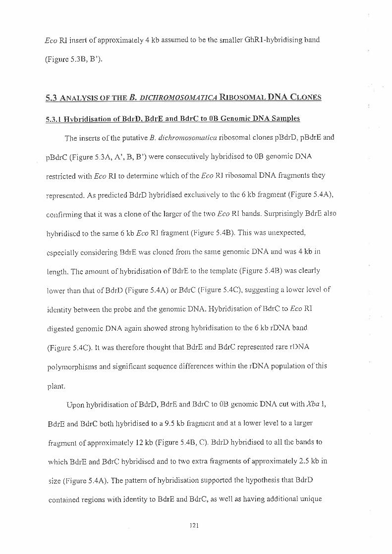
EcoRl. insert of approxirnately 4 kb assumed to be the smaller Glr.Rl-hybridising band
(Figure 5.38, B').
( 2 Älrl,rr^E'
P r\rîrrDlt tl MATICA RTNOS rr Tìl\JA l-r nlrr.cMOSYSIS
5.3.1 Hvbridisation of BdrD. BdrE and BdrC to 0B Gen omic DNA Samnles
The inserts of the putative B. dichrontosontatica ribosomal clones pBdrD, pBdrE and
pBdrC (Figure 5.34, A', B,B') were consecutively liybridised to 0B genomic DNA
restricted witlt Eco RI to deternine which of the EcoP.I ribosomal DNA fragments they
¡epresented. As pledicted BdrD hybridised exclusively to the 6 kb fi-agment (Figure 5.44),
confinning that it was a clone of the larger of the Two Eco RI bands. Surplisingly BdrE also
hybridised to the same 6 kb Eco RI fragnent (Figure 5.48). This was unexpected,
especially considering BdrE was cloned from the same genontic DNA and was 4 kb in
lerrgth. The amount of hybridisation of BdrE to the template (Figure 5.48) was clearly
lorver than that of BdrD (Figure 5.44) or BdrC (Figure 5.4C), suggesting a lower level of
identity between the probe and the genomic DNA. Hybridisation of BdrC to Eco RI
digested genomic DNA again showed strong hybridisatiort to the 6 kb rDNA band
(Figule 5.4C). It was therefore thought that BdrE and BdrC represented rare rÐNA
polyntorphisnts and significant sequence differences within tlie rDNA population of this
plant.
Upon hybridisation of BdrD, BdrE and BdrC to 0B genomic DNA cut with XbaI,
BdrE and BdrC both hybridised to a 9.5 kb fragment and at a lower level to a larger
fi'agnrent of approxirnafely 12 kb (Figure 5.48, C). BdrD hybridised to all the bands to
which BdrE and BdrC hybridised and to two extra fi'agments of approximately 2.5 kb in
size (Figure 5.44). The pattern of hybridisation supported the hypothesis that BdrD
contained regions with identity to BdrE and BdrC, as well as having additional unique
121

Figure 5.4:
A. Southern hybridisation of ,8. dichrontosontatica 0B genomic DNA probed witl-r the insert
from pBdrD. Each lane is loaded with 2 ¡rg of total genomic DNA without B chromosomes
restricted rvith tlie endonucleases indicated. Tlie numbers on the left represent the molecular
sizes (kb) of lambda bacteriophage DNA restricted with ¡/ind III.
B. Soutlrem hybridisation of .8. dichrontosontatica 0B genomic DNA probed with the insert
from pBdrE. Each lane is loaded with 2 ¡rg of total genomic DNA without B chromosomes
restrictecl rvith the endonucleases indicated. The numbers on the left represent the tlolecular
sizes (kb) of lambda bacteriophage DNA restricted with HiÍd III.
C. Southem hybridisation of ,8. clichromosontatica 0B genomic DNA probed with the insert
from pBdrC. Each lane is loaded rvith 2 ¡rg of total genonric 0B DNA restricted with the
ellclollrcleases indicated. The nuntbers on the left represent the molecular sizes (kb) of
lan-rbda bacteriophage DNA restricted with -f1i¡¡d III.
D. Sonthem hybridisation of ,8. clichrontosomalica genonlc DNA probed witli the insert of
B. clichrontosotttcttica rDNA clone pBdrD.
Each lane u,as loaded with 3 ¡rg of total genomic DNA derived fi'oln a 0B or 38
B. clichrontosontatica plant and digested with the restriction endonucleases indicated. The
numbers on the left represent the molecular sizes (kb) of larnbda bacteriophage DNA
restricteci rvith Hird III.
122

A
23.1 -
9.4 -6.5 -
4.3 -
23.1 -
9.4 -6.5 -
{;
23.1 -
9.4 -6.5 -
4.3 -
r"åt*qt B
t
O
;2.3 -
2.3 -2.0 -
2.3 -
D EcoRjl Xbøl Cløl I Sacl BamHl
23.1 -
9.4 -
6.5 -4.3
-
2.3 -2.0 -

regions cortmensurate with its larger size. The level of BdrE hybridisation was again lower
tlran that of BdrD or BdrC (Figure 5.4A., B, and C), supporting the idea that there was
sequence diversity between the BdrE probe and the rnajority of rDNA sequences.
It was therefore apparent that all the available B. dichrontosontatica rDNA clones
obtained contained regions which showed identity to the 6kb Eco RI fragment and no
clone represented tlie 4 kb EcoP.I fragment (Figure 5'1).
5.3.2 Comoal'ison of BdrD Hvbridisation to 0B and tB Genomic DNA Samples
The hybridisation pattern of BdrD to 0B and *B genomic DNA cut with EcoP.I,
Xbct 1, Cla I, Spe I, Sac I and Bant HI was indistinguishable between samples with alld
u,ithout B chromosorles (Figure 5.4D) supporling pleviotts evidence that the rDNA
regio¡s on A and B chromosomes could not be distinguislied by this method (Donald et ul.,
1 ees).
As discussed in Chapter3, aB cluonlosome contains approxintately 3% of the total
I-DNA i¡ a plant with one B chromosome. The greater proportion of I-DNA on the A
chronrosomes is likely to overshadow any minor length r,ariation in this regiotl specific to
t¡e B chronlosolres. Of parlicular interest rvas the IGS regiori whicli contains RNA
polytrerase I and other rRNA gene regulatory elemetrts. Mappirlg of the
B. dichrontosonntica rDNA clones was undeftaken to defilie and then subclone the IGS
reglol'ì.
5.4 M¡,pplxc BnnD AND BDRE
Restrictio¡ el'ìzynles which cut within BdrD and BdrE once or twice, namely Eco P.I,
XbaI, Xtol, ClaI (pBdrD) and þe I (pBdrE), were chosen to map the clones. Physical
maps were also constructed using probes specihc for each rRNA-coding region derived
from the cotton ÐNA repeat unit (GhR1, Figure 5.5).
r24

Fieure 5.5:
Physical map of the cotton ribosomal repeat unit GhRI showing recognitiotl sites fol the
errdonucleases Eco RI and XbaL Tlie regions containing sequence identity to 18S and 25S
IRNA coding regious are indicated (adapted from Orford, 1996).
125

ÌõÈf
ú,ì
-úrì
útlì
H
ñÈf
2.8 kb 1.7 kb
25S
3.8 kb
Direction of transcription
18S
l.s kb

Initially pGhRl was digested with Eco Rf and Xba I and the resulting GhRl products
of 3.8 kb,2.8 kb, 1.7 kb and 1,5 kb (Figure 5.5) were individually eluted from gel sections
and used as probes to the B. dichrontosontatica rDNA clones. The 1 .5 kb Eco RII Xba I
GhRl fragment, which contains the 18S rRNA gene, hybridised to the 1.2 kb EcoP.Il XbaI
fragrnent of BdrD (Figure 5.68) and the 1.6 kb XbaIl Eco RJ. fragment of BdrE
(Figure 5.78). Hybridisation of the 1.7 kb Eco RI GhRl fragment, containing part of the
intergenic spacer region, was confined to the 1.5 kb EcoP.Il Xba I fragment of Bdt'D
(Figure 5.6C) and the 0.2 kb EcoRIl XhoI fragment of BdrE (Figure 5.7C). The 2.8 kb
.Xbu Il Eco RI fi'agrlent of GhRl, which also contains a portion of the intelgenic spacer
regiolr, displayed a signifrcant level of hybridisation to the 2.5 kb Xba I fragurent of BdrD
(Figure 5.6D) as well as to the 0.3 kb Spell XbaI fragurent of BdrE (Figure 5.7D).
The 1 .7 kl¿ Eco RI and 2.8 kb XbaIlEco RI fragments of GhRi contain mostly
iutergenic spacer (Figure 5.5) and, due to the lorv level of sequence colrsen¡ation in the IGS
region across species, it rvas not expected that this region from cotton rvould hybridise to
tlre B. clichromosontatìca rDNA clones. The signifìcant levels of hybridisation seetr, despite
stringent rvashing conditions, implied the presence of some 18S, 5.8S and/or 25S rDNA
sequences rvithin each of these regions of GhRl.
Of significance is the obserr¿ation that neither BdrD or BdrE showed hybridisation to
the 3.8 kb GhRl fragnent which contains a significant proportion of the 25S IRNA gene of
cotton or to either the ITSl or ITS2 regions of B. dichrontosonrutica (results not shown). It
rvas therefore confirmed that the 25S, 5.8S and ITS regions were not cloned and it was
probable that the uncloned 4kb B. cliclromosomaÍicct EcoFtI fì'agment rvhich hybridised to
GhRl (Figure 5.1), contained these regiolts.
121

Figure 5.6:
A. An agarose gel showing DNA fragments resulting from restriction of pBdrD.
The nu¡rbers on the left represent the molecular sizes (kb) of lambda bacteriophage DNA
restricted with Hi¿d iIL The remaining lanes each contain approximately 3 pg of pBdrD
DNA digested with the following restriction enzymes'. L Eco RI; 2. XbaI;3. Xho I; 4. CIa
I; 5. EcoFlI + Xbu I;6. EcoRI * XtoI;7. Eco RI + C/4 I;8. XbaI + XltoI;9. XbaI +
Cta I;10. Xho I + ClaI;71. EcoRI + XbaI * XtoI; 12. EcoRI + XbaI + ClaI;13. Eco
RI + Xto I + Clu I; 14. Xba | + Xlto I + Clo I.
B. Southem hybriclisation of the gel in A with the 1.5 kb GhRl fragment.
Lanes and nrolecular size rnarkers are as above
C. Southern hybridisatiorl of the gel in A u'ith the 1.7 kb GhRl fragment
Lanes and llolecular size nrarkers are as abol'e
D. Southe¡r hybridisatiori of the gel depicted in A rvith the 2.8 kb GhRl fragment.
Lanes and ntolecular size nrarkers are as above
128
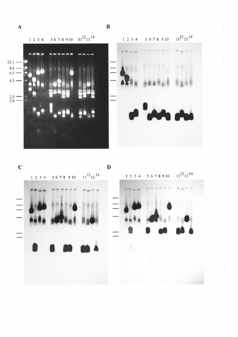
A
23.1 -9.4 -6.5 -
4.3 -
2.3 -2.0 -
B
t234 s6t89t0
1234 5678910
ßr4t'tl2 tz14 r234 s678910 1112
t2
hlf +lt*** ,#
tt¡¡¡ âr
Dcttr2 tzl4 1234 5678910 ll tzl4
¡rlrt
It tI - sö
I I I l¡
.:a¡G- ¡õ-4-- ----
IT
I)
I
It l -'lf , rl t* t¿t
,-tta
lr-

Fisure 5.7:
A. An agarose gel showing DNA fragments resulting from restdction of pBdrE.
The numbers on the left represent the molecular sizes (kb) of lambda bacteriophage DNA
restricted with l1ind III. The remaining lanes each contain approximately 3 pg of pBdrE
DNA digested with the following restriction enzymes: 7. Eco RI; 2. Xba I; 3. Xlto I;
4. SpeI;5. EcoF{I+ Xbal;6. EcoRI+ XhoI;7. Eco RI +.þe I;8. XbaI+ XtoI;9. XbaI
+ SpeI; I0. XtoI+ SpeI; TL Ecoyl+ XbaI-r XhoI;12. EcoRI+ XbaI+ SpeI;
13. EcoRI'r XtoI+ SpeI;14. Xbal+ XtoI+ SpeI.
B. Southern h¡,þ¡i¿itution of the gel in A witli the 1.5 kb GhRl fragment.
Lanes aud molecular size markers are as above.
C. Southem hybridisation of the gel in A with tlie 1.7 kb GhRl fragment.
Lanes and lnolecular size markers are as above.
D. Southenr hybridisation of the gel in A with the 2.8 kb GhRl fì'agment.
Lanes and molecular size markers are as above
130

BA
23,1-
9.4-6.5 -+.3
2.3-2.0-
0.56 -
t 2 3 4 5 6 7 8 91011121314
r234567891017121314
123456789t011 121314
!
o o. ¡
.ol|.
| 23 4 567 891011 121314
to
I
ofÕ
DC
â:rt ryn¡*ñt-%
ot'*
ot t¡oI oI
. oa
5.5 Spounxcn AN¿.r.vsrs or BnnD
Tlie physical map of BdrD was confirmed by subcloning and sequencing
(Figure 5.8). Sequencing was carried out from both ends of the pBdrD(4) subclone
(Figure 5.8). The 550 bp of sequence obtained using the reverse sequencing primer (RSP)
(Figure 5.9) showed greater thang0o/o identity to previously characterised 18S IRNA genes
fi'om other plants (GenBank and EMBL,1997) and temrinated approximately 200 bp short
of the 3' end of the 18S IRNA gene. Sequence obtained frorn BdrD(4) using the (-20)
prir.ner showed no significant identity rvith any previously characterised sequences
(GeriBank and EMBL,1997). The (-20) primer sequence of subclone pBdrD(2.5) resulted
ir-r a furlher 800 bp of sequence, 160 bp of rvliich corresponded to the 5'end of the 18S
rRNA gene (Figure 5.13). The renraining BdrD(2.5) sequence shorved a low level of
sintilarity to the intergerric spacer region of previously characterised plant rRNA gelles
(GerrBank and EMBL, 1997).
The sequence data obtailled disagreed "r,ith
previous physical positioning of the
cotton 18S coding region within the 1.5 kb Eco P.II Xba I fragtlent of GhRl (Orford,
1996). The sequence of the I .5 kb Eco RII Xba I fragment of BdrD, rvhich hybridised to
the 1.7 kb Eco RI GhRl fragment (Figure 5.6C), indicated that it contained tlie 18S coding
region. Restriction of pGhR1, pBdrD and pBdrE with Eco RI and XbaI, followed by
hybridisation with the cucumber l8S coding region (Kavanagh and Timmis, 1986)
revealed strong probe identity to the 1.7 kb fragrnent of GhRl, the 1.5 kb fragment of
BdrD and the 1.6 kb fragrnent of BdrE (Figure 5.104, A'). The implication of this result
was that the coding regions of the cotton rRNA repeat unit were not ananged as previously
tlrought and located the 18S in the 1.7 kb Eco RI fragmeut rather than the 1.5 kb Eco PtIl
XbaI (Figure 5.5; Orford,1996).
132

Figure 5.8:
Physical map of BdrD with restriction sites for the endonucleases Eco RI, Xba I, Xto I and
Clalnarked.
The fragment sizes given are in kb and the regions containing sequence identity to 18S and
25S rRNA coding regions are indicated. The relative positions of BdrC and the subcloned
DNA fi'agrnents, in addition to the ends of the subclones which were sequenced with the
reverse (R) and (-20) forward (F) sequencing primers, are indicated.
133

Direction of
FqXasSSÈ-XsÈli
Fq(ì
Èli
5Sl-t
5'
XXssÈa-l-t
5Sri-25S at
J
\¡(¿Jo\
l¡(.,I
1.2 2.4
185
1.5
R+ +F R+
)ftF+
+F BdrC0.66
E )ft Xb
+ +FXb
)ftR
E)ft

1 TTCCTCGTTT AAGACCAACA ATTGCAATGA TCTATCCCCA TCACGATGAA
51 ATTTCNU\GA TTTCCCGGGC CTGTCGGCCA AGGCTATATA CTCGTTGAAT
101 ACATCAGTGT AGCGCGCGTG CGGCCCAGAA CATCTAAGGG CATCACAGAC
151 CTGTTATTGC CTCNU\CTTC CGTGGCCTAA AAGGCCATAG TCCCTCTAAG
207 AAGCTGGCCG TGGAGGGATA CCTCCACATA GCTAGTTAGC AGGCTGAGGT
25T CTCGTTCGTT AACGGAATTA ACCAGACAiU\ TCGCTCCACC AACTAAGAAC
301 GGCCATGCAC CACCACCCAT AGAATCAAGA AAGAGCTCTC AGTCTGTCAA
351 TCCTTACTAT GTCTGGACCT GGTAAGTTTC CCCGTGTTGA GTCNU\TTAA
401 GCCGCAGGCT CCACTCCTGG TGGTGCCCTT CCGTCAATTC CTTTAAGTTT
451 CAGCCTTGCG ACCATACTCC CCCCGGAACC AAAAACTTTG ATTTCTCATA
501 AGGTGCCAGC GGAATCCTAA AAGCAACATC CGCTGATCCC TGGTCGGCAT
Figure 5.9:
Partial sequence of the pBdrD(4), 4 kb subclone, ft'om the RSP priner of
pBluescript@SK(-). The reverse complement of the sequence depicted showed greater than
90% identity to plant 18S rRNA sequences.
135

Figure 5.10:
A. An agarose gel of pGhRl, pBdrD and pBdrE restricted with Eco RI and XbaI.
The numbers on the left represent tlie molecular sizes (kb) of lambda bacteriophage DNA
restricted with,É/ind III. The renaining lanes contain approxirnately 3 ¡-tg of the digested
recombinant plasmid DNA indicated.
A'. Southern hybridisation of tlie gel in A with the 18S region of pCU18
Lanes aud molecttlar size nrarkers are as above.
B. Arr agarose gel of pBdrE, and pBdrD restricted with Eco P.I, Xba I, Xto I and þe I (pBdrE) or
C/a I (pBdrD). The nurrrbers on the left represent the molecular sizes (kb) of lar¡bda
bacter.iopitage DNA restricted with Hi¡rd IIL The remaining lanes contairl approximately 3 ¡"rg of
the digested recontbirtant plaslnid DNA indicated.
B'. Southern hybridisatiort of the gel in B with end-labelled total RNA flom
B. rlichrontosontctlica. Lanes alrd rnoleculal size lnarkers are as above.
C. An agarose gel of pGhRl, and pCU18 restricted rvith Eco RI and XbaI.
The ¡umbers on the left represent the molecular sizes (kb) of larnbda bacteriophage DNA
restricted r¡,ith H¡nd IIL The remaining lanes contaír¡2-3 ¡rg of the digested recombinant plasrnid
DNA indicated
C'. Southern hybridisation of the gel in C with end-labelled total RNA fi'om
B. clichrontosotncttica. Lanes and molecular size markers are as above.
136

A Ar
ot'esC ".&Psrr }O
23.1-9A-6.5-4.3-2.3_2.0-
Br crCB
23.r -
9.4 -6.5 -
2.3 -2.0 -
0.56 -
fsfot CE' ."$s {,s.
t¡
23.r -9.4 -6.5 -
4.3 -
2.3 -2.0 -
-}l ta
Èi
rÕrntsÞ
GT
ta
-ag
TUg
Ë-I
0.56 -

Sequencing of subclone BdrD(1.2) with tlie RSP indicated that the clone consisted of
a sequence of 483 bp which showed greater than 90% similarity to the extreme 3' end of
the 25S rRNA gene of other plants (see Figure 5.13), beyond which the sequence showed
no signifrcant identity with any sequences in the database (GenBarik and EMBL,1997).
Hybridisation of the 1.5 kb GhRl EcoRIl XbaI fragment to the 1.2 kb XItoIl.Eco RI
fragment of pBdrD suggested that this fragment coutained the 3' region of the cotton 25S.
This agreed with the original physical position of 25S rDNA in pGhRl (see Figure 5.5) but
placed it in the opposite orientation to that proposed, consistent with the 18S being located
in the i.7 kb Eco RI GhRl fragment and not in the 1.5 kb fi'agment.
Seque¡cing of pBdrD(2.5) using the RSP and pBdrD(1.2) using tlie (-20) forward
pri¡¡er, showed no sigr-rificant identity with any sequences in the database (GenBank and
EMBL, 1997; Figure 5.13). The small size of subclone pBdrD(0.6) enabled the couplete
sequence of both strands to be obtained. The total 592 bp of this clone also showed no
significant identity with the database (GenBank and EMBL,1997;. Figure 5.13).
Hybridisation of end-labelled total B. dichrontosontatica RNA to pBdrD DNA
restricted witlt Eco F{I, Xba I, Xho I and Cla I and pBdr E DNA restl'icted with .Ðco RI,
Xbct I, Xto I at'td Spe I enabled confimration of the regions of the clones which were
represented in the 25S, 5.8S and i8S rRNAs. Hybridisation of labelled RNA to the 1.5 kb
and 1.2 kb fragr¡ents of BdrD (Figure 5.10B, B') supported tlie sequencing information
described above, suggesting that these fragments contained the i8S and part of the 25S
codi¡g regions respectively. Tliere was also a lower level of RNA hybridisation to the
2.5 kb XbaI fragnent (Figure 5.108, B') accounted for by the presence of 160 bp of the
18S coding region in tliis fragment (Figure 5.8). Hybridisation of RNA was only seen to
the 1.6 kb fragment of BdrE, which contains the 18S region of the clone. Labelled RNA
was also hybridised to pGhRl and pCU18 (Kavanagh and Timmis, 1986) restricted with
r38

EcoRf and Xba I (Figure 5.10C). Strong hybridisation was seen to the 1.7 kb ar-rd 1.5 kb
fragrne¡ts of pGhRl as well as to the 3.8 kb fragrnent (Figure 5.10C') thought to contain a
large proporlion of the 25S coding region (Figure 5.8). The hybridisation seen to GhRl
supported the evidence presented, suggesting that the 1.7 kb Eco F.I' fragrnent contained the
i8S codi¡g region. RNA hybridisation was observed to all CUl8 fragrnents of the rDNA
repeat u¡it (Figure 5.10C, C'), consistent with the physical rnap (Kavanagh and Timrnis,
1 986).
The presence of approxirnately 4 kb of sequeltce rvithin pBdrD, betu'een the 5' end
of the 18S coding region and the 3' end of the 25S coding legion (Figure 5'8), which
displayed no apparellt identity with the database, supporled the physical rnap and suggested
that this region contained the IGS region of B. dichrontosotttctlicr¿ rDNA.
5.6 SBor IENCE ANAIYSIS OF BDRB
The physical rnap of BdrE was also confimred by subcloning and sequencing (r'esult
'ot shown). Reverse and (-20) primer sequences of pBdrE(1.6) shou'ed extensive similarity
to the I 8S rRNA gene of a range of fungal, rathet' than plant, 18S rRNA genes' RSP
sequence of pBdrE(3.9) revealed that it contained the 3'end of the 25S rRNA gene, which
agai¡ showed high levels of identity with fungal rRNA sequerlces. The remaining sequence
obtained showed no apparent identity with any sequences in the database, suggesting that
this clone contained the intergenic spacer of a fungal rRNA gene. Tlie predicted size of the
IGS of pBdrE was approximately 1.9 kb (result not shown) which is very small in
comparison to the 4 kb IGS of pBdrD (Figure 5.8). The smaller size of the IGS of pBdrE,
along witl-r the database search results obtained suggested that pBdrE was not from
B. clichrontosontatica DNA but probably fi-orn contaminant fur-rgal DNA.
r39

5.7 SnounNcn AN¡.r,vsIs on BnnC
Sequencing of pBdrC confirmed that the fragment resulted from a Eco RI
polymorphism within BdrD coresponding to the 3' 665 bp of pBdrD(4) (Figure 5.8). The
additional -Ðco RI site was identified as the deletion of an A residue witliin the sequence
GAAATTC of BdrD.
5.8 ATTEMPTS TO CLONE TITE RNM¡.TNNNN OF THE RTNO DNA
Rnpn¿.T IJNIT
Further attentpts to clone the remainder of the rDNA repeat unit rvithin the 4 kb
Eco RI fragment were carried out. The probe chosen was the 2.95 kb Eco P.II Xlto I
fragment of pCUl8 which contains the 25S and 5.8S coding regions plus ITSI and ITS2
(Kavanagh and Timmis, 1986) which therefore rvould not be expected to hybridise to arty
of the previously cloned regions in BdrD (Figure 5.8). Total 0B genoniic DNA was
restricted witli Eco RI and ligated ínto Eco Rl-restricted pBluescriptG'SK(-). Screening of
approximately 1500 recombinants yielded trvo positives uamed pBdr85 and pBdr89. Uporl
digestion rvith Eco RI, pBdr85 was revealed to have an inseft of approximately 150 bp and
the insert in pBdr89 could not be excised from the vector using Eco RL Subsequent
sequencing of pBdr85 revealed that it contained 148 bp of tlie 18S rRNA gene not present
in pBdrD (result not shown). The clone pBdr89 contained a 451 bp inserl that rvas thought
to be a cloning artefact. The sequence of Bdr89 coresponded to tlie extreme 3'end of the
18S ge¡e plus ITSl and the 5' end of the 5.8S rRNA coding region (result not shown). No
other putative ribosomal clones were obtained. The inability to clone the complete region
between the 3' end of the 18S and the 5' end of tlie 25S therefore necessitated a PCR
approach to allow comparison of these regions on the A and B cluomosomes.
140

( O Þl-R Ä'r¡ALYSIS OF rrrn TTS1 ¡run ( RS R rrNs rìN tr¡u. Tl
CnRouosouns
\trr\ Chapters 3 and 4, microdissected B cluomosorne DNA enabled the ITS2
sequence of the B cluomosome to be analysed, This approach overcame problems
associated with the presence of the A chromosomes in whole plant getlomic preparations.
A PCR based approach was undertaken to exar¡ine the B chromosome rDNA
sequence in the region between the 3' end of the 18S and the 5' end of the 25S using
genomic DNA containing Bs. Difficulty in identifying sequertces uuique to the Bs, due to
the prese¡ce of A chromosome DNA in the template, was overcome by using the.tfc I site
rvithin the ITS2, which was previously shown to be a specific identifier of B chlomosolÌle
rDNA (Chapter 3).
Initially the region between the 18S and 25S, which contains the ITS1, 5.8S and
lTS2 (see Figure 3.14) was amplified from 0B, 1B, 28,38 and 48 B. dícltrontosotttatica
genomic DNA samples using tlie XF18S and ER25S plimers (Figure 3.2,5.114)' The
resulti¡g product was approximately 700 bp in lengtli (Figure 5.1lA, B). Taking into
account tlie 218 bp ITS2, 300 bp ITSI (Section 3.2) and prirner lengths, this suggested that
the 5.8S coding region is approximately 150 bp tliat is in accoldance with the nomral range
i¡ pla¡ts (Kavanagh and Timmis, 1988). The B chromosotne sequences were selected by
restricti¡g the 700 bp product amplified from the lB,2B,38 and 4B ternplates with,!fc I
rvhich resulted itr three fragments, the largest one (700 bp) representing the A chromosome
product which did not contain the Sfc I site and hence was unrestricted. The second band of
approximately 600 bp was the B chromosome region frorn the.!fc I site of the ITS2 to the
XF185 primer (Figure 5.1 1A). The remaining 98 bp fragrnent, which was difficult to
141

Figure 5.11:
A. A diagrarn of the PCR products in part B of this figure. The positions of the XFl8S and
ER25 S primers used to amplify the region, the relative positions of the Sfc I and Spe I
restriction enzyme sites and the sizes of the ,lfc I and ,þe I restriction fragments are
indicated.
B. An agarose gel of the PCR products resulting from amplification of .8. dichrontosontatica
genonric DNA with XF18S and ER25S.
T¡e first lane contains molecular size markers (100 bp DNA ladder), consisting of fragments
tìtat r-ange in size from 100 bp to 1000 bp in 100 bp increments, plus an additional fragment
of 1500 bp. The remaining lanes each contain 10 ¡rl of a 25 pl PCR reaction nrix. The
cluomosol¡e constitntions of tlle ternplate DNA and restriction enzymes used are:
1. 0B(1) unrestricted.
2. 1B restricted with.!fc I.
3. 48 restricted witli .ffc I.
4. 3B restricted with ^!fc I.
5.28 lestricted with,Sfc I.
6. 0B(2) unrestricted.
7. 0B(1) restricted with Spe I.
8. 0B(2) restricted with.þe I.
The double headed affow ( .*) indicates the 700 bp PCR product amplified with
XFl85 and ER25S. The 600 bp fragment, which resulted when the 700 bp PCR products
arnplifiecl fror¡ *B ternplates were digested with ^!fc I, is anowed and labelled' The alrows
on t¡e right indicate the 580 bp and 120 bp fragments produced when the 700 bp PCR
products arnplified from the 0B templates were restricted witli lþe I'
142

A
xtr18s
18S 255
ER25S
700 bp llP{tâ' ø'
B
600 bp
580 bp
r2345 6 7I
120 bp
<- 580 bp
+ t20bp
500 bP
-\
600 bp
5.8S
r ll*

visualise on all agarose gel, contained the region from the ER25S primer to the,ffc I site of
the B clrromosomes.
The 600 bp fragment isolated from 18, 28,38 and 4B PCR products restricted with
,!fc I was gel purified and cloned in pBluescript@SK(-). Parallel experiments using 0B
tenrplate DNA utilised an Spe I site in the ITS2 region, 5' of the B chromosome,!fc I site,
to clone and sequerrce the equivalent region containing the 5.8S and ITSI region derived
fronr the A chromosorne (Figure 5.114). A CIa I restriction site witliin the 5.8S region
(Figure 5.12) errabled subcloning and confinnation of sequence data.
Sequerrcing revealed that this region of the B chromosome showed no apparent
rearrangements or sequence polymorphisms that were B-specific. There was, however, a
lorv level of polyrrorphisrn seen between individual products derived from A alid B
chronrosor.r'les (Figure 5.12). The nucleotides that rvere polymorpliic in diffelent samples
s¡orved no apparent regularity and therefore nlay represent PCR-based el:rors, rather than
true polymorpliism in the region.
T¡e 149 bp of 5.8S sequence obtained was greater than 95% identical to other plant
5.8S sequeuces in the database (GenBank and EMBL, 1998). The 5.8S sequence is thought
to be only l0% divergent across angiosperms (Hershkovitz and Lewis, 1996) and therefore
rvould be expected to show high levels of identity between closely related species. The
lrigli levels of identity of the 5.8S sequence from B. clichrontosotnatica A and B
chromosomes, despite the inactivity of the rRNA genes, suggests that the B-rRNA genes
remain under strong selection.
The design of the PCR prirners used in the analysis of the ITSl and 5.8S regiolls was
based o¡ the standard organisation of the rDNA repeat unit. It is therefore possible that the
pCR products obtained were not representative of the entire population of B rRNA genes
between 18S and 25S-encoding regions. For example, if large realrangements in tlie rDNA
144

Figure 5.12:
Alignrnent of the 1TS1-5.8S-iTS2 nucleotide sequence derived fi'om a range of individual
B. dichrontosontalica template DNAs'
Tlie cluomosolne constitutions of the plants from which the ternplate DNAs were prepared
are indicated a¡d the sequences of the ITS2 region from A (Al) and B chromosomes (81)
incorporated (see Figure 3.4). Polymorphisms between individual clones are marked-
Due to the way in rvhich the A and B chromosome regions were selected for cloning the
sequences are different lengths. The A cluomosome derived clones (081 and 0B2)
te¡¡iltate at the ,þe I site within the ITS2, whereas the B chronlosome derived clones (1B,
2P.,38 and 48) tenninate at the,!fc i site within the ITS2.
145

xF18S0B1oB21B2B3B4B
0B1oB21B2B3B48
TAACAAGGTTTAACAAGGTTTAACAAGGTTTAACAAGGTTTAACAAGGTTTAACAAGGTT
TCCGTAGGTGTCCGTAGGTGTCCGTAGGTGTCCGTAG-TGTCCGTAGGTGTCCGTAGGTG
AACCTGCGGAAACCTGCGGAAACCTGCGGAAACCTGCGGAAACCTGCGGAAACCTGCGGA
AGGATCATTGAGGATCATTGAGGATCATTGAGGATCATTGAGGATCATTGAGGATCATTG
TCGAAGCCTGTCGAAGCCTGTCGAAGCCTGTCGAAGCCTGTCGAAGCCTGTCGAAGCCTG
CNU\GCAGru\CNU\GCAGAJ\CA'U\GCAGN\CAJU\GCAGAi\CfuU\GCAGA;\CruU\G,TAGA'\
ACATGTTATAACATGTTATAACATGTTATAACATGTTATAACATGTTATAACATGTTATA
ACAACAATGCACAACAATGCACAACAATGCACAACAATGCACAACGATGCACAACAATGC
CAGGATGGGGCAGGATGGGGCAGGATGGGG
CAGGATGGGGCAGGATGGGGCAGGATGGGG
081oB21B283B4B
GCGTCAGTTCGCGTCAGTTCGCGTCAGTTCGCGTCAGTTCGCGTCAGTTTGCGTCAGTTC
GATCCTCGTGGATCCTCGTGGATCCTCGTGGATCCTCGTGGATCCTCGTGGATCCTCGTG
GCACACCGTCGCACACCGTCGCACACCGTCGCACACCGTCGCACACCGTCGCACACCGTC
AGTGTGCGTCAGTGTGCGTCAGTGTGCGTCAGTGTGCGTCAGTGTGCGTCAGTGTGCGTC
CCAGATGACCCCAGATGACCCCGGATGACCCCAGATGACCCCAGATGACC
CCAGATGACC
081oB21B293B4B
0B10821B283B4B
081oB.21B283B4B
081oB,21B283B48
CATTGGGTGTCATTGGGTGTCATTGGGTGTCATTGGGTGTCATTGGGTGTCATTGGGTGT
CTTGGTCGTTCTTGGTCGTTCTTGGTCGTTCTTGGTCGTTCTTGGTCGTTCTTGGTCGTT
TCTTTGACGTTCTTTGACGTTCTTTGACGTTCTTTGACGTTCTTTGACGTTCTTTGACGT
A'\CAJUU\CCCA]\CA,UU\CCCA'\CNUU\CCCA'\CNUU\CCCA;\CAJUU\CCCfu\CA'UU\CCC
CGGCACGATGCGGCACGATGCGGCACGATGCGGCACGATG
GGGCACGATGGGGCACGATG
TCA'\GGruU\TTCA'\GGruU\TTCA]\GGAJU\TTCzu\GGNU\TTCA'\GGNU\TTCN\GGA'U\T
TTNU\TTGzu\TTNU\TTGAJ\TTruU\TTGAJ\TTAiU\TTGA'\TTA,U\TTGAJ\TTNU\TTGA;\
GAATÎGCTTGGAATGGCTTGGAATGGCTTGGAATGGCTTGGAATGGCTTGGAATGGCTTG
TTCCATGATGTTCCATGATGTTCCATGATGTTCCATGATGTTCCATGATGTTCCATGATG
TCCCGTTCGCTCCCGTTCGCTCCCGTTCGCTCCCGTTCGCTCCCGTTCGCTCCCGTTCGC
^^ñ^m^dñ^^\J\f J- lr I \rU l- U¿{
GGTGTGCTCAGGTGTGCTCAGGTGTGCTCAGGTGTGCTCAGGTGTGCTCA
TGGGGCGTGGTGGGGCGTGGTGGGGCGTGGTGGGGCGTGGTGGGGCGTGGTGGGGCGTGG
CTTCTTTGTACTTCTTTGTACTTCTTTGTACTTCTTTGTACTTCTTTGTACTTCTTTGTA
ATCTAIUU\CGATCTA'UU\CGATCTNUU\CGATCTNUU\CGATCTNUU\CGATCTA,UU\CG
ACTCTACTCTACTCTACTCTACTCTACTC
trLf\
r-a
CAQC
ER58SACGGGATATCACGGGATATCACGGGACATCACGGGATATCACGGGATATCACGGGATATC
TCGCGTCACGTCGCGTCACGTCGCGTCACGTCGCGTCACGTCGCGTCACGTCGCGTCACG
CATCGATGAACATCGATGAACATCGATGAACATCGATGAACATCGATGAACATCGATGAA
CIa 1
GAACGTAGCAGAACGTAGCAGAACGTAGCAGAACGTAGCAGAACGTAGCAGAACGTAGCA
NU\TGCGATAAiU\TGCGATAA,U\TGCGATAAiU\TGCGATAA,U\TGCGATAA,U\TGCGATA

0810821B283B4B
081oB21B2B3B4BA1B1
081oB21B2B3B4BA1B1
081oB21B283B4BA1B1
0810821B293B48A1B1
081oB21B2B3B48A1B1
CTTGGTGTGACTTGGTGTGACTTGGTGTGACTTGGTGTGACTTGGTGTGACTTGGTGTGA
ATTGCAGAATATTGCAGAATATTGCAGAATATTGCAGAATATTGCAGAATATTGCAGAAT
ATCGAGTTTTATCGAGTTTTATCGAGTTTTATCGAGTTTTATCGAGTTTTATCGAGTTTT
TGAACGCAACTGAACGCAAGTGAACGCAAGTGAACGCAAGTGAACGCAAGTGAACGCAAG
GTGCGCCCGATTGCGCCCGATTGCGCCCGATTGCGCCCGAññ^^^^^^^1I l- Lr\-\JUUU\ffI
TTGCGCCCGA
AGCCACTCGGAGCCACTCGGAGCCACTCGGAGCCACTCGGAGCCACTCGGAGCCACTCGG
CCGAGGGCACCCGAGGGCACCCGAGGGCACCCGAGGGCAC
CCGAGGGCACCCGAGGGCAC
GTCTGCCTGGGTCTGCCTGGGTCTGCCTGG
GTCTGCCTGGGTCTGCCTGG
xFs8SGCGTCACGCGTCAC
GCGTCACGCGTCACGCGTCAC
GTCTGCCTGG GCGT
CACA
TCGCGTCGCCTCGCGTCGCCTCGCGTCGCC
TCGCGTCGCCTCGCGTCGCCTCGCGTCGCCTCGCGTCGCCTCGCGTCGCC
CCCCACCATTCCCCACCATTCCCCAGCATTCCCCAGCATTCCCCACCATTCCCCACCATTCCCCACCGTTUU\-U¡\UU¡IJ- ]
CTTTCCTTCGCTTTCCTTCGCTTTCCTTCGCTTTCCTTCGCTTTCCTTCGCTTTCCTTCGCTTTCCTTCGCTTTCCTTCG
GGATGATTGGGGATGATTGGGGATGATTGGGGATGATTGGGGATGATTGGGGATGATTGGGGATGATTGGGGATGATTGG
5.85
CTGGGGGCGG
CTGGGGGCGG
CTGGGGGCGG
CTGGGGGCGG
CTGGGGGCGG
CTGGGGGCGG
CTGGGGGCGG
CTGGGGGCGG
ATACTGGTCTATACTGGTCTATACTGGTCTATACTGGTCTATACTGGTCTATACTGGTCTATACTGGTCTATACTGGTCT
CCCGTTCCTCCCCGTTCCTCCCCGTTCCTCCCCGTTCCTCCCCGTTCCTCCCCGTTCCTCCCCGTTCCTCCCCGTTCCTC
ACCGAGCGGTACCGAGCGGTACCGAGCGGT
ACAGAGCGGTACCGAGCGGTACCGAGCGGTACCGAGCGGTACCGAGCGGT
TGGCCA7UU\CTGGCCNUU\CTGGCCA'UU\CTGGCCA'UU\CTGGCCNUU\CTGGCCA,¡UU\CTGGCCA]UU\CTGGCCNUU\C
AAGAGTCCCTAAGAGTCCCTAAGAGTCCCTAAGAGTCCCTAAGAGTCCCTAAGAGTCCCTAAGAGTCCCCAAGAGTCCCT
TTTGAAGGATTTTGAAGGATTTTGAAGGATTTTGAAGGATTTTGAAGGATTTTGAAGGATTTTGAAGGATTTTGAAGGAT
GCACGA....GCACGA....GCACGACTAG TGGTGGTTGAGCACGACTAG TGGTGGTTGAGCACGACTAG TGGTGGTTGAGCACGACTAG TGGTGGTTGAGCACGACTAG TGGTGGTTGAGCACGACTAG TGGTGGTTGA
Spe I
CNUU\CTACG GAATCGTGTCCAAGACTACA GAATCGTGTC
Sfc I
GTGTGTCTTT TCAiUUU\GGT GCTTCTTAAT AGACCCAACG TGTTGTCAT1GTGTGTCTTT TCNUUU\GGT GCATCTTA.AT AGACCCAACG TGTTGTCATT

0810821B283B4BA1B1
TGACAACGCTT CGACC
TGACAACGCTT CGACq-ER25S

were present on the Bs they would not necessarily have been arnplified. Therefore
heterogeneity in this region on the B chromosomes, though unlikely, cannot be completely
ruled out. However, it was concluded that a proportion of the ITS2 regions which contains
the B-specific Sfc I site are present in apparently intact rDNA repeat units in the region
between the 18S and 25S coding regions and hence sequence diversity and rearrangements
in this region do not account for the transcriptional inactivity
( 1O Ârr^r verq rìE' THE INTERGENIC SPICNN OX 'nr¡n A f-r¡pnnrnerìN'tr c
The organisation of eukaryotic IGS regions, including the relative positions of the
rRNA promoter and associated elements, is broadly conserved (Wu and Fallon, 1998).
Relative to the rDNA coding regions however, the nucleotide sequeuces of the IGS evolve
rapidly and show both inter- and intra-specific variability. Variation in IGS length has also
been seen within individual plants (Kavanagh and Timnris, 1986; Rogers artd Bendich,
1987a; Rogers and Bendich, 1987b; Vy'u and Fallon, 1998)
5 1O 1 Spnrrenc eAn ;rlvsis of fhe Co lcfe A Chrnrnnsnrne IGS rvifhin RdrD
Tlie corrplete sequence of the A chromosome IGS was obtained fi'om pBdrD,
tlrrough subcloning and the design of a series of intemal prirners rvithiri The2.5 kb clone
(Figure 5.13).
A search of the GenBank database, with the complete A chromosome derived IGS
sequence, sliowed low levels of identity with other plant IGS regions but the organisation
of tlie region overall was highly conserved in comparison with other plant IGS regions.
Ribosomal RNA transcription initiation sites (TIS) have been detennined within the IGS of
a number of different plant species (Sollner-\Webb and Tower, 1986; Delcasso-
Trenrousaygtre et a1.,1988; Doelling et al., 1993; Perry el a1.,1990; Borisjuk and
Hemleben, 1993; Cordesse et a1.,1993; Polanco and de la Vega, 1994, Capesius, 1997)
t47

Figure 5.13:
Complete sequence of the B. dichrontosontatica A chromosome iGS region.
Initially sequence was obtained from pBdrD using the (-20) forward primer and RSP from
pBluescript@SK(-). Additional primers designed internally to sequence the region are
underlined in blue and the primers PIGSl, PIGS2, PIGS3 and P18S are underlined in red.
The three repeat regions (conesponding to the regions identified in Figure 5.14) are
underlined in pink, green and black respectively. Putative TIS regions are highlighted in
greerÌ and restriction sites used for subcloning are boxed (Xba I: TCTAGA, Xlto I:
CTCGAG, ClaI: ATCGAT). The 3' end of the 25S and 5' end of the 18S are marked.
148

1
51
101
151
20r
25r
301
351
40r
451
501
551
601
651
10r
15L
801
851
901
951
1_001
10 51
1101
1151
L20L
125L
13 01
13 51
L40L
TTCACCAAGT
TTAGACCGTC
AATAGTAATT
CATCGCGCTT
TATGACTGAA
CTCGCCGCCT
CGTGTCGTTG
AÄ.GTACAATT
GTTGGATTGT
GTGAGACAGG
CAACCTAGTA
GGTTGruUU\G
CGCCTCTAAG
GTTTGCCGAC
GCNU\GCCTG
CTCATCAAGC
TCACCCACCA
TTAGTTTTAC
CGAGAGGAAC
CCAGTGGCGC
TCAGAATCCG
CAGCAGTAGG
TTCGACGGAC
GGCGAGTAGA
ATAGGGAACG
CCTACTGATG
CGTTGATTCG
GAAGCTACCG
GGCTAGAAGC
GGCCTCGGCC
GAGTCGTTCA
ATCCTTTGCA
TGAGCTGGGT
ACAGTGTTGC
CACAATTGGT
TGCGCTGGAT
GACGCGTGTG
CCCAJU\GGCA
GGCCGCCTTG
GACGACTTAA
ATACGCGACG GGGTATTGTA AGTGGCAJUU\ TGGCCTTGCT GCCACGATCUPIGSl 255
ACTGAGATTC AGCCCTTT UU CTCCC CCCCATCTAC
ACATCTAATT ATTTCCCC
TAGAGAJU\TT T AAGTGTT GGGATNUUU\ AJU\CCT AC GGTAACAACT
ACGTTCCAAC CAAqGCCTAA N\GNU\CryU\ GGAT GGGTTê EATIJIA1TTGT
GAGGCTGTTC CCCATACTTA GAAAAATTTA CAAGTGTTTG GAT/\¡V\AÄAA
AACCTACACG GTAACAACTA TGTACGTTCC AACCAAqCCC TAGACGA,¡U\T
NU\GGGCCGA TTGCGTTGGG TTACCTTTAT GAACTTGGAA AGTCCAAGGT
AGACCAAGAT CAATCCATAC GGGGTCAAGT GruU\CGACCT ACGGTAGTTT
GCCCCCCCCC CCCCCCCA'U\ ACTGCGAGTC GAJU\CGGGCC CAACACATAC
TAGACATNU\ CGAGTCCGAA CGGGCCCGAC ACATCCGAAT AACGAGCCAT
CACAGATAAG ACCCACGTAA GGTTGGAAGA CAACCCGACA TCGTTGTATC
CTTTTCGTTC GTCATACAGA A€GCCGAqAG GGGCCTGACC CGTCCGAACT
CGATATTCGG GCCGACTCGA CCCGAJUU\CC AACTTCTCGA TTTGGAAGCC
-
CCTCTATGTG TGTTAGATAA GTGTTTCGCA AGTTGCATCA CATTGTTGAC
TA¡UU\TGAGT CGNV\CGGTC CCGTCACGTC CTAACCCGGG GTGACGGGCC
CATTCGACAC GAIUVU\CATG TTffiÏTT GA,U\GTGGCG ATGTGTGTTG
GATAAGTGCC N\TTNU\GAT TCGGGTATAT TCTTTATTAA CATACGTTGT
CGTAACGGCC GGACCCGTCC CAACCCGACA AACGGGCTGA ATGGACCAAG
ATCCAAcTTACCGAGATGAAGTA@GTATGCGTTGGATGAGTGTT
TCGTATCTTG TATCACCCTC TTGACATA;U\ CGAGCCGA;U\ ACGGTCCCGA

14 51
15 01_
15 51
1601
1 651
17 01
17 51
18 01
18 51
1901
1_951
200L
2057
2LOI
2L5L
220r
225r
230r
235r
240L
2451
250t
255L
2601
265I
21 0r
21 57
2807
2B5L
GCCATCCTAA CGGGCCGATT CGACCCGGAG
GACGTAAGCG AGTCGNU\CG GGCCCAACTT
TTTTCATGTA TCACGTTCTT TACATAiUU\T
ATCCTAACCC GACAACG GACC
ATGTCTTGGG ATTGCTTTTT CGTA'U\GCCG
CGACCTT CGAC CCGGTCCAACPIGS2
ACCAACTTCC CGATTTGGAA
GTAAGTTGCA TCACGCTCTT
CCCGATAAGT CCCN\GTNU\
G GGCCCGACCC
CGA]UU\CCAA CGCTCGATGT
TATCACCI]ffiGþATAiu\
TTGGAA TCTAC CATTTGTGACPIGS3
AATGTTGGTG TTTCGTAJ\,\G
CCCTCTT ACACC
ACAACGGGCC GAATCGACCC
TATATTTTTG TTTGCGAAGA
AACTTC TGATTA
CTGCTTTTTC GCCAATATGT
TTACAGCTCG ACCCGACAAC
GCCCGGAACG GCCCCACATG
GNUUUU\TAA AACGTCTTTT
CACAC TGTGCA
TTCA,\GruU\T CAATGTCTGT
AA
GGGTAACCCG
CGruU\GTTAi\
AATTGGACAT
TCCTGTCACT
GTGGTGTT CCCCAAGCAA
GGTTGCCGAG CNUU\GGTGT CATGACACCG CACACAGCCC CATTTCCTTG
GTCGTTTC GT GTCATGACAC CGCACACAGC CCCATTTCCT
TGGTCGTTTC CGAGCA7UU\G GTGTC GTGTCACGAA
CTCGTTCACA GCCCCATTTC CTTGGTCGTT GCCGAGCAAA AGGTGTCATG
ACCACCTTTT 9gTTGGTCGT TGCCGAGCAA AAGGTGTCAT GACACCCATT
T>QCTTGGTCG TTGCCGAGCA AAAGGTGTCA TGACACCCTT TTç{]TTGGTC
GTTGCCGAGC NUU\GGTGTC ATGACACCCT TTTCçTTGGT CGTTGCCGAG
CAAAAGGTGT CATGACACCC TTTTqçTTGG TCGTTGCCGA GCAAAAGGTG
TCATG
1 Lr\.-\- TTTT T
TGGTCGTTGC CGAGCNUU\G GTGTCATGAC ACCCATTTç€ TTGGTCGTTG
CCGAGCAAAA GGTGTCATGA CACCCTTTTç <CTTGGTCGTT GCCGAACAAA
AGGTGTCATG ACACCCTTTT ççTTGGTCGT TGCCGAACAA AAGGTGTCAT
GACACCCTTT TCCTTGCCCA GACGACGTCC TGATCA'UUU\ AACTTTGATT
ATTCN\GA,U\ TCAATGTCTG TTCCTGTCAC TTTGCTTTTT CTCCAATATG

29OI T
295I GGGGATCTTC TAGAAGGGGG AGGGGTGTTT
TTCGGTGTTG
ATCAAGTCGG
GTCGTATCTT
AAGCTATCGA
GTAGTCCCTC
TGCGACNU\G
ATGGGGACTC
TCTCTTGCCC
CCGAGGGGAC
NU\GATCTTG
GGCTAGGCAT
GATCGTCA;U\
GGTTTTAT TTTGTGGTGT TGGCCACACC
CTGTTCGTTT AATGATGCAT TCTTGTCGAA
ACGTCCCTAG ATCNU\CCCG GATTGCGTTA
ATGAGGGTAA GAGTCTCAAC GTGTGACCTT
TTCTTGCACC GACCAGTCAT ACAACGATCG
TGCCTTAGTG TTGGTGGCTT TTGTTCAACT
GGTACATTTT GGGGTCGCAT ATGGGTTTCG
GCCTTGCGGG TGGTTGTTTC CCAAGGCGGC
AAGATGCATG AGTGGTGTTT GGTTTGTTAC
TGT TCCTT
TGTATCGTTT TGATCGTATA TGAGGGTTTA
ATGGCATCGC ATTGATAJVU\ ruU\TGTACCT
AGCAACGTCG GATGAACCAC GAAGCATTCT
GGTTAGTTCT GTTGATTGTT TGGCCCGTTG
ATATGGAAGC CA;U\GTGGGC ATGGGTCTTA
CAGTGG GTGTTGGCTG GTGGGTGATC TTGAAGTCAC
3001
30sl
3t_01
3151
320L
3257
3301
3351
3401
3451
3501
3551_
3601
3 651
3701
3751
3801
3851
3901
3 951
4001
4051
41- 01
4151
GGCTTAACGT ATAAG
GGA'U\GGNU\ TAGGGGTGGG
TAGGATGGGC AGGTACCTGG
TTTCACAAGA TTCATGCGTG
ACGC
GGGAATACTG
AGGCTAGTAG
AATGATGTAT
TTGTAGGATT
TAAGACGCAT
TGGTTCTCTT
GTTGTTCGCT
TTCGGGGATG
GGGTGGTTGG
TTCAGTTTTG
CTGTGTTGCA
TTTTCGTCAG
CTTTGATCCT
AGGGTGGCTT
TGATCCTTAT
CGA'UU\TGAT
ATTGCTGAGA
GTGGATGTGG
GACGTGTTTC
TTCGGGTGGA
CGTATCGTGC
TGGCTCTTGA
TATTTCGTAA
CTCCCCTGCT
CGGCAAGACT
TACCTGATCA
GCGTTGCACC
TACGATTGAT
GGTCTCTTGG
ACTGCCCTCG
TTGAGCGTTC TCGCTTCTCT TACACGACCG CCTTTCACGT ATGGCTCGTL8S
TCAGCGTGTC TGGCTACGTC GAAGAGGAATPl8S
TGTCTC AiU\GATTA'\G CCATGCATGT GTAAGTATGA
ACA'U\TTCAG ACTGTGA,U\C TGCGAATGGC TCATTAAATC AGTTATAGTT
ACCTGGT T
TGTTTGATGG TACCTGCTAC TCGGATAACC GTAGTAATTC T

and they generally contain a conserved region with the sequence TATATAGGGG or
TATATAAGGG. Two putative transcription initiation sites of the consensus sequence
TATATAGGGG were identified at nucleotide numbers 2122 and 2903 of the
B. clichrontosomatica A chromosorne IGS (Figure 5.13) and it was assuned that the one
closest to the 5' end of the 18S was functional based on the position of the TIS determined
in otlrer species (McMullen et al., 1986; Delcasso-Trernousaygue et al.,1988; Gerstner e/
a1.,7988; Perry and Palukaitis, 1990; Zentgraf et a\.,1990; Borisjuk and Hemleben, 1993).
Furthermore, the region downstream of the second putative TIS exhibited conseruation
rvith other members of the Asteraceae (GenBank and EMBL, 1998).
Additional sequences, in common with other plant rDNAs, were located 3' to the 25S
a¡rd 5' to the 1 8S rRNA domains. The pyrimidine-rich region, consistirrg of ten cytosine
residues out of eleven nucleotides, directly following the 3'temrinus of the 25S, as well as
the te¡ nucleotides (AGAGGAATGC) immediately upstream of the 18S domain (Figure
5.13), were contmon to a range of other plant species (GeriBank and EMBL, 1998;
McMulle¡ et a\.,1986; Appels et a1.,1986; Delcasso-Tremousaygue et a|.,1988, Peny
and Palukaitis, 1990). Tliese sequences appear to be ultder selection in comparison rvith
other sequences within the IGS, suggesting a role in the processing of the 40S precursor
rRNA.
Dot matrix analysis enabled the complete intergenic spacer sequence to be compared
within itself to determine regions which contain liigh levels of repetitive elements and to
aid in defining the boundaries of the repeats (Figure 5.L4;Maizel and Lenk, 1981). The
matrix helped identify three major regions of repeats. The first, commencing directly after
the 3' e¡d of the 18S, consisted of two imperfect repeats of approximately 100 bp in lengtli
(Figure 5.13, 5.14). The second region, from bases 1000 to approximately base 2000,
included of a number of 6-8 bp sequences present twice within approxirnately 50
150

Figure 5.14:
Dot matrix analysis of the DNA sequence of the intergenic spacer of a
B. dichrontosontatica A chromosome rRNA gene.
A self conrparison of the IGS sequence (nucleotides 483 - 4032 in Figure 5.13) was made
and the three blocks of repetitive regions are indicated by sets of diagonal lines. A
schematic representation of the spacer is shown under the plot.
151

2,000rl
,p00I
1I
3,000 4,000rlrrr
+"--4,000
- 3,000
-2,000
- 1,000
trrlll
'¿ ,ì ! !'Í Í
.¡ {tt '¡Ì
't
rrrrllllll
Region I Region 2 Region 3

nucleotides (Figure 5.13, 5.14). The third region, fiom base 232I fo approximately 28i 1,
co¡sisted e¡tirely of 12 repeats of a 41 bp sequence, which was often irnperfect differing
by one or a few bases (Figure 5.13, 5.14). Overall the structure of the A chromosome
B. clicltrontosontatica IGS was consistent with all other plant species studied where at least
one array of ta¡dem repeats is found upstream of the TIS. Repeats withili tlte IGS region,
such as tlrose identified in B. dichrontosontaticaby dot nratrix analysis (Figure 5'14), are
ubiquitous in plants and their function remains unknown. However, they liave been
hypothesised to function as RNA polymerase I enhancers therefore playing an important
role in the regulation of rRNA transcription (Flavell et ctl., i986;Hernleben el a|.,1988;
Sardaua el. al.,1993).
11 ON +B CD
Localisation of the IGS within pBdrD (Figure 5.8) enabled the inserls of subclones
co¡tairring IGS sequences to be used as probes to genouric DNA from 0B and *B plants, to
look for variations in hybridisation pattems associated rvith the presellce of Bs. The
genonric DNA of twenty nine plants with and without B chromosollles, selected fronr a
ra¡ge of B. clichrontosontatica populations, was screelred for polymorphisrns in the IGS
regio¡ by Southem hybridisation. The DNA samples were separately restricted with,þe I
ard Xl:aI and probed with BdrD(2.5) or BdrD(0.6), which contain different segments of
tlre IGS of B. dichrontosontatica (see Figure 5.8).
Ana e IGS +B A with
Genornic DNA restricted with.!þe I was run on an agarose gel (Figure 5'15A)'
Sourhe¡r blotted and initially hybridised with BdrD(2.5) which contained 160 bp of the
18S coding region and approxirnately 2.4kb of IGS sequence (Figure 5'8)' Hybridisation
rvas observed to two main bands one greater than23.1 kb and the other approximately
F
153

Figure 5.15:
A. An agarose gel of .8. dichrotnosotnatica genomic DNA restricted with þe I.
The numbers on the left represent the molecular sizes (kb) of lambda bacteriophage DNA
restricted with lli¡rd III. The relative quantities and degree of enzyme digestion can be seen
between different sarnples. The presence (+) or absence (-) of B chromosomes, which was
confirmed by Bd49 hybridisation (John et al., i990), is indicated at the top of individual
lanes
B. Southenr hybridisation of the gel in A with BdrD(2.5)
Lanes, clrromosonte constitutions and nrolecular size markers are as above
C. Southern hybridisation of the gel in A with BdrD(0'6)'
Lanes, chromosolne constitutions and molecular size markers are as above.
154

A
23.1 -9.4 -6.5 -4.3 -
2.3 -2.0 -
0.56 -
C
B
+- - -++-- --- ++- -++-+++++-++-+-
+ -- - ++-- - - -+ +- - ++-+++++-++-+-
+- - -++---- -++-- ++-+++++-++-+-IO-o?- t

9.5 kb (Figure 5.158), the larger of which was thought to be due to partial DNA digestion.
In several tracks there was also an additional hybridising band of approximately 6 kb.
However, there were no polymorphisms detected that were consistently present when the
DNA was from plants with Bs and absent from plants without Bs (Figure 5.158).
The same Southern blot was hybridised with BdrD(0.6) which contained 600 bp of
IGS sequence upstream of the putative start of transcription (Figure 5.8), One major
BdrD(0.6)-hybridising fragment of 9.5 kb was obseled, as well as a larger one in several
tracks resulting from partial digestion of some samples (Figure 5.15C). Although there
were BdrD(0.6) hybridising fragrnents that were polymorphic between individual samples,
none were specific to tlie presence of B chromosones.
5-11.2 Analvsis of the IGS in 0B and +B Genomic DNA Restricted with XbaI
Hybridisation of BdrD(2.5) to genomic DNA restricted with XbaI (Figure 5.164)
was consistently to two bands of approxirna|ely 2.5 kb (Figure 5.168). hi addition the
probe hybridised to a fragment of approximately 9.5 kb (Figure 5.168)' The larger
hybridising band present in several tracks was likely to represent a large llumber of repeats
lacking rl'te Xba I site at one end of BdrD(2.5) (Figure 5.8). In several samples there were
also one or two larger hybridising bands thought to be due to additional polymorphisms or
to parlial XbaI digestion. Horvever, there were no BdrD(2.5) hybridising fragments
associated specifically with the presence of B chromosomes.
Hybridisation of the XbaL-digested genomic DNA samples with BdrD(O.6) detected
hybridisation to the same bands as BdrD(2.5) (Figure 5.168) due to sequences present in
comrl'ìon to the two probes (Figure 5.16C). hr addition, BdrD(0.6) also hybridised to a
fragment of approximately 6.7 kb in all samples (Figure 5.16C). hnportantly there was also
a polymorphic band of approximately 4.4 kb present in all +B samples, that was absent
from every 0B sarnple examined (Figure 5.16C), suggesting the presence of an Xbct I
156

Figure 5.16:
A. An agarose gel of B. dicltrontosontatica genomic DNA restricted withXbaI.
The numbers on the left represent the molecular sizes (kb) of larnbda bacteriophage DNA
restricted with l/ind III. The relative quantities and degree of enzyme digestion can be seen
between different samples. The presence (+) or absence (-) of B chromosome DNA, which
was confirmed by Bd49 hybridisation (John et a1.,1990), is indicated at the top of
individual lanes
B. Soutliem hybridisation of tlie gel in A with BdrD(2.5).
Lanes, clrromosome constitutions and molecular size markers are as above.
C. Southem hybridisation of the gel in A with BdrD(0.6).
Lanes, chl'omosonre constitutions and molecular size markers are as above. The putative B
chromosonre specihc band is arrowed
D. Southem blot of eight samples (marked with * in C) restricted with XbaI and
hybridised r¡,ith the 500 bp Xba Il Xto I fragment in BdrD(0.6)
The nurnbers on the left represent the molecular sizes (kb) of lanlbda bacteriophage DNA
restricted with,ÉIird III. The putative B chromosome specific band is arrowed.
151

ÉJ
Ê
ü
BA
++ - - ++- - - - - ++ -++++- ++++-++ - - -
23.1-9.4 -6.s -4.3 -
C
-O-- ?- r ¿ aa -
lÞ rff*rlrr F22
0.56 -
.3
.0
45 67 8
-++++51**
D
+
| 236 23 7* ** * * *
++ -- ++- - - - - ++ -++++-++++-++ - - -
8 4
t 23.t -9.4-6.5 -4.3- <
iát,2.2.
J0
0.56 -

polymorphism in this region that was specific to the B chromosorres. Variations in DNA
loadings made it difficult to visualise the putative +B specific hybridising band in some
tracks (Figure 5.16C). Subsequent selection of four 0B and four lB samples enabled
confirmation of the +B-specific , XbaI polymorphism (Figure 5.16D).
The cluomosome constitution of each genomic DNA sample was checked by
subsequent liybridisation of the same filters with Bd49, a B. dichrontosontatica B-specific
repeat sequence (John et a\.,799I), expected to hybr-idise only to +B samples. All the +B
sarnples hybridised strongly to this probe and the 0B samples were negative (result not
shown).
{ 1? pl- ÄruALYcrs nìr TllS ôlr ,t ,r NDB f-r¡r¡n f\C¡.ìNíú\Q
5.12.1 A nted Identification of th e R Chromosome IGS P rnhism
To identify the origin of the putative +B-specific XbaI polymorphism (Figure 5.16C,
D), which was possibly located within a B-specific IGS, a PCR approach rvas undertaken.
Primers were designed to the 5'end of the 25S coding region (PIGS1, Table 2.4b) and to
the 5' end of the pBdrD(0.6) sequence (PIGS2, Table 2.4b) of the A chromosome rDNA.
In cornbination, PIGSl and PIGS2 were designed to arnplify a 1.3 kb product from the IGS
regiorr represerrted in BdrD (Figure 5.13).
PCR with PIGSl and PIGS2 resulted in two rnajor arnplification products, of
approximately 1.3 kb and 1.2kb in length, from all0B and fB templates analysed
(Figure 5.l7A,lanes 1-4). In contrast, only the 1.2 kb fragrnent and several smaller
fragrrents were amplified from microdissected B chromosome DNA, with no trace of the
1.3 kb amplicon (Figure 5.I7 A,lane 5). To detennine which of the products were speciñc
to the IGS region, the gel was Southern blotted and probed with the 500 bp Xba 1l XIto I
fragment of the BdrD(O.6) subclone (Figure 5.174). Hybridisation was seen to both the
159

Fisure 5.17:
A. An agarose gel and Southern hybridisation of PCR products resulting from
anrplification with PIGSl and PIGS2, hybridised with the 500 bp XbaIl XtoI fragment of
BdrD(0.6).
Tlie hrst lane contains molecular size markers (100 bp ladder) which consists of fragments
from 100 bp to 3000 bp in 100 bp increments. The remaining lanes each contain 10 ¡rl of a
25 ¡tl reacfion mix. The numbers on the riglit indicate the sizes of hybridising fragments.
The.B. dichrontosontatica template DNAs used were: 1. 0B genomic DNA; 2. 0B genomic
DNA; 3. +B genomic DNA; 4. +B genomic DNA; 5. microdissected B clrromosome DNA;
6. no template DNA (-ve control).
B. All agarose gel and Southern hybridisation of PCR products resulting frorn
amplification with PIGS3 and P18S, hybridised with BdrD(2.5).
The first lane contains molecular size nrarkers (100 bp ladder) which consists of fragrnents
fi-oln 100 bp to 3000 bp in 100 bp increments. The remaining lanes each coutain 10 ¡rl of a
25 ¡.rl reaction mix, The numbers on the right indicate the sizes of liybridising fragments.
TIre B. tlichrontosontatica template DNAs used were: 1. 0B genomic DNA; 2. 0B genomic
DNA; 3. +B genomic DNA; 4. +B genomic DNA; 5. microdissected B cluomosorne DNA;
6. pBdrD (*ve control); 7. no template DNA (-ve control)
160

A
3.0 -
1.0 -
12 3 4 5 67
123456 123456
*
1234567
<- 1.3 kb+ 1.2kb
t + o.6kb
+ 2.2kb
# o.gtu
B
3.0 -
1.0
-
5å5'-
¡+¡a
1.3 kb and I.2 kb products amplified from all samples tested and there was no apparent
difference in hybridisation between 0B and +B samples (Figure 5.174). The 1 .2 kb
product, amplified from the microdissected B-DNA, also hybridised the probe in addition
to a smaller band of 600 bp (Figure 5. 174). The 1 .3 kb and 1 .2 kb products amplified fi'om
a 0B ternplate, and The 1.2 kb and 0.6 kb products fi'om microdissected B-DNA, were
cloned and sequenced.
The nucleotide sequence of the 1.3 kb clone (pBdK3) was closely sirnilar to the
sequence of tlie IGS from BdrD (Figure 5.1 8). The sequence data showed that the 1 .2 kb
product (pBdK2) was the result of a second PIGS2 binding site, intemal to the site to
which the primer was designed. The 1.2 kb product amplified from nricrodissected B-DNA
(BdBIGSI) was identical in sequence to that from the 0B plant (Figure 5.18). One possible
reason that tlie 1.3 kb fragment was not able to be amplifìed fi'om the microdissected
B-DNA is tliat the IGS region is truncated on the Bs. It is also possible, although unlikely,
that the region was too large to be represented within the DOP-PCR-arnplified DNA
sample.
The sequence of the microdissected B-DNA-derived 0.6 kb PCR product (BdBIGS2)
was similar to the BdBIGS I but tenninated 600 bp into the sequeuce. The last 1 1
nucleotides of BdBIGS2, leading into the PIGSl primer region, differed from the sequence
of BdBIGSI, BdK3 and BdK2 suggesting a DNA sequence rnorph specific to the B
chromosomes (Figure 5.1 8).
The region arnplifìed using PIGSl and PIGS2 included the sequence of BdrD(0.6).
Therefore it is possible that the variations between the A and B-derived PCR products,
detected using these primers (Figure 5.174), were responsible for the XbaI, +B-specific
polymorphisrn identified by Southem liybridisation with BdrD(0.6) (Section 5.1 1.2) but
this could not be experirnentally established.
162

Figure 5.18:
Alignrnent of sequences of BdBIGS2, BdBIGSI, BdK3 and BdK2 with the IGS sequence
(Figure 5.13). Primer binding sites of PIGS2 are underlined in red and PIGS3 in green.
Primer binding site of PIGSI, within BdBIGS2, is underlined in blue.
163

BIGSl
BIGS2BIGSlK3R2IGS
BIGS2BIGSlK3K2IGS
BIGS2BIGSlK3K2IGS
BIGS2
BIGS2BIGSlK3K2IGS
BIGS2BIGSlK3K2IGS
BIGS2
TTCAGCCCTTTGTCGCTCCGAT GACAT GT GGCGAT GT GT GCTT GATAAGT G
CCCATTCGACACGAAAAACATGTTCTCGAGTTGAAAGTGGCGATGTGTGTTGGATAAGTGCCCATTCGACACGAAA.AACATGTTCTCGAGTTGAAAGTGGCGATGTGTGTTGGATAAGTGCCCATTCGACACGAAAAACAT GTTTTCGAGTTGAAAGTGGCGATGTGTGTTGGATAAGTGCCCATTCGACACGAAÄAACATGTTCTCGAGTT GAAAGTGGCGATGTGTGTTGGATAAGTG
************************
CAATTAAAGO"'"""O'O"CT"O"OO.'O'O'"""""'*CGGCCGGACCCGTCCAATTAÄAGATTCGGGTATAT TCTTTATTAACATACGTT GT CGTAACGGCCGGACCCGTC
CAATTAAAGATTCGGGTATATTCTTTATTAACATACGTTGTCGTAACGGCCGGACCCGTCCAATTAAAGATTCGGGTATATTCTTTATTAACATACGTTGTCGTAACGGCCGGACCCGTCCAATTAAAGATTCGGGTATATTCTTTATTAACATACGTTGTCGTAACGGCCGGACCCGTC**************************************r<*********************
cc.AACCCGÅaoooa"""a, "*, ""oaaÅo"or
a aAACT TACCGA.AT GAAGTACAT cG
CCCAACCCGAC A.AACGGGCTGAATGGACCAAGATCCAACT TACCGAGATGAAGTACATCG
CCCAACCCGACAAACGGGCTGAATGGACCAAGATCCAACTTACCGAGATGAAGTACATCGCCCAACCCGACAÄACGGGCTGAATGGACCAAGATCCAACTTACCGAGATGAAGTACATCGCCCAACCCGACAÀACGGGCTGAATGGACCAAGATCCAACTTACCGAGAT GAAGTACATCG************************************************************
ATGTATGcGrr""or"o"rårrr."ror.rraror.o..ðr.rr"oaoro*."oaaaaÅATGTATGCGTTGGATGAGTGTTTCGTATCTTGTATCACCCTCTTGACATAAACGAGCCGAATGTAT GCGTTGGATGAGTGTTTCGTATCTTGTATCACCCTCTTGACATAAACGAGCCGAATGTATGCGTTGGATGAGTGTTTCGTATCTTGTATCACCCTCTTGACATA.AACGAGCCGAATGTATGCGTT GGATGAGTGT T TCGTATCTTGTATCACCCTCTTGACATAÄACGAGCCGA*********************************************)k**************
AAACGGTCCCGAGCCATCCTAACGGGCCGATTCGACCCGGAGACCAACTTCCCGATTTGGAAACGGTCCCGAGCCATCCTAACGGGCCGATTCGACCCGGAGACCAACTTCCCGATTTGGA.AACGGTCCCGAGCCATCCTAACGGGCCGATTCGACCCGGAGACCAACTTCCCGATTTGGA.AACGGTCCCGAGCCATCCTAACGGGCCGATTCGACCCGGAGACCAACTTCCCGATTTGGAÄACGGTCCCGAGCCATCCTAACGGGCCGATTCGACCCGGAGACCAACTTCCCGATTTGG************************************************************
A-AACccACcror"r ar"orÅ"ororararrranroo"rraaoraocccrcrrGACGTAAGAAACCCACCTATGTGTGATAGATATGTGTTTCGTAAGTTGCATCACGCTCTTGACGTAAGAAACCCACCTATGTGT GATAGATATGTGTTTCGTAAGTTGCATCACGCTCTTGACGTAAGAAACCCACCTATGTGTGATAGATATGTGTTTCGTAAGTTGCATCACGCT CTTGACGTAAGA.AACCCACCTATGTGTGATAGATATGTGTTTCGTAAGTTGCATCACGCTCTTGACGTAAG************************************************************
."o"ra"oúccGGcccAAòrraaa"oroo"r..aooaro*rrrraoräror.o."ttòCGAGTCGAAACGGGCCCAACTTCCCGATAAGTCCCAAGTAAATTTTCATGTATCACGTTCCGAGTCGAAACGGGCCCAACTTCCCGATAAGTCCCAAGTAAATTTTCATGTATCACGTTCCGAGTCGAAACGGGCCCAACTT CCCGATAAGTCCCAAGTAAATTTTCATGTATCACGTTCCGAGTCGAAACGGGCCCAACTTCCCGATAAGTCCCAAGTAAATTTTCATGTAT CACGTTC************************************************************
T T TACATAAAAT GGCC GAAACGGGCCC GACCCAT CC TAACCCGACAAC GGGCCGAAAT GA
TTTACATAAAATGGCCGAAACGGGCCCGACCCATCCTAACCCGACAACGGGCCGA.AATGATTTACATAAAATGGCCGAÄACGGGCCCGACCCATCCTAACCCGACAACGGGCCGAAATGATTTACATAAÄATGGCCGAÂACGGGCCCGACCCAT CCTAACCCGACAACGGGCCGAAATGA
T T TACATAAAAT GGCCGAAACGGGCCCGACC CAT CCTAACCCGACAACGGGCCGAAAT GA************************************************************
CC C GAAAACCAACGC T CGAT GT GGAA-CC C GAÄÄACCAAC GCT C GAT GT GGAA_
CCCGAAAACCAACGCTCGATGTATGTCTTGGGATT GCTTTTTCGT AI\'\GCCGTATCACCG
CCC GAAAAC CAAC GC T CGAT GT GGAA-C CTTGGGATTGCTTTTTCGTAAAGCCGTATCACCG**********************
K3K2IGS
BIGS2BIGSlK3K2IGS
BIGS2BIGSlK3K2IGS
BIGSlK3K2IGS

BIGS2BIGSlK3K2IGS
BIGS2BIGSlK3R2IGS
T CTAGACATA.AACGACCT T AAACGGGCC CGACCCGGT GCAACCCGACAACGGGCCGAAT C
TCTAGACATAAACGACCTT AAACGGGCCCGACCC GGTC CAACC CGACAACGGGC CGAATC
GAC C CGAAAC C CAAC GC T C GAGAT GGAA
GAC GACGC TCGCTCTACCATTTGTGACAATGTTGG

5.12.2 Further PCR An alvsis of the B some IGS
The primers PIGS3 and Pl8S (Figure 5.13, 5.18; Table 2.4b) were designed to
further elucidate the structure of the iGS on the B chromosomes and, in particular, to
compare the RNA polymerase I promoter regions of the A and B chromosomes. PCR with
p¡imers PIGS3 and Pl8S from 0B and *B genomic DNA ternplates amplified a product of
approximately 2.2 kb: equivalent in length to the region in an A chromosome rDNA repeat
u¡it (Figure 5. 13). A smaller product of approxirnately 800 bp was pt'esent in all 0B and
*B tracks (Figure 5.178). When the microdissected B-DNA was used as a template for
tþese printers only the 800 bp product was amplifìed. Cloning and sequencing of the
microdissected B-DNA-derived 800 bp PCR product revealed that it was due to a second
binding site for the primer PIGS3. The results obtained using the PIGS3 and P18S primers
rvere therefore inconclusive. The inability to aniplify lb,e2.2 kb product from the
nricrodissected B-DNA n-ray represent a B cluomosome polymorpìrism or alternatively the
s¡rall size of DNA in the template. In support of the latter, Soutliern hybridisation \Nas
nnable to identify any B-specific polymorphisms when BdrD(2.5) was used as a probe to
Xba I and Spe I digested 0B and *B genon-ric DNA (Figure 5. I 5, 5. 16), suggesting tlìat no
sequence variatiolr exists in this region of the IGS between tlie A and B chromosontes.
{ 1? frucrr¡¡ TrìTs^'rrrìN AN¡r Ysrs OF THE TllS Rn rìN rìN rr¡u' Â ,rrunHvRr¿
B CnnouosoMEs
Previously, fluorescence in situ hybridisation (FISH) using the cotton ribosornal
repeat unit as a probe revealed the presence of rDNA at a major cluster near the satellited
end of the shoft amr of the B. dichrontosontatica B chromosome and, to alesserextent,
proxinrally on the same ann (Donald et a\.,1995; Houben et a|.,1998). The heterologous
cotton probe would only be expected to hybridise to the conserved coding regions and not
165

to the IGS region, due to the high degree of divergence in this region even between closely
related organisms (Section 3.1, 5.1). Therefore FISH, using BdrD(0.6) and BdrD(2.5) as
probes, was used to confirm the presence of iGS sequences on the Bs of
B. dichrontosontatica. In addition, Southem hybridisation and PCR analysis suggested that
the organisation of the B chromosome IGS rnay differ frorn that on the As (Section 5.11,
5.I2).In the light of these results it was also of interest to detemine if there were any
detectable differences between the hybridisation of BdrD(O.6) and BdrD(2.5) to the Bs, If
similar results were obtained, this would strengthen the evidence that the rDNA on the B is
in cornplete, essentially nonnal, genes rather than fragments of disorganised rDNA-related
sequences.
As expected hybridisation of both BdrD(2.5) and BdrD(O.6) rvas visualised at the
NOR on the larger of the two A cluomosomes (Figure 5.19). In the case of the B
chrornosonre BdrD(2.5) and BdrD(0.6) both hybridised to the secondary constriction at the
end of one arm and, to a lesserdegree, aregion closerto the centronere (Figure 5.19),
identical to the hybridisation position of the heterologous cotton rDNA probe (Donald el
al.,1995). When the hybridisation signals of the BdrD(2.5) and BdrD(0.6) were overlayed
there were no regions where either one of the probes hybridised differentially
(Figure 5.19). Tlie results indicated that the rDNA on the B chromosomes contains intact
IGS regions at a level proportionate to the signal seen when tliey were hybridised witli
GhRl (Donald et a|.,1995).
The hybridisation of BdrD(2.5) and BdrD(0.6) to the B chromosome at equivalent
levels and pattems showed lhat in silu hybridisation was not able to detect differences in
this region. What was clear was that the proxirnal region of the Bs, which hybridised the
cotton rDNA probe, also contained IGS sequences and it was therefore confirmed that there
was a second locus which most likely contained intact rRNA genes on the B chromosomes.
166

Figure 5.19:
In situ hybridisation with BdrD(O.6) and BdrD(2.5) as probes to rnetaphase spreads of
B. dich rontos ontatica.
BdrD(O.6) was labelled with biotin and hybridisation is seen as a green signal. BdrD(2.5)
was labelled with DIG and hybridisation is seen as a red signal. Using Adobe Photoshop
the irnages were overlayed and positions where both probes hybridise appear yellow
A. Corr-rplete 28 metapliase spread (2r:4+28) with B chrornosorles arrowed. Signals from
hybridisation of BdrD(2.5) and BdrD(O.6) are overlayed
B. Contplete 2B nietaphase spread with B chromosomes anowed. The DAPI stained spread
and the probes visualised in each ft'ame are indicated
C. Contplete 2B nretaphase spread plus an interphase cell and an incornplete cell, with B
cht'olrosontes anowed. The DAPI stained spread and the probes visualised in each frame
are indicated
D. Incomplete metaphase spread with two A chronrosomes and one B chromosome which
is arrorved. The DAPI stained spread and the probes visualised for each frarne are
indicated
r67

A B
BdrD(2.5)/BdrD(0.6)
BdrD(2.5)/BdrD(O.6)C
t),A.1, I
r'
r,
lìtl l l)(2.5) lìrlll)(0.ó)
lltlrlX2.5) BrlrlX0.6)
l\
I I
BdrD(2.5)/BdrD(0.6)

5.13 CONCLUSION
Cloning of part of the A chromosome rDNA repeat unit enabled the detection of an
IGS-related Xba I polymorphism that was exclusively associated with the presence of
B chromosones. PCR and in situ analysis was not successful in detennining the origin of
the polymorphism but PCR experiments demonstrated that there was a degree of sequence
variation between the regions amplified from the A and microdissected B-DNA templates'
Although the level of hybridisation of BdrD(O.6) to the +B-specific, XbaI fragment
was low, it is possible that the polymorphism is representative of every IGS sequence on
the B chrornosorrìes, especially as previously it was estimated that the Bs contain only
approxirnately 3o/o of the rRNA genes in a plant with one B chromosome (Chapter 3).
Ultimately the identification of the source of the XbaIpolyrnorphism, that is present in the
IGS region of the B chromosollles, is cmcial in detenlinirrg whether it has any role in
rDNA i¡activity. However, the low level of probe hybridisation to the polymorphic band in
geuomic DNA, which suggests low copy number in the genome or high sequence
divergence from the majority of the rDNA sequences, have so far precluded its
characterisation.
169

CHAPTER 6:
Isolation of Non-Repetitive Sequences
from the B Chromosomes
t70
.1 Inrn
Previously, in B. dichrontosornatica, differential hybridisation and sequence
em-ichment methods isolated a B chromosome-specific sequence named Bd49 (Section
1.6.1b; John et a1.,1991). The nature of differential hybridisation causes a bias towards the
isolatioti of high copy nurnber repeat sequences and in accordance with this Bd49 was
found to be a 149 bp, tandemly repeated sequence. In situ hybridisation enabled the
localisation of Bd49 to the proximal region of the B chromosones but no transcriptional
activity has been detected and no functions have been deduced from tlie sequence (Section
1.6.1b; Leacli et al., 1995; Franks et a|.,1996).It was thought that the evolutionary origin
of tlie B clrromosomes n'ìay have been revealed by characterisation of Bd49 and its native
sequence context rvithin the B chromosome DNA. However, the flanking sequences were
either highly repeated on the A chrornosomes, retroviral or chloroplast derived (Franks el
al., 1996) and thus were not useful in determinirrg B chrorrosonle origin.
Despite efforts using subtractive cloning, differential screening techniques and
arralysis of sequences associated with Bd49 on the B, no furlher B. dichrontosomaticn,
B-specific sequences have since been isolated from B. clichrontosontatica (Franks et al.,
1996). There is therefore a requirement for non-repetitive sequences, specific to the Bs or
present on the A and B chromosomes to be isolated to help in determining the or-igin of the
B, clichrontosontalica B chromosome and to shed light on its function.
This chapter discusses the use of two PCR-based approaches that were undertaken to
isolate non-repetitive sequences specific to the B clrromosomes and/or those that are
located on both the A and B chromosomes. The techniques chosen involve the comparison
of PCR-alnplification products from genomic DNA samples with and without B
chromosomes, in order to identify amplification products correlated with the presence of
Bs
111
Ideally, the PCR template DNA samples should be hornogeneous with respect to A
chrornosome sequences, so that the only difference between the 0B and +B plarrts is the
presence of B cluomosomes, eliminating the possibility that differences found between the
amplification products from 0B and +B genornic DNA are due to polymorphisms between
the As of different individuals. However, due to the outbreeding nature of
B. dichrontosomatica it is difficult to obtain a population of 0B and *B plants with
homogeneous A cluomosomes. The advantage of using individuals heterogeneous for As is
tliat polymorphisrns between these chromosomes are unlikely to be inconectly associated
u,ith the presence of Bs because they will only be arnplified from one or a few individual
plant DNA samples. The +B genomic DNA samples used in the following experiments
u,ere derived fi'onr plarits which were cultivated from an individual plant with a single B
chrotrrosol¡e suggestiug that, although the A chror-ltosol-ltes were heterogeneous, the Bs
u,ere homogeneous. It was therefore not expected that there would be any polyrnorphisms
betrveen the B chromosorìes frorn the individual +B samples. In addition, the use of 38
gerromic DNA meant that B chromosome sequerlces were well represeltted in the tentplate
DNA and any B chromosome-specific sequences rvould be rnore readily detected.
6.2 Auplrnrnn Fn¡cunNr Lnxcrs PoL\'I{oRPHISMS
Anplified fragrnent length polymorphism (AFLP) analysis is a method by which a
selection of restriction fragrnents of total genomic DNA are detected by amplification
using PCR (Vos et a1.,1995). The genomic DNA is initially cut with two different
restriction eltzymes (in this case Psl I, a rare cutter, and Mse I, a fiequent cutter) and two
double stranded adaptors (one specific for each enzyrre) are ligated to the digested DNA.
Following adaptor ligation, tlie mixture contains a selection of fi'agments, most of which
have MseI- Mse I ends. In addition, small proportions of fragments have PstI-Mse I ends
t72

and even less have PstI-Pst I ends. Fragments with Psr I ends would conceivably represent
low copy sequences, in comparison to MseI-Mse I fragrnents which would be more likely
to represent repetitive regions due to the frequent cutting nature of Mse L
AFLP analysis subsequently employs PCR amplification using two oligonucleotide
primers, one corresponding to each adaptor. To allow the visualisation of AFLP products
after electrophoresis and autoradiography, the primer conesponding to the rare-cutting
enzyrne's adaptor (Pst I) is radioactively labelled in the final amplification step (Section
2.3.5),thereby reducing the complexity of the mixture so that only tlie fragments with a
rare cutting enzyme site on one or trvo ends (representing mainly low copy sequences) are
visualised. TIle AFLP 'profiles' of different genomic samples are then conrpared to look
for polymorphisns.
It rvas considered possible that the AFLP technique would ertable detection of
sequences unique to Bs as well as low copy polynorphic sequences present on both the A
and B chromosomes. The latter were expected to be preseut at greater copy number in a +B
plant as rvell as showing polymorphisrn associated rvith location on the two chromosonle
types.
The AFLP products arnplified from individual 0B and 3B templates, using all
possible primel conrbinations (Table 2.4c), were rutl on acrylatnide gels. The polyrlorphic
¡ature of the population was highlighted by a number of bands being amplified from one or
a few genornic DNA samples and not from others (Figure 6.1). However, the AFLP
pattems were overall very sirnilar for all primer combinations suggesting that the A
chromosonres withi¡ the 0B and +B samples were, to a certain extent, homogeneous and
that the B chromosome consists predominantly of sequences also on the A chromosomes.
The AFLP bands, 15 in all, which were present in 38 and absent in 0B amplification
products (examples indicated in Figure 6.1) were gel excised, eluted from the gel slice and
113

Figure 6.1:
Examples of polyacrylamide gels of AFLP products using the primer Psl I+AA rn
combination with MseI+C.LT and MseI+CGA.
The template DNA samples were derived from individual B. dichrontosomatica plants
rvithout (08) or vyith (+B) B chromosomes as indicated at the top of the gels. Examples of
putative B chromosome specific AFLP products are arrowed. The band 34, which was
analysed further, is arrowed and labelled.
t74

*flr
\-4-
+B
++34
+
+
OB +B OB
FI'. I.:-
iFJ
.--}lÞ*"{}'-'
{¡, læ+ _r?
-s
\....- --t-'-
-
*
MseI+CAT MseI+CGA

then reamplified using the same primers that were used for the original arnplification. The
rearnplified products were subsequently labelled during PCR with ¡cr-t'PldATP and used
individually as probes to 0B and *B genomic DNA restricted with a range of
endonucleases to determine B chromosome specif,rcity (results not shown).
Hybr-idisation of most of the bands excised slrowed no differential hybridisation to
0B and *B ge¡ornic DNA. However one AFLP fragment, named 3A (Figure 6.1), which
was arnplified using the primers Psl I+AA and Mse I+CGA (Table 2.4c), hybridised to a
band i¡ Íhe HùÅ III +B track which was absent from the coresponding 0B track
(Figule 6.2A, band a). A range of 0B and +B samples was subsequently restricted rvith
ÍlindIII to detemine if tlie 3A polymorphism seen \ tas consistently corelated rvith the
presence of B chrontosomes. Unlike the example shown in Figure 6.2A, the hybridisatiolr
patterri of 3A was indistinguishable between 0B and lB genorttic DNA restricted with
Hindlll (Figure 6.28) suggesting that the 0B DNA sattrple used in Figr.rre 6.24 u,as not
representative of all 0B plants. Holever, hybridisation levels of 3A to the putative
polymorphic band (Figure 6.2A, band a) appeared to be higher to *B than 0B getromic
DNA, when corlrpared to the hybridisation levels of 3A to a larger band rvhich hybridised
the plobe at an equal level in all samples (Figure 6.2B, band b). The higher level of
hybridisation to *B than 0B genomic DNA in Southem blots (Figure 628) suggested that
sequences contained in band 3A (Figure 6,1) are present on both the B and A cluomosomes
of B. di chromosotnatica.
Tl're reamplified 3A PCR product was then cloned into pGEM*-T for further analysis.
Putative positive clones were size-selected by PCR amplificatiolt across the insert with T7
and SP6 primers (Table 2.4a) and Southern hybridisation with the original 3A PCR product
(Figure 6.3). The levels of hybridisation to the probe varied, indicating a mixed popr.rlation
176

Figure 6.2:
A. Sotrthern hybridisation of B. dichromosomatica genomic DNA without (08) and with
(+B) B chromosomes probed with 32P-labelled AFLP product 34.
Each lane was loaded with 2 pg of total genomic 0B or +B DNA from single plants
restricted with the endonucleases indicated. The putative B chromosome specific HindIII
restriction fragntent is arrowed and corresponds to the similarly-labelled band in part B of
this figure. The numbers on the left represent the molecular sizes (kb) of lambda
bacteriophage DNA restricted witlì lli¡?d III.
B. Southem liybridisation of a range of B. dichrontosonrutica genomic DNAs without (08)
ancl with (+B) B chrorlosonres restricted with HindIlI and probed with 32P-labelled AFLP
product 34.
Tlie relative quantities of DNA and degree of enzyme digestion can be seen between
different sanrples. The 3A hybridising band which conesponds to the putative B
chromosome specific fi'agment is arrorved and labelled (a), and the 3A hybridising barld
identified in all sarnples is arorved a¡ld labelled (b).
171

A
2.0
B 0B DNA samples fromindividual olants
\u
+B DNA samples fromindividual plants
23.t -
9.4
-6.5 -
4.3 -
PstIl-oB +Bl
HindIIIfor .Bl
23.1-9.4
-6.5 -
4.3 -
2.3 -2.0 -
t** ..\
b
a
23.1-9.4
-6.5 -
4.3 -
2.3 -2.0-
\
b
a

Figure 6.3:
Southerr hybridisation of PCR products produced by amplification across the inserts of
lrutative paflp3A clones (1-16) with the universal T7 and SP6 primers (Table 2.4a) and
probed witli AFLP product 34.
The clones selected for sequencing are marked with an asterisk and their numbering
corresponds with the names of the clones. Lanes were equally loaded with DNA, as
determined by EtBr staining of the agarose gel.
179

¡&*{.*
9 10111213141516

of clones, seven of which were selected for further analysis (p2af1p3, p6aflp3, p7aflp3,
p9aflp3, p1 1aflp3, p15aflp3 and p16aflp3; Figure 6.3).
Sequence analysis revealed that clones p2af1p3 and p 1 1aflp3 had inserts similar to
each otlrer, wrthg4o/o nucleotide identity across the 105 bp (Figure 6,4). Clones p16aflp3
and p15aflp3 also had similar inserts which showed 990lo nucleotide identity (Figure 6.4),
þowever their low level of hybridisation to the original 3A-PCR product (Figure 6.3)
suggested that they were not responsible for the +B polyn-rorphism and they were not
furtlrer analysed. The inserts of p6aflp3, p7af1p3 and p9aflp3 were different from each
other and from the other cloned 3A inserts (Figure 6.4). As expected, all the clones
sequenced had one Ps/ I adaptor sequence and one MseI adaptor sequence, including the
selective ¡ucleotides (CGA on the MseI end and TT on the Ps/ I end). Tlie heterogeneity
betrveen sequences obtained from cloning the reamplihed fragment 3A implied that withill
the region originally excised from the gel there was a range of fiagmeuts of sin-rilar size. It
was unclear ivhicli of the cloned inserts were responsible for the 3A polyr-norphisnr
between PCR products amplified from 0B and +B templates (Figure 6.1).
hrsefts ofthe four clones pZaflp3 (l 1aflp3), p6aflp3, p7aflp3 and p9aflp3
(Figure 6.3) were subsequently PCR-labelled and hybridised to Southeur blots of AFLP-
PCR products from 0B and *B sarnples resolved on agarose gels. Labelled 2aflp3 (11aflp3)
hybridised to AFLP products amplified from all the +B samples and at a lower level to one
0B sarnple (Figure 6.54). Hybridisation of 6aflp3 was limited to AFLP products derived
fi'onr three of the 0B sarnples (Figure 6.58). Taflp3 hybridised to all +B- and no OB-derived
AFLP products (Figure 6.5C) and 9aflp3 hybridised at varying levels to all 0B and all +B
arnplifi cation products (Figure 6. 5D).
The hybridisation of 2af7p3 and 7aflp3 predominaritly to AFLP products arnplified
from *B samples (Figure 6.5) indicated that the ability to arnplify Zaflp3 (93% identical to
l8l

Figure 6.4:
Sequence of putative paflp3 clones selected from the Southern hybridisation in Figure 6.3.
The sequence of 2aflp3 is aligned with 1 1aflp3, and 15aflp3 is aligned with 16aflp3, and
nucleotide differences are marked. Primers designed to arnplify 2af1p3 (P2aflp3-1,
P2af1p3-2) and 7aflp3 (P7aflp3-1 and P7aflp3-2) are underlined in red in their respective
sequences.
182

2afLp,11aflp
2afLp11af1p
2afLp1lafJ.p
CGATAGT GT GGAAAGGGA.AGAAGGCCCT,C-.T GAACCACTTT T CGCT CGT
GAAGGITTACCAAT GAACCACT T T T CGC T C GT
P2aflp3-1
G CAGAT AT T T CAAAC C GT T CAAAAT C T G C C TAGT GT AC T C C GC GAC T T G G
GCAGATATTT CAAÄ,CC GT T C AJUV\T CT GCCTAGT GTAP2afJ:p3-2
GAATT
105
15aflp16afJ.p
15afJ-p16af1p
1Saf1p16aflp
C GAAACT TACCT T GAT TATT CCTAGCAGT GAATAGTATAGTCCCAGT CT C
CGAAÄ,CTTACCTT GATTATT CCTAGCAGT GAATAGTATAGT CCCACT CT C
GT CT CCAAC CAAACAT T CT GCAAT T CGCAT T T GAC GAT CGT CAGGAC GGC
GT CT C CAAC CAÄACAT T CT GCAAT T C GCAT T T GAC GAT C GT CAGGAC GGC
CCTTCCTT 16a
6afJ.p CGAGT GGTACCGGCTACAT T GCT GGCAGAAGT GTAGCT GT T GGCCAGTAÀ
6af1p TCGA.AACTTGTGGCTCTTGGAATGCAGTTCACAAACCTTATGGCGTTGGT
6afJ.p AATACTTT 1ss
Taf1p
7af1p
TafJ-p 107
GCAT GAGCTT CTT TGCCT T GCACGAT CG
PTafJ.p3-1
A.AAGAGGTAGGCTAAGGAÄA,T GGAAT GCT T T GGGGTTPTafJ.p3-2
9afIp
9afIp
CGACTAATATT GAGGT CACGGTTACGGT GAACAAT CGT GGT CCCCAACCA
T T GGCAATCCGCAGTT GATT TTACTT GCACGAGCGGACGCAGGGAT GT TA
9afJ.p CGTGGAATT TO9
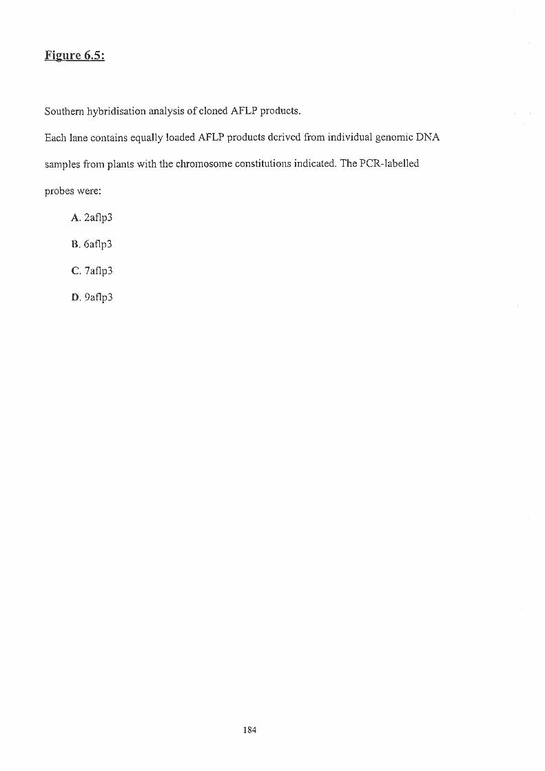
Figure 6.5:
Southem hybridisation analysis of cloned AFLP products.
Each lane contains equally loaded AFLP products derived from individual genomic DNA
samples frorn plants with the chromosome constitutions indicated. The PCR-labelled
probes were:
A,.2aflp3
B.6aflp3
C.7af1p3
D.9aflp3
184

B
A
D
Õ
OB
OB
OB
+B
**- ,l} {}
+B
+B
C
OB +B

1 i aflp3) and 7aflp3 from genomic DNA was closely related to the presence of B
chromosomes in the template and may have been responsible for the original AFLP
polyrnorphism (Figure 6.1). It was therefore thought that the 2af1p3 and 7aflp3 sequences
may be present on the B chromosomes. The additional hybridisation of 2aflp3 to the AFLP
product amplified from one 0B sample (Figure 6.5) suggested that this sequence may also
be present on some A chromosomes.
Results fi'orn genomic DNA Southem hybridisations using 2af7p3 and 7aflp3 as
probes rvere difficult to interpret as the levels of hybridisation were vety low, suggesting
that the sequences were at low copy number within the B. clichrontosontatica genome
(result not shown).
6.2.1 PCR Ana lvsis of Putative B Chromosom e AFLP Products
The inability to obtain clear results from genomic Southern hybr:idisations te[d to a
¡
PCR approach in contbination with microdissection (Sectìon3.2.2) to detennine whether
2aflp3 and 7aflp3 sequences were in fact present on the B chromosontes. Prinlers were
desig¡ed to 2aflp3 and 7aflp3 (Figure 6.4, Table 2.4c), so that the conesponding seqllences
could be arnplified frorn genomic DNA. As expected, primers P2aflp-I and P2aflp-2
arriplified a 100 bp product from genornic DNA witli B chrornosol't.les and fi'om the clone
p2aflp3 (Figure 6.6). Initially a smear was anrplified fror.n nricrodissected B chromosotne
DNA telnplate, but reamplification resulted in a product of equal size to 2aflp3
(Figure 6.6). The negative control containing no DNA tenrplate confinned that there was
no contamination of the PCR reaction constituents (Figure 6.6). Cloning and sequencing of
the 2aflp3 product amplified from microdissected B-DNA revealed that the inserl of the
B-derived 2af1p3 clone showed 100% nucleotide identity to the original 2aflp3 sequence,
i¡dicating that the sequence \ilas present on the B chromosomes (result not showrl).
186

Figure 6.6:
Agarose gel of PCR products amplihed with 2aflp3 specific prinrers (P2aflp3-l and
P2aflp3-2).
The first lane contains molecular size markers (100 bp DNA ladder), consisting of
fi'agments that range from 100 bp to 3000 bp in 100 bp increments.
The template DNA for the remaining lanes were:
1. Microdissected B chromosome DNA.
2. Preamplified microdissected B chromosome DNA from lane l.
3. B. dichrontosontatica *B genomic DNA.
4. Clone p2af1p3 (+ve control).
5. No DNA tenrplate (-ve control).
187
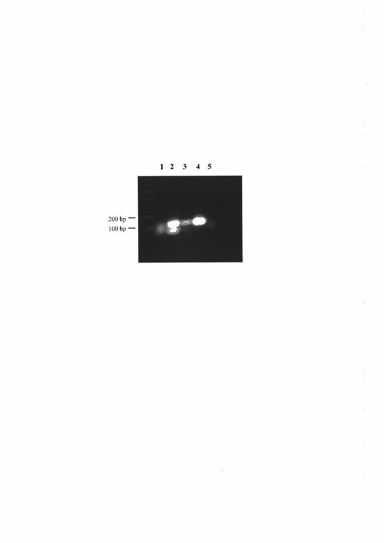
t2 3 4 5
200 bp -100 bp - ÇË*.}

Primers, P7aflp3-1 and P7aflp3-2, designed to amplify 7aflp3-like sequences from
genomic DNA were not able to arnplify a product from the microdissected B-DNA, despite
several attempts (result not shown), suggesting that the sequence was not present on the B
chromosomes. The hybridisation of 7aflp3 exclusively to the AFLP products amplified
from +B samples in Figure 6.4C may be the result of a polymorphism which is specific to
the A cluomosomes present in the fB samples in the experiment. Alternatively, it may be
that the sequence could not be arnplified from the microdissected B chromosome DNA due
to unknown technical difficulties.
6.2.2 Screenine of 3B Genomic Librarv with 2aflp3
The sr¡all size of 2aflp3lead to attempts to isolate larger clones, wliich would be
nlore re\/ealing in database searches and in studies of differences between the sequence on
the A and B chromosontes, rvhich in turn nray be useful in detenuining the origin and
evolutionary path of the B chromosomes. Horvever, screeuing of 50,000 plaques of a 3B
B. dichrontosontutica genomic library was unsuccessful in isolating genomic clones of
2af7p3, supporting tlie idea that the sequence rvas of low copy in the B. clichrontosotnatica
genome of plants in wliich they were present.
6.2.3 Summary of AFLP Analysis
In sumrnary, the AFLP technique enabled isolation of a sequence (2aflp3) which rvas
present in all +B sarnples examined as well as in DNA derived from microdissected B
chronosonres. The srnall size and difficulty in perfoming Southem analysis with 2aflp3 as
a probe made it difficult to determine the extent of the sequence in the population and
fufther analysis requires a larger genomic clone to be isolated.
189

6.3 RlNnou Atvrpr,rrrnn Por-rruonpsrc DNA SBounNcBs
Random arnplified polyrnorpliic DNA sequences (RAPDs) are DNA fragments
arnplified from genomic DNA by PCR using 1O-base oligonucleotides as prirners
(V/illiams et al., i990). The object of the work presented in this section was to isolate B
clrronrosome-specific RAPDs from.B. dicltrontosontalica. The urajority of RAPDs
amplified with individual primers will be shared between 0B and *B sarnples, representing
sequences on the A chromosomes which may or may not also be present on the B
chromosonres. Repeat and low copy sequences specific to the B chronosomes which
co¡tain, or are flanked by, primer recognition sites, will also be amplified and
distinguished as additional RAPDs, arnplified only from genoutic DNA terrrplates
containing B chromosomes.
lnitially, collaborators in Germany used 300 1O-base priurers, to arnplify DNA frorn
0B and 38 pools of genomic DNA ternplates, in order to detenline u,hich primers rvere
caudidates for arnplifying polymorphisms consistent u,ith the presellce of B chrourosomes.
Fifteell of the primers tested were selected because they each an-rplified PCR fi'agmerlts
front *B and not from 0B templates (result not shorvn) and these primers fomred tire focus
of tl-re following experinents. During atternpts at repeating the t'esults obtained in Genrany
only four of the original hfteen primers showed consistent amplifìcation of
B. clichrontosonntica genomic DNA (Figure 6.1A). These prirnet's (Vi2, Y2,26 and AC5)
rvere used to arnplify from'pools'of 0B and 38 genomic DNA samples and the PCR
products were resolved on agarose gels (Figure 6.7 A, B). RAPDs which were consistently
amplified from *B pooled DNA templates and not from 0B pooled DNA (namely 2R, 4R,
6R , 7R, CR, ER and FR; Figure 6.7 A, B) were excised from the gel, freeze-squeezed and
cloned into the vector pGEM@-T. Inserts of putative clones were confinned as
190

Figure 6.7:
A. Agarose gels of PCR products amplified with the 1O-mer primers indicated, fi'om 0B
and +B pooled B. dichrontosomatica genomic DNA samples
The RAPD bands that were gel excised for further analysis are arowed and labelled. The
first lanes contain molecular size markers (100 bp DNA ladder), consisting of fragments
that range fi-om 100 bp to 3000 bp in 100 bp increments.
B. Agarose gel of PCR products amplified with the lO-mer primers indicated, from 0B and
+B poolecl B. clichrontosomatica genomic DNA santples.
The RAPD bands that were gel excised for further analysis are atrowed and labelled. The
first lanes contain nrolecular size markers (100 bp DNA ladder), consisting of fragr.trents
that range fronr 100 bp to 3000 bp in 100 bp increments.
191

Q12 T3r0B+Br bB-JF
1.0 -
Z6 ACsfg3+Bl l0B+Bt
a2 u9 vt2 v13 Y2
A
3.0 -
1.0 -
3.0
1.0
Y3 YO9 ZI 7.6 ZII AA8 AB1
6R
Y2
roÌ+Bl
B2R
6R
FR

representative of the RAPD fragment by hybridisation with the initial freeze-squeezed
products (result not shown).
6.3.1 Analysis of Putative B Chromosome Specific RAPDs
All the putative B-specific RAPDs were analysed to determine their B chromosome
status by Southem hybridisation. The inserts from clones pBd2R, pBd4R, pBd6R and
pBd7R, originally obtained using RAPD primers Y72,Y2 and AC5 (Figure 6.7), were used
as probes to Eco RI digested genomic DNA with and without B cluomosomes to look for
differences in hybridisation pattems associated with the presence of B ch¡omosomes
(Figure 6.8).
The insert frorr clone pBd2R hybridised to Eco RI fragments of both 0B and +B
genomic DNA samples (Figure 6.88). High levels of hybridisation of Bd2R occuned to a
ladder of balrds, suggesting that the sequerìce was a high copy number repeat. The lack of
differentiation bettveen 0B and *B sarnples suggested that the sequence was present on the
A chrontosonles, or on the A and B chromosones in a similar organisation (Figure 6.88).
The inserl from clone pBd4R displayed lower levels of hybridisation to,Eco RI
digested genomic DNA (Figure 6.8C) rvhen compared with Bd2R (Figure 6.8B) and
showed a snìear with several superirnposed hybridising fragments. Agaiu, no difference in
overall hybridisation pattern was detected betrveen 0B and *B samples, indicating that
RAPD fragment 4R was not B-specific. Despite equal amounts of DNA being present in
each track (Figure 6.84) the levels of hybridisation of Bd4R varied considerably between
individual genomic DNA samples, suggesting tliat the sequence valied in copy number
between different plants (Figure 6.8C). The apparent heterogeneous nature of the sequence
may account for the initial differential 4R RAPD amplification product (Figure 6.64).
Similarly, no differential hybridisation of BdTR was apparent between 0B and *B genorlic
DNA samples (Figure 6.8D). The additional band in one of the 38 tracks
193

Figure 6.8:
A. Agarose gel showin g B. dichrontosontatica genomic DNA lestricted with Eco RL
Each lane was loaded with 3 pg of genomic DNA. The numbers on the left represent the
nrolecular sizes (kb) of lambda bacteriophage DNA restricted witlt Hind III. The
chromoson-re constitutions of the plants from which the genornic DNA was prepared are
indicated.
B. Southem hybridisation of the agarose gel in A rvith the insert from pBd2R.
Laues and nrolect¡lar size markers are as above.
C, Southem hybridisation of the agarose gel in A rvith the inseft frotl pBd4R.
Lanes and nrolecular size markers are as above
D. Southenr hybridisation of the agarose gel in A rvith the insert fronl pBd7R.
La¡es a¡d ntolecular size narkers are as above. The polymorphìc band present in orte 38
sample is alrorved.
E. Southem hybridisation of the agarose gel in A with the insert frorn pBd6R.
Laues and molecular size urarkers are as above
194

A OB OB 3B 3B
23.1 -9.4
-6.5 -+.J
2.3 -2.0 -
B OB OB 3B 38 C OB OB 3B 3B
tt*
t#
iÊ

3BOB OB 3B 3B E OB OBD
*
*i}*
t

(Figure 6.8D) was initially thought to represent a polymorphism between different plants
but a longer exposure (not shown) indicated that the band was present in all tracks and was
increased in copy number in the DNA of one plant examined'
Bd6R showed increased levels of hybridisation to the genomic DNAs (Figure 6.8E)
in cornparison to Bd4R (Figure 6.8C) and BdTR (Figure 6.8D). The smear of hybridisation
(Figure 6.8E) suggested that pBd6R contained a repetitious sequence, dispersed throughout
the genome. No differences were seen in patterns or levels of hybridisation between 0B and
*B genomic DNA.
The inserls from pBdCR, pBdFR and pBdER, all initially amplified with tlie RAPD-
primer Y2 (Figure 6.7), were used as probes to Southem blots of the RAPD-PCR products
fron-r 0B and +B pooled samples. BdCR and BdFR, as expected, hybridised strongly to the
parent RAPD band, but also showed hybridisation to both 0B and *B ploducts with no
apparent differences in the liybridisation pattems between the.sarnples (Figure 6.9A, B). In
contrast, hybridisation of BdER to the sanre Southem blot of the RAPD-PCR gel rvas
confined to the B-containing Y2 RAPD-PCR product, with Ilo sign of any hybridisation to
PCR products an-rplified from 0B samples with the same primer (Figure 6.9C). The
hybridisation obserryed rvas not specific to the BdER band, but was also seen to a number
of other bands within the PCR product, implying tha|Y2 was an-rplifying a rtumbel of
regions from within the +B genome with BdER identity. The implication of the
hybridisation of BdER to RAPD products amplifìed, with prirner Y2, from *B and not 0B
genomic DNA was that the clone pBdER contained a sequence specific to the B
chromosomes.
To deten¡ine the extent of BdER within B-containitrg samples, aud to confinn the
absence of the sequence in 0B plants, primer Y2 was used in PCRs with DNA templates
prepared from the six individual 0B and +B plants present in the original pools. The
196

Fieure 6.9:
Southem hybridisation analysis of cloned RAPD products amplified with the Y2 RAPD
primer from 0B and +B pooled B. dichrontosontatica genomic DNA templates.
Each lane contains equally loaded RAPD products amplified with the Y2 primer from 0B
and +B pooled B. dichrontosomatica genomic DNA templates.
The probes were:
A. BdCR
B. BdFR
C. BdER. The RAPD representing BdER, amplifìed from the *B pool of samples, is
arrowed and labelled
t97

CBAOB +B OB +B
OB +B
1.0 -
3.0 -
1.0 -
3.0 -
1.0 -
BdER
/r/
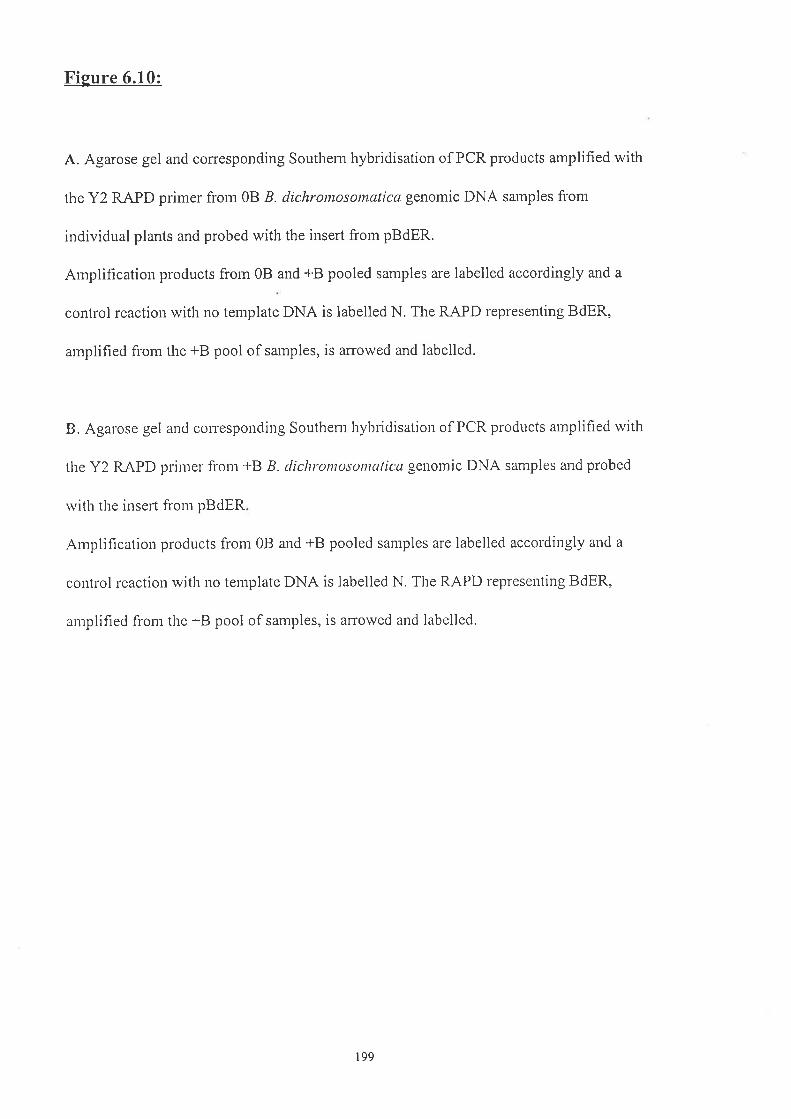
Figure 6.10:
A. Agarose gel and coresponding Southern hybridisation of PCR products amplified with
the Y2 RAPD primer frorn 0B B. dichrontosontatica genomic DNA sarnples frorn
individual plants and probed with the insert frorn pBdER.
Arnplification products froln 0B and +B pooled samples are labelled accordingly and a
control reaction with no template DNA is labelled N. The RAPD representiug BdER,
amplified from the +B pool of sarnples, is arrowed and labelled.
B. Agarose gel and conesponding Southern hybridisation of PCR products amplified with
tlre Y2 RAPD prir-ner from *B B. clichrontosontcttica genomic DNA samples and probed
rvith the inseft from pBdER
Anrplìfication products from 0B and +B pooled samples are labelled accordingly and a
control reaction with no template DNA is labelled N. The RAPD rept'eseuting BdER,
anrplified fi'om the +B pool of samples, is arowed and labelled.
199
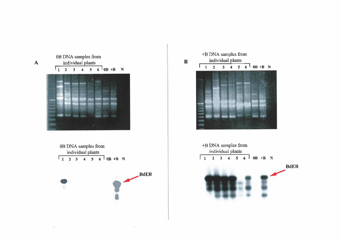
A0B DNA samples from
individual plantsfr z 3 4 s olon+g N
0B DNA samples fromindividual plants
t23 4 5 6
B
+B DNA samples fromindividual plants
ñoB+BN
+B DNA samples fromindividual plants
lt 2 3 4 s e l oB +B NOB+B N
BdER
BdER r'TÕIttO r'
{h"'s4 Wrs -' *t *phrs
qo¡ñ
*.{p

number and size of the PCR products amplified with prirnet'Y2 varied slightly between
individual samples (Figure 6.104, B) suggesting that the plants from which the DNA was
derived were heterogeneous, as would be expected for an outbreeding population. Southern
analysis of the PCR products with BdER as a probe showed hybridisation to the products
a¡rplified from all six +B templates (Figure 6.108). Hybridisation at a lower level was also
seen to tlie products amplified from one sample from within the 0B pool (Figure 6.104).
The positive and negative controls were the parallel aniplification and hybridisation from
the 0B a¡d +B pooled samples and the incorporation of a reactiotl lacking DNA ternplate to
check for contamination of the PCR constituents (Figure 6.104, B).
The lack of BdER hybridisation to RAPDs anrplified frorn the 0B pool, despite the
hybridisation to products amplified from one of the individual samples preseut within the
pool, suggested that there was a low copy number of the BdER sequence in the genonle of
o¡e of the 0B plants. In an-rplificationreactions from tlie 0B pool it is likely that the high
proportion of non-BdER sequences with primer Y2 identity outcompeted the BdER
sequences for the plirler. However, within the individual 0B sample, the percentage of
BdER in the total DNA would be greater and, therefore, lllore readily arnplifiable. The total
¡l¡mber of BdER-hybridising bands was decreased in the 0B sample, compared wìth the
*B sa¡rples (Figure 6.10), indicating lower copy number and less variation amongst the
copies of the sequence in DNA from the plant u,ithout Bs than the plants with Bs.
In order to asceftain the extent of BdER witliin the B. dichromosontatica population,
a¡rplification with prirner Y2 was repeated on a number of additional samples with and
without B chromosoûìes. The genornic DNA samples chosen were derived fi'orn different
populations of ,8. dichromosomatica than the samples used in the initial 0B and +B pools
and were therefore expected to be divergent in A, and possibly B, chrotnosolne sequences
201

Figure 6.11:
Agarose gel of RAPD products amplified with primer Y2 from a range of B
dichromosotnatica genomic DNA samples without (08) and with (+B) B chromosomes, in
addition to the 0B and +B pooled samples and a no DNA template (N) control.
The first lanes contain molecular size markers (100 bp DNA ladder), consisting of
fragrnents that range from 100 bp to 3000 bp in 100 bp incrernents. The RAPD
representing BdER, amplified from the +B pool of samples, is arrowed and labelled
202

0B and +B DNA samples fromindividual plants
+B OB +B OB +B OB +B +B OB+B N
BdER
1000 bp -
it r;t ;{ : trr r*'å
trl.<l ¡ë3'
I I I ' t:+ Fa¡
âr
lrr
}{
3.'

due to the outbreeding nature of the species. Amplification products resulted from all the
samples tested and were resolved on agarose gels. Controls included were parallel
arnplifications from the original 0B and +B pools and a reaction with no DNA template
(Figure 6.11).
Overall the number and sizes of the RAPD products amplified were fairly uniform
across all samples, including those from the 0B and +B pools (Figure 6.1 1). The low level
of visible polymorphism between the RAPDs amplified from individual templates was
indicative of the degree of heterogeneity within the population. Hybridisation of the RAPD
products with BdER was strong to the +B pool-PCR products as was seen previously
(Figure 6.9, 6.10). However, minimal levels of BdER hybridisation were obtained to all
other RAPD products (result not shown). The BdER sequence could be present on the B
and/or the A chromosornes, but it rvas clear that it rvas not a sequence ubiquitous to either
clrronrosome type in B. clichromosontalica. Sequencing of pBdER with (-20) and revetse
sequencing primers enabled the full sequence of the insert to be deten¡ined (Figure 6'12).
The 1275 bp of nucleotide sequence obtained shorved significarrt levels (55-70%) of
nucleotide identity with retroviral and mitochondrial sequeuces iu the database (GenBank
and EMBL , IggT), suggesting invasion, followed by sequence divergence, of a
subpopulation of A and, possibly, B chromosorìes with mobile elements or mitochondrial
DNA.
Inmaize,RAPD analysis isolated several putative B-specific RAPDs (Gourmet and
Rayburn, 1996; Stark el al.,1996; Lin and Chou, 1997), including one with signihcant
nucleic acid similarity to a maize retroviral element called PREM-1 (Stark et a1.,1996).
However, the overall composition of the maize B was similar to that of the standard
clrromosome complement (Stark et a1.,1996). Initial RAPD and AFLP analysis in rye
suggested that low copy sequences are on average under represented on the B clrromosome
204
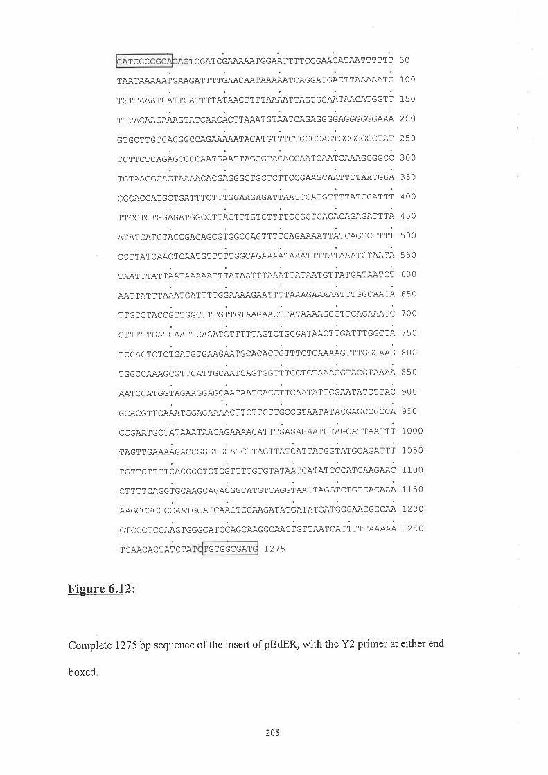
TCGC T GGATCGAAAÀATGGAAT T TTCCGAACATAATTTTT T
TAATAAAAATGAAGAT T T TGzu\Cru\T AJUUU\TCAGGATGACTTAÀÄAATG
TGTTA,CATCATTCATTTTATAACTTTT NUU\TTAGTGGAATAACATGGTT
T T TAcAAGAÅAG TAr cAACÀc r rAAAr G Til, co.o.. 6i466 6 6 66 Nry;q
GTGCTTGTCACGGCCAG A'UU\i\TACATGTTTCT GCCCAGTGCGCGCCTAT
TcrrcrcAGÅccc.coorcÅerracccrococco¡rça;qiç Nqi\6cGGcð
TGTAACGGAGT AJU\,\CACGAGGGCTGCTCTTCCGAAGCAATTCTAACGGA
cccAccATGò,co* rc, r rcceacaco,t*,cce,"r, rtatccetri
TTCCTCTGGAGATGGCCTTACTTTGTCTTTTCCGCTGAGACAGAGATTTA
ATAT cATCTÅcccacecccrccccacrrrt cacoeaot rÅr cecc c t r tiCCTTATCAÄCTCAÀTGTTTTTGGCAGAJUUqTAAÀTTTTATAAATGTAATA
TAAT T TAT Ti¡1 e;ry;qiuq1 r ior*r r roit rat*t c r ro, cat*t c iAAT TATT TAAATGAT T T T GG AJUÐqGÄIqT T TT AJU\G AiUUN\T CTGG CAACA
TTGCcrACCcrrccctrrcircre¡ceeçi1a1ruq,u\ccòrrceca¡erð
C T T T T T GAT CAAÎ T CAGAT G T T T T TAG T C T G C GATA.AC T T GAT T T G G C TA
I c cAG T G r c i co, c, c*c.Å¡r ccecec, à r m c r. rury;úc r r r c c c*ð
TGGCCAÄAGCGTTCATTGCAATCAGTGGTTTCCTCT AJ\'\CGTACGTAi\iU\
AAT ccATGG ieceeccecc.ilre¡rce..i t c*rer r.coarot.ttoð
GCACGTTCAAATGGAGAJU\'\CTTGTTGTTGCCGTAATATACGAGCCGCCA
cccAAT GcrÅra¡¡reecai e;ry;ry¡rça1 t räaceceercrÅccnr root t tTAG TTGAAAAGACCGGGTG CATCTTAGT TATCAT TATGG TATGCAGAT TT
T G T T c r r r r òoc. "c
r c, c c r r r r c r c roraÀr cerer c ð cer ceecooò
CTT TT CAGGTGCAAGCAGACG GCAT GT CAGG TAAT TAGG T CTG TCACAAA
AAGccc c ccò*raaor ada raa*aeratcerercatcc ceaccccoÅ
G T C C C T C CAAG T G G G CAT C CAG CAAG G CAAC T G T TAAT CAT T T T TAAÀAA
50
100
150
200
250
300
350
400
450
500
550
600
650
700
750
800
8s0
900
950
1000
1050
110 0
115 0
1200
l-250
TCAACACTATCTA I21 5
Figure 6.12:
Complete I27 5 bp sequence of the insert of pBdER, with the Y2 primer at either end
GCGGCGAT
boxed
205

and that the euchromatic component of the B sliows similar repetitive sequence
cornposition as that of the As (Langdon et a\.,1997). Similar to the RAPD results obtained
in B. dichrontosontatica,ihe pericentromeric region of the rye B cluonosone was found to
contain organelle related DNA (Langdon et al.,1997).
Sequences which were retroviral or organelle-derived have previously been found on
tlre B clrromosomes of B. diclromosomatica, flanking Bd49 (Franks et al., 1996) and tlie
wasp, Nasonia vitripennis, which contains a B-specific transposable element (NATE)
(Section 1.7 .2). Therefore, it is evident that, in addition to B. dichrontosontatica,theB
chromosor¡es from a range of species contaiu retroviral and/or other transposable elements
but it is unceftain whether the Bs are more or less prone to accu¡ulation of such
sequences. TIie significance, if any, of these sequences on the Bs is unknotvn, howet'er, i11
light of their essentially inert genetic nature it is not surprising that B chromosomes are
parlicularly tolerant of DNA transposition and reamangement.
6.4 ConcLUSION
The results obtained using RAPD and AFLP techniques suggest that a large
proportion of the B cluomosome in B. dichrontosontatica is composed of sequences u4tich
are colnmon to the A clrromosomes. This hypothesis is supporled by the fact that, aparl
from tlre repeat 8d49, no other sequences specific to the B. dichrontosontatica Bs have
been isolated (Franks et a\.,1996). Further, the only other sequences located on the Bs of
B. clichrontosontatica are rDNA (Section 1.6.2b; Donald et a\.,1995) and Bdm29 (Section
1.6.1b, 1.8;Houbenet a1.,1997b) both of which are also present on the A chromosomes.
The results presented in this chapter are complemented by comparisons of the sequence
con-rpositions of the A and B chromosomes in a wide range of species, which supporl the
idea tliat the B chromosomes consist predominantly of sequences present on both
206

chromosome types (John et a1.,1991; Tsujimoto and Niwa, 1992; Blunden et al., 1993;
Cuardro and Jouve, 1994; Jamilena et a1.,I994;López-Le6n et al.,1994; McQuade et al.,
7994; Bougourd et a1.,1995; Stark et a1.,1996).
207

R
Concluding Discussion
aa
208

7.1 OnCnNrs¡,rrON tNn ExpnnSSrON or Pr,nNr B CnnOvrOSOnrns
Previously, fluorescence in situ hybridisation revealed the presence of sequences
with ¡DNA identity on the B chromosomes of B. dichromosotnatica. The preliminary aim
of the rDNA work presented in this thesis was to determine if these B chromosome
sequences were transcriptionally active.
Initially, analysis of ITS2 identified two nucleotide differences between the
sequences derived from the A and B chromosomes. One of these differences involved a
^ffc I restriction site present in the ITS2 of B-derived rDNA that enabled the ITS2 on the A
and B chromosomes to be distinguished. The inability to detect the B-specific ITS2 regions
witliin the 40S precursor rRNA suggested that the B-rDNA was not transcribed in leaf or
floral tissue. This was the first molecular evidence of the transcriptional inactivity of rDNA
on B chronlosomes which had previously only been examined by silver staining
techniques. The mechanism for the B-rDNA inactivation is not known but it was of interest
to detennine if there were any differences in the sequellce and/or organisation of the rDNA
on the A and B cluomosomes which could; (1) explain tlie inactivity of the B cluomosome
sequences, (2) shed light on the evolution of these sequences on the Bs, and their
relationship with those on the As and (3) suggest a possible origin for the B chromosomes
themselves.
Comparison of the region separating the i 8S and 25S coding regions of the rDNA on
the A and B chromosomes, which is comprised of the ITSl, 5.8S and ITS2, suggested that
a proportion, and possibly all, the regions on the Bs were intact and, with the exception of
the identified B-specific polyrnorphisms at bases I42 and 175 within the ITS2, identical in
sequence to the corresponding region on the A chromosomes. In addition, comparisons of
Soutliern hybridisation after probing with cotton (Donald et a|.,1995) and
209

B. dichromosotnatica rDNA was unable to detect differences in hybridisation between 0B
and +B genomic DNA, indicating overall homogeneity within and between the rRNA
coding regions on the A and B chrornosomes,
To examine the B chromosome IGS region, which is of parlicular interest as it
contains the RNA polymerase I transcription-initiation site as well as other elements
important in rRNA transcriptional regulation, Southern hybridisation with IGS-derived
probes revealed essentially very similar pattems of hybridisation to 0B and iB genornic
DNA. A single restriction fragment polymorphisrn associated witli the presence of Bs rvas
identified but the source of the polymorphism is yet to be detenniued. Experiments which
looked specifically at tlie B cluomosome IGS region, usitig PCR, supporled the hypothesis
that there were low levels of variation in the IGS region on the Bs, but these may have
reflected tecl-rnical difficulties associated with the presence of inconrplete DNA fragnrents
in the DOP-PCR-amplified microdissected B-DN A. In sittt hybridisation confirmed the
preserìce of IGS sequences on the Bs and no variation in the hybridisation of the differellt
IGS-subcìones was detected'
In summary, evidence from ITS2 characterisation, Southenr hybridisation, PCR
analysis, nucleotide sequence comparison and in silu hybridisation supported the
preposition that overall the rDNA on the B chromosornes of .8. clichrontosontatica is
organised in intact repeat units. The identification of several putative B-specific
polymorphisrns in the IGS region implied that variation between A- and B-rÐNA nay
exist and could possibly be associated with the B-rDNA inactivity.
The apparent sirnilarity between A and B chlomosome rDNA suggests that despite
the inactivity of the rDNA on the Bs, they are under the control of a homogenising
mechanisrn which controls the non-independent evolution of these repetitive DNA
sequences. The irnplication is therefore that tlie B cluomosome rDNA is maintained by
210

mechanisms similar to those on the As. However, differences between tlie A and Bs in the
region suggest that the two chromosome types may not be evolving in concert with each
other
1.2 Isol on- uen chro
Attempts to isolate non-repetitive sequences from the B chromosomes using RAPD
and AFLP analysis were not successful in isolating B-specihc sequences but did identify
sequences polymorphic within the species, present on the A and possibly the B
chromosomes of some individuals. It was apparent from RAPD and AFLP analyses that
t¡e number and size of the PCR products amplified from 0B and *B genomic DNA
samples, using these teclmiques, were overall siniilar. Any polynrorphisrns that were
detected generally represented genetic heterogeueity expected ofan outbreeding species
and did not involve sequelÌces unique to the B chromosomes, indicating that the A and B
chromosomes were overall very similar.
7.1.3 Concludins Discussion
Due to the lack of transcription and in the absence of selection, many of the sequences
that co¡rprise Bs would be expected to show high rates of evolutionary change' Horvever,
tlre results fror¡ this work indicate that despite the inactivity of the B. dichrontosontalicaB
cluomosomes, insinuated by the sequence analysis of rRNA and histone acetylation
pattems (Houben et al.,1997a), these chromosones contain a high proportion of DNA,
including rDNA, that is both comrnon to the A chromosomes and consen¡ed in sequence.
Previously, it was suggested that degeneration of supemumerary chromosomes may
proceed via the mechanism of Muller's Ratcliet (Green, 1990). This hypothesis predicts
that supernurrerary chromosomes should lose functional loci, lose sequence homology
with the regular genorne, and gain heterochromatin over time, resulting in multiple
heteromorphic fonns of degenerate supernumeraries within and between populations.
2rl

Although there are two different B chromosome types present in -8. dicltrontosomcttica
(Section 1.8), evidence suggests the micro B ch¡omosomes are more likely to be
forerunners of the larger Bs than degenerate morphs (Houben et al., I997b). The Bs in .8.
clichrontosomatica have no apparent functional loci but still seem to maintain significant
sequence identity with the A chromosomes and this scenario is similar in a wide range of
other species where the Bs are apparently transcriptionally inactive, but the sequences on
the Bs are predominantly common with the As (Chapter 6).
One of the major questions raised by the results in this project is: How are sequences
on the Bs maintained when they appear to be inactive and is there any evolutionary or other
requirenrent for this sequence conservation? One possible answer is that the entire B
chromosome is under evolutionary control similar to that proposed for rDNA and other
repetitive sequences, whereby the intra-genomic diversity is generally low due to concefted
evolution within and between loci (Dover, 1982). Altematively the Bs may be 'young' and
have had illsufficient time to diverge. However, tliere is currently no evidence for Bs at
different stages of degeneration, which would be predicted if this was the case. Other
possibilities are that the structure of the B is important in ensuring its owtl transmission
and this is somehow selected for or that Bs that vary considerably from the As are more
detrimental to the organism and therefore selected against.
7.2 FrN¿,r, CouvtnNr
Many of the airns outlined prior to the commencement of this research have been
achieved, but a number of important questions remain unanswered;
(1) What is the significance, if any, of the very fi'equent presence of rDNA sequences
on B chromosomes in a large number of species?
212

(2) How is meiotic pairing between the A and B chu'omosotnes prevented at the
molecular level, despite the apparent presence of massive sharing of sequences between the
two ch¡omosome types?
(3) Is there a stage of the life cycle when the sequences on the B chromosomes are
expressed?
Further molecular analysis of B chromosomes is likely to help answer these questions,
in addition to increasing general understanding of the universally important processes
involved in the control of chromosorne pairing, division and evolution. Perhaps the next
important step towards this end is to extend the analyses started here to give detailed
sequence comparisons of DNA that is presetrt oli both chromosome types.
2r3

APPENDICES
214
AppnNorx 1
Phosphorimage analysis of the proportion of ITS2-PCR products, amplihed from
genornic DNA derived from.B. dichromosotnatica plants with different chromosome
constitutions, which was unrestricted and restricted with,!fc I'
Chromosomeconstitutions
Unrestricted(PSL)
Restricted(PSL)
% of ITS2Restricted withSfc I
OB 20760.0 0 0
30680.0 0 0
2l 180.0 0 0
1B 17980.0 109.20 3.8
37790.0 1234.60 3.2
18890.0 651.9 3.4
2B 13010.0 931.90 6.1
17500.0 1050.4 5.6
18150.0 I 160.0 6.0
3B 23540.0 2099.0 8.2
28630.0 3298.6 10.3
41060.0 4509.0 9.9
4B 28750.0 4631.0 13.8
17070.0 2584 13.3
r 6390 2569.5 1 3.5
Usi¡g the MacBas Version 2.X computer program (Fuji Photo Film, Co', Ltd) the
radioactivity witliin specified regions was measured in units of photostimulated
luninescence (PSL) and the percentage of the total ITS2 PCR product which was restricted
with ^!fc I was calculated.
215

Appnxnrx 2
Donald TM, Houben A, Leach CR and Timmis JN. 1997. Ribosomal RNA genes
specific to the B chromosomes in Brachyconte dichrontosontatica are not transcribed in
leaf tissue. Genotne 40:674-68I.
216

A Donald, T., Houben, A., Leach, C.R. & Timmis, J.N. (1997) Ribosomal RNA genes specific to the B chromosomes in Brachycome dichromosomatica are not transcribed in leaf tissue. Genome, v. 40(5), pp. 674-681
NOTE:
This publication is included after page 216 in the print copy of the thesis held in the University of Adelaide Library.


RE,FERENCES
211

Alfenito MR and Birchler JA. 1993. Molecular characterisation of amaize B chromosome
centric sequence. Genetics 135: 589-597.
Altshul SF, Gish W, Miller W, Myers E\il and Lipman DJ. 1990. Basic local alignment
search tool. Journal of Molecular Biology 215: 403-410.
Appels R and Honeycutt RL. 1986. rDNA: Evolution over a billion years, pp. 81-135, in
DNA Systetnatics, Vol II. Plants, edited by Dutta SK. CRC Press. Boca, Raton, FL.
Arnheim N. 1993. Concerted evolution of multigene families, pp. 38-61, in Evolution of
Genes and Proteirrs, edited by Nei M and Koehn B. Sinauer Associates. Sunderland,
Massachussetts.
Ayonadu U and Rees H. 1968. The influence of B chromosomes on chiaslna frequencies in
Black Mexican corn. Genetica 39:75-81.
Baldrvin BG, Sanderson N{J, Porter JM, Wojciechon'ski MF, Campbell CS and
Donoghue MJ. 1 995. Ann. Missouri Botanic Garclens 82: 247 -277 .
Beckett JB. 1991. Cytogenetic, genetic and plant breeding application of B-A translocation in
rr;iize,pp.493-529,in Chrotnosome Engineering in Plants, Purt,4, edited by Gupta PK
and Tsuchiya T. Elsevier Science Publishers, Amsterdanl.
Bell G and Burt A. 1990. B-chromosome: genn-line parasites which indnce changes in host
recombination. Parasitology 100: S l9-S26.
Bestor TH. 1998. Methylation meets acetylation . Nature 393: 3 II-312.
Beukeboom LW and Werren JH. 1993. Deletion analysis of the Selfish.B Chromosome,
Paternal Sex Ratio (PSÄ), in the Parasitic Wasp Nasonia vitripetmis. Geneîics 133: 637-
648.
Beukeboom LW. 1994. Bewildering Bs: an impression of the 1st B-chromosorte conference.
Heredity 73:328-336.
Blunden R, Wilkes TJ, Forster JW, Jiminex MM, Sandery MJ, Kemp A and Jones RN.
1993. Identification of the 83900 farnily, a second farnily of rye B chromosome specific
repeated sequence. Genonte 36: 7 06-7 12.
218

Borisjuk N and Hemleben V. 1993. Nucleotide sequence of the potato rDNA intergenic
spacer. Plant Molecular Biology 21: 381-384.
Bougourd SM and Jones RN. 1997. B chromosomes: a physiological enigma. New
Phytologist 137 : 43-54.
Bougourd SM and Parker JS. 1979. The B chromosome system of Allium Schoenoprasunt
II. Stability, inheritance and phenotypic effects. Chromosoma 75:369-383.
Bougourd SM and Plowman AB. 1996. The inheritance of B cluotnosomesinAlliunt
Schoenoprasunt L. Chromosome Research 4: 1 5 1-1 58.
Bougourd SM, Plowman AB, Ponsford NR, Elias ML, Holms DS, Taylor S. 1995. The
case of unselfish B-cluomosomes: evidence fromAlliutn Schoenoprasum.In Bralidam PE,
Berinett MD eds. Kew Chromosome Conference IY:2I-34.
Buescher PJ, Phillips RL and Brambi R. 1984. Ribosomal RNA content of maize
genotrophs with different ribosomal gene numbers. Biochenical Genetics 22:923-930.
Bullock 'WO, Fernandez JM and Short JM. 1987. XLI-Blue: A high effrciency plasmid
transformin g recA, Escherichia coli strain with beta-galactosidase selection. Bioteclutiques
5:376-378.
Cabrero J, Alche JD, and Camacho JPM. 1987. Effects of B chronosomes on the activity
of nucleolar organizer regions in the grasshopper Eltprepocnentis plorans: activation of a
latent nucleolar organizer region on a B cluomosome fused to an autosonte. Gettome 29'.
r16-12r.
Cabrero JrLopez-Leon MD, Gomez R, Castro AJ, Martin-Alganza and Camacho JPM.
1997. Geographical distribution of B chrornosoÍìes in the grasshopper Eyrepoarcnús
plorans, along the river basin, is rnainly shaped by non-selective historical events.
Chromosotne Research 5: 194-198.
Cameron FM and Rees H. 1967. The influence of B cluornosomes on meiosis irt Loliunt.
Heredity 22:446-450.
Capesius l. 1997. Analysis of the ribosomal RNA gene repeat from the vrcss Funaria
hygrontetrica. Plant Molecular Biology 33: 559-564.
2t9

Carlson WR and Roseman RR. 1992. A new property of the maize B chromosome. Genetics
137:217-223.
Carlson \üR. 1986. The B chromosome of maize. CRC Critical Reviews in Plant Sciences 3:
20r-226.
Carter CR and Smith-White S. 1912. The cytology of Brachycome lineariloba 3. Accessory
chromosom es. Chrotnosoma 39: 36I-379.
Carter CR. 1978a.The cytolo gy of Brachycome 8. The inheritance, frequency and distribution
of B chromosomes in B. dichrontosomatica, formerly included in B. lineariloba.
Chrontosonta 67 :109 -l2I'
Carter CR. 1978b. Taxonomy of the Bracltycotne lineariloba complex (Asteraceae). Telopea
7:387-393.
Cebria A, Navarro ML and Puertas MJ. 1994. Genetic Control of B ch¡omosome
transmission in Aegilops Speltoides (Poaceae). Atnerican Jountal of Botany 81 : 1502-
1 507.
Charlesworth B, Sniegowski P and Stephen W. 1994. The evolutionary dynamics of
repetitive DNA in eukaryotes. Nature 371:215-220.
Chen ZJ and Pikaard CS. 1997. Transcriptional analysis of nucleolar dominance in
polyploid plants: Biased expression/silencing of progenitor rRNA genes is
developrnentally regulated in Brassica. Proceedittgs of the National Acadenty of Science,
USA 94 3442-3447.
Chen ZJ, Comai L and Pikaard CS. 1998. Gene dosage and stochastic effects determine the
severity and direction of uniparental ribosornal RNA gene silencing (nucleolar dominance)
in Arabidiopsrs allopolyploids. Proceedings of the National Acadenty of Science, USA 95:
14891-14896.
Chilton MD and McCarthy B,J.1973. DNA from maize with and without B chromosomes: a
comparative study. Genetics 7 4: 605-614.
220

Chirgwin JM, Przybyla AE, MacDonald RJ and Rutter WJ. 1979. Isolation of biologically
active ribonucleic acid from sources enriched in ribonuclease. Biocltemistry 18: 5294-
5299.
Ctough A.1992. The origins and effects of B chromosomes in Brachycome
dichrontosontatica. B.Sc (Hons) Thesis. University of Adelaide.
Cordesse F, Cooke R, Tremousaygue D, Grellet F and Delseny M. 1993. Fine structure and
evolution of the rDNA intergenic spacer in rice and other cereals. Journal of Molecular
Evolutíon 36:369-379.
Covert SF. 1998. Supernumerary chromosomes in hlamentous fungi. Curuent Genetics 33:
3 1 1-3 19.
Cuadrado A and Jouve N. 1994. Highly repetitive sequences in B chromosomes of Secale
cereale revealed by fluorescence in situ hybridisation. Genonte 37:709-712.
Delcasso-Tremousaygue D, Grellet F, Panabieres F and Ananiev ED. 1988. Structural and
transcriptional characterization of the extemal spacer of a ribosomal RNA nuclear gene
frorrr a higlier plant, European Jouructl of Biochentisty 172:767-776.
Doelling JH, Gaudino RJ and Pikaard CS. 1993. Functional analysis of Arabidopsis
thaliana rRNA gene and spacer promoters in vivo and by transient expression.
Proceeclings of the National Acadenty of Science, USA 90:7528-1532'
Donald TM, Leach CR. Clough A. Timmis JN. 1995. Ribosomal RNA genes and the B
clrronrosom e of Brachl)come dichrontosontatica. Heredity 7 4: 556-56I.
Dover GA and Riley R.1972. Prevention of pairing of homoelogous meiotic chromosomes of
wheat by activity of supernumerary clromosomes of Aegilops. Nature 240: 159-160.
Dover GA. 1982. Molecular drive: a cohesive mode of species evolution. Nature299: l1I-
117.
Dover GA. 1993. The evolution of genetic redundancy for advanced players. Curuent
Opinions in Genetic Development 3:902-910'
Duggleby RG, Kinns H and Rood JI. 1981. A computer program for determining the size of
DNA restriction fragment. Analytical Biocltentistry 170: 49-55 '
221
Eikbush DG, Eikbush TH and'Werren JH. 1992. Molecular characterisation of repetitive
DNA sequence from a B chromosome. Chrotnosonta 101: 575-583'
Etlis THN, Goldsbrough PB and Castleton JA. 1983. Transcription and methylation of flax
rDNA. Nucleic Acids Research 11:3047-3064.
Feinberg AP and Vogelstein B. 1983. A technique for radiolabeling DNA restriction
fragments to high specific activity. Analytical Biocltentistry 732: 6-13'
Flavell RB, O'Dell M and Thompson WF. 1988. Regulation of cytosine methylation in
ribosomal DNA and nucleolus organiser expression in wheat. Journal of Molecular
Biology 204: 523-534.
Flavell RB, O'Dell M, Vincentz, Sardana R and Barker RF. 1986. The differential
expression of ribosomal RNA genes. Philosophical transnctions of the Roltsl Society of
Lotrclon, Series B 374:385-397.
Franks TK, Houben A, Leach CR and Timmis JN. 1996. The molecular organisation of a B
chlonrosome tandem repeat sequellce from Brachl)come clicltrontosomatica. Cltrontosonta
105:223-230.
Friebe B. 1989. Nucleolar activity of B-chron'ìosonle in Allùm cernuum (Alliaceae). Plant
Syst emics and Evolution 763: 87 -92.
Ganley ARD and Scott B. 1998. Extraordinary ritrosonral spacer length heterogeneity in a
Neotypodium endophyte liybrid: Implications for concerted evolution. Genetics 150: 1625-
1637.
Gerstner J, Schiebel K, VonWaldburg G and Hemleben V. 1998. Complex organization of
tlre length heterogeneous 5'external spacer of mung bean (Vigna radìata) ribosomal DNA.
Genonte 30:723-733.
Gish W and States DJ. 1993. Identification of protein coding regions by database similarity
searclr. Nature Genetics 3: 266-272.
Givens JF and Phillips RL.1976. The nucleolus organizer region of maize (ZeamaysL.):
Ribosomal RNA gene distribution and nucleolar interactions. Chrontosoma fl,103-117.
222

Gourmet C and Rayburn AL. 1996. Identification of RADP markers associated with the
presence of B chromosorrres in maize. Heredity 77:240-244.
Green DM. 1990. Muller's Ratchet and the evolution of supernumerary chromosomes.
Get¡onte 33: 818-824.
Grunstein M and Hogness D.I975. Colony hybridisation: a method for the isolation of
cloned DNAs that contain a specific gene. Proceedings of the National Academy of
Science, USA 72: 3961.
Gutknecht J, Sperlich D and Bachmann L. 1995. A specific satellite DNA family of
Drosophila subsilvestt'rs appearing predominantly in B chromosomes. Cltonrcsonta 703
s39-s44.
Hanahan D. 1983. Studies on transformation of Esclterichia coli with plasmids. Jountal of
Molecular Biologlt 166: 557-580.
Hay B and Short JM. 1992. ExAssistrM helper phage and SOLRTM cells for Lambda ZAP@II
excisions. Strategies 5: 16-18.
Hemleben V, Ganal M, Gerstner J, Schiebel K and Torres RA. 1988. Organization and
lengtlr lreterogeneity of plant ribosomal RNA geries, pp. 371-383, in Architecture of
Eukuryotic Genes, edited by Kahl G. VCH, Weinheim.
Hershkovitzl4A. and Lewis LA. 1996. Deep-level diagnostic value of tlie rDNA-ITS region.
Molecular Biology and Evolution 73'. 127 6-1295.
Hershkovitz MA and Zimmer EA. 1996. Conservation pattems in angiospenn rDNA ITS2
sequences. Nucleic Acids Researclt 24: 2857 -2861 .
Hewitt GM. 1973. Variable transmission rates of a B-chromosome in Myrnteleotettix
nta cul atu s (Thunb.) (Acridae : Orlhoptera). Ch ronto s o nta 40 : 8 3 - 1 06.
Hewitt GM. 1976. Meiotic Drive for B-Ch¡omosomes in the Prirnary Oocytes of
M1n'meleotettix ntaculatus (Orlhoptera: Acrididae). Cltontosonta 56'.381-391.
Holmes DS and Bougourd SM. 1989. B-chromosome selectionin Alliutn Schoenoprasuml.
Natural populations. Heredity 63: 83-87.
223

Holmes DS and Bougourd SM. 1991. B-chromosome selection in Allium SchoenoprasuntlL
Experimental populat ions. H er e dity 67 : I 17 -122.
Holmes DS and Quigley M. 1981. A rapid boiling rnethod for the preparation of bacterial
plasmids. Anal Biochent ll4:193-197 '
Houben A, Belyaev ND, Leach CR and Timmis JN. 1997a. Differences of histone H4
acetylation and replication timing between A and B cluomosomes of -Braclqtç6771¿
di chr onto s o nt at i c a. Ch r om o s ottt e Res e ar ch 5 : 233 -237 .
Houben A, Leach CR, Verlin D, Rofe R and Timmis JN. 1997b. A repetitive DNA
sequence common to the different B chromosomes of the genus Brachycome.
Chrontosotna 106: 5 I 3-5 1 9.
Houben A, Thompson N, Ahne R, Leach CR, Verlin D and Timmis JN. i998. A
nionophyletic origin of the B chromosomes of ,Brachyconrc dichrontosontatica. Plant
S),stentics and Evolution. In press'
Hubbell HR. i985. Silver staining as an indicator of active riboson-ral RNA genes. Staùt
Techn olog1, 60: 285 -29 4.
Hutclrinson J.I975. Selection of B cluomosomes ín Seccile cereale and Loliunt perenne.
Heredity 34:39-52.
Ingle J, Timmis JN and Sinclair J.1975. The relationship betweelt satellite DNA, ribosomal
RNA gene redundancy and genome size in plants. Plant PhltsiologlL 55: 496-501.
Jamilena M, Garrido-Ramos M, Ruiz Rejon C and Ruiz Rejon M. 1994. Molecular
relationship between the A and B chromosomes of Crepis capillaris. Heredity 73: 527'
531.
Jenkins G. 1986. Synaptonemal cornplex formation in hybrids of Loliunt tentulentunt x
Loliunt pererme (L.). III Tetraploid. Chrontosotna 93: 4I3-4I9.
Jeppesen P and Turner BM. 1993. The inactive X chromosome in female mammals is
disti¡guised by a lack of histone H4 acetylation, a cytogenetic marker for gene expressiotl.
Cell74:281-289.
224

John B and Hewitt GM. L965a. The B ch¡omosome system of Myrnteleotettix maculatus
(THUNB.) L The mechanics. Cltomosotna 16: 548-578.
Jolrn B and Hervitt GM' 1965b' The B chromosome system of M)tntteleotettix maculatus
(THUNB.) II. The statistics. Chromosotna !7:121-138.
John UP, Leach CR and Timmis JN. 1991. A sequence specific to the B chromosomes of
Brachyconte dichrontosomatica. Gettonte 34: 7 39-7 44.
Jones RN and Rees H. 1967. Genotypic control of chrolnosolne behaviour in rye. XI The
influence of B chromosome on meiosis. Heredity 22:333-347 .
Jones RN and Rees H. 1982. B Chrontosotn¿s. London: Academic Press.
Jon es RN. 1 97 5 . B chromosorte systems in flowering plants and animal species. Intentational
Reviev' of Cytology 40: l-100.
Jorres RN. 1995. B chromosomes in plants. Netu Phytslsg¡tr 131: 41I-434.
Jorgenson AL, Laursen HB, Jones C and Bak AL. 1992. Evolutionarily different alphoid
repeat DNA on hornologous clrromosofires in human and cltimpanzee. Proceedings on the
National Acadenqt of Science, USA 89: 33 l0-33 14.
Jupe ER and Zimmer EA. 1993. DNasel-sensitive and undenriethylated rDNA is
preferentially expressed in a maize hybrid. Plant Molecular Biology 21: 805-821.
I(akeda K, Fukui K and Yamagata H. 1991. Heterochromatica differentiation in a barley
cluomosonre revealed by C- and N- banding techniques. Tlteoretical and Applied Genetics
81:144-150.
Kavanagh TA and Timmis JN. 1986. Heterogeneity in cucun.tber ribosomal DNA.
Tlteoretical and Applied Genetics 72: 337 -345.
Kavanagh TA and Timmis JN. i988. Structure of melon rDNA and nucleotide sequence of
the 17-25S spacer region. Theoretical and Applied Gettetics 76: 673-680.
Kayano H. 1957 . Cytogeneti cs of Lilliunt callosum III. Preferential segregation of a
supernumerary chromosoûte in EMCs. Proceedings of the Japan Acadenty 33: 553-558.
Kishikarva H. 1965. Cytogenetic studies of B chromosomes in rye, Secale cereale L. in Japan
Agricultural Bulletin Saga University 27: l-81.
225

Kylros D'W, Carter CR and Smith-White. 1977.The cytology of Braclryconte lineariloba'
Chromosotna 65: 81-101.
Langdon T, Jenkins G, \ililliams H, Edwards C, Forster J, Ougham H, Thomas H and
Jones N. 1997. Sequence organisation of the rye B chrornosome. Experimental Biolog
Online.l'ttto ://linl<. sprineer. de/l inldservice/ì ournal s/0089 8 aber97 hall<lindex
Lapitan NLV. 1991. Organization and evolution of higher plant nuclear genomes. Genome
35: 17I-179.
Leach CR, Donald TM, Franks TK, Spiniello SS, Hanrahan CF and Timmis JN. 1995.
Organisation and origin of a B chromosome centromeric sequence from Bracltycome
dichrontosomatica. Chromosotna 103:. 7 08-1 1 4.
Lin BY and Chou HP.1997. Physical mapping of four RAPDs in the B chromosome of
tnaize. Theoretical and Applied Genetics 94: 534-538.
Lôpez-Le(tn MD, Cabrero and Camcho JPM. 1995. Changes in DNA methylation during
developrnent in the B chromosome NOR of the grassliopper Eltprepocnentis plorans.
Heredit¡t 74:296-302.
Lôpez-Leln MD, Cabrero J and Camacho JPM. I 991. A nucleolus organizer regiori in a B
clrromosome inactivated by DNA nethylation . Cltromosonta 100: 1 34- I 38.
L|pez-León MD, Neves N, Schwarzacher T, Heslop-Harrison JS, Hervitt GM, and
Camacho J-PM. 1994. Possible origin of a B chromosome deduced from its DNA
corrposition using double FISH technique. Chrontosotne Research 2: 87 -92.
Mabuchi T. 1991. Nucleolus organization and meiotic behaviour of B chromosomes in
Hepatica nobilis. Genome 34: 853-859.
Maizel JV and Lenk RP. 1981. Enhanced Graphic Matrix Analysis of Nucleic Acid and
Protein Sequences. Proceeding of the National Acaclenty of Science, USA 78l- 7 665-7 669 .
Ntlaluszynska J and Schweizer D. 1989. Ribosomal RNA genes in B cluomosomes of Crepis
capillaris detected by non-radioactive in situ hybridisation. Heredity 62:59-65.
Marilley M and Pasero P. 1996. Common DNA structural features exhibited by eukaryotic
libosornal gene promoters. Nucleic Acids Researclt 24:2204-2211..
226

Martini G, O,Dell M and Flavell RB. 1982. Partial inactivation of wheat nucleolus
organisers by the nucleolus organiser chromosomes of Aegilops urnbellulata. Cltrontosoma
84: 687-700.
Matthews RB and Jones RN. 1982. Dynamics of the B-chromosome polymorphism in rye I.
Simulated populations. Heredity 48: 345-369.
McAllister BF and Werren J}J.1997. Hybrid origin of a B chromosome (PSR) in the
parasitic wasp Ncso nia vitrip ettnis. Chrot?los on't a 106: 243 -253 .
McAllister BF. 1995. Isolation and characterisation of a retroelement from B chromosome
(PSR) in the parasitic wasp Nasonia vitripermis. htsect Molecular Biology 4:253-262.
McMullen MD, Hunter B, Phillips RL and Rubenstein I. 1986. Nucleic Acids Research 14'.
4953-4968.
N{cQuade LR, Hill RJ, Francis D. 1994. B-chromosome systems in the greater glider,
Petauroídes tolans (Marsupialia: Pseudocheiridae). IL lnvestigation of B-chromosome
DNA sequences isolated by micromanipulation and PCR. Cytogenetics attd Cell Genetics
66:155-161.
Mendelson D and Zohary D. 1972. Behaviour and transmission of supemumerary
clrromosornes in Aegilops speltoides. Hereclity 29: 329-339.
Miao VP, Covert SF and Van Etten HD. 1991. A fungal gene for antibiotic resistauce on a
dispensable ("8") chromosome. Science 254: 1773-1776.
N4üntzing A. 1943. Genetical effects of duplicated fragntent chromosomes in rye. Hereditas
29:91-112.
N{üntzing A. 1963. Effects of accessory chromosomes in diploid and tetraploid rya Hereditas
49:371-426.
N{urphy G and Kavanagh T. 1988. Speeding-up the sequencing of double-stranded DNA.
Nucleic Acids Research 16: 5198.
Neves N, Heslop-Harrison JS and Viegas \il. 1995. rRNA gene activity and control of
expression mediated by methylation and irnprinting during embryo development in wheat
x rye lrybrids. Theoretícal and Applied Genetics 91:529-533'
221

Nur U and Brett BLH. 1985. Genotypes suppressing meiotic drive of a B chromosome in the
rnealybug P s eudoco ccus ffinis (ob s curus). G enetics ll0:7 3 -92.
Nur U and Brett BLH. 1987. Control of meiotic drive of B chromosomes in the mealybug
Pseudococcus affinis (obscurus). Genetics I15: 499-510.
Nur U and Brett BLH. 1988. Genotypes affecting the condensation and transmission of
heterochromatic B chromosomes in the mealybtg Pseudococcus ffinis (obscurus).
Genetics 115: 499-510.
Nur U,'Werren JH, Burke WD and Eikbush TH. 1988, A "selfish' B chromosome that
enhances its transmission by eliminating the paternal genome. Science 240: 5I2-514.
Ohta S. 1991. Phylogenetic relationship of Aegilops ntutica BOISS witli the diploid species of
congenic Aegilops-Triticutn complex, based on the new nrethod of genome analysis using
its B-chromosortes. Mentoirs of the College of Agriculture, Klts¡s University 137: 1-116.
Ohta S. 1996. Mechariisms of B-chromosome accu¡nulationin Aegilops mutica Boiss. Genes
and Genetic Slts¡¿¡ls 71: 23-29.
Orford SJ. 1996. Analysis of gene expression during cotton fibre development. PhD Thesis.
University of Adelaide.
Parker JS, Jones GH, Edgar L and Whitehouse C. 1989. The population cytogenetics of
Crepis capillaris.Il The stability and inheritance of B-chromosomes. Heredity 63:19-27.
Parker JS, Jones GH, Edgar L and Whitehouse C. 1991 . Tlie population cytogenetics of
Crepis capillaris IV. The distribution of B-cluomosomes in British populations. Heredíty
66: 21 1-21 8.
Perry I(L and Palukaitis P. 1990. Transcription of libosomal DNA and the organisation of
tlre intergenic spacer. Molecular and General Genetics 221: 102-112.
Pich U, Houben A, Fuchs J, Meister A and Schubert I.1994. Utility of DNA amplified by
degenerate oligonucleotide-primed PCR (DOP-PCR) from the total genome and defined
clrrornosomal regions of the field bean. Molecular and General Genetics 243:173-177.
Polanco C and de la Vega MP. 1994 The structure of the rDNA intergenic spacer of Avena
sativa L.: a comparative study. 1994. Plant Molecular Biology 25: 7 51-756.
228

Puertas MJ, González-Sánch ezM, Manzanero S, Romera F and Jiménez MM. 1998.
Genetic control of the rate of transmission of rye B chromosomes. IV. Localization of the
genes controlling B transmission rate. Heredity 80:209-213'
Puertas MJ, Jiménez MM and Romera F. 1993. Rye B chromosome transmission depends
on the B, the carrier of the B and the mother of the carrier. Clromosotnes Today l1: 391-
399.
Randolph LF. i928. Types if supernumerary chrornosomes inmaize. Anatotnical Record 4l:
r02.
Randolph LF.lg4L Genetic characteristics of the B ch¡omosomes in maize. Genetics 26'.
608-63 1.
Reed KM, Beukeboom LW, Eikbush DG and Werren JH. 1994. Junctions between
repetitive DNAs on the PSR chromosome of Nasonia vitripennis: Association of
Palindromes with recombination. Journal of Molecular Evolution 38: 352-362.
Reed KM. I 993. Cytogenetic analysis of the paternal sex ratio chromosom e in Nasonia
vitripennis. Genonte 36: I57-161.
Rees H and Hutchison J. 1973. Nuclear DNA variation due to B chromosones. Cold Spring
Harbor Syntposiurtt on Quantitative Biology 38: 175-182.
Riley R and Chapman V. 1958. Genetic control of the cytologically diploid behaviour of
lrexaploid wheat. Nctture 182: 713-715.
Robles J and Doers M. 1994. pGEM@-T vector systems troubleshooting guide. Promega
Notes 45:19-20.
Rogers SO and Bendich AJ. i987a. Ribosomal RNA genes in plants: variability in copy
number and in tlie intergenic spacer. Plant Molecular Biology 2: 509-520.
Rogers SO and Bendich AJ. 1987b. Heritability in ribosomal RNA genes of Viciafaba.
Genetics 177:285-295.
Roman H. 1948a. Selective fertilisation in maize. Genetics 27: 167.
Roman H. 1948b. Directed fertilisation in maize. Proceedings of the National Acadenry of
Sci ence, USA. 34: 36-42.
229

Romera F, Jimenez MM and Puertas MJ. 199i. Genetic control of the rate of transmission
of rye B chromosomes. I. Effects in 28 x 0B crosses. Heredity 66: 61-65.
Romera F, Vega JM, Diez M and Puertas MJ. 1989. B-chromosorne polyrnorphism in
Korean rye populations. Heredity 62: II7-I2I.
Rusche ML, Mogensen HL, Shi L, Rougier M, Chaboud A and Dumas C.1997.8
clrronrosome behaviour in maize pollen as detemined by a molecular probe. Gettetics 147:
r9r5-192r.
Rutishauser A and Röthlisberger E. 1966. Boosting nrechanism of B chromosomes in
Crepis capillaris. Chrontosotnes Today 1: 28-30'
Sambrook J, Fritsch EF and Maniatis T. i989. Molecular cloning: A laboratory ntanual,
2"d ed,. Cold Spring Harbour Press, Cold Spring Harbour, N.Y'
Sandery MJ, Forster JW, Blunden R and Jones RN. 1990. Identification of a family of
repeated sequences on the rye B chronosomes. Genonte 33: 908-913.
Sanger F, Nicklen S and Coulson AR. 1977. DNA sequettcing with cllain-tenninating
inhibitors. Proceedings of the National Acaclenty of Science, USA 74: 5463-5467.
Santos JL, Del Cerro AL, Fernández A and Díez N{. 1993. Meiotic Behaviour of B
clrrorrrosolnes in the grasshopper Ontoceslus burri: A case of drive in females. Hereclitas
118: I 39-143.
Sapre B and Despande S. 1987. Origin of B chromosolr'ìes in Coix L. through spontaneous
interspecific hybridization. Jountal of Heredity 78, 1 91- I 96.
Sardana R, O'Dell M and Flavell R. 1993. Conelation between the size of the intergenic
legulatory region, the status of cytosine methylation of rRNA genes and nucleolar
expression in wheat. Molecular and General Gettetics 236: I55-162.
Sawadogo M and Van Dyke MW. 1991. A rapid method for the purification of deprotected
oligodeoxynucleotides . Nucleic Acids Researclt 19: 67 4.
Sears ER and Okamoto M. 1958. Intergenomic chromosome relationships in hexaploid
wlreat. Proceeclings of the Tenth International Congress of Genetics /:258-259.
230

Sharbel TF, Green DM and Houben A. 1998. B-cluomosotne origin in the endemic New
Zealand frog Leiopelnta hochstetteri ll'trough sex chromosotne devolution. Genotne 4l: 14-
22.
Sharv MW and Hewitt GM. 1985. The genetic control of meiotic drive acting on the B
clrromosom e of Myrnteleotettix ntaculatus (Orthoptera: Acridae). Heredity 54:187 -I94.
Shaw MW, Hewitt GM and Anderson DA. 1985. Polymorphism in the rate of meiotic drive
acting on the B chromosonte of Myrnteleotettix ntaculatus. Heredity 55: 61-68.
Short JM, Fernandez JM Sorge JA and Huse WD. 1988. )"ZAP: a bacteriophage I
expression vector with ir¡ vivo excision properlies. Nucleic Acids Researclt 16:1583-7600.
Smith-White S and Carter CR. 1970. The cytolo gy of Brachyconte lineariloba 2. The
clrromosome species and their relationships. Chromosonta 30'. I29-I53.
Srnith-White S and Carter CR. 1981. The maintenance of B chroniosomes in Bracltyconte
clichrontosontalica, pp 335-355 ,in Evoltttion and Speciatiott.
Sollner-Webb B and Torver J. 1986. Transcription of cloned eukaryotic ribosomal RNA
genes. Annual Review of Biochentistty 55:801-830.
Stark EA, Connerton I, Bennett ST, Barnes SR, Parker JS and Forster JW. 1996.
Molecular analysis of the structure of the maize B-chromosonte. Chromosonte Researclt 4:
t5-23.
Staub RW. 1987. Leaf striping correlated ivith the presence of B chromosotnes in nraize.
Journal of Heredity 78:7I-74.
Steete-Scott N, Kavanagh TA and Timmis JN. 1984. Methylation of IRNA gelles in some
liigher plants. Plant Sciettce Letters,35:213-217 '
Tetenius H, Nigel PC, Bebb CE, Nordenskjold M, Ponder B and Tunnacliffe ^J.1992.
Degenerate oligonucleotide-prin,ed PCR: general amplification of target DNA by a single
degenerate primer. Genonùcs 13: 7 I8-725.
Thompson JD, Higgins DG and Gibson TJ.1994. CLUSTAL'W: irnproving the sensitivity
of progressive rnultiple sequence aligrunent tluough sequence weighting, position-specific
gap penalties and weight matrix choice. Nucleic Acids Research 22 4673-4680.
231

Thompson WF and Flavell RB. 1988. DNaseI sensitivity of ribosomal RNA genes m
cluomatin and nucleolar dominance in wheat. Journal of Molecular Biology 204: 535-548.
Thuring R'WJ, Sanders JPM and Borst P.I975. A freeze-sqûeeze method for recovering
long DNA from agarose gels. Analytical Bíochentistry 66:213-220.
Timmis JN, Ingle J, Sinclair J and Jones RN. 1975. The genorne quality of rye B
clrrornosom es. Journal of Experimental Biology 26: 367 -37 8 .
Torres-Ruiz RA and Hemleben V. 1994.Paltenn of methylation in ribosomal RNA genes of
Cucurbita pepo L. Plant Molecular Biology 26: 116l -II79.
Tsujimoto H and Niwa K.1992. DNA stmcture of the B chrornosome of rye revealed by in
situ hybridisation using repetitive sequences. Japattese Jourual of Genetics 67: 233-241 .
von Kalm L,Yize PD and Smyth DR. 1986. An under-methylated region in the spacer of
ribosomal RNA geries of Liliunt henryi. Planl Molecular Biolog1,6:33-39.
Vos P, Hogers R, Bleeker M, Reijans M, van de Lee T, Hornes M, Frijters A, Pot J,
Peleman J, Kuiper M and Zabeau M. 1995. AFLP: a new technique for DNA
fingerprinting. Nucleic Acids Research 23 4407 -441 4.
Watanabe K and Smith-White S. 1987. Phyletic and evolutionary relationships of
Brach)¡sconte lineariloba (Conrpositae). Plant Syslenùcs ancl Evolutiott 757'. I2I-I41r
Watanabe K, Carter CR and Smith-White S. 1915.The cytology of Braclryconte
lineariloba 5. Chromosome relationships and phylogeny of the race A cytodernes (n:2) x
B. contpylscarpa A (n:4). Chrontosotna 57: 3 l9-331 .
Wienand H and Feix C. 7980. Zein-specific restriction enzyme fragnents of maize DNA.
FEBS Letter 116: 14-16.
Wilkes TM, Francki MG, Langridge P, Karp A, Jones RN and Forster JW. 1995.
Analysis of rye B-chromosome structure using in situ hybridisation (FISH). Cltromosonte
Research 3: 466-472.
Williams JDK, Kubelik AR, Livak KJ, Rafalski JA and Tingey SV. 1990. DNA
polymorphisms amplified by arbitrary primers are useful as genetic markers. Nucleic Acids
Researclt 1 8: 653 1 -6535.
232

Wilson EB. 1907. The supemumerary chromosomes of Hemiptera. Science 26: 870-871.
Wintersberger E. 1994. DNA amplification: new insights into its mechanism. Chromosoma
103:73-81.
Wu CCN and Fallon AM. 1998. Analysis of a ribosomal DNA intergenic spacer region from
the yellow fever mosquito, Aedes aegypti. Insect Molecular Biology !: 19-29.
Zentgraf U, Ganal M and Hemleben V. 1990. Length heterogeneity of the rRNA precursor
in cucumber (Cucuntis sativus). Plant Molecular Biology 15: 465-475.
233