Optogenetic EB1 inactivation shortens metaphase spindles by ...

Eur Biophys J (2009) 38:503–522
DOI 10.1007/s00249-008-0401-1ORIGINAL PAPER
Dense chromatin plates in metaphase chromosomes
Isaac Gállego · Pablo Castro-Hartmann · Juan Manuel Caravaca · Silvia Caño · Joan-Ramon Daban
Received: 26 September 2008 / Revised: 11 December 2008 / Accepted: 19 December 2008 / Published online: 3 February 2009© European Biophysical Societies’ Association 2009
Abstract In a previous work we observed multilayeredplate-like structures surrounding partially denatured HeLachromosomes at metaphase ionic conditions. This unex-pected Wnding has led us to carry out an extensive investi-gation of these structures. Our results show that plates canalso be found in metaphase chromosomes from chickenlymphocytes. We have used atomic force microscopy(AFM) to image and investigate the mechanical propertiesof plates in aqueous solution. Plates are thin (»6.5 nm eachlayer) but compact and resistant to penetration by the AFMtip: their Young’s modulus is »0.2 GPa and the stressrequired for surface penetration is »0.03 GPa in the pres-ence of Mg2+ (5–20 mM). Low-ionic strength conditionsproduce emanation of chromatin Wbers from the edges ofuncrosslinked plates. These observations and AFM resultsobtained applying high forces indicate that the chromatin
Wlament is tightly tethered inside the plates. Images ofmetal-shadowed plates and cryo-electron microscopyimages of frozen-hydrated plates suggest that nucleosomesare tilted with respect to the plate surface to allow an inter-digitation between the successive layers and a thicknessreduction compatible with the observed plate height. Thesimilarities between denatured plates from chicken chro-mosomes and aggregates of puriWed chromatin fromchicken erythrocytes suggest that chromatin has intrinsicstructural properties leading to plate formation. Scanningelectron micrographs and images obtained with the 200-kVtransmission microscope show that plates are the dominantcomponent of compact chromatids. We propose that meta-phase chromosomes are formed by many stacked platesperpendicular to the chromatid axis.
Keywords Metaphase chromosome structure · Chromatin higher order structure · Chromatin plates · DNA condensation · Atomic force microscopy (AFM) · Cryo-electron microscopy (Cryo-EM)
AbbreviationsAFM Atomic force microscopyCryo-EM Cryo-electron microscopySEM Scanning electron microscopyTEM Transmission electron microscopy
Introduction
It has been known for a long time that DNA moleculeshaving an enormous length are packaged in metaphasechromosomes, but the detailed path of DNA within chro-mosomes is a major unresolved issue in molecular biology.In eukaryotic cells, DNA is associated with histone proteins
A preliminary report of this work was presented at the 23rd Reunión Bienal de la Sociedad de Microscopía de España, Bilbao, July 2007.
J. M. Caravaca and S. Caño contributed equally to this work.
Electronic supplementary material The online version of this article (doi:10.1007/s00249-008-0401-1) contains supplementary material, which is available to authorized users.
I. Gállego · P. Castro-Hartmann · J. M. Caravaca · S. Caño · J.-R. Daban (&)Departament de Bioquímica i Biologia Molecular, Facultat de Biociències, Universitat Autònoma de Barcelona, 08193 Bellaterra, Spaine-mail: [email protected]
Present Address:J. M. CaravacaFox Chase Cancer Center, 333 Cottman Avenue, Philadelphia, PA 19111, USA
123
504 Eur Biophys J (2009) 38:503–522
forming nucleosomes (WolVe 1998). Chromatin Wlamentsare composed of nucleosome cores connected by a variablelength of linker DNA that is associated with histone H1.Since, in the nucleosome core (Luger et al. 1997; Harpet al. 2000; White et al. 2001), the wrapping of 147 bp ofDNA forming 1.7 turns around the histone octamer(H2A2H2B2H32H42) reduces only sevenfold the length ofthe DNA, it is obvious that the nucleosome Wlament mustbe folded to yield the high linear packing ratio [»10,000(Sumner 2003)] and the high local DNA concentration[»0.17 g/ml (Daban 2003)] found in condensed chromo-somes.
In principle, the dimensions of the nucleosome core (cyl-inder of 5.7 nm height and 11 nm diameter) are largeenough to allow the visualization of these particles usingtransmission electron microscopy (TEM) and atomic forcemicroscopy (AFM). Unfortunately, however, individualnucleosomes can only be seen in extended Wbers preparedin low concentration buVers without divalent cations(Thoma et al. 1979; Leuba et al. 1994); in particular, nucle-osomes are clearly visible in extended chromatin withoutlinker histones (Yang et al. 1994). At cation concentrationsapproaching those found in vivo, TEM (Thoma et al. 1979;Bartolomé et al. 1994), cryo-electron microscopy (Cryo-EM; Robinson et al. 2006), and AFM (Zlatanova et al.1994; Caño et al. 2006) results show that nucleosomes arenot visible as separated units in the condensed Wbers. MorediYculties have been found for the visualization of thechromatin Wlament within the living cell. At present thereare no clear imaging evidences demonstrating that the 30-nm chromatin Wber is present in vivo in the interphasenuclei (Horowitz-Scherer and Woodcock 2006; Tremethick2007; van Holde and Zlatanova 2007).
These problems have led diVerent laboratories to usediverse approaches to investigate the organization of nucle-osomes in chromatin Wbers. Two main kinds of structuralmodels for the 30-nm chromatin Wber have been suggestedfrom these studies. In crossed-linker models, the nucleo-some Wlament forms a zigzag structure and linker DNA isextended in the Wber interior (Makarov et al. 1985; Subiranaet al. 1985; Bordas et al. 1986; Williams et al. 1986; Leubaet al. 1994; Bednar et al. 1998; Mozziconacci and Victor2003; Staynov and Proykova 2008). In the solenoid model(Widom and Klug 1985; Bradbury and Baldwin 1986),linker DNA is folded and the nucleosome Wlament forms asimple helix. Recently, Schalch et al. (2005) have obtainedthe X-ray crystal structure of a tetranucleosome with a shortlinker DNA and lacking histone H1; from this structurethese authors have suggested a crossed-linker model inwhich two helices are formed by the stacking of nucleo-some cores. The most compact models described so far forchromatin Wbers are solenoids in which the nucleosomes ofthe successive helical turns are interdigitated (Bartolomé
et al. 1994; Daban and Bermúdez 1998; Robinson et al.2006; Wong et al. 2007; Kepper et al. 2008); whereas thetypical solenoid contains »6 nucleosomes per 11 nm Wber,the interdigitation considered in these models gives rise todensities up to 15 nucleosomes per 11 nm. In addition, ithas to be taken into account that compact chromatin Wbersmay be discontinuous (Zentgraf and Franke 1984; Caravacaet al. 2005). It has also been proposed that less compactWbers with a zigzag folding are also capable of formingdense structures by lateral association involving interdigita-tion of nucleosomes (Grigoryev 2004; Engelhardt 2007).At high cation concentrations, model nucleosome arrays(Hansen 2002) and puriWed nucleosome core particles(Mangenot et al. 2003) give rise to diverse types of supra-molecular aggregates.
Several structural models have been proposed for meta-phase chromosomes. In the loop-sacaVold model (Paulsonand Laemmli 1977; Marsden and Laemmli 1979; Saitohand Laemmli 1994) and, generally, in other models includ-ing loops (Pienta and CoVey 1984; Manuelidis and Chen1990; Filipski et al. 1990), the loops of 50–100 bp of DNAanchored to a protein scaVold form Wbers of 30 nm whichare further helically folded giving rise Wrst to Wbers of inter-mediate diameter (200–300 nm) and Wnally to the coiledmetaphase chromatid (»600 nm). In the model of multiplehelical coiling without loops of Taniguchi and Takayama(1986), the 30-nm chromatin Wber forms directly a superhe-lix of about 200 nm and then the coiled chromatid. In amore recent study about chromosome condensation duringprophase, Kireeva et al. (2004) have suggested a model inwhich an intermediate 100–130 nm chromonema Wber foldsinto a 200–250 nm chromatin Wber; in metaphase this Wberforms the Wnal chromatid, which is stabilized by axialcondensin.
Nonhistone proteins may play diVerent roles in thecontrol of chromosomes structure during the cell cycle(Nasmyth and Haering 2005). Topoisomerase II� and con-densin I, which constitute the two main components of thescaVold in histone-depleted chromosomes (Maeshima andLaemmli 2003), condensin II which is also located in thechromosome axis (Ono et al. 2003), and cohesins (Ishiguroand Watanabe 2007) have been considered important ele-ments for the assembly and segregation of metaphase chro-mosomes. Nevertheless, the exact structural role of theseproteins is controversial (Belmont 2002; Gassmann et al.2004). For instance, the results obtained by Poirier andMarko (2002a) in stretching experiments indicated thatchromosomes from somatic cells have a mechanical integ-rity that is essentially dependent on DNA, and that the pro-tein scaVold does not form a mechanically continuousbackbone within chromatids. Furthermore, from the resultsobtained in studies performed with condensin-depletedcells it has been suggested that condensin is not essential
123

Eur Biophys J (2009) 38:503–522 505
for mitotic chromosome condensation (Hudson et al. 2003;Savvidou et al. 2005). Considering these apparently contra-dictory observations, in agreement with Hirano (2005) andBelmont (2006), it now seems clear that to elucidate theactual role of the diVerent nonhistone proteins found inchromosomes it will be necessary to know the actual struc-ture of chromatin within condensed chromatids.
As described above, all the structural models for meta-phase chromosomes consider that chromatids are built withchromatin Wbers. However, in a previous work of our labo-ratory (Caravaca et al. 2005), in which we investigated thebulk structure of HeLa metaphase chromosomes as a func-tion of the ionic conditions, we found that chromatin Wberscan only be detected by TEM when chromosomes are dena-tured by incubation with deionized water or with buVerscontaining extremely low cation concentrations. Similarlow ionic strength conditions were used in the early experi-ments that led to the proposal of the initial Wbrillar (DuPraw1966) and loop-scaVold (Marsden and Laemmli 1979)models for chromosome structure. In contrast, we observedhighly compact chromosomes in the presence of a widerange of cation concentrations. Moreover, after incubationat 37°C under these ionic conditions, we detected multilay-ered plate-like structures protruding from partially dena-tured HeLa metaphase chromosomes. Since the conditionsin which we observed these planar structures include thecation concentrations (determined using ion microscopy byStrick et al. 2001) corresponding to metaphase chromo-somes, we decided to carry out an extensive investigationof these structures, which had never been described beforein the chromatin literature. The results obtained in thisinvestigation open a completely new perspective on thestructure of chromatin within metaphase chromosomes.
Materials and methods
Metaphase chromosomes and chromatin fragments
Chromosomes from HeLa cells were isolated and puriWedfollowing previously described procedures (Caravaca et al.2005). Cells were incubated at 37°C for 10 min with ahypotonic KCl (75 mM) solution, centrifuged at 275g for5 min at room temperature, and then resuspended andDounce-homogenized (15 times) in ice-cold TE buVer(15 mM Triethanolamine–HCl, pH 7.5, 2 mM EDTA, and0.5 mM EGTA) containing 20 mM NaCl, 80 mM KCl,0.2 mM spermine, 0.5 mM spermidine, and 1 mg/ml digito-nin. The homogenate was centrifuged at 200g for 10 min at4°C, and the pellet was homogenized and centrifuged usingthe same conditions. The two supernatants, containing thechromosomes, were pooled. As indicated in the text, insome experiments Triton X-100 (0.2% v/v) was used
instead of digitonin; in other experiments detergents wereomitted. The chromosome suspension was used directly inthe electron microscopy experiments or was further puriWedusing sucrose or glycerol step gradients in PE buVer (5 mMPipes, pH 7.2, and 1 mM EGTA) containing 5 mM NaCland 5 mM MgCl2. In some experiments, the puriWed chro-mosomes were dialyzed extensively against PE buVer con-taining 5 mM NaCl and 5 mM MgCl2, but without glyceroland sucrose, and mechanically disrupted by sonication,Polytron homogenization, or rapid passage through a 22-gauge syringe needle. Chicken chromosomes were obtainedfrom peripheral blood lymphocyte cultures stimulated withphytohemagglutinin (Macgregor and Varley 1983). Thepreparation of chromosomes in TE buVer containing mono-valent cations and polyamines was carried out followingthe procedure described for HeLa cells.
Chromatin fragments containing 20–30 nucleosomes wereobtained from chicken erythrocytes as described elsewhere(Bartolomé et al. 1994; Caño et al. 2006). In some experi-ments, the chromatin samples were layered onto 5–20%sucrose gradients containing 10 mM Triethanolamine–HCl,pH 7.5, and 0.8 mM MgCl2 prepared in small tubes (4 ml)and centrifuged at 100,000g for 2 h at 4°C; the selected frac-tions corresponding to aggregates of relatively low molecularmass were crosslinked overnight at 4°C with 0.15% glutaral-dehyde before the spreading and electron microscopy obser-vation (Fig. 8b–d, h–j). In other experiments, the chromatinsample was dialyzed overnight at 4°C against 10 mM Trieth-anolamine–HCl, pH 7.5, and 0.8 mM MgCl2; the resultingsamples were spread directly (without performing any previ-ous centrifugation to remove aggregates) and crosslinked onthe grid at room temperature with 2.5% glutaraldehyde for2.5 h (samples corresponding to Fig. 8e–g, k).
Electron microscopy
The spreading of the chromosomes on carbon-coated gridswas generally carried out by centrifugation. The grid wasplaced on top of a solidiWed layer of agarose (3%) or in thecap of an inverted tube, and the chromosome suspensionabove the grid was centrifuged at 4°C for 10 min at 1,500g.In some experiments mechanically disrupted chromosomeswere spread by simple diVusion for 15 min to 48 h. In theindicated experiments, after spreading, the grids wereplaced on tubes containing 4 ml of the solutions describedin the Wgure legends and incubated at 37°C for 30 min.Chromosomes were Wxed directly on the electron micros-copy grids with 2.5% glutaraldehyde for 10 min at roomtemperature. The spreading of chromatin samples wasperformed by direct deposition (Aragay et al. 1991). Thegrids with the glutaraldehyde-crosslinked extensions ofchromosomes and chromatin were washed by immersion ina large volume of deionized water (0.5–4 ml) for 10–20 min,
123
506 Eur Biophys J (2009) 38:503–522
dehydrated in ethanol for 2–3 s, and allowed to dry on aWlter paper. The results obtained with control samples pre-pared using the diVerent conditions described below, butwithout chromosomes and chromatin, showed that the Wnalwashing with water eliminates all the salts (NaCl, KCl, andMgCl2) and other components of the buVers that after dry-ing could produce crystalline precipitates. Generally, thegrids were rotary-shadowed with platinum; in unidirec-tional shadowing experiments performed to measure theplate height, spherical latex particles (Bal-Tec) were addedfor the determination of the shadowing angle. Unless other-wise indicated carbon-coated copper grids were pretreatedwith Alcian blue. In some experiments the carbon Wlm wasactivated by glow-discharge. Micrographs were obtainedusing the Hitachi transmission H7000 and scanning S570electron microscopes and the Jeol 200-kV JEM-2011 and120-kV JEM-1400 transmission microscopes. To facilitatethe visualization of plate edges and other structures, imageswere prepared in negative contrast. Additional experimen-tal details have been described elsewhere (Aragay et al.1991; Bartolomé et al. 1994; Caravaca et al. 2005).
Cryo-EM
Frozen-hydrated preparations of mechanically disruptedchromosomes were obtained essentially following the gen-eral procedures described for aqueous suspensions of diVer-ent biological materials (Dubochet et al. 1988; Bednar andWoodcock 1999). In these experiments we have usedperforated Quantifoil R2/4 Wlm (hole diameter »2 �m) on a400-mesh copper grid. After glow-discharge to makehydrophilic de holey Wlm, a 5-�l drop of the sample wasdeposited onto the Wlm, the grid was mounted on a plunger(Leica EM-CPC) and blotted with Whatman No. 4 Wlterpaper. The aqueous suspension of chromatin plates withinthe holes was immediately vitriWed by rapid immersion inliquid ethane. The specimen grid under liquid nitrogen wasmounted on a Gatan 914 cryotransfer holder. Images wereobtained with the Jeol JEM-1400 cryo-electron microscopeoperated at 100 kV, under low-dose conditions, and usingdiVerent degrees of defocus (500–700 nm) to obtain anadequate phase contrast (Dubochet et al. 2007).
AFM
For the visualization of plates, chromosome preparationsmechanically disrupted by passage through a syringe nee-dle were adsorbed on freshly cleaved mica for 5 min andwashed with the buVer corresponding to each experiment.Prior to use, the mica sheets (3 £ 3 mm) were glued on topof a TeXon disc (9 mm diameter). For the visualization ofwhole chromosomes the freshly cleaved mica was placed inthe cap of an inverted tube, and the chromosome suspen-
sion was spread by centrifugation as described above.Imaging was performed with a Nanoscope-IV Multimodemicroscope (Digital Instruments) equipped with a tapping-mode liquid cell and silicon nitride 100-�m long triangularcantilevers OMCL-TR400PSA (Olympus). All imagingand force spectroscopy experiments were performed inaqueous solution at room temperature. As expected, takinginto account the high solubility of NaCl and MgCl2 inwater, we have not observed crystalline precipitates in thecontrol samples (prepared without chromatin and chromo-somes) containing PE buVer and these salts at the concen-trations used in our experiments (see legends of Fig. 5; Fig.S3 in the supplementary material).
The spring constants of the individual cantilevers used inour force spectroscopy studies were determined from ther-mal noise measurements performed with the MFP-3Dmicroscope (Asylum Research); the values obtained were:0.07 and 0.10 nN/nm for the tips used in experiments in thepresence of 5 and 20 mM Mg2+, respectively (the nominalmanufacture’s value is 0.08 nN/nm). The raw data plots(detector voltage vs. piezo displacement) were used toobtain the cantilever sensitivity (V/nm) and the correctedforce curves showing the force versus tip displacement(Heinz and Hoh 1999). In these plots the force (F) valueswere calculated from the product of the cantilever springconstant and the deXection; the tip displacement wasobtained by subtracting the cantilever deXection from thepiezo displacement. The indentation (�) of the tip into thesurface of a soft sample can be modeled with the Hertzequation (Radmacher 2002): F = 4/3 E/(1 ¡ �2) R1/2 �3/2;where E is the elastic or Young’s modulus, � the Poissonratio (considered 0.5 in our calculations), and R the radiusof the spherical tip apex used in our experiments (»15 nm,according to the manufacturer). We obtained the elasticmodulus of chromatin plates under diVerent ionic condi-tions by Wtting this equation to the experimental values ofF and � in the region ranging from the point of the apparentbeginning of the elastic indentation up to the point in whichthe abrupt plastic deformation is observed (see Fig. 6c). Toobtain the stress at which the plate begins to exhibit thisplastic behavior, we used the approximate contact areabetween the tip apex and the plate calculated according toWeisenhorn et al. (1993).
Results
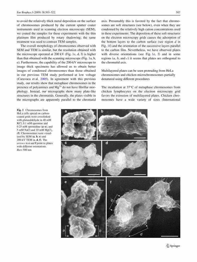
SEM and 200-kV TEM images show that compact metaphase chromosomes contain multilayered plate-like structures
We have not used organic media [typically methanol–aceticacid (Sumner 1989)] for chromosome Wxation. Furthermore,
123
Eur Biophys J (2009) 38:503–522 507
to avoid the relatively thick metal deposition on the surfaceof chromosomes produced by the current sputter coaterinstruments used in scanning electron microscopy (SEM),we coated the samples for these experiments with the thinplatinum Wlm produced by rotary shadowing; the sametreatment was used to contrast TEM samples.
The overall morphology of chromosomes observed withSEM and TEM is similar, but the resolution obtained withthe microscope operated at 200 kV (Fig. 1c, d, f) is higherthan that obtained with the scanning microscope (Fig. 1a, b,e). Furthermore, the capability of the 200-kV microscope toimage thick specimens has allowed us to obtain betterimages of condensed chromosomes than those obtainedin our previous TEM study performed at low voltage(Caravaca et al. 2005). In agreement with this previousstudy, our results show that metaphase chromosomes in thepresence of polyamines and Mg2+ do not have Wbrillar mor-phology. Instead, our micrographs show many plate-likestructures in the chromatids. Generally, the plates visible inthe micrographs are apparently parallel to the chromatid
axis. Presumably this is favored by the fact that chromo-somes are soft structures (see below), even when they arecondensed by the relatively high cation concentrations usedin these experiments. The deposition of these soft structureson the electron microscopy grids causes the adsorption ofthe bottom layers to the carbon surface (see region d inFig. 1f) and the orientation of the successive layers parallelto the carbon Wlm. Nevertheless, we have observed plateswith diverse orientations (see Fig. 1e, f) and in someregions (a, b, and c) it seems that plates are orthogonal tothe chromatid axis.
Multilayered plates can be seen protruding from HeLa chromosomes and chicken microchromosomes partially denatured using diVerent procedures
The incubation at 37°C of metaphase chromosomes fromchicken lymphocytes on the electron microscopy gridfavors the extrusion of multilayered plates. Chicken chro-mosomes have a wide variety of sizes (International
Fig. 1 Chromosomes from HeLa cells spread on carbon-coated grids were crosslinked with glutaraldehyde in 40 mM KCl, 0.1 mM spermine and 0.25 mM spermidine (a–e), and 5 mM NaCl and 10 mM MgCl2 (f). Chromosomes were visual-ized by SEM (a, b, e) and 200-kV TEM (c, d, f). The arrows in e and f point to plates with diVerent orientations. Bars 500 nm
123
508 Eur Biophys J (2009) 38:503–522
Chicken Genome Sequencing Consortium 2004). TheTEM images presented in Fig. 2a and b correspond topartially denatured chicken microchromosomes. The
plates extruded from these chromosomes show the samestructural characteristics as those associated with HeLachromosomes.
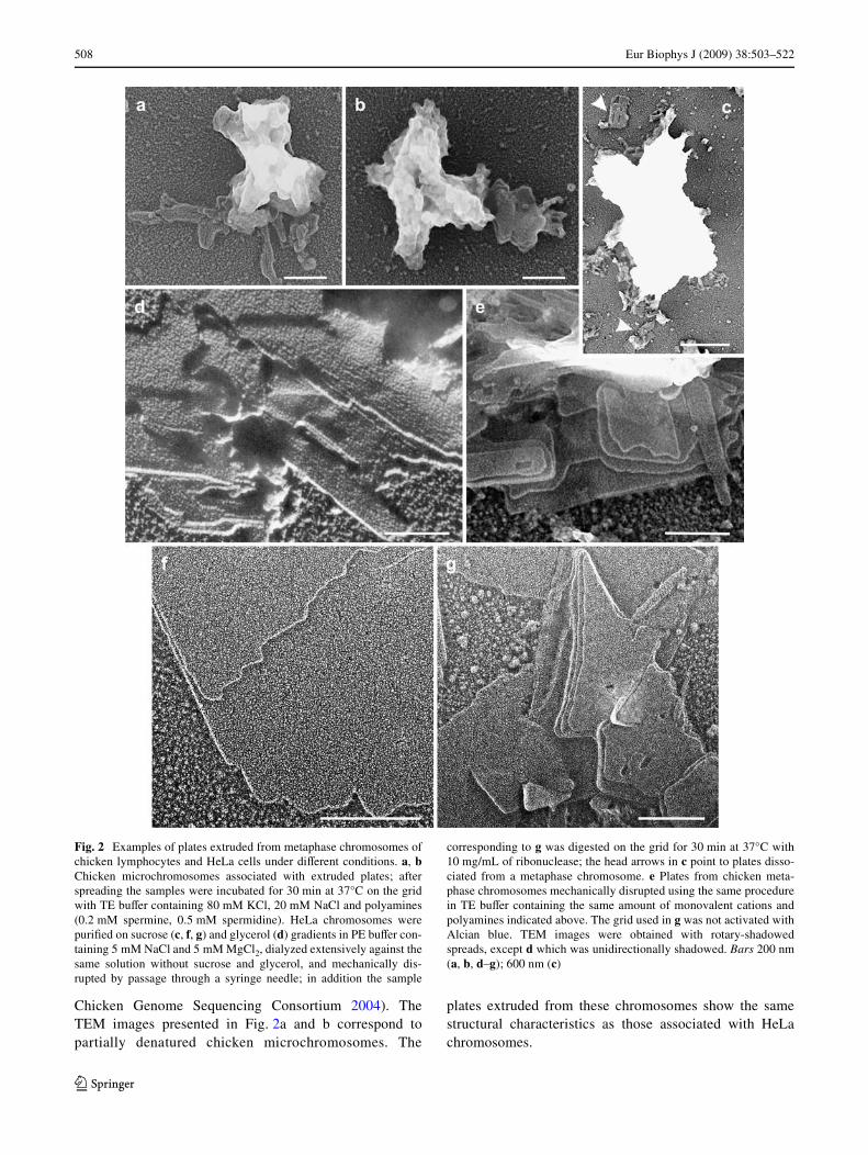
Fig. 2 Examples of plates extruded from metaphase chromosomes ofchicken lymphocytes and HeLa cells under diVerent conditions. a, bChicken microchromosomes associated with extruded plates; afterspreading the samples were incubated for 30 min at 37°C on the gridwith TE buVer containing 80 mM KCl, 20 mM NaCl and polyamines(0.2 mM spermine, 0.5 mM spermidine). HeLa chromosomes werepuriWed on sucrose (c, f, g) and glycerol (d) gradients in PE buVer con-taining 5 mM NaCl and 5 mM MgCl2, dialyzed extensively against thesame solution without sucrose and glycerol, and mechanically dis-rupted by passage through a syringe needle; in addition the sample
corresponding to g was digested on the grid for 30 min at 37°C with10 mg/mL of ribonuclease; the head arrows in c point to plates disso-ciated from a metaphase chromosome. e Plates from chicken meta-phase chromosomes mechanically disrupted using the same procedurein TE buVer containing the same amount of monovalent cations andpolyamines indicated above. The grid used in g was not activated withAlcian blue. TEM images were obtained with rotary-shadowedspreads, except d which was unidirectionally shadowed. Bars 200 nm(a, b, d–g); 600 nm (c)
123

Eur Biophys J (2009) 38:503–522 509
Our results indicate that diVerent mechanical treatmentssuch as chromosome sonication, Polytron homogenization,and rapid passage through a syringe needle also favor theextrusion of plates. In particular, we observed that themechanical stress produced by the latter procedure is ableto split oV part of the extruded plates from the chromatids(see Fig. 2c). Some examples of multilayered plates frommetaphase chromosomes of HeLa cells and chicken lym-phocytes mechanically disrupted, using a syringe needle,are presented in Fig. 2d–g. These plates have large areaswith a very smooth surface, but we also observed largeextrusions from mechanically disrupted chromosomecontaining many small plates (see Fig. S1A in the supple-mentary material). In most microscopic Welds of our prepa-rations of partially denatured chromosomes (from chickenlymphocytes and HeLa cells) we observed many multilay-ered plates of diVerent sizes connected to chromatids anddissociated from them.
In order to exclude the possibility that the observedplates could be produced artefactually, we prepared oursamples using diverse procedures. In our current prepara-tion method we used digitonin and Dounce homogenizationto break the cells, but we also observed plates in prepara-tions performed using Triton X-100 instead of digitonin;we observed plates even in preparations without any deter-gent. Crude preparations of chromosomes without furtherpuriWcation on density gradients allowed us the visualiza-tion of plates (see examples in Fig. 2a, b, e). Plates werefound in chromosome preparations containing high concen-trations of sucrose or glycerol due to the puriWcation ondensity gradients; we also observed plates when we usedthese chromosomes after an extensive dialysis to removecompletely sucrose and glycerol (see examples in Fig. 2c,d, f, g; Fig. S1A). Plates remained unchanged when theywere extensively digested with ribonuclease (Fig. 2g), indi-cating that RNA (which could be a chromosome compo-nent) is not involved in plate formation. Usually in ourTEM and SEM experiments the spreading was by centrifu-gation, but in samples spread by simple diVusion we alsoobserved plates (Fig. 3a). The carbon Wlm on the electronmicroscopy grids was generally activated with Alcian blue.However, our results show that this treatment was not nec-essary for the observation of plates (Fig. 2g); we alsoobserved plates using carbon Wlms activated using theglow-discharge technique. Finally, although generally wecarried out crosslinking of spread samples with glutaralde-hyde, we detected plates even in uncrosslinked samples.In fact, in our AFM experiments (see below) plates wereimaged without crosslinking. In addition, in the case ofAFM the substrate was freshly cleaved mica instead of thecarbon Wlm used in our TEM and SEM experiments.
We have also observed plates in unWxed and unstainedfrozen-hydrated preparations. An example of Cryo-EM
images of these samples is presented in Fig. 3. In thesepreparations there is no substrate and the plates in suspen-sion in aqueous media are not oriented by any surface.
Fig. 3 a TEM micrograph corresponding to plates spread on carbon-coated grids without centrifugation; the sample was adsorbed for 1 h.b, c Cryo-EM images of frozen-hydrated plates (unstained and un-Wxed). In b the Quantifoil Wlm surrounding a hole with vitriWed icecontaining plates is indicated with an asterisk. Plates are shown at ahigher magniWcation in c; large arrows point to the edges of someplates with several layers and small arrows point to the edges corre-sponding to monolayer plates. All the samples in this Wgure were pre-pared from HeLa chromosomes puriWed on sucrose gradients in PEbuVer containing 5 mM NaCl and 5 mM MgCl2, dialyzed extensivelyagainst the same solution without sucrose, and mechanically disruptedby passage through a syringe needle. Bars 200 nm (a, b); 100 nm (c)
123

510 Eur Biophys J (2009) 38:503–522
Nevertheless, as can be seen in Fig. 3c, plates and in partic-ular their edges are clearly distinguishable even in thesepreparations (see also Fig. 7). Table 1 summarizes all theassayed procedures and conditions leading to the observa-tion of plates. This extensive study demonstrates that platesare native structures.
Extreme denaturing conditions cause the emanation of chromatin Wbers from uncrosslinked plates
In order to characterize the plates we tried to Wnd proce-dures capable of producing a signiWcant denaturation, butwithout giving rise to a complete unfolding. In these studieswe found that even uncrosslinked plates are highly stable.For instance, in the micrograph presented in Fig. S1B of thesupplementary material, it can be seen that treatment ofuncrosslinked plates on the electron microscopy grid with arelatively low concentration of the detergent SDS in thepresence of Mg2+ produced a splitting into many smallplates, but did not cause the complete disappearance ofthese planar structures.
We obtained a higher degree of denaturation by the incu-bation of the uncrosslinked plates with buVers of low ionicstrength without divalent cations. Some micrographs
obtained under these conditions are presented in Fig. 4. Itcan be seen that partially unfolded plates are associatedwith chromatin Wbers of diVerent compaction degrees ema-nating from them. Part of the surface of some plates (in par-ticular see Fig. 4b) show unfolded layers with a grainytexture. The dimensions of the observed granules suggestthat these unfolded layers could be formed by nucleosomesrandomly placed on top of smooth layers. However, evenunder these denaturing conditions most of the visible layershave a compact texture.
As can be seen in Fig. 4 and in Fig. S2 of the supplemen-tary material, the Wbers emanate from the edges of theplates, indicating that the chromatin Wlament is so tightlytethered inside the plates that it can only be liberated at theedges. We have also observed that chromatin Wbers caninteract laterally with the edges of the plates; in contrast,Wbers can be on top of a plate without being absorbed bythe chromatin in the top layer (see examples in Fig. S2).This suggests that the forces holding the chromatin Wlamentwithin a layer are stronger than the interactions that existbetween adjacent layers. As observed for diVerent materialsforming planar sheets (Callister and Rethwisch 2008), suchaYnity diVerences within and between layers are requiredfor the formation of multilaminar structures. Furthermore,
Table 1 Conditions of existence of chromatin plates
Summary of results presented in this work and in a previous publication of our laboratory (Caravaca et al. 2005)
Procedures Conditions leading to the observation of plates
Chromosome preparation Cell lysis using digitonin or Triton X-100, and without detergentsPreparation in polyamine-containing buVers without further puriWcationPuriWcation on sucrose or glycerol gradientsDialysis to remove sucrose and glycerol after gradient puriWcation
Chromosome treatments favoring plate extrusion
Incubation at 37°C for 30 min on the electron microscopy gridMechanical disruption: passage through a syringe needle; sonication; homogenization
BuVers 5 mM Pipes, pH 7.25–10 mM Phosphate, pH 7.47 mM Triethanolamine–HCl, pH 7.590 mM Triethanolamine–borate, pH 8.6
Cations 5–20 mM Na+ + 120 mM K+ (with or without Mg2+ or polyamimes)1.7–40 mM Mg2+ (or mixtures of Mg2+ and Ca2+)0.1 to 0.2 mM spermidine + 0.25 to 0.5 mM spermine
Spreading Centrifugation at 1,500g for 10 min (in TEM and in some AFM experiments)Simple diVusion (in AFM and in some TEM experiments)
Substrates Carbon Wlm activated with Alcian blue or glow-discharge, and without activation (TEM)Freshly cleaved mica (AFM)Without any substrate (Cryo-EM)
Fixation Glutaraldehyde crosslinking on grid or in solution before spreading (TEM)Without any Wxative (Cryo-EM and AFM)
Contrast agents Shadowing with platinum in TEM and SEMWithout any contrast agent (Cryo-EM and AFM)
Hydration state during imaging Dry sample in TEM and SEMFrozen-hydrated sample in Cryo-EMSample in aqueous solution at room temperature in AFM (tapping- and contact-mode),
and in force spectroscopy analysis
123

Eur Biophys J (2009) 38:503–522 511
these observations are consistent with the sliding betweenlayers that presumably occurs in the plates surroundingpartially denatured chromatids (see “Discussion”). ThediVerences in the length of relative sliding between layersprobably facilitate the clear visualization of several stackedlayers with well-deWned edges in many plates (see forinstance region d in Fig. 1f and Fig. 2e, g).
AFM experiments demonstrate that chromatin plates are stable in aqueous solution at room temperature
Even in the presence of divalent cations, metaphase chro-mosomes are very soft structures in aqueous solution. This,together with their large size, makes very diYcult the imag-ing of chromosomes spread on mica by tapping-modeAFM; the resolution obtained using this technique withwhole chromosomes is very poor (Fig. 5e). In contrast,
plates adsorbed on mica can be imaged without anyproblem using tapping-mode AFM in the presence of5 mM Mg2+ (Fig. 5a–d, g) and higher concentrations of thisdivalent cation (Fig. S3A–E, G in the supplementarymaterial). In agreement with our TEM observations, thetopographical images (Fig. 5a, c, g; Fig. S3A, C, E–G), thecorresponding cross-section proWles (see examples inFig. 5f), and in particular the amplitude images (Fig. 5b;Fig. S3B, D) indicate that the surface of plates is verysmooth.
Since all these images were obtained with uncrosslinkedpreparations, our results indicate that plates are stable bythemselves in aqueous solution. In fact, plates have amechanical stability high enough to allow contact-modeimaging (Fig. S3F). Furthermore, when higher forces wereapplied with the AFM tip to internal regions of diVerentplates, there was no apparent change in their structure.
Fig. 4 Chromatin Wbers ema-nating from plates partially denatured by treatment with solutions of low ionic strength. a–d HeLa metaphase chromo-somes puriWed on sucrose gradi-ents containing 5 mM NaCl and 5 mM MgCl2, were mechani-cally disrupted by passage through a syringe needle, spread on electron microscopy grids and incubated for 30 min at 37°C with 10 mM EDTA (pH 7.5) and 5 mM NaCl, cross-linked with glutaraldehyde and rotary shadowed. All TEM micrographs are at the same scale (bar 200 nm)
123

512 Eur Biophys J (2009) 38:503–522
In contrast, when high forces were applied to regionsincluding the edges, the plates become severely damaged.One of such experiments is presented in Fig. 5c (image
obtained before the high force was applied to the regionmarked with a square) and Fig. 5d (image obtained after theapplication of the force). These results are in agreement
Fig. 5 AFM study of plates from HeLa metaphase chromosomes inaqueous solution in the presence of 5 mM Mg2+. (a, c–e) Topographi-cal images (tapping-mode); b Amplitude image of the same sampleshown in a. g Three-dimensional image. The image in d was obtainedafter the application of a high force in the region marked with a squarein c. f Height proWles generated along the lines ab, cd, and ef indicatedin a, c, and d, respectively. The plates shown in a–d and g wereobtained from chromosomes puriWed on sucrose gradients containing
PE buVer, 5 mM NaCl and 5 mM MgCl2, dialyzed (at room tempera-ture for 4 h) against the same solution without sucrose, mechanicallydisrupted by passage through a syringe needle, and then spread on micaand imaged without crosslinking. The chromosomes in e were spreadby centrifugation on mica, crosslinked with glutaraldehyde, andwashed with PE buVer without glucose containing 5 mM NaCl and5 mM MgCl2. Bars: 200 nm (a–d); 2 �m (e)
123
Eur Biophys J (2009) 38:503–522 513
with our observations presented in the preceding sectionindicating that the chromatin Wlament is tightly bound inthe internal regions of the plates and more prone to beunfolded in the periphery.
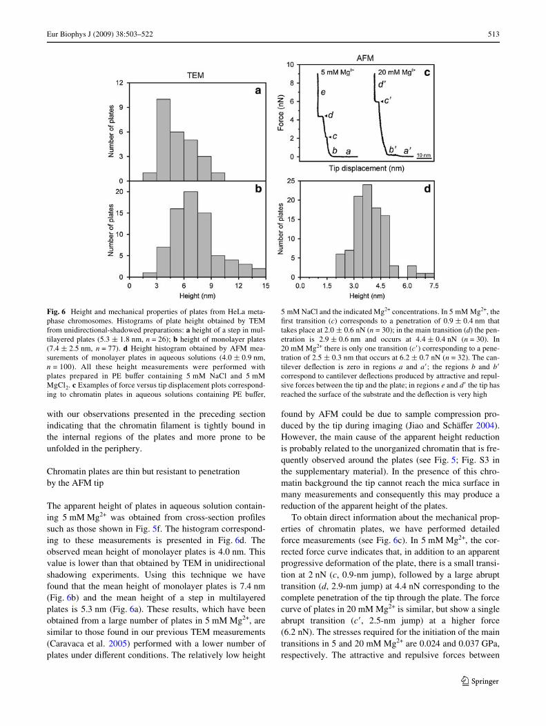
Chromatin plates are thin but resistant to penetration by the AFM tip
The apparent height of plates in aqueous solution contain-ing 5 mM Mg2+ was obtained from cross-section proWlessuch as those shown in Fig. 5f. The histogram correspond-ing to these measurements is presented in Fig. 6d. Theobserved mean height of monolayer plates is 4.0 nm. Thisvalue is lower than that obtained by TEM in unidirectionalshadowing experiments. Using this technique we havefound that the mean height of monolayer plates is 7.4 nm(Fig. 6b) and the mean height of a step in multilayeredplates is 5.3 nm (Fig. 6a). These results, which have beenobtained from a large number of plates in 5 mM Mg2+, aresimilar to those found in our previous TEM measurements(Caravaca et al. 2005) performed with a lower number ofplates under diVerent conditions. The relatively low height
found by AFM could be due to sample compression pro-duced by the tip during imaging (Jiao and SchäVer 2004).However, the main cause of the apparent height reductionis probably related to the unorganized chromatin that is fre-quently observed around the plates (see Fig. 5; Fig. S3 inthe supplementary material). In the presence of this chro-matin background the tip cannot reach the mica surface inmany measurements and consequently this may produce areduction of the apparent height of the plates.
To obtain direct information about the mechanical prop-erties of chromatin plates, we have performed detailedforce measurements (see Fig. 6c). In 5 mM Mg2+, the cor-rected force curve indicates that, in addition to an apparentprogressive deformation of the plate, there is a small transi-tion at 2 nN (c, 0.9-nm jump), followed by a large abrupttransition (d, 2.9-nm jump) at 4.4 nN corresponding to thecomplete penetration of the tip through the plate. The forcecurve of plates in 20 mM Mg2+ is similar, but show a singleabrupt transition (c�, 2.5-nm jump) at a higher force(6.2 nN). The stresses required for the initiation of the maintransitions in 5 and 20 mM Mg2+ are 0.024 and 0.037 GPa,respectively. The attractive and repulsive forces between
Fig. 6 Height and mechanical properties of plates from HeLa meta-phase chromosomes. Histograms of plate height obtained by TEMfrom unidirectional-shadowed preparations: a height of a step in mul-tilayered plates (5.3 § 1.8 nm, n = 26); b height of monolayer plates(7.4 § 2.5 nm, n = 77). d Height histogram obtained by AFM mea-surements of monolayer plates in aqueous solutions (4.0 § 0.9 nm,n = 100). All these height measurements were performed withplates prepared in PE buVer containing 5 mM NaCl and 5 mMMgCl2. c Examples of force versus tip displacement plots correspond-ing to chromatin plates in aqueous solutions containing PE buVer,
5 mM NaCl and the indicated Mg2+ concentrations. In 5 mM Mg2+, theWrst transition (c) corresponds to a penetration of 0.9 § 0.4 nm thattakes place at 2.0 § 0.6 nN (n = 30); in the main transition (d) the pen-etration is 2.9 § 0.6 nm and occurs at 4.4 § 0.4 nN (n = 30). In20 mM Mg2+ there is only one transition (c�) corresponding to a pene-tration of 2.5 § 0.3 nm that occurs at 6.2 § 0.7 nN (n = 32). The can-tilever deXection is zero in regions a and a�; the regions b and b�correspond to cantilever deXections produced by attractive and repul-sive forces between the tip and the plate; in regions e and d� the tip hasreached the surface of the substrate and the deXection is very high
123
514 Eur Biophys J (2009) 38:503–522
the tip and the plate are responsible for the cantileverdeXections observed in the Wrst part (regions b and b�) ofthe force curves (Heinz and Hoh 1999; Zlatanova et al.2000). The region ranging from the point of the apparentbeginning of the elastic indentation up to the point in whichan abrupt plastic deformation is observed has allowed us toestimate the Young’s modulus of chromatin plates underdiVerent conditions using the Hertz equation (see “Materi-als and methods”). The values obtained for this elasticmodulus are 0.16 § 0.06 GPa (n = 24) and 0.23 § 0.06GPa (n = 29) in 5 and 20 mM Mg2+, respectively. In5 mM Mg2+, the Young’s modulus corresponding to theregion between the Wrst abrupt transition (c) and the maintransition (d) is 0.16 § 0.06 GPa (n = 24).
Although it is not possible to know exactly which frac-tion of the cantilever deXection is due to the initial interac-tions in regions b and b�, the force curves can also be usedto estimate the plate thickness. In 5 and 20 mM Mg2+, thedisplacements of the tip from the apparent beginning of theindentation up to the point of contact between the tip andthe substrate are 6.8 § 0.9 (n = 30) and 6.1 § 1.3 (n = 32)nm, respectively. These estimates of plate thickness arecompatible with the height measurements obtained byTEM. Thus, considering all our results, it can be concludedthat the height of monolayer plates is approximately6.5 nm.
Internal structure of chromatin plates
The plate thickness is similar to the height of a single nucleo-some (»5.7 nm). This suggests that each step could beformed by a monolayer of nucleosomes with their Xat facesoriented parallel to the plate surface. However, since such aregular organization should produce plates with many circularstructures (having the nucleosome diameter: »11 nm) clearlyvisible in the micrographs, but we do not see this repetitivestructure in the surface of the plates, our results exclude thissimple organization of nucleosomes in the plates.
In the edges of plates imaged by Cryo-EM and conven-tional TEM (Fig. 7), there are pairs of dense bars separatedby about 3 nm. Since this distance corresponds approxi-mately to the pitch of the helix formed by DNA in thenucleosome (Finch et al. 1977; Richmond et al. 1984;Uberbacher and Bunick 1985), we have interpreted thateach pair of bars belongs to the two DNA turns of a nucleo-some. These bar pairs can be seen roughly parallel or form-ing V-shaped Wgures. This is in agreement with the wedgeshape of nucleosomes. The same structural features wereobserved previously in aggregates of puriWed nucleosomecore particles imaged using conventional TEM (Finch et al.1977) and Cryo-EM (Leforestier et al. 1999).
The bars are more easily seen in the periphery than in theinternal regions of the plates (see arrows in Fig. 7a–e). At
high magniWcation, the surface of the plates has an irregulartexture, similar to that observed by other authors that havestudied irregular aggregates of nucleosome core particles(Leforestier et al. 1999). We have performed distance mea-surements in many regions containing several consecutivedense bars in the edges of the plates. The average distancebetween bars in these regions is »3.7 nm (see Fig. 7f andthe legend of this Wgure). This distance is signiWcantlylarger than the distance (»2.8 nm) between the turns ofDNA in the columns of stacked nucleosome core particlesobserved in TEM prepartions (Finch et al. 1977; Dubochetand Noll 1978), in core particle crystals (Uberbacher andBunick 1985; White et al. 2001), and in ordered liquid crys-talline phases formed by aggregates of these particles(Leforestier et al. 1999, 2001). Thus, our results indicatethat nucleosomes in the plates have diverse orientations andfollow a pattern more complex than the simple stackingobserved in other laboratories with puriWed core particles.
On the other hand, taking into account that our measure-ments indicate that the plate height is lower than the nucle-osome diameter, it can be suggested that nucleosomes aretilted to allow the interdigitation of nucleosomes betweenadjacent layers. The apparent height of each interdigitatedlayer can be lower than the nucleosome diameter and con-sequently may produce planar structures compatible withthe observed plate thickness. There are other experimentalresults that favor this compact structure: (1) We know atpresent that the successive helical turns of nucleosomesforming the chromatin Wber can be interdigitated, and thatthe apparent height of each interdigitated turn with tiltednucleosomes is only a fraction of the nucleosome diameter(Daban and Bermúdez 1998; Robinson et al. 2006; Wonget al. 2007; Kepper et al. 2008; see next section); (2) Tiltednucleosomes in each layer could interact through their faceswith the nucleosomes of the adjacent layers, and at presentthere are many experimental evidences indicating that thislateral interaction between nucleosomes is the driving forcefor the formation of condensed chromatin structures (see“Discussion”).
PuriWed chromatin fragments in the presence of Mg2+ can associate to form small plate-like structures
We observed using TEM (Bartolomé et al. 1994; Bermúdezet al. 1998), nondenaturing gel electrophoresis in the pres-ence of Mg2+ (Bartolomé et al. 1995), and AFM in aqueoussolution (Caño et al. 2006) that small chromatin fragmentsfrom chicken erythrocytes in solutions containing diVerentconcentrations of mono- and divalent cations form compactcircular structures of about 35 nm in diameter. Some ofthese compact bodies are indicated with white arrows inFig. 8b. We modeled these structures as interdigitated com-pact solenoids (Daban and Bermúdez 1998). Rhodes and
123

Eur Biophys J (2009) 38:503–522 515
coworkers (Robinson et al. 2006) have recently performeda very detailed study, using TEM and Cryo-EM, of thestructure of reconstituted chromatin fragments containingDNA of deWned sequence. The results obtained by theseauthors have led them to propose an interdigitated compactmodel for chromatin Wbers similar to that suggested in our
early studies. More recently, other laboratories have studiedthe conformational and dynamic properties of interdigitatedsolenoids (Wong et al. 2007; Kepper et al. 2008).
We have observed that metaphase chromosomes with arelatively high degree of denaturation may contain, in addi-tion to many small plates, compact circular structures
Fig. 7 Cryo-EM images of frozen-hydrated plates (a, b) are shown in reverse contrast to facilitate the comparison with TEM images of plates contrasted by metal shadowing (c–e). The arrows indicate some regions in the edges of the plates in which the dense bars that presumably correspond to the turns of DNA in nucleosomes are visible (see “Results”). All the samples shown in this Wgure were pre-pared, as described in Fig. 3, from HeLa chromosomes in PE buVer containing 5 mM NaCl and 5 mM MgCl2. f Histogram corresponding to the distance between bars in the edges of rotary-shadowed plates from HeLa chromosomes (average distance 3.7 § 0.5 nm; n = 247); the average distance obtained from Cryo-EM images of the same samples is 3.9 § 0.4 nm (n = 72). Plates from chromo-somes of chicken lymphocytes have an average distance between the bars in the edges of 3.5 § 0.4 nm (n = 154). Bars 20 nm (a–c); 50 nm (d, e)
123

516 Eur Biophys J (2009) 38:503–522
similar to those observed in experiments performed withpuriWed small chromatin fragments. This can be seen par-ticularly clear in Fig. 8a, in which some of these circularstructures are indicated with white arrows. Note that circu-lar structures are on top of small chromatin plates or later-ally associated with them. In addition, note that many smallplates show a grainy texture similar that observed in theunfolded layers considered above (Fig. 4b); the dimensions
of the observed granules are compatible with the size ofnucleosomes placed with diverse relative orientations.
On the other hand, we found previously that intermedi-ate Mg2+ concentrations (2–20 mM) cause the associationof small chromatin fragments producing very large aggre-gates (Caravaca et al. 2005; Caño et al. 2006). In this work,in order to obtain small aggregates, we have used lowerMg2+ concentrations. We have observed in diVerent
Fig. 8 Unfolded chromatin plates from chicken metaphase chromo-somes (a) contain structural elements similar to those seen inaggregates of small chromatin fragments from chicken erythrocytes(b–k). a After spreading, chromosomes of chicken lymphocytes wereincubated in the presence of polyamines for 30 min at 37°C (the sameconditions as in Fig. 2a, b). b–k DiVerent aggregation degrees of smallchromatin fragments from chicken erythrocytes in 10 mM Triethanol-
amine–HCl, pH 7.5, and 0.8 mM MgCl2. The white arrows in b pointto the highly folded chromatin fragments described previously in ourlaboratory (similar structures are observed in a and some of themare also indicated with arrows); the association of these compactfragments gives rise to dimers (some of them are indicated witharrowheads in b) and higher aggregates (b–k). All TEM micrographsshown in this Wgure are at the same scale (bar 200 nm)
123

Eur Biophys J (2009) 38:503–522 517
experiments that, in the presence of 0.8 mM Mg2+, theincrease of the chromatin concentration and the incubationtime favor the aggregation of the puriWed small chromatinfragments. As shown in the selected micrographs presentedin Fig. 8b–k, chromatin fragments can associate laterallygiving rise to dimers (indicated with white arrowheads) andto relatively small aggregates. Note that in these aggregatesthe 35-nm circular bodies change signiWcantly their shapeand form small plate-like structures (see micrographs 8c, d,g–i); in some cases it seems that the aggregated structurescontain several layers of small plates (see micrographs 8e,f, j, k). Since these structures produced by puriWed chroma-tin are similar to those seen in the denatured plates pre-sented in Fig. 8a, it can be suggested that chromatin hasintrinsic structural properties that lead to the formation ofplates.
Discussion
Plate structure
Plates associated with metaphase chromosomes werereported for the Wrst time in a previous work of our labora-tory (Caravaca et al. 2005). This initial TEM study was per-formed exclusively with human chromosomes (HeLa cells).Here we have observed plates in chromosomes of chicken,a species with a large evolutionary distance from humans.The very diVerent buVers, ionic conditions, preparationprocedures, spreading techniques, and substrates assayed inour work allow us to conclude that plates are not producedartefactually (see Table 1). In particular, our AFM andCryo-EM results showing that plates exist in aqueous solu-tion and are stable without crosslinking indicate that platesare native structures. The chromatin Wbers that emanatefrom plates incubated at low ionic strength (this work) andwith high NaCl concentrations (Caravaca et al. 2005),together with the high similarities observed between par-tially denatured plates and the aggregates produced by thespontaneous association of puriWed chromatin fragmentsindicate that the chromatin Wlament is able to fold givingrise to planar structures. The denaturation experiments andAFM studies performed applying high forces show that thechromatin Wlament is tightly tethered inside the plates, butcan be more easily unfolded in the peripheral regions.
Although almost all the structural studies in the chroma-tin Weld have been devoted to Wbers, in a previous study ofa simpliWed system (nucleosome cores lacking histone H1and linker DNA), Leforestier et al. (2001) using Cryo-EMtechniques demonstrated that isolated nucleosome core par-ticles at a high concentration associate giving rise to plate-like structures with many layers formed by columns of coreparticles; the electrostatic interactions involved in this
lamellar structure has been studied in a theoretical work byCherstvy and Everaers (2006). More recently, Engelhardt(2007) has proposed that in extended chromatin Wbers thelateral association produced by interdigitation of nucleo-somes could form dense structures composed of severalsheets, and Hancock (2008) has found experimentally that,in the presence of high concentrations of crowding agents(25% polyethylene glycol), puriWed polynucleosomes canself-associate forming large sheets of chromatin.
Our TEM and Cryo-EM results exclude the possibilitythat chromatin plates are formed by monolayers ofnucleosomes with their Xat faces oriented parallel to theplate surface. Plates could also be formed by columns ofnucleosomes having their axes parallel to the plate surface.This structure was observed in lamellar aggregates obtainedwith isolated core particles (Leforestier et al. 2001; seeabove). However, this simple pattern is also excludedbecause the plate thickness found in our TEM and AFMmeasurements (»6.5 nm) is lower than the column diame-ter (equal to the nucleosome diameter: »11 nm). Moreover,columns of nucleosomes in the plates should be clearly vis-ible in the electron micrographs, but our images and dis-tance measurements show that nucleosomes in the platesfollow a complex orientation pattern. Our results stronglysuggest that nucleosomes are tilted with respect to the platesurface to allow the interdigitation between layers and areduction of thickness compatible with the observed plateheight. This thickness reduction is consistent with theremarkable reduction of Wber length observed in interdigi-tated solenoids (Daban and Bermúdez 1998; Robinsonet al. 2006; Wong et al. 2007). Note, in addition, that tiltednucleosomes in each layer may interact through their faceswith the nucleosomes of the adjacent layers. Results fromdiVerent laboratories (McDowall et al. 1986; Mangenotet al. 2003; Daban 2000, 2003), suggesting that face-to-faceinteractions between nucleosomes is the driving force forthe formation of chromatin structures with a high local con-centration of DNA, also favor our interpretation. Further-more, Kepper et al. (2008) have demonstrated using MonteCarlo dynamic simulations that interdigitated solenoidshaving nucleosomes with a relatively high tilt angle, favor-ing face-to-face interactions, are more stable than otherpossible conformations. Lateral association of nucleosomecores has also been found in a tetranucleosome crystal (see“Introduction”; Schalch et al. 2005) and high-resolutionX-ray diVraction analysis (Luger et al. 1997; Harp et al. 2000;White et al. 2001) indicate that this association involvesand acidic surface formed by histones H2A and H2B andbasic residues of the N-termimal tail of histone H4. Finally,more recently, it has been demonstrated (Zhou et al. 2007;Chodaparambil et al. 2007) that this acidic region is respon-sible for the regulation of both the folding of well-deWnednucleosome arrays and their self-association leading to
123
518 Eur Biophys J (2009) 38:503–522
condensed three-dimensional structures (Caterino andHayes 2007).
Plates in chromosomes
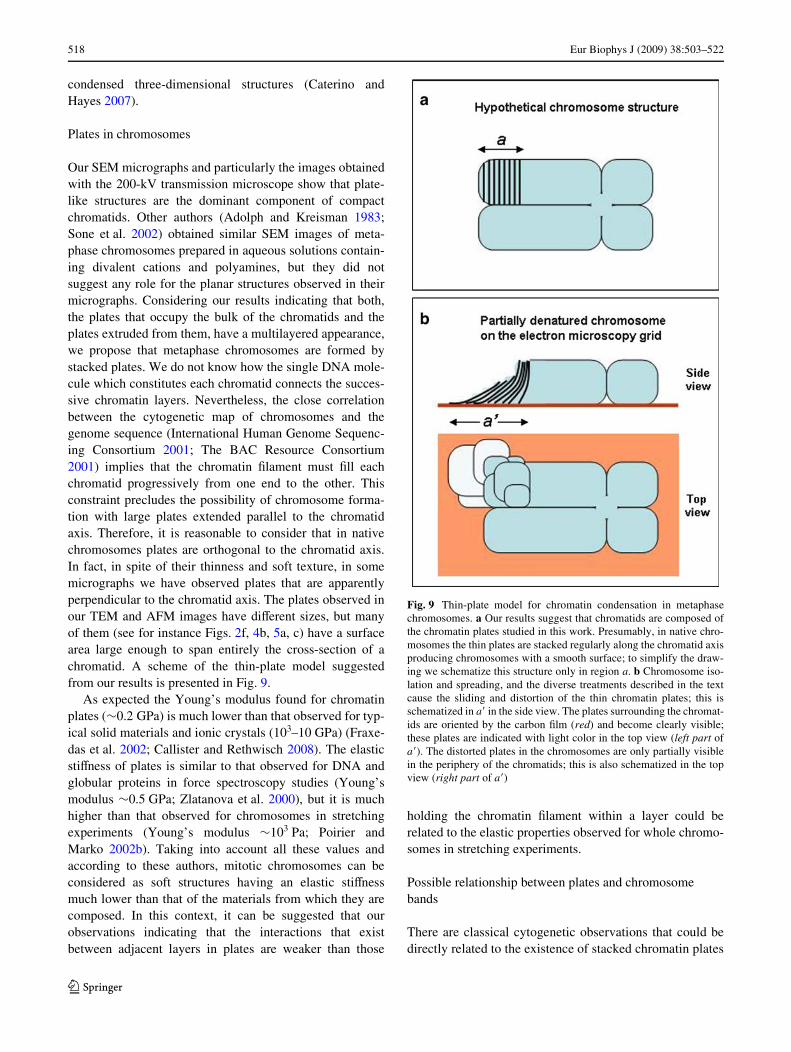
Our SEM micrographs and particularly the images obtainedwith the 200-kV transmission microscope show that plate-like structures are the dominant component of compactchromatids. Other authors (Adolph and Kreisman 1983;Sone et al. 2002) obtained similar SEM images of meta-phase chromosomes prepared in aqueous solutions contain-ing divalent cations and polyamines, but they did notsuggest any role for the planar structures observed in theirmicrographs. Considering our results indicating that both,the plates that occupy the bulk of the chromatids and theplates extruded from them, have a multilayered appearance,we propose that metaphase chromosomes are formed bystacked plates. We do not know how the single DNA mole-cule which constitutes each chromatid connects the succes-sive chromatin layers. Nevertheless, the close correlationbetween the cytogenetic map of chromosomes and thegenome sequence (International Human Genome Sequenc-ing Consortium 2001; The BAC Resource Consortium2001) implies that the chromatin Wlament must Wll eachchromatid progressively from one end to the other. Thisconstraint precludes the possibility of chromosome forma-tion with large plates extended parallel to the chromatidaxis. Therefore, it is reasonable to consider that in nativechromosomes plates are orthogonal to the chromatid axis.In fact, in spite of their thinness and soft texture, in somemicrographs we have observed plates that are apparentlyperpendicular to the chromatid axis. The plates observed inour TEM and AFM images have diVerent sizes, but manyof them (see for instance Figs. 2f, 4b, 5a, c) have a surfacearea large enough to span entirely the cross-section of achromatid. A scheme of the thin-plate model suggestedfrom our results is presented in Fig. 9.
As expected the Young’s modulus found for chromatinplates (»0.2 GPa) is much lower than that observed for typ-ical solid materials and ionic crystals (103–10 GPa) (Fraxe-das et al. 2002; Callister and Rethwisch 2008). The elasticstiVness of plates is similar to that observed for DNA andglobular proteins in force spectroscopy studies (Young’smodulus »0.5 GPa; Zlatanova et al. 2000), but it is muchhigher than that observed for chromosomes in stretchingexperiments (Young’s modulus »103 Pa; Poirier andMarko 2002b). Taking into account all these values andaccording to these authors, mitotic chromosomes can beconsidered as soft structures having an elastic stiVnessmuch lower than that of the materials from which they arecomposed. In this context, it can be suggested that ourobservations indicating that the interactions that existbetween adjacent layers in plates are weaker than those
holding the chromatin Wlament within a layer could berelated to the elastic properties observed for whole chromo-somes in stretching experiments.
Possible relationship between plates and chromosome bands
There are classical cytogenetic observations that could bedirectly related to the existence of stacked chromatin plates
Fig. 9 Thin-plate model for chromatin condensation in metaphasechromosomes. a Our results suggest that chromatids are composed ofthe chromatin plates studied in this work. Presumably, in native chro-mosomes the thin plates are stacked regularly along the chromatid axisproducing chromosomes with a smooth surface; to simplify the draw-ing we schematize this structure only in region a. b Chromosome iso-lation and spreading, and the diverse treatments described in the textcause the sliding and distortion of the thin chromatin plates; this isschematized in a� in the side view. The plates surrounding the chromat-ids are oriented by the carbon Wlm (red) and become clearly visible;these plates are indicated with light color in the top view (left part ofa�). The distorted plates in the chromosomes are only partially visiblein the periphery of the chromatids; this is also schematized in the topview (right part of a�)
123
Eur Biophys J (2009) 38:503–522 519
oriented perpendicular to the chromatid axis. The identiW-cation of individual chromosomes is based on diversestaining procedures that give rise to characteristic bandingpatterns (Sumner 1989; Castleman 2000), in which thevisible bands are roughly orthogonal to the chromatid axis.Even the thinnest bands show this orientation. Thetransverse orientation of all the bands is schematized in thewell-established ideograms corresponding to the bandingpatterns of human chromosomes (ISCN 2005). Typicalbanding procedures use chromosomes Wxed with methanol-acetic acid. This could alter the native structure of chromo-somes. Nevertheless, equivalent patterns having bandsroughly orthogonal to the chromatid axis have beenobtained with native chromosomes (not treated withorganic solvents) stained with diVerent Xuorescent dyes(Saitoh and Laemmli 1994). The cytogenetic bands aremuch thicker than the thin chromatin plates studied here,but it has been reported that chromosome stretching causesthe splitting of the typical bands into much thinnersubbands which are also oriented perpendicular to the chro-matid axis (Hliscs et al. 1997). Taken together, these obser-vations suggest that chromosome bands could be producedby selective staining of chromatin plate clusters having theDNA base composition adequate for the interaction withthe dyes used in the diVerent banding protocols. Therelative weakness of the interactions between chromatinlayers (see above) could be related to the easy band split-ting observed in stretching experiments. The thinness, softtexture, and high tendency of chromatin plates to associateforming multilayered structures make impossible to deter-mine experimentally the number of plates in a chromatid.However, taking into account our plate height measure-ments and assuming a close contact between consecutiveplates in highly condensed chromosomes, we can estimatethat a cytogenetic band having a thickness of 300 nmshould correspond to a cluster of about 45 chromatinplates.
Functional implications
The folding of the chromatin Wlament forming plates withinchromosomes at metaphase ionic conditions is not compati-ble with the consideration that the 30-nm chromatin Wber isthe fundamental structural element of metaphase chromo-somes. Nevertheless, our model for chromatin condensa-tion does not exclude the possible existence of additionalorganization levels of the chromatin Wlament. In particular,the attachment of speciWc DNA sequences to nonhistoneproteins may give rise to the loops that become visible inthe presence of the divalent cation chelator EDTA (Marsdenand Laemmli 1979) and after the removal of histones (Paulsonand Laemmli 1977). Our denaturation experiments suggestthat chromatin Wbers and plates are interconvertible;
presumably, in the future, it will be possible to integrateboth structural forms into a dynamic model for chromatinfunction during the cell cycle.
The dense aspect of chromatids in the presence of diva-lent cations and polyamines, together with the large relativeamount of histones (Uchiyama et al. 2005) and DNA(Daban 2000) in metaphase chromosomes, suggest thatchromatin plates occupy most of the volume and conse-quently may be responsible for the basic physical propertiesof the metaphase chromosome. This is in agreement withthe fact that the observed condensation/decondensationbehavior of metaphase chromosomes in aqueous solutioninduced by diVerent cations (Poirier et al. 2002) can be cor-related with the compaction/unfolding behavior observedfor puriWed chromatin samples in diVerent laboratories(Zlatanova et al. 1994; Bartolomé et al. 1995; Hansen2002; Mangenot et al. 2003). Thus, according to these con-siderations, nonhistone proteins in condensed metaphasechromosomes have to interact with a dense chromatinstructure instead of the extended loops generally consideredin the current chromatin literature. Nevertheless, theobserved condensation/decondensation properties of meta-phase chromosomes also indicate that a signiWcant part ofthe chromosome volume is occupied by water, which couldfacilitate the diVusion of proteins with diVerent functions.This possibility is consistent with in vivo photobleachingexperiments showing that there is a rapid exchange of his-tone H1 (Lever et al. 2000; Misteli et al. 2000) and topoiso-merase II (Tavormina et al. 2002; Christensen et al. 2002)in mitotic chromosomes. Therefore, although chromatin ishighly condensed in chromosomes, the resulting structure isdynamic from a functional point of view.
At present it is not possible to describe the structuralrelationships between chromatin plates and characteristicchromosome elements such as centromeres and telomeres.Nor is it known how chromatin plates are related to otherlarge-scale properties of chromosome structure such as theobserved diameter of chromatids and the possible existenceof a hole along the chromatid axis for the location of non-histone proteins. Many questions remain open, but we thinkthat the study of chromatin plates will inspire completelynew experimental approaches that will be very useful forthe understanding of the structure and functional propertiesof eukaryotic chromosomes.
Acknowledgments We thank Francesca Vidal and Gerard Oncinsfor advice about preparation of chicken chromosomes and force curveanalysis, respectively, and José M. Valpuesta and Montserrat Samsófor advice on Cryo-EM techniques. We also acknowledge the help ofIsmael Díez and Jordi Díaz (Serveis CientíWco-Tècnics, UB) in theAFM experiments. TEM and SEM images were obtained in the Serveide Microscòpia (UAB). Chicken blood was obtained in the Servei deGranges i Camps Experimentals (UAB) and in Gimave S. A. (Ripollet,Barcelona). Cell culture was performed in the Servei de CultiusCel·lulars (IBB). I.G. and P.C.-H. were supported by predoctoral
123

520 Eur Biophys J (2009) 38:503–522
fellowships from the Generalitat de Catalunya, and S.C. by a predoc-toral fellowship from the Ministerio de Educación y Ciencia (MEC).This work was supported in part by grant BFU2005-3883 (MEC).
References
Adolph KW, Kreisman LR (1983) Surface structure of isolated meta-phase chromosomes. Exp Cell Res 147:155–166. doi:10.1016/0014-4827(83)90280-X
Aragay AM, Fernandez-Busquets X, Daban JR (1991) DiVerentmechanisms for in vitro formation of nucleosome core particles.Biochemistry 30:5022–5032. doi:10.1021/bi00234a026
Bartolomé S, Bermúdez A, Daban JR (1994) Internal structure of the30 nm chromatin Wber. J Cell Sci 107:2983–2992
Bartolomé S, Bermúdez A, Daban JR (1995) Electrophoresis ofchromatin on nondenaturing agarose gels containing Mg2+. Self-assembly of small chromatin fragments and folding of the 30-nmWber. J Biol Chem 270:22514–22521. doi:10.1074/jbc.270.38.22514
Bednar J, Woodcock CL (1999) Cryoelectron microscopic analysis ofnucleosomes and chromatin. Methods Enzymol 304:191–213.doi:10.1016/S0076-6879(99)04012-4
Bednar J, Horowitz RA, Grigoryev SA, Carruthers LM, Hansen JC,Koster AJ, Woodcock CL (1998) Nucleosomes, linker DNA, andlinker histone form a unique structural motif that directs the high-er-order folding and compaction of chromatin. Proc Natl Acad SciUSA 95:14173–14178. doi:10.1073/pnas.95.24.14173
Belmont AS (2002) Mitotic chromosome scaVold structure: newapproaches to an old controversy. Proc Natl Acad Sci USA99:15855–15857. doi:10.1073/pnas.262672799
Belmont AS (2006) Mitotic chromosome structure and condensation.Curr Opin Cell Biol 18:632–638. doi:10.1016/j.ceb.2006.09.007
Bermúdez A, Bartolomé S, Daban JR (1998) Partial denaturation ofsmall chromatin fragments: direct evidence for the radial distribu-tion of nucleosomes in folded chromatin Wbers. J Cell Sci111:1707–1715
Bordas J, Perez-Grau L, Koch MHJ, Vega MC, Nave C (1986) Thesuperstructure of chromatin and its condensation mechanism.II Theoretical analysis of the X-ray scattering patterns andmodel calculations. Eur Biophys J 13:175–185. doi:10.1007/BF00542561
Bradbury EM, Baldwin JP (1986) Neutron scatter and diVractiontechniques applied to nucleosome and chromatin structure. CellBiophys 9:35–66
Callister WD, Rethwisch G (2008) Fundamentals of materials scienceand engineering. Wiley, New York
Caño S, Caravaca JM, Martín M, Daban JR (2006) Highly compactfolding of chromatin induced by cellular cation concentrations.Evidence from atomic force microscopy studies in aqueous solu-tion. Eur Biophys J 35:495–501. doi:10.1007/s00249-006-0057-7
Caravaca JM, Caño S, Gállego I, Daban JR (2005) Structural elementsof bulk chromatin within metaphase chromosomes. ChromosomeRes 13:725–743. doi:10.1007/s10577-005-1008-3
Castleman KR (2000) Image analysis: quantitative interpretation ofchromosome images. In: Balbock R, Graham J (eds) Image pro-cessing and analysis. A practical approach. Oxford UniversityPress, Oxford, pp 69–109
Caterino TL, Hayes JJ (2007) Chromatin structure depends on what’sin the nucleosome’s pocket. Nat Struct Mol Biol 14:1056–1058.doi:10.1038/nsmb1107-1056
Cherstvy AG, Everaers R (2006) Layering, bundling, and azimuthalorientations in dense phases of nucleosome core particles. J PhysCondens Matter 18:11429–11442. doi:10.1088/0953-8984/18/50/003
Chodaparambil JV, Barbera AJ, Lu X, Kaye KM, Hansen JC, Luger K(2007) A charged and contoured surface on the nucleosome regu-lates chromatin compaction. Nat Struct Mol Biol 14:1105–1107.doi:10.1038/nsmb1334
Christensen MO, Larsen MK, Barthelmes HU, Hock R, Andersen CL,Kjeldsen E, Knudsen BR, Westergaard O, Boege F, Mielke C(2002) Dynamics of human DNA topoisomerases II� and II� inliving cells. J Cell Biol 157:31–44. doi:10.1083/jcb.200112023
Daban JR (2000) Physical constraints in the condensation of eukary-otic chromosomes. Local concentration of DNA versus linearpacking ratio in higher order chromatin structures. Biochemistry39:3861–3866. doi:10.1021/bi992628w
Daban JR (2003) High concentration of DNA in condensed chromatin.Biochem Cell Biol 81:91–99. doi:10.1139/o03-037
Daban JR, Bermúdez A (1998) Interdigitated solenoid model for com-pact chromatin Wbers. Biochemistry 37:4299–4304. doi:10.1021/bi973117h
Dubochet J, Noll M (1978) Nucleosome arcs and helices. Science202:280–286. doi:10.1126/science.694532
Dubochet J, Adrian M, Chang JJ, Homo JC, Lepault J, McDowall AW,Schultz P (1988) Cryo-electron microscopy of vitriWed speci-mens. Q Rev Biophys 21:129–228
Dubochet J, Zuber B, Eltsov M, Bouchet-Marquis C, Al-Amoudi A,Livolant F (2007) How to “read” a vitreous section. Methods CellBiol 79:385–406. doi:10.1016/S0091-679X(06)79015-X
DuPraw EJ (1966) Evidence for a ‘folded-Wbre’ organization in humanchromosomes. Nature 209:577–581. doi:10.1038/209577a0
Engelhardt M (2007) Choreography for nucleosomes: the conforma-tional freedom of the nucleosomal Wlament and its limitations.Nucleic Acids Res 35:e106. doi:10.1093/nar/gkm560
Filipski J, Leblanc J, Youdale T, Sikorska M, Walker PR (1990)Periodicity of DNA folding in higher order chromatin structures.EMBO J 9:1319–1327
Finch JT, Lutter LC, Rhodes D, Brown RS, Rushton B, Levitt M, KlugA (1977) Structure of nucleosome core particles of chromatin.Nature 269:29–36. doi:10.1038/269029a0
Fraxedas J, Garcia-Manyes S, Gorostiza P, Sanz F (2002) Nanoinden-tation: toward the sensing of atomic interactions. Proc Natl AcadSci USA 99:5228–5232. doi:10.1073/pnas.042106699
Gassmann R, Vagnarelli P, Hudson D, Earnshaw WC (2004) Mitoticchromosome formation and the condensin paradox. Exp Cell Res296:35–42. doi:10.1016/j.yexcr.2004.03.006
Grigoryev SA (2004) Keeping Wngers crossed: heterochromatinspreading through interdigitation of nucleosome arrays. FEBSLett 564:4–8. doi:10.1016/S0014-5793(04)00258-3
Hancock R (2008) Self-association of polynucleosome chains bymacromolecular crowding. Eur Biophys J 37:1059–1064.doi:10.1007/s00249-008-0276-1
Hansen JC (2002) Conformational dynamics of the chromatin Wber insolution: determinants, mechanisms, and functions. Annu RevBiophys Biomol Struct 31:361–392. doi:10.1146/annurev. biophys.31.101101.140858
Harp JM, Hanson BL, Timm DE, Bunick GJ (2000) Asymmetries inthe nucleosome core particle at 2.5 Å resolution. Acta CrystllogrD 56:1513–1534. doi:10.1107/S0907444900011847
Heinz WF, Hoh JH (1999) Spatially resolved force spectroscopy ofbiological surfaces using the atomic force microscope. TrendsBiotechnol 17:143–150. doi:10.1016/S0167-7799(99)01304-9
Hirano T (2005) Condensins: organizing and segregating the genome.Curr Biol 15:R265–R275. doi:10.1016/j.cub.2005.03.037
Hliscs R, Mühlig P, Claussen U (1997) The nature of G-bands ana-lyzed by chromosome stretching. Cytogenet Cell Genet 79:162–166. doi:10.1159/000134710
Horowitz-Scherer RA, Woodcock CL (2006) Organization of inter-phase chromatin. Chromosoma 115:1–14. doi:10.1007/s00412-005-0035-3
123
Eur Biophys J (2009) 38:503–522 521
Hudson DF, Vagnarelli P, Gassmann R, Earnshaw WC (2003) Con-densin is required for nonhistone protein assembly and structuralintegrity of vertebrate mitotic chromosomes. Dev Cell 5:323–336. doi:10.1016/S1534-5807(03)00199-0
International Chicken Genome Sequencing Consortium (2004)Sequence and comparative analysis of the chicken genomeprovide unique perspectives on vertebrate evolution. Nature432:695–716. doi:10.1038/nature03154
International Human Genome Sequencing Consortium (2001) Initialsequencing and analysis of the human genome. Nature 409:860–921. doi:10.1038/35057062
ISCN (2005) An international system for human cytogenetic nomen-clature. ShaVer LG, Tommerup N (eds) S Karger AG, Basel
Ishiguro K, Watanabe Y (2007) Chromosome cohesion in mitosis andmeiosis. J Cell Sci 120:367–369. doi:10.1242/jcs.03324
Jiao Y, SchäVer TE (2004) Accurate height and volume measurementson soft samples with the atomic force microscope. Langmuir20:10038–10045. doi:10.1021/la048650u
Kepper N, Foethke D, Stehr R, Wedemann G, Rippe K (2008) Nucleo-some geometry and internucleosomal interactions control the chro-matin Wber conformation. Biophys J 95:3692–3705. doi:10.1529/biophysj.107.121079
Kireeva N, Lakonishok M, Kireev I, Hirano T, Belmont AS (2004)Visualization of early chromosome condensation: a hierarchicalfolding, axial glue model of chromosome structure. J Cell Biol166:775–785. doi:10.1083/jcb.200406049
Leforestier A, Fudaley S, Livolant F (1999) Spermidine-inducedaggregation of nucleosome core particles: evidence for multipleliquid crystalline phases. J Mol Biol 290:481–494. doi:10.1006/jmbi.1999.2895
Leforestier A, Dubochet J, Livolant F (2001) Bilayers of nucleosomecore particles. Biophys J 81:2414–2421
Leuba SH, Yang G, Robert C, Samori B, van Holde K, Zlatanova J,Bustamante C (1994) Three-dimensional structure of extendedchromatin Wbers as revealed by tapping-mode scanning forcemicroscopy. Proc Natl Acad Sci USA 91:11621–11625.doi:10.1073/pnas.91.24.11621
Lever MA, Th’ng JPH, Sun X, Hendzel MJ (2000) Rapid exchange ofhistone H1.1 on chromatin in living human cells. Nature408:873–876. doi:10.1038/35048603
Luger K, Mäder AW, Richmond RK, Sargent DF, Richmond TJ (1997)Crystal structure of the nucleosome core particle at 2.8 Å resolu-tion. Nature 389:251–260. doi:10.1038/38444
Macgregor HC, Varley JM (1983) Working with animal chromo-somes. Wiley, New York
Maeshima K, Laemmli UK (2003) A two-step scaVolding model formitotic chromosome assembly. Dev Cell 4:467–480. doi:10.1016/S1534-5807(03)00092-3
Makarov V, Dimitrov S, Smirnov V, Pashev I (1985) A triple helixmodel for the structure of chromatin Wber. FEBS Lett 181:357–361. doi:10.1016/0014-5793(85)80292-1
Mangenot S, Leforestier A, Durand D, Livolant F (2003) Phase dia-gram of nucleosome core particles. J Mol Biol 333:907–916.doi:10.1016/j.jmb.2003.09.015
Manuelidis L, Chen TL (1990) A uniWed model of eukaryotic chromo-somes. Cytometry 11:8–25. doi:10.1002/cyto.990110104
Marsden MPF, Laemmli UK (1979) Metaphase chromosome struc-ture: evidence for a radial loop model. Cell 17:849–858.doi:10.1016/0092-8674(79)90325-8
McDowall AW, Smith JM, Dubochet J (1986) Cryo-electronmicroscopy of vitriWed chromosomes in situ. EMBO J 5:1395–1402
Misteli T, Gunjan A, Hock R, Bustin M, Brown DT (2000) Dynamicbinding of histone H1 to chromatin in living cells. Nature408:877–881. doi:10.1038/35048610
Mozziconacci J, Victor JM (2003) Nucleosome gaping supports afunctional structure for the 30 nm chromatin Wber. J Struct Biol143:72–76. doi:10.1016/S1047-8477(03)00102-3
Nasmyth K, Haering CH (2005) The structure and function of SMC andkleisin complexes. Annu Rev Biochem 74:595–648. doi:10.1146/annurev.biochem.74.082803.133219
Ono T, Losada A, Hirano M, Myers MP, Neuwald AF, Hirano T(2003) DiVerential contributions of condensin I and condensin IIto mitotic chromosome architecture in vertebrate cells. Cell115:109–121. doi:10.1016/S0092-8674(03)00724-4
Paulson JR, Laemmli UK (1977) The structure of histone-depletedmetaphase chromosomes. Cell 12:817–828. doi:10.1016/0092-8674(77)90280-X
Pienta KJ, CoVey DS (1984) A structural analysis of the role of thenuclear matrix and DNA loops in the organization of the nucleusand chromosome. J Cell Sci Suppl 1:123–135
Poirier MG, Marko JF (2002a) Mitotic chromosomes are chromatinnetworks without a mechanically contiguous protein scaVold.Proc Natl Acad Sci USA 99:15393–15397. doi:10.1073/pnas.232442599
Poirier MG, Marko JF (2002b) Micromechanical studies of mitoticchromosomes. J Muscle Res Cell Motil 23:409–431. doi:10.1023/A:1023402321367
Poirier MG, Monhait T, Marko JF (2002) Reversible hypercondensa-tion and decondensation of mitotic chromosomes studied usingcombined chemical-micromechanical techniques. J Cell Biochem85:422–434. doi:10.1002/jcb.10132
Radmacher M (2002) Measuring the elastic properties of living cellsby the atomic force microscope. Methods Cell Biol 68:67–90.doi:10.1016/S0091-679X(02)68005-7
Richmond TJ, Finch JT, Rushton B, Rhodes D, Klug A (1984) Struc-ture of the nucleosome core particle at 7 Å resolution. Nature311:532–537. doi:10.1038/311532a0
Robinson PJJ, Fairall L, Huynh VA, Rhodes D (2006) EM measure-ments deWne the dimensions of the “30-nm” chromatin Wber: evi-dence for a compact, interdigitated structure. Proc Natl Acad SciUSA 103:6506–6511. doi:10.1073/pnas.0601212103
Saitoh Y, Laemmli UK (1994) Metaphase chromosome structure:bands arise from a diVerential folding path of the highly AT-richscaVold. Cell 76:609–622. doi:10.1016/0092-8674(94)90502-9
Savvidou E, Cobbe N, SteVensen S, Cotterill S, Heck MMS (2005)Drosophila CAP-D2 is required for condensin complex stabilityand resolution of sister chromatids. J Cell Sci 118:2529–2543.doi:10.1242/jcs.02392
Schalch T, Duda S, Sargent DF, Richmond TJ (2005) X-ray structureof a tetranucleosome and its implications for the chromatin Wbre.Nature 436:138–141. doi:10.1038/nature03686
Sone T, Iwano M, Kobayashi S, Ishihara T, Hori N, Takata H, UshikiT, Uchiyama S, Fukui K (2002) Changes in chromosomal surfacestructure by diVerent isolation conditions. Arch Histol Cytol65:445–455. doi:10.1679/aohc.65.445
Staynov DZ, Proykova YG (2008) Topological constraints on the pos-sible structures of the 30 nm chromatin Wbre. Chromosoma117:67–76. doi:10.1007/s00412-007-0127-3
Strick R, Strissel PL, Gavrilov K, Levi-Setti R (2001) Cation-chroma-tin binding as shown by ion microscopy is essential for the struc-tural integrity of chromosomes. J Cell Biol 155:899–910.doi:10.1083/jcb.200105026
Subirana JA, Muñoz-Guerra S, Aymamí J, Radermacher M, Frank J(1985) The layered organization of nucleosomes in 30 nm chro-matin Wbers. Chromosoma 91:377–390. doi:10.1007/BF00291012
Sumner AT (1989) Chromosome banding. In: Lacey AJ (ed) Lightmicroscopy in biology. A practical approach. IRL Press, Oxford,pp 279–314
123

522 Eur Biophys J (2009) 38:503–522
Sumner AT (2003) Chromosomes: organization and function. Black-well, Oxford
Taniguchi T, Takayama S (1986) High-order structure of metaphasechromosomes: evidence for a multiple coiling model. Chromoso-ma 93:511–514. doi:10.1007/BF00386792
Tavormina PA, Côme MG, Hudson JR, Mo YY, Beck WT, GorbskyGJ (2002) Rapid exchange of mammalian topoisomerase II� atkinetochores and chromosome arms in mitosis. J Cell Biol158:23–29. doi:10.1083/jcb.200202053
The BAC Resource Consortium (2001) Integration of cytogeneticlandmarks into the draft sequence of the human genome. Nature409:953–958. doi:10.1038/35057192
Thoma F, Koller T, Klug A (1979) Involvement of histone H1 in theorganization of the nucleosome and of the salt-dependent super-structures of chromatin. J Cell Biol 83:403–427. doi:10.1083/jcb.83.2.403
Tremethick DJ (2007) Higer-order structure of chromatin: the elusive30 nm Wber. Cell 128:651–654. doi:10.1016/j.cell.2007.02.008
Uberbacher EC, Bunick GJ (1985) X-ray structure of the nucleosomecore particle. J Biomol Struct Dyn 2:1033–1055
Uchiyama S, Kobayashi S, Takata H, Ishihara T, Hori N, Higashi T,Hayashihara K, Sone T, Higo D, Nirasawa T, Takao T, Matsuna-ga S, Fukui K (2005) Proteome analysis of human metaphasechromosomes. J Biol Chem 280:16994–17004. doi:10.1074/jbc.M412774200
Van Holde KE, Zlatanova J (2007) Chromatin Wber structure: where isthe problem now? Semin Cell Dev Biol 18:651–658. doi:10.1016/j.semcdb.2007.08.005
Weisenhorn AL, Khorsandi M, Kasas S, Gotzos V, Butt HJ (1993)Deformation and height anomaly of soft surfaces studied with anAFM. Nanotechnology 4:106–113. doi:10.1088/0957-4484/4/2/006
White CL, Suto RK, Luger K (2001) Structure of the yeast nucleosomecore particle reveals fundamental changes in internucleosome
interactions. EMBO J 20:5207–5218. doi:10.1093/emboj/20.18.5207
Widom J, Klug A (1985) Structure of the 300 Å chromatin WlamentX-ray diVraction from oriented samples. Cell 43:207–213.doi:10.1016/0092-8674(85)90025-X
Williams SP, Athey BD, Muglia LJ, Schappe RS, Gough AH,Langmore JP (1986) Chromatin Wbers are left-handed doublehelices with diameter and mass per unit length that depend onlinker length. Biophys J 49:233–248
WolVe AP (1998) Chromatin structure and function, 3rd edn. Aca-demic Press, London
Wong H, Victor JM, Mozziconacci J (2007) An all-atom model of thechromatin Wber containing linker histones reveals a versatilestructure tuned by the nucleosome repeat length. PLoS One2:e877. doi:10.1371/journal.pone.0000877
Yang G, Leuba SH, Bustamante C, Zlatanova J, van Holde K (1994)Role of linker histones in extended chromatin Wbre structure. NatStruct Biol 1:761–763. doi:10.1038/nsb1194-761
Zentgraf H, Franke WW (1984) DiVerences of supranucleosomalorganization in diVerent kinds of chromatin: cell type-speciWcglobular subunit containing diVerent numbers of nucleosomes.J Cell Biol 99:272–286. doi:10.1083/jcb.99.1.272
Zhou J, Fan JY, Rangasamy D, Tremethick DJ (2007) The nucleosomesurface regulates chromatin compaction and couples it with tran-scriptional repression. Nat Struct Mol Biol 14:1070–1076.doi:10.1038/nsmb1323
Zlatanova J, Leuba SH, Yang G, Bustamante C, van Holde K (1994)Linker DNA accessibility in chromatin Wbers of diVerent confor-mations: a reevaluation. Proc Natl Acad Sci USA 91:5277–5280.doi:10.1073/pnas.91.12.5277
Zlatanova J, Lindsay SM, Leuba SH (2000) Single molecule forcespectroscopy in biology using the atomic force microscope. ProgBiophys Mol Biol 74:37–61. doi:10.1016/S0079-6107(00)00014-6
123
Copyright © 2022 FDOKUMEN