A high-yield procedure for isolation of metaphase chromosomes from root tips of Vicia faba L
6
Planta (1992)188:93-98 P l a n t a Springer-Verlag1992 A high-yield procedure for isolation of metaphase chromosomes from root tips of Vicia faba L. J. Dole~el 1, J. ~ihalikovfi 1, and S. Lucretti 2 1 Institute of ExperimentalBotany,Department of Plant Biotechnology, Sokolovskfi 6, CS-77200 Olomouc, Czechoslovakia 2 ENEA, C.R.E. Casaccia, GeneticEngineeringDivision, C.P. 2400, 1-00100 Rome, Italy Received 11 February; accepted7 March 1992 Abstract. A new method is described for the isolation of large quantities of Vicia faba metaphase chromo- somes. Roots were treated with 2.5 mM hydroxyurea for 18 h to accumulate meristem tip cells at the G1/S interface. After release from the block, the cells re-en- tered the cell cycle with a high degree of synchrony. A treatment with 2.5 p~M amiprophos-methyl (APM) was used to accumulate mitotic cells in metaphase. The highest metaphase index (53.9%) was achieved when, 6 h after the release from the hydroxyurea block, the roots were exposed to APM for 4 h. The chromosomes were released from formaldehyde-fixed root tips by chopping with a scalpel in LB01 lysis buffer. Both the quality and the quantity of isolated chromosomes, exam- ined microscopically and by flow cytometry, depended on the extent of the fixation. The best results were achieved after fixation with 6% formaldehyde for 30min. Under these conditions, 1.106 chromosomes were routinely obtained from 30 root tips. The chromo- somes were morphologically intact and suitable both for high-resolution chromosome studies and for flow-cyto- metric analysis and sorting. After the addition of hexy- lene glycol, the chromosome suspensions could be stored at 4~ C for six months without any signs of deterioration. Key words: Cell cycle synchronization - Chromosome isolation (plant) - Flow cytometry - Metaphase arrest - Root tip - Vicia Introduction Reliable and efficient methods for isolation of intact plant chromosomes are urgently needed in many areas of research. High-resolution chromosome studies rang- ing from chromatin structure to a detection of low copy sequences by in-situ hybridization are difficult to per- Abbreviations: APM = amiprophos-methyl; DAPI = 4',6-diami- dino-2-phenylindole form using classical squash preparations because of the presence of cytoplasm and cell wall. Increased resolution has been reported after the enzymatic digestion of the cell wall (Ambros et al. 1986; Dill6 et al. 1990). How- ever, cytoplasmic remnants associated with chromo- somes may still cause nonspecific binding of probes and prevent undisturbed observation. These problems may be overcome if suspensions of isolated chromosomes are used. Another attractive feature of this approach is that thousands of chromosomes can be dried and then ana- lysed on a small area of a slide. In addition to high-resolution chromosome studies, the availability of suspensions of intact chromosomes permits the analysis and sorting of single chromosome types by flow cytometry (Gray and Cram 1990). Sorted chromosome fractions can be used for construction of chromosome-specific gene libraries and gene mapping (Van Dilla and Deaven 1990). In plants, the progress in flow-cytometric sorting of single chromosome types has been slow, mainly as a consequence of difficulties in the preparation of high-quality suspensions of chro- mosomes suitable for flow cytometry (De Laat and Blaas 1984; Conia et al. 1987; Arumuganathan et al. 1991). Most authors have obtained plant chromosomes after hypotonic lysis and-or mechanical rupture of mitotic protoplasts prepared enzymatically from cells cultured in vitro (Hadlaczky et al. 1983; De Laat and Blaas 1984; Conia et al. 1987; Mii et al. 1987; Arumuganathan et al. 1991). Unfortunately, a more general application of this approach is seriously limited by the fact that reproduc- ible protocols for the establishment of rapidly growing cell cultures are available only for some species or even some genotypes within them. Moreover, cells cultured in vitro are karyologically unstable (Karp and Bright 1985; Lee and Phillips 1988) and so they probably can- not be considered a reliable source of chromosomes for construction of chromosome-specific gene libraries or gene mapping. In contrast to in-vitro cultures, plant root tips repre- sent an experimental system which is cheap, easy to han- dle and karyologically stable. Interestingly, only few au-
Transcript of A high-yield procedure for isolation of metaphase chromosomes from root tips of Vicia faba L
Planta (1992)188:93-98 P l a n t a
�9 Springer-Verlag 1992
A high-yield procedure for isolation of metaphase chromosomes from root tips of Vicia faba L. J. Dole~el 1, J. ~ihalikovfi 1, and S. Lucretti 2
1 Institute of Experimental Botany, Department of Plant Biotechnology, Sokolovskfi 6, CS-77200 Olomouc, Czechoslovakia 2 ENEA, C.R.E. Casaccia, Genetic Engineering Division, C.P. 2400, 1-00100 Rome, Italy
Received 11 February; accepted 7 March 1992
Abstract. A new method is described for the isolation of large quantities of Vicia faba metaphase chromo- somes. Roots were treated with 2.5 mM hydroxyurea for 18 h to accumulate meristem tip cells at the G1/S interface. After release from the block, the cells re-en- tered the cell cycle with a high degree of synchrony. A treatment with 2.5 p~M amiprophos-methyl (APM) was used to accumulate mitotic cells in metaphase. The highest metaphase index (53.9%) was achieved when, 6 h after the release from the hydroxyurea block, the roots were exposed to APM for 4 h. The chromosomes were released from formaldehyde-fixed root tips by chopping with a scalpel in LB01 lysis buffer. Both the quality and the quantity of isolated chromosomes, exam- ined microscopically and by flow cytometry, depended on the extent of the fixation. The best results were achieved after fixation with 6% formaldehyde for 30min. Under these conditions, 1.106 chromosomes were routinely obtained from 30 root tips. The chromo- somes were morphologically intact and suitable both for high-resolution chromosome studies and for flow-cyto- metric analysis and sorting. After the addition of hexy- lene glycol, the chromosome suspensions could be stored at 4 ~ C for six months without any signs of deterioration.
Key words: Cell cycle synchronization - Chromosome isolation (plant) - Flow cytometry - Metaphase arrest - Root tip - Vicia
Introduction
Reliable and efficient methods for isolation of intact plant chromosomes are urgently needed in many areas of research. High-resolution chromosome studies rang- ing from chromatin structure to a detection of low copy sequences by in-situ hybridization are difficult to per-
Abbreviations: APM = amiprophos-methyl; DAPI = 4',6-diami- dino-2-phenylindole
form using classical squash preparations because of the presence of cytoplasm and cell wall. Increased resolution has been reported after the enzymatic digestion of the cell wall (Ambros et al. 1986; Dill6 et al. 1990). How- ever, cytoplasmic remnants associated with chromo- somes may still cause nonspecific binding of probes and prevent undisturbed observation. These problems may be overcome if suspensions of isolated chromosomes are used. Another attractive feature of this approach is that thousands of chromosomes can be dried and then ana- lysed on a small area of a slide.
In addition to high-resolution chromosome studies, the availability of suspensions of intact chromosomes permits the analysis and sorting of single chromosome types by flow cytometry (Gray and Cram 1990). Sorted chromosome fractions can be used for construction of chromosome-specific gene libraries and gene mapping (Van Dilla and Deaven 1990). In plants, the progress in flow-cytometric sorting of single chromosome types has been slow, mainly as a consequence of difficulties in the preparation of high-quality suspensions of chro- mosomes suitable for flow cytometry (De Laat and Blaas 1984; Conia et al. 1987; Arumuganathan et al. 1991).
Most authors have obtained plant chromosomes after hypotonic lysis and-or mechanical rupture of mitotic protoplasts prepared enzymatically from cells cultured in vitro (Hadlaczky et al. 1983; De Laat and Blaas 1984; Conia et al. 1987; Mii et al. 1987; Arumuganathan et al. 1991). Unfortunately, a more general application of this approach is seriously limited by the fact that reproduc- ible protocols for the establishment of rapidly growing cell cultures are available only for some species or even some genotypes within them. Moreover, cells cultured in vitro are karyologically unstable (Karp and Bright 1985; Lee and Phillips 1988) and so they probably can- not be considered a reliable source of chromosomes for construction of chromosome-specific gene libraries or gene mapping.
In contrast to in-vitro cultures, plant root tips repre- sent an experimental system which is cheap, easy to han- dle and karyologically stable. Interestingly, only few au-

94
tho r s have r e p o r t e d successful i s o l a t i o n o f c h r o m o s o m e s f r o m r o o t t ips ( G r i e s b a c h et al. 1982). Th i s m i g h t be e x p l a i n e d by di f f icul t ies in o b t a i n i n g suf f ic ien t q u a n t i t i e s o f p r o t o p l a s t s f r o m r o o t - t i p cells.
In this pape r , we desc r ibe a n o r ig ina l a n d r e p r o d u c - ible p r o c e d u r e for i s o l a t i o n o f large q u a n t i t i e s o f m o r - p h o l o g i c a l l y i n t ac t m i t o t i c c h r o m o s o m e s f r o m r o o t t ips o f Viciafaba. The p r o c e d u r e e m p l o y s a nove l a p p r o a c h in w h i c h the c h r o m o s o m e s are re leased d i rec t ly in to the i so l a t i on buf fe r by c h o p p i n g u p f o r m a l d e h y d e - f i x e d r o o t t ips.
Material and methods
Cell-cycle synchronization and metaphase accumulation. All incuba- tions were performed at 25 ~ C in darkness, and all solutions were aerated. Seedlings of Vieia faba L. cv. Inovec with main roots about 2 cm long were incubated for 18 h in a Hoagland solution (Gamborg and Wetter 1975) containing 2.5 mM hydroxyurea. Then the roots were washed in distilled water and immersed in hydroxyurea-free Hoagland solution. Samples of root tips were taken at 1-h intervals up to 12 h for the analysis of mitotic activity and cell-cycle kinetics.
To accumulate mitotic cells in metaphase, the seedlings were transferred 6, 7, or 8 h after incubation in hydroxyurea-free Hoag- land solution to containers with 2.5 I~M amiprophos-methyl (APM) in Hoagland solution. This concentration was found to be optimal for metaphase arrest (data not shown). Samples of root tips were taken 3 or 4 h after the incubation in APM for analysis of the frequency of metaphases.
J. Dole2el et al. : Isolation of Viciafaba chromosomes
Mitotic activity and metaphase frequency were analysed on squash preparations. Samples of root tips were fixed overnight in ethanol:acetic acid (3:1, v/v) and then prepared according to the standard Feulgen procedure (Dole~el and Novfik 1984). On each slide, 1000 cells were analysed and five preparations were analysed in each variant. The whole experiment was repeated three times.
Chromosome isolation. Immediately after the APM treatment, the roots were cut I cm from the root tip, rinsed in distilled water and fixed for periods from 10 to 60 min at 5 ~ C in 6% (v/v) formal- dehyde made up in Tris buffer (10 mM Tris, 10 mM NazEDTA, 100 mM NaCI, pH 7.5) with 0.1% Triton X-100. After three washes (20 min each) in Tris buffer, the meristem tips (1.5-2.0 mm) of 30 roots were chopped with a sharp scalpel in a glass Petri dish containing 1 ml LB01 lysis buffer (Dole~el et al. 1989) of the fol- lowing composition: 15 mM Tris, 2 mM Na2EDTA, 80 mM KC1, 20 mM NaC1, 0.5 mM spermine, 15 mM mercaptoethanol, 0.1% Triton X-100, pH 7.5. The suspension of released chromosomes and nuclei was passed through a 50-gm-pore-size nylon filter to remove large tissue and cellular fragments. Then the suspension was carefully syringed twice through a 22G hypodermic needle and finally filtered through a 15-gm-pore-size nylon filter.
To remove interphase nuclei and chromosome clumps, 750 gl of the suspension was layered over 700 ~tl of 40% (w/v) sucrose in Tris buffer in a 10-mt glass centrifuge tube and centrifuged at 200 rpm for 15 min. The supernatant was transferred into a sterile 1.5-ml Eppendorf tube.
Fluorescence microscopy. Approximately 10 ILl of chromosome sus- pension was dried on a microscope slide. After mounting in LB01 buffer containing 5 I-tM 4',6-diamidino-2-phenylindole (DAPI), the slides were analysed using a Leitz Orthoplan microscope. A Ploe-
12
4
0
i 8
cl h
J
G1
jJ 0 128
G2
4_ G2 E
Early S
G1 i
Late S
j G2
B
G1
0 128 256
F
G1
J G1
s C
LotQ S
G G2
_ J L....,. . . . .
255 384 384 O 128 256 384 0
G2
Late S~,
G1
G1
G2
i /J 12B 256
RELATIVE NUCLEAR DNA CONTENT (CHANNEL NUMBER)
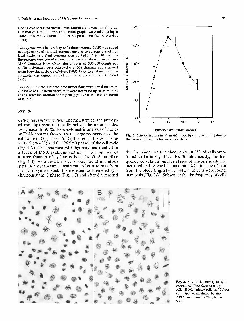
Fig. 1 A-H. Histograms of relative nuclear DNA contents obtained by flow-cytometric analysis of DAPI-stained nuclei released from Vicia faba root tips. The nuclei were isolated before a treatment with hydroxyurea (A) and immediately after the treatment with
D
H
384 512
2.5 mM hydroxyurea for 18 h (B). Further samples of nuclei were isolated from root tips during the recovery from the hydroxyurea block after 2 h (C), 3 h (D), 4 h (E), 6 h (F), 10 h (G), and 12 h (H)
mopak epifluorescent module with filterblock A was used for visu- alization of DAPI fluorescence. Photographs were taken using a Vario Orthomat 2 automatic microscope camera (Leitz, Wetzlar, FRG).
Flow cytometry. The DNA-specific fluorochrome DAPI was added to suspensions of isolated chromosomes or to suspensions of iso- lated nuclei to a final concentration of 5 ~tM. After 30 min, the fluorescence intensity of stained objects was analysed using a Leitz MPV Compact Flow Cytometer at rates of 100-200 counts per s. The histograms were collected over 512 channels and analysed using Flowstar software (Dole~el 1989). Prior to analysis, the flow cytometer was aligned using chicken red-blood-cell nuclei (Dole~el 1991).
Long-term storage. Chromosome suspensions were stored for sever- al days at 4 ~ C. Alternatively, they were stored for up to six months at 4 ~ C after the addition of hexylene glycol to a final concentration of 0.75 M.
Results
X a Z
0 t- o
Cell-cycle synchronization. The meristem cells in untreat- ed roo t tips were mitotically active, the mitot ic index being equal to 9.1%. F low-cytomet r ic analysis o f nucle- ar D N A conten t showed that a large p ropo r t i on o f the cells were in G1 phase (45.1%) the rest o f the cells being in the S (28.4%) and G2 (26.5%) phases o f the cell cycle (Fig. 1 A). The t rea tment with hyd roxyurea resulted in a block o f D N A synthesis and in an accumula t ion o f a large f ract ion o f cycling cells at the G1/S interface (Fig. 1 B). As a result, no cells were found in mitosis after 18 h hydroxyurea treatment. After a release f rom the hydroxyurea block, the meris tem cells entered syn- chronous ly the S phase (Fig. 1 C) and after 6 h reached
50
40
30
2 0
1 0
0 4 14
I i I i
6 8 10 12
J. Dole~el et al. : Isolation of Vieiafaba chromosomes 95
RECOVERY TIME (hours)
Fig. 2. Mitotic indices in Viciafaba root tips (mean _ SE) during the recovery from the hydroxyurea block
the G2 phase. At this time, only 10.2% o f cells were found to be in G1 (Fig. 1 F). Simultaneously, the fre- quency o f cells in various stages o f mitosis gradual ly increased and reached its m a x i m u m 8 h after the release f rom the block (Fig. 2) when 44.5% o f cells were found in mitosis (Fig. 3 A). Subsequently, the f requency o f cells
Fig. 3. A Mitotic activity of syn- chronized Vieia faba root tip cells. B Metaphase cells in V. faba root tips accumulated by the APM treatment. • 260; bar= 50 ~tm
96
7o t 6O
t~ uJ 5 0
I - 40 - iii
h 0 3 0 >. o z iii
2 0 0 I,u n,- I,,I,.
10-
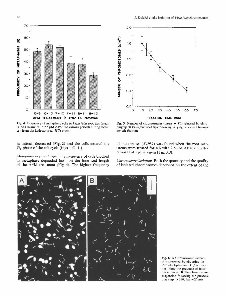
0 6 - 9 6 - 1 0 7 - 1 0 7 - 1 1 8 - 1 1 8 - 1 2
A P M T R E A T M E N T (h a f t e r HU r e m o v a l )
Fig. 4. Frequency of metaphase cells in Viciafaba root tips (mean _+ SE) treated with 2.5 pM APM for various periods during recov- ery from the hydroxyurea (HU) block
J. Dole~el et al. : Isolation of Viciafaba chromosomes
2.0
,,O 0
1.6
nl ~i 0
1 . 2 0
0 n- z
0 . 8
0
14,1 m 3E O A '
Z
O~ i i , i i i
0 10 2 0 30 40 50 6 0 7 0
F I X A T I O N T I M E (rain)
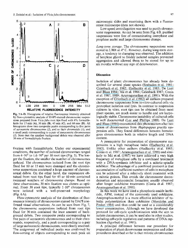
Fig. 5. Number of chromosomes (mean + SE) released by chop- ping up 30 Viciafaba root tips following varying periods of formal- dehyde fixation
in mitosis decreased (Fig. 2) and the cells entered the GI phase of the cell cycle (Figs. 1 G, H).
Metaphase accumulation. The frequency of cells blocked in metaphase depended both on the time and length of the APM treatment (Fig. 4). The highest frequency
of metaphases (53.9%) was found when the root mer- istems were treated for 4 h with 2.5 pM APM 6 h after removal of hydroxyurea (Fig. 3 B).
Chromosome isolation. Both the quantity and the quality of isolated chromosomes depended on the extent of the
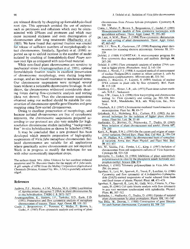
Fig. 6. A Chromosome suspen- sion prepared by chopping up formaldehyde-fixed V. faba root tips. Note the presence of inter- phase nuclei. B The chromosome suspension following the purifica- tion step. • 390; bar=25 ~tm
J. Dole~el et al. : Isolation of Viciafaba chromosomes 97
8t 2 A i o
uz z C D
'Li, RELATIVE FLUORESCENCE INTENSITY
Fig. "/A-D. Histograms of relative fluorescence intensity obtained by flow-cytometric analysis of DAPI-stained chromosome suspen- sions prepared from Viciafaba root tips fixed with 6% formalde- hyde for 15 min (A), 30 min (B), 45 min (C), and 60 rain (D). All histograms show two composite peaks corresponding to five pairs of acrocentric chromosomes (2), and to their chromatids (1), and a small peak corresponding to a pair of metacentric chromosomes (3). Note that the smallest background debris was observed after 30 rain of formaldehyde fixation
fixation with formaldehyde. Under our experimental conditions, the number of isolated chromosomes ranged from 4" 105 to 1.6-106 per 30 root tips (Fig. 5). The lon- ger the fixation, the smaller the number of chromosomes isolated. The chromosomes isolated from the root tips fixed for 10 or 15 min were damaged and the chromo- some suspensions contained a large amount of chromo- somal debris. On the other hand, the suspensions ob- tained from root tips fixed for 45 or 60 min contained increased numbers of chromosome clumps. Based on these observations, 30 min fixation was chosen as opti- mal. From 30 root tips, typically 1 "10 6 chromosomes were isolated with a well-preserved morphology (Fig. 6A).
Flow-cytometric analysis of the distribution of fluo- rescence intensity of chromosomes stained by DAPI con- firmed visual observations. As can be seen from Fig. 7, the chromosome suspensions isolated from root tips fixed for 30 min contained the least amount of back- ground debris. Two composite peaks corresponding to five pairs of acrocentric chromosomes and to their chro- matids, respectively, and a peak corresponding to a pair of large metacentric chromosomes, were clearly resolved. The assignment of individual peaks was confirmed by flow-sorting of objects corresponding to each peak on
microscopic slides and examining them with a fluores- cence microscope (data not shown).
Low-speed centrifugation was used to purify chromo- some suspensions. As can be seen from Fig. 6 B, purified suspensions were free of contaminating interphase and prophase nuclei and large chromosome clumps.
Long-term storage. The chromosome suspensions were stored in LB01 at 4 ~ C. However, during long-term stor- age, a tendency to clumping was observed. The addition of hexylene glycol to freshly isolated samples prevented aggregation and allowed them to be stored for up to six months without any sign of deterioration.
Discussion
Isolation of plant chromosomes has already been de- scribed for several plant species (Szabados et al. 1981; Griesbach et al. 1982; Hadlaczky et al. 1983; De Laat and Blaas 1984; Mii et al. 1986; Griesbach 1987; Conia et al. 1987, 1989; Arumuganathan et al. 1991). With the exception of Griesbach et al. (1982) all authors prepared chromosome suspensions from in-vitro-cultured cells via protoplast isolation and lysis. In contrast to suspension cultures in vitro, root tips can be obtained from most plant species, are much easier to handle and are karyo- logically stable. Chromosome instability of cultured cells is well documented (Lee and Phillips 1988). De Laat and Blaas (1984) encountered this phenomenon after iso- lation of chromosomes from Haplopappus gracilis sus- pension cells. They found differences between homolo- gous chromosomes both in relative length and DNA content.
A prerequisite for preparation of chromosome sus- pensions is a high metaphase index (Hadlaczky et al. 1983). Unlike other authors (Hadlaczky etal. 1983; Conia et al. 1987; Arumuganathan et al. 1991) and simi- larly to Mii et al. (1987) we have achieved a very high frequency of metaphase cells by a combined treatment with a DNA-synthesis inhibitor and a mitotic-spindle inhibitor. The advantage of a combined protocol is that accumulation of sufficient numbers of cells in metaphase can be achieved after a relatively short treatment with a mitotic poison. This avoids the chromosome decon- densation and micronuclei formation usually observed after longer colchicine treatments (Conia etal. 1987; Arumuganathan et al. 1991),
In this work we have used a phosphoric-amide herbi- cide, APM, instead of the commonly used colchicine. Amiprophos-methyl is a more effective inhibitor of tu- bulin polymerization than colchicine (Morejohn and Fosket 1984) and thus could be used at a considerably lower concentration. Although we have developed the protocol for cell-cycle synchronization with the aim to isolate chromosomes, it can be used also in other studies, including cell-cycle regulation and patterns of DNA rep- lication (Sgorbati et al. 1991).
A principal difference between our procedure for preparation of plant chromosome suspensions and other procedures described so far is that mitotic chromosomes
98 J. Dole2el et al. : Isolation of Viciafaba chromosomes
are released directly by chopp ing up formaldehyde-f ixed root tips. This a p p r o a c h avoided the use o f enzymes such as pectinases and cellulases which m a y be conta- minated with DNases and proteases and which may cause increased stickness and even disintegration o f ch romosomes after long t reatments (Hadlaczky et al. 1983). We have found that opt imal fixation was crucial for release o f sufficient numbers o f morphologica l ly in- tact ch romosomes . Similarly, Sgorbati et al. (1986) re- por ted an up to sixfold increase in the number o f nuclei isolated by crushing o f formaldehyde-f ixed Pisum sati- rum roo t tips as c o m p a r e d with non-fixed material.
While non-f ixed plant c h rom osom e s are sensitive to mechanical stress ( A r u m u g a n a t h a n et al. 1991), opt imal fo rmaldehyde fixation results in excellent preservat ion o f c h r o m o s o m e morpho logy , even during long- term storage, and an increased resistance to mechanical stress. Our c h r o m o s o m e suspensions were syringed several times wi thout a noticeable c h r o m o s o m e breakage. In ad- dition, the ch romosom e s wi ths tood considerable shear- ing forces dur ing f low-cytometr ic analysis and sort ing (data not shown). Thus the c h r o m o s o m e suspensions obta ined using our me thod should be suitable for con- s truct ion o f chromosome-speci f ic gene libraries and gene mapp ing using f low-sorted chromosomes .
Owing to excellent preservat ion o f morpho logy , and because isolated c h r o m o s o m e s are free o f cytoplasmic remnants , the c h r o m o s o m e suspensions prepared ac- cord ing to our p ro toco l are also very suitable for high- resolut ion c h r o m o s o m e studies including " b a c k g r o u n d - f ree" in-situ hybr id iza t ion as shown by Schubert (1992).
It m a y be concluded that a new pro tocol has been developed which permits p repara t ion o f high-quali ty suspensions o f Viciafaba metaphase chromosomes . Iso- lated ch romosomes are suitable for all applications where genetically active ch romosomes are not required. Work is in progress to modi fy the technique for use with other economical ly impor tan t crops.
The authors thank Mrs. Jifina Eli/t~ov~i for her excellent technical assistance and Dr. Slavomir Ondro for the supply of V.faba seeds. A gift sample of APM from the Mobay Corporation (Agricultural Chemicals Division, Kansas City, Mo., USA) is gratefully acknowl- edged.
References
Ambros, P.F., Matzke, A.J.M., Matzke, M.A. (1986) Localization of Agrobaeterium rhizogenes T-DNA in plant chromosomes by in situ hybridization. EMBO J. 5, 2073-2077
Arumuganathan, K., Slattery, J.P., Tanksley, S.D., Earle, E.D, (1991) Preparation and flow cytometric analysis of metaphase chromosomes of tomato. Theor. Appl. Genet. 82, 101-111
Conia, J., Bergounioux, C., Perennes, C., Muller~ P., Brown, S., Gadal, P. (1987) Flow cytometric analysis and sorting of plant
chromosomes from Petunia hybrida protoplasts. Cytometry 8, 500 508
Conia, J., Muller, P., Brown, S., Bergounioux, C., Gadal, P. (1989) Monoparametric models of flow cytometric karyotypes with spreadsheet software. Theor. Appl. Genet. 77, 295-303
De Laat, A.M.M., Blaas, J. (1984) Flow-cytometric characteriza- tion and sorting of plant chromosomes. Theor. Appl. Genet. 67, 463467
Dill6, J.E., Ross, K., Gustavson, J.P. (1990) Preparing plant chro- mosomes for scanning electron microscopy. Genome 33, 333- 339
Dole~el, J. (1989) FLOWSTAR: A microcomputer program for flow cytometric data manipulation and analysis. Biol6gia 44, 287-291
Dole~el, J. (1991) Flow cytometric analysis of nuclear DNA con- tent in higher plants. Phytochem. Anal. 2, 143-154
Dole~el, J., Nov~k, F.J. (1984) A standard method of measurement of nuclear Feulgen-DNA content in Allium sativum L. cells by absorption cytophotometry. Mikroskopie 41,335 34l
Dole~el, J., Binarov~, P., Lucretti, S. (1989) Analysis of nuclear DNA content in plant cells by flow cytometry. Biol. Plant. 31, 113-120
Gamborg, O.L., Wetter, L.R., eds. (1975) Plant tissue culture meth- ods. N.R.C., Saskatoon
Gray, J.W., Cram, L.S. (1990) Flow karyotyping and chromosome sorting. In: Flow cytometry and sorting, pp. 503-529, Me- lamed, M.R., Mendelsohn, M.L. eds. Wiley-Liss, Inc., New York
Griesbach, R.J. (1987) Chromosome-mediated transformation via microinjection. Plant Sci. 50, 69 77
Griesbach, R.J., Malmberg, R.L., Carlson, P.S. (1982) An im- proved technique for the isolation of higher plant chromo- somes. Plant Sci. Lett. 24, 55-60
Hadlaczky, G., Bisztray, G., Praznovsky, T., Dudits, D. (1983) Mass isolation of plant chromosomes and nuclei. Planta 157, 278 285
Karp, A., Bright, S.W.J. (1985) On the causes and origins of soma- clonal variation. Oxford Surv. Plant Mol. Cell Biol. 2, 199-234
Lee, M., Phillips, R.L. (1988) The chromosomal basis of somaclon- al variation. Annu. Rev. Plant Physiol. and Plant Mol. Biol. 39, 413~437
Mii, M., Saxena, P.K., Fowke, L.C., King, J. (1987) Isolation of chromosomes from cell suspension cultures of Vicia hajastana. Cytologia 52, 523 528
Morejohn, L., Fosket, D. (1984) Inhibition of plant microtubule polymerization in vitro by the phosphoric amide herbicide ami- prophos-methyl. Science 224, 874
Schubert, I. (1992) Telomeric polymorphism in Vicia faba. Biol. Zbl. (in press)
Sgorbati, S., Levi, M., Sparvoli, E., Trezzi, F., Lucchini, G. (1986) Cytometry and flow cytometry of 4',6-diamidino-2-phenylin- dole (DAPI)-stained suspensions of nuclei released from fresh and fixed tissues of plants. Physiol. Plant. 68, 471-476
Sgorbati, S., Sparvoli, E., Levi, M., Galli, M.G., Citterio, S., Chia- tante, D. (1991) Cell cycle kinetic analysis with flow cytometry in pea root meristem synchronized with aphidicolin. Physiol. Plant. 81,507-512
Szabados, L., Hadlaczky, G., Dudits, D. (1981) Uptake of isolated plant chromosomes by plant protoplasts. Planta 151, 141-145
Van Dilla, M., Deaven, L. (1990) Construction of gene libraries for each human chromosome. Cytometry 11,208-218