CENP-E Is a Plus End–Directed Kinetochore Motor Required for Metaphase Chromosome Alignment
10
Cell, Vol. 91, 357–366, October 31, 1997, Copyright 1997 by Cell Press CENP-E Is a Plus End–Directed Kinetochore Motor Required for Metaphase Chromosome Alignment and Salmon, 1994). Initially, the oscillatory movements of bioriented chromosomes have a net bias toward the spindle equator. Once an equatorial position is Kenneth W. Wood,* Roman Sakowicz, ² Lawrence S. B. Goldstein, ² and Don W. Cleveland* ‡ achieved, chromosomes continue to oscillate much in * Laboratory of Cell Biology the same manner as during initial movement to the spin- Ludwig Institute for Cancer Research dle midzone, except that these movements yield little ² Howard Hughes Medical Institute net displacement from the metaphase plate (Skibbens Division of Cellular and Molecular Medicine et al., 1993; Khodjakov and Rieder, 1996). Stable meta- University of California at San Diego phase chromosome positioning and the preceding pro- La Jolla, California 92093-0660 metaphase chromosome movements that establish metaphase alignment are thus a consequence of the balance or imbalance, respectively, of poleward and Summary antipoleward movements and are almost certainly tem- porally distinct halves of the same kinetochore-depen- Mitosis requires dynamic attachment of chromo- dent process. somes to spindle microtubules. This interaction is me- In many meiotic spindles, as well as early embryonic diated largely by kinetochores. During prometaphase, mitotic spindles, such as in Drosophila (Theurkauf and forces exerted at kinetochores, in combination with Hawley, 1992; Matthies et al., 1996; Wilson et al., 1997), polar ejection forces, drive congression of chromo- spindle assembly differs from the more typical mitotic somes to the metaphase plate. A major question has examples in that the spindles form by an “inside-out” been whether kinetochore-associated microtubule mo- mechanism in which microtubules organize in the ab- tors play an important role in congression. Using im- sence of centrosomes around a centrally located mass munodepletion from and antibody addition to Xenopus of chromatin. Bipolarity is achieved by the action of egg extracts, we show that the kinetochore-associ- multivalent minus end–directed microtubule motor com- ated kinesin-like motor protein CENP-E is essential plexes, including NuMA, cytoplasmic dynein, and dy- for positioning chromosomes at the metaphase plate. nactin in frogs (Heald et al., 1996, 1997; Merdes et al., We further demonstrate that CENP-E powers move- 1996) and the kinesin-like motor Ncd in Drosophila (Mat- ment toward microtubule plus ends in vitro. These thies et al., 1996). These motor complexes tether parallel findings support a model in which CENP-E functions microtubule bundles and stabilize converging microtu- in congression to tether kinetochores to dynamic mi- bules into spindle poles on either side of the centrally crotubule plus ends. located chromatin. Compared to typical mitotic spin- dles, initial congression of chromosomes to the spindle Introduction equator in these meiotic and early mitotic spindles would seem to play at most a minor role in establishing Accurate segregation of genetic material is mediated by metaphase chromosome alignment. However, the sec- the microtubules of the mitotic spindle. Spindle microtu- ond phase of congression, maintenance of alignment bules have a defined polarity, with their slow-growing prior to anaphase, is nonetheless of undiminished im- minus ends anchored at or near the spindle pole, and portance. their dynamic, fast-growing plus ends interacting with The two predominant and opposing forces currently chromosomes and with microtubules emanating from thought to be responsible for chromosome movement the opposite pole (McIntosh and Euteneur, 1984). Inter- during congression are an antipoleward polar ejection action of chromosomes with spindle microtubules is force associated with regions of high microtubule den- mediated by kinetochores, specialized microtubule at- sity near the spindle poles and forces generated directly tachment structures located at the centromeric region at the kinetochore by microtubule-dependent motors. of each sister chromatid (reviewed in Rieder, 1982; Studies in vitro have demonstrated the presence of both Mitchison, 1988). During prometaphase, kinetochores plus end– and minus end–directed microtubule motor capture and stabilize dynamically unstable microtubules activities on prometaphase kinetochores (Mitchison and growing from the poles (Nicklas and Kubai, 1985; Mitchi- Kirschner, 1985; Hyman and Mitchison, 1991). However, son et al., 1986). Once attached to microtubules, kineto- microsurgical separation of sister kinetochores of con- chores and chromosomes exhibit oscillatory move- gressing bioriented chromosomes results in continued ments, switching between poleward and antipoleward poleward movement of the kinetochore that was leading movement (Roos, 1976; Bajer, 1982; Rieder et al., 1986; at the time of severance (Khodjakov and Rieder, 1996). Skibbens et al., 1993; Khodjakov and Rieder, 1996; Wa- The trailing kinetochore ceases to move. Laser ablation ters et al., 1996) and culminating in alignment at the of the leading kinetochore also causes the trailing kinet- metaphase plate. These movements are collectively re- ochore to halt. Furthermore, examination of the distance ferred to as congression, and are thought to be, at least separating sister kinetochores during congression re- in part, a consequence of forces generated by micro- veals that pushing forces rarely result in compression tubule motors localized to the kinetochore (Rieder of the intrakinetochore distance (Waters et al., 1996). Together these findings suggest that during congres- sion kinetochore-associated minus end–directed pulling ‡ To whom correspondence should be addressed.
Transcript of CENP-E Is a Plus End–Directed Kinetochore Motor Required for Metaphase Chromosome Alignment
Cell, Vol. 91, 357–366, October 31, 1997, Copyright 1997 by Cell Press
CENP-E Is a Plus End–Directed KinetochoreMotor Required for Metaphase Chromosome Alignment
and Salmon, 1994). Initially, the oscillatory movementsof bioriented chromosomes have a net bias towardthe spindle equator. Once an equatorial position is
Kenneth W. Wood,* Roman Sakowicz,†Lawrence S. B. Goldstein,†and Don W. Cleveland*‡
achieved, chromosomes continue to oscillate much in*Laboratory of Cell Biologythe same manner as during initial movement to the spin-Ludwig Institute for Cancer Researchdle midzone, except that these movements yield little†Howard Hughes Medical Institutenet displacement from the metaphase plate (SkibbensDivision of Cellular and Molecular Medicineet al., 1993; Khodjakov and Rieder, 1996). Stable meta-University of California at San Diegophase chromosome positioning and the preceding pro-La Jolla, California 92093-0660metaphase chromosome movements that establishmetaphase alignment are thus a consequence of thebalance or imbalance, respectively, of poleward andSummaryantipoleward movements and are almost certainly tem-porally distinct halves of the same kinetochore-depen-Mitosis requires dynamic attachment of chromo-dent process.somes to spindle microtubules. This interaction is me-
In many meiotic spindles, as well as early embryonicdiated largely by kinetochores. During prometaphase,mitotic spindles, such as in Drosophila (Theurkauf andforces exerted at kinetochores, in combination withHawley, 1992; Matthies et al., 1996; Wilson et al., 1997),polar ejection forces, drive congression of chromo-spindle assembly differs from the more typical mitoticsomes to the metaphase plate. A major question hasexamples in that the spindles form by an “inside-out”been whether kinetochore-associated microtubule mo-mechanism in which microtubules organize in the ab-tors play an important role in congression. Using im-sence of centrosomes around a centrally located massmunodepletion from and antibody addition to Xenopusof chromatin. Bipolarity is achieved by the action ofegg extracts, we show that the kinetochore-associ-multivalent minus end–directed microtubule motor com-ated kinesin-like motor protein CENP-E is essentialplexes, including NuMA, cytoplasmic dynein, and dy-for positioning chromosomes at the metaphase plate.nactin in frogs (Heald et al., 1996, 1997; Merdes et al.,We further demonstrate that CENP-E powers move-1996) and the kinesin-like motor Ncd in Drosophila (Mat-ment toward microtubule plus ends in vitro. Thesethies et al., 1996). These motor complexes tether parallelfindings support a model in which CENP-E functionsmicrotubule bundles and stabilize converging microtu-in congression to tether kinetochores to dynamic mi-bules into spindle poles on either side of the centrallycrotubule plus ends.located chromatin. Compared to typical mitotic spin-dles, initial congression of chromosomes to the spindleIntroductionequator in these meiotic and early mitotic spindleswould seem to play at most a minor role in establishing
Accurate segregation of genetic material is mediated bymetaphase chromosome alignment. However, the sec-
the microtubules of the mitotic spindle. Spindle microtu-ond phase of congression, maintenance of alignment
bules have a defined polarity, with their slow-growingprior to anaphase, is nonetheless of undiminished im-
minus ends anchored at or near the spindle pole, andportance.
their dynamic, fast-growing plus ends interacting withThe two predominant and opposing forces currently
chromosomes and with microtubules emanating fromthought to be responsible for chromosome movement
the opposite pole (McIntosh and Euteneur, 1984). Inter- during congression are an antipoleward polar ejectionaction of chromosomes with spindle microtubules is
force associated with regions of high microtubule den-mediated by kinetochores, specialized microtubule at-
sity near the spindle poles and forces generated directlytachment structures located at the centromeric region
at the kinetochore by microtubule-dependent motors.of each sister chromatid (reviewed in Rieder, 1982; Studies in vitro have demonstrated the presence of bothMitchison, 1988). During prometaphase, kinetochores plus end– and minus end–directed microtubule motorcapture and stabilize dynamically unstable microtubules activities on prometaphase kinetochores (Mitchison andgrowing from the poles (Nicklas and Kubai, 1985; Mitchi- Kirschner, 1985; Hyman and Mitchison, 1991). However,son et al., 1986). Once attached to microtubules, kineto- microsurgical separation of sister kinetochores of con-chores and chromosomes exhibit oscillatory move- gressing bioriented chromosomes results in continuedments, switching between poleward and antipoleward poleward movement of the kinetochore that was leadingmovement (Roos, 1976; Bajer, 1982; Rieder et al., 1986; at the time of severance (Khodjakov and Rieder, 1996).Skibbens et al., 1993; Khodjakov and Rieder, 1996; Wa- The trailing kinetochore ceases to move. Laser ablationters et al., 1996) and culminating in alignment at the of the leading kinetochore also causes the trailing kinet-metaphase plate. These movements are collectively re- ochore to halt. Furthermore, examination of thedistanceferred to as congression, and are thought to be, at least separating sister kinetochores during congression re-in part, a consequence of forces generated by micro- veals that pushing forces rarely result in compressiontubule motors localized to the kinetochore (Rieder of the intrakinetochore distance (Waters et al., 1996).
Together these findings suggest that during congres-sion kinetochore-associated minus end–directed pulling‡To whom correspondence should be addressed.
Cell358
forces dominate, with plus end–directed pushing forces than that shared between XCENP-E and its nearest phy-logenetic neighbors (Moore and Endow, 1996). Outsideplaying at most a minor role in driving kinetochore-
mediated chromosome movement. The outstanding is- XCENP-E’s amino-terminal domain lie three additionalregions of greater than 25% identity with hCENP-E, butsue, however, has been the identity of the molecules at
the kinetochore that generate the force for initial chro- not with other kinesin-like proteins (Figure 1A). On thebasis of these regions of identity and its comparablymosome movement and maintenance at the mitotic
midzone. large predicted size, we conclude that XCENP-E is theXenopus homolog of human CENP-E.A prominent candidate for powering one or more as-
pects of chromosome movement in mitosis is centro-mere-associated protein-E (CENP-E), a member of the XCENP-E Associates with Xenopus Centromereskinesin superfamily of microtubule motor proteins that In Vivo and In Vitrois an integral component of kinetochore corona fibers To verify that Xenopus CENP-E exhibits a pattern ofthat link centromeres to spindle microtubules (Yen et al., cell cycle-dependent kinetochore association similar to1992; Yao et al., 1997). CENP-E localizes to kinetochores human CENP-E, polyclonal antibodies were raised againstthroughout all phases of mitotic chromosome move- two recombinant antigens, one spanning the tail andment (early prometaphase through anaphase A) (Yen et C-terminal portion of the rod (a-XCENP-ETAIL, Figure 1B)al., 1992; Lombillo et al., 1995a; Brown et al., 1996). and the other corresponding to a portion of the N termi-Previous efforts have suggested a role for CENP-E in nus of the rod domain (a-XCENP-EROD; Figure 1B). Immu-mitosis and meiosis. Microinjection of a monoclonal an- noblotting of Xenopus egg extract reveals that thetibody directed against CENP-E into cultured human
a-XCENP-ETAIL antibody specifically recognizes XCENP-Ecells delays anaphase onset (Yen et al., 1991). Anti- as a single band of greater than 300 kDa (Figure 2B).CENP-E antibody injection into maturing mouse oocytes The a-XCENP-EROD antibody specifically recognizesinduces arrest at the first reductional division of meiosis XCENP-E (arrowhead, Figure 2B) and another protein(Duesbery et al., 1997). Antibodies against CENP-E of slightly lower molecular weight (indicated by dot inblock microtubule depolymerization-dependent minus Figure 2B) that may be a distinct isoform of XCENP-Eend–directed movement of purified chromosomes in lacking the tail domain or XCENP-E that has lost its tailvitro (Lombillo et al., 1995a). Recently, CENP-E was re- domain due to partial proteolysis. Immunostaining ofported to be associated with minus end–directed micro- cultured Xenopus XTC cells using a-XCENP-ETAIL anti-tubule motor activity (Thrower et al., 1995), raising the body (Figure 2C) revealed patterns of cell cycle–depen-possibility that CENP-E might be responsible for pole- dent localization similar to that observed for mammalianward kinetochore movements in prometaphase and CENP-E (Yen et al., 1992; Brown et al., 1996) with twoanaphase A. exceptions: (1) during interphase XCENP-E was local-
We report here that quantitative removal of Xenopus ized to the nucleus (Figure 2C, panels 1 and 2), consis-CENP-E from Xenopus egg extracts, normally capable tent with the presence of a nuclear localization signalof assembling metaphase mitotic spindles in vitro, dis- (Boulikas, 1993) at the C-terminal end of the rod domainrupts metaphase chromosome alignment. We also dem- (Figures 1A and 1C), and (2) a proportion of XCENP-Eonstrate that the kinesin-like motor domain of Xenopus could occasionally be found localized to mitotic spindleCENP-E powers movement toward microtubule plus poles (e.g., Figure 2C, panels 5 and 6). a-XCENP-ETAILends. Together these findings suggest that CENP-E immunostaining of metaphase spindles assembled infunctions in vivo at the trailing kinetochore to link anti- vitro using cytostatic factor (CSF)-arrested Xenopus eggpoleward movement to microtubule growth and at the extracts cycled through interphase (Murray, 1991; Sawinleading kinetochore to couple microtubule disassembly and Mitchison, 1991) revealed that XCENP-E was almostdynamics to minus end–directed movement. exclusively associated with kinetochores (Figure 2A,
panel 2). Unlike in cultured cells, XCENP-E was infre-quently found localized to poles of spindles assembledResultsin vitro.
Identification of Xenopus CENP-ETo investigate the role of CENP-E in mitotic spindle XCENP-E Is Required for Metaphase
Chromosome Alignmentformation in vitro using extracts of Xenopus eggs, weused low stringency hybridization followed by library To determine the aspect(s) of mitosis for which
XCENP-E is required, a-XCENP-ETAIL antibody was usedrescreening to clone the Xenopus homolog of CENP-E.The full-length sequence encodes a protein of 2954 to deplete XCENP-E from Xenopus egg extracts natu-
rally arrested in metaphase (CSF-extract). Immunoblot-amino acids with a predicted molecular mass of 340kDa (Figures 1A and 1C). The predicted structure of ting of control and XCENP-E-depleted CSF-extracts re-
vealed that of XCENP-E could bequantitatively removedXenopus CENP-E (XCENP-E) is very similar to humanCENP-E (hCENP-E), consisting of an amino-terminal re- by immunodepletion with this antibody (Figure 3A, lane
2). Unrelated antigens, such as XNuMA (Merdes et al.,gion containing a kinesin-like microtubule motor do-main, linked to a globular tail domain by a region pre- 1996), were unaffected by depletion of XCENP-E (Figure
3A, lower panel). The specificity of depletion was furtherdicted to form a long, discontinuous a-helical coiled coil(Lupas et al., 1991; Bergeret al., 1995) (Figure 1A). Within confirmed by comparison of the proteins recovered in
the a-XCENP-ETAIL precipitate (Figure 3D, lane 2) withthe core of the motor domain (residues 1–324), XCENP-Eand hCENP-E share 74% identity, significantly greater those present in the control IgG precipitate (Figure 3D,
CENP-E Is Required for Chromosome Alignment359
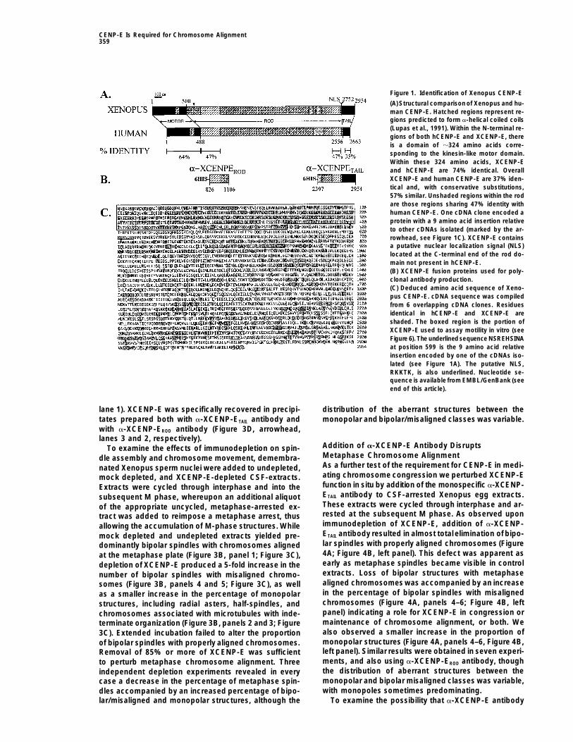
Figure 1. Identification of Xenopus CENP-E
(A) Structural comparisonof Xenopus and hu-man CENP-E. Hatched regions represent re-gions predicted to form a-helical coiled coils(Lupas et al., 1991). Within the N-terminal re-gions of both hCENP-E and XCENP-E, thereis a domain of z324 amino acids corre-sponding to the kinesin-like motor domain.Within these 324 amino acids, XCENP-Eand hCENP-E are 74% identical. OverallXCENP-E and human CENP-E are 37% iden-tical and, with conservative substitutions,57% similar. Unshaded regions within the rodare those regions sharing 47% identity withhuman CENP-E. One cDNA clone encoded aprotein with a 9 amino acid insertion relativeto other cDNAs isolated (marked by the ar-rowhead, see Figure 1C). XCENP-E containsa putative nuclear localization signal (NLS)located at the C-terminal end of the rod do-main not present in hCENP-E.(B) XCENP-E fusion proteins used for poly-clonal antibody production.(C) Deduced amino acid sequence of Xeno-pus CENP-E. cDNA sequence was compiledfrom 6 overlapping cDNA clones. Residuesidentical in hCENP-E and XCENP-E areshaded. The boxed region is the portion ofXCENP-E used to assay motility in vitro (seeFigure 6). The underlinedsequence NSREHSINAat position 599 is the 9 amino acid relativeinsertion encoded by one of the cDNAs iso-lated (see Figure 1A). The putative NLS,RKKTK, is also underlined. Nucleotide se-quence is available from EMBL/GenBank (seeend of this article).
lane 1). XCENP-E was specifically recovered in precipi- distribution of the aberrant structures between thetates prepared both with a-XCENP-ETAIL antibody and monopolar and bipolar/misaligned classes was variable.with a-XCENP-EROD antibody (Figure 3D, arrowhead,lanes 3 and 2, respectively).
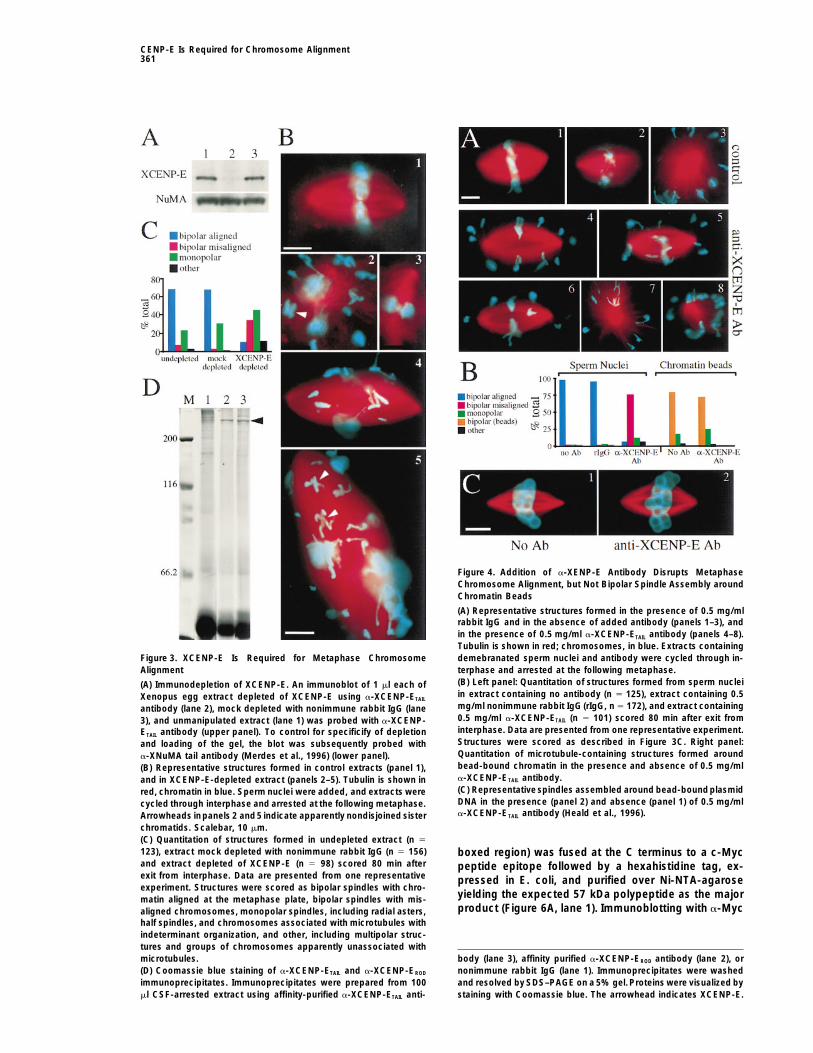
Addition of a-XCENP-E Antibody DisruptsTo examine the effects of immunodepletion on spin-Metaphase Chromosome Alignmentdle assembly and chromosome movement, demembra-As a further test of the requirement for CENP-E in medi-nated Xenopus sperm nuclei were added to undepleted,ating chromosome congression we perturbed XCENP-Emock depleted, and XCENP-E-depleted CSF-extracts.function in situ by addition of the monospecific a-XCENP-Extracts were cycled through interphase and into theETAIL antibody to CSF-arrested Xenopus egg extracts.subsequent M phase, whereupon an additional aliquotThese extracts were cycled through interphase and ar-of the appropriate uncycled, metaphase-arrested ex-rested at the subsequent M phase. As observed upontract was added to reimpose a metaphase arrest, thusimmunodepletion of XCENP-E, addition of a-XCENP-allowing the accumulation of M-phase structures. WhileETAIL antibody resulted in almost totalelimination of bipo-mock depleted and undepleted extracts yielded pre-lar spindles with properly aligned chromosomes (Figuredominantly bipolar spindles with chromosomes aligned4A; Figure 4B, left panel). This defect was apparent asat the metaphase plate (Figure 3B, panel 1; Figure 3C),early as metaphase spindles became visible in controldepletion of XCENP-E produced a 5-fold increase in theextracts. Loss of bipolar structures with metaphasenumber of bipolar spindles with misaligned chromo-aligned chromosomes was accompanied by an increasesomes (Figure 3B, panels 4 and 5; Figure 3C), as wellin the percentage of bipolar spindles with misalignedas a smaller increase in the percentage of monopolarchromosomes (Figure 4A, panels 4–6; Figure 4B, leftstructures, including radial asters, half-spindles, andpanel) indicating a role for XCENP-E in congression orchromosomes associated with microtubules with inde-maintenance of chromosome alignment, or both. Weterminate organization (Figure 3B, panels 2 and 3; Figurealso observed a smaller increase in the proportion of3C). Extended incubation failed to alter the proportionmonopolar structures (Figure 4A, panels 4–6, Figure 4B,of bipolar spindles with properly aligned chromosomes.left panel). Similar results were obtained in seven experi-Removal of 85% or more of XCENP-E was sufficientments, and also using a-XCENP-EROD antibody, thoughto perturb metaphase chromosome alignment. Threethe distribution of aberrant structures between theindependent depletion experiments revealed in everymonopolar and bipolar misaligned classes was variable,case a decrease in the percentage of metaphase spin-with monopoles sometimes predominating.dles accompanied by an increased percentage of bipo-
lar/misaligned and monopolar structures, although the To examine the possibility that a-XCENP-E antibody
Cell360
of beads, followed by tethering of the distal microtubuleminus ends by a complex of cytoplasmic dynein andNuMA (Heald et al., 1996, 1997; Merdes et al., 1996).While a-XCENP-ETAIL antibody addition induced chro-mosome misalignment in spindles assembled aroundsperm nuclei (Figure 4B, left panel), this antibody didnot perturb assembly of chromatin-bead spindles (Fig-ure 4B, right panel; Figure 4C). Thus, the disruption ofspindle bipolarity and chromosome alignment observedupon addition of a-XCENP-E antibody is not due to adefect in spindle assembly unrelated to XCENP-E func-tion at kinetochores.
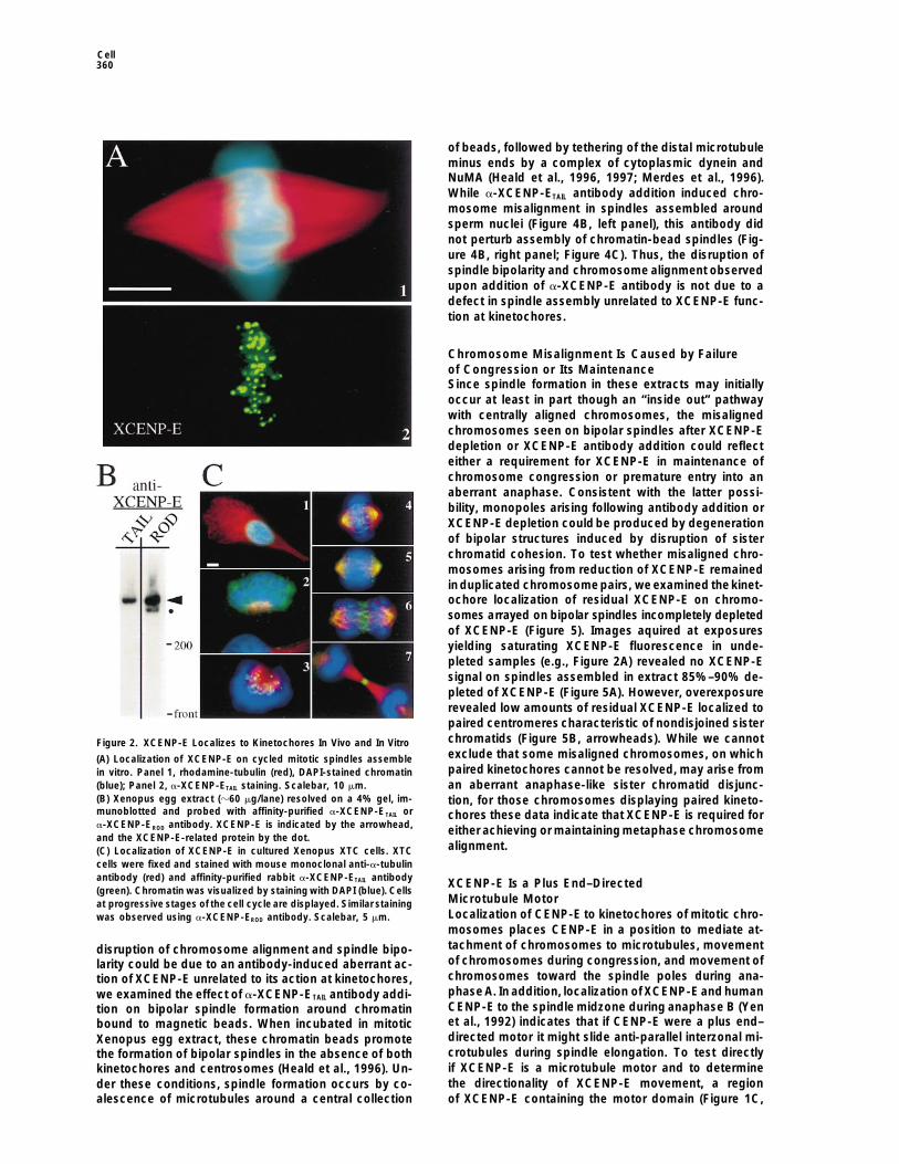
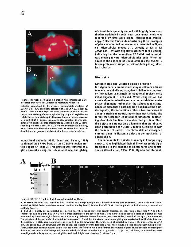
Chromosome Misalignment Is Caused by Failureof Congression or Its MaintenanceSince spindle formation in these extracts may initiallyoccur at least in part though an “inside out” pathwaywith centrally aligned chromosomes, the misalignedchromosomes seen on bipolar spindles after XCENP-Edepletion or XCENP-E antibody addition could reflecteither a requirement for XCENP-E in maintenance ofchromosome congression or premature entry into anaberrant anaphase. Consistent with the latter possi-bility, monopoles arising following antibody addition orXCENP-E depletion could be produced by degenerationof bipolar structures induced by disruption of sisterchromatid cohesion. To test whether misaligned chro-mosomes arising from reduction of XCENP-E remainedinduplicated chromosomepairs, we examined thekinet-ochore localization of residual XCENP-E on chromo-somes arrayed on bipolar spindles incompletely depletedof XCENP-E (Figure 5). Images aquired at exposuresyielding saturating XCENP-E fluorescence in unde-pleted samples (e.g., Figure 2A) revealed no XCENP-Esignal on spindles assembled in extract 85%–90% de-pleted of XCENP-E (Figure 5A). However, overexposurerevealed low amounts of residual XCENP-E localized topaired centromeres characteristic of nondisjoined sisterchromatids (Figure 5B, arrowheads). While we cannotFigure 2. XCENP-E Localizes to Kinetochores In Vivo and In Vitroexclude that some misaligned chromosomes, on which(A) Localization of XCENP-E on cycled mitotic spindles assemblepaired kinetochores cannot be resolved, may arise fromin vitro. Panel 1, rhodamine-tubulin (red), DAPI-stained chromatin
(blue); Panel 2, a-XCENP-ETAIL staining. Scalebar, 10 mm. an aberrant anaphase-like sister chromatid disjunc-(B) Xenopus egg extract (z60 mg/lane) resolved on a 4% gel, im- tion, for those chromosomes displaying paired kineto-munoblotted and probed with affinity-purified a-XCENP-ETAIL or chores these data indicate that XCENP-E is required fora-XCENP-EROD antibody. XCENP-E is indicated by the arrowhead, either achieving or maintaining metaphase chromosomeand the XCENP-E-related protein by the dot.
alignment.(C) Localization of XCENP-E in cultured Xenopus XTC cells. XTCcells were fixed and stained with mouse monoclonal anti-a-tubulinantibody (red) and affinity-purified rabbit a-XCENP-ETAIL antibody XCENP-E Is a Plus End–Directed(green). Chromatin was visualized by staining with DAPI (blue). Cells
Microtubule Motorat progressive stages of the cell cycle are displayed. Similar stainingLocalization of CENP-E to kinetochores of mitotic chro-was observed using a-XCENP-EROD antibody. Scalebar, 5 mm.mosomes places CENP-E in a position to mediate at-tachment of chromosomes to microtubules, movementdisruption of chromosome alignment and spindle bipo-of chromosomes during congression, and movement oflarity could be due to an antibody-induced aberrant ac-chromosomes toward the spindle poles during ana-tion of XCENP-E unrelated to its action at kinetochores,phase A. In addition, localization of XCENP-E and humanwe examined the effect of a-XCENP-ETAIL antibody addi-CENP-E to the spindle midzone during anaphase B (Yention on bipolar spindle formation around chromatinet al., 1992) indicates that if CENP-E were a plus end–bound to magnetic beads. When incubated in mitoticdirected motor it might slide anti-parallel interzonal mi-Xenopus egg extract, these chromatin beads promotecrotubules during spindle elongation. To test directlythe formation of bipolar spindles in the absence of bothif XCENP-E is a microtubule motor and to determinekinetochores and centrosomes (Heald et al., 1996). Un-the directionality of XCENP-E movement, a regionder these conditions, spindle formation occurs by co-
alescence of microtubules around a central collection of XCENP-E containing the motor domain (Figure 1C,
CENP-E Is Required for Chromosome Alignment361
Figure 4. Addition of a-XENP-E Antibody Disrupts MetaphaseChromosome Alignment, but Not Bipolar Spindle Assembly aroundChromatin Beads
(A) Representative structures formed in the presence of 0.5 mg/mlrabbit IgG and in the absence of added antibody (panels 1–3), andin the presence of 0.5 mg/ml a-XCENP-ETAIL antibody (panels 4–8).Tubulin is shown in red; chromosomes, in blue. Extracts containing
Figure 3. XCENP-E Is Required for Metaphase Chromosome demebranated sperm nuclei and antibody were cycled through in-Alignment terphase and arrested at the following metaphase.
(B) Left panel: Quantitation of structures formed from sperm nuclei(A) Immunodepletion of XCENP-E. An immunoblot of 1 ml each ofin extract containing no antibody (n 5 125), extract containing 0.5Xenopus egg extract depleted of XCENP-E using a-XCENP-ETAILmg/ml nonimmune rabbit IgG (rIgG, n 5 172), and extract containingantibody (lane 2), mock depleted with nonimmune rabbit IgG (lane0.5 mg/ml a-XCENP-ETAIL (n 5 101) scored 80 min after exit from3), and unmanipulated extract (lane 1) was probed with a-XCENP-interphase. Data are presented from one representative experiment.ETAIL antibody (upper panel). To control for specificify of depletionStructures were scored as described in Figure 3C. Right panel:and loading of the gel, the blot was subsequently probed withQuantitation of microtubule-containing structures formed arounda-XNuMA tail antibody (Merdes et al., 1996) (lower panel).bead-bound chromatin in the presence and absence of 0.5 mg/ml(B) Representative structures formed in control extracts (panel 1),a-XCENP-ETAIL antibody.and in XCENP-E-depleted extract (panels 2–5). Tubulin is shown in(C) Representative spindles assembled around bead-boundplasmidred, chromatin in blue. Sperm nuclei were added, and extracts wereDNA in the presence (panel 2) and absence (panel 1) of 0.5 mg/mlcycled through interphase and arrested at the following metaphase.a-XCENP-ETAIL antibody (Heald et al., 1996).Arrowheads inpanels 2 and5 indicate apparently nondisjoined sister
chromatids. Scalebar, 10 mm.(C) Quantitation of structures formed in undepleted extract (n 5
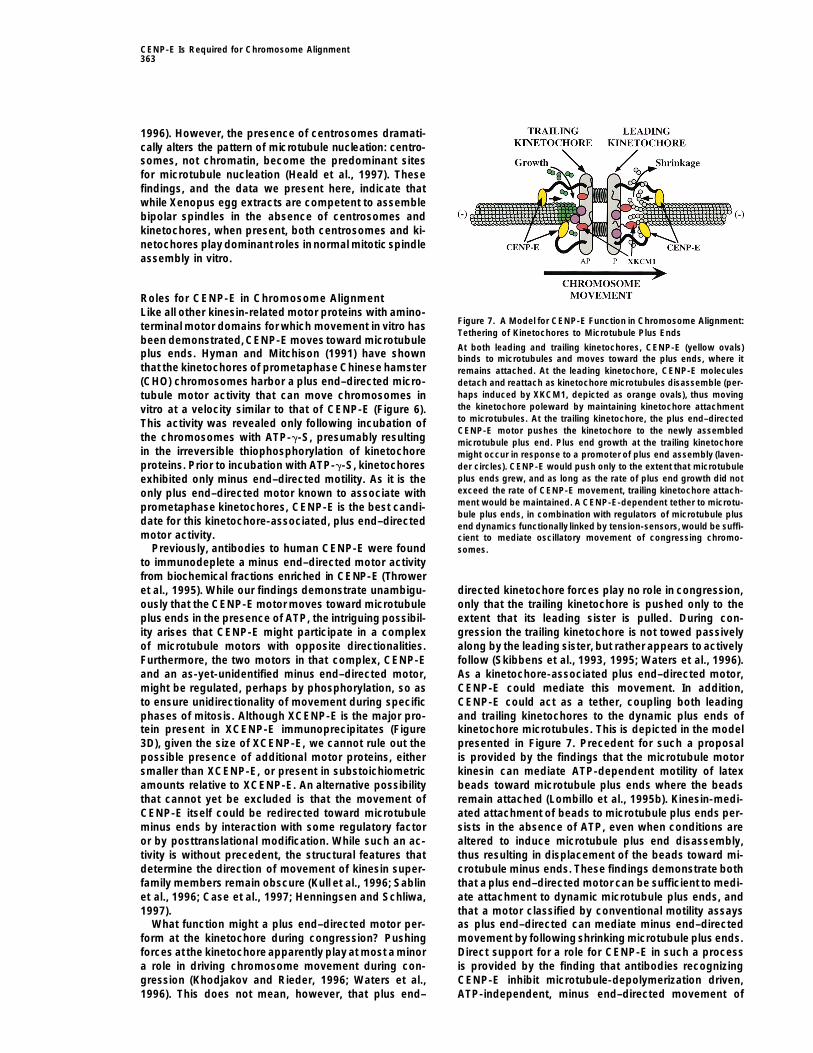
123), extract mock depleted with nonimmune rabbit IgG (n 5 156) boxed region) was fused at the C terminus to a c-Mycand extract depleted of XCENP-E (n 5 98) scored 80 min after peptide epitope followed by a hexahistidine tag, ex-exit from interphase. Data are presented from one representative pressed in E. coli, and purified over Ni-NTA-agaroseexperiment. Structures were scored as bipolar spindles with chro-
yielding the expected 57 kDa polypeptide as the majormatin aligned at the metaphase plate, bipolar spindles with mis-product (Figure 6A, lane 1). Immunoblotting with a-Mycaligned chromosomes, monopolar spindles, including radial asters,
half spindles, and chromosomes associated with microtubules withindeterminant organization, and other, including multipolar struc-tures and groups of chromosomes apparently unassociated with
body (lane 3), affinity purified a-XCENP-EROD antibody (lane 2), ormicrotubules.(D) Coomassie blue staining of a-XCENP-ETAIL and a-XCENP-EROD nonimmune rabbit IgG (lane 1). Immunoprecipitates were washed
and resolved by SDS–PAGE on a 5% gel.Proteins were visualized byimmunoprecipitates. Immunoprecipitates were prepared from 100ml CSF-arrested extract using affinity-purified a-XCENP-ETAIL anti- staining with Coomassie blue. The arrowhead indicates XCENP-E.
Cell362
of microtubules polarity markedwith brightly fluorescentrhodamine-labeled seeds near their minus ends wasrecorded by time-lapse digital fluorescence micros-copy. Selected frames demonstrating three examplesof plus end–directed movement are presented in Figure6B. Microtubules moved at a velocity of 5.1 6 1.7mm/min (n 5 49) with brightly fluorescent seeds leading,indicating that the immobilized XCENP-E fusion proteinwas moving toward microtubule plus ends. When as-sayed in the absence of a-Myc antibody the XCENP-Efusion protein also supported microtubule gliding, albeitless robustly.
Discussion
Kinetochores and Mitotic Spindle FormationMisalignment of chromosomes may result from a failureto reach the spindle equator, that is, failure to congress,or from failure to maintain an equatorial position onceinitial alignment is achieved. While congression has
Figure 5. Disruption of XCENP-E Function Yields Misaligned Chro- classically referred to the process that establishes meta-mosomes that Have Not Undergone Premature Anaphase phase alignment, rather than the subsequent mainte-Spindles assembled in the extracts incompletely depleted of nance of metaphase chromosome position at the spin-XCENP-E (85–90% depletion), stained with a-XCENP-ETAIL antibody.
dle equator, the separation of these two processes isImages collected with exposure times sufficient to yield saturatedalmost certainly temporal, rather than mechanistic. Thekinetochore staining of control spindles (e.g., Figure 2A) yielded noforces that establish equatorial chromosome position-visible kinetochore staining (A). However, longer exposure revealed
residual XCENP-E, present in paired spots characteristic of nondis- ing also likely function to maintain that position. Thus,joined prometaphase sister chromatids ([B], panels 1 and 2, corre- the defect in chromosome alignment that we observesponding to boxes 1 and 2 in [A]). From the fluorescence intensity, upon perturbation of XCENP-E function, combined withwe estimate that kinetochore-associated XCENP-E has been re-
the presence of paired sister chromatids on misalignedduced 6-fold or greater, consistent with the extent of depletion.chromosomes, indicates a defect in the mechanics ofcongression.
Recent models for spindle assembly in Xenopus eggmonoclonal antibody (9E10; Evans and Bishop, 1985)extracts have highlighted their ability to assemble bipo-confirmed the 57 kDa band as the XCENP-E fusion pro-lar spindles in the absence of kinetochores and centro-tein (Figure 6A, lane 2). This protein was tethered to a
glass coverslip using the a-Myc antibody, and gliding somes (Heald et al., 1996, 1997; Hyman and Karsenti,
Figure 6. XCENP-E Is a Plus End–Directed Microtubule Motor
(A) XCENP-E residues 1–473 fused at the C terminus to a c-Myc epitope and a hexahistidine tag (see schematic). Coomassie blue stain ofpurified XCENP-E fusion protein (arrowhead) used for motility (lane 1); immunoblot of XCENP-E fusion protein probed with a-Myc monoclonalantibody (lane 2).(B) XCENP-E motility assay. Microtubules marked near their minus ends with brightly fluorescent seeds were added with ATP to a flowchamber containing purified XCENP-E fusion protein tethered to the coverslip with a-Myc monoclonal antibody. Gliding of microtubules wasmonitored by time-lapse digital fluorescence microscopy. Selected frames from one time lapse series, spaced 90 sec apart, are presented.The positions of the plus ends of microtubules numbered 1, 2, and 3 at the start of continuous gliding are marked with solid white dots, andthe position of a stationary microtubule end is marked by the arrowhead. The bright seed of microtubule 3 enters the plane of focus at 1.5min, and glides 13.6 mm downward with the bright seed leading over the following 3 min. Microtubule 2 moves continuously during the first3 min, after which point it detaches and reattaches further toward the bottom of the frame. Microtubule 1 glides minus-end leading throughoutthe entire time course. The average microtubule velocity of all microtubules was 5.1 mm/min 6 1.7 (n 5 49). Of those, 33 microtubules wereunambiguously polarity marked, and all glided with their bright seeds leading. Scalebar, 5 mm.
CENP-E Is Required for Chromosome Alignment363
1996). However, the presence of centrosomes dramati-cally alters the pattern of microtubule nucleation: centro-somes, not chromatin, become the predominant sitesfor microtubule nucleation (Heald et al., 1997). Thesefindings, and the data we present here, indicate thatwhile Xenopus egg extracts are competent to assemblebipolar spindles in the absence of centrosomes andkinetochores, when present, both centrosomes and ki-netochores play dominant roles innormal mitotic spindleassembly in vitro.
Roles for CENP-E in Chromosome AlignmentLike all other kinesin-related motor proteins with amino-
Figure 7. A Model for CENP-E Function in Chromosome Alignment:terminal motor domains for which movement in vitro hasTethering of Kinetochores to Microtubule Plus Ends
been demonstrated,CENP-E moves toward microtubuleAt both leading and trailing kinetochores, CENP-E (yellow ovals)
plus ends. Hyman and Mitchison (1991) have shown binds to microtubules and moves toward the plus ends, where itthat the kinetochores of prometaphase Chinese hamster remains attached. At the leading kinetochore, CENP-E molecules(CHO) chromosomes harbor a plus end–directed micro- detach and reattach as kinetochore microtubules disassemble (per-
haps induced by XKCM1, depicted as orange ovals), thus movingtubule motor activity that can move chromosomes inthe kinetochore poleward by maintaining kinetochore attachmentvitro at a velocity similar to that of CENP-E (Figure 6).to microtubules. At the trailing kinetochore, the plus end–directedThis activity was revealed only following incubation ofCENP-E motor pushes the kinetochore to the newly assembled
the chromosomes with ATP-g-S, presumably resulting microtubule plus end. Plus end growth at the trailing kinetochorein the irreversible thiophosphorylation of kinetochore might occur in response to a promoter of plus end assembly (laven-proteins. Prior to incubation with ATP-g-S, kinetochores der circles). CENP-E would push only to the extent that microtubule
plus ends grew, and as long as the rate of plus end growth did notexhibited only minus end–directed motility. As it is theexceed the rate of CENP-E movement, trailing kinetochore attach-only plus end–directed motor known to associate withment would be maintained. A CENP-E-dependent tether to microtu-prometaphase kinetochores, CENP-E is the best candi-bule plus ends, in combination with regulators of microtubule plus
date for this kinetochore-associated, plus end–directed end dynamics functionally linked by tension-sensors,would be suffi-motor activity. cient to mediate oscillatory movement of congressing chromo-
Previously, antibodies to human CENP-E were found somes.to immunodeplete a minus end–directed motor activityfrom biochemical fractions enriched in CENP-E (Throweret al., 1995). While our findings demonstrate unambigu- directed kinetochore forces play no role in congression,
only that the trailing kinetochore is pushed only to theously that the CENP-E motor moves toward microtubuleplus ends in the presence of ATP, the intriguing possibil- extent that its leading sister is pulled. During con-
gression the trailing kinetochore is not towed passivelyity arises that CENP-E might participate in a complexof microtubule motors with opposite directionalities. along by the leading sister, but rather appears toactively
follow (Skibbens et al., 1993, 1995; Waters et al., 1996).Furthermore, the two motors in that complex, CENP-Eand an as-yet-unidentified minus end–directed motor, As a kinetochore-associated plus end–directed motor,
CENP-E could mediate this movement. In addition,might be regulated, perhaps by phosphorylation, so asto ensure unidirectionality of movement during specific CENP-E could act as a tether, coupling both leading
and trailing kinetochores to the dynamic plus ends ofphases of mitosis. Although XCENP-E is the major pro-tein present in XCENP-E immunoprecipitates (Figure kinetochore microtubules. This is depicted in the model
presented in Figure 7. Precedent for such a proposal3D), given the size of XCENP-E, we cannot rule out thepossible presence of additional motor proteins, either is provided by the findings that the microtubule motor
kinesin can mediate ATP-dependent motility of latexsmaller than XCENP-E, or present in substoichiometricamounts relative to XCENP-E. An alternative possibility beads toward microtubule plus ends where the beads
remain attached (Lombillo et al., 1995b). Kinesin-medi-that cannot yet be excluded is that the movement ofCENP-E itself could be redirected toward microtubule ated attachment of beads to microtubule plus ends per-
sists in the absence of ATP, even when conditions areminus ends by interaction with some regulatory factoror by posttranslational modification. While such an ac- altered to induce microtubule plus end disassembly,
thus resulting in displacement of the beads toward mi-tivity is without precedent, the structural features thatdetermine the direction of movement of kinesin super- crotubule minus ends. These findings demonstrate both
that a plus end–directed motor can be sufficient to medi-family members remain obscure (Kull et al., 1996; Sablinet al., 1996; Case et al., 1997; Henningsen and Schliwa, ate attachment to dynamic microtubule plus ends, and
that a motor classified by conventional motility assays1997).What function might a plus end–directed motor per- as plus end–directed can mediate minus end–directed
movement by following shrinking microtubule plus ends.form at the kinetochore during congression? Pushingforces at the kinetochore apparently play at most a minor Direct support for a role for CENP-E in such a process
is provided by the finding that antibodies recognizinga role in driving chromosome movement during con-gression (Khodjakov and Rieder, 1996; Waters et al., CENP-E inhibit microtubule-depolymerization driven,
ATP-independent, minus end–directed movement of1996). This does not mean, however, that plus end–
Cell364
Fusion Protein Expression, Antibody Production,isolated CHO chromosomes, while antibodies recogniz-and Immunoblottinging two other kinetochore motors, cytoplasmic dyneinXCENP-ETAIL (aa 2396–2954) and XCENP-EROD (aa 826–1106) antigensand MCAK, have no effect (Lombillo et al., 1995a).were produced as hexahistidine fusion proteins using the pRSETBexpression plasmid (Invitrogen). Following induction with IPTG in-clusion bodies were purified and solubilized in 8 M urea, 0.1 MA Model for Chromosome Congressionsodium phosphate (pH 8.0). XCENP-EROD fusion protein was furtherKinetochores of congressing chromosomes oscillatepurified over Ni-NTA agarose (Qiagen). XCENP-ETAIL protein wasbetween poleward (P) and antipoleward (AP) motilityisolated from preparative SDS–PAGE gels. These antigens were
states, exhibiting what has been called directional insta- used to raise polyclonal antibodies in rabbits.bility (Skibbens et al., 1993). Switching of kinetochores Antibodies were affinity purified overantigen coupled to cyanogen
bromide–activated Sepharose (Pharmacia), eluting with 0.2 M gly-between P and AP motility states is regulated in a man-cine (pH 2.5), dialyzed into 10 mM K-HEPES (pH 7.8), 100 mM KCl,ner that appears tobe influenced by tension (Skibbens et1 mM MgCl2 and concentrated using Nanospin filter concentratorsal., 1993, 1995; Cassimeris et al., 1994) and may involve(Gelman Sciences). Immunoblots were blocked with TBS (20 mMphosphorylation of kinetochore proteins (Nicklas et al.,Tris [pH 7.6], 150 mM NaCl) containing 5% w/v nonfat dried milk
1995). Poleward movement of leading kinetochores is (TBS/NFDM) andprobed with 2 mg/ml affinity-purifiedantibody over-accompanied by loss of tubulin subunits from kineto- night in TBS/NFDM 0.05% Tween. Primary antibody was visualized
using 125I-Protein A (Amersham) oralkaline phosphatase–conjugatedchore-bound microtubule plus ends, while antipolewardgoat anti-rabbit antibody (Promega). Quantitative phosphorimagingmovement of trailing kinetochores is accompanied bywas performed using a Molecular Dynamics model 445 SI phos-microtubule growth (Figure 7; reviewed in Inoue andphorimager.Salmon, 1996). Chromosome movement during con-
gression could be driven by a tension-sensitive, leadingSpindle Assembly In Vitrokinetochore-induced destabilization of the attached mi-CSF-arrested extract was prepared from Xenopus eggs essentiallycrotubule plus ends, rather than by the regulated activityas described in (Murray, 1991; Sawin and Mitchison, 1991). Rhoda-of kinetochore-associated, minus end–directed micro-mine-labeled bovine brain tubulin (Hyman et al., 1992) was added
tubule motors. As a plus end–directed motor, CENP-E at 1 ml/300 ml of extract. For immunodepletion, 100 mg of affinity-could tether kinetochores to both growing and shrinking purified a-XCENP-E antibody or nonimmune rabbit IgG (Calbio-
chem) was bound for 1 hr to 30 ml slurry of protein A Affiprep beadsmicrotubule plus ends, thus coupling chromosome(BioRad), the beads sedimented and washed with CSF-XB (Murray,movement directly to the dynamics of kinetochore mi-1991), CSF-XB containing 0.5 M NaCl, and CSF-XB. CSF extractcrotubule plus ends (yellow ovals, Figure 7).This scheme(100 ml) was added to the bead-bound antibody and rocked 1 hr atrequires that a molecule capable of inducing microtu-48C. Beads were sedimented by brief centrifugation, and the de-
bule depolymerization, such as XKCM1 (Walczak et al., pleted extract recovered. The extent of XCENP-E depletion was1996), also be localized to kinetochores (orange ovals, variable. Immunoprecipitates were washed with CSF-XB/1% Triton
X-100 and CSF-XB/0.5 M NaCl, then examined by SDS–PAGE andFigure 7), perhaps complemented by an activity promot-Coomassie blue staining. Demembranated sperm were added to aing microtubule growth at the trailing kinetochore (laven-portion of each extract, and exit from metaphase arrest induced byder circles, Figure 7). Disruption of the CENP-E linkageaddition of CaCl2. Progress of extracts through the cell cycle waswould be expected to result in defective alignment, asmonitored by fluorescence microscopic examination of 1 ml aliquots
we have reported here, and could potentially cause dis- squashed under a coverslip (Murray, 1991). At 80 min following exitsociation of chromosomes from the mitotic spindle. from metaphase, one half volume of the appropriate extract was
added and the reaction incubated for an additional 80–120 min.While we do not observe a dramatic reduction in theM-phase structures accumulating in extracts were scored inassociation of chromosomes with microtubules uponsquashed samples taken after 160–200 min total elapsed time.XCENP-E depletion or addition of anti-XCENP-E anti-
For antibody addition experiments, purified anti-XCENP-E anti-bodies (see Figures 3B and 4A), the ability of chromatinbody or nonimmune rabbit IgG at 10 mg/ml was added to extract
to associate with microtubules in the apparent absence at a 1:20dilution, followed by sperm nucleiand CaCl2. Eighty minutesof kinetochores in this system (Heald et al., 1996) later a half volume of CSF-arrested extract was added, containing
a proportional amount of the appropriate antibody. Structures weremight mask a substantial defect in kinetochore-micro-scored as describe above.tubule association, resulting in chromosome misalign-
Chromatin bead spindles were assembled as described by Healdment rather than dissociation. Our findings offer compel-et al. (1996), using plasmid DNA and a kilobaseBINDER kit (Dynal).ling evidence that CENP-E mediates chromosomeMicrotubule-containing structures were scored following sedimen-
movement during metaphase alignment, consistent with tation onto coverslips as described below.CENP-E powering plus end–directed movement of trail-ing kinetochores and transducing forces generated by
Immunofluorescence Microscopykinetochore microtubule plus end dynamics.Extract containing mitotic spindles was diluted z30-fold in BRB80(80 mM K-PIPES [pH 6.8], 6 mM MgCl2, 1 mM EGTA) containing0.5% Triton X-100 and 30% glycerol, and spindles sedimented ontoExperimental Proceduresa coverslip through a cushion of BRB80 containing 0.5% TritonX-100 and 40% glycerol at 7000 rpm in a Sorvall HS4 rotor. Cov-Isolation of XCENP-E cDNA and DNA Constructs
Fragments spanning nucleotides 1–1707 and 6376–8080 of erslips were fixed in 2208C methanol, rehydrated in TBS-Tx (TBS,0.1 % Triton X-100), blocked for 1 hr with 1% BSA in TBS-Tx, andhCENP-E cDNA (Yen et al., 1992) were used to screen a lgt10
Xenopus cDNA library (Rebagliati et al., 1985), hybridizing at 428C probed with 5 mg/ml affinity-purified antibody in 1% BSA in TBS-Tx. Primary antibody was visualized using by FITC-conjugated sec-(Church and Gilbert, 1984). Multiple cDNA clones spanning the full
length of XCENP-E were isolated and DNA sequence determined by ondary goat anti-rabbit antibody (Cappel). Chromatin was visualizedby staining with DAPI.a combination of automated cycle sequencing (AppliedBiosystems,
Perkin Elmer) and manual sequencing using Sequenase version 2.0 Xenopus XTC cells cultured on coverslips in 60% L15 mediumcontaining 5% FBS were rinsed in TBS, fixed, and stained with(USB). Overlapping sequence was compiled using MacVector soft-
ware (Kodak Scientific Imaging Systems, Rochester, NY). affinity-purified antibody as described above. Tubulin was stained
CENP-E Is Required for Chromosome Alignment365
with monoclonal anti-alphatubulin antibody DM1A(Sigma), followed Brown, K.D., Wood, K.W., and Cleveland, D.W. (1996). The kinesin-like protein CENP-E is kinetochore-associated throughout polewardby Texas red goat anti-mouse secondary antibody (Cappel).
Fluorescent images were collected using a Princeton Instruments chromosome segregation during anaphase-A. J. Cell Sci. 109,961–969.cooled CCD mounted on a Zeiss Axioplan microscope and con-
trolled by Metamorph software (Universal Imaging). Image pro- Case, R.B., Pierce, D.W., Hom-Booher, N., Hart, C., and Vale, R.D.cessing was performed using both Metamorph and Adobe Pho- (1997). The directional preference of kinesin motors is specified bytoshop software. an element outside of the motor catalytic domain. Cell 90, 959–966.
Cassimeris, L., Rieder, C.L., and Salmon, E.D. (1994). MicrotubuleExpression and Purification of XCENP-E Motor Domain assembly and kinetochoredirectional instability in vertebrate mono-E. coli strain BL21(DE3) pLysS, carrying a plasmid consisting of polar spindles: implications for the mechanism of chromosome con-pEt23d expression vector (Novagen) containing a fragment of the gression. J. Cell Sci. 107, 285–297.XCENP-E cDNAencoding amino acids 1–473 fused at the C terminus
Church, G.M., and Gilbert, W. (1984). Genomic sequencing. Proc.to a c-Myc epitope tag followed by a hexahistidine tag, was grown
Natl. Acac. Sci. USA 81, 1991–1995.at 378C in a modified LB medium. At OD600nm of z1, expression of
Duesbery, N., Choi, T., Brown, K.D., Wood, K.W., Resau, J., Fuka-fusion protein was induced at room temperature for 4 hr with 0.5sawa, K., Cleveland, D.W., and Vande Woude, G.F. (1997). CENP-EmM IPTG. Bacterial pellets were suspended in lysis buffer (50 mMis an essential kinetochore motor in maturing oocytes and is maskedTris-HCl [pH 7.5], 50 mM NaCl, 1 mM PMSF, 0.1 mM ATP), andduring Mos-dependent cell cycle arrest in metaphase II. Proc. Natl.lysed by 3 passages through a french press. Insoluble debris wasAcad. Sci. USA 94, 9165–9170.removed by a ultracentrifugation, andsoluble protein bound in batchEvans, G.I., and Bishop, J.M. (1985). Isolation of monoclonal anti-at 48C to 0.5 ml of Ni-NTI-agarose resin (Qiagen). The resin wasbodies specific for human c-myc proto-oncogene product. Mol.washed with 5 ml of lysis buffer containing 20 mM Imidazole, andCell. Biol. 5, 3610–3616.XCENP-E fusion protein eluted in lysis buffer containing 100 mM
Imidazole and 1 mM DTT. Freshly prepared protein was used to Heald, R., Tournebize, R., Blank, T., Sandaltzopoulos, R., Becker, P.,assay motility. Hyman, A., and Karsenti, E. (1996). Self-organization of microtubules
into bipolar spindles around artificial chromosomes in Xenopus eggextracts. Nature 382, 420–425.Preparation of Polarity-Marked Microtubules
and Motility Assay Heald, R., Tournebize, R., Haberman, A., Karsenti, E., and Hyman,Fluorescent, taxol stabilized, polarity-marked microtubules were A. (1997). Spindle assembly in Xenopus egg extracts: respectiveprepared essentially as described (Howard and Hyman, 1993) using roles of centrosomes and microtubule self-organization. J. Cell Biol.unlabeled bovine brain tubulin, rhodamine-labeled tubulin and 138, 615–628.N-ethyl maleimide modified tubulin (Hyman et al., 1992). Flow cham- Henningsen, U., and Schliwa, M. (1997). Reversal in the direction ofbers prepared from coverslips sealed with an Apiezon grease were movement of a molecular motor. Nature 389, 93–96.preadsorbed with a 1:10 dilution of anti-Myc monoclonal antibody
Howard, J., and Hyman, A.A. (1993). Preparation of marked microtu-9E10 ascities fluid (Evans and Bishop, 1985), washed with PEM80bules for the assay of the polarity of microtubule-based motors by(80 mM PIPES pH 6.9, 1 mM EGTA, 1 mM MgCl2), incubated withfluorescence microscopy. In Motility Assays for Motor Proteins, J.M.0.1 mg/ml XCENP-E motor protein, and unbound protein removedScholey, ed. (San Diego, CA: Academic Press, Inc.), pp. 105–113.by rinsing with PEM80. Polarity-marked microtubules in PEM80 con-Hyman, A.A., and Karsenti, E. (1996). Morphogenetic properties oftaining 10 mM taxol, 2 mM MgATP, and an oxygen scavenging sys-microtubules and mitotic spindle assembly. Cell 84, 401–410.tem (0.1 mg/ml catalase, 0.03 mg/ml glucose oxidase, 10 mM glu-
cose, 0.1% b-mercapoethanol [Kishino and Yanagida, 1989]) were Hyman, A.A., and Mitchison, T.J. (1991). Two different microtubule-then flowed into the chamber. Time-lapse image acquisition and based motor activities with opposite polarities in kinetochores. Na-data analysis were performed at room temperature using a Zeiss ture 351, 206–211.Axioplan fluorescence microscope, a Princeton Instruments cooled Hyman, A.A., Drechsel, D., Kellogg, D., Salser, S., Sawin, K., Steffen,CCD and the MetaMorph software package (Universal Imaging, P., Wordeman,L., andMitchison, T.J. (1992). Preparationof modifiedWest Chester, PA). tubulins. In Methods in Enzymology, R.B. Vallee, ed. (San Diego,
CA: Academic Press), pp. 478–485.Acknowledgments Inoue, S., and Salmon, E.D. (1996). Force generation by microtubule
assembly/disassembly in mitosis and related movements. Mol. Biol.We thank members of the Cleveland laboratory mitosis group for Cell 6, 1619–1640.stimulating discussions and helpful comments on the manuscript,
Khodjakov, A., and Rieder, C.L. (1996). Kinetochores moving awayespecially Andreas Merdes, Jennifer Yucel, and Xuebiao Yao.from their associated pole to not exert a significant pushing forceThanks also to Sandra Holloway, Ken Sawin, Andrew Murray, andon the chromosome. J. Cell Biol. 135, 315–327.Tim Mitchison for help with Xenopus extracts and to John NewportKishino, A., and Yanagida, T. (1989). Force measurements by manip-for advice on frogs. Special thanks to Rebecca Heald for adviceulation of a single actin filament. Nature 334, 74–76.and reagents for making bead spindles. K. W. W. and R. S. were
supported by postdoctoral fellowships from the Cancer Research Kull, F.J., Sablin, E.P., Lau, R., Fletterick, R.J., and Vale, R.D. (1996).Fund of the Damon Runyon-Walter Winchell Foundation. This work Crystal structure of the kinesin motor domain reveals a structuralwas supported by the Ludwig Institute for Cancer Research and a similarity to myosin. Nature 380, 550–555.grant from the NIH to D. W. C. L. S. B. G. is an investigator of the Lombillo, V.A., Nislow, C., Yen, T.J., Gelfand, V.I., and McIntosh,Howard Hughes Medical Institute. J.R. (1995a). Antibodies to the kinesin motor domain and CENP-E
inhibit microtubule depolymerization-dependent motion of chromo-Received June 30, 1997; revised September 26, 1997. somes in vitro. J. Cell Biol. 128, 107–115.
Lombillo, V.A., Stewart, R.J., and McIntosh, R.J. (1995b). KinesinReferences supports minus end-directed, depolymerization-driven motility of
microspheres coupled to shortening microtubules. Nature 373,Bajer, A.S. (1982). Functional autonomy of monopolar spindle and 161–164.evidence for oscillating movement in mitosis. J. Cell Biol. 93, 33–48. Lupas, A., Van Dyke, M., and Stock, J. (1991). Predicting coiled coilsBerger, B., Wilson, D.B., Wolf, E., Tonchev, T., Milla, M., and Kim, from protein sequences. Science 252, 1162–1164.P.S. (1995). Predicting coiled coils by use of pairwise residue corre- Matthies, H.J.G., McDonald, H.B., Goldstein, L.S.B., and Theurkauf,lations. Proc. Natl. Acad. Sci. USA 92, 8259–8263. W.E. (1996). Anastral meiotic spindle morphogenesis: role of theBoulikas, T. (1993). Nuclear localization signals. Crit. Rev. Eukar. non-claret disjunctional kinesin-like protein. J. Cell Biol. 134,
455–464.Gene Express. 3, 193–227.
Cell366
McIntosh, J.R., and Euteneur, U. (1984). Tubulin hooks as probes Wilson, P.G., Fuller, M.T., and Borisy, G.G. (1997). Monastral spin-for microtubule polarity: an analysis of the method and an evaluation dles: implications for dynamic centrosome organization. J. Cell Sci.of data on microtubule polarity in the mitotic spindle. J. Cell Biol. 110, 451–464.98, 525–533. Yao, X., Anderson, K.L., andCleveland, D.W. (1997). The microtubuleMerdes, A., Ramyar, K., Vechio, J.D., and Cleveland, D.W. (1996). motorCENP-E is an integral component of kinetochorecorona fibersA complex of NuMA and cytoplasmic dynein is essential for mitotic that link centromeres to spindle microtubules. J. Cell Biol., in press.spindle assembly. Cell 87, 447–458. Yen, T.J., Compton, D.A., Wise, D., Zinkowski, R., Brinkley, B.R.,Mitchison, T.J. (1988). Microtubule dynamics and kinetochore func- Earnshaw, W.C., and Cleveland, D.W. (1991). CENP-E, a novel cen-tion in mitosis. Annu. Rev. Cell Biol. 4, 527–549. tromere associated protein required for progression from meta-
phase to anaphase. EMBO J. 10, 1245–1254.Mitchison, T.J., and Kirschner, M.W. (1985). Properties of the kineto-chore in vitro. II. Microtubule capture and translocation. J. Cell Biol. Yen, T.J., Li, G., Schaar, B.T., Szilak, I., and Cleveland, D.W. (1992).101, 766–777. CENP-E is a putative kinetochore motor that accumulates just be-
fore mitosis. Nature 359, 536–539.Mitchison, T.J., Evans, L., Schultz, E., and Kirschner, M.W. (1986).Sites of microtubule assembly and disassembly in the mitotic spin-
EMBL/GenBank Accession Numberdle. Cell 45, 515–527.
Moore, J.D., and Endow, S.A. (1996). Kinesin proteins: a phylum ofThe nucleotide sequence of Xenopus CENP-E is available frommotors for microtubule-based motility. Bioessays 18, 207–219. SeeEMBL/GenBank under accession number AF027728.also Kinesin Home Page http://www.blocks.fhcrc.org/zkinesin/
KinesinTree.html.Note Added in ProofMurray, A.W. (1991). Cell cycle extracts. In Methods in Cell Biology,
B.K. Kay and H.B. Peng, eds. (San Diego, CA: Academic Press), pp.Schaar et al. have obtained similar results upon perturbation of581–605.CENP-E function in cultured human cells: Schaar, B.T., Chan, G.K.T.,
Nicklas, R.B., and Kubai, D.F. (1985). Microtubules, chromosome Maddox, P., Salmon, E.D., and Yen, T.J. (1997). CENP-E functionmovement, and reorientation after chromosomes are detached from at kinetochores is essential for chromosome alignment. J. Cell Biol.,the spindle by micromanipulation. Chromosoma 92, 313–324. in press.Nicklas, R.B., Ward, S.C., and Gorbsky, G.J. (1995). Kinetochorechemistry is sensitive to tension and may link mitotic forces to acell cycle checkpoint. J. Cell Biol. 130, 929–939.
Rebagliati, M.R., Weeks, D.L., Harvey, R.P., and Melton, D.A. (1985).Identification and cloning of localized maternal mRNAs from Xeno-pus eggs. Cell 42, 769–777.
Rieder, C.L. (1982). The formation, structure, and composition ofthe mammalian kinetochore and kinetochore fiber. Int. Rev. Cytol.79, 1–57.
Rieder, C.L., and Salmon, E.D. (1994). Motile kinetochores and polarejection forces dictate chromosome position. J. Cell Biol. 124,223–233.
Rieder, C.L., Davison, E.A., Jensen, L.C., Cassimeris, L., and Salmon,E.D. (1986). Oscillatory movements of monooriented chromosomes,and their position relative to the spindle pole, result from the ejectionproperties of the aster and half spindle. J. Cell Biol. 103, 581–591.
Roos, U.-P. (1976). Light and electron microscopy of rat kangaroocells in mitosis. III. Patterns of chromosome behavior during pro-metaphase. Chromosoma 54, 363–385.
Sablin, E.P., Kull, F.J., Cooke, R., Vale, R.D., and Fletterick, R.J.(1996). Crystal structure of the motor domain of the kinesin-relatedmotor ncd. Nature 380, 555–559.
Sawin, K.E., and Mitchison, T.J. (1991). Mitotic spindle assembly bytwo different pathways in vitro. J. Cell Biol. 112, 929–940.
Skibbens, R.V., Skeen, V.P., and Salmon, E.D. (1993). Directionalinstability of kinetochore motility during chomosome congressionand segregation in mitotic newt lung cells: a push-pull mechanism.J. Cell Biol. 122, 859–876.
Skibbens, R.V., Rieder, C.L., and Salmon, E.D. (1995). Kinetochoremotility after severing betweensister centromeres using laser micro-surgery: evidence that kinetochore directional instability and posi-tion is regulated by tension. J. Cell Sci. 108, 2537–2548.
Theurkauf, W.E., and Hawley, R.S. (1992). Meiotic spindle assemblyin Drosophila females: behavior of non-exchange chromosome andthe effects of mutations in the nod kinesin-like protein. J. Cell Biol.116, 1167–1180.
Thrower, D.A., Jordan, M.A., Schaar, B.T., Yen, T.J., and Wilson, L.(1995). Mitotic HeLa cells contain a CENP-E-associated minus end-directed microtubule motor. EMBO J. 14, 918–926.
Walczak, C.E., Mitchison, T.J., and Desai, A. (1996). XKCM1: a Xeno-pus kinesin-related protein that regulates microtubule dynamicsduring mitotic spindle assembly. Cell 84, 37–47.
Waters, J.C., Skibbens, R.V., and Salmon, E.D. (1996). Oscillatingmitotic newt lung kinetochores are, on average, under tension andrarely push. J. Cell Sci. 109, 2823–2831.