A new system for comparative functional genomics of Saccharomyces yeasts
Upload
independentCategory
view
0download
0
M IN I R E V I EW
The process of kinetochore assembly in yeasts
Babhrubahan Roy, Neha Varshney, Vikas Yadav & Kaustuv Sanyal
Molecular Mycology Laboratory, Molecular Biology and Genetics Unit, Jawaharlal Nehru Centre for Advanced Scientific Research, Bangalore, India
Correspondence: Kaustuv Sanyal, Molecular
Mycology Laboratory, Molecular Biology and
Genetics Unit, Jawaharlal Nehru Centre for
Advanced Scientific Research, Jakkur Post,
Bangalore 560 064, India. Tel: +91 80
2208 2878; fax: +91 80 2208 2766;
e-mail: [email protected]
Received 1 September 2012; revised 29
September 2012; accepted 1 October 2012.
DOI: 10.1111/1574-6968.12019
Editor: Derek Sullivan
Keywords
centromere; evolution; kinetochore
recruitment; spindle; microtubules; yeast.
Abstract
High fidelity chromosome segregation is essential for efficient transfer of the
genetic material from the mother to daughter cells. The kinetochore (KT),
which connects the centromere DNA to the spindle apparatus, plays a pivotal
role in this process. In spite of considerable divergence in the centromere DNA
sequence, basic architecture of a KT is evolutionarily conserved from yeast to
humans. However, the identification of a large number of KT proteins paved
the way of understanding conserved and diverged regulatory steps that lead to
the formation of a multiprotein KT super-complex on the centromere DNA in
different organisms. Because it is a daunting task to summarize the entire spec-
trum of information in a minireview, we focus here on the recent understand-
ing in the process of KT assembly in three yeasts: Saccharomyces cerevisiae,
Schizosaccharomyces pombe and Candida albicans. Studies in these unicellular
organisms suggest that although the basic process of KT assembly remains the
same, the dependence of a conserved protein for its KT localization may vary
in these organisms.
Introduction
The precise transmission of the genetic information from
one generation to the next during the mitotic cell cycle
is extremely important for a eukaryotic organism. This
process involves faithful duplication of the whole genome
during S phase followed by segregation of the duplicated
genome with high fidelity during mitosis. The molecular
mechanisms that ensure equal distribution of duplicated
chromosomes in mitosis require proper assembly of a
large multiprotein complex at the centromere (CEN),
known as the kinetochore (KT). The primary function of
a KT is to attach the chromosome to the dynamic plus
ends of spindle microtubules (MTs), a crucial step in seg-
regation of chromosomes. KTs are also associated with
the formation of heterochromatin at the centromeric/
pericentric regions and maintenance of cohesion between
sister chromatids till anaphase onset (Cleveland et al.,
2003; Cheeseman & Desai, 2008). Additionally, a KT is
involved in the recruitment of the spindle assembly
checkpoint machinery that monitors the KT-MT attach-
ment and initiates signals to prevent cell cycle progression
if an error persists. Once all the chromosomes are
bi-orientated, separation of two sister chromatids marks
the onset of anaphase. Any defect in the KT structure can
disrupt KT–MT interaction that may result in an unequal
distribution of chromosomes leading to aneuploidy.
Cellular events associated with themitotic cell cycle
In metazoan cells, the nuclear envelope breaks down
during mitosis that allows KT–MT interaction to facilitate
bi-oriented chromosomes to arrange on a plane known as
the metaphase plate (Nasmyth, 2001; Guttinger et al.,
2009). In contrast, the nuclear envelope never breaks
down in budding yeasts and thus cells undergo closed
mitosis without formation of a metaphase plate (Straight
et al., 1997; Sazer, 2005; De Souza & Osmani, 2007).
Existence of a metaphase plate is unlikely in Schizosac-
charomyces pombe and Candida albicans as well. Interest-
ingly, a semi-open mitosis has been reported recently in
fission yeast Schizosaccharomyces japonicus (Aoki et al.,
2011; Yam et al., 2011). The nuclear envelope breaks
down only during anaphase in this organism. The nuclear
envelope virtually breaks down by increasing its perme-
ability during both mitosis and meiosis in S. pombe as
well (Asakawa et al., 2010, 2011). These studies indicate
FEMS Microbiol Lett && (2012) 1–11 ª 2012 Federation of European Microbiological SocietiesPublished by Blackwell Publishing Ltd. All rights reserved
MIC
ROBI
OLO
GY
LET
TER
S

that an intermediate type of cell division takes place in
fission yeasts.
The microtubule organizing centres (MTOCs) remain
outside the nuclear envelope throughout the cell cycle in
metazoans. This allows KT–MT interaction to take place
only during mitosis. A metazoan KT is typically associ-
ated with multiple MTs (20–30 in humans; McDonald
et al., 1992). MTOCs, known as the spindle pole bodies
(SPBs) in yeast, remain attached to the nuclear envelope
throughout the cell cycle in Saccharomyces cerevisiae
(Byers & Goetsch, 1975). In addition, KTs remain
attached to the MTs throughout the cell cycle in this
organism. However, a temporary detachment of chromo-
somes from MTs occurs (for 1–2 min) at the time
of CEN DNA replication in S phase in S. cerevisiae
(Kitamura et al., 2007). After completion of CEN replica-
tion, a KT reassembles and reestablishes its attachment
with MTs. Subsequently, sister CENs precociously
separate from each other (Goshima & Yanagida, 2000;
Jaspersen & Winey, 2004; Sazer, 2005; Kitamura et al.,
2007; Tanaka & Tanaka, 2009). This transient detachment
between KT and MT may be specific in organisms (such
as S. cerevisiae and C. albicans) in which only one MT
interacts with a KT (Ding et al., 1993; Joglekar et al.,
2008; Thakur & Sanyal, 2011). Interestingly, SPBs in
S. pombe remain outside the nuclear envelope during
interphase. Following mitotic initiation, the duplicated
SPBs penetrate the nuclear membrane (Ding et al., 1997).
The KT is associated with 2–3 MTs in fission yeast
(Winey et al., 1995). Therefore, budding yeasts, fission
yeasts and metazoans exhibit obvious divergence in tim-
ing of commencement of KT–MT interaction, the num-
ber of MTs associated with a KT and the fate of the
nuclear membrane during the cell cycle.
The kinetochore structure
Early microscopy of mitotic chromosomes in human cells
revealed that the human KT is a tri-layered structure
(Brinkley & Stubblefield, 1966; McEwen et al., 2007):
inner and outer layers that are bridged by a middle layer.
Proteins that form the inner layer interact directly with
the CEN DNA, while the outer layer proteins form the
chromosomal attachment sites of the MT plus ends. Pro-
teins in the middle layer act as linkers between the inner
and outer KT (Cheeseman & Desai, 2008). However, in
unicellular organisms like yeasts, the structure of a KT
cannot be ascertained due to the small-sized cells. Immu-
nostaining of a KT protein in these organisms appears as
a single conspicuous focus of clustered KTs at nuclear
peripheral regions and close to spindle pole bodies
(Meluh et al., 1998; Takahashi et al., 2000; Sanyal &
Carbon, 2002). All KTs remain clustered together
throughout the cell cycle in S. cerevisiae (Anderson et al.,
2009; Duan et al., 2010) and C. albicans (Sanyal & Carbon,
2002; Roy et al., 2011; Thakur & Sanyal, 2011; Fig. 1a).
Clustered KTs are found in S. pombe as well except at
metaphase where multiple foci of KT proteins were
observed (Goshima et al., 1999; Tanaka et al., 2009;
Jakopec et al., 2012). Although the exact nature of KT
architecture in yeasts is uncertain, various genetic and
biochemical studies indicate the presence of functional
homologs of several KT proteins at distinct layers of a
human KT in these yeasts (Table 1). Determination of
relative positions of different proteins at the respective
KTs by ‘single molecule high-resolution colocalization’
demonstrates that axial localization of proteins at the KT
at distinct phases of mitosis in S. cerevisiae (Joglekar
et al., 2009) and humans is largely conserved (Wan et al.,
2009; Fig. 1b). However, such studies are yet to be carried
out in S. pombe and C. albicans. Nevertheless, the
difference in the cross-linking time of KT proteins of
(a)
(b)
Fig. 1. Spatial organization of different proteins/protein complexes at
the KT. (a) KTs are clustered at the nuclear periphery as demonstrated
by immunostained CENP-A/CaCse4 dot-like signals (red) on the DAPI-
stained nuclei (blue) in fixed cells of Candida albicans which were
spheroplasted and immunostained with anti-CaCse4 Ab (Sanyal &
Carbon, 2002) and DAPI. Imaging was performed with a 1009
magnification objective on a confocal laser scanning microscope (LSM
510 META, Carl Zeiss). The image was processed by ZEN 2008
software (LSM) to provide the three-dimensional view. (b) A model
showing the organization of different protein subcomplexes in a yeast
KT. Proteins are placed horizontally according to their probable 3D
location with respect to CEN DNA.
ª 2012 Federation of European Microbiological Societies FEMS Microbiol Lett && (2012) 1–11Published by Blackwell Publishing Ltd. All rights reserved
2 B. Roy et al.

C. albicans with CEN chromatin indicates a structural
similarity between C. albicans (Sanyal et al., 2004; Roy
et al., 2011; Thakur & Sanyal, 2011) and metazoans KTs.
Timing of kinetochore assembly
Dynamics of assembly of KT proteins is dissimilar in
yeasts and metazoans. In metazoans, only the CEN-spe-
cific histone H3 variant and an inner KT-associated
super-complex, commonly known as constitutive centro-
mere-associated network, remain localized at the KT
throughout the cell cycle (Foltz et al., 2006; Liu et al.,
2006; Okada et al., 2006). Localization/delocalization
dynamics of middle and outer KT proteins is specific to
stages of the cell cycle. For example, a middle KT pro-
tein and a MT interacting protein are loaded at the KT
at late interphase and delocalize from the KT during
transition of late anaphase to telophase in metazoans
(Liu et al., 2006; Cheeseman & Desai, 2008; Cheeseman
et al., 2008). In contrast, proteins from all layers of a
KT exhibit constitutive localization at the CEN in S. ce-
revisiae (Meluh et al., 1998; Goshima & Yanagida, 2000)
and C. albicans (Sanyal & Carbon, 2002; Roy et al.,
2011; Thakur & Sanyal, 2011). All the outer KT proteins
of S. pombe localize at the CEN only during mitosis
except one component, which remains localized at the
Table 1. Kinetochore proteins/protein complexes in yeasts
Layers in
KT structure KT protein Complex
Organism
H. sapiens S. cerevisiae S. pombe C. albicans
Inner Ndc10 CBF3 – Ndc10 – –
Scm3 # HJURP Scm3 Scm3 Orf19.1668*
CENP-A # CENP-A Cse4 Cnp1 CaCse4
CENP-C # CENP-C Mif2 Cnp3 CaMif2
CENP-T CENP-S/T/W/X CENP-T Cnn1 SPBC800.13/Cnp20 N. A.
CENP-W CENP-W YDR374W-A/Wip1 SPAC17G8.15 N. A.
CENP-S CENP-S YDLO86-A/Mhf1 SPBC2D10.16 N. A.
CENP-X CENP-X YDL160C-A/Mhf2 SPCC576.12C/Mhf2 N. A.
Ctf19 Ctf19 CENP-P Ctf19 Fta2 N. A.
Okp1 CENP-Q Okp1 Fta7 N. A.
Mcm21 CENP-O Mcm21 Mal2 Ctf5/Orf19.3494*
Ame1 CENP-U Ame1 Mis17 N. A.
Mis6 CENP-I Ctf3 Mis6 Orf19.5701*
Sim4 CENP-K Mcm22 Sim4 N. A.
Mis15 CENP-N Chl4 Mis15 Chl4/Orf19.6851*
Fta1 CENP-L Iml3 Fta1 N. A.
Middle Mis12 Mis12/MIND Mis12 Mtw1 Mis12 CaMtw1
Dsn1 Dsn1 Dsn1 Mis13 N. A.
Nsl1 Nsl1 Nsl1 Mis14 Orf19.6537*
Nnf1 Nnf1 Nnf1 Nnf1 Orf19.2519*
Spc105 Spc105 Blinkin Spc105 Spc7 Orf19.4557*
Kre28 Zwint1 Kre28 – N. A.
Sos7 – – Sos7 N. A.
Ndc80 Ndc80 Hec1 Ndc80 Ndc80 Ndc80
Nuf2 Nuf2 Nuf2 Nuf2 CaNuf2
Spc24 Spc24 Spc24 Spc24 Orf19.1484*
Spc25 Spc25 Spc25 Spc25 Orf19.6628*
Outer Dam1 Dam1 – Dam1 Dam1 CaDam1
Duo1 – Duo1 Duo1 CaDuo1/Orf19.1428*
Dad1 – Dad1 Dad1 CaDad1
Dad2 – Dad2 Dad2 CaDad2
Dad3 – Dad3 Dad3 CaDad3/Orf19.3871*
Dad4 – Dad4 Dad4 CaDad4/Orf19.596.2*
Spc34 – Spc34 Spc34 CaSpc34/Orf19.3788*
Ask1 – Ask1 Ask1 CaAsk1
Hsk3 – Hsk3 Hsk3 CaHsk3/Orf19.1482*
Spc19 – Spc19 Spc19 CaSpc19
*As annotated in www.candidagenome.org.
#, not applicable; N. A., not annotated; –, absent.
FEMS Microbiol Lett && (2012) 1–11 ª 2012 Federation of European Microbiological SocietiesPublished by Blackwell Publishing Ltd. All rights reserved
Kinetochore assembly in yeasts 3
KT throughout the cell cycle (Liu et al., 2005; Sanchez-
Perez et al., 2005).
The organization of yeast centromeres
Organization of CENs in different fungi including several
yeast species can be classified into three categories: point,
large regional and small regional CENs (Roy & Sanyal,
2011; Sanyal, 2012). S. cerevisiae has short point CENs
(< 400 bp) with conserved DNA motifs for protein bind-
ing, and thus, they are genetically defined (Fitzgerald-
Hayes et al., 1982; Hieter et al., 1985). In contrast, S. pom-
be has longer regional CENs (� 40 kb) consisting of
repetitive as well as unique DNA elements (Clarke et al.,
1986; Nakaseko et al., 1987; Fishel et al., 1988; Takahashi
et al., 1992; Steiner et al., 1993; Baum et al., 1994; Wood
et al., 2002). C. albicans possesses small regional CENs
that span a 3- to 5-kb unique DNA sequence without any
repeat elements (Sanyal et al., 2004). Unlike point CENs,
regional CENs are epigenetically defined as they do not
possess any exclusive CEN-specific protein binding
sequence motifs (Steiner & Clarke, 1994; Baum et al.,
2006).
A series of experimental evidence gathered from (1)
in silico analysis, (2) genetic analysis of KT localization
interdependence, (3) biochemical purification of protein
complexes and (4) advanced microscopic observations
facilitate a comparative analysis of the process of KT
assembly in S. cerevisiae, S. pombe and C. albicans – each
having a distinct class of CENs as discussed above. Several
genetic and biochemical studies identified > 60 proteins
that are present at the KT in S. cerevisiae. In contrast, fewer
studies were performed on the KT proteins in C. albicans
and S. pombe. Thus, we mostly restrict this comparative
analysis to only a few KT protein families and their known
interacting partners that were studied in all three yeasts –the CENP-A, CENP-C, Mis12 and Dam1 complex. We
compare and contrast the processes that lead to KT–MT
interaction to facilitate chromosome segregation in these
organisms.
Centromeric chromatin properties inyeasts
CEN chromatin properties have been studied in different
yeasts. In S. cerevisiae, partial micrococcal nuclease
(MNase) digestion along with DNase I digestion of chro-
matin revealed that there are more distinct ladder patterns
at CEN chromatin as compared with that in bulk chroma-
tin (Bloom & Carbon, 1982). In this experiment, mapping
exact cleavage sites discovered a distinctly protected region
of 220–250 bp of CEN chromatin flanked by a highly phased
nucleosome structure with several nuclease sensitive sites.
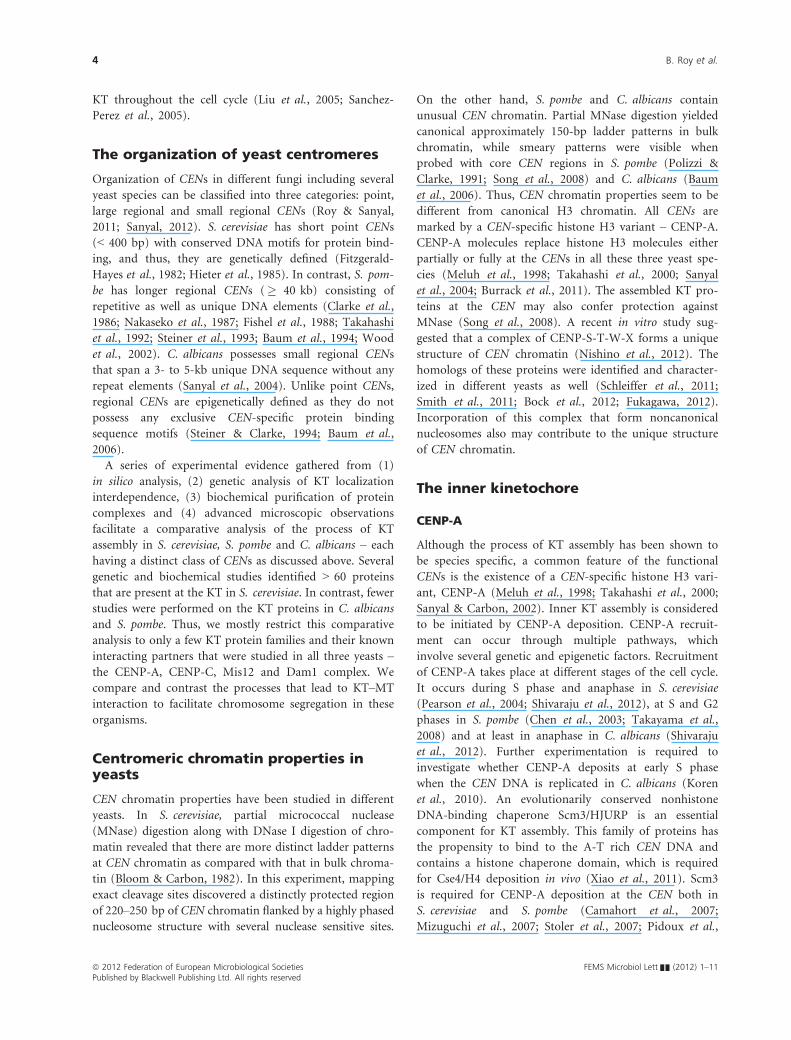
On the other hand, S. pombe and C. albicans contain
unusual CEN chromatin. Partial MNase digestion yielded
canonical approximately 150-bp ladder patterns in bulk
chromatin, while smeary patterns were visible when
probed with core CEN regions in S. pombe (Polizzi &
Clarke, 1991; Song et al., 2008) and C. albicans (Baum
et al., 2006). Thus, CEN chromatin properties seem to be
different from canonical H3 chromatin. All CENs are
marked by a CEN-specific histone H3 variant – CENP-A.
CENP-A molecules replace histone H3 molecules either
partially or fully at the CENs in all these three yeast spe-
cies (Meluh et al., 1998; Takahashi et al., 2000; Sanyal
et al., 2004; Burrack et al., 2011). The assembled KT pro-
teins at the CEN may also confer protection against
MNase (Song et al., 2008). A recent in vitro study sug-
gested that a complex of CENP-S-T-W-X forms a unique
structure of CEN chromatin (Nishino et al., 2012). The
homologs of these proteins were identified and character-
ized in different yeasts as well (Schleiffer et al., 2011;
Smith et al., 2011; Bock et al., 2012; Fukagawa, 2012).
Incorporation of this complex that form noncanonical
nucleosomes also may contribute to the unique structure
of CEN chromatin.
The inner kinetochore
CENP-A
Although the process of KT assembly has been shown to
be species specific, a common feature of the functional
CENs is the existence of a CEN-specific histone H3 vari-
ant, CENP-A (Meluh et al., 1998; Takahashi et al., 2000;
Sanyal & Carbon, 2002). Inner KT assembly is considered
to be initiated by CENP-A deposition. CENP-A recruit-
ment can occur through multiple pathways, which
involve several genetic and epigenetic factors. Recruitment
of CENP-A takes place at different stages of the cell cycle.
It occurs during S phase and anaphase in S. cerevisiae
(Pearson et al., 2004; Shivaraju et al., 2012), at S and G2
phases in S. pombe (Chen et al., 2003; Takayama et al.,
2008) and at least in anaphase in C. albicans (Shivaraju
et al., 2012). Further experimentation is required to
investigate whether CENP-A deposits at early S phase
when the CEN DNA is replicated in C. albicans (Koren
et al., 2010). An evolutionarily conserved nonhistone
DNA-binding chaperone Scm3/HJURP is an essential
component for KT assembly. This family of proteins has
the propensity to bind to the A-T rich CEN DNA and
contains a histone chaperone domain, which is required
for Cse4/H4 deposition in vivo (Xiao et al., 2011). Scm3
is required for CENP-A deposition at the CEN both in
S. cerevisiae and S. pombe (Camahort et al., 2007;
Mizuguchi et al., 2007; Stoler et al., 2007; Pidoux et al.,
ª 2012 Federation of European Microbiological Societies FEMS Microbiol Lett && (2012) 1–11Published by Blackwell Publishing Ltd. All rights reserved
4 B. Roy et al.

2009; Williams et al., 2009). Moreover, over-expression of
Scm3 results in a reduction in Cse4 at the CEN in S. cere-
visiae (Mishra et al., 2011). Although Scm3 is required
for Cse4 localization at the CEN, but its own localization
at the CEN is independent of Cse4 in both S. cerevisiae
and S. pombe (Williams et al., 2009; Luconi et al., 2011).
Similarly, another KT protein essential for CENP-A local-
ization is CENP-C. The localization of CENP-A is depen-
dent on CENP-C in both S. pombe (Tanaka et al., 2009)
and C. albicans (Thakur & Sanyal, 2012). In addition to
these proteins, epigenetic regulation of CENP-A deposi-
tion (reviewed in Roy & Sanyal, 2011) has been demon-
strated in S. pombe (Steiner & Clarke, 1994) and
C. albicans (Baum et al., 2006).
Ndc10, a part of the point CEN-specific CBF3 complex,
has been shown to influence the recruitment of most of
the KT proteins including CENP-A in S. cerevisiae (Ortiz
et al., 1999; Russell et al., 1999; Goshima & Yanagida,
2000; He et al., 2001; Janke et al., 2001, 2002). It is not
clear that Ndc10 is required only in S. cerevisiae because
an obvious homolog is not identified in S. pombe or
C. albicans. On the other hand, Ams2 at S phase (Chen
et al., 2003) and Hip1 at G2 phase (Takayama et al., 2008)
influence CENP-A loading in S. pombe. The cell cycle
phase–specific loading of CENP-A has also been shown to
be affected by Mis6 through Sim3 in S. pombe (Takahashi
et al., 2000; Dunleavy et al., 2007). Interestingly, proteins
from the middle and outer KT affect the localization of
CENP-A in C. albicans (Roy et al., 2011; Thakur & Sanyal,
2012). The Dam1 complex, a fungal-specific outer KT
protein complex, which has no known role in CENP-A
recruitment in S. cerevisiae or in S. pombe, influences the
localization and stability of CENP-A in C. albicans
(Thakur & Sanyal, 2012).
CENP-C
Members of the evolutionarily conserved CENP-C family
contain a c. 25-amino acid-long conserved region, known
as the CENP-C box, which is essential for its KT localiza-
tion (Meluh & Koshland, 1995; Yu et al., 2000; Suzuki
et al., 2004). CENP-C localization at the KT is mediated
by CENP-A in both S. cerevisiae (Westermann et al.,
2003) and S. pombe (Tanaka et al., 2009). CENP-C
requires Mis12 for its recruitment at the KT in both
S. cerevisiae (Westermann et al., 2003) and C. albicans
(Roy et al., 2011). Ndc10 and Nnf1 influence CENP-C
localization in S. cerevisiae (Meluh & Koshland, 1997;
Collins et al., 2005). However, the dependence of CENP-
C on Nnf1 has not been studied in S. pombe and C. albi-
cans. Interestingly, subunits of the Dam1 complex are
essential for CENP-C localization at the KT in C. albicans
(Thakur & Sanyal, 2012).
The middle kinetochore
The Ndc80-MIND-Spc105 (NMS) super-complex
The yeast counterpart of the KNL1-Mis12-Ndc80 (KMN)
network, identified in higher eukaryotes, consists of the
Ndc80 complex, MIND/Mis12 complex and Spc105/Spc7
complex.
The Ndc80 complex
The requirement of CENP-A for KT localization of the
Ndc80 complex is similar in budding yeasts, S. cerevisiae
(Collins et al., 2005) and C. albicans (Burrack et al.,
2011). Moreover, Cnn1/CENP-T and Ndc10 were
reported to influence the assembly of the Ndc80 complex
in S. cerevisiae (He et al., 2001; Janke et al., 2001;
Schleiffer et al., 2011; Bock et al., 2012; Nishino et al.,
2012). Middle KT components including Mis12 and Nnf1
were shown to affect the localization of this complex at
the KT (Westermann et al., 2003). In S. pombe, depen-
dence as well as localization of the Ndc80 complex is not
well established. The Dam1 complex subunits influence
the loading of Nuf2, a constituent of the Ndc80 complex
in C. albicans (Thakur & Sanyal, 2012).
The MIND/Mis12 complex
CENP-A plays an important role in recruiting Mis12 at
the KT both in S. cerevisiae (Pinsky et al., 2003; Wester-
mann et al., 2003; Collins et al., 2005) and C. albicans
(Burrack et al., 2011; Roy et al., 2011) but Mis12 and
CENP-A are independent of each other for their KT
recruitment in S. pombe (Takahashi et al., 2000).
Ndc10 is essential for the KT localization of each of
the constituents of the MIND complex in S. cerevisiae
(Goshima & Yanagida, 2000; Nekrasov et al., 2003; Pinsky
et al., 2003). KT localization of the Mis12 complex is
independent of Spc105 in S. cerevisiae (Pagliuca et al.,
2009) but Mis12, Mis13/Dsn1 and Mis14/Nsl1 require
Spc7 and Sos7 for their KT localization in S. pombe
(Kerres et al., 2007; Pagliuca et al., 2009; Jakopec et al.,
2012). Depletion of a subunit of the Dam1 complex
affects Mis12 localization in C. albicans (Thakur &
Sanyal, 2012).
The Spc105/Spc7 complex
The Spc105 complex of S. cerevisiae consists of two
subunits, which are Spc105 and Kre28. Ndc10 influences
KT recruitment of both the components of this complex
(Nekrasov et al., 2003; Pagliuca et al., 2009). The recruit-
ment of Spc105 at the KT is independent of the MIND
FEMS Microbiol Lett && (2012) 1–11 ª 2012 Federation of European Microbiological SocietiesPublished by Blackwell Publishing Ltd. All rights reserved
Kinetochore assembly in yeasts 5
and Ndc80 complex in S. cerevisiae (Pagliuca et al.,
2009).
However, Spc7/Spc105 forms complex with Sos7, which
has been identified recently as a KT protein in fission
yeast S. pombe (Jakopec et al., 2012). Spc7 and Sos7 are
interdependent for their KT localization (Jakopec et al.,
2012). Both the proteins are dependent on Mis12 for
their loading at the KT (Kerres et al., 2007; Jakopec et al.,
2012).
The outer kinetochore
The Dam1 complex
The Dam1 complex is essential for cell viability and local-
ized at the KT throughout cell cycle in both budding
yeasts, S. cerevisiae (Hofmann et al., 1998; Cheeseman
et al., 2001a, b; Enquist-Newman et al., 2001) and
C. albicans (Burrack et al., 2011; Thakur & Sanyal, 2011).
CENP-A influences the KT recruitment of this complex
in both the budding yeasts (Collins et al., 2005; Burrack
et al., 2011).
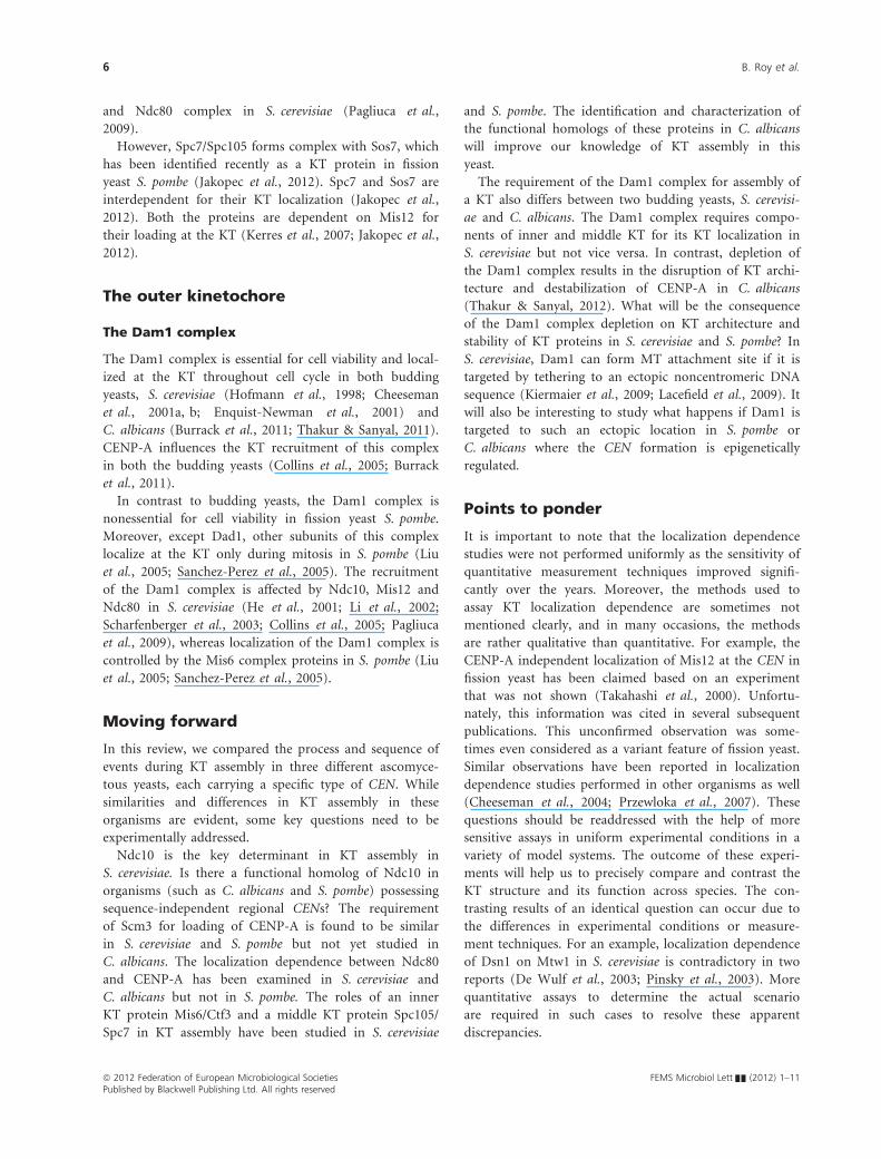
In contrast to budding yeasts, the Dam1 complex is
nonessential for cell viability in fission yeast S. pombe.
Moreover, except Dad1, other subunits of this complex
localize at the KT only during mitosis in S. pombe (Liu
et al., 2005; Sanchez-Perez et al., 2005). The recruitment
of the Dam1 complex is affected by Ndc10, Mis12 and
Ndc80 in S. cerevisiae (He et al., 2001; Li et al., 2002;
Scharfenberger et al., 2003; Collins et al., 2005; Pagliuca
et al., 2009), whereas localization of the Dam1 complex is
controlled by the Mis6 complex proteins in S. pombe (Liu
et al., 2005; Sanchez-Perez et al., 2005).
Moving forward
In this review, we compared the process and sequence of
events during KT assembly in three different ascomyce-
tous yeasts, each carrying a specific type of CEN. While
similarities and differences in KT assembly in these
organisms are evident, some key questions need to be
experimentally addressed.
Ndc10 is the key determinant in KT assembly in
S. cerevisiae. Is there a functional homolog of Ndc10 in
organisms (such as C. albicans and S. pombe) possessing
sequence-independent regional CENs? The requirement
of Scm3 for loading of CENP-A is found to be similar
in S. cerevisiae and S. pombe but not yet studied in
C. albicans. The localization dependence between Ndc80
and CENP-A has been examined in S. cerevisiae and
C. albicans but not in S. pombe. The roles of an inner
KT protein Mis6/Ctf3 and a middle KT protein Spc105/
Spc7 in KT assembly have been studied in S. cerevisiae
and S. pombe. The identification and characterization of
the functional homologs of these proteins in C. albicans
will improve our knowledge of KT assembly in this
yeast.
The requirement of the Dam1 complex for assembly of
a KT also differs between two budding yeasts, S. cerevisi-
ae and C. albicans. The Dam1 complex requires compo-
nents of inner and middle KT for its KT localization in
S. cerevisiae but not vice versa. In contrast, depletion of
the Dam1 complex results in the disruption of KT archi-
tecture and destabilization of CENP-A in C. albicans
(Thakur & Sanyal, 2012). What will be the consequence
of the Dam1 complex depletion on KT architecture and
stability of KT proteins in S. cerevisiae and S. pombe? In
S. cerevisiae, Dam1 can form MT attachment site if it is
targeted by tethering to an ectopic noncentromeric DNA
sequence (Kiermaier et al., 2009; Lacefield et al., 2009). It
will also be interesting to study what happens if Dam1 is
targeted to such an ectopic location in S. pombe or
C. albicans where the CEN formation is epigenetically
regulated.
Points to ponder
It is important to note that the localization dependence
studies were not performed uniformly as the sensitivity of
quantitative measurement techniques improved signifi-
cantly over the years. Moreover, the methods used to
assay KT localization dependence are sometimes not
mentioned clearly, and in many occasions, the methods
are rather qualitative than quantitative. For example, the
CENP-A independent localization of Mis12 at the CEN in
fission yeast has been claimed based on an experiment
that was not shown (Takahashi et al., 2000). Unfortu-
nately, this information was cited in several subsequent
publications. This unconfirmed observation was some-
times even considered as a variant feature of fission yeast.
Similar observations have been reported in localization
dependence studies performed in other organisms as well
(Cheeseman et al., 2004; Przewloka et al., 2007). These
questions should be readdressed with the help of more
sensitive assays in uniform experimental conditions in a
variety of model systems. The outcome of these experi-
ments will help us to precisely compare and contrast the
KT structure and its function across species. The con-
trasting results of an identical question can occur due to
the differences in experimental conditions or measure-
ment techniques. For an example, localization dependence
of Dsn1 on Mtw1 in S. cerevisiae is contradictory in two
reports (De Wulf et al., 2003; Pinsky et al., 2003). More
quantitative assays to determine the actual scenario
are required in such cases to resolve these apparent
discrepancies.
ª 2012 Federation of European Microbiological Societies FEMS Microbiol Lett && (2012) 1–11Published by Blackwell Publishing Ltd. All rights reserved
6 B. Roy et al.

Concluding remarks
It is evident that although most of the proteins assemble at
the CEN are functionally conserved across species, the CEN
DNA is diverged even in closely related species. Compara-
tive genomic analyses in different yeasts revealed that the
CEN DNA is hyper-variable even in closely related species
(Bensasson et al., 2008; Padmanabhan et al., 2008; Rhind
et al., 2011). The phenomenon of hyper-variability of the
DNA sequence at the CEN despite its conserved function in
chromosome segregation was previously designated as the
‘centromere paradox’ (Henikoff et al., 2001).
In this review, we analysed the similarities and differ-
ences in the process of KT assembly in yeasts. While the
(a) (b) (c)
Fig. 2. The recruitment of proteins during KT assembly in yeasts. The recruitment of various proteins and their interaction during formation of
the KT structure has been shown in (a) Saccharomyces cerevisiae, (b) Schizosaccharomyces pombe and (c) Candida albicans. Top panels show
relative presence of various KT proteins from outer to inner layers (top to bottom). Arrows indicate the localization dependence of a protein/
protein complex to another during the assembly process. Arrowheads point towards the component, which is dependent on the other protein.
Bottom panels show composition of various major KT complexes. Dotted arrows show intracomplex dependency. The double-headed arrows
indicate mutual dependency between proteins and protein complexes. The interdependence of various KT proteins shown here is based on the
data assembled from the literature – for the S. cerevisiae KT (Meluh & Koshland, 1997; Ortiz et al., 1999; Russell et al., 1999; Goshima &
Yanagida, 2000; He et al., 2001; Janke et al., 2001, 2002; Li et al., 2002; De Wulf et al., 2003; Nekrasov et al., 2003; Pinsky et al., 2003;
Scharfenberger et al., 2003; Westermann et al., 2003; Collins et al., 2005; Camahort et al., 2007; Mizuguchi et al., 2007; Pagliuca et al., 2009;
Schleiffer et al., 2011; Bock et al., 2012; Nishino et al., 2012), for the S. pombe KT (Takahashi et al., 2000; Chen et al., 2003; Hayashi et al.,
2004; Liu et al., 2005; Saitoh et al., 2005; Sanchez-Perez et al., 2005; Kerres et al., 2006, 2007; Dunleavy et al., 2007; Takayama et al., 2008;
Pidoux et al., 2009; Tanaka et al., 2009; Williams et al., 2009; Jakopec et al., 2012) and for the C. albicans KT (Burrack et al., 2011; Roy et al.,
2011; Thakur & Sanyal, 2011, 2012).
FEMS Microbiol Lett && (2012) 1–11 ª 2012 Federation of European Microbiological SocietiesPublished by Blackwell Publishing Ltd. All rights reserved
Kinetochore assembly in yeasts 7
organization of a KT is conserved, there appears to be
subtle divergence in regulation of KT assembly in these
organisms. Whether this process has evolved uniquely in
different organisms to keep pace with the fast evolving
CEN DNA is not clear. If the process of KT assembly
occurs in a step-wise manner, a master regulator may
control the recruitment of most of the KT proteins. In
fact, an inner KT protein Ndc10 plays the central role in
S. cerevisiae (Fig. 2a), while the middle KT proteins –Mis6 and Spc7 – play governing roles to a great extent in
S. pombe (Fig. 2b). This process is remarkably diverged
with a complex interdependence among many essential
KT proteins from various layers in C. albicans (Fig. 2c).
Unravelling this fascinating molecular mechanism of KT
assembly in many organisms will improve our under-
standing of how the KT assembly pathways coevolved
with the CEN DNA during speciation.
Acknowledgements
We thank B. Suma (Central instrumentation facility,
Molecular Biology and Genetics Unit, Jawaharlal Nehru
Centre for Advanced Scientific Research) for confocal
microscopy and image processing. We are thankful to the
members of Sanyal laboratory for insightful comments.
We express our regret to our colleagues whose work
could not be cited due to space limitations.
References
Anderson M, Haase J, Yeh E & Bloom K (2009) Function and
assembly of DNA looping, clustering, and microtubule
attachment complexes within a eukaryotic kinetochore. Mol
Biol Cell 20: 4131–4139.Aoki K, Hayashi H, Furuya K, Sato M, Takagi T, Osumi M,
Kimura A & Niki H (2011) Breakage of the nuclear
envelope by an extending mitotic nucleus occurs during
anaphase in Schizosaccharomyces japonicus. Genes Cells 16:
911–926.Asakawa H, Kojidani T, Mori C, Osakada H, Sato M, Ding DQ,
Hiraoka Y & Haraguchi T (2010) Virtual breakdown of the
nuclear envelope in fission yeast meiosis. Curr Biol 20: 1919–1925.
Asakawa H, Hiraoka Y & Haraguchi T (2011) Physical
breakdown of the nuclear envelope is not necessary for
breaking its barrier function. Nucleus 2: 523–526.Baum M, Ngan VK & Clarke L (1994) The centromeric K-type
repeat and the central core are together sufficient to
establish a functional Schizosaccharomyces pombe
centromere. Mol Biol Cell 5: 747–761.Baum M, Sanyal K, Mishra PK, Thaler N & Carbon J (2006)
Formation of functional centromeric chromatin is specified
epigenetically in Candida albicans. Proc Natl Acad Sci 103:
14877–14882.
Bensasson D, Zarowiecki M, Burt A & Koufopanou V (2008)
Rapid evolution of yeast centromeres in the absence of
drive. Genetics 178: 2161–2167.Bloom KS & Carbon J (1982) Yeast centromere DNA is in a
unique and highly ordered structure in chromosomes and
small circular minichromosomes. Cell 29: 305–317.Bock LJ, Pagliuca C, Kobayashi N et al. (2012) Cnn1 inhibits
the interactions between the KMN complexes of the yeast
kinetochore. Nat Cell Biol 14: 614–624.Brinkley BR & Stubblefield E (1966) The fine structure of the
kinetochore of a mammalian cell in vitro. Chromosoma 19:
28–43.Burrack LS, SE A & Berman J (2011) The requirement for the
Dam1 complex is dependent upon the number of kinetochore
proteins and microtubules. Curr Biol 21: 889–896.Byers B & Goetsch L (1975) Behavior of spindles and spindle
plaques in the cell cycle and conjugation of Saccharomyces
cerevisiae. J Bacteriol 124: 511–523.Camahort R, Li B, Florens L, Swanson SK, Washburn MP &
Gerton JL (2007) Scm3 is essential to recruit the histone H3
variant Cse4 to centromeres and to maintain a functional
kinetochore. Mol Cell 26: 853–865.Cheeseman IM & Desai A (2008) Molecular architecture of the
kinetochore-microtubule interface. Nat Rev Mol Cell Biol 9:
33–46.Cheeseman IM, Enquist-Newman M, T M-R, Drubin DG &
Barnes G (2001a) Mitotic spindle integrity and kinetochore
function linked by the Duo1p/Dam1p complex. J Cell Biol
152: 197–212.Cheeseman IM, Brew C, Wolyniak M, Desai A, Anderson S,
Muster N, Yates JR, Huffaker TC, Drubin DG & Barnes G
(2001b) Implication of a novel multiprotein Dam1p
complex in outer kinetochore function. J Cell Biol 155:
1137–1146.Cheeseman IM, Niessen S, Anderson S, Hyndman F, Yates JR,
Oegema K & Desai A (2004) A conserved protein network
controls assembly of the outer kinetochore and its ability to
sustain tension. Genes Dev 18: 2255–2268.Cheeseman IM, Hori T, Fukagawa T & Desai A (2008) KNL1
and the CENP-H/I/K complex coordinately direct
kinetochore assembly in vertebrates. Mol Biol Cell 19:
587–594.Chen ES, Saitoh S, Yanagida M & Takahashi K (2003) A cell
cycle-regulated GATA factor promotes centromeric
localization of CENP-A in fission yeast. Mol Cell 11: 175–187.Clarke L, Amstutz H, Fishel B & Carbon J (1986) Analysis of
centromeric DNA in the fission yeast Schizosaccharomyces
pombe. Proc Natl Acad Sci 83: 8253–8257.Cleveland DW, Mao Y & Sullivan KF (2003) Centromeres and
kinetochores: from epigenetics to mitotic checkpoint
signaling. Cell 112: 407–421.Collins KA, Castillo AR, Tatsutani SY & Biggins S (2005)
De novo kinetochore assembly requires the centromeric
histone H3 variant. Mol Biol Cell 16: 5649–5660.De Souza CP & Osmani SA (2007) Mitosis, not just open or
closed. Eukaryot Cell 6: 1521–1527.
ª 2012 Federation of European Microbiological Societies FEMS Microbiol Lett && (2012) 1–11Published by Blackwell Publishing Ltd. All rights reserved
8 B. Roy et al.
De Wulf P, McAinsh AD & Sorger PK (2003) Hierarchical
assembly of the budding yeast kinetochore from multiple
subcomplexes. Genes Dev 17: 2902–2921.Ding R, McDonald KL & McIntosh JR (1993) Three-
dimensional reconstruction and analysis of mitotic spindles
from the yeast, Schizosaccharomyces pombe. J Cell Biol 120:
141–151.Ding R, West RR, Morphew DM, Oakley BR & McIntosh JR
(1997) The spindle pole body of Schizosaccharomyces pombe
enters and leaves the nuclear envelope as the cell cycle
proceeds. Mol Biol Cell 8: 1461–1479.Duan Z, Andronescu M, Schutz K, McIlwain S, Kim YJ, Lee C,
Shendure J, Fields S, Blau CA & Noble WS (2010) A three-
dimensional model of the yeast genome. Nature 465:
363–367.Dunleavy EM, Pidoux AL, Monet M, Bonilla C, Richardson W,
Hamilton GL, Ekwall K, McLaughlin PJ & Allshire RC
(2007) A NASP (N1/N2)-related protein, Sim3, binds CENP-
A and is required for its deposition at fission yeast
centromeres. Mol Cell 28: 1029–1044.Enquist-Newman M, Cheeseman IM, Van Goor D, Drubin
DG, Meluh PB & Barnes G (2001) Dad1p, third component
of the Duo1p/Dam1p complex involved in kinetochore
function and mitotic spindle integrity. Mol Biol Cell 12:
2601–2613.Fishel B, Amstutz H, Baum M, Carbon J & Clarke L (1988)
Structural organization and functional analysis of
centromeric DNA in the fission yeast Schizosaccharomyces
pombe. Mol Cell Biol 8: 754–763.Fitzgerald-Hayes M, Clarke L & Carbon J (1982) Nucleotide
sequence comparisons and functional analysis of yeast
centromere DNAs. Cell 29: 235–244.Foltz DR, Jansen LET, Black BE, Bailey AO, Yates JR &
Cleveland DW (2006) The human CENP-A centromeric
nucleosome-associated complex. Nat Cell Biol 8:
458–469.Fukagawa T (2012) Formation of a centromere-specific
chromatin structure. Epigenetics 7: 672–675.Goshima G & Yanagida M (2000) Establishing biorientation
occurs with precocious separation of the sister kinetochores,
but not the arms, in the early spindle of budding yeast. Cell
100: 619–633.Goshima G, Saitoh S & Yanagida M (1999) Proper metaphase
spindle length is determined by centromere proteins Mis12
and Mis6 required for faithful chromosome segregation.
Genes Dev 13: 1664–1677.Guttinger S, Laurell E & Kutay U (2009) Orchestrating nuclear
envelope disassembly and reassembly during mitosis. Nat
Rev Mol Cell Biol 10: 178–191.Hayashi T, Fujita Y, Iwasaki O, Adachi Y, Takahashi K &
Yanagida M (2004) Mis16 and Mis18 are required for
CENP-A loading and histone deacetylation at centromeres.
Cell 118: 715–729.He X, Rines DR, Espelin CW & Sorger PK (2001) Molecular
analysis of kinetochore-microtubule attachment in budding
yeast. Cell 106: 195–206.
Henikoff S, Ahmad K & Malik HS (2001) The centromere
paradox: stable inheritance with rapidly evolving DNA.
Science 293: 1098–1102.Hieter P, Pridmore D, Hegemann JH, Thomas M, Davis RW
& Philippsen P (1985) Functional selection and analysis of
yeast centromeric DNA. Cell 42: 913–921.Hofmann C, Cheeseman IM, Goode BL, McDonald KL, Barnes
G & Drubin DG (1998) Saccharomyces cerevisiae Duo1p and
Dam1p, novel proteins involved in mitotic spindle function.
J Cell Biol 143: 1029–1040.Jakopec V, Topolski B & Fleig U (2012) Sos7, an essential
component of the conserved Schizosaccharomyces pombe
Ndc80-MIND-Spc7 complex, identifies a new family of
fungal kinetochore proteins. Mol Cell Biol 32: 3308–3320.Janke C, Ortiz J, Lechner J, Shevchenko A, Shevchenko A,
Magiera MM, Schramm C & Schiebel E (2001) The budding
yeast proteins Spc24p and Spc25p interact with Ndc80p and
Nuf2p at the kinetochore and are important for kinetochore
clustering and checkpoint control. EMBO J 20: 777–791.Janke C, Ortiz J, Tanaka TU, Lechner J & Schiebel E (2002)
Four new subunits of the Dam1-Duo1 complex reveal novel
functions in sister kinetochore biorientation. EMBO J 21:
181–193.Jaspersen SL & Winey M (2004) The budding yeast spindle
pole body: structure, duplication, and function. Annu Rev
Cell Dev Biol 20: 1–28.Joglekar AP, Bouck D, Finley K, Liu X, Wan Y, Berman J, He X,
Salmon ED & Bloom KS (2008) Molecular architecture of the
kinetochore-microtubule attachment site is conserved
between point and regional centromeres. J Cell Biol 181: 587–594.
Joglekar AP, Bloom K & Salmon ED (2009) In vivo protein
architecture of the eukaryotic kinetochore with nanometer
scale accuracy. Curr Biol 19: 694–699.Kerres A, Jakopec V, Beuter C, Karig I, Pohlmann J, Pidoux A,
Allshire R & Fleig U (2006) Fta2, an essential fission yeast
kinetochore component, interacts closely with the conserved
Mal2 protein. Mol Biol Cell 17: 4167–4178.Kerres A, Jakopec V & Fleig U (2007) The conserved Spc7
protein is required for spindle integrity and links
kinetochore complexes in fission yeast. Mol Biol Cell 18:
2441–2454.Kiermaier E, Woehrer S, Peng Y, Mechtler K & Westermann S
(2009) A Dam1-based artificial kinetochore is sufficient to
promote chromosome segregation in budding yeast. Nat
Cell Biol 11: 1109–1115.Kitamura E, Tanaka K, Kitamura Y & Tanaka TU (2007)
Kinetochore-microtubule interaction during S phase in
Saccharomyces cerevisiae. Genes Dev 21: 3319–3330.Koren A, Tsai HJ, Tirosh I, Burrack LS, Barkai N & Berman J
(2010) Epigenetically-inherited centromere and
neocentromere DNA replicates earliest in S-phase. PLoS
Genet 6: e1001068.
Lacefield S, Lau DTC & Murray AW (2009) Recruiting a
microtubule-binding complex to DNA directs chromosome
segregation in budding yeast. Nat Cell Biol 11: 1116–1120.
FEMS Microbiol Lett && (2012) 1–11 ª 2012 Federation of European Microbiological SocietiesPublished by Blackwell Publishing Ltd. All rights reserved
Kinetochore assembly in yeasts 9
Li Y, Bachant J, Alcasabas AA, Wang Y, Qin J & Elledge SJ
(2002) The mitotic spindle is required for loading of the
DASH complex onto the kinetochore. Genes Dev 16: 183–197.Liu X, McLeod I, Anderson S, Yates JR & He X (2005)
Molecular analysis of kinetochore architecture in fission
yeast. EMBO J 24: 2919–2930.Liu S-T, Rattner JB, Jablonski SA & Yen TJ (2006) Mapping
the assembly pathways that specify formation of the
trilaminar kinetochore plates in human cells. J Cell Biol 175:
41–53.Luconi L, Araki Y, Erlemann S & Schiebel E (2011) The
CENP-A chaperone Scm3 becomes enriched at kinetochores
in anaphase. Cell Cycle 10: 3369–3378.McDonald KL, O’Toole ET, Mastronarde DN & McIntosh JR
(1992) Kinetochore microtubules in PTK cells. J Cell Biol
118: 369–383.McEwen BF, Dong Y, VandenBeldt KJ & McIntosh JR (2007)
Using electron microscopy to understand functional
mechanisms of chromosome alignment on the mitotic
spindle. Methods Cell Biol 79: 259–293. Academic Press.
Meluh PB & Koshland D (1995) Evidence that the MIF2 gene
of Saccharomyces cerevisiae encodes a centromere protein
with homology to the mammalian centromere protein
CENP-C. Mol Biol Cell 6: 793–807.Meluh PB & Koshland D (1997) Budding yeast centromere
composition and assembly as revealed by in vivo cross-
linking. Genes Dev 11: 3401–3412.Meluh PB, Yang P, Glowczewski L, Koshland D & Smith MM
(1998) Cse4p is a component of the core centromere of
Saccharomyces cerevisiae. Cell 94: 607–613.Mishra PK, Au W-C, Choy JS, Kuich PH, Baker RE, Foltz DR
& Basrai MA (2011) Misregulation of Scm3p/HJURP causes
chromosome instability in Saccharomyces cerevisiae and
human cells. PLoS Genet 7: e1002303.
Mizuguchi G, Xiao H, Wisniewski J, Smith MM & Wu C
(2007) Nonhistone Scm3 and histones CenH3-H4 assemble
the core of centromere-specific nucleosomes. Cell 129:
1153–1164.Nakaseko Y, Kinoshita N & Yanagida M (1987) A novel
sequence common to the centromere regions of
Schizosaccharomyces pombe chromosomes. Nucleic Acids Res
15: 4705–4715.Nasmyth K (2001) Disseminating the genome: joining,
resolving, and separating sister chromatids during mitosis
and meiosis. Annu Rev Genet 35: 745.
Nekrasov VS, Smith MA, Peak-Chew S & Kilmartin JV (2003)
Interactions between centromere complexes in
Saccharomyces cerevisiae. Mol Biol Cell 14: 4931–4946.Nishino T, Takeuchi K, Gascoigne KE, Suzuki A, Hori T,
Oyama T, Morikawa K, Cheeseman IM & Fukagawa T
(2012) CENP-T-W-S-X forms a unique centromeric
chromatin structure with a histone-like fold. Cell 148: 487–501.
Okada M, Cheeseman IM, Hori T, Okawa K, McLeod IX,
Yates JR III, Desai A & Fukagawa T (2006) The CENP-H-I
complex is required for the efficient incorporation of newly
synthesized CENP-A into centromeres. Nat Cell Biol 8:
446–457.Ortiz J, Stemmann O, Rank S & Lechner J (1999) A putative
protein complex consisting of Ctf19, Mcm21, and Okp1
represents a missing link in the budding yeast kinetochore.
Genes Dev 13: 1140–1155.Padmanabhan S, Thakur J, Siddharthan R & Sanyal K
(2008) Rapid evolution of Cse4p-rich centromeric DNA
sequences in closely related pathogenic yeasts, Candida
albicans and Candida dubliniensis. Proc Natl Acad Sci 105:
19797–19802.Pagliuca C, Draviam VM, Marco E, Sorger PK & De Wulf P
(2009) Roles for the conserved Spc105p/Kre28p complex in
kinetochore-microtubule binding and the spindle assembly
checkpoint. PLoS ONE 4: e7640.
Pearson CG, Yeh E, Gardner M, Odde D, Salmon ED &
Bloom K (2004) Stable kinetochore-microtubule attachment
constrains centromere positioning in metaphase. Curr Biol
14: 1962–1967.Pidoux AL, Choi ES, Abbott JKR et al. (2009) Fission yeast
Scm3: a CENP-A receptor required for integrity of
subkinetochore chromatin. Mol Cell 33: 299–311.Pinsky BA, Tatsutani SY, Collins KA & Biggins S (2003) An
Mtw1 complex promotes kinetochore biorientation that is
monitored by the Ipl1/Aurora protein kinase. Dev Cell 5:
735–745.Polizzi C & Clarke L (1991) The chromatin structure of
centromeres from fission yeast: differentiation of the central
core that correlates with function. J Cell Biol 112: 191–201.Przewloka MR, Zhang W, Costa P, Archambault V, D’Avino
PP, Lilley KS, Laue ED, McAinsh AD & Glover DM (2007)
Molecular analysis of core kinetochore composition and
assembly in Drosophila melanogaster. PLoS ONE 2: e478.
Rhind N, Chen Z, Yassour M et al. (2011) Comparative
functional genomics of the fission yeasts. Science 332:
930–936.Roy B & Sanyal K (2011) Diversity in requirement of genetic
and epigenetic factors for centromere function in fungi.
Eukaryot Cell 10: 1384–1395.Roy B, Burrack LS, Lone MA, Berman J & Sanyal K (2011)
CaMtw1, a member of the evolutionarily conserved Mis12
kinetochore protein family, is required for efficient inner
kinetochore assembly in the pathogenic yeast Candida
albicans. Mol Microbiol 80: 14–32.Russell ID, Grancell AS & Sorger PK (1999) The unstable F-box
protein p58-Ctf13 forms the structural core of the CBF3
kinetochore complex. J Cell Biol 145: 933–950.Saitoh S, Ishii K, Kobayashi Y & Takahashi K (2005) Spindle
checkpoint signaling requires the Mis6 kinetochore
subcomplex, which interacts with Mad2 and mitotic
spindles. Mol Biol Cell 16: 3666–3677.Sanchez-Perez I, Renwick SJ, Crawley K, Karig I, Buck V,
Meadows JC, Franco-Sanchez A, Fleig U, Toda T & Millar
JB (2005) The DASH complex and Klp5/Klp6 kinesin
coordinate bipolar chromosome attachment in fission yeast.
EMBO J 24: 2931–2943.
ª 2012 Federation of European Microbiological Societies FEMS Microbiol Lett && (2012) 1–11Published by Blackwell Publishing Ltd. All rights reserved
10 B. Roy et al.

Sanyal K (2012) How do microbial pathogens make CENs?
PLoS Pathog 8: e1002463.
Sanyal K & Carbon J (2002) The CENP-A homolog CaCse4p
in the pathogenic yeast Candida albicans is a centromere
protein essential for chromosome transmission. P Natl Acad
Sci USA 99: 12969–12974.Sanyal K, Baum M & Carbon J (2004) Centromeric DNA
sequences in the pathogenic yeast Candida albicans are all
different and unique. P Natl Acad Sci USA 101:
11374–11379.Sazer S (2005) Nuclear envelope: nuclear pore complexity.
Curr Biol 15: R23–R26.Scharfenberger M, Ortiz J, Grau N, Janke C, Schiebel E &
Lechner J (2003) Nsl1p is essential for the establishment of
bipolarity and the localization of the Dam-Duo complex.
EMBO J 22: 6584–6597.Schleiffer A, Maier M, Litos G, Lampert F, Hornung P,
Mechtler K & Westermann S (2011) CENP-T proteins are
conserved centromere receptors of the Ndc80 complex. Nat
Cell Biol 14: 604–613.Shivaraju M, Unruh JR, Slaughter BD, Mattingly M, Berman J
& Gerton JL (2012) Cell-cycle-coupled structural oscillation
of centromeric nucleosomes in yeast. Cell 150: 304–316.Smith KM, Phatale PA, Sullivan CM, Pomraning KR & Freitag
M (2011) Heterochromatin is required for normal
distribution of Neurospora crassa CenH3. Mol Cell Biol 31:
2528–2542.Song JS, Liu X, Liu XS & He X (2008) A high-resolution map
of nucleosome positioning on a fission yeast centromere.
Genome Res 18: 1064–1072.Steiner NC & Clarke L (1994) A novel epigenetic effect can
alter centromere function in fission yeast. Cell 79: 865–874.Steiner NC, Hahnenberger KM & Clarke L (1993)
Centromeres of the fission yeast Schizosaccharomyces pombe
are highly variable genetic loci. Mol Cell Biol 13: 4578–4587.Stoler S, Rogers K, Weitze S, Morey L, Fitzgerald-Hayes M &
Baker RE (2007) Scm3, an essential Saccharomyces cerevisiae
centromere protein required for G2/M progression and Cse4
localization. P Natl Acad Sci USA 104: 10571–10576.Straight AF, Marshall WF, Sedat JW & Murray AW (1997)
Mitosis in living budding yeast: anaphase A but no
metaphase plate. Science 277: 574–578.Suzuki N, Nakano M, Nozaki N, Egashira S-i, Okazaki T &
Masumoto H (2004) CENP-B interacts with CENP-C
domains containing Mif2 regions responsible for centromere
localization. J Biol Chem 279: 5934–5946.Takahashi K, Murakami S, Chikashige Y, Funabiki H, Niwa O
& Yanagida M (1992) A low copy number central sequence
with strict symmetry and unusual chromatin structure in
fission yeast centromere. Mol Biol Cell 3: 819–835.
Takahashi K, Chen ES & M Y (2000) Requirement of Mis6
centromere connector for localizing a CENP-A-like protein
in fission yeast. Science 288: 2215–2219.Takayama Y, Sato H, Saitoh S, Ogiyama Y, Masuda F &
Takahashi K (2008) Biphasic incorporation of centromeric
histone CENP-A in fission yeast. Mol Biol Cell 19: 682–690.Tanaka K & Tanaka TU (2009) Live cell imaging of
kinetochore capture by microtubules in budding yeast.
Methods in Molecular Biology, Vol. 545 (McAnish AD, ed.),
pp. 233–242. Humana Press.
Tanaka K, Li Chang H, Kagami A & Watanabe Y (2009) CENP-C
functions as a scaffold for effectors with essential kinetochore
functions in mitosis and meiosis. Dev Cell 17: 334–343.Thakur J & Sanyal K (2011) The essentiality of the fungus-
specific Dam1 complex is correlated with a one-
kinetochore-one-microtubule interaction present throughout
the cell cycle, independent of the nature of a centromere.
Eukaryot Cell 10: 1295–1305.Thakur J & Sanyal K (2012) A coordinated interdependent
protein circuitry stabilizes the kinetochore ensemble to
protect CENP-A in the human pathogenic yeast Candida
albicans. PLoS Genet 8: e1002661.
Wan X, O’Quinn RP, Pierce HL et al. (2009) Protein
architecture of the human kinetochore microtubule
attachment site. Cell 137: 672–684.Westermann S, Cheeseman IM, Anderson S, Yates JR, Drubin
DG & Barnes G (2003) Architecture of the budding yeast
kinetochore reveals a conserved molecular core. J Cell Biol
163: 215–222.Williams JS, Hayashi T, Yanagida M & Russell P (2009)
Fission yeast Scm3 mediates stable assembly of Cnp1/CENP-
A into centromeric chromatin. Mol Cell 33: 287–298.Winey M, Mamay CL, O’Toole ET, Mastronarde DN,
Giddings TH, McDonald KL & McIntosh JR (1995) Three-
dimensional ultrastructural analysis of the Saccharomyces
cerevisiae mitotic spindle. J Cell Biol 129: 1601–1615.Wood V, Gwilliam R, Rajandream MA et al. (2002) The
genome sequence of Schizosaccharomyces pombe. Nature 415:
871–880.Xiao H, Mizuguchi G, Wisniewski J, Huang Y, Wei D & Wu
C (2011) Nonhistone Scm3 binds to AT-rich DNA to
organize atypical centromeric nucleosome of budding yeast.
Mol Cell 43: 369–380.Yam C, He Y, Zhang D, Chiam KH & Oliferenko S (2011)
Divergent strategies for controlling the nuclear membrane
satisfy geometric constraints during nuclear division. Curr
Biol 21: 1314–1319.Yu H-G, Dawe RK & Hiatt EN (2000) The plant kinetochore.
Trends Plant Sci 5: 543–547.
FEMS Microbiol Lett && (2012) 1–11 ª 2012 Federation of European Microbiological SocietiesPublished by Blackwell Publishing Ltd. All rights reserved
Kinetochore assembly in yeasts 11
Copyright © 2022 FDOKUMEN