Copper tolerant yeasts isolated from polluted area of Argentina
Upload
univ-brestCategory
view
1download
0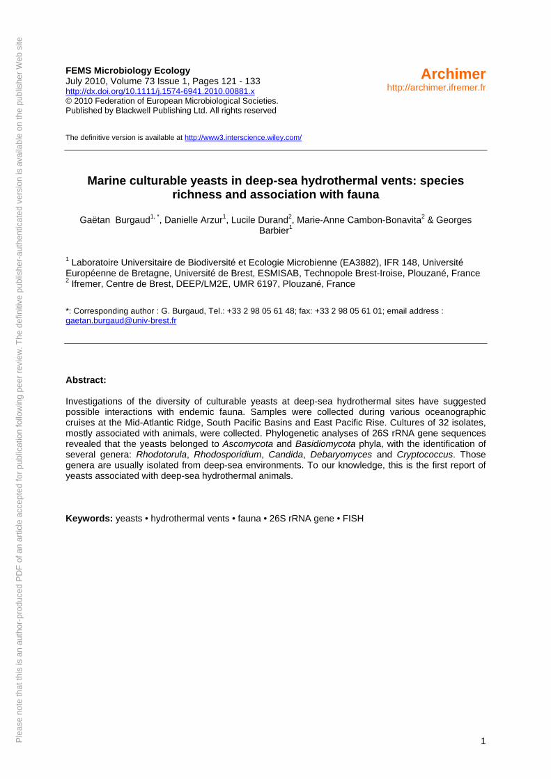
Ple
ase
note
that
this
is a
n au
thor
-pro
duce
d P
DF
of a
n ar
ticle
acc
ept
ed fo
r pu
blic
atio
n fo
llow
ing
peer
rev
iew
. The
def
initi
ve p
ub
lish
er-a
uthe
ntic
ated
ve
rsio
n is
ava
ilab
le o
n th
e pu
blis
her
Web
site
1
FEMS Microbiology Ecology July 2010, Volume 73 Issue 1, Pages 121 - 133 http://dx.doi.org/10.1111/j.1574-6941.2010.00881.x © 2010 Federation of European Microbiological Societies. Published by Blackwell Publishing Ltd. All rights reserved The definitive version is available at http://www3.interscience.wiley.com/
Archimerhttp://archimer.ifremer.fr
Marine culturable yeasts in deep-sea hydrothermal vents: species richness and association with fauna
Gaëtan Burgaud1, *, Danielle Arzur1, Lucile Durand2, Marie-Anne Cambon-Bonavita2 & Georges
Barbier1 1 Laboratoire Universitaire de Biodiversité et Ecologie Microbienne (EA3882), IFR 148, Université Européenne de Bretagne, Université de Brest, ESMISAB, Technopole Brest-Iroise, Plouzané, France 2 Ifremer, Centre de Brest, DEEP/LM2E, UMR 6197, Plouzané, France *: Corresponding author : G. Burgaud, Tel.: +33 2 98 05 61 48; fax: +33 2 98 05 61 01; email address : [email protected]
Abstract: Investigations of the diversity of culturable yeasts at deep-sea hydrothermal sites have suggested possible interactions with endemic fauna. Samples were collected during various oceanographic cruises at the Mid-Atlantic Ridge, South Pacific Basins and East Pacific Rise. Cultures of 32 isolates, mostly associated with animals, were collected. Phylogenetic analyses of 26S rRNA gene sequences revealed that the yeasts belonged to Ascomycota and Basidiomycota phyla, with the identification of several genera: Rhodotorula, Rhodosporidium, Candida, Debaryomyces and Cryptococcus. Those genera are usually isolated from deep-sea environments. To our knowledge, this is the first report of yeasts associated with deep-sea hydrothermal animals. Keywords: yeasts • hydrothermal vents • fauna • 26S rRNA gene • FISH
Ple
ase
note
that
this
is a
n au
thor
-pro
duce
d P
DF
of a
n ar
ticle
acc
ept
ed fo
r pu
blic
atio
n fo
llow
ing
peer
rev
iew
. The
def
initi
ve p
ub
lish
er-a
uthe
ntic
ated
ve
rsio
n is
ava
ilab
le o
n th
e pu
blis
her
Web
site
2
Introduction Yeasts are ubiquitous microorganisms that represent a part of the microbiota in all
natural ecosystems, such as soils, freshwaters and marine waters from the ocean
surface to the deep sea. Marine yeasts are divided into obligate and facultative
groups. Obligate marine yeasts are yeasts that have never been isolated from
anywhere other than the marine environment, whereas facultative marine yeasts are
also known from terrestrial habitats (Kohlmeyer & Kohlmeyer, 1979). Based on these
definitions, Kohlmeyer & Kohlmeyer (1979) examined yeasts occurring in marine
environments and gathered a list of 176 species isolated from diverse marine
habitats. Of those, only 25 were obligate marine yeasts, widely represented by the
genera Metschnikowia, Rhodosporidium, Candida and Torulopsis.
Hawksworth (2002) hypothesized the existence of 1.5 million fungal species; this
estimate is now a commonly used and accepted figure. If this is correct, <5% of the
fungi have been described up to now and these almost exclusively from terrestrial
environments. In that ecosystem, fungi are known to utilize a wide spectrum of simple
and more complex organic compounds. The decomposition activities of fungi are
clearly important in relation to the redistribution of elements among organisms and
environmental compartments (Gadd, 2007). Bearing in mind these parameters, our
hypothesis is that deep sea and especially hydrothermal vents, which remain
underexplored habitats for fungi, could be ecological niches hosting specific fungal
communities.
Deep-sea hydrothermal vents are localized at seafloor spreading centers called rifts,
where seawater seeps into cracked regions caused by the presence of hot basalt
and magma.
For Peer Review
Seawater carrying dissolved minerals is then emitted from springs. Two major types of 57
emissions have been found. Warm fluids diffuse at temperatures ranging from 6 to 23°C into 58
seawater at 2-4°C when hot vents called black smockers emit hydrothermal fluid at 270-59
380°C (Munn, 2003). Thermal gradients in hydrothermal vents are so important that just a 60
few centimeters away, the temperature can fall to 2-4°C allowing mesophilic or psychrophilic 61
organisms as well as thermophilic and hyperthermophilic prokaryotes to grow and interact 62
with all biotic or abiotic components of these ecosystems. Dense animal communities cluster 63
around those hot springs. These communities are supported by the chemolithoautothrophic 64
activities of prokaryotes (Jorgensen and Boetius, 2007). 65
The occurrence of fungi (filamentous fungi and yeasts) at deep-sea hydrothermal vents 66
remains an underexplored topic. Over the last years, the interest for the diversity of microbial 67
eukaryotes in these ecosystems emerged using PCR amplification of SSU ribosomal RNA 68
genes and sequence analysis (Edgcomb et al. 2002; Lopez-Garcia et al., 2003; 2007). These 69
papers revealed a scarce fungal diversity but some sequences were novel. Only two papers 70
have specifically dealt with fungal diversity at deep-sea hydrothermal vents based on culture-71
dependent methods (Gadanho & Sampaio, 2005; Burgaud et al, 2009). Culturable yeasts 72
affiliated to Ascomycota and Basidiomycota phyla were reported from hydrothermal waters. 73
Some papers assessing fungal diversity at deep-sea vents were also published. Bass et al. 74
(2007) reported the presence of sequences affiliated to Debaryomyces hansenii and novel 75
sequences closed to Malassezia furfur in hydrothermal sediments. Le Calvez et al. (2009) 76
reported that fungal diversity from deep-sea vent animals was widely constituted of sequences 77
affiliated to Chytridiomycota and Basidiomycota phyla. The latter phylum was mostly 78
represented by yeasts with, for example, the Cryptococcus and Filobasidium genera that form 79
dense clusters. 80
81
The occurrence of yeasts in other deep-sea environments has been much more studied. 82
Nagahama et al. (2001b) reported that culturable fungal diversity was dominated by 83
ascomycetous yeasts in surface sediments in water depths exceeding 2000 meters (Candida, 84
Debaryomyces, Kluyveromyces, Saccharomyces and Williopsis). Inversely, diversity was 85
dominated by basidiomycetous yeasts on the subsurface of sediments in water depths 86
exceeding 2000 meters and from deep-sea clams, tubeworms and mussels (Rhodotorula, 87
Sporobolomyces, Cryptococcus and Pseudozyma). Recent studies have clearly demonstrated 88
that Cryptococcus was the dominant genus sequenced from sediments collected at deep 89
methane cold seeps (Takishita et al., 2006; 2007). Those observations are in agreement with 90
Page 3 of 37
ScholarOne Support 1-434/817-2040 ext 167
FEMS Microbiology Ecology
123456789101112131415161718192021222324252627282930313233343536373839404142434445464748495051525354555657585960
For Peer Review
Bass et al. (2007) who suggest that yeast forms dominate fungal diversity in deep oceans. 91
Several yeasts mostly isolated from deep-sea sediments represented new species in the 92
Ascomycota or Basidiomycota phyla (Nagahama et al., 1999; 2001a; 2003a; 2003b; 2006a; 93
2008). 94
95
In this study, we decided to assess the presence of yeasts at deep-sea hydrothermal vents 96
based on a culture-based approach with an emphasis on yeasts in interactions with the 97
endemic animal fauna thriving in these extreme ecosystems. A recent paper (Gadanho and 98
Sampaio, 2005) has dealt with the diversity of yeasts in deep-sea vent waters but, to our best 99
knowledge, this is the first report of the culturable yeasts isolated from deep-sea animals. 100
Those interactions with the fauna are discussed based on the cultures obtained from the 101
samples collected during different oceanographic the cruises at Mid-Atlantic Ridge, South-102
West Pacific Lau Basin and East Pacific Rise. 103
Materials and methods 104
Environmental sampling 105
210 hydrothermal samples were collected during 6 oceanographic cruises at several dates and 106
locations (For hydrothermal vents locations, see Tivey, 2007): (i) BIOLAU in the Lau Basin, 107
South-west Pacific (12/05/1989–27/05/1989; 20°3.0′S, 176°7.8′W; -2620 m); (ii) 108
DIVANAUT2 (19/06/ 1994–01/07/1994) on the MAR at Menez Gwen (37°51′N, 31°31′W; -109
900 m) and Lucky Strike (37°17′N, 32°16′W; -1650 m) hydrothermal sites; (iii) HERO on the 110
EPR at Elsa site (30/09/1991–04/11/1991; 12°48′N, 103°57′W; -2630 m); (iv) MARVEL 111
(29/08/1997–13/09/1997) on the MAR at Menez Gwen and Lucky Strike sites; (v) EXOMAR 112
(25/07/2005–28/08/2005) on the MAR at Rainbow (36°08′N, 34°00′W, -2300 m), TAG 113
(26°02′N, 44°54′W, -3630 m) and Lost City (30°04′N, 42°12′W, -900 m) sites; (vi) 114
MoMARDREAM-Naut (08/07/2007–19/07/2007) on the MAR at Rainbow site. Depending 115
on cruises, deep-sea sampling was performed using the Deep Submergence Vehicle “Nautile” 116
or the Remote Operated Vehicle (ROV) “Victor 6000” and the N/O “Atalante” and 117
“Pourquoi Pas?” research vessel. 118
The deep-sea samples were processed as described by Burgaud et al (2009) taking care to 119
avoid contamination in applying strict sterile sampling conditions. 120
121
Enrichment conditions and isolation 122
Page 4 of 37
ScholarOne Support 1-434/817-2040 ext 167
FEMS Microbiology Ecology
123456789101112131415161718192021222324252627282930313233343536373839404142434445464748495051525354555657585960
For Peer Review
The samples were processed directly after the Nautile or ROV recovery. The ollected samples 123
mainly composed of deep-sea hydrothermal vent animals (Rimicaris exoculata and 124
Chorocaris chacei shrimps and Bathymodiolus azoricus mussels) were used to inoculate the 125
GYPS culture medium that led to the best isolation rate during a previous study (Burgaud et 126
al., 2009). This medium contained per liter: glucose (Sigma) 1 g, yeast extract (AES) 1 g, 127
peptone (AES) 1 g, starch (Fisher) 1 g, sea salts (Sigma) 30 g. This medium was 128
supplemented per litre with agar 15 g and chloramphenicol (Sigma) 500 mg, pH was also 129
adjusted to 7.5. Cultures were done aerobically at 4°C, 15°C, 25°C (ambient temperature), 35 130
and 45°C (only during EXOMAR) at atmospheric pressure until fungal strains were 131
visualized. During the MoMARDREAM-Naut cruise, some dissections were realized on 132
board on animal samples in order to investigate the yeast location. 133
Each purified strain from our collection (Table 1) has been integrated to the ‘Souchothèque de 134
Bretagne’ culture collection 135
(http://www.ifremer.fr/souchotheque/internet/htdocs/generique.php?pagebody=catalogue.php136
) and are available with an accession number associated to their GenBank number. 137
138
Physiological characterization and statistical analysis 139
All experiments were done in triplicate. The yeasts were grown in liquid GYPS broth media. 140
The effect of temperature on growth was determined at 5°C, 15°C, 25°C and 35°C at 30 g.L-1 141
sea salts. The effect of salinity was analyzed modifying sea salts concentrations in media from 142
0 to 60 g.L-1 with steps of 15 g.L-1 at optimal temperature for each strain. Optical densities 143
(OD) were measured at 600 nm with Nanocolor 100D (Macherey-Nagel, Hoerdt, France) at 144
17, 22, 25 and 28 hours of growth under each condition of salinity and temperature. 145
146
DNA extraction and 26S rDNA sequencing 147
DNA of each strain was extracted using FastDNA Spin Kit (MP Biomedicals, Illkirch, 148
France) specific for fungi and yeasts. Amplifications of the D1/D2 region of 26S rDNA were 149
carried out with rDNA primers ITS5 (5’-GGA AGT AAA AGT CGT AAC AAG-3’), LR6 150
(5’-CGC CAG TTC TGC TTA CC-3’), NL1 (5’-GCA TAT CAA TAA GCG GAG GAA 151
AAG-3’) and NL4 (5’-GGT CCG TGT TTC AAG ACG G-3’) as described by Gadanho & 152
Sampaio (2005). All PCR reactions were performed in 20 µL reaction volumes containing 19 153
µL of 1X PCR Buffer (Promega), 2 mM of MgCl2, 0.2 mM of each dNTPs (Promega), 0.6 154
Page 5 of 37
ScholarOne Support 1-434/817-2040 ext 167
FEMS Microbiology Ecology
123456789101112131415161718192021222324252627282930313233343536373839404142434445464748495051525354555657585960
For Peer Review
µM of primers (forward and reverse), 1.25 U of Taq DNA Polymerase (Promega) and 1µL of 155
DNA. The polymerase chain reactions were performed on PTC-200 (Biorad, France). The 156
amplification consisted in an initial denaturation step at 94°C for 2 min, followed by 30 157
iterations of 15 sec at 94°C, 30 sec at 54°C, 1 min at 72°C and a final extension step of 2 min 158
at 72°C. A negative control with sterile distilled water replacing DNA was added. Two kinds 159
of amplification were generated using ITS5-NL4 and NL1-LR6 primers. The amplified DNA 160
fragments were separated by electrophoresis in 0.8% agarose (w/v) gel (Promega) in 0.5X 161
Tris-Borate-EDTA (TBE) Buffer at 90 V for 1h and stained with ethidium bromide. A 162
molecular size marker was used for reference (Lambda DNA/EcoR1+Hind III Markers, 163
Promega). The DNA banding patterns were visualized under UV transillumination and 164
picture files were generated using Gel-Doc 2000 (Biorad, France). 165
The sequencing of the D1/D2 region of the 26S rDNA was then realized using NL1 on the 166
ITS5-NL4 fragments and NL4 on the NL1-LR6 fragments. The sequences were obtained by 167
“Big Dye Terminator” technology (Applied Biosystems). This work was done at 168
“Biogenouest” sequencing facility in the “Station Biologique de Roscoff” (www.sb-169
roscoff.fr). 170
Phylogenetic analyses 171
Sequences were edited and cleaned using Sequencher v 4.8 (Gene Codes). Sequences were 172
then imported to MEGA 4.0 software (Tamura et al., 2007). Each sequence was analyzed in 173
order to find GenBank sequences with close BLAST-N hits (Altschul et al., 1990). 174
Similarities between sequences were assessed using pairwise distance calculation with 175
MEGA 4.0. The sequences were trimmed to ensure that all sequences had the same start and 176
end-point. All the D1/D2 regions of the 26S rDNA sequences were aligned using ClustalW 177
v.1.83 (Thompson et al., 1994). After visual checking and manual curation, an alignment 178
composed of 62 taxa and 579 characters was analysed for the Bayesian estimation of 179
phylogeny using MrBayes v.3.1.2 software (Ronquist and Huelsenbeck, 2003). A two-million 180
generation option has been set to run the Metropolis-coupled Monte Carlo Markov Chain 181
method (mcmc). After generation 2 000 000, the standard deviation of split frequencies was P 182
= 0.005997 indicating that a convergence had occurred (P-value of < 0.05). The alignment 183
was analysed using MODELTEST v.3.7 (Posada and Crandall, 1998), in order to obtain the 184
more realistic evolutionary model used for phylogenetic analyses (GTR + G model; gamma-185
distribution shape parameter = 0.3978). Phylogeny was then evaluated using two different 186
Page 6 of 37
ScholarOne Support 1-434/817-2040 ext 167
FEMS Microbiology Ecology
123456789101112131415161718192021222324252627282930313233343536373839404142434445464748495051525354555657585960
For Peer Review
methods: (i) Bayesian inference with MrBayes v.3.1.2 analysis using 2 000 000 generations 187
and the mcmc method. The tree search included two mcmc searches with four chains (setting 188
default temperature for heating the chains) and a sampling frequency of 100 generations. A 189
‘burnin’ of 5000 (25% of the 2 000 000 generations/100 sample frequency) was set in order to 190
exclude the first 5000 trees generated. (ii) Maximum likelihood with 100 bootstrap iterations 191
using PHYML (Guindon et al., 2005) and the parameters obtained with MODELTEST v.3.7. 192
The final phylogenetic tree topology was realized using MrBayes v.3.1.2 analysis results. 193
Nodes in the tree show Bayesian posterior probabilities and ML bootstraps respectively. 194
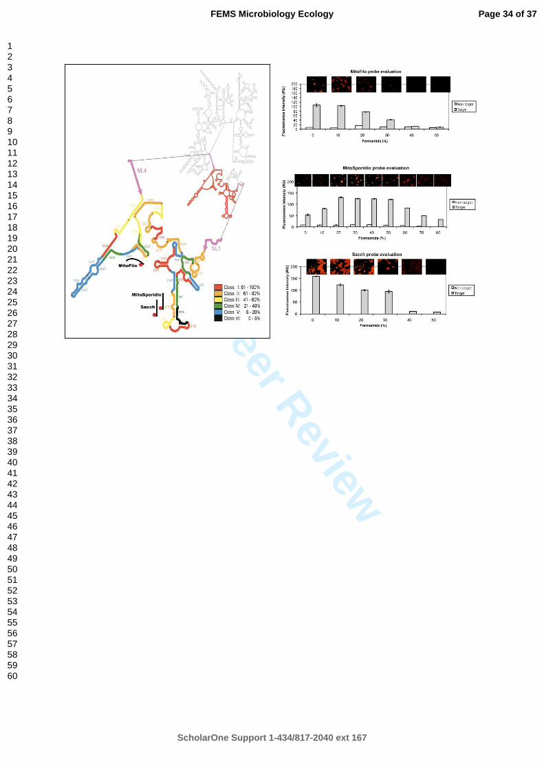
Fluorescent probe design and evaluation 195
196
For the detection of yeasts isolated from deep-sea vent animals by FISH, we designed 197
oligonucleotide probes using the Primrose software (http://www.bioinformatics-198
toolkit.org/Primrose/index.html) as described by Ashelford et al. (2002) using a set of high-199
quality, full-length rRNA sequences of probe target organisms. The PrimRose design tool 200
permitted to produce oligonucleotide probes for the three principal clusters of our collection 201
(Table 3). These probes exhibited no mismatches with the target organisms but exhibited 202
mismatches with the next most similar sequences in the GenBank database proving that the 203
designed probes were in silico highly specific. The target sites of newly designed probes were 204
checked for accessibility using the prediction maps based on the 26S rRNA of Saccharomyces 205
cerevisiae (Inacio et al., 2003). Each probe was in a relative accessible area of the 26S rRNA 206
secondary structure (Fig S1). As it was not possible to test the probes with culture isolates that 207
exhibited zero or one mismatch with the probes, we used an alternative method and tested the 208
probes against all strains from our collection displaying two or more mismatches with the 209
oligonuceotides. All newly designed probes were labelled at the 5’ terminus with the 210
fluorescent marker Cy3. All probes were synthesized by (Proligo, France) and stored in sterile 211
distilled water at -20°C. The newly designed probes were checked under in situ conditions 212
with target and non-target species. The universal probe Euk516-Fluorescein (5’-213
ACCAGACTTGCCCTCC-3’; Amann et al., 1995) and the non-Euk516-Cy3 (5’-214
CCTCCCGTTCAGACCA-3’) probes were used as positive and negative control respectively. 215
The average cell brightness was measured using different formamide concentrations from 0 to 216
80% with steps of 10%. Systematic evaluation of the signal intensities was done by recording 217
images of independent visual fields (encompassing at least 100 cells), followed by digital 218
image analysis using the daime software (Daims et al., 2006). During this step, the intensities 219
Page 7 of 37
ScholarOne Support 1-434/817-2040 ext 167
FEMS Microbiology Ecology
123456789101112131415161718192021222324252627282930313233343536373839404142434445464748495051525354555657585960
For Peer Review
of the image pixels analyzed enable determination of single cell fluorescence in relative units 220
(RU). 221
Fluorescence In Situ Hybridization 222
On environmental samples. Interior branchiostegites of Rimicaris exoculata shrimps and 223
byssus of Bathymodiolus azoricus mussels were processed for FISH analyses. Following 224
harvest and dissections, animal subsamples were fixed with 4% paraformaldehyde solution in 225
phosphate-buffered saline (PBS) for 3 hours at 4°C in a dark room. After fixation, tissues 226
were washed three times with PBS and stored at -20°C in a storage buffer containing PBS and 227
96% ethanol (1:1). 228
On membrane filters. The seawater surrounding shrimps (MoMAR08, Rainbow) was sampled 229
in 5 L sterile sampling bags using a peristaltic pump. Immediately after dives, seawater 230
samples for in situ hybridizations were mixed with 3 % formaldehyde (final concentration) 2 231
hours at 4°C. Fixed seawater was then filtered on polycarbonate membranes 0.22 µm 232
(Nuclepore®, 47 mm diameter; Whatman, Maidstone, Kent, UK) and rinsed with a PBS 2X - 233
sterile seawater (v:v) buffer. Then filters were dehydrated using ethanol series (50 %, 80 % 234
and absolute, 3 min each). Dried filters were stored at -20°C until hybridization treatments. 235
Three membranes were treated in this study. The filtered volume was 0.8 L for membrane A, 236
1 L for membrane B and 1.5 L for membrane C. The filtered seawater on membranes A and B 237
was from the same sample. 238
The samples (environmental samples and membrane filters) were cut in squares and paste 239
with one drop of 0.2% low-gelling point agarose (35-40°C) on slides (Menzel-Glaser, 240
Germany). All slides were then dipped in 0.2% agarose and air dried. Samples were then 241
subjected to dehydratation with increasing concentrations of ethanol (50, 80, and 96%, for 3 242
min each). Working solutions of probes had a concentration of 30 ng of DNA per liter. The 243
hybridization buffer, containing 0.9 M NaCl, 20 mM Tris-HCl (pH 7.2), 0.03% SDS, and 0, 244
10, 20, 30, 40, 50, 60, 70 and 80% formamide, and the fluorescent probe were gently mixed 245
in a ratio of 10:1 (vol/vol) to get a final oligonucleotide concentration of 3 ng per liter. For 246
hybridization, slides were placed in sampling tubes and incubated at 46°C in the dark for 247
exactly 3 hours. Following hybridization, the slides were washed with prewarmed washing 248
buffer (20 mM Tris/HCl, 5 mM EDTA (pH 8.0) and 900, 450, 215, 102, 46, 18, 5, 0,6 and 0 249
mM NaCl corresponding respectively to 0, 10, 20, 30, 40, 50, 60, 70 and 80% formamide 250
stringencies) for 20 min at 48°C. Slides were rinsed with double-distilled water, air dried, 251
Page 8 of 37
ScholarOne Support 1-434/817-2040 ext 167
FEMS Microbiology Ecology
123456789101112131415161718192021222324252627282930313233343536373839404142434445464748495051525354555657585960
For Peer Review
DAPI stained (final concentration 1 µg/ml) and mounted with the antifading reagent Citifluor 252
AF 2 (Citifluor, France) before observations under fluorescent microscope. 253
Results 254
Yeast isolation 255
Yeasts were not found in all the studied sites as shown in Table 1. No yeast was isolated from 256
samples collected during HERO (on the East Pacific Rise at Elsa site), DIVANAUT2 and 257
MARVEL (Menez-Gwenn and Lucky Strike) cruises or at TAG site during the EXOMAR 258
oceanographic cruise. The hydrothermal site that yielded the highest number of isolates was 259
clearly Rainbow (29 isolates out of 32 strains). Rainbow is also the site where the highest 260
number of samples was processed (97/210). The yeast collection obtained from deep-sea 261
samples raised thirty-two isolates that could be divided in pigmented yeasts (18) and non-262
pigmented yeasts (14). Pigmented yeasts consisted widely of red-pigmented yeasts (16), 263
black-pigmented yeast (1) and brown-pigmented yeast (1). 264
Regarding yeast isolation versus type of substrate, strains were obtained mostly from 265
hydrothermal shrimps Rimicaris exoculata (11), Chorocaris chacei (3), Mirocaris fortunata 266
(1) and from hydrothermal mussels Bathymodiolus azoricus (7). Carbonate colonization 267
modules deployed for 1 year near Rainbow vent yielded a few yeasts (4); sponges led to the 268
isolation of three yeasts. Finally, seawater, gastropods and coral samples permitted to obtain 269
one strain each (Table 1). Those results indicate that yeasts were much more associated with 270
animals rather than mineral substrates. Statistical distribution tests have been performed in 271
order to find out the distribution type of yeasts in hydrothermal sites. The variance to mean 272
ratio (s2/m) was calculated for each site (Cancela da Fonseca, 1966). Values of s2/m 273
significantly different of 1 corresponds with (s2/m) - 1 > 2(2n/(n - 1)2)1/2 and were obtained 274
only for Rainbow site. For this hydrothermal site, an aggregate distribution was observed 275
(s2/m=1.32) indicating that the culturable yeasts isolated were located in specific niches in 276
this hydrothermal site (mainly shrimps and mussels). 277
278
During the MoMARDREAM-Naut cruise, dissections of body components were processed 279
for all shrimps (branchiostegites, scaphognathites, exopodites, gills, stomach and digestive 280
tract) and mussels (interior and external faces of shells) to investigate the localization of 281
yeasts in deep-sea animals. For shrimps, a large majority of strains were grown from the inner 282
side of the branchiostegites that can be divided in 3 different compartments: (a) an antero-283
Page 9 of 37
ScholarOne Support 1-434/817-2040 ext 167
FEMS Microbiology Ecology
123456789101112131415161718192021222324252627282930313233343536373839404142434445464748495051525354555657585960
For Peer Review
ventral area, which was relatively clear; (b) a posterior area, which always remained light 284
beige; (c) an antero-dorsal area with an intensely rusty coloration (for schematic views, see 285
Zbinden et al., 2004; Corbari et al, 2008). Yeast isolates resulted from this study were all 286
cultivated from the antero-dorsal area characterized by high amounts of minerals and a dense 287
bacterial mat. 288
289
The yeasts were also isolated from Bathymodiolus azoricus (7) during the MoMADREAM-290
Naut oceanographic cruise (Table 1). Most of them (6) were cultivated from external face of 291
the mussel shells and more precisely from the byssus that is a network of filaments allowing 292
attachment to rocks. This tangle gathers a lot of particles and organic matter in decomposition 293
(personal observation). Only one yeast was isolated from the interior of a mussel (Mo32). 294
295
Physiological analysis 296
297
Three categories of strains were identified (Table 2) based on the definition of halotolerant 298
and halophilic microorganisms (Margesin & Schinner, 2001; Kushner, 1978). Non halophiles 299
are strains with maximal growth without sea salts and a decreasing growth rate with increased 300
sea salts concentration in media. Halotolerant yeasts are strains able to grow in the absence as 301
well as in the presence of salt. Halophiles required salt for an optimal growth. Regarding 302
halophily, optimal salinities, optimal temperatures and OD measurement, 9 physiological 303
groups were defined. Most of the isolated strains were non halophiles (23 strains) and 304
halotolerant (2 strains, maximal OD at 30 g/l sea salts) growing efficiently at an optimal 305
temperature of 25°C. Four strains had poor maximal growth at 25°C including 1 non 306
halophile, 2 halotolerant (maximal OD at 30 and 60 g/l sea salts) and 1 halophile (maximal 307
OD at 30 g/l sea salts). Three strains had maximal and efficient growth at 35°C, including 1 308
non halophile, 1 halotolerant (maximal OD at 45 g/l sea salts) and 1 halophile (maximal OD 309
at 30 g/l sea salts). 310
311
Identification 312
313
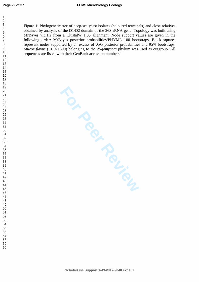
For species identification, a sequence analysis of the D1/D2 domain of the 26S rRNA gene 314
was done (Fig 1). A total of 12 phylotypes was found among the collection of yeasts isolated 315
from deep-sea hydrothermal vents. Eleven phylotypes could be assigned to a known yeast 316
species and one represents a new yeast species. 317
Page 10 of 37
ScholarOne Support 1-434/817-2040 ext 167
FEMS Microbiology Ecology
123456789101112131415161718192021222324252627282930313233343536373839404142434445464748495051525354555657585960
For Peer Review
318
Within Basidiomycota, the Sporidiobolales order was the dominant cluster composed of 16 319
strains. A majority of strains (Ex2, Ex3, Ex4, Ex5, Ex6, Ex7, Ex9, Ex11, Ex12, Mo32, Mo35 320
and Mo37) was identified as Rhodotorula mucilaginosa (100% similarity). A large majority 321
of R. mucilaginosa was isolated from deep-sea shrimps (14) and the others from deep-sea 322
mussels (2). As member of the Sporidiobolales order, isolates affiliated to Rhodosporidium 323
diobovatum were also isolated (Mo24, Mo33 and Mo38) with 100% similarity. These 3 324
strains were isolated respectively from Rimicaris exoculata exuviae in decomposition on 325
smocker rocks, Bathymodiolus azoricus and a sponge. One strain isolated from R. exoculata 326
was identified as Sporobolomyces roseus based on 26S rRNA genes (Mo22) with 100% 327
similarity with the reference strain. Four strains (Mo26, Mo27, Mo28 and Mo29) were 328
affiliated to the Filobasidiales order and identified as Cryptococcus uzbekistanensis (100% 329
similarity). These four strains were all isolated from a carbonate colonization module. Finally, 330
one isolate (Mo36) from B. azoricus mussel was identified as Leucosporidium scottii in the 331
Leucosporidiales order. 332
The Ascomycota phylum gathered 9 strains belonging to the Saccharomycetales order. Within 333
this order, 4 strains (Mo20, Mo21, Mo40 and Bio2) isolated respectively from R. exoculata, 334
Mirocaris fortunata, a deep-sea coral and the gills of the gastropod Ifremeria nautilei were 335
identified as Debaryomyces hansenii (100% similarity). Candida atlantica isolates were 336
found in R. exoculata exuviae in decomposition (Mo25) and B. azoricus (Mo31). One strain 337
isolated from a deep-sea sponge (Ex15) was identified as Pichia guilliermondii (100% 338
similarity). Finally, among the Saccharomycetales order, one strain was identified as Candida 339
viswanathii (Bio1) with 100% similarity. One halophilic strain (Mo39) isolated from a deep-340
sea coral represents a new species in the Candida genus and thus was identified as Candida 341
sp. This strain has 95% similarity with the reference sequence of Candida atmosphaerica (23 342
mismatches on 505 bp). Mo30 isolated from Bathymodiolus azoricus was identified as 343
Phaeotheca triangularis (mitosporic Ascomycota) with 100% similarity. In the Dothideales 344
order, one strain (Mo34) isolated from Bathymodiolus azoricus was identified as Hortaea 345
werneckii with 99.98% similarity (one mismatch on 560bp). 346
Sequencing of the 26S rRNA genes indicated the presence of Ascomycota and Basidiomycota 347
in our culture collection. In term of quantity, the phylum Basidiomycota (21) was two times 348
higher than the Ascomycota (11). In term of species richness, ascomycetous yeasts belonged 349
to 7 different clusters while basidiomycetous yeasts belonged to 5 clusters. 350
351
Page 11 of 37
ScholarOne Support 1-434/817-2040 ext 167
FEMS Microbiology Ecology
123456789101112131415161718192021222324252627282930313233343536373839404142434445464748495051525354555657585960
For Peer Review
Fluorescence in situ hybridizations 352
We processed numerous assays to detect fungi on deep-sea hydrothermal vent animal samples 353
using different existing fluorescent probes from different databases. The Euk516-Cy3 probe 354
gave positive results on pure cultures but strong background fluorescence on hydrothermal 355
samples led to the renouncement of its use. The probe MY1574 targeting Eumycota 356
organisms (Baschien et al., 2008) showed very weak fluorescence on pure cultures. Thus, we 357
decided to design our own probes (Table 3) based on our culture collection that was divided 358
in 3 main clusters: MitoFilo (Cryptococcus / Mitosporic Filobasidiales order), MitoSporidio 359
(Rhodotorula, Rhodosporidium / Mitosporic Sporidiobolales order) and Sacch 360
(Debaryomyces, Pichia / Saccharomycetales order). The probes designed revealed a strong 361
specificity for the target organisms. The optimal conditions for the in situ hybridization 362
protocol use stringent conditions of 20% formamide (Fig S1). 363
Our aim was to check the applicability of the FISH method to the in situ detection of yeasts in 364
deep-sea hydrothermal fauna samples. Hydrothermal body components of endemic shrimps 365
(Rimicaris exoculata) and mussels (Bathymodiolus azoricus) were fixed for FISH 366
experiments directly after dissection. The pieces of shrimps and mussels that gave the higher 367
number of fungi isolation (interior branchiostegites of shrimps and byssus of mussels) were 368
analyzed for yeast cell fluorescence. Although shrimp and mussel samples from Rainbow site 369
led to the highest rate of isolation, no FISH signal was ever observed. The FISH detection 370
limit of 103-104 target cells per ml is relatively high (Daims et al., 2005) and thus, the absence 371
of FISH signals does not necessarily mean that the target organisms were not present in the 372
samples. 373
To test this hypothesis, several volumes of water were concentrated on polycarbonate 374
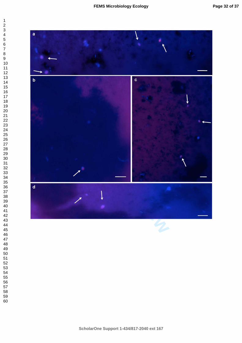
membrane filters to yield sufficient cells for FISH experiments with these new probes. 375
Membrane filters were embedded in low gelling-point agarose to minimize cell loss. Yeast 376
cells could be visualized in a low quantity on these membrane filters (Fig 2). Such results are 377
another evidence of the yeast cells presence in hydrothermal vents but at low concentration. 378
Using FISH on membrane filters, yeast cells detected were affiliated to 3 genera: 379
Rhodosporidium, Rhodotorula and Cryptococcus. 380
381
Discussion 382
383
Page 12 of 37
ScholarOne Support 1-434/817-2040 ext 167
FEMS Microbiology Ecology
123456789101112131415161718192021222324252627282930313233343536373839404142434445464748495051525354555657585960
For Peer Review
Occurrence of yeasts in deep-sea hydrothermal vents 384
In this study, the main aim was to isolate yeast strains from deep-sea hydrothermal endemic 385
fauna knowing that yeasts can be isolated from seawater surrounding hydrothermal fauna 386
(Gadanho and Sampaio, 2005). Yeast isolation was successful even if the retrieved species 387
richness was relatively low. Thirty-two strains were isolated mostly from Rimicaris exoculata 388
shrimps. The association with shrimps is probably favorable for yeasts that could benefit from 389
nutrients due to the water circulation in the gill chamber. Most of our strains were isolated 390
from the Rainbow hydrothermal site which confirms previous results (Gadanho and Sampaio, 391
2005). The Rainbow hydrothermal field hosted in ultramafic rocks is a unique vent enriched 392
in CH4, H2, CO, Fe and depleted in H2S (Charlou et al., 2002). The high yeast isolation ratio 393
may indicate that yeasts thrive in hydrothermal sites depleted in H2S. The isolation rate of 394
non-pigmented yeasts on sulfur-free media significantly higher than those on sulfur-based 395
media in a previous study (Gadanho & Sampaio, 2005) support such hypothesis. 396
397
Several yeasts were also isolated from mussels and more precisely from the byssus 398
constituted of filaments with a high concentration of minerals and organic matter. These 399
yeasts may have a role in the decomposition of organic material entrapped in mussel byssi in 400
deep-sea vents. These results seem promising as they confirm the data obtained in previous 401
studies and suggest that yeasts may interact with deep-sea hydrothermal vent fauna. 402
403
Pattern of the culturable yeast communities 404
New species. 405
The yeast that was firstly isolated from stomach of a marine fish was described as D. hansenii 406
and deposited in the Centraalbureau voor Schimmelcultures (CBS 5307) database. In a recent 407
paper, based on the intergenic spacer (IGS) region of the rRNA gene, this strain was re-408
evaluated as Candida sp. (Nguyen et al., 2009). This strain is identical to another one isolated 409
from deep-sea hydrothermal vent waters and annotated MARY089 (Gadanho and Sampaio, 410
2005). These two strains isolated from different marine environments were finally reported as 411
a single new undescribed species within the Candida genus. In our collection, strain Mo39, 412
isolated from deep-sea coral near Rainbow hydrothermal vents (Table 1), has the same 26S 413
rRNA gene sequence as CBS 5307 and MARY089. Mo39 is halophilic and thus supposed to 414
be an autochtonous marine yeast species. This new ecotype can be characterized as an 415
obligate marine yeast and its complete description is currently under progress. 416
Page 13 of 37
ScholarOne Support 1-434/817-2040 ext 167
FEMS Microbiology Ecology
123456789101112131415161718192021222324252627282930313233343536373839404142434445464748495051525354555657585960
For Peer Review
417
Known species 418
Two strains (Mo25 and Mo31) isolated from Rimicaris exoculata and Bathymodiolus azoricus 419
samples were identified as Candida atlantica. This result seems in keeping with previous 420
published reports that have isolated this species from coastal seawater in the South of 421
Portugal (Gadanho et al., 2003) and in deep-sea hydrothermal vent waters (Gadanho and 422
Sampaio, 2005). The very fisrt C. atlantica strain was isolated from shrimp eggs in the North 423
Atlantic Ocean (Siepmann and Höhnk, 1962). C. atlantica seemed to be a marine obligate 424
yeast and some interactions with shrimps seemed to occur. Our physiological analysis has 425
revealed that Mo25 and Mo31 were non-halophiles, which does not mean that they are unable 426
to grow in marine environments. They may have a role in deep-sea environments in 427
interaction with endemic crustaceans even if they are not in optimal growth conditions. One 428
isolate (Bio1) isolated from seawater surrounding mussels at Lau Basin in the South-West 429
Pacific was clearly identified as Candida viswanathii. Kohlmeyer & Kohlmeyer (1979) 430
characterized this yeast as marine facultative. More recently, C. viswanathii was isolated from 431
a shrimp (Peneaus braziliensis) in the Gulf of Mexico. Its synonym, Candida lodderae was 432
recently reported in deep-sea hydrothermal vent waters at Rainbow site (Gadanho and 433
Sampaio, 2005) and characterized as the most abundant yeast. 434
435
Leucosporidium scottii isolates (Mo36) were retrieved only in the oceanic regions close to 436
Antarctica and are known to be psychrophilic and probably autochthonous marine species 437
(Lachance and Starmer, 1998). Such strains known for their presence in cold polar marine 438
environments could be another evidence that confirms the hypothesis of global exchanges 439
from polar environments to deep-sea vents based on results from bacteria (Maruyama et al., 440
2000) and filamentous fungi (Burgaud et al., 2009). Hortaea werneckii (Mo34) was 441
characterized as halophilic in our physiological study. This is not surprising as this black 442
yeast-like fungus was characterized as halophilic or extremely halotolerant in different studies 443
(Gunde-Cimerman et al., 2000; Kogej et al, 2005) where it was frequently isolated from 444
hypersaline waters of solar salterns. In a molecular survey, H. werneckii was identified (based 445
on internal transcribed spacers and 5.8 S rRNA gene) in deep-sea methane seep sediments at a 446
depth of 2965 meters (Lai et al., 2007). Phaeotheca triangularis (Mo30) was also frequently 447
isolated from salted environments (Gunde-Cimerman et al., 2000) and characterized as 448
halophile. This confirmed previous results on P. triangularis showing a better growth with 449
5% additional salts (Zalar et al., 1999). In our study, Mo30 was characterized as halotolerant 450
Page 14 of 37
ScholarOne Support 1-434/817-2040 ext 167
FEMS Microbiology Ecology
123456789101112131415161718192021222324252627282930313233343536373839404142434445464748495051525354555657585960
For Peer Review
with 4.5% sea salts optimal concentration and thus hypothesized as marine adapted yeast. 451
This is the first report about the presence of Phaeotheca triangularis at deep-sea vents. 452
Mo22 is described as Sporobolomyces roseus. The genus Sporobolomyces is composed of 453
strains mainly isolated from the pyllophane (Bai et al., 2002). However, a previous study has 454
proved that strains of the genus Sporobolomyces are frequently isolated from marine 455
ecosystems and the frequency of isolation increases when distance from the coastline and 456
depth increase (Hernandez-Saavedra et al, 1992). Moreover, yeasts from this genus were 457
found in benthic invertebrates collected from deep-sea floor in the Pacific Ocean (Nagahama 458
et al, 2001b). Our strain was isolated from a deep-sea hydrothermal shrimp in the Atlantic 459
Ocean and characterized as halotolerant with an optimal salinity of 6% sea salts. This may 460
indicate that yeasts of this genus are also able to live in deep-sea vents and interact with 461
endemic crustaceans. 462
463
A previous study of yeasts in oceanic environments (Fell, 1976) reported that yeast 464
communities appeared to be constituted of ubiquitous and endemic species. Typical 465
ubiquitous strains were the ascomycetous yeast Debaryomyces hansenii and the 466
basidiomycetous ones Cryptococcus and Rhodotorula. Kohlmeyer and Kohlmeyer (1979) 467
confirmed this statement and characterized these genera mainly as facultative marine yeasts. 468
Some of these results, especially for Rhodotorula yeasts showing a strong ubiquity, were 469
confirmed based on their presence in several habitats such as deep-sea vents (Gadanho and 470
Sampaio, 2005), deep-sea sediments (Nagahama et al., 2001b), coastal waters (Gadanho et 471
al., 2003; 2004) and oligotrophic lakes (Libkind et al., 2003). Our results confirm their 472
ubiquity and indicate that these strains seem to be allochtonous. Strain Ex15 identified as 473
Pichia guilliermondii has also been characterized as non halophile and may be another 474
allochtonous yeast strain as reported by Kohlmeyer and Kohlmeyer (1979). 475
476
The members of the genus Rhodosporidum have been characterized as non halophiles (Mo24 477
and Mo33) and halotolerant (Mo38). Based on previous reports, this genus seemed to be 478
restricted to marine environments (Gadanho and Sampaio, 2005). R. diobovatum in deep-sea 479
vents seemed to be able to colonize different substrates (shrimps, mussels and sponges). The 480
isolation of a strain from shrimp exuviae in decomposition may indicate a role as a recycler of 481
organic material and so a probable implication in carbon cycle in deep-sea environments. 482
483
Adaptation to marine conditions 484
Page 15 of 37
ScholarOne Support 1-434/817-2040 ext 167
FEMS Microbiology Ecology
123456789101112131415161718192021222324252627282930313233343536373839404142434445464748495051525354555657585960
For Peer Review
The isolation of culturable yeasts led to an old question about marine yeasts “Are there any 485
indigenous marine yeasts ?” (Kohlmeyer & Kohlmeyer, 1979) and to the resulting question 486
“Which are the indigenous species ?”. Based on our results, one can suggest that halophilic 487
strains are marine indigenous yeasts and that others, halotolerant and non-halophiles, are 488
ubiquitous terrestrial strains present in deep-sea waters due to sedimentation or other natural 489
or anthropogenic phenomena. But almost all yeast species can grow well in media with NaCl 490
concentrations exceeding those normally present in the sea (Kohlmeyer & Kohlmeyer, 1979). 491
Few yeast species with a physiological dependence on NaCl or other seawater components 492
have been reported (Nagahama, 2006b). Thus, our results appeared in good agreement with 493
such statements. Only 2 strains described as halophiles (Mo34 and Mo39) in our study can be 494
described as obligate marine yeasts. 495
FISH observations 496
497
FISH using labeled oligonucleotide probes targeting rRNA has been used as a powerful 498
technique for assessing both microbial identity and spatial distributions in situ in complex 499
environmental contexts (Yang et al., 2008). Our results indicate a very low-level of yeasts at 500
deep-sea vents. As a first conclusion, regarding diversity and quantification (added to 501
previous results of Gadanho and Sampaio, 2005), it seems that yeasts at deep-sea vents 502
represent a minor community that might not be major actors in biogeochemical cycles. 503
However, fluorescent signals are correlated to the cellular content of ribosomes and 504
consequently to the microbial growth rates. Recently, the detection limits of conventional 505
FISH with Cy3-labeled probe EUB338 were found to be approximately 370 16S rRNA 506
molecules per cell for Escherichia coli hybridized on glass microscope slides and 1,400 16S 507
rRNA copies per E. coli cell in environmental samples (Hoshino et al., 2008). So, in addition 508
to a low concentration of yeast cells, low detection of yeasts may be caused by low ribosome 509
content of most yeasts in the deep-sea environment due to low-level metabolic activities of 510
yeasts living under extreme environmental abiotic factors (high hydrostatic pressure, low 511
temperatures,…). Our attempts to cultivate the yeast strains resulted from this study under 512
elevated hydrostatic pressure have been successful, but ribosomal activities were lower under 513
high hydrostatic pressure than at atmospheric pressure. Such results may account for the low 514
fungal detection using FISH (unpublished data). Consequently, care must be taken when 515
dealing with diversity and biomass estimations when using FISH alone. 516
Page 16 of 37
ScholarOne Support 1-434/817-2040 ext 167
FEMS Microbiology Ecology
123456789101112131415161718192021222324252627282930313233343536373839404142434445464748495051525354555657585960
For Peer Review
The quantification of yeasts using FISH has been impossible due to a non homogeneous 517
repartition of microorganisms on filters. Moreover, bacterial and yeast cells were only visible 518
in some regions of the filters without minerals due to strong autofluorescence. However we 519
can say that yeast concentrations are really low, as shown by the only few cells visualized 520
after filtration of seawater surrounding shrimps. This result is in keeping with the relatively 521
low diversity revealed by Gadanho and Sampaio (2005) ranging from 0 to 10 cfu/L for pink 522
yeasts and from 0 to 6000 cfu/L for non-pigmented yeasts. To better analyze the fungal 523
presence in deep-sea animals, one could work with phylum-specific probes on histological 524
sections of animals and use the CARD-FISH (Amann & Fuchs, 2008) or the DOPE-FISH 525
(Stoecker et al., 2010) methods to amplify probe signals. 526
These data raise emerging questions regarding the ecological role of such microorganisms in 527
deep-sea vents and about the old question of the ubiquity or endemism of those strains. Yeasts 528
at deep-sea vents may be facultative parasites or opportunistic pathogens of endemic deep-sea 529
animals as it has already been hypothesized in previous works (Van Dover et al., 2007; 530
Burgaud et al, 2009). However, a role in th edecomposition of abundant organic material may 531
occur. 532
Considering all the results obtained, we can say that yeasts may seem to interact with deep-533
sea hydrothermal endemic fauna even if the density is low. These yeasts are mainly composed 534
of ubiquitous species but obligate marine yeasts have also been harvested. However, the 535
results obtained using in situ hybridization have allowed us to visualize these ubiquist species 536
showing that they are able to live and grow in deep-sea hydrothermal vents. Yeasts associated 537
with endemic animals in deep-sea vents may be exposed to favorable conditions and could 538
benefit from a stable source of nutrients (Nagahama et al., 2001b). Yeasts were reported from 539
dead and healthy individuals which may also indicate their facultative saprophytism and so 540
emphasize the wide role of fungi in the decomposition of organic matter from terrestrial 541
environments to deep-sea hydrothermal vents. Even if yeasts were isolated from animal body 542
components, they were not visualized using FISH. To better understand the interaction with 543
animals and fungi in deep-sea vents, we need to work on tissues as in Van Dover et al. (2007) 544
and also with probes specific to fungal phyla (Ascomycota, Basidiomycota and 545
Chytridiomycota). In conclusion, several questions regarding the role of yeasts in deep-sea 546
hydrothermal vents and the endemism or ubiquity of the isolated yeasts remain a difficult task 547
without clear answers. Their culture under high hydrostatic pressures would be an interesting 548
study to better characterize their lifestyle and role at deep-sea vents. 549
Page 17 of 37
ScholarOne Support 1-434/817-2040 ext 167
FEMS Microbiology Ecology
123456789101112131415161718192021222324252627282930313233343536373839404142434445464748495051525354555657585960
For Peer Review
550
Acknowledgements 551
552
We thank the chief scientists of the BIOLAU, DIVANAUT2, HERO, MARVEL, EXOMAR 553
and MoMARDREAM-Naut cruises, the pilots and support crews of the oceanographic vessels 554
and Deep Submergence Vehicles of Ifremer. We greatly acknowledge Dr Jerome Mounier for 555
his valuable help and comments on FISH and Jerome Lepioufle for valuable advice on the 556
manuscript. We thank all the members of the GDR Ecchis for their advice and suggestions. 557
We would like to thank the editor and the anonymous reviewers who have provided helpful 558
comments on the refinement of this manuscript. We also thank ANR Deep-Oases, Ifremer, 559
Region Bretagne and French Research Ministry for their financial support. We finally thank 560
Françoise Gaill and the CHEMECO/European Science Foundation EURODEEP for 561
discussions and financial support. 562
563
Page 18 of 37
ScholarOne Support 1-434/817-2040 ext 167
FEMS Microbiology Ecology
123456789101112131415161718192021222324252627282930313233343536373839404142434445464748495051525354555657585960
For Peer Review
References 564
Altschul S, Gish W, Miller W, Myers E & Lipman D (1990) Basic local alignment search tool. J Mol Bio 215: 565 403–410. 566 567 Amann RI, Ludwig W & Schleifer K (1995) Phylogenetic identification and in situ detection of individual 568 microbial cells without cultivation. Microbiol Rev, 59: 143-169. 569 570 Amann RI & Fuchs BM (2008) Single-cell identification in microbial communities by improved fluorescence in 571 situ hybridization techniques. Nat Rev Microbiol, 6: 339-348. 572 573 Ashelford KE, Weightman AJ, & Fry JC (2002) PRIMROSE: a computer program for generating and estimating 574 the phylogenetic range of 16S rRNA oligonucleotide probes and primers in conjunction with the RDP-II 575 database. Nucleic Acids Res 30: 3481-3489. 576 577 Bai F, Zhao JH, Takashima M, Jia JH, Boekhout T & Nakase T (2002) Reclassification of the Sporobolomyces 578 roseus and Sporidiobolus pararoseus complexes, with the description of Sporobolomyces phaffii sp. nov. Int J 579 Syst Evol Microbiol 52: 2309-2314. 580 581 Baschien C, Manz W, Neu T, Marvanova L & Szewzyk U (2008) In Situ Detection of Freshwater Fungi in an 582 Alpine Stream by New Taxon-Specific Fluorescence In Situ Hybridization Probes. Appl Environ Microb 74: 583 6427-6436. 584 585 Bass D, Howe A, Brown N, Barton H, Demidova M, Michelle H, Li L, Sanders H, Watkinson SC, Willcock S & 586 Richards TA (2007) Yeast forms dominate fungal diversity in the deep oceans. Proc R Soc B 274: 3069-3077. 587 588 Burgaud G, Le Calvez T, Arzur D, Vandenkoornhuyse, P & Barbier G (2009) Diversity of culturable marine 589 filamentous fungi from deep-sea hydrothermal vents. Environ Microbiol 11: 1588-1600. 590 591 Cancela da Fonseca JP (1966) L’outil statistique en biologie du sol. III. Indices d’intérêt écologique. Revue 592 d’Ecologie et de Biologie du Sol 3: 381–407. 593 594 Charlou JL, Donval JP, Fouquet Y, Jean-Baptiste P & Holm N (2002) Geochemistry of high H2 and CH4 vent 595 fluids issuing from ultramafic rocks at the Rainbow hydrothermal field (36j14VN, MAR). Chem Geol, 191: 345-596 359. 597 598 Corbari L, Zbinden M, Cambon-Bonavita MA, Gaill F & Compère P (2008) Bacterial symbionts and mineral 599 deposits in the branchial chamber of the hydrothermal vent shrimp Rimicaris exoculata: Relationship to moult 600 cycle. Aquat Biol, 1: 225-238. 601 602 Daims H, Stoecker K & Wagner M (2005) Fluorescence In situ Hybridisation for the Detection of Prokaryotes. 603 In: Advanced Methods in Molecular Microbial Ecology. BIOS Scientific Publishers, Abingdon, UK. pp. 213-604 239. 605
Daims H, Lücker S, & Wagner M (2006) daime, a novel image analysis program for microbial ecology and 606 biofilm research. Environ Microbiol 8: 200-213. 607 608 Edgcomb VP, Kysela DT, Teske A, & de Vera Gomez A (2002) Benthic eukaryotic diversity in the Guaymas 609 Basin hydrothermal vent environment. Proc Natl Acad Sci USA 99: 7658-7662. 610 611 Fell JW (1976) Yeasts in oceanic regions. In: Jones EBG (ed) Recent advances in aquatic mycology. Elec, 612 London, pp 93-124. 613
Gadanho M, Almeida JM, & Sampaio JP (2003) Assessment of yeast diversity in a marine environment in the 614 south of Portugal by microsatellite-primed PCR. Antonie van Leeuwenhoek 84: 217-227. 615 616 Gadanho M & Sampaio JP (2004) Application of temperature gradient gel electrophoresis to the study of yeast 617 diversity in the estuary of the Tagus river, Portugal. FEMS Yeast Res 5: 253-261. 618 619 Gadanho M & Sampaio J (2005) Occurrence and Diversity of Yeasts in the Mid-Atlantic Ridge Hydrothermal 620 Fields Near the Azores Archipelago. Microb Ecol 50: 408-417. 621
Page 19 of 37
ScholarOne Support 1-434/817-2040 ext 167
FEMS Microbiology Ecology
123456789101112131415161718192021222324252627282930313233343536373839404142434445464748495051525354555657585960
For Peer Review
622 Gadd GM (2007) Geomycology: biogeochemical transformations of rocks, minerals and radionuclides by fungi, 623 bioweathering and bioremediation. Mycol Res 111: 3-49. 624 625 Guindon S, Lethiec F, Duroux P & Gascuel O (2005) PHYML online – a web server for fast maximum 626 likelihoodbased phylogenetic inference. Nucleic Acids Res 33: 557–559. 627 628 Gunde-Cimerman N, Zalar P, de Hoog S & Plemenitas A (2000) Hypersaline waters in salterns - natural 629 ecological niches for halophilic black yeasts. FEMS Microbiol Ecol 32: 235-240. 630 631 Hawksworth DL (2002) The magnitude of fungal diversity: the 1.5 million species estimate revisited. Mycol Res 632 105: 1422-1432. 633 634 Hernandez-Saavedra NY, Hernandez- Saavedra D & Ochoa JL (1992) Distribution of Sporobolomyces (Kluyver 635 et van Niel) genus in the western coast of Baja California Sur, Mexico. Syst Appl Microbiol 15: 319-322. 636 637 Hoshino T, Yilmaz LS, Noguera DR, Daims H & Wagner M (2008) Quantification of Target Molecules Needed 638 To Detect Microorganisms by Fluorescence In Situ Hybridization (FISH) and Catalyzed Reporter Deposition-639 FISH. Appl Environ Microb 74: 5068-5077. 640 641 Joergensen BB, & Boetius A (2007) Feast and famine-microbial life in the deep-sea bed. Nat Rev Microbiol 5: 642 770-781. 643 644 Kogej T, Ramos J, Plemenitas A, & Gunde-Cimerman N (2005) The halophilic fungus Hortaea werneckii and 645 the halotolerant fungus Aureobasidium pullulans maintain low intracellular cation concentrations in hypersaline 646 environments. Appl Environ Microb 71: 6600-6605. 647 648 Kohlmeyer J & Kohlmeyer E (1979) Marine Mycology: The Higher Fungi. New-York, USA; Academic Press. 649 650
Inacio J, Behrens S, Fuchs BM, Fonseca A, Spencer-Martins I & Amann R (2003) In situ accessibility of 651 Saccharomyces cerevisiae 26S rRNA to Cy3-labeled oligonucleotide probes comprising the D1 and D2 652 domains. Appl Environ Microb 69: 2899-2905. 653
Kushner DJ (1978) Life in high salt and solute concentrations. In: Kushner DJ (ed) Microbial life in extreme 654 environments. Academic Press, London, pp 317-368. 655
Lachance MA & Starmer WT (1998) Ecology and yeasts. In: Kurtzman CP, Fell JW (eds) The yeasts, a 656 taxonomic study, 4th edn. Elsevier, Amsterdam, The Netherlands, pp 21-30. 657
Lai X, Cao L, Tan H, Fang S, Huang Y & Zhou S (2007) Fungal communities from methane hydrate-bearing 658 deep-sea marine sediments in South China Sea. ISME J 1: 756-762. 659 660 Le Calvez T, Burgaud G, Mahe S, Barbier G & Vandenkoornhuyse P (2009) Fungal Diversity in Deep Sea 661 Hydrothermal Ecosystems. Appl Environ Microb 75: 6415-6421. 662 663 Libkind D, Brizzio S, Ruffini A, Gadanho M, van Broock M & Sampaio JP (2003) Molecular characterization of 664 carotenogenic yeasts from aquatic environments in Patagonia, Argentina. Antonie van Leeuwenhoek 84: 313-665 322. 666 667 Lopez-Garcia P, Philippe H, Gail F & Moreira D (2003) Autochthonous eukaryotic diversity in hydrothermal 668 sediment and experimental microcolonizers at the Mid-Atlantic Ridge. Proc Natl Acad Sci USA 100: 697-702. 669 670 Lopez-Garcia P, Vereshchaka A & Moreira D (2007) Eukaryotic diversity associated with carbonates and fluid-671 seawater interface in Lost City hydrothermal field. Environ Microbiol 9: 546-554. 672 673 Margesin R & Schinner F (2001) Potential of halotolerant and halophilic microorganisms for biotechnology. 674 Extremophiles 5: 73-83. 675 676 Maruyama A, Honda D, Yamamoto H, Kitamura K & Higashihara T (2000) Phylogenetic analysis of 677 psychrophilic bacteria from the Japan Trench, including a description of the deep-sea species Psychrobacter 678
Page 20 of 37
ScholarOne Support 1-434/817-2040 ext 167
FEMS Microbiology Ecology
123456789101112131415161718192021222324252627282930313233343536373839404142434445464748495051525354555657585960
For Peer Review
pacificensis sp. nov. Int J Syst Evol Microbiol 50: 835-846. 679 680 Munn, CB (2003) Marine Microbiology – Ecology and Applications. Oxford, UK: Bios-Garland Scientific. 681 682 Nagahama T, Hamamoto M, Nakase T & Horikoshi K (1999) Kluyveromyces nonfermentans sp. nov., a new 683 yeast species isolated from the deep sea. Int J Syst Evol Microbiol 49: 1899-1905. 684 685 Nagahama T, Hamamoto M, Nakase T & Horikoshi K (2001a) Rhodotorula lamellibrachii sp. nov., a new yeast 686 species from a tubeworm collected at the deep-sea floor in Sagami Bay and its phylogenetic analysis. Antonie 687 van Leeuwenhoek 80: 317-323. 688 689 Nagahama T, Hamamoto M, Nakase T, Takami H & Horikoshi K (2001b) Distribution and identification of red 690 yeasts in deep-sea environments around the northwest Pacific Ocean. Antonie van Leeuwenhoek 80: 101-110. 691 692 Nagahama T, Hamamoto M, Nakase T, Takaki Y & Horikoshi K (2003a) Cryptococcus surugaensis sp. nov., a 693 novel yeast species from sediment collected on the floor of Suruga Bay. Int J Syst Evol Microbiol 53: 2095-694 2098. 695 696 Nagahama T, Hamamoto M, Nakase T & Horikoshi K (2003b) Rhodotorula benthica sp. nov. and Rhodotorula 697 calyptogenae sp. nov., novel yeast species from animals collected from the deep-sea floor, and Rhodotorula 698 lysiniphila sp. nov., which is related phylogenetically. Int J Syst Evol Microbiol 53: 897-903. 699 700 Nagahama T, Hamamoto M & Horikoshi K (2006a) Rhodotorula pacifica sp. nov., a novel yeast species from 701 sediment collected on the deep-sea floor of the north-west Pacific Ocean. Int J Syst Evol Microbiol 56: 295-299. 702 703 Nagahama T (2006b) Yeast biodiversity in freshwater, marine and deep-sea environments. Biodiversity and 704 Ecophysiology of Yeasts. 705 706 Nagahama T, Abdel-Wahab MA, Nogi Y, Miyazaki M, Uematsu K, Hamamoto M & Horikoshi K (2008) 707 Dipodascus tetrasporeus sp. nov., an ascosporogenous yeast isolated from deep-sea sediments in the Japan 708 Trench. Int J Syst Evol Microbiol 58: 1040-1046. 709 710 Nguyen H, Gaillardin C & Neuvéglise C (2009) Differentiation of Debaryomyces hansenii and Candida famata 711 by rRNA gene intergenic spacer fingerprinting and reassessment of phylogenetic relationships among D. 712 hansenii, C. famata, D. fabryi, C. flareri (=D. subglobosus) and D. prosopidis: description of D. vietnamensis sp. 713 nov. closely related to D. nepalensis. FEMS Yeast Res 9: 641-662. 714 715 Posada D & Crandall K (1998) Applications note. MODELTEST: testing the model of DNA substitution. 716 Bioinformatics 14: 817–818. 717 718 Ronquist F & Huelsenbeck JP (2003) MrBayes 3: Bayesian phylogenetic inference under mixed models. 719 Bioinformatics 19: 1572–1574 720 721 Siepmann R & Höhnk W (1962) Über Hefen und einige Pilze (Fungi imp., Hyphales) aus dem Nordatlantik. 722 Veroeff Inst für Meeresforschung in Bremerhaven 8: 79-97. 723 724 Stoecker K, Dorninger C, Daims H & Wagner M (2010) Double Labeling of Oligonucleotide Probes for 725 Fluorescence In Situ Hybridization (DOPE-FISH) Improves Signal Intensity and Increases rRNA Accessibility. 726 Appl Environ Microb 76: 922-926. 727 728 Takishita K, Tsuchiya M, Reimer JD & Maruyama T (2006) Molecular evidence demonstrating the 729 basidiomycetous fungus Cryptococcus curvatus is the dominant microbial eukaryote in sediment at the 730 Kuroshima Knoll methane seep. Extremophiles 10: 165-169. 731 732 Takishita K, Yubuki N, Kakizoe N, Inagaki Y & Maruyama T (2007) Diversity of microbial eukaryotes in 733 sediment at a deep-sea methane cold seep: surveys of ribosomal DNA libraries from raw sediment samples and 734 two enrichment cultures. Extremophiles 11: 563-576. 735 736 Tamura K, Dudley J, Nei M & Kumar, S (2007) MEGA4: molecular evolutionary genetics analysis (MEGA) 737 software, version 4.0. Mol Biol Evol 24: 1596–1599. 738 739
Page 21 of 37
ScholarOne Support 1-434/817-2040 ext 167
FEMS Microbiology Ecology
123456789101112131415161718192021222324252627282930313233343536373839404142434445464748495051525354555657585960
For Peer Review
Tivey, MK (2007) Generation of seafloor hydrothermal vent fluids and associated mineral deposits. 740 Oceanography 20: 50-65. 741 742 Thompson JD, Higgins DG & Gibson, T.J. (1994) CLUSTAL W: improving the sensitivity of progressive 743 multiple sequence alignment through sequence weighting, position-specific gap penalties and weight matrix 744 choice. Nucleic Acids Res 22: 4673–4680. 745 746 Van Dover CL, Ward ME, Scott JL, Underdown J, Anderson B, Gustafson C, Whalen M & Carnegie RA (2007) 747 A fungal epizootic in mussels at a deep-sea hydrothermal vent. Mar Ecol 28: 54-62. 748 749 Zalar P, de Hoog GS & Gunde-Cimerman N (1999) Ecology of halotolerant dothideaceous black yeasts. Stud 750 Mycol 43: 38-48. 751 752 Zbinden M, Le Bris N, Gaill F & Compere P (2004) Distribution of bacteria and associated minerals in the gill 753 chamber of the vent shrimp Rimicaris exoculata and related biogeochemical processes. Mar Ecol Progr Ser 284: 754 237-251. 755
Page 22 of 37
ScholarOne Support 1-434/817-2040 ext 167
FEMS Microbiology Ecology
123456789101112131415161718192021222324252627282930313233343536373839404142434445464748495051525354555657585960
For Peer Review
Tables and Figures 756
757
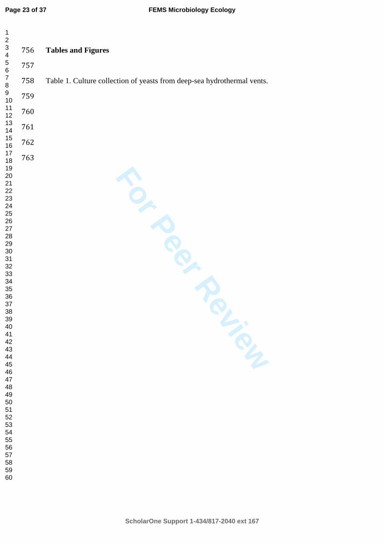
Table 1. Culture collection of yeasts from deep-sea hydrothermal vents. 758
759
760
761
762
763
Page 23 of 37
ScholarOne Support 1-434/817-2040 ext 167
FEMS Microbiology Ecology
123456789101112131415161718192021222324252627282930313233343536373839404142434445464748495051525354555657585960
For Peer Review
Location (Depth) Sample processed (type) Strain 764 South Pacific West; B2E07: Seawater surrounding mussels Bio1 765 (Lau Basin; -2620m) B9E07: Gastropod (Ifremeria nautilei) gills Bio2 766 Mid-Atlantic Ridge EX6E01 to EX6E04: Rimicaris exoculata Ex2 to Ex7 767 (Rainbow; -2300m) EX6E05: Chorocaris chacei Ex9, Ex11 and Ex12 768 MoPR1: Rimicaris exoculata Mo20 769 MoPR1: Mirocaris fortunata Mo21 770 MoPR2: Rimicaris exoculata Mo22 771 MoPR3: Sloughs of shrimp on smocker rocks Mo24 and Mo25 772 MoPR5: Colonization module TRAC (Carbonates) Mo26 to Mo29 773 MoPR6: Bathymodiolus azoricus Mo30 to Mo36 774 MoPR8: Rimicaris exoculata Mo37 775 MoPR9: Sponge Mo38 and Mo39 776 MoPR9: Coral Mo40 777 Mid-Atlantic Ridge EX18E02: Siliceous sponge Ex15 778 (Lost-City; -700m) 779
780
781
782
783
784
785
786
787
788
789
790
791
Page 24 of 37
ScholarOne Support 1-434/817-2040 ext 167
FEMS Microbiology Ecology
123456789101112131415161718192021222324252627282930313233343536373839404142434445464748495051525354555657585960
For Peer Review
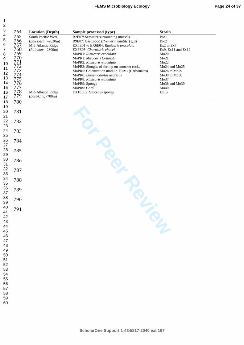
Table 2. Physiological analysis of the yeast collection. This table shows distribution of 792
halotolerant and halophilic strains of the collection depending on their optimal salinities (g/l 793
sea salts), optimal temperatures (°C) and maximal optical densities of cultures on GYPS broth 794
medium (120 rpm on a rotary shaker) measured at 600nm at 4 different incubation times (17h, 795
22h, 25h and 28h). 796
797
Page 25 of 37
ScholarOne Support 1-434/817-2040 ext 167
FEMS Microbiology Ecology
123456789101112131415161718192021222324252627282930313233343536373839404142434445464748495051525354555657585960
For Peer Review
798
Low OD (<1.1) High OD (>2.0)
Optimum 25°C 25°C 35°C
Non
halophile
0-15 g/l
Mo25
Bio1, Bio2, Ex2, Ex3, Ex4,
Ex5, Ex6, Ex7, Ex9, Ex11,
Ex12, Mo20, Mo21,
Mo24, Mo26, Mo27,
Mo28, Mo29, Mo31,
Mo32, Mo33, Mo35,
Mo40
Ex15
30 g/l Mo36 Mo37, Mo38
45 g/l Mo30 Halotolerant
60 g/l Mo22
Halophile 30 g/l Mo34 Mo39
799
Page 26 of 37
ScholarOne Support 1-434/817-2040 ext 167
FEMS Microbiology Ecology
123456789101112131415161718192021222324252627282930313233343536373839404142434445464748495051525354555657585960
For Peer Review
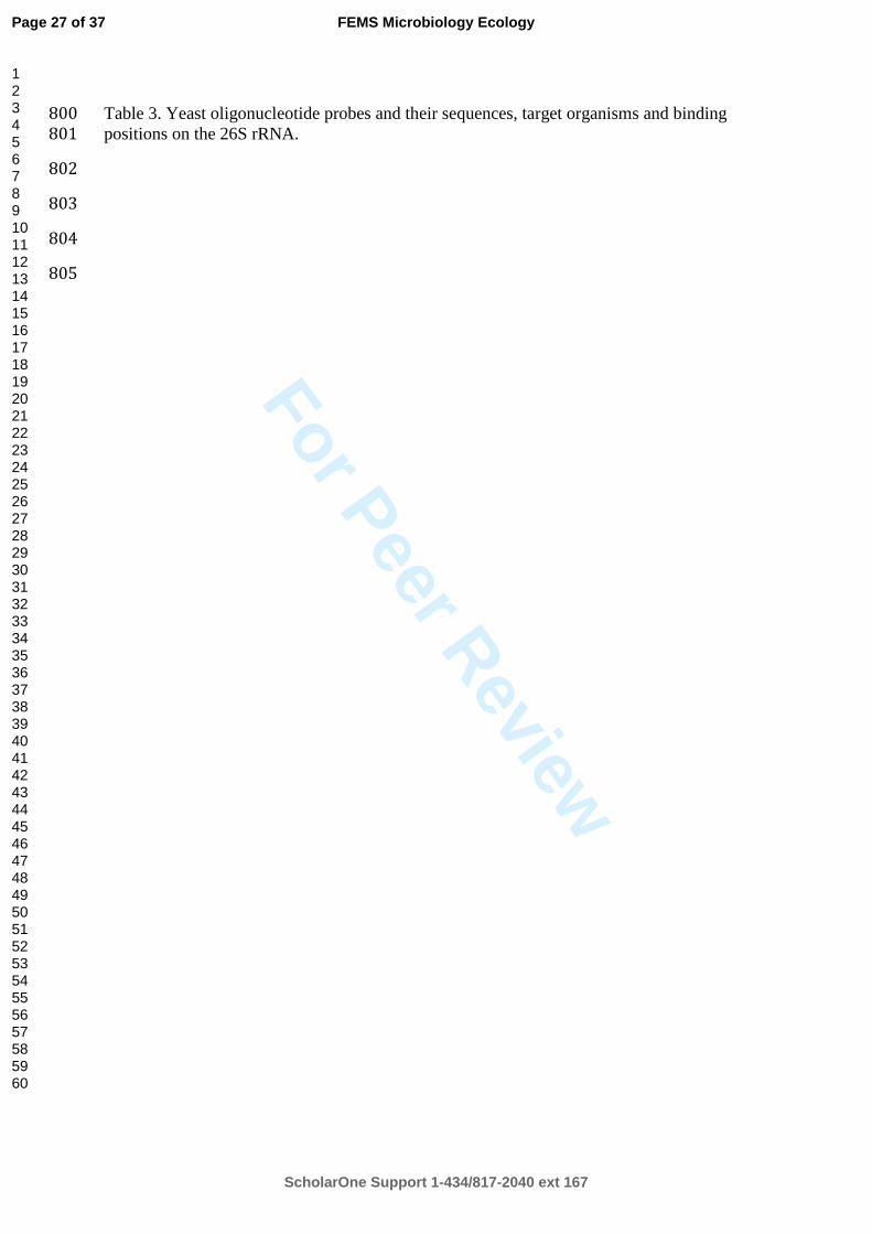
Table 3. Yeast oligonucleotide probes and their sequences, target organisms and binding 800
positions on the 26S rRNA. 801
802
803
804
805
Page 27 of 37
ScholarOne Support 1-434/817-2040 ext 167
FEMS Microbiology Ecology
123456789101112131415161718192021222324252627282930313233343536373839404142434445464748495051525354555657585960
For Peer Review
Probe Hybridization stringency rRNA subunit, binding position (a)
Probe sequence Target organisms
(% formamide) and relative probe accessibility (b)
(5’-3’) (Genus/Species)
Sacch 20 26S; 162-177 ; 44 to 66% GGCATCTCATCGCACG Debaryomyces Pichia
MitoFilo 10 26S; 397-412 ; 60% ACACCGCAGAACGGCT Members of the genus Cryptococcus (c)
MitoSporidio 20 26S; 164-179 ; 44 to 66% TGGGCGTCCGCACCAT Members of the genera Rhodotorula
and Rhodosporidium (d)
(a) Nucleotide position according to Saccharomyces cerevisiae 26Sr RNA between NL1 and NL4 primers.
(b) According to Inacio et al., 2003.
(c) Cryptococcus saitoi, C. randhawii, C. uzbekistanensis, C. adeliensis, C. vishniacii, C. socialis, C. friedmannii and C. uniguttulatus.
(d) Rhodotorula mucilaginosa, R. glutinis, R. graminis, R. dairenensis, Rhodosporidium babjevae and R. diobovatum.
Page 28 of 37
ScholarOne Support 1-434/817-2040 ext 167
FEMS Microbiology Ecology
123456789101112131415161718192021222324252627282930313233343536373839404142434445464748495051525354555657585960
For Peer Review
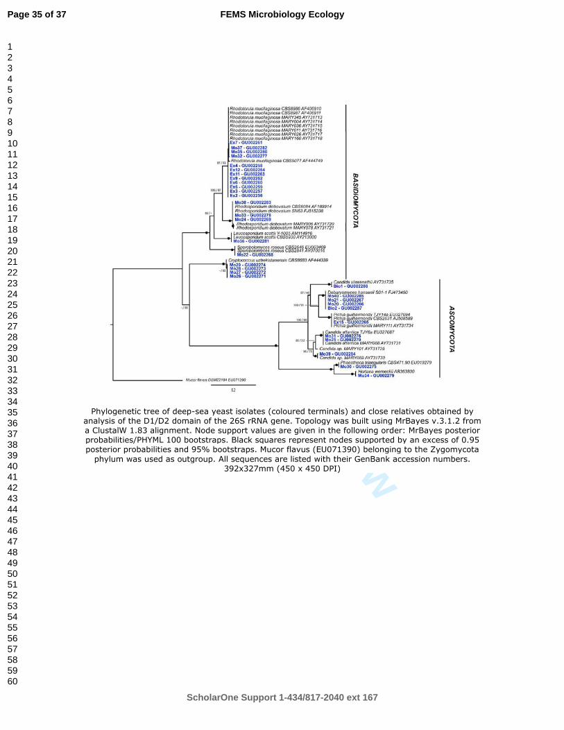
Figure 1: Phylogenetic tree of deep-sea yeast isolates (coloured terminals) and close relatives obtained by analysis of the D1/D2 domain of the 26S rRNA gene. Topology was built using MrBayes v.3.1.2 from a ClustalW 1.83 alignment. Node support values are given in the following order: MrBayes posterior probabilities/PHYML 100 bootstraps. Black squares represent nodes supported by an excess of 0.95 posterior probabilities and 95% bootstraps. Mucor flavus (EU071390) belonging to the Zygomycota phylum was used as outgroup. All sequences are listed with their GenBank accession numbers.
Page 29 of 37
ScholarOne Support 1-434/817-2040 ext 167
FEMS Microbiology Ecology
123456789101112131415161718192021222324252627282930313233343536373839404142434445464748495051525354555657585960
For Peer Review
Page 30 of 37
ScholarOne Support 1-434/817-2040 ext 167
FEMS Microbiology Ecology
123456789101112131415161718192021222324252627282930313233343536373839404142434445464748495051525354555657585960
For Peer Review
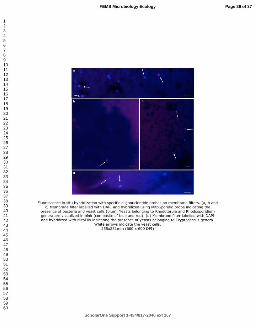
Figure 2: Fluorescence in situ hybridization with specific oligonucleotide probes on membrane filters. (a, b and c) Membrane filter labelled with DAPI and hybridized using MitoSporidio probe indicating the presence of bacteria and yeast cells (blue). Yeasts belonging to Rhodotorula and Rhodosporidium genera are vizualized in pink (composite of blue and red). (d) Membrane filter labelled with DAPI and hybridized with MitoFilo indicating the presence of yeasts belonging to Cryptococcus genera. White arrows indicate the yeast cells.
Page 31 of 37
ScholarOne Support 1-434/817-2040 ext 167
FEMS Microbiology Ecology
123456789101112131415161718192021222324252627282930313233343536373839404142434445464748495051525354555657585960
For Peer Review
Page 32 of 37
ScholarOne Support 1-434/817-2040 ext 167
FEMS Microbiology Ecology
123456789101112131415161718192021222324252627282930313233343536373839404142434445464748495051525354555657585960
For Peer Review
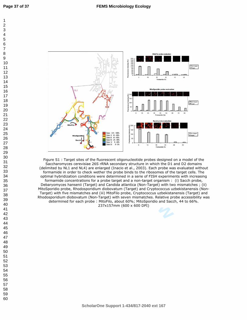
Figure S1 : Target sites of the fluorescent oligonucleotide probes designed on a model of the Saccharomyces cerevisiae 26S rRNA secondary structure in which the D1 and D2 domains (delimited by NL1 and NL4) are enlarged (Inacio et al., 2003). Each probe was evaluated without formamide in order to check wether the probe binds to the ribosomes of the target cells. The optimal hybridization conditions were determined in a serie of FISH experiments with increasing formamide concentrations for a probe target and a non-target organism : (i) Sacch probe, Debaryomyces hansenii (Target) and Candida atlantica (Non-Target) with two mismatches ; (ii) MitoSporidio probe, Rhodosporidium diobovatum (Target) and Cryptococcus uzbekistanensis (Non-Target) with five mismatches and (iii) MitoFilo probe, Cryptococcus uzbekistanensis (Target) and Rhodosporidium diobovatum (Non-Target) with seven mismatches. Relative probe accessibility was determined for each probe : MitoFilo, about 60%; MitoSporidio and Sacch, 44 to 66%.
Page 33 of 37
ScholarOne Support 1-434/817-2040 ext 167
FEMS Microbiology Ecology
123456789101112131415161718192021222324252627282930313233343536373839404142434445464748495051525354555657585960
For Peer Review
Page 34 of 37
ScholarOne Support 1-434/817-2040 ext 167
FEMS Microbiology Ecology
123456789101112131415161718192021222324252627282930313233343536373839404142434445464748495051525354555657585960
For Peer Review
Phylogenetic tree of deep-sea yeast isolates (coloured terminals) and close relatives obtained by analysis of the D1/D2 domain of the 26S rRNA gene. Topology was built using MrBayes v.3.1.2 from a ClustalW 1.83 alignment. Node support values are given in the following order: MrBayes posterior probabilities/PHYML 100 bootstraps. Black squares represent nodes supported by an excess of 0.95 posterior probabilities and 95% bootstraps. Mucor flavus (EU071390) belonging to the Zygomycota
phylum was used as outgroup. All sequences are listed with their GenBank accession numbers. 392x327mm (450 x 450 DPI)
Page 35 of 37
ScholarOne Support 1-434/817-2040 ext 167
FEMS Microbiology Ecology
123456789101112131415161718192021222324252627282930313233343536373839404142434445464748495051525354555657585960
For Peer Review
Fluorescence in situ hybridization with specific oligonucleotide probes on membrane filters. (a, b and c) Membrane filter labelled with DAPI and hybridized using MitoSporidio probe indicating the
presence of bacteria and yeast cells (blue). Yeasts belonging to Rhodotorula and Rhodosporidium genera are vizualized in pink (composite of blue and red). (d) Membrane filter labelled with DAPI and hybridized with MitoFilo indicating the presence of yeasts belonging to Cryptococcus genera.
White arrows indicate the yeast cells. 255x231mm (600 x 600 DPI)
Page 36 of 37
ScholarOne Support 1-434/817-2040 ext 167
FEMS Microbiology Ecology
123456789101112131415161718192021222324252627282930313233343536373839404142434445464748495051525354555657585960
For Peer Review
Figure S1 : Target sites of the fluorescent oligonucleotide probes designed on a model of the Saccharomyces cerevisiae 26S rRNA secondary structure in which the D1 and D2 domains
(delimited by NL1 and NL4) are enlarged (Inacio et al., 2003). Each probe was evaluated without formamide in order to check wether the probe binds to the ribosomes of the target cells. The
optimal hybridization conditions were determined in a serie of FISH experiments with increasing formamide concentrations for a probe target and a non-target organism : (i) Sacch probe,
Debaryomyces hansenii (Target) and Candida atlantica (Non-Target) with two mismatches ; (ii) MitoSporidio probe, Rhodosporidium diobovatum (Target) and Cryptococcus uzbekistanensis (Non-Target) with five mismatches and (iii) MitoFilo probe, Cryptococcus uzbekistanensis (Target) and Rhodosporidium diobovatum (Non-Target) with seven mismatches. Relative probe accessibility was
determined for each probe : MitoFilo, about 60%; MitoSporidio and Sacch, 44 to 66%. 237x157mm (600 x 600 DPI)
Page 37 of 37
ScholarOne Support 1-434/817-2040 ext 167
FEMS Microbiology Ecology
123456789101112131415161718192021222324252627282930313233343536373839404142434445464748495051525354555657585960
Copyright © 2022 FDOKUMEN