Critical Motions for AutoCalibration When Some Intrinsic Parameters Can Vary
Upload
khangminh22Category
view
1download
0
1/8Brazilian Journal of Biology, 2024, vol. 84, e253156 | https://doi.org/10.1590/1519-6984.253156
Original Article
THE INTERNATIONAL JOURNAL ON NEOTROPICAL BIOLOGYTHE INTERNATIONAL JOURNAL ON GLOBAL BIODIVERSITY AND ENVIRONMENT
ISSN 1519-6984 (Print)ISSN 1678-4375 (Online)
This is an Open Access article distributed under the terms of the Creative Commons Attribution License, which permits unrestricted use, distribution, and reproduction in any medium, provided the original work is properly cited.
1. Introduction
The fungal diversity has been estimated at up to 5.1 to 12 million based on high-throughput sequencing methods and culture-dependent culture-independent survey (O’Brien et al., 2005; Wu et al., 2019). Approximately 100,000 fungal species have been described, and countless are uncover every year (Blackwell, 2011). Many of these fungi have been associated with plants and divided into five main functional groups: mycorrhizal, pathogenic, saprotrophic, epiphytic, and endophytic fungi (Porras-
Alfaro and Bayman, 2011). Of these, endophytic fungi are special interest in the agricultural area.
Fungal endophytes are inhabiting plant tissue without causing apparent harm to the host (Petrini, 1991). They have a symbiotic-mutualistic relationship with plants, colonizing different above-ground tissues such as leaves, stems, seeds, bark, petioles, and reproductive structures, which differentiates them from mycorrhizal groups that inhabit roots (Faeth and Fagan, 2002; Shearin et al., 2018). Every
AbstractEndophytic fungi are a ubiquituos group that colonize all plant species on earth. Studies comparing the location of endophytic fungi within the leaves and the sampling time in Manihot esculenta Crantz (cassava) are limited. In this study, mature leaves of M. esculenta from Panama were collected in order to compare the cultivable diversity of endophytic fungi and to determine their distribution within the leaves. A total of one hundred sixty endophytes belonging to 97 species representing 13 genera and 8 morphospecies determined as mycelia sterilia that containing 63 isolates were isolated. Cladosporium, Nigrospora, Periconia, and mycelia sterilia 1 and 3 were the most predominant isolated endophytes. We detected that endophytes varied across the sampling time, but not amongst locations within leaves. The endophytes composition across sampling and the location of endophytes within leaf was similar, except for Periconia and mycelia sterilia 3 and 7. The data generated in this study contribute to the knowledge on the biodiversity of endophytic fungi in Panama, and establish the bases for future research focused on understanding the function of endophytes in M. esculenta crops.
Keywords: ascomycota, endophytes, biodiversity, cassava.
ResumoOs fungos endofíticos são um grupo ubiquituo que colonizam todas as espécies de plantas na terra. Os estudos que comparam a localização dos fungos endofíticos dentro das folhas de Manihot esculenta Crantz (mandioca) e o tempo de amostragem são muito escassos. Neste estudo, folhas maduras de M. esculenta foram coletadas do Panamá com a finalidade de comparar a diversidade cultivável de endófitos e determinar sua distribuição dentro das folhas. Um total de 170 endófitos foram isolados de 97 espécies que representam 13 gêneros e 8 morfoespécies determinadas como micélios esterilizados contendo 63 isolados. Os fungos Cladosporium, Nigrospora, Periconia e mycelia sterilia 1 e 3 foram os isolados mais predominantes. Também detectamos que os endófitos variaram ao longo do tempo de amostragem, mas não entre os locais dentro das folhas. A composição de endófitos na amostragem e localização de endófitos dentro da folha foi semelhante, exceto para Periconia e mycelia sterilia 3 e 7. Os dados gerados neste estudo contribuem para o conhecimento da biodiversidade de fungos endofíticos no Panamá e estabelecem as bases para pesquisas sobre o entendimento da função de endófitos em culturas de M. esculenta.
Palavras-chave: ascomycota, endófitos, biodiversidade, mandioca.
Diversity of culturable endophytic fungi vary through time in Manihot esculenta CrantzA diversidade de fungos endofíticos cultiváveis varia ao longo do tempo em Manihot esculenta Crantz
L. A. Ramírez-Camejoa,b* aCenter for Biodiversity and Drug Discovery, Institute of Scientific Research and High Technology Services (INDICASAT-AIP), City of Knowledge, Clayton, Panama, Republic of Panama
bCoiba Scientific Station (COIBA AIP), City of Knowledge, Clayton, Panama, Republic of Panama
*e-mail: [email protected]: June 10, 2021 – Accepted: September 27, 2021

Brazilian Journal of Biology, 2024, vol. 84, e2531562/8
Ramírez-Camejo, L.A.
2. Material and Methods
2.1. Collection of Manihot esculenta Crantz leaves and isolation of fungal endophytes
Healthy leaves of M. esculenta were collected during the summer season (Jan to Mar) in a plantation in Ocú, Herrera (Panama). Five trees of ~10 months old were randomly selected, and from each tree, three mature leaves (5–7 lobes) were chosen randomly but visually healthy and free of damage by herbivores or pathogens. Samples were packaged and preserved at 4 °C, and same trees were collected five times during 2 ½ months with 15 days of interval.
The isolation method was carried out in the Mycological laboratory of the Instituto de Investigación Agropecuaria de Panama (I.D.I.A.P.), located in Divisa, Herrera (Panama). The collected leaves samples were divided as lower, middle and top parts within 24 h of collection and washed under running tap water for 1 min. Sixteen segments of 2 mm2 were cut from each leaf part and surface-sterilized through sequential immersion in 70% alcohol (1 min), 0.5% sodium hypochlorite (3 min), 70% ethanol (30 s), and another 40% alcohol (30–40 s) (Bethancourt, 2000). The leaf samples were soaked and shaken with sterile distilled water three times for each 3 min. The sterilized samples were dried with sterile tissue paper for ~30 min and plated on 2% potato dextrose agar (PDA) medium supplemented with 50 mg of Vancomycin Hydrochloride and an infusion of 30 g/L of M. esculenta leaves. PDA is a common and nonselective media made from potato infusion, and dextrose as a carbohydrate source that provides enough nutrients to encourage the growth of a range of fungi; the infusion of M. esculenta leaves could increase the growth of fungal endophytes while the Vancomycin Hydrochloride avoid bacterial contamination. Also, PDA has been used extensively for the growth of different fungal endophytes in other tropical and crop plants (Gamboa and Bayman, 2001; Santamaría and Bayman, 2005; Hartanti et al., 2021). Uninoculated plates were used as controls. Endophytes were incubated at 25–28 °C for 2 weeks in 12 h of light-dark cycles. Mycelium from distinct colonies emerging from leaf segments were subcultured on new PDA plates to obtain pure colonies. Endophytes colonies were submitted to microculture on glass slides for examination of reproductive structures (Riddell, 1950). For sporulating fungi, we used morphology-based taxonomy (Barnett and Hunter, 1972). Pure colonies were transferred to a 2 mL glass vial with 2% of malt extract agar (MEA) supplemented with the antibiotic above described and grouped by morphospecies based on morphological characteristics in the mycelium growing in tubes (Ramírez-Camejo et al., 2017).
2.2. Fungal diversity analysis
Diversity analysis were performed using the endophyte species (morphospecies) isolated based on two criteria: 1) sampling time and 2) position in the lobed leaf (lower vs middle vs top). Species diversity was calculated using the sample size- and coverage-based rarefaction and extrapolation (R/E) of the Hill numbers of species, i.e.,
plant species on earth has a community of endophytes, e.g., leaf segments of 2mm2 in cacao (Theobroma cacao) trees and two understory (Heisteria concinna and Ouratea lucens) trees harbor naturally abundant, dense, and diverse endophyte infections ranged from 82% to 100% (Arnold et al., 2000, 2003). However, such proportion and diversity of fungi in plants could be dependent upon the host, age, nutrient, the environment, season, or the interaction with other microorganisms that surround them (Porras-Alfaro and Bayman, 2011). Because fungal endophytes are everywhere, their uses as biological control agents against pathogens, and their pharmaceutical potential, are undeniable reasons for their study (Mejía et al., 2008; Sridhar, 2019; Adeleke and Babalola, 2021). In agricultural crops, endophytes play crucial roles by protecting plants against many biotic (conferring pest and disease resistance) and abiotic stresses (including impacts of climate change), increasing their resilience, and for improved management of post-harvest control (Lugtenberg et al., 2016). Hence, understanding their diversity in plants of agricultural importance, such as Manihot esculenta Crantz (cassava) is a first step to understand its functionality in that host.
Manihot esculenta (Phanerogama, Euphorbiaceae) is an ancient plant that has been extensively cultivated in tropical and subtropical regions in the world (Liu et al., 2014). Its worldwide production ended in approximately 277 million tons in 2015 with the main production areas being Africa (55%), Asia (33%), and Latin America (12%) (FAO, 2017). The domesticated species M. esculenta is grown for its starch-rich storage roots, providing food and energy security for about 800 million people (Burns et al., 2010; FAO 2013; Parmar et al., 2017).
In this study, two questions were addressed: (1) Are culturable endophytic fungi distinctly diverse across sampling time and the position of endophytes within leaves. Because communities of endophytic fungi can vary across time and within leaf parts (Suryanarayanan and Thennarasan, 2004; Porras-Alfaro and Bayman, 2011), we hypothesized that fungal endophytes differ between sampling time and surveyed endophytes within the leaves. (2) How is the mycobiota of M. esculenta distributed spatially? The distribution of endophytes within leaves and across time is typically heterogeneous (Gamboa and Bayman, 2001; Cannon and Simmons, 2002; Arnold and Herre, 2003; Suryanarayanan and Thennarasan, 2004). We hypothesized that some endophytic fungi are found in all location within the leaf parts and sampling time; however, some endophytes will be limited to a specific location within the leaf or sampling time in M. esculenta.
These questions would be valid and novel for any plant. M. esculenta was used because is naturalized in Panama, produced in small farms, and the relationships between fungal endophytes have important implications for fungal biodiversity and plant health. For example, endophytes may potentially be useful for biocontrol of pathogens (Arnold et al., 2003; Ezra et al., 2009), and several cassava pathogens are very devastating (Lozano and Booth, 1974; Legg and Alvarez, 2017).
Brazilian Journal of Biology, 2024, vol. 84, e253156 3/8
Fungal endophytes in Manihot esculenta Crantz
richness (q = 0), Shannon diversity (q = 1, the exponential of Shannon entropy), and Simpson diversity (q = 2, the inverse of Simpson concentration), as implemented in the R package Interpolation and Extrapolation for species diversity (iNext) v2.0.20 (Chao and Jost, 2012; Hsieh et al., 2016).
PAleontological STatistics (PAST) v4.04 was used to compare fungal diversity in both datasets using one-way ANOVA and Kruskal-Wallis tests followed by Tukey’s Pairwise to test sample means and medians, respectively (Hammer et al., 2001). Additionally, Mann-Whitney pairwise and Dunn’s post hoc tests were used to identify which means were significantly different from the others. The composition of the communities of fungal endophytes within leaves and between sampling time was performed using Whittaker index. Chi square (𝜒2) tests were used to compare differences between endophytes collected in a different position within leaves and sampling time.
3. Results
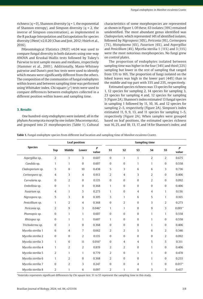
One hundred-sixty endophytes were isolated; all in the phylum Ascomycota except by one isolate (Mucoromycota), and grouped into 21 morphospecies (Table 1). Cultural
characteristics of some morphospecies are represented as shown in Figure 1. Of these, 63 isolates (39%) remained unidentified. The most abundant genus identified was Cladosporium, which represented 14% of identified isolates, followed by Nigrospora (10%), Periconia (9%), Corinespora (7%), Histoplasma (6%), Fusarium (6%), and Aspergillus and Penicillium (4%). Mycelia sterilia 1 (11%) and 3 (11%) were the most notorious morphospecies. No fungi grew on control plates.
The proportion of endophytes isolated between sampling time was higher in the four (34%) and third (22%) sampling but lower in the rest of sampling that ranged from 13% to 16%. The proportion of fungi isolated on the lobed leaves was high in the lower part (44%) than in the middle and top part with 33% and 23%, respectively.
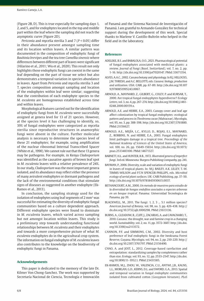
Estimated species richness was 13 species for sampling 1, 12 species for sampling 2, 14 species for sampling 3, 23 species for sampling 4 and, 12 species for sampling 5 (Figure 2A). Shannon’s index estimated 13 fungal species in sampling 1 followed by 11, 10, 16, and 12 species for sampling 2–5, respectively (Figure 2A). Simpson‘s index estimated 11, 9, 9, 13, and 11 species for sampling 1–5, respectively (Figure 2A). When samples were grouped based on leaf positions, the estimated species richness was 16, 25, and 18; 13, 17, and 14 for Shannon’s index; and
Table 1. Fungal endophyte species from different leaf location and sampling time of Manihot esculenta Crantz.
Species
Leaf position Sampling time
Top Middle LowerP
valueS1 S2 S3 S4 S5
P value
Aspergillus sp. 2 1 3 0.607 0 1 1 2 2 0.675
Candida sp. 1 1 0 0.607 0 0 1 1 0 0.558
Cladosporium sp. 5 8 10 0.438 5 3 6 6 3 0.736
Corinespora sp. 4 3 4 0.913 2 4 3 2 0 0.406
Curvularia sp. 0 2 0 0.135 0 0 0 2 0 0.092
Embellisia sp. 0 1 0 0.368 1 0 0 0 0 0.406
Fusarium sp. 4 1 5 0.273 1 0 4 4 1 0.136
Nigrospora sp. 5 3 8 0.305 3 1 6 5 1 0.165
Penicillium sp. 1 2 4 0.368 0 2 0 3 2 0.273
Periconia sp. 2 9 3 0.046* 1 1 0 9 3 0.001*
Phomopsis sp. 0 1 1 0.607 0 0 0 1 1 0.558
Rhizopus sp. 0 1 1 0.607 1 0 0 1 0 0.558
Trichoderma sp. 0 1 0 0.368 0 0 0 1 0 0.406
Mycelia sterilia 1 6 4 7 0.662 2 2 5 6 2 0.346
Mycelia sterilia 2 0 0 2 0.135 0 0 0 0 2 0.092
Mycelia sterilia 3 1 6 11 0.016* 0 4 4 5 5 0.311
Mycelia sterilia 4 1 2 2 0.819 2 2 0 1 0 0.406
Mycelia sterilia 5 2 1 1 0.779 2 0 1 1 0 0.478
Mycelia sterilia 6 1 2 0 0.368 2 0 0 1 0 0.255
Mycelia sterilia 7 0 2 3 0.247 0 0 4 1 0 0.017*
Mycelia sterilia 8 2 1 6 0.097 2 1 0 3 3 0.437
*Asterisks represents significant differences by Chi square test. S1 to S5 represent the sampling time in this study.

Brazilian Journal of Biology, 2024, vol. 84, e2531564/8
Ramírez-Camejo, L.A.
Figure 1. Colony morphology of some endophytic fungi isolated from Manihot esculenta Crantz grown on PDA at 25–28 °C.
Figure 2. Diversity of fungal endophytes isolated from Manihot esculenta Crantz among sampling days and location of fungi within leaves, following Chao and Jost (2012): A-C: Sample‐size‐based rarefaction and extrapolation (R/E) curves. Plots shown three measures of species diversity: species richness, Shannon diversity (the exponential of Shannon entropy), and Simpson diversity (the inverse of Simpson concentration). Solid parts of the curves represent rarefaction while dashed curves represent extrapolation beyond observed samples. B-D: Sampling completeness curves. Shaded areas, 95% confidence intervals (bootstrapped).

Brazilian Journal of Biology, 2024, vol. 84, e253156 5/8
Fungal endophytes in Manihot esculenta Crantz
11, 12, and 12 for Simpson‘s index between top, middle, and lower position, respectively (Figure 2C).
The sampling completeness curve increases as the number of fungal isolates increases (Figure 2B, D). The sample completeness was highest for sampling 3 and 5 (0.89%) followed by sampling 4 (0.87%), and decreasing on sampling 1 and 2 (0. 85% and 0.82%) (Figure 2B). Lower sampling contributed 0.96% of sampling completeness while 0.87% and 0.83% was for top and middle sampling (Figure 2D).
Species diversity differed significantly among samplings (ANOVA, F = 2.597, df = 4, 99, P = 0.041) but not between the position of fungal endophytes within the leaf (ANOVA, F = 1.93, df = 38.32, P = 0.159). A significant difference was found between sampling 2 and 4 (Tukey’s pairwise, 0.045; Mann-Whitney pairwise tests, 0.011, and Dunn’s post hoc tests, 0.010). No significant differences were showed among the position of fungal endophytes within the leaf during Tukey’s pairwise tests, Mann-Whitney pairwise tests and Dunn’s post hoc tests (P ≥ 0.05).
The species composition did not differ significantly between sampling (Whittaker = 0.693) or position of endophytes in the leaf (Whittaker = 0.26). Only the fungus Periconia (𝜒2 = 4.7, 𝑃 < 0.046) and mycelia sterilia 3 (𝜒2 = 6, 𝑃 < 0.016) was significantly more common in some part of the leaf than in other leaf parts (Table 1). Also, Periconia (𝜒2 = 2.8, 𝑃 < 0.001) and mycelia sterilia 7 (𝜒2 = 1, 𝑃 < 0.017) was significantly more common in some sampling than others (Table 1). The remaining endophytes did not differ significantly in frequency where endophytic fungi was isolated by sampling or within leaves (𝑃 > 0.05).
4. Discussion
This study explores the culturable endophytic fungi isolated from M. esculenta leaves and omitted nonculturable fungi or those that require special culture media or cultural conditions (Bayman, 2006). This implies a portion of the total fungal diversity present in M. esculenta are revealed. On the other hand, most endophytes are culturable and grow well with the method used here. No endophytic fungi grew in control plates during the culturing procedure, indicating that the manipulation during the field trip and isolation techniques were effective in preventing contamination.
Cladosporium, Nigrospora, Periconia, and mycelia sterilia 1 and 3 were the most representative endophyte taxa identified in M. esculenta leaves and these genera have also been shown to be the dominant genera in endophytes of other plants such as Camellia sinensis, Mentha sp., and palm trees (Table 1) (Song et al., 2016; Zakaria and Wan Aziz, 2018; Wu et al., 2020). Interestingly, the first three genera and mycelia sterilia 1 were found across all sampling days and in all locations within leaves, suggesting that common taxa may come from a similar source (e.g., airborne spores and soil spores) or leaves are favorable substrates for colonization of certain endophytes than others.
In this study, species of Candida, Curvularia, Embellisia, Phomopsis, Rhizopus, and Trichoderma, were the least abundant endophytes (Table 1). This result could reflect
differences in the culture media used (PDA + Vancomycin Hydrochloride) or methods of sampling. Furthermore, it could represent that every sampling event is different, and the abundance and diversity of endophytes in the leaf is driven by the tree or environmental factors such as temperature and water stress.
Our results suggest that the overall species richness of endophytes in M. esculenta among sampling time ranged from 12 and 23 species, with Shannon diversity between 10–16 species, and Simpson diversity from 9–13 species (Figure 2A). These values increased from sampling 1 and 4 and decreased from sampling 2, 3, and 5, reflect the fungal succession that occurred as the leaves receive new endophytes across time. Fungi isolated from the top part of the leaf in M. esculenta had a similar estimated diversity of fungal species than middle and lower parts for Richness, Shannon diversity, and Simpson diversity, respectively (Figure 2C). This indicated the persistence of the fungi associated with adult leaves within M. esculenta.
Overall, species diversity was similar across sampling but with significant differences on Sampling 2 vs Sampling 4 (Figure 2A), but not for fungi isolated between locations within leaves. This supported the hypothesis that communities of endophytes vary across sampling days of M. esculenta, but not all the time (Porras-Alfaro and Bayman, 2011). This is possibly due to the physiological changes that commonly occur in the plant as it progresses in its biological cycle (Alves, 2002; El-Sharkawy, 2004). For example, in different stages of plant development, changes occur in the distribution of photosynthates and energetic compounds demanded by the different organs of the plant, as well as by changes in the climatic patterns and that occur as the growing season progresses (Yoshioka, 1986; Gray and Brady, 2016). This study was accomplished during the summer season where lack of water and high-temperature stresses can decrease the rate of leaf formation in M. esculenta (El-Sharkawy, 2004), which affect directly the abundance and diversity of fungi (Talley et al., 2002). Also, we cannot discard the possibility that differences in fungal diversity on sampling time could be due to leaf nutrient availability as showed in Pinus muricata and Vaccinium ovatum plants (Oono et al., 2020).
The highest number of isolates were recorded from the lower part of the M. esculenta lobe, but such abundance was not significant across other parts of the leaf. Possibly the proximal leaf part close to the base provides favorable conditions for the germination of spores, since the moisture may be higher than other parts. Similar results were found in Betula pubescens var. tortuosa where a greater number of endophytes (over 30%) was shown in the basal part of the leaf (Helander et al., 1993). In contrast, tropical forests reported that the largest endophytic population is found in the midrib and tip region compared to the base of the leaf (Cannon and Simmons, 2002; Suryanarayanan et al., 2002). These reports indicate that the pattern described for simple leaves (a single lobe) does not fit in the case of palm-split leaves such as M. esculenta.
Further analysis on increasing the number of samples per time and per leaf segment would improve the strength of the inferences that can be drawn, as suggested by the sampling completeness curves for M. esculenta
Brazilian Journal of Biology, 2024, vol. 84, e2531566/8
Ramírez-Camejo, L.A.
(Figure 2B, D). This is true especially for sampling days 1, 2, and 5, and for endophytes located in the top and middle part within the leaf where the sampling did not reach the asymptotic curve (Figure 2D).
Periconia and mycelia sterilia 3 and 7 (P < 0.05) differ in their abundance present amongst sampling time and its location within leaves. A similar pattern was documented in the composition of endophytic fungi of Bauhinia brevipes and the tea tree Camellia sinensis where differences between different part of leaves were significant (Hilarino et al., 2011; Wu et al., 2020). This result not only highlights those endophytic fungi can coexist in the same leaf depending on the part of tissue we select but also demonstrates a temporal variation in species abundance in leaves. Apart from Periconia and mycelia sterilia 3 and 7, species composition amongst sampling and location of the endophytes within leaf were similar; suggesting that the contribution of each endophyte species to the M. esculenta are homogeneous established across time and within leaves.
Morphological features carried out for the identification of endophytic fungi from M. esculenta were successfully assigned at genera level for 13 of 21 species. However, at the species level it has challenging to identify, so, 39% of fungal endophytes were categorized as mycelia sterilia since reproductive structures in anamorphic fungi were absent in the culture. Further molecular analysis is necessary to determine the species level of these 21 endophytes; for example, using amplification of the nuclear ribosomal Internal Transcribed Spacer (White et al., 1990). We cannot rule out that these 13 genera may be pathogens. For example, in Kenya Cladosporium was identified as the causative agents of brown leaf spot in M. esculenta leaves with a relative prevalence of 24%. In our study, Cladosporium was the most important genera isolated, and its abundance may reflect either the presence of many avirulent endophytes or dormant pathogens and the lack of the environmental conditions that stimulate signs of diseases as suggested in another endophyte (Ek-Ramos et al., 2013).
In conclusion, the sampling strategy used for the isolation of endophytes using leaf segments of 2 mm2 was successful for estimating the diversity of endophytic fungal communities based on a culture dependent approach. Different endophyte species were found to dominate in M. esculenta leaves, which varied across sampling but not amongst location within leaves. This study is a preliminary step towards understanding functional relationships between M. esculenta and their endophytes and towards a more comprehensive picture of what M. esculenta endophytes do in the agricultural crop system. The information on fungal endophyte of M. esculenta leaves also contributes to the knowledge on the biodiversity of endophytic fungi in Panama.
Acknowledgements
This paper is dedicated to the memory of the late Dr. Kilmer Von Chong-Sanchez. The work was supported by Secretaría Nacional de Ciencia, Tecnología e Innovación
of Panamá and the Sistema Nacional de Investigación of Panamá. I am grateful to Armando González for technical support during the development of this work. Special thanks to Marlene V. Castillo-Bultrón who helped in the field and in the laboratory.
References
ADELEKE, B.S. and BABALOLA, O.O., 2021. Pharmacological potential of fungal endophytes associated with medicinal plants: a review. Journal of Fungi (Basel, Switzerland), vol. 7, no. 2, pp. 1-16. http://dx.doi.org/10.3390/jof7020147. PMid:33671354.
ALVES, A.A.C., 2002. Cassava botany and physiology. In R.J. HILLOCKS, J.M. THRESH, and A.C. BELLOTTI, eds. Cassava: biology, production and utilization. USA: CABI, pp. 67-89. http://dx.doi.org/10.1079/9780851995243.0067.
ARNOLD, A., MAYNARD, Z., GILBERT, G., COLEY, P. and KURSAR, T., 2000. Are tropical fungal endoyphytes hyperdiverse? Ecology Letters, vol. 3, no. 4, pp. 267-274. http://dx.doi.org/10.1046/j.1461-0248.2000.00159.x.
ARNOLD, A.E. and HERRE, E.A., 2003. Canopy cover and leaf age affect colonization by tropical fungal endophytes: ecological pattern and process in Theobroma cacao (Malvaceae). Mycologia, vol. 95, no. 3, pp. 388-398. http://dx.doi.org/10.2307/3761880. PMid:21156627.
ARNOLD, A.E., MEJÍA, L.C., KYLLO, D., ROJAS, E.I., MAYNARD, Z., ROBBINS, N. and HERRE, E.A., 2003. Fungal endophytes limit pathogen damage in a tropical tree. Proceedings of the National Academy of Sciences of the United States of America, vol. 100, no. 26, pp. 15649-15654. http://dx.doi.org/10.1073/pnas.2533483100. PMid:14671327.
BARNETT, H.L. and HUNTER, B.B., 1972. Illustrated genera of imperfect fungi. 3rd ed. Minnesota: Burgess Publishing Companhy, pp. 241.
BAYMAN, P., 2006. Diversity, scale and variation of endophytic fungi in leaves of tropical plants. In: M.J. BAILEY, A.K. LILLEY, T.-W. TIMMS-WILSON and P.T.N SPENCER-PHILLIPS, eds. Microbiol ecology of aerial plant surfaces. UK: CABI Publishing, pp. 37-50. http://dx.doi.org/10.1079/9781845930615.0037.
BETHANCOURT, A.M., 2000. Un metodo de muestreo para estudio de la diversidad de hongos endofitos asociados a especies arboreas en un bosque tropical. Panama: Universidad Tecnologica de Panama, pp 39.
BLACKWELL, M., 2011. The fungi: 1, 2, 3 ... 5.1 million species? American Journal of Botany, vol. 98, no. 3, pp. 426-438. http://dx.doi.org/10.3732/ajb.1000298. PMid:21613136.
BURNS, A., GLEADOW, R., CLIFF, J., ZACARIAS, A. and CAVAGNARO, T., 2010. Cassava: the drought, war and famine crop in a changing world. Sustainability, vol. 2, no. 11, pp. 3572-3607. http://dx.doi.org/10.3390/su2113572.
CANNON, P.F. and SIMMONS, C.M., 2002. Diversity and host preference of leaf endophytic fungi in the Iwokrama Forest Reserve, Guyana. Mycologia, vol. 94, no. 2, pp. 210-220. http://dx.doi.org/10.2307/3761797. PMid:21156490.
CHAO, A. and JOST, L., 2012. Coverage-based rarefaction and extrapolation: standardizing samples by completeness rather than size. Ecology, vol. 93, no. 12, pp. 2533-2547. http://dx.doi.org/10.1890/11-1952.1. PMid:23431585.
EK-RAMOS, M.J., ZHOU, W., VALENCIA, C.U., ANTWI, J.B., KALNS, L.L., MORGAN, G.D., KERNS, D.L. and SWORD, G.A., 2013. Spatial and temporal variation in fungal endophyte communities isolated from cultivated cotton (Gossypium hirsutum). PLoS

Brazilian Journal of Biology, 2024, vol. 84, e253156 7/8
Fungal endophytes in Manihot esculenta Crantz
One, vol. 8, no. 6, pp. e66049. http://dx.doi.org/10.1371/journal.pone.0066049. PMid:23776604.
EL-SHARKAWY, M.A., 2004. Cassava biology and physiology. Plant Molecular Biology, vol. 56, no. 4, pp. 481-501. http://dx.doi.org/10.1007/s11103-005-2270-7. PMid:15669146.
EZRA, D., LOUSKY, T., and ELAD, Y., 2009. Endophytes as biological control agents for plant pathogens. Biological Control of Fungal and Bacterial Plant Pathogens, vol. 43, pp. 11-14.
FAETH, S.H. and FAGAN, W.F., 2002. Fungal endophytes: common host plant symbionts but uncommon mutualists. Integrative and Comparative Biology, vol. 42, no. 2, pp. 360-368. http://dx.doi.org/10.1093/icb/42.2.360. PMid:21708729.
FOOD AND AGRICULTURE ORGANIZATION – FAO, 2013. Save and grow: cassava, a guide to sustainable production intensification. Geneva: FAO. Cassava, a 21st century crop, pp. 1-18.
FOOD AND AGRICULTURE ORGANIZATION – FAO, 2017. Food outlook october 2015: biannual report on global food markets. Geneva: FAO, pp. 2015.
GAMBOA, M.A. and BAYMAN, P., 2001. Communities of endophytic fungi in leaves of a tropical timber tree (Guarea guidonia: meliaceae). Biotropica, vol. 33, no. 2, pp. 352-360. http://dx.doi.org/10.1111/j.1744-7429.2001.tb00187.x.
GRAY, S.B. and BRADY, S.M., 2016. Plant developmental responses to climate change. Developmental Biology, vol. 419, no. 1, pp. 64-77. http://dx.doi.org/10.1016/j.ydbio.2016.07.023. PMid:27521050.
HAMMER, Ø., HARPER, D.A.T. and RYAN, P.D., 2001. Past: paleontological statistics software package for education and data analysis. Palaeontologia Electronica, vol. 4, no. 1, pp. 9.
HARTANTI, A.T., SUSANTI, F.N., PRASASTY, V.D. and RADIASTUTI, N., 2021. Culturable endophytic fungal diversity in Cassava tubers of Indonesia. Biodiversitas (Surakarta), vol. 22, no. 3, pp. 1250-1260. http://dx.doi.org/10.13057/biodiv/d220322.
HELANDER, M.L., NEUVONEN, S., SIEBER, T. and PETRINI, O., 1993. Simulated acid rain affects birch leaf endophyte populations. Microbial Ecology, vol. 26, no. 3, pp. 227-234. http://dx.doi.org/10.1007/BF00176955. PMid:24190092.
HILARINO, M.P.A., SILVEIRA, F.A., OKI, Y., RODRIGUES, L., SANTOS, J.C., CORRÊA JUNIOR, A., FERNANDES, G.W. and ROSA, C.A., 2011. Distribution of the endophytic fungi community in leaves of Bauhinia brevipes (Fabaceae). Acta Botanica Brasílica, vol. 25, no. 4, pp. 815-821. http://dx.doi.org/10.1590/S0102-33062011000400008.
HSIEH, T.C., MA, K.H. and CHAO, A., 2016. iNEXT: an R package for rarefaction and extrapolation of species diversity (Hill numbers). Methods in Ecology and Evolution, vol. 7, no. 12, pp. 1451-1456. http://dx.doi.org/10.1111/2041-210X.12613.
LEGG, J., and ALVAREZ, E., 2017. Diseases affecting cassava. In: C. HERSHEY. Achieving sustainable cultivation of cassava. Cambridge: Burleigh Dodds Science Publishing, vol. 2, pp. 213-244.
LIU, Q., LIU, J., ZHANG, P. and HE, S., 2014. Root and tuber crops. In: N.K. VAN ALFEN, ed. Encyclopedia of agriculture and food systems. Cambridge: Academic Press. vol. 5, pp. 46-61.
LOZANO, J.C. and BOOTH, R.H., 1974. Diseases of cassava (Manihot esculenta Crantz). PANS Pest Articles & News Summaries, vol. 20, no. 1, pp. 30-54. http://dx.doi.org/10.1080/09670877409412334.
LUGTENBERG, B.J.J., CARADUS, J.R. and JOHNSON, L.J., 2016. Fungal endophytes for sustainable crop production. FEMS Microbiology Ecology, vol. 92, no. 12, pp. fiw194. http://dx.doi.org/10.1093/femsec/fiw194. PMid:27624083.
MEJÍA, L.C., ROJAS, E.I., MAYNARD, Z., BAEL, S.V., ARNOLD, A.E., HEBBAR, P., SAMUELS, G.J., ROBBINS, N. and HERRE, E.A., 2008. Endophytic fungi as biocontrol agents of Theobroma cacao
pathogens. Biological Control, vol. 46, no. 1, pp. 4-14. http://dx.doi.org/10.1016/j.biocontrol.2008.01.012.
O’BRIEN, H.E., PARRENT, J.L., JACKSON, J.A., MONCALVO, J.M. and VILGALYS, R., 2005. Fungal community analysis by large-scale sequencing of environmental samples. Applied and Environmental Microbiology, vol. 71, no. 9, pp. 5544-5550. http://dx.doi.org/10.1128/AEM.71.9.5544-5550.2005. PMid:16151147.
OONO, R., BLACK, D., SLESSAREV, E., SICKLER, B., STROM, A. and APIGO, A., 2020. Species diversity of fungal endophytes across a stress gradient for plants. The New Phytologist, vol. 228, no. 1, pp. 210-225. http://dx.doi.org/10.1111/nph.16709. PMid:32472573.
PARMAR, A., STURM, B. and HENSEL, O., 2017. Crops that feed the world: production and improvement of cassava for food, feed, and industrial uses. Food Security, vol. 9, no. 5, pp. 907-927. http://dx.doi.org/10.1007/s12571-017-0717-8.
PETRINI, O., 1991. Fungal endophytes of tree leaves. In: J.H. ANDREWS and S.S. HIRANO, eds. Microbial ecology of leaves. New York: Springer, pp. 179-197. http://dx.doi.org/10.1007/978-1-4612-3168-4_9.
PORRAS-ALFARO, A. and BAYMAN, P., 2011. Hidden fungi, emergent properties: endophytes and microbiomes. Annual Review of Phytopathology, vol. 49, no. 1, pp. 291-315. http://dx.doi.org/10.1146/annurev-phyto-080508-081831. PMid:19400639.
RAMÍREZ-CAMEJO, L.A., MALDONADO-MORALES, G. and BAYMAN, P., 2017. Differential microbial diversity in Drosophila melanogaster: are fruit flies potential vectors of opportunistic pathogens? International Journal of Microbiology, vol. 2017, pp. 8526385. http://dx.doi.org/10.1155/2017/8526385. PMid:29234354.
RIDDELL, R.W., 1950. Permanent stained mycological preparations obtained by slide culture. Mycologia, vol. 42, no. 2, pp. 265-270. http://dx.doi.org/10.1080/00275514.1950.12017830.
SANTAMARÍA, J. and BAYMAN, P., 2005. Fungal epiphytes and endophytes of coffee leaves (Coffea arabica). Microbial Ecology, vol. 50, no. 1, pp. 1-8. http://dx.doi.org/10.1007/s00248-004-0002-1. PMid:16132426.
SHEARIN, Z.R.C., FILIPEK, M., DESAI, R., BICKFORD, W.A., KOWALSKI, K.P. and CLAY, K., 2018. Fungal endophytes from seeds of invasive, non-native Phragmites australis and their potential role in germination and seedling growth. Plant and Soil, vol. 422, no. 1-2, pp. 183-194. http://dx.doi.org/10.1007/s11104-017-3241-x.
SONG, J.J., PONGNAK, W. and SOYTONG, K., 2016. Isolation and identification of endophytic fungi from 10 species palm trees. Agricultural Technology (Thailand), vol. 12, no. 2, pp. 349-363.
SRIDHAR, K.R., 2019. Diversity, ecology, and significance of fungal endophytes. In S. JHA, ed. Endophytes and secondary metabolite. Cham: Springer, pp. 61-100. http://dx.doi.org/10.1007/978-3-319-90484-9_5.
SURYANARAYANAN, T.S. and THENNARASAN, S., 2004. Temporal variation in endophyte assemblages of Plumeria rubra leaves. Fungal Diversity, vol. 15, pp. 197-204.
SURYANARAYANAN, T.S., MURALI, T.S. and VENKATESAN, G., 2002. Occurrence and distribution of fungal endophytes in tropical forests across a rainfall gradient. Canadian Journal of Botany, vol. 80, no. 8, pp. 818-826. http://dx.doi.org/10.1139/b02-069.
TALLEY, S.M., COLEY, P.D. and KURSAR, T.A., 2002. The effects of weather on fungal abundance and richness among 25 communities in the Intermountain West. BMC Ecology, vol. 2, no. 1, pp. 7-10. http://dx.doi.org/10.1186/1472-6785-2-7. PMid:12079496.
WHITE, T.J., BRUNS, T., LEE, S. and TAYLOR, J., 1990. Amplification and direct sequencing of fungal ribosomal RNA Genes for phylogenetics. In: M.A. INNIS, D.H. GELFAND, J.J. SNINSKY, eds. PCR protocols: a guide to methods and applications, pp. 315-322.

Brazilian Journal of Biology, 2024, vol. 84, e2531568/8
Ramírez-Camejo, L.A.
WU, B., HUSSAIN, M., ZHANG, W., STADLER, M., LIU, X. and XIANG,
M., 2019. Current insights into fungal species diversity and
perspective on naming the environmental DNA sequences of
fungi. Mycology, vol. 10, no. 3, pp. 127-140. http://dx.doi.org/
10.1080/21501203.2019.1614106. PMid:31448147.
WU, Z., SU, Q., CUI, Y., HE, H., WANG, J., ZHANG, Y., ZHAO, Y., ABUL,
H., YANG, Y. and LONG, Y., 2020. Temporal and spatial pattern
of endophytic fungi diversity of Camellia sinensis (cv. Shu Cha
Zao). BMC Microbiology, vol. 20, no. 1, pp. 1-11. http://dx.doi.org/10.1186/s12866-020-01941-1. PMid:32859152.
YOSHIOKA, H., 1986. Translocation and distribution of photosynthates in tomato plants. Japan Agricultural Research Quarterly, vol. 19, no. 4, pp. 266-270.
ZAKARIA, L. and WAN AZIZ, W.N., 2018. Molecular identification of endophytic fungi from banana leaves (Musa spp.). Tropical Life Sciences Research, vol. 29, no. 2, pp. 201-211. http://dx.doi.org/10.21315/tlsr2018.29.2.14. PMid:30112150.
Copyright © 2022 FDOKUMEN