Formation of a Dynamic Kinetochore- Microtubule Interface through Assembly of the Dam1 Ring Complex
14
Molecular Cell, Vol. 17, 277–290, January 21, 2005, Copyright ©2005 by Elsevier Inc. DOI 10.1016/j.molcel.2004.12.019 Formation of a Dynamic Kinetochore- Microtubule Interface through Assembly of the Dam1 Ring Complex is played by the microtubule binding proteins of the outer kinetochore. Kinetochores must attach to the spin- dle with high fidelity to avoid chromosome gain or loss. Importantly, this attachment must be maintained even though the kinetochore microtubules are highly dynamic Stefan Westermann, 2 Agustin Avila-Sakar, 3 Hong-Wei Wang, 3 Hanspeter Niederstrasser, 2,4 Jonathan Wong, 2 David G. Drubin, 2 Eva Nogales, 1,2,3 and Georjana Barnes 2, * 1 Howard Hughes Medical Institute (Maiato et al., 2004; Pearson et al., 2004). During growth 2 Department of Molecular and Cell Biology and shrinkage of kinetochore microtubules, the attached 3 Lawrence Berkeley National Laboratory chromosomes move vigorously back and forth, thus un- University of California, Berkeley dergoing “directional instability.” How the kinetochore Berkeley, California 94720 recognizes the plus end of a microtubule, allows the exchange of subunits while maintaining attachment, and mechanically couples the depolymerization of kineto- Summary chore microtubules to the generation of force necessary for chromosome movement are fundamental unan- How kinetochore proteins form a dynamic interface swered questions in kinetochore biology. with microtubules is largely unknown. In budding Detailed molecular analysis of chromosome segrega- yeast, the 10-protein Dam1 complex is an Aurora ki- tion in budding yeast suggested an important role for nase target that plays essential roles maintaining the nonmotor microtubule-associated proteins (MAPs) in integrity of the mitotic spindle and regulating interac- chromosome segregation (He et al., 2001). Among these tions with the kinetochore. Here, we investigated the MAPs are Stu2p, Bim1p, and Bik1p, the budding yeast biochemical properties of purified Dam1 complex. The homologs of XMAP215, EB1, and CLIP-170, respec- complex oligomerized into rings around microtubules. tively. Stu2p is essential for the dynamics of kinetochore Ring formation was facilitated by microtubules but microtubules (Kosco et al., 2001; Pearson et al., 2003), could occur in their absence. Mutant alleles led to as loss-of-function mutants lead to a reduction in the partially assembled complexes or reduced microtu- transient separation of sister chromatids (He et al., bule binding. The interaction between rings and micro- 2001). Recent biochemical evidence suggests that tubules is mediated by the C termini of both Dam1 Stu2p can directly target and destabilize the plus end of and -tubulin. Ring formation promotes microtubule a microtubule (van Breugel et al., 2003). The biochemical assembly, stabilizes against disassembly, and pro- activities of other microtubule binding proteins at the motes bundling. A GTP-tubulin lattice is the preferred kinetochore are less well defined. Of special importance binding partner for the complex, and Dam1 rings can for budding yeast spindle function is the Dam1 complex. exhibit lateral mobility on microtubules. These obser- Dam1p and Duo1p have been identified as essential vations suggest a mechanism by which the kineto- proteins for mitotic spindle function (Hofmann et al., chore can recognize and stay attached to the plus 1998). Temperature-sensitive mutations in both proteins ends of microtubules. show two general classes of phenotypes. The first class is defined by broken-down, disorganized or collapsed Introduction spindles and suggests an important function in assem- bly and maintenance of the mitotic spindle (Cheeseman The inheritance of genetic material depends on the faith- et al., 2001a; Hofmann et al., 1998; Jones et al., 1999). ful segregation of chromosomes during mitosis. The The second class of mutant phenotypes show massive physical connection between centromeres and microtu- defects in chromosome segregation (Cheeseman et al., bules is formed by the kinetochore, a complex structure 2001a; Janke et al., 2002; Jones et al., 2001). After the that consists of more than 60 proteins in budding yeast initial characterization of Dam1p and Duo1p, additional (Cheeseman et al., 2002a; McAinsh et al., 2003). Bio- subunits of the complex were identified by two-hybrid chemical characterizations of kinetochore proteins have screens (Enquist-Newman et al., 2001) or affinity purifi- allowed them to be grouped into distinct subcomplexes cations (Cheeseman et al., 2001b, 2002b; De Wulf et al., (Cheeseman et al., 2002b; De Wulf et al., 2003; West- 2003; Janke et al., 2002; Li et al., 2002), bringing the ermann et al., 2003). The organization of these subcom- total number of subunits to ten. Importantly, the complex plexes and their interdependencies in associating with has been recognized as a key target of the yeast Aurora centromeric DNA seem to be widely conserved from kinase Ipl1p in regulating kinetochore-microtubule at- budding yeast to higher animals (De Wulf et al., 2003; tachments. Mutations that prevent Ipl1p phosphoryla- Westermann et al., 2003; Wieland et al., 2004), allowing tion of Dam1p mimic the phenotype of Ipl1 temperature- sensitive alleles. Conversely, dam1 mutations that mimic the investigation of broadly applicable features of kinet- constitutive Ipl1 phosphorylation are able to partially ochore biology in a genetically tractable organism. suppress an Ipl1 temperature-sensitive allele (Cheese- A particularly important, albeit poorly understood, role man et al., 2002b), demonstrating the importance of Dam1p in the regulation of the microtubule-kinetochore *Correspondence: [email protected] interface in vivo. 4 Present address: Department of Cell Biology and Physiology, The low amount of functional Dam1 complex that can Washington University School of Medicine, St. Louis, Missouri 63110. be purified from yeast cells and the fact that individual
-
Upload
independent -
Category
Documents
-
view
0 -
download
0
Transcript of Formation of a Dynamic Kinetochore- Microtubule Interface through Assembly of the Dam1 Ring Complex
Molecular Cell, Vol. 17, 277–290, January 21, 2005, Copyright ©2005 by Elsevier Inc. DOI 10.1016/j.molcel.2004.12.019
Formation of a Dynamic Kinetochore-Microtubule Interface through Assemblyof the Dam1 Ring Complex
is played by the microtubule binding proteins of theouter kinetochore. Kinetochores must attach to the spin-dle with high fidelity to avoid chromosome gain or loss.Importantly, this attachment must be maintained eventhough the kinetochore microtubules are highly dynamic
Stefan Westermann,2 Agustin Avila-Sakar,3
Hong-Wei Wang,3 Hanspeter Niederstrasser,2,4
Jonathan Wong,2 David G. Drubin,2 Eva Nogales,1,2,3
and Georjana Barnes2,*1Howard Hughes Medical Institute
(Maiato et al., 2004; Pearson et al., 2004). During growth2Department of Molecular and Cell Biologyand shrinkage of kinetochore microtubules, the attached3 Lawrence Berkeley National Laboratorychromosomes move vigorously back and forth, thus un-University of California, Berkeleydergoing “directional instability.” How the kinetochoreBerkeley, California 94720recognizes the plus end of a microtubule, allows theexchange of subunits while maintaining attachment, andmechanically couples the depolymerization of kineto-Summarychore microtubules to the generation of force necessaryfor chromosome movement are fundamental unan-How kinetochore proteins form a dynamic interfaceswered questions in kinetochore biology.with microtubules is largely unknown. In budding
Detailed molecular analysis of chromosome segrega-yeast, the 10-protein Dam1 complex is an Aurora ki-tion in budding yeast suggested an important role fornase target that plays essential roles maintaining thenonmotor microtubule-associated proteins (MAPs) inintegrity of the mitotic spindle and regulating interac-chromosome segregation (He et al., 2001). Among thesetions with the kinetochore. Here, we investigated theMAPs are Stu2p, Bim1p, and Bik1p, the budding yeastbiochemical properties of purified Dam1 complex. Thehomologs of XMAP215, EB1, and CLIP-170, respec-complex oligomerized into rings around microtubules.tively. Stu2p is essential for the dynamics of kinetochoreRing formation was facilitated by microtubules butmicrotubules (Kosco et al., 2001; Pearson et al., 2003),could occur in their absence. Mutant alleles led toas loss-of-function mutants lead to a reduction in thepartially assembled complexes or reduced microtu-transient separation of sister chromatids (He et al.,bule binding. The interaction between rings and micro-2001). Recent biochemical evidence suggests thattubules is mediated by the C termini of both Dam1Stu2p can directly target and destabilize the plus end ofand ��-tubulin. Ring formation promotes microtubulea microtubule (van Breugel et al., 2003). The biochemicalassembly, stabilizes against disassembly, and pro-activities of other microtubule binding proteins at themotes bundling. A GTP-tubulin lattice is the preferredkinetochore are less well defined. Of special importancebinding partner for the complex, and Dam1 rings canfor budding yeast spindle function is the Dam1 complex.exhibit lateral mobility on microtubules. These obser-Dam1p and Duo1p have been identified as essentialvations suggest a mechanism by which the kineto-proteins for mitotic spindle function (Hofmann et al.,
chore can recognize and stay attached to the plus1998). Temperature-sensitive mutations in both proteins
ends of microtubules.show two general classes of phenotypes. The first classis defined by broken-down, disorganized or collapsed
Introduction spindles and suggests an important function in assem-bly and maintenance of the mitotic spindle (Cheeseman
The inheritance of genetic material depends on the faith- et al., 2001a; Hofmann et al., 1998; Jones et al., 1999).ful segregation of chromosomes during mitosis. The The second class of mutant phenotypes show massivephysical connection between centromeres and microtu- defects in chromosome segregation (Cheeseman et al.,bules is formed by the kinetochore, a complex structure 2001a; Janke et al., 2002; Jones et al., 2001). After thethat consists of more than 60 proteins in budding yeast initial characterization of Dam1p and Duo1p, additional(Cheeseman et al., 2002a; McAinsh et al., 2003). Bio- subunits of the complex were identified by two-hybridchemical characterizations of kinetochore proteins have screens (Enquist-Newman et al., 2001) or affinity purifi-allowed them to be grouped into distinct subcomplexes cations (Cheeseman et al., 2001b, 2002b; De Wulf et al.,(Cheeseman et al., 2002b; De Wulf et al., 2003; West- 2003; Janke et al., 2002; Li et al., 2002), bringing theermann et al., 2003). The organization of these subcom- total number of subunits to ten. Importantly, the complexplexes and their interdependencies in associating with has been recognized as a key target of the yeast Auroracentromeric DNA seem to be widely conserved from kinase Ipl1p in regulating kinetochore-microtubule at-budding yeast to higher animals (De Wulf et al., 2003; tachments. Mutations that prevent Ipl1p phosphoryla-Westermann et al., 2003; Wieland et al., 2004), allowing tion of Dam1p mimic the phenotype of Ipl1 temperature-
sensitive alleles. Conversely, dam1 mutations that mimicthe investigation of broadly applicable features of kinet-constitutive Ipl1 phosphorylation are able to partiallyochore biology in a genetically tractable organism.suppress an Ipl1 temperature-sensitive allele (Cheese-A particularly important, albeit poorly understood, roleman et al., 2002b), demonstrating the importance ofDam1p in the regulation of the microtubule-kinetochore*Correspondence: [email protected] in vivo.4 Present address: Department of Cell Biology and Physiology,
The low amount of functional Dam1 complex that canWashington University School of Medicine, St. Louis, Missouri63110. be purified from yeast cells and the fact that individual

Molecular Cell278
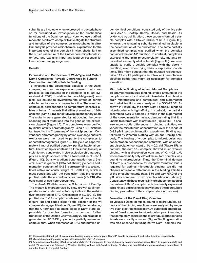
Figure 1. Expression and Characterization of Wild-Type and Mutant Dam1 Complexes
(A) Schematic representation of the mutations introduced into Dam1 and Duo1.(B) Coomassie-stained 13.5% gel of the purified complexes showing their respective subunit compositions.(C) Size-exclusion chromatography of Dam1 wild-type (wt), dam1-19, and duo1-2 complexes on a Superose 6 column. Void volume and elutionvolumes of standard proteins (Thyroglobulin, Stokes Radius 8.5 nm, Catalase, Stokes Radius 5.2 nm) are given at the top.
Structure and Function of the Dam1 Ring Complex279
subunits are insoluble when expressed in bacteria have der identical conditions, consisted only of the five sub-units Ask1p, Spc19p, Dad2p, Dad4p, and Hsk3p. Asso far precluded an investigation of the biochemical
functions of the Dam1 complex. Here, we use purified, evidenced by gel filtration, these subunits formed a sta-ble complex with a Stokes radius of 60 A (Figure 1C),reconstituted Dam1 complex to investigate the structure
and function of the complex on microtubules in vitro. whereas the remaining subunits remained insoluble inthe pellet fraction of the purification. The same partiallyOur analysis provides a biochemical explanation for the
important roles of this complex in vivo, sheds light on assembled complex was purified when the complexcontained the duo1-2 mutation. In contrast, complexesthe structural nature of the kinetochore-microtubule in-
terface, and explains important features essential for expressing the Ipl1p phosphorylation-site mutants re-tained full assembly of all subunits (Figure 1B). We werekinetochore biology in general.unable to purify a soluble complex with the dam1-1mutation, even when trying various expression condi-Resultstions. This might suggest that the mutated residue cys-teine 111 could participate in intra- or intermolecularExpression and Purification of Wild-Type and Mutant
Dam1 Complexes Reveals Differences in Subunit disulfide bonds that might be necessary for complexformation.Composition and Microtubule Binding
To investigate the biochemical activities of the Dam1complex, we used an expression plasmid that coex- Microtubule Binding of Wt and Mutant Complexes
To analyze microtubule binding, limited amounts of thepresses all ten subunits of the complex in E. coli (Mi-randa et al., 2005). In addition to the wild-type (wt) com- complexes were incubated with taxol-stabilized bovine
brain microtubules and centrifuged, and supernatantplex, we sought to investigate the effect of certainselected mutations on complex function. These mutant and pellet fractions were analyzed by SDS-PAGE. As
shown in Figure 1D, the entire Dam1 complex binds tocomplexes corresponded to temperature-sensitive al-leles or to dam1 mutants that either prevent (dam1 S4A) microtubules with high affinity. In contrast, the partially
assembled duo1-2 complex is found in the supernatantor mimic (dam1 S4D) constitutive Ipl1p phosphorylation.The mutants were generated by introducing the corre- of the cosedimentation assay, demonstrating that it is
unable to interact with microtubules (Figure 1E). To ana-sponding point mutations into the gene on the expres-sion plasmid (Figure 1A). The complexes were purified lyze more subtle differences in binding affinities, we
varied the microtubule concentration over the range ofby nickel-affinity chromatography using a 6� histidinetag fused to the C terminus of the Hsk3p subunit. Con- 0–3.8 �M in a cosedimentation experiment. Binding was
followed by Western blotting with an anti-Dam1p anti-ventional chromatography by cation exchange and sizeexclusion were then used to purify the complexes to body. The binding of wt complex to microtubules was
concentration dependent and saturable, with an appar-apparent homogenity (Figure 1B). The yield was approxi-mately 1 mg of purified complex per liter bacterial cul- ent dissociation constant of KD �0.2 �M (Figure 1F). In
contrast, the dam1-19 complex showed much weakerture. The wt complex contained all ten subunits in equalstoichiometry and eluted in size exclusion chromatogra- binding, with a dissociation constant of KD �0.8 �M,
whereas maximally only 70% of the mutant complex wasphy as a single species with a Stokes radius of �97 A(Figure 1C). Density gradient centrifugation on a 5%– bound to microtubules. Thus, the C-terminal domain
of Dam1p is dispensable for complex formation but is40% sucrose gradient (data not shown) yielded a sedi-mentation constant of 10.2 S, corresponding to a calcu- required for optimal microtubule binding. We did not
observe noticeable differences in the binding affinitieslated native molecular weight of �390 kDa, which ismost consistent with the conclusion that the species of the phosphomutants dam1S4A and dam1S4D of the
Ipl1 sites compared to wt complex (data not shown).purified under these conditions is a dimer (2 � 218 kDa)consisting of two heterodecamers. Consistent with these results, in vitro phosphorylation of
recombinant Dam1 complex with bacterially expressedThe dam1-19 allele lacks the C terminus of Dam1p.The mutant is characterized by slow growth at all tem- Ipl1p kinase did not significantly change the microtubule
binding properties of the complex (data not shown).peratures and collapsed mitotic spindles at the restric-tive temperature of 37�C (Cheeseman et al., 2001a). Thepurified dam1-19 complex contained all ten subunits Formation of the Dam1 Ring Complex
To visualize Dam1 complex bound to microtubules, ali-(Figure 1B) and eluted close to the position of the wtcomplex during gel filtration (Figure 1C), demonstrating quots of the binding reactions were analyzed by nega-
tive stain electron microscopy. At substoichiometric ra-that the C-terminal 138 amino acids of Dam1p are dis-pensable for complex formation. In contrast, further tios of Dam1 complex to microtubules, prominent rings
that completely encircled the microtubule orthogonal totruncation of the Dam1p C terminus by 20 amino acids togenerate dam1Q185Stop yielded a partially assembled its axis were readily observed (Figure 2A). Ring formation
was also observed by using native Dam1 complex iso-complex that, when expressed at 37�C and purified un-
(D) Coomassie-stained gel of microtubule binding assay of wt complex. S and P denote supernatant and pellet fraction, respectively.(E) Microtubule binding assay of partially assembled duo1-2 complex.(F) Determination of binding affinities for wt and dam1-19 complexes to microtubules by cosedimentation assay. Dam1 in supernatant (S) andpellet (P) fractions was followed by Western blotting with an anti-Dam1 antibody. Binding was quantified and expressed as a percentage ofcomplex found in the pellet fraction.

Molecular Cell280
Figure 2. Electron Microscopy Reveals Decoration, Composition, and Self-Assembly of the Dam1 Ring Complex
(A) Purified Dam1 complex was bound to taxol-stabilized bovine brain microtubules and visualized by electron microscopy (EM) by usingnegative stain. Bar, 50 nm.(B) At higher concentrations of Dam1 complex, complete decoration is observed.(C) Microtubule binding reactions were diluted prior to EM and visualized with negative stain. Dam1 rings in various states of disassemblycan be seen in the vicinity of the microtubule. Dashed box shows smallest subunit corresponding to the basic building block of the ring. Solidbox shows half ring assembled by longitudinal association of the small building blocks. Bar, 10 nm.(D) Pure Dam1 complex at 0.2 mg/ml was incubated on a phospholipid monolayer for 48 hr and visualized by EM using negative stain. Self-assembled Dam1 rings are visible. Bar, 40 nm.
lated directly from yeast cells, the dam1 phosphomu- about 70 A. This size is not compatible with tubulinsubunits, instead the width is the same as observed intants, and the dam1-19 complex (data not shown).
With saturating amounts of complex, complete deco- fragmented rings of the complex seen in the vicinity(Figure 2C, solid box). Thus, these subunits are likelyration of microtubules could be seen (Figure 2B). With-
out additional systematic image analysis of well-ordered the basic building block of the ring and associate longi-tudinally to form rings around microtubules.assemblies, it is not absolutely clear whether at these
higher concentrations Dam1 complex is forming rings The observation of Dam1 ring assembly raised thequestion of whether these structures would only formthat are closely stacked or a continuous helical filament
(or both). We were able to reveal apparent Dam1 ring in the presence of microtubules. In the absence of micro-tubules, Dam1 preparations showed a few small, curveddisassembly intermediates in the vicinity of microtu-
bules by diluting the binding reactions 1000-fold prior aggregates, reminiscent of the disassembly productshown in Figure 2C. However, when the protein wasto electron microscopy (EM) (Figure 2C). These apparent
disassembly products suggest a possible mode of ring bound to lipid monolayers during 2D-crystallization at-tempts, both fragments and full rings were readily ob-subunit organization. The smallest structures visible
were fairly regular in size and shape (Figure 2C, dashed served (Figure 2D). Rings formed under these conditionswere fairly regular in size but somewhat smaller thanbox) with a length of about 105 A and a diameter of
Structure and Function of the Dam1 Ring Complex281
those associated with microtubules (see below), with an Alexa-488 to recombinant Dam1 complex. Labeled com-plex and nonincorporated dye were separated by gelouter diameter of about 450 A and an inner diameter of
about 290 A. Ring assembly was also observed when filtration. Analysis of the Coomassie-stained gel of thispreparation by UV illumination revealed that the labelNi bound NTA-lipids (that interacted with the His-tag)
or negatively charged phospholipids were used, sug- was incorporated into the Spc34p and Spc19p subunitsof the Dam1 complex (Figure 4C). Microtubule bindinggesting that ring assembly is likely the result of concen-
trating the Dam1 complex on a surface. assays and analysis by EM demonstrated that the Alexa-488-Dam1 complex formed normal rings around micro-tubules (Supplemental Figure S1 available online at http://Cryo-EM of GMPCPP Microtubules Decoratedwww.molecule.org/cgi/content/full/17/2/277/DC1)with Dam1 Complexand bound to microtubules with comparable affinity toTo analyze the decoration of microtubules with Dam1wt complex. We digested taxol-stabilized, rhodaminecomplex under native conditions, we used cryo-EM ofmicrotubules with the protease subtilisin to remove thefrozen hydrated samples in vitreous ice. We tested twoC termini of �- and �-tubulin and tested binding of Alexa-types of stabilized microtubule substrates for this analy-labeled Dam1 complex to the microtubules by fluores-sis: taxol-stabilized bovine brain microtubules and mi-cence microscopy. Control microtubules showed robustcrotubules assembled in the presence of the slowlybinding of fluorescently-labeled Dam1 complex (Figureshydrolyzable GTP analog GMPCPP (Guanosine-5�-[(�,�)-4D and 4F). Microtubules decorated with the complexmethyleno]triphosphate). Previous studies have demon-were often organized into thick bundles (see below). Instrated that these microtubules differ in average numbercontrast, subtilisin-digested microtubules showed little,of protofilaments and the precise axial repeat of tubulinif any, decoration with the labeled complex (Figures 4Emonomers along the protofilament (Hyman et al., 1995).and 4G). We conclude that complete removal of theWe noticed that the decoration of GMPCPP microtu-C termini of �- and �-tubulin prevents Dam1 complexbules was more uniform and that the Dam1 rings ap-binding and formation of the Dam1 ring complex on mi-peared more ordered, with long stretches of microtu-crotubules.bules showing regular ring spacing and dimensions
compared to the decoration of taxol-stabilized microtu-bules (data not shown). We therefore used GMPCPP The Dam1 Ring Complex Promotes Tubulinmicrotubules decorated with Dam1 complex to calcu- Polymerization and Microtubule Stability In Vitrolate the dimensions and spacing of the rings. We ana- The Dam1 complex has an important role in spindlelyzed the diffraction pattern of selected stretches of integrity in vivo, as temperature-sensitive mutants of itsdecorated microtubules (Figure 3A). The Fourier trans- subunits are unable to assemble or maintain a normalformation (Figure 3B) revealed layer lines at 43 and 21 A, mitotic spindle. We therefore investigated the effects ofcorresponding to the microtubule lattice (white arrows), the Dam1 complex on microtubule assembly in vitro.and an additional, broader layer line corresponding to Polymerization of pure brain tubulin into microtubulesthe axial repeat of the bound rings around the microtu- was followed by an increase in light scattering at 350bule at about 100 A (white arrowheads). A filtered image nm. Under our conditions, 10 �M tubulin (1 mg/ml) alonegenerated with these three layer lines plus the equator did not assemble into microtubules over the course ofis shown in Figure 3C. The density profile obtained by the experiment. However, addition of Dam1 complex inintegrating this filtered image along the microtubule axis substoichiometric amounts led to a rapid, concentra-is shown beneath. From the major peaks in the density tion-dependent increase in light scattering (Figure 5A).profile we estimate the outer diameter of the rings to To confirm that this increase was due to the formationbe about 540 A and the inner diameter to be 320 A. of microtubules, an aliquot of the reaction was fixed
with glutaraldehyde and centrifuged onto coverslips,and the microtubules were visualized by immunofluores-Complex Binding and Ring Formation Depends
on the Presence of the C Terminus of ��-Tubulin cence with a tubulin antibody. This analysis confirmedthe formation of numerous microtubules in the presenceThe wrapping of the Dam1 rings around the microtubule
strongly suggested an involvement of the C-terminal of Dam1 complex, but not in the control reaction (Fig-ure 5B).tails of �� and �-tubulin in its interaction with the micro-
tubule. To test this possibility, we analyzed the decora- A complex that forms a ring around a microtubulemight increase the stability of the polymer. We testedtion of subtilisin-digested or control microtubules by
cryo-EM. Extended subtilisin digest resulted in the re- this possibility by assembling microtubules from 15 �Mtubulin in the presence and absence of Dam1 complex.lease of the C termini of both �- and �-tubulin (Figure 4A).
Complete digestion was confirmed by Western blotting These microtubules were then rapidly diluted, and theirdisassembly was followed by light scattering and bywith a �-tubulin antibody whose epitope is located in
the C-terminal domain (data not shown). Control micro- a sedimentation assay. Figure 5C demonstrates thatcontrol microtubules rapidly depolymerized when theytubules showed strong decoration by the Dam1 ring
complex, whereas only bare microtubules were found in were diluted below the critical concentration and thatthe bulk of tubulin was found in the supernatant afterthe subtilisin-digested sample (Figure 4B). Upon shorter
digests, which only removed the C terminus of �-tubulin, centrifugation (Figure 5D). In contrast, microtubules as-sembled in the presence of Dam1 complex depoly-we still observed significant ring formation (data not
shown). merized more slowly, retained a higher light scatteringsignal, and had more tubulin in the pellet of the centrifu-To further analyze binding of the Dam1 complex to
microtubules, we covalently linked the fluorescent dye gation. Thus, the Dam1 complex promotes microtubule

Molecular Cell282
Figure 3. Analysis of Dam1 Ring Decoration on GMPCPP Microtubules by Cryo-EM
(A) Analysis of Dam1 decoration. Cryo-EM micrograph of Dam1 decorated microtubule. Black arrowheads point to adjacent Dam1 rings.(B) Fourier transform of microtubule as shown in (A). White arrows point to a series of horizontal lines (43 and 21 A layer lines) generated bydiffraction of the microtubule. White arrowheads point to the layer line at 100 A generated by diffraction from the Dam1 complex.(C) Computer-filtered image of the analyzed microtubule. Density profile of the filtered image across the microtubule and estimation of thedimensions of the Dam1 ring complex. Bars, 100 nm.
assembly and stabilizes microtubules against dilution- insert), suggesting that bundling could be aided by di-rect ring-to-ring interactions.induced disassembly.
The Dam1 Ring Complex Organizes Microtubules Binding of Alexa 488-Labeled Dam1 Ring Complexto Various Microtubule Substrates:into Long Bundles In Vitro
Next, we investigated the effects of the Dam1 complex Preference for a GTP LatticeWe used Alexa-labeled Dam1 complex to analyze furtheron the assembly of higher concentrations of tubulin.
In our assembly assay, 15 �M tubulin showed a slow aspects of its binding to various microtubule substrates.When rhodamine-labeled GMPCPP microtubules wereincrease in light scattering with an extended lag period
(Figure 5E). Increasing the amount of Dam1 complex incubated with limiting amounts of Alexa-Dam1 complexand then visualized by fluorescence microscopy, a strik-increased the rate of tubulin assembly and reduced the
lag period. Importantly, fixed aliquots of the assembly ing punctate decoration of microtubules was observed(Figures 6A and 6B). Accordingly, a line scan of the Alexareactions that were sedimented onto coverslips and
stained with an anti-tubulin antibody revealed distinct signal along a single GMPCPP microtubule revealeddiscrete signals that often displayed the same intensitymicrotubule morphologies: whereas the control reac-
tions displayed single microtubules with an average (Figure 6C). As the same binding conditions revealedsingle ring decoration by EM, the fluorescent signalslength of 10–15 �m (Figure 5F), microtubules assembled
in the presence of the Dam1 complex were frequently likely corresponded to individual rings.To fulfill their function, kinetochores must associateorganized into long bundles (Figure 5G) with an average
length of 35–45 �m. Analysis of these microtubules by with and remain attached to microtubule ends. Kineto-chore proteins assembled on beads coated with centro-EM revealed the presence of bundles of typically four to
six microtubules that were decorated to varying degrees meric DNA in vitro can preferentially bind to the GTPlattice of a microtubule (Severin et al., 1997). However,with Dam1 rings (Figure 5I). Sometimes rings on adjacent
microtubules showed direct lateral contacts (Figure 5I which subunits of the kinetochore are responsible for

Structure and Function of the Dam1 Ring Complex283
Figure 4. Dam1 Complex Binding and Ring Formation Depend on the �- and �-Tubulin C Termini
(A) Taxol-stabilized bovine brain microtubules were digested with subtilisin. Coomassie-stained gel (containing 6 M urea to enhance separationof �- and �-tubulin) of the digest showing shift of �- and �-tubulin mobility.(B) Cryo-EM analysis of Dam1 complex binding to subtilisin-digested microtubules. Control microtubules show normal decoration (top),whereas only bare microtubules are observed in the protease treated preparation (bottom). Bar, 50 nm.(C) Preparation of Alexa-labeled Dam1 complex. Coomassie-stained gel of preparation is on the left. UV illumination of the gel reveals covalentlabeling of the Spc34 and Spc19 subunits of the Dam1 complex by the dye.(D) Rhodamine-labeled control microtubules (red) bind Alexa488-labeled Dam1 complex (green) and get bundled (F), whereas subtilisin-digested microtubules (E) do not (G). Bar, 10 �m.
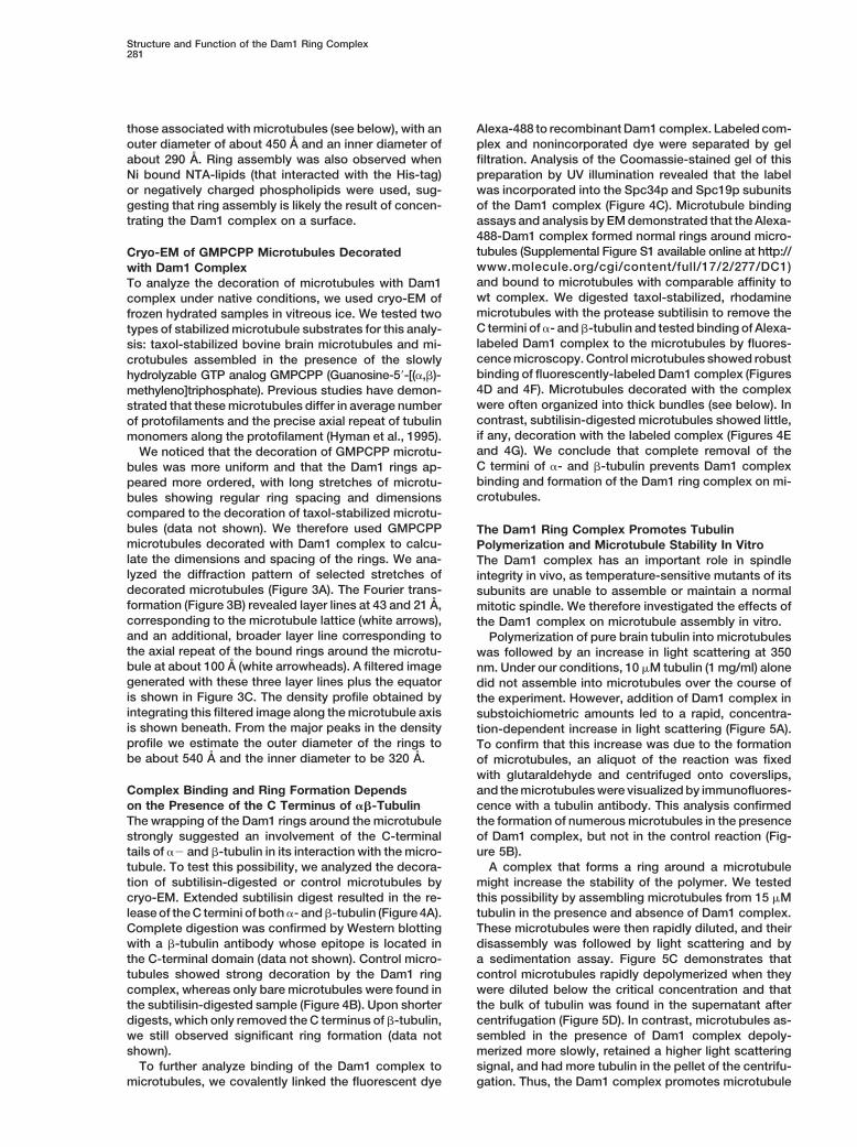
this important feature is not known. As GMPCPP micro- The corresponding intensity scan further revealed thisdistribution of the complex (Figure 6F). We quantifiedtubules, which mimic the GTP lattice of a microtubule,
seemed to be the preferred substrate for Dam1 decora- the decoration by counting clearly segmented microtu-bules in randomly chosen fields and ordered them intotion in our cryo-EM analysis, we sought to analyze fur-
ther whether the Dam1 ring complex showed a prefer- three categories (Figure 6G); more than 50% (n � 100)of the segmented microtubules showed a strong deco-ence for this type of lattice over a GDP lattice. For this
experiment, we prepared segmented microtubules that ration of the GMPCPP segment and one end of themicrotubule (Figure 6G). In most cases (�80%), thisconsisted of an unlabeled GMPCPP seed that was elon-
gated by using rhodamine-labeled GTP-tubulin. After end decoration was on the longer rhodamine segment,suggesting that it corresponds to the plus end of theincorporation into the lattice, the GTP is rapidly hy-
drolyzed, thus producing segmented microtubules con- microtubule. In about 35% of the microtubules, thestrong decoration of just the GMPCPP segment wassisting of an unlabeled GMPCPP segment and two la-
beled GDP segments. The segmented microtubules apparent, whereas only 12% of microtubules displayedevenly distributed Dam1 rings. The preferred binding towere mixed with limiting amounts of Alexa-labeled Dam1
complex, fixed, and visualized by fluorescence micros- the GMPCPP segement was not due to the lack of arhodamine label on this part of the lattice, as binding tocopy. Strikingly, a stronger decoration of the segmented
microtubules with Alexa488-Dam1 complex was found segmented control microtubules that were completelystabilized with taxol showed no bias for the unlabeledon the GMPCPP parts of the lattice (Figures 6D and 6E).

Molecular Cell284
Figure 5. Biochemical Activities of the Dam1 Ring Complex on Microtubules In Vitro
(A) Light-scattering assay of tubulin assembly in the absence and presence of the Dam1 complex. Addition of Dam1 complex increases rateof tubulin assembly.(B) Reactions from (A) were fixed, centrifuged, and pelleted onto coverslips. Staining with an anti-tubulin antibody demonstrates that microtu-bules are only formed in the presence of Dam1 complex.(C) Microtubule stability experiment. Microtubules were assembled from 15 �M tubulin in the presence and absence of Dam1 complex andthen rapidly diluted.(D) Reactions from (C) were centrifuged, supernatants (S) and pellets (P) were separated, run on a 12.5% SDS-PAGE gel, and stained withCoomassie. Black dots indicate subunits of the Dam1 complex.(E) Assembly of 15 �M tubulin incubated with increasing amounts of Dam1 complex followed by light scattering.
Structure and Function of the Dam1 Ring Complex285
Figure 6. Analysis of Microtubule Binding with the Alexa488-Dam1 Complex
(A) GMPCPP-stabilized, rhodamine microtubules were incubated with limiting amounts of Alexa-Dam1 complex and visualized by fluores-cence microscopy.(B) Note the punctate decoration of microtubules with the Alexa-labeled complex.(C) Intensity scan of the microtubule reveals discrete signals.(D) Decoration of segmented GMPCPP/GDP microtubules with Alexa-Dam1 complex. Microtubules are composed of an unlabeled segmentassembled with GMPCPP and two rhodamine labeled segments containing GDP-tubulin.(E) The Alexa-labeled complex preferentially binds to the GMPCPP segments of the microtubules. Bar, 5 �m.(F) Intensity scan shows stronger decoration on the GMPCPP segment and on one end of the microtubule.(G) Quantitation of the decoration of segmented microtubules. 100 clearly segmented microtubules in random fields were counted and orderedinto three categories: (1) microtubules with a stronger decoration of the GMPCPP segment or one end, (2) microtubules with a strongerdecoration only of the GMPCPP segment, and (3) microtubules with an evenly distributed decoration.
segments (data not shown). Thus, the cryo-EM analysis crotubules are not stable, they rapidly disassembledupon dilution. When the distribution of Dam1 rings wastogether with the decoration of segmented GTP/GDP
microtubules suggest that a GTP lattice is the preferred analyzed after disassembly of the GDP segments, wenoticed that both ends of the remaining GMPCPP seg-binding partner for the Dam1 ring complex.ments were strongly labeled for Dam1 rings, suggestingthat during microtubule disassembly, rings remainedBehavior of the Dam1 Ring Complex
on Depolymerizing Microtubules: attached to the shortening microtubules (data notshown). We wanted to analyze this behavior of the Dam1Evidence for Ring Sliding
During the course of the previous experiments, we no- ring complex in a more controlled manner. GMPCPPmicrotubules were loaded with limiting amounts of Dam1ticed that because the GDP segments of the mixed mi-
(F) Control reaction from (E) centrifuged onto coverslips and stained with a tubulin antibody.(G) Assembly in the presence of Dam1 complex leads to the formation of long bundles of microtubules. Bar, 10 �m.(H) Quantitation of bundling activity. Clearly recognizable bundles in randomly chosen fields were counted and compared to a control.(I) Bundling visualized by EM using negative stain. Insert shows rings on adjacent microtubules directly contacting each other. Bar, 100 nm.

Molecular Cell286
Figure 7. Dynamic Behavior of the Dam1 Ring Complex on Depolymerizing Microtubules
(A and B) Distribution of Alexa-labeled Dam1 complex (green) on GMPCPP microtubules before and after depolymerization induced byXMCAK1. Microtubules were incubated with limiting amounts of Alexa-labeled complex. At t � 0 microtubules show weak punctate decorationwith no preference for ends (A). After incubation with the depolymerizing kinesin XKCM1, only short microtubules with brightly-labeled endsare found (B). Bar, 5 �m.(C) In a parallel experiment, samples were visualized by EM using negative stain. After 2.5 min incubation with XMCAK1, rings accumulateon both ends behind the peeling protofilaments. After 5 min, only very short microtubules that are densely packed with Dam1 rings are found.After 10 min, microtubules disintegrate and release the Dam1 complex. Bar, 50 nm.

Structure and Function of the Dam1 Ring Complex287
rings. To induce disassembly of these microtubules, we self-assembly into rings in the absence of microtubules.At this point, it is not clear how binding to the lipidadded the kinesin XMCAK1, which catalyzes depoly-
merization by peeling protofilaments from both ends of monolayer contributes to ring formation. It is likely thatthe effect is the result of an increase in local concentra-the microtubules (Desai et al., 1999). The reactions were
fixed at various times and visualized by fluorescence tion of the protein complex as it binds to the monolayer.The microtubule could have a similar effect, interactingmicroscopy. Figure 7A shows a representative example
of the decoration of GMPCPP microtubules at t � 0: a with the Dam1 complexes and thus increasing their localconcentration at the microtubule surface above the criti-long microtubule is sparsely labeled with Dam1 rings
that show no obvious preference for the ends (see also cal concentration for self-assembly. In addition, the neg-ative charge on the C terminus of tubulin could alsoFigure 6B). After 2.5 min incubation with the enzyme,
the bulk of microtubules have depolymerized, and the neutralize the overall positive charge in the Dam1 com-plexes and thus facilitate their interaction. Indeed, itremaining short microtubules show a decoration with a
characteristic accumulation of rings on both ends of the seems likely that electrostatic interactions play an im-portant role in this process as high-salt conditions pre-microtubules (Figure 7B). Out of 50 short microtubules
analyzed after XMCAK1 incubation, 41 showed a strong vent oligomerization and microtubule binding, whereaslow-salt conditions support them (S.W., D.G.D., andAlexa signal on both ends (82%). To confirm that the
strong Alexa signal indeed corresponded to an accumu- G.B., unpublished data). The behavior of the complexin this regard is very reminiscent of the GTPase dynamin,lation of rings on the ends of depolymerizing microtu-
bules, we performed a parallel experiment in which sam- which self-assembles into rings and helical stacks underlow-salt conditions and is thought to form these struc-ples were analyzed by EM using negative stain. Figure
7C shows a representative microtubule after 2.5 min tures around the necks of invaginated coated pits tosupport constriction (Hinshaw and Schmid, 1995). Inter-incubation with XMCAK1. The ends of the microtubule
show a dense decoration with Dam1 ring complex just estingly, dynamin was first purified as a microtubule-associated protein (Shpetner and Vallee, 1989).behind the peeling protofilaments, corresponding well
to the fluorescence image in Figure 7B. After 5 min,only a few short microtubules were visible. These were Why Form a Ring?completely decorated with Dam1 rings along their The assembly of ten small proteins into a complex, andlength. After 10 min, the remaining microtubules disinte- the subsequent oligomerization of multiple complexesgrate and release the complex. Taken together, these into a ring, seems to be a very complicated way toobservations support the conclusion that upon depoly- construct a microtubule binding structure. However, amerization of the microtubule, the Dam1 rings do not microtubule binding ring might be uniquely suited toimmediately dissociate or disassemble but remain fulfill the functions of the Dam1 complex within the cell.attached to the microtubule ends and slide back with We demonstrated that the Dam1 ring complex is anpeeling protofilaments. excellent microtubule stabilizer and that it strongly pro-
motes microtubule growth through the addition of fur-ther subunits. Because the stability of a microtubule isDiscussionthought to be largely governed by the lateral interactionsbetween adjacent protofilaments (Nogales et al., 1999),Assembly of the Dam1 Ring Complex
The rings formed around microtubules by the Dam1 rings that bind orthogonal to the microtubule axis areexpected to strengthen interprotofilament interactionscomplex are remarkable structures that are clearly not
formed by any other microtubule binding protein investi- and additionally to prevent protofilament peeling, whichin turn would encourage further growth (Figure 7D, top).gated to date. All other microtubule binding proteins
whose association with microtubules has been visual- Additional observations identify another potentiallysignificant property of Dam1 ring complexes. We dem-ized follow the path of the protofilament and/or bind
with the axial repeat of tubulin dimers. One exception onstrated that binding between the ring and the microtu-bule is mediated by flexible domains, involving the Cis -tubulin and its associated proteins in the -tubulin
ring complex, which form a ring that binds to the micro- terminus of Dam1p and the C terminus of ��-tubulin,and is largely electrostatic in nature. These observationstubule minus end (Zheng et al., 1995). Our studies indi-
cate that the Dam1 rings are formed by longitudinal self- open as a possibility the lateral sliding or diffusion ofDam1 rings along microtubules. It should be noted thatassembly of multiple copies of the Dam1 complex upon
the microtubule surface. Although the presence of mi- some single-headed kinesins are thought to move byundirected lateral diffusion along the microtubule andcrotubules strongly facilitates the oligomerization pro-
cess, ring assembly seems to be an intrinsic property that this type of interaction is also mediated by theflexible C-terminal domains of ��-tubulin (Okada andof the Dam1 complex, as we have been able to induce
(D) Model for the molecular functions and behaviors of the Dam1 ring complex. Top, preferred binding of the complex to the GTP cap ofgrowing microtubules could stabilize the cap and promote the addition of further tubulin subunits. Lateral diffusion of the rings or de novoassembly can ensure continued association of Dam1 rings with the GTP cap. In case of a catastrophe, peeling protofilaments shorten themicrotubule, but the rings do not disassemble but instead slide back on the lattice. Thus, Dam1-associated chromosomes could maintainassociation with growing and shrinking microtubules. Bottom, lateral mobility of the Dam1 ring complex could allow dynamic bundling ofmicrotubules. Bundling of microtubules with opposite polarity by ring-to-ring interaction could allow motors to move the microtubules againsteach other while maintaining integrity of the bundle. This could be important for spindle elongation during anaphase B.

Molecular Cell288
Hirokawa, 2000). Moreover, we provide evidence for this destabilization of the Dam1 complex at the restrictivetype of dynamic ring behavior. When we analyzed the temperature in vivo. Interestingly, three subunits of thisdistribution of fluorescent Dam1 rings after disassembly partial complex, Ask1p, Spc19p, and Dad2p, are allof microtubules, we found an accumulation of rings at acidic proteins (pI � 4.47, 4.59, and 3.97, respectively),the ends of the microtubule, suggesting that the rings and we demonstrated that they are unable to bind todo not disassemble but slide back as the protofilaments microtubules, indicating that the microtubule bindingpeel away from the ends (Figure 7D). module of the Dam1 complex is formed by the highly
We furthermore demonstrated that the Dam1 ring com- basic proteins Dam1p, Duo1p, and Spc34p (pI � 9.97,plex bundles microtubules in vitro. Lateral mobility of the 10.76, and 8.6, respectively). This suggestion of sub-rings could be important for “dynamic bundling” of micro- complexes within the Dam1 complex agrees well withtubules in which rings on bundled microtubules of mixed results from two-hybrid and direct binding assays, whichpolarity interact directly with each other. This would allow indicate that Dam1p, Duo1p, and Spc34p interactmotors to slide these bundled microtubules against closely with each other (Ikeuchi et al., 2003; Shang eteach other (Figure 7D, bottom) without disrupting ana- al., 2003).phase spindle integrity. Whereas the wt Dam1 complex bound strongly to
microtubules with a dissociation constant of Kd �The Role of a Ring at the Kinetochore 0.2 �M, which is in the range determined for brain MAPsTo explain how kinetochores can remain attached to like tau (Goode and Feinstein, 1994), binding of thegrowing and shortening microtubules, the existence of dam1-19 complex to microtubules was markedly re-sleeves or collars into which the kinetochore microtu- duced, implicating the C-terminal domain of Dam1p inbules insert have been hypothesized (Hill, 1985). The establishing the contact to the microtubule. Importantly,Dam1 ring complex could form such a sleeve in budding dam1 mutants that mimic or prevent Ipl1p phosphoryla-yeast. Moreover, our cryo-EM analysis, as well as the tion of the complex showed regular subunit compositiondecoration of mixed lattice microtubules with fluores- and wt microtubule binding activities. Thus, Auroracent Dam1 complex, suggest that a GMPCPP lattice phosphorylation does not seem to destabilize or influ-(which mimics the GTP or GDP-Pi bound state of the ence the microtubule binding activities of the complex.microtubule) is the preferred binding partner for the This is in agreement with the observation that dam1Dam1 ring complex. As one consequence, ring forma- phosphomutants and ipl1 ts mutants do not show grosstion may be sensitive to the small structural changes in spindle integrity phenotypes (Cheeseman et al., 2002a;the lattice that accompany GTP hydrolysis or Pi release. Kang et al., 2001). An important future aim will be to mapSuch a feature of the Dam1 complex would allow the onto the ring structures the locations of the individualkinetochore to recognize and preferentially bind to the subunit proteins and the surfaces that are phosphory-microtubule plus end. Upon attachment of the kineto- lated by Ipl1p and other kinases.chore to the microtubule via the Dam1 ring complex,the microtubule plus end would be stabilized by the ring
Experimental Procedurescomplex (Figure 7A). Similar to a proposal based onin vitro studies of kinetochore-microtubule interactions
Purification of Recombinant Dam1 Complex from Bacteria(Severin et al., 1997), GTP hydrolysis is expected toTo express recombinant Dam1 complex, plasmid pC43HSK3H (Mi-produce a lattice that is less favorable for Dam1 ringranda et al., 2005) was transformed into BL21Rosetta cells (Nova-
formation, and the kinetochore would therefore occupy gen) containing the pRARE plasmid. 2.5 l Eschericia Coli bacterialthe newly formed GTP cap by lateral diffusion or de novo culture was grown at 37�C to OD 0.4, and protein expression wasring assembly (Figure 7A). induced by the addition of 1 mM IPTG. Bacteria were harvested after
4 hr, washed in phosphate buffer saline (PBS), and resuspended inIn future studies, it will be important to analyze the20 mM sodium phosphate (pH 6.8), 500 mM NaCl, 1 mM EDTA,dynamic behavior of the Dam1 ring complex on microtu-20 mM Imidazol, and 0.5% (v/v) Triton X-100. Cells were lysed bybules, the attachment of further kinetochore complexessonication, and the lysate was cleared by centrifugation. The super-to this binding interface, and the molecular details ofnatant was incubated with Ni-NTA Agarose (Qiagen), equilibrated
the phospho regulation of this attachment by the Aurora- in PBS, and rotated for 2 hr at 4�C. Agarose beads were collectedkinase Ipl1p. Furthermore, whether a similar ring com- by low-speed centrifugation, washed twice, and resuspended inplex is formed by the kinetochores of higher animals 6 ml 20 mM sodium phosphate, 500 mM NaCl, 1 mM EDTA, and
200 mM imidazole. After 2 hr rotation at 4�C, the supernatant waswill be an important question for the future.collected and dialyzed overnight against 20 mM sodium phosphate,150 mM NaCl, and 1 mM EDTA. The dialysate was then loaded ontoInsights into Organization and Functionsa 1 ml HiTrap SP sepharose cation exchange column (Amerham
of Dam1 Subunits Pharmacia), and proteins were eluted by using a 10 ml linear gradientThe biological functions and regulatory interactions of of 0.15 to 1 M NaCl in Buffer A. Dam1 complex was eluted at 600the Dam1 complex have been extensively studied by mM NaCl. The corresponding fractions were collected, concen-
trated with a Centricon 30 (Amicon) device (preblocked with 2%genetic analysis. Our biochemical and structural analy-BSA), and applied to a Superose 6 gel filtration column. The columnses of wt and mutant Dam 1 complexes have allowedwas developed with 25 mM sodium phosphate (pH 6.8), 500 mMus to analyze the contributions of specific subunits toNaCl, and 1 mM EDTA. Dam1 containing fractions were snap frozencomplex function and to relate biological and biochemi-in liquid N2 and stored at �80�C.
cal activities. Mutations like duo1-2 or dam1Q185Stop Mutations in Dam1 and Duo1 were introduced by Quikchangelead to the expression of a partially assembled complex site-directed mutatgenesis on the pC43HSK3H expression plasmdconsisting of Ask1p, Spc19p, Dad2p, Dad4p, and Hsk3p. (Stratagene). They were confirmed by DNA sequencing, and the
mutant complexes were expressed and purified as described above.This suggests that the duo1-2 mutation could lead to a

Structure and Function of the Dam1 Ring Complex289
Fluorescent Labeling of Dam1 Complex with Alexa-488 Limited Proteolysis of Tubulin with SubtilisinTo remove the C terminus of both �- and �-tubulin, 15 �M taxol-500 �l HiTrapS purified Dam1 complex (�500 �g) was incubated
with 100 �M Alexa-488-C5-maleimide (Molecular Probes) for 1 hr stabilized microtubules were incubated with 40 �g/ml subtilisin inPEM buffer for 1 hr at 30�C. The reaction was stopped by the additionat 0�C in the dark. The reaction was stopped by the addition of 1 mM
DTT, and the sample was purified by gel filtration on a Superose of 1 mM PMSF, the microtubules were pelleted by centrifugation(TLA100, 15 min, 60 K, and 25�C), washed twice, and resuspended6 column (Amersham Pharmacia). The labeling stoichiometry was
calculated from the absorption spectrum and found to be 1.9 mol in the original volume of PEM-buffer.dye/mole complex. Alexa-488 Dam1 complex was snap frozen in50 �l aliquots and stored at �80�C. After thawing, the sample was Acknowledgmentskept at 4�C, as repeated freezing and thawing led to decreasedfluorescence and degradation of the complex. The authors wish to thank J.J. Miranda and Stephen Harrison for
generously providing the Dam1 expression plasmid and Claire Walz-cak for the kind gift of recombinant XMCAK1. The authors thank allMicrotubule Polymerization Assaysmembers of the Barnes/Drubin Lab for discussions. This work wasTubulin polymerization was followed by right angle light scatteringsupported by grants from the National Institute of General Medicalat 350 nm with a band pass of 1.78 nm in a Fluoromax-Spectropho-Sciences to G.B. (GM-47842), and E.N. (GM51487-09), the Office oftometer (Horiba). The absorption was recorded every 10 s for 1600 s.Basic Energy Science of the US Department of Energy under con-70 �l polymerization reactions containing the indicated amounts oftract DE-AC03-76F00098, and a postdoctoral fellowship of thetubulin and Dam1 complex in 80 mM PIPES/KOH, (pH 6.8), 1 mMDeutsche Forschungsgemeinschaft (DFG) to S.W. E.N. is a HowardMgCl2, and 1 mM EGTA (PEM) with 25% (v/v) glycerol were pipettedHughes Medical Institute Investigator. Research in this article wason ice and transferred to the prewarmed 37�C cuvette chamber.supported in part by a grant to D.G.D. from Philip Morris USA Inc.After the reaction, an aliquot was fixed by the addition of glutaralde-and Philip Morris International.hyde to 0.1% (v/v) and centrifuged onto coverslips as described
(Evans et al., 1985). The coverslips were fixed for 3 min in methanol,Received: December 2, 2004rehydrated in PBS 1 mg/ml BSA 0.1% NP-40 (PBN) and incu-Revised: December 29, 2004bated with Tub2.1-FITC conjugated anti-�-tubulin antibody (SigmaAccepted: December 30, 20041:50 in PBN) for 20 min at RT.Published: January 20, 2005To follow dilution-induced disassembly, microtubules were as-
sembled from 15 �M tubulin in the presence and absence of Dam1Referencescomplex. The polymerization reactions were diluted 1:1 by the addi-
tion of 70 �l warm PEM buffer, and disassembly was followed by lightCheeseman, I.M., Enquist-Newman, M., Muller-Reichert, T., Drubin,scattering. The reaction was transferred to TLA 100 tubes (Beckman)D.G., and Barnes, G. (2001a). Mitotic spindle integrity and kineto-and centrifuged for 15 min at 60 K. Supernatant and pellet fractionschore function linked by the Duo1p/Dam1p complex. J. Cell Biol.were analyzed on a 12.5% SDS-PAGE.152, 197–212.
Cheeseman, I.M., Brew, C., Wolyniak, M., Desai, A., Anderson, S.,Preparation of Segmented GMPCPP/GDP Microtubules Muster, N., Yates, J.R., Huffaker, T.C., Drubin, D.G., and Barnes, G.3 �M unlabeled tubulin was assembled with 100 �M GMPCPP (Jena (2001b). Implication of a novel multiprotein Dam1p complex in outerBioscience) in a 10 �l volume for 15 min at 37�C. To elongate these kinetochore function. J. Cell Biol. 155, 1137–1146.GMPCPP seeds, 10 �l of 50 �M tubulin (5:1 unlabeled tubulin:rho-
Cheeseman, I.M., Drubin, D.G., and Barnes, G. (2002a). Simple cen-damine tubulin) with 1 mM GTP was added and elongation wastromere, complex kinetochore: linking spindle microtubules andallowed to continue for 15 min. 2 �l of the segmented microtubulecentromeric DNA in budding yeast. J. Cell Biol. 157, 199–203.mix was incubated with 2 �l Alexa-488 Dam1 complex (dialyzedCheeseman, I.M., Anderson, S., Jwa, M., Green, E.M., Kang, J.,against 25 mM potassium phosphate [pH 6.8], 150 mM KCl, and 1Yates, J.R., 3rd, Chan, C.S., Drubin, D.G., and Barnes, G. (2002b).mM EDTA). Binding was allowed for 10 min, then 1 �l of the bindingPhospho-regulation of kinetochore-microtubule attachments by thereaction was fixed with 1 �l 1% glutaraldehyde, 7 �l PEM, and 1 �lAurora kinase Ipl1p. Cell 111, 163–172.10� Oxygen-Scavenging-Mix (1 � OSM:200 �g/ml glucose oxidase,
35 �g/ml catalase, 4.5 mg/ml glucose, and 0.5% �-mercaptoetha- De Wulf, P., McAinsh, A.D., and Sorger, P.K. (2003). Hierarchicalnol). 2 �l of this mix was squashed under an 18 � 18 mm coverslip assembly of the budding yeast kinetochore from multiple subcom-and viewed by fluorescence microscopy with a 60� Nikon objective plexes. Genes Dev. 17, 2902–2921.on a Nikon Eclipse fluorescence microscope. Desai, A., Verma, S., Mitchison, T.J., and Walczak, C.E. (1999). Kin
To visualize Alexa-labeled Dam1 complex on depolymerizing mi- I kinesins are microtubule-destabilizing enzymes. Cell 96, 69–78.crotubules, 1.5 �l GMPCPP microtubules loaded with Alexa-Dam1
Enquist-Newman, M., Cheeseman, I.M., Van Goor, D., Drubin, D.G.,complex were incubated with 2 �l recombinant XKCM1 (residues
Meluh, P., and Barnes, G. (2001). Dad1p, third component of the187–592, 0.3 �M), 1.5 �l 10 mM MgATP, 3 �l PEM, and 1 �l 10x OSM.
Duo1p/Dam1p complex involved in kinetochore function and mitoticThe reactions were stopped by addition of 1 �l 1% glutaraldehyde.
spindle integrity. Mol. Biol. Cell 12, 2601–2613.
Evans, L., Mitchison, T., and Kirschner, M. (1985). Influence of theMicrotubule Binding Assays centrosome on the structure of nucleated microtubules. J. Cell Biol.Binding assays were performed essentially as described (Cheese- 100, 1185–1191.man et al., 2001a). Prior to binding, the Dam1 complex was dialyzed
Goode, B.L., and Feinstein, S.C. (1994). Identification of a novelin Slide-A-Lyzer (Pierce) minidialysis units against 20 mM sodium
microtubule binding and assembly domain in the developmentallyphosphate (pH 6.8), 150 mM NaCl, and 1 mM EDTA. The dialysate
regulated inter-repeat region of tau. J. Cell Biol. 124, 769–782.was diluted with PEM buffer and centrifuged at 60,000 rpm for 15 min
He, X., Rines, D.R., Espelin, C.W., and Sorger, P.K. (2001). Molecularin a TL100 rotor to remove aggregates. Binding to taxol-stabilizedanalysis of kinetochore-microtubule attachment in budding yeast.microtubules was allowed for 10 min at RT. Microtubules were thenCell 106, 195–206.sedimented by centrifugation at 25,000 rpm in a TL100 rotor for 20Hill, T.L. (1985). Theoretical problems related to the attachment ofmin at RT.microtubules to kinetochores. Proc. Natl. Acad. Sci. USA 82, 4404–4408.EMHinshaw, J.E., and Schmid, S.L. (1995). Dynamin self-assemblesElectron microscopy of Dam1-decorated microtubules or self-into rings suggesting a mechanism for coated vesicle budding. Na-assembled Dam1 complex, as well as cryo-EM of samples frozenture 374, 190–192.in vitreous ice, was performed according to published protocols.
Detailed descriptions of these procedures are available as Supple- Hofmann, C., Cheeseman, I.M., Goode, B.L., McDonald, K.L.,Barnes, G., and Drubin, D.G. (1998). Saccharomyces cerevisiaemental Data online.

Molecular Cell290
Duo1p and Dam1p, novel proteins involved in mitotic spindle func- Drubin, D.G., and Barnes, G. (2003). Architecture of the buddingyeast kinetochore reveals a conserved molecular core. J. Cell Biol.tion. J. Cell Biol. 143, 1029–1040.163, 215–222.Hyman, A.A., Chretien, D., Arnal, I., and Wade, R.H. (1995). StructuralWieland, G., Orthaus, S., Ohndorf, S., Diekmann, S., and Hemmerich,changes accompanying GTP hydrolysis in microtubules: informationP. (2004). Functional complementation of human centromere proteinfrom a slowly hydrolyzable analogue guanylyl-(alpha,beta)-methyl-A (CENP-A) by Cse4p from Saccharomyces cerevisiae. Mol. Cell.ene-diphosphonate. J. Cell Biol. 128, 117–125.Biol. 24, 6620–6630.Ikeuchi, A., Sasaki, Y., Kawarasaki, Y., and Yamane, T. (2003). Ex-Zheng, Y., Wong, M.L., Alberts, B., and Mitchison, T. (1995). Nucle-haustive identification of interaction domains using a high-ation of microtubule assembly by a gamma-tubulin-containing ringthroughput method based on two-hybrid screening and PCR-con-complex. Nature 378, 578–583.vergence: molecular dissection of a kinetochore subunit Spc34p.
Nucleic Acids Res. 31, 6953–6962.
Janke, C., Ortiz, J., Tanaka, T.U., Lechner, J., and Schiebel, E. (2002).Four new subunits of the Dam1-Duo1 complex reveal novel func-tions in sister kinetochore biorientation. EMBO J. 21, 181–193.
Jones, M.H., Bachant, J.B., Castillo, A.R., Giddings, T.H., and Winey,M. (1999). Yeast Dam1p is required to maintain spindle integrityduring mitosis and interacts with the Mps1p kinase. Mol. Biol. Cell10, 2377–2391.
Jones, M.H., He, X., Giddings, T.H., and Winey, M. (2001). YeastDam1p has a role at the kinetochore in assembly of the mitoticspindle. Proc. Natl. Acad. Sci. USA 98, 13675–13680.
Kang, J.-s., Cheeseman, I.M., Kallstrom, G., Velmurugan, S., Barnes,G., and Chan, C.S.M. (2001). Functional cooperation of Dam1, Ipl1,and the inner centromere protein (INCENP)-related protein Sli15during chromosome segregation. J. Cell Biol. 155, 763–774.
Kosco, K.A., Pearson, C.G., Maddox, P.S., Wang, P.J., Adams, I.R.,Salmon, E.D., Bloom, K., and Huffaker, T.C. (2001). Control of micro-tubule dynamics by Stu2p is essential for spindle orientation andmetaphase chromosome alignment in yeast. Mol. Biol. Cell 12, 2870–2880.
Li, Y., Bachant, J., Alcasabas, A.A., Wang, Y., Qin, J., and Elledge,S.J. (2002). The mitotic spindle is required for loading of the DASHcomplex onto the kinetochore. Genes Dev. 16, 183–197.
Maiato, H., Deluca, J., Salmon, E.D., and Earnshaw, W.C. (2004).The dynamic kinetochore-microtubule interface. J. Cell Sci. 117,5461–5477.
McAinsh, A.D., Tytell, J.D., and Sorger, P.K. (2003). Structure, func-tion, and regulation of budding yeast kinetochores. Annu. Rev. CellDev. Biol. 19, 519–539.
Miranda. J.J.L., De Wulf, P., Sorger, P.K., and Harrison, S.C. (2005).The yeast DASH complex forms closed rings on microtubules. Nat.Struct. Mol. Biol., in press.
Nogales, E., Whittaker, M., Milligan, R.A., and Downing, K.H. (1999).High-resolution model of the microtubule. Cell 96, 79–88.
Okada, Y., and Hirokawa, N. (2000). Mechanism of the single-headedprocessivity: diffusional anchoring between the K-loop of kinesinand the C terminus of tubulin. Proc. Natl. Acad. Sci. USA 97, 640–645.
Pearson, C.G., Maddox, P.S., Zarzar, T.R., Salmon, E.D., and Bloom,K. (2003). Yeast kinetochores do not stabilize Stu2p-dependentspindle microtubule dynamics. Mol. Biol. Cell 14, 4181–4195.
Pearson, C.G., Yeh, E., Gardner, M., Odde, D., Salmon, E.D., andBloom, K. (2004). Stable kinetochore-microtubule attachment con-strains centromere positioning in metaphase. Curr. Biol. 14, 1962–1967.
Severin, F.F., Sorger, P.K., and Hyman, A.A. (1997). Kinetochoresdistinguish GTP from GDP forms of the microtubule lattice. Nature388, 888–891.
Shang, C., Hazbun, T.R., Cheeseman, I.M., Aranda, J., Fields, S.,Drubin, D.G., and Barnes, G. (2003). Kinetochore protein interactionsand their regulation by the Aurora kinase Ipl1p. Mol. Biol. Cell 14,3342–3355.
Shpetner, H.S., and Vallee, R.B. (1989). Identification of dynamin, anovel mechanochemical enzyme that mediates interactions be-tween microtubules. Cell 59, 421–432.
van Breugel, M., Drechsel, D., and Hyman, A. (2003). Stu2p, thebudding yeast member of the conserved Dis1/XMAP215 family ofmicrotubule-associated proteins is a plus end-binding microtubuledestabilizer. J. Cell Biol. 161, 359–369.
Westermann, S., Cheeseman, I.M., Anderson, S., Yates, J.R., 3rd,