The kinetochore protein Cenp-F is a potential novel target for zoledronic acid in breast cancer...
13
Introduction Zoledronic acid, a nitrogen-containing bisphosphonate (NBP), is a potent inhibitor of bone resorption commonly used to treat cancer- induced bone disease from solid tumours and multiple myeloma [1]. Zoledronic acid induces apoptosis of osteoclasts through inhi- bition of key enzymes of the mevalonate pathway, which is respon- sible for post-translational modification of a wide range of proteins including signalling GTPases [2]. The molecular target for NBPs has been identified as farnesyl diphosphate (FPP) synthase, and binding of NBP to FPP synthase prevents the formation of FPP and its down- stream metabolite geranyl geranyl diphosphate (GGPP) [3]. The mevalonate pathway constitutes an important part of the metabolic process resulting in cholesterol synthesis, which is ubiquitous to all nucleated cells, and zoledronic acid may therefore also induce apoptosis of other cell types, including tumour cells. In support of this, there is increasing evidence from both in vitro and in vivo model systems for a role of NBPs as potential anti- tumour agents, reviewed in Ref. [4]. The majority of the reported effects can be overcome by replenishing the tumour cells with far- nesol or genanylgeraniol, providing substrates for isoprenylation. Small GTPases of the Rab, Ras and Rho families have been the most studied targets for NBPs, as they play important roles in maintaining normal osteoclast function. However, with approxi- mately 300 peptides with prenylation motifs identified in the human proteome, there are a large number of additional potential targets for drugs that interfere with this form of post-translational modification [5]. An interesting protein in this context is the cen- tromeric protein Cenp-F (mitosin) that has been shown to undergo farnesylation, and together with other proteins such as Cenp-E, cytoplasmic dynein, MAD1, MAD2, Bub1 and BubR1 form the protein complex responsible for kinetochore assembly, micro- tubule attachment, microtubule dynamics and spindle checkpoint The kinetochore protein Cenp-F is a potential novel target for zoledronic acid in breast cancer cells Hannah K. Brown, Penelope D. Ottewell, Robert E. Coleman, Ingunn Holen * Academic Unit of Clinical Oncology, Medical School, University of Sheffield, Sheffield, UK Received: September 15, 2009; Accepted: December 02, 2009 Abstract The anti-resorptive agent zoledronic acid inhibits key enzymes in the mevalonate pathway, disrupting post-translational modification and thereby correct protein localization and function. Inhibition of prenylation may also be responsible for the reported anti-tumour effects of zoledronic acid, but the specific molecular targets have not been identified. Cenp-F/mitosin, a kinetochore-associated protein involved in the correct separation of chromosomes during mitosis, has been shown to undergo post-translational prenylation and may therefore be a novel target contributing to the anti-tumour effects of zoledronic acid. We investigated whether zoledronic acid causes loss of Cenp-F from the kinetochore in breast cancer cells, to determine if the reported anti-tumour effects may be mediated by impairing correct chro- mosome separation. MDA-MB-436, MDA-MB-231 and MCF-7 breast cancer cells and MCF-10A non-malignant breast epithelial cells were treated with zoledronic acid in vitro, and the effect on Cenp-F localization was analysed by immunoflourescence microscopy. Zoledronic acid caused loss of Cenp-F from the kinetochore, accompanied by an increase in the number of cells in pro-, /prometa- and metaphase in all of the cancer cell lines. There was also a significant increase in the number of lagging chromosomes in mitotic cells. The effects of zoledronic acid could be reversed by inclusion of an intermediary of the mevalonate pathway, showing that the loss of Cenp-F from the kinetochore was caused by the inhibition of farnesylation. In contrast, no effect was seen on Cenp-F in non-malignant MCF-10A cells. This is the first report showing a specific effect of zoledronic acid on a protein involved in the regulation of chromosome segregation, identifying Cenp-F as a potential new molecular target for NBPs in tumour cells. Keywords: zoledronic acid • Cenp-F • mitosin • breast cancer • prenylation J. Cell. Mol. Med. Vol 15, No 3, 2011 pp. 501-513 *Correspondence to: Dr. Ingunn HOLEN, DU39, School of Medicine and Biomedical Sciences, University of Sheffield, Beech Hill Road, Sheffield S10 2RX, UK. Tel.: 44 (0)114 271 3854 Fax: 44 (0)114 271 1711 E-mail: [email protected] © 2011 The Authors Journal of Cellular and Molecular Medicine © 2011 Foundation for Cellular and Molecular Medicine/Blackwell Publishing Ltd doi: 10.1111/j.1582-4934.2009.00995.x
Transcript of The kinetochore protein Cenp-F is a potential novel target for zoledronic acid in breast cancer...

Introduction
Zoledronic acid, a nitrogen-containing bisphosphonate (NBP), is apotent inhibitor of bone resorption commonly used to treat cancer-induced bone disease from solid tumours and multiple myeloma[1]. Zoledronic acid induces apoptosis of osteoclasts through inhi-bition of key enzymes of the mevalonate pathway, which is respon-sible for post-translational modification of a wide range of proteinsincluding signalling GTPases [2]. The molecular target for NBPs hasbeen identified as farnesyl diphosphate (FPP) synthase, and bindingof NBP to FPP synthase prevents the formation of FPP and its down-stream metabolite geranyl geranyl diphosphate (GGPP) [3].
The mevalonate pathway constitutes an important part of themetabolic process resulting in cholesterol synthesis, which is
ubiquitous to all nucleated cells, and zoledronic acid may thereforealso induce apoptosis of other cell types, including tumour cells.In support of this, there is increasing evidence from both in vitroand in vivo model systems for a role of NBPs as potential anti-tumour agents, reviewed in Ref. [4]. The majority of the reportedeffects can be overcome by replenishing the tumour cells with far-nesol or genanylgeraniol, providing substrates for isoprenylation.Small GTPases of the Rab, Ras and Rho families have been themost studied targets for NBPs, as they play important roles inmaintaining normal osteoclast function. However, with approxi-mately 300 peptides with prenylation motifs identified in thehuman proteome, there are a large number of additional potentialtargets for drugs that interfere with this form of post-translationalmodification [5]. An interesting protein in this context is the cen-tromeric protein Cenp-F (mitosin) that has been shown to undergofarnesylation, and together with other proteins such as Cenp-E,cytoplasmic dynein, MAD1, MAD2, Bub1 and BubR1 form the protein complex responsible for kinetochore assembly, micro-tubule attachment, microtubule dynamics and spindle checkpoint
The kinetochore protein Cenp-F is a potential novel target
for zoledronic acid in breast cancer cells
Hannah K. Brown, Penelope D. Ottewell, Robert E. Coleman, Ingunn Holen *
Academic Unit of Clinical Oncology, Medical School, University of Sheffield, Sheffield, UK
Received: September 15, 2009; Accepted: December 02, 2009
Abstract
The anti-resorptive agent zoledronic acid inhibits key enzymes in the mevalonate pathway, disrupting post-translational modification andthereby correct protein localization and function. Inhibition of prenylation may also be responsible for the reported anti-tumour effectsof zoledronic acid, but the specific molecular targets have not been identified. Cenp-F/mitosin, a kinetochore-associated protein involvedin the correct separation of chromosomes during mitosis, has been shown to undergo post-translational prenylation and may thereforebe a novel target contributing to the anti-tumour effects of zoledronic acid. We investigated whether zoledronic acid causes loss of Cenp-Ffrom the kinetochore in breast cancer cells, to determine if the reported anti-tumour effects may be mediated by impairing correct chro-mosome separation. MDA-MB-436, MDA-MB-231 and MCF-7 breast cancer cells and MCF-10A non-malignant breast epithelial cellswere treated with zoledronic acid in vitro, and the effect on Cenp-F localization was analysed by immunoflourescence microscopy.Zoledronic acid caused loss of Cenp-F from the kinetochore, accompanied by an increase in the number of cells in pro-, /prometa- andmetaphase in all of the cancer cell lines. There was also a significant increase in the number of lagging chromosomes in mitotic cells.The effects of zoledronic acid could be reversed by inclusion of an intermediary of the mevalonate pathway, showing that the loss ofCenp-F from the kinetochore was caused by the inhibition of farnesylation. In contrast, no effect was seen on Cenp-F in non-malignantMCF-10A cells. This is the first report showing a specific effect of zoledronic acid on a protein involved in the regulation of chromosomesegregation, identifying Cenp-F as a potential new molecular target for NBPs in tumour cells.
Keywords: zoledronic acid • Cenp-F • mitosin • breast cancer • prenylation
J. Cell. Mol. Med. Vol 15, No 3, 2011 pp. 501-513
*Correspondence to: Dr. Ingunn HOLEN, DU39, School of Medicine and Biomedical Sciences, University of Sheffield, Beech Hill Road, Sheffield S10 2RX, UK.Tel.: �44 (0)114 271 3854Fax: �44 (0)114 271 1711E-mail: [email protected]
© 2011 The AuthorsJournal of Cellular and Molecular Medicine © 2011 Foundation for Cellular and Molecular Medicine/Blackwell Publishing Ltd
doi:10.1111/j.1582-4934.2009.00995.x
502
signalling during mitosis [6]. The mature kinetochore plays a crit-ical role in chromosome segregation, mediating the establishmentand maintenance of kinetochore-microtubule attachments and theregulation of the spindle assembly checkpoint (SAC). Cenp-F is a367 kD protein expressed in a cell cycle dependent manner. Onlylow levels of Cenp-F are present in S-phase, which accumulatesduring the cell cycle resulting in a peak at G2/M-phase. In S/G2-phase, Cenp-F is mainly distributed in the nucleus but is excludedfrom the nucleoli [7, 8]. There is a dynamic spatial and temporaldistribution of Cenp-F during mitosis progression. In late G2, asub-pool of protein is localized at the nuclear envelope with thebulk of Cenp-F distributed within the nuclear matrix. It is firstdetectable at the kinetochores in late G2 to early prophase, and isreported to be one of the first proteins to associate with the kine-tochores, suggesting a role for Cenp-F in its initial assembly [6].Cenp-F exhibits several motifs that have been proposed to con-tribute to its function and localization, including farnesylationsites. Between late S-phase and prophase the protein is modifiedby farnesylation at a CAAX box motif at the C-terminus [9, 10].Hussein and Taylor showed that the localization of Cenp-F to thenuclear envelope in G2/M and the kinetochores in early mitosis, aswell as Cenp-F degradation, is dependent on its farnesylation [9].Studies with farnesyltransferase inhibitors (FTIs) suggested thatfarnesylation of Cenp-F is also necessary for its function at theG2/M transition [11]. Taken together, there is emerging evidencethat inhibition of Cenp-F farnesylation results in disruption of cor-rect kinetochore assembly, subsequently leading to delayed cellcycle progression and inhibition of cell proliferation. As NBPs havebeen shown to affect protein prenylation, as well as inhibit tumourcell proliferation [12, 13], we have investigated whether zoledronicacid causes delocalization of Cenp-F from the kinetochore inbreast cancer cells. We found that cancer cells treated with zole-dronic acid displayed loss of Cenp-F from the kinetochore, accom-panied by an accumulation of cells in early stages of mitosis anddisrupted chromosome alignment, whereas this does not happenin non-malignant MCF-10A cells. The effects could be counter-acted by addition of farnesol (FOH), showing that they were medi-ated through inhibition of the mevalonate pathway. Our data arethe first to show an effect of zoledronic acid on kinetochoreassembly in breast cancer cells, and we propose that Cenp-F maybe a novel target protein contributing to the anti-tumour effects ofzoledronic acid.
Materials and methods
Cell lines and tissue culture
The hormone-independent human breast cancer cell lines MDA-MB-436and MDA-MB-231 and the hormone-dependent human breast cancer cellline MCF-7 were obtained from the European collection of cell cultures(Salisbury, UK). MDA-MB-436, MDA-MB-231 and MCF-7 cells were cul-tured in RPMI-1640 medium supplemented with 10% foetal calf serum
(both from Gibco, Invitrogen, Paisley, UK) and harvested with trypsin(0.05%)–EDTA (0.02%) (PAA Laboratories GmbH, Pasching, Germany).MCF-10-A cells were cultured in DMEM:HAMS nutritient mix F12 �
Glutamax medium (Gibco, Invitrogen, Paisley, UK) supplemented with 5%horse serum (Invitrogen, Paisley, UK) 10 �g/ml insulin, 20 ng/ml epider-mal growth factor and 0.1 �M hydrochortisol (all from SIGMA-Aldrich,Steinheim, Germany).
Drugs and chemicals
Zoledronic acid ([2-(imidazol-1-yl)-hydroxy-ethylidene-1,1-bisphosphonicacid, disodium salt, 4.5 hydrate]) supplied as the hydrated di-sodium saltby Novartis Pharma AG, Basel, Switzerland. A 10 mM stock solution wasprepared in PBS and stored at �20�C. Farnesol (trans-trans farnesol,(E,E)-3,7,11-Trimethyl-2,6,10-dodecatrien-1-ol trans,trans-3,7,11-Trimethyl-2,6,10-dodecatrien-1-ol) was stored at room temperature andgeranylgeraniol (trans-3,7,11–15-Tetramethyl-2,6,10,14-hexadecatetraen-1-ol), stored at �20�C, were both obtained from SIGMA-Aldrich. A 50 mMstock solution of Clodronate (dichloromethylene bisphosphonate disodiumtetrahydrate) in dH2O was prepared and stored at 4�C. Clodronate wasfrom Leiras Oy, Turku, Finland.
Cell treatment
After harvesting, cells were seeded (MDA-MB-231 and MDA-MB-436 at 2 � 104 cells/ml, MCF-7 at 4 � 104 cells/ml and MCF 10-A at 4 �
104 cells/ml, 0.5 ml per chamber) into chamber slides (Lab-Tek, Nalge Nunc International, NY, USA). Cells were incubated for 24 hrs at 37�C and 5% CO2 prior to treatment. Subsequently, medium was discardedand fresh medium supplemented with the drug and/or chemical of interest was added at the concentration and exposure time indicated in the result section.
Immunofluorescence staining
Cells were fixed in 3.7% formalin for 10 min., washed three times in PBSand permeabilized in 0.1% Triton X-100 for 5 min. After three wash steps,non-specific binding sites were blocked with 2% BSA and 5% normalhorse serum in PBS. Cells were incubated over night at 4�C with anti-Cenp-F (610768, BD Transduction Laboratories, Erembodegen, Belgium,1:400), washed three times, then incubated with a biotinylated anti-mouse (BA-2000, Vector Laboratories Inc., Burlingame, CA, USA, 1:100) for 1 hrat ambient temperature, washed three times and then incubated for 1 hr inFluorescein-Avidin D at ambient temperature (Fluorescein A-2001, VectorLaboratories Inc. 1:250). Cells were mounted with Prolong Gold anti-fadereagent with DAPI (Molecular Probes, Invitrogen, OR, USA).
Microscopy and scoring
Immunofluorescence microscopy was performed with a fluorescence andphase contrast inverted microscope (Leica DMI 4000B, Germany) with a40.0�:0.60 DRY objective. Images were acquired with LAS AF software(Leica Microsystems CMS GmbH) and a DFC 300F� R2 Camera(Version:1.8.0).
© 2011 The AuthorsJournal of Cellular and Molecular Medicine © 2011 Foundation for Cellular and Molecular Medicine/Blackwell Publishing Ltd

J. Cell. Mol. Med. Vol 15, No 3, 2011
503
Forty cells in pro-, prometa- and metaphase were scored for Cenp-Flocalization at or not at the kinetochore. Only cells with a clear punctuatepattern of Cenp-F localized within the DAPI stained DNA were scored as‘cells with Cenp-F localized at the kinetochore’. Mitotic cells of each mitoticstage (pro-, prometa-, meta-, ana-, telophase) were counted for mitosisprogression experiments. The degree of chromosome condensation(DAPI) was used as criteria for mitotic stages. For quantification of mitoticcells with lagging chromosomes, 100 mitotic cells were counted per drugconcentration.
Statistical analysis
Statistical analysis was performed with GraphPad Prism software (Version5.01). Experiments with various drug exposure times and concentrationswere analysed by two-way ANOVA and Bonferroni post test. For differencesbetween means in experiments with only one exposure time, one-wayANOVA and Dunnett post test was performed. In all cases, a P-value of�0.05 was considered significant.
Results
Zoledronic acid inhibits Cenp-F/mitosin localizationat the kinetochore in breast cancer cells
The farnesylated protein Cenp-F, involved in regulation of mitosis,represents a potential novel molecular target for zoledronic acid.Displacement and/or dysfunction of mitotic proteins may be
involved in mediating the reported anti-tumour effects of NBPs(Fig. 1). We therefore investigated the effects of zoledronic acid onCenp-F kinetochore localization in MDA-MB-436, MDA-MB-231and MCF-7 human breast cancer cells. Cells were exposed toincreasing doses of zoledronic acid for either 24 or 48 hrs, fixedand Cenp-F localization in mitotic cells was assessed using fluo-rescence microscopy. Effects of the used treatment schedules oncell viability were ruled out by initial experiments in which noeffect on MDA-MB-436 cell apoptosis was observed after treat-ment with 25 �M zoledronic acid for 72 hrs or on MCF-7 cellsafter 48 hrs at 25 �M (data not shown).
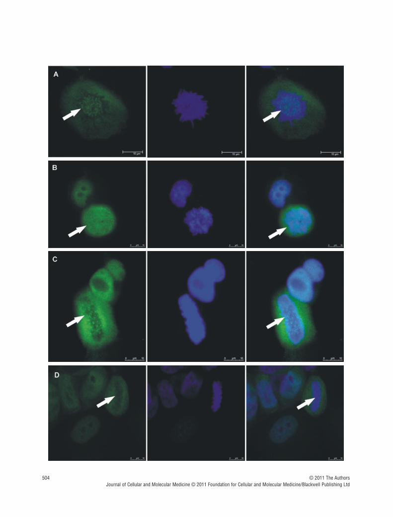
Cenp-F staining of untreated MDA-MB-436 cells at differentstages of mitosis showed localization of the protein as reported inprevious studies [7, 14], with clear kinetochore staining in pro-,prometa- and metaphase. Following treatment with zoledronic acid,there was a marked redistribution of Cenp-F from the kinetochore,resulting in a dispersed staining pattern (Fig. 2). The punctuate pat-tern of Cenp-F staining was evident in control cells (Fig. 2A and C)whereas the distinct pattern was completely abolished in zole-dronic acid treated cells (Fig. 2B and D) which exhibited diffusestaining for Cenp-F.
Quantitation showed that compared to untreated control, allof the cancer cell lines displayed a significant decrease in thenumber of mitotic cells in either prophase, prometaphase ormetaphase with Cenp-F localized at the kinetochore following 24 hrs treatment with 10 �M and 25 �M zoledronic acid (Fig. 3).The reduction was 50.0% and 33.3% vs. 91.7%, both P �
0.001, MDA-MB-436 (Fig. 3A); 58.3% and 58.8% vs. 94.2%, P � 0.01 and P � 0.01, MDA-MB-231 (Fig. 3B); and 46.7% and36.7% vs. 81.7%, P � 0.01 and P � 0.001, MCF-7 (Fig. 3C).
© 2011 The AuthorsJournal of Cellular and Molecular Medicine © 2011 Foundation for Cellular and Molecular Medicine/Blackwell Publishing Ltd
Fig. 1 Schematic representation of the potentialeffects on Cenp-F following zoledronic acid treat-ment. Cenp-F farnesylation, which is crucial forCenp-F localization to the kinetochores, takesplace during late S-phase. This enables the pro-tein to take an active part in kinetochore assem-bly, chromosome alignment/segregation andG2/M transition. The potential effects of inhibi-tion of Cenp-F farnesylation by zoledronic acidand the resulting consequences for the cell aredepicted in the left panel.
504 © 2011 The AuthorsJournal of Cellular and Molecular Medicine © 2011 Foundation for Cellular and Molecular Medicine/Blackwell Publishing Ltd

J. Cell. Mol. Med. Vol 15, No 3, 2011
505© 2011 The AuthorsJournal of Cellular and Molecular Medicine © 2011 Foundation for Cellular and Molecular Medicine/Blackwell Publishing Ltd
Fig. 2 Zoledronic acid induces delocalization of Cenp-F from the kinetochore. In untreated MDA-MB-436 cells, Cenp-F is localized at the kinetochore (A, C). (B) and (D) show images of MDA-MB-436 cells treated with zoledronic acid (10 �M for 24 hrs � 24 hrs incubation in drug-free medium) at therespective mitotic stages to (A) and (C), but lacking Cenp-F associated with the kinetochore. Cells were stained using a specific antibody to Cenp-F (green)and DNA visualized by DAPI (blue). White arrows indicate cells of interest.
Fig. 3 Zoledronic acid significantly reduces the percentage of cells with Cenp-F localized at the kinetochore. MDA-MB-436, MDA-MB-231 and MCF-7 cellswere treated with 0, 10 or 25 �M zoledronic acid for 1, 4, 6, 8, 24 (followed by incubation in medium for up to 48 hrs) or cells were continuously exposedto zoledronic acid for 48 hrs. The percentage of cells in pro-, prometa- and metaphase with Cenp-F localized at the kinetochore was determined. Resultsare the mean � SEM of three experiments. *P � 0.05, **P � 0.01, ***P � 0.001, for differences between each treatment group compared to corre-sponding control. *P � 0.05, for differences between corresponding treatment groups.

506
Following 24 hrs exposure, there was no significant differencein the effect caused by the two drug concentrations, a dose of10 �M was sufficient to cause significant levels of Cenp-F delo-calization. This effect was further enhanced following 48 hrscontinuous exposure to the drug. A total of 10 and 25 �M zole-dronic acid caused a decrease in the percentage of MDA-MB-436 cells with Cenp-F localized at the kinetochore to 25.8% and10.8%, respectively, (both P � 0.001 vs. control 90.0%),49.2% and 20.0% in MDA-MB-231 cells (both P � 0.001 vs.control 96.7%) and 24.2% and 20.0% in MCF-7 cells (both P �
0.001 vs. control 80.8%).These data show that 24 hrs exposure to 10 �M zoledronic
acid is sufficient to significantly reduce the number of mitoticbreast cancer cells with Cenp-F localized at the kinetochore andthis effect was increased with higher concentrations and pro-longed treatment times. Because MDA-MB-436 cells appear tohave the greatest sensitivity to zoledronic acid treatment, the cellline was chosen for a range of experiments. Furthermore a GFPtransfected version of this cell line has been used in previous in vivo studies exploring the potential anti-tumour effects ofzoledronic acid [15].
To confirm that the observed effect on Cenp-F delocalization isdue to zoledronic acid’s ability to inhibit protein prenylation,experiments with the non-NBP clodronate were performed.Because clodronate is known to be less potent at inhibiting boneresorption than zoledronic acid [1] a dose of 500 �M was usedto treat MDA-MB-436 cells for 48 hrs. Clodronate treatment didnot cause any change in Cenp-F localization at the kinetochore,supporting that this effect is due to inhibition of FPP synthase byzoledronic acid (data not shown).
Zoledronic acid is rapidly cleared from the circulation to bonefollowing in vivo infusion, and peripheral tissues, includingtumours, will therefore only be exposed to this drug for a limitedperiod of time. To establish the minimum exposure time required
to affect Cenp-F localization, we determined the percentage ofmitotic breast cancer cells with Cenp-F at the kinetochore follow-ing exposure to 10 or 25 �M zoledronic acid for 1, 4, 6 and 8 hrs.As shown in Fig. 3, treating MDA-MB-436 cells with 25 �M zole-dronic acid for 4 hrs significantly decreased the proportion of cellswith Cenp-F at the kinetochore compared to control (72.5% vs.90.8%, P � 0.01, Fig. 3D). Similar decreases were observed after6 hrs treatment of MDA-MB-231 cells with 10 and 25 �M zole-dronic acid (76.7% and 74.2% vs. 92.5%, P � 0.01 and P � 0.01,Fig. 3E) and in MCF-7 cells treated with 10 �M (68.3% vs. 85.0%,P � 0.05, Fig. 3F). Increasing the zoledronic acid exposure timeto 8 hrs caused significant reductions in kinetochore-associatedCenp-F-levels in all cell lines, with the highest effect found inMDA-MB-436 cells (65% vs. 91.7% in the control P � 0.001, Fig. 3D). These data show that treatment of breast cancer cellswith zoledronic acid for as short as 6�8 hrs induced a significantdecrease of cells with Cenp-F localized at the kinetochore.Although prolonged exposure (24–48 hrs) resulted in a more pronounced effect, shorter exposure times (6–8 hrs) may be sufficient to affect tumour cells by this mechanism.
Doses of zoledronic acid used in some in vitro experiments(10–25 �M for up to 48 hrs) are not clinically achievable asplasma levels of the drug, following 4 mg i.v. infusion, only reachup to 1–2 �M. Therefore, we investigated whether lower concen-trations of zoledronic acid could affect the localization of Cenp-F.MDA-MB-436 cells were treated with 0, 4, 6, 10 or 25 �M zole-dronic acid for 48 hrs continuously and were analysed for Cenp-Flocalization. As shown in Fig. 4, treatment with 6 �M zoledronicacid for 48 hrs significantly decreased the percentage of cells withCenp-F at the kinetochore when compared to untreated control(59.2% vs 90%, P � 0.05), and a dose-dependent increase in thenumber of cells without Cenp-F at the kinetochore was observed(33.3% and 21.2% vs control 90%, P � 0.001 and P � 0.001).
These data show that a concentration of zoledronic acid as lowas 6 �M is sufficient to induce loss of Cenp-F at the kinetochore,suggesting that high drug concentrations are not required to affectfast growing cancer cells.
Effects of zoledronic acid on mitosis progressionin breast cancer cells
Localization of Cenp-F to the kinetochore is dependent on farnesy-lation of the CAAX motif at the C-terminus of the protein, and stud-ies using siRNA have found that Cenp-F-depleted cells show adelay in mitosis progression [9, 16]. Inhibition of Cenp-F farnesy-lation caused by FTI treatment is reported to induce a similar inhi-bition in cell cycle progression to that seen in Cenp-F-depletedcells [17]. We therefore investigated whether zoledronic acidinduced depletion of Cenp-F from the kinetochore had an effect onmitosis progression in breast cancer cells. This was assessed fol-lowing treatment of MDA-MB-231, MDA-MB-436 and MCF-7 cellswith increasing doses of zoledronic acid for up to 48 hrs, anddetermination of the number of cells in each mitotic stage(prophase, prometaphase, metaphase, anaphase and telophase)
© 2011 The AuthorsJournal of Cellular and Molecular Medicine © 2011 Foundation for Cellular and Molecular Medicine/Blackwell Publishing Ltd
Fig. 4 Low zoledronic acid doses affect Cenp-F localization at the kineto-chore. MDA-MB-436 cells were treated with 0, 4, 6, 10 and 25 �M zole-dronic acid for 48 hrs prior to DNA and Cenp-F staining. Cells were thenanalysed by microscopy for Cenp-F localization. Results are the mean �SEM of three experiments. *P � 0.05, ***P � 0.001 for differencesbetween each treatment group compared to control.

J. Cell. Mol. Med. Vol 15, No 3, 2011
507
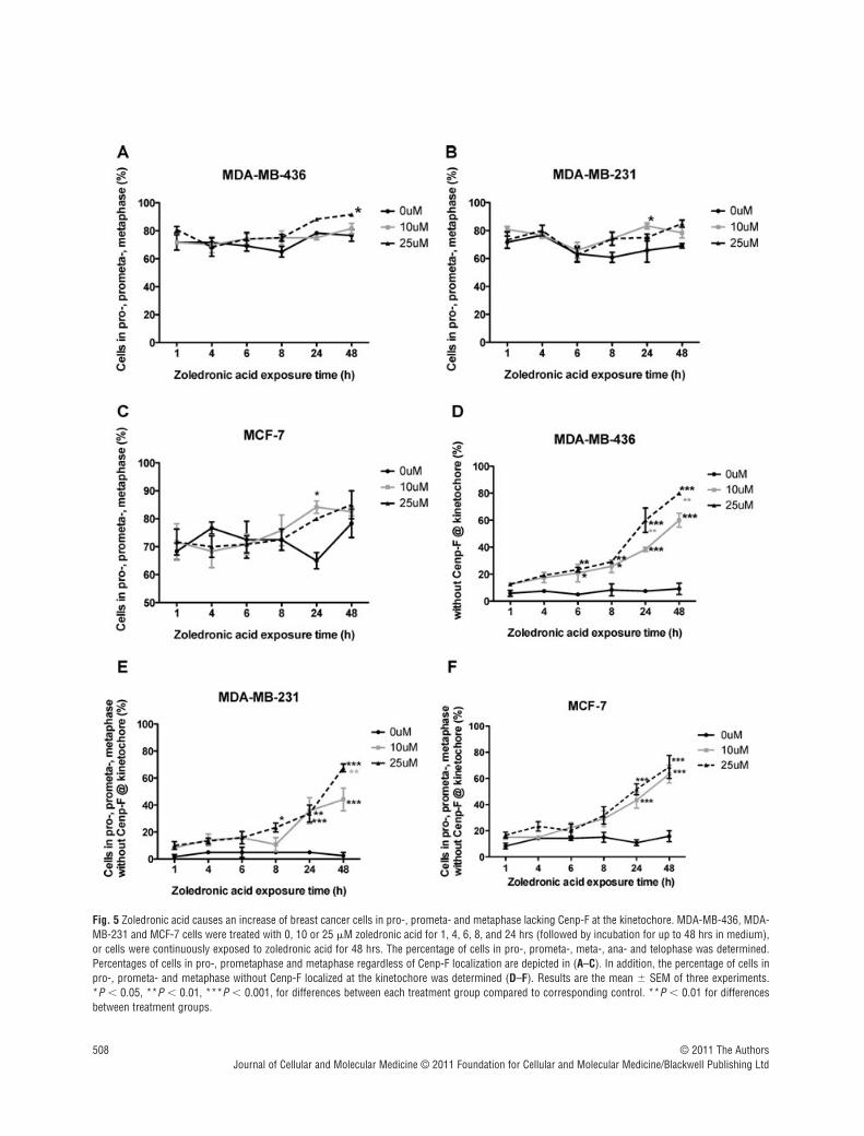
compared to untreated control. A significant increase of MDA-MB-231 cells in pro-, prometa- and metaphase was observed afterexposure to 10 �M zoledronic acid for 24 hrs (83.3% vs. 65.8%,P � 0.05, Fig. 5B) and in MDA-MB-436 cells after 48 hrs treat-ment with 25 �M zoledronic acid (91.6% vs. 76.6%, P � 0.05,Fig. 5A). MCF-7 cells in prophase, prometaphase and metaphaseincreased significantly after 24 hrs treatment with 10 �M zole-dronic acid (84.2% vs. 65%, P � 0.05, Fig. 5C) but not at 48 hrsto either drug concentration. These data suggest that 25 �M zole-dronic acid for 48 hrs has little effect on mitosis progression inhuman breast cancer cells when determining the effect regardlessto Cenp-F localization of the cells.
We next investigated whether progression of cells in earlystages of mitosis without Cenp-F at the kinetochore was affectedto a greater extend compared to cells with correct Cenp-F localiza-tion. Breast cancer cells were treated with zoledronic acid for upto 48 hrs, stained for Cenp-F, and the number of cells in pro-,prometa- and metaphase lacking Cenp-F at the kinetochore wasdetermined. The number of cells in ana- and telophase was alsoassessed. In MDA-MB-436 cells, 10 and 25 �M zoledronic acidinduced a significant increase in the percentage of pro-, prometa-and metaphase cells without Cenp-F at the kinetochore after 6 hrs,compared to control (20.8% and 23.3% vs. 5%, P � 0.05 and P � 0.01, Fig. 5D). This was further enhanced with increasingtreatment times (60% and 80% vs. 9.2% at 48 hrs, P � 0.001 andP � 0.001). Although an effect of treatment was seen in all celllines, it was less pronounced in MDA-MB-231 and MCF7, com-pared to MDA-MB-436 cells. A significant increase in MDA-MB-231 cells in pro-, prometa and metaphase without Cenp-F at thekinetochore could be seen following 8 hrs treatment with 25 �Mzoledronic acid compared to control (23.3% vs. 5%, P � 0.05,Fig. 5E). This increased to 44.2% and 67.5% (both P � 0.001compared to control of 2.5%), following 48 hrs treatment with 10and 25 �M. In MCF-7 cells, 24 hrs exposure to 10 and 25 �Mzoledronic acid increased the cells in pro-, prometa- andmetaphase to 43.3% and 51.7% vs. control levels of 10.8%, bothP � 0.001 (Fig. 5F), reaching maximum after 48 hrs continuousexposure to 10 and 25 �M (63.3% and 68.75% vs. 15.8%, P �
0.001 and P � 0.001). These data show that exposing breast can-cer cells to 10 �M zoledronic acid for a period of 6–8 hrs is suffi-cient to induce a significant increase in the number of MDA-MB-231 and MDA-MB-436 cells lacking Cenp-F at the kinetochorewith a shift towards prophase, prometaphase and metaphase. Incomparison, the estrogen-dependent cell line MCF-7 seems to beless sensitive to the drugs effect on the cell.
In order to confirm that the effect of zoledronic acid on Cenp-F is specific to fast proliferating cell lines, such as cancer cells,the same experiment was performed with the non-malignantbreast cancer cell line MCF-10A. In these cells no change in Cenp-F localization or mitosis progression was observed afterzoledronic acid treatment (25 �M for 8 or 24 hrs followed byincubation in drug-free medium up to a total of 48 hrs, or 48 hrscontinuous exposure), suggesting that rapidly proliferating cancer cells are more sensitive to the effects of zoledronic acidtreatment (data not shown).
The mevalonate pathway intermediary FOHreverses zoledronic acid induced Cenp-F delocalization from the kinetochore
Zoledronic acid causes inhibition of FPP synthase, a key enzymeof the mevalonate pathway, resulting in reduced levels of preny-lated proteins [3]. In order to determine whether the zoledronicacid induced reduction of Cenp-F associated with the kinetochorewas mediated via inhibition of the mevalonate pathway, we inves-tigated the effects of combining zoledronic acid with the meval-onate pathway intermediates GGOH and FOH.
MDA-MB-436 cells were treated with 25 �M zoledronic acid �50 �M /100 �M FOH or 50 �M /100 �M GGOH for 48 hrs, andthe number of cells with Cenp-F localized at the kinetochore wasdetermined. As expected, zoledronic acid caused a reduction in thepercentage of cells with Cenp-F at the kinetochore, but there wasno significant effect of either FOH or GGOH alone compared tocontrol (Fig. 6). Treatment with 25 �M zoledronic acid combinedwith either 50 �M or 100 �M FOH showed a significant increasein cells with Cenp-F localized at the kinetochore, compared to cellstreated with zoledronic acid alone (49.2% and 60.0% vs. 16.7%,P � 0.01 and P � 0.01, Fig. 6A). In contrast, inclusion of GGOHdid not significantly affect the ability of zoledronic acid to causeloss of Cenp-F (Fig. 6B) when used at identical concentrations toFOH. When using higher GGOH doses (150 �M) together with 25 �M zoledronic acid, the effect of the drug on Cenp-F localiza-tion at the kinetochore could be reversed (data not shown).
These data show that lower doses of FOH are superior to GGOHin reversing the effect on Cenp-F induced by zoledronic acid,which is in agreement with the proposed importance of farnesyla-tion for Cenp-F localization and function.
Zoledronic acid disrupts chromosome alignmentand spindle morphology in breast cancer cells
Several siRNA studies have reported that depletion of Cenp-F resultsin chromosome misalignment as well as in increased numbers ofcells with multipolar spindles [8, 16, 18, 19]. We therefore investi-gated whether the zoledronic acid induced reduction of kinetochore-associated Cenp-F was associated with changes in chromosomealignment and/or spindle morphology. MDA-MB-436 cells wereexposed to 4, 6, 10 and 25 �M zoledronic acid for 48 hrs andstained for Cenp-F and DNA. The cells that scored positive for mis-alignment had chromosomes distributed broadly around theprometaphase-, and metaphase plates and/or chromosomesremained far outside of these structures. We found that the numberof mitotic cells without Cenp-F at the kinetochore and misalignedchromosomes was significantly increased following treatment with10 and 25 �M zoledronic acid when compared to control (4.7% and7.3% vs. 0.3%, P � 0.05 and P � 0.001, Fig. 7). In addition to thedrug-induced disruption of chromosome alignment (Fig. 7A) we alsoobserved cells containing tripolar spindles (Fig. 7B), but the increasewas not statistically significant for any of the drug concentrations
© 2011 The AuthorsJournal of Cellular and Molecular Medicine © 2011 Foundation for Cellular and Molecular Medicine/Blackwell Publishing Ltd
508 © 2011 The AuthorsJournal of Cellular and Molecular Medicine © 2011 Foundation for Cellular and Molecular Medicine/Blackwell Publishing Ltd
Fig. 5 Zoledronic acid causes an increase of breast cancer cells in pro-, prometa- and metaphase lacking Cenp-F at the kinetochore. MDA-MB-436, MDA-MB-231 and MCF-7 cells were treated with 0, 10 or 25 �M zoledronic acid for 1, 4, 6, 8, and 24 hrs (followed by incubation for up to 48 hrs in medium),or cells were continuously exposed to zoledronic acid for 48 hrs. The percentage of cells in pro-, prometa-, meta-, ana- and telophase was determined.Percentages of cells in pro-, prometaphase and metaphase regardless of Cenp-F localization are depicted in (A–C). In addition, the percentage of cells inpro-, prometa- and metaphase without Cenp-F localized at the kinetochore was determined (D–F). Results are the mean � SEM of three experiments. *P � 0.05, **P � 0.01, ***P � 0.001, for differences between each treatment group compared to corresponding control. **P � 0.01 for differencesbetween treatment groups.

J. Cell. Mol. Med. Vol 15, No 3, 2011
509
tested. The same experiment was undertaken using the non-malig-nant breast cell line MCF-10A but no effect on either chromosomealignment or spindle morphology was observed (data not shown).In agreement with other published data, our results show thatdepletion of Cenp-F from the kinetochore may cause failure in chro-mosome alignment and abnormalities in spindle morphology.
Discussion
The direct anti-tumour effects of NBPs, in particular zoledronic acid,are still controversial, and the specific molecular mechanismsresponsible remain to be identified. There is, however, an agreementthat in a range of tumour models, zoledronic acid does inhibit cellgrowth as well as induce apoptosis, and these effects can bereversed by incorporation of intermediaries of the mevalonate pathway [20, 21, 22]. Regardless of the cell type, NBPs inhibit FPPsynthase, resulting in a depletion of the pool of prenylated forms ofproteins required for a variety of cellular functions. The effects ofzoledronic acid on prenylated proteins such as the small GTPases,including Ras, Rab and Rho, have been studied in some detail.These GTPases are involved in the regulation of vesicular trafficking,cytoskeletal arrangement and cell survival, and are therefore key tar-gets for NBPs in osteoclasts, recently reviewed by Coxon et al. [23].So far no studies of the effects of zoledronic acid on other types ofprenylated proteins have been reported, despite more than 300 pep-tides with prenylation motifs having been identified in the humanproteome [17], together with 60 farnesylated eukaryotic proteins[11]. Studies aiming to identify the molecular effects of zoledronicacid are important to increase our understanding of its proposedanti-tumour action. One example is the study by Marra et al. [24]who found a zoledronic acid induced decrease in Cyr61, an angio-genic inducer, in prostate cancer cells. We have investigated theeffects of zoledronic acid on Cenp-F, a farnesylated protein implicatedin the assembly of the kinetochore structures required for correctchromosome alignment and separation during mitosis (Table 1). Wefound that in both hormone-dependent and -independent breastcancer cells, zoledronic acid does cause loss of Cenp-F from thekinetochore. Our findings are in agreement with the results reportedby Schafer-Hales et al., who investigated the effect of the FTI lona-farnib on 15 human cancer cell lines, including the breast cancer celllines MCF-7, MDA-MB-231 and MDA-MB-436 [11]. They reported areduction in the percentage of Cenp-F at the kinetochore after drugtreatment, suggesting that farnesylation is required for the correctlocalization of Cenp-F. In contrast, exposure of zoledronic acid to thenon-malignant breast cell line MCF-10-A did not affect Cenp-F local-ization and the conflicting results may be influenced by several fac-tors including a difference in drug uptake, different effects on FPPsor the effect could be tumour cell specific possibly associated withthe increased proliferation rate found in malignant cell types.
We could show that the zoledronic acid induced delocalizationof Cenp-F in cancer cells was caused by inhibition of the meval-onate pathway, because the presence of FOH, but not GGOH,
© 2011 The AuthorsJournal of Cellular and Molecular Medicine © 2011 Foundation for Cellular and Molecular Medicine/Blackwell Publishing Ltd
Fig. 6 The delocalization of Cenp-F from the kinetochore is reversed by lowconcentrations of FOH but not GGOH. MDA-MB-436 cells were treated with25 �M zoledronic acid in the presence or absence of 50 �M/100 �M FOH(A) or 50 �M/100 �M GGOH (B) for 48 hrs. Cells in pro-, prometa- andmetaphase with Cenp-F localized at the kinetochore were determined.Results are the mean � SEM of three experiments. **P � 0.01 for differ-ences between each combined treatment group compared to zoledronicacid alone.

510
effects of zoledronic acid on Cenp-F localization were significantlyreversed. The requirement for farnesylation of Cenp-F for kineto-chore localization has also been reported by Hussein and Taylor[9]. Post-translational prenylation is mainly involved in ensuringcorrect membrane association of proteins [25], and it is not clearwhy lack of farnesylation should cause Cenp-F redistribution, asthe kinetochore does not include any membrane structures. It hasbeen suggested that farnesylation is also involved in protein–proteininteractions, and that accumulation of unprenylated forms of pro-teins may compete for effectors [23]. In this context, it may be ofimportance that two proteins closely associated with both Cenp-Fand the kinetochore, Cenp-E and lamin B, are also farnesylated andtherefore likely to be affected by zoledronic acid treatment [17, 26, 27].Hence it is possible that in addition to Cenp-F, several other pro-teins involved in ensuring correct chromosome segregation will beaffected by zoledronic acid, but this remains to be established.
The standard dosing regimen of zoledronic acid used in treat-ment of cancer-induced bone disease is a 4 mg infusion every
3–4 weeks, and the high affinity of NBPs for bone and their rapidclearance from the general circulation means that peripheraltumours will be exposed to these drugs for no more than a fewhours [20]. This has been one of the main arguments against theability of zoledronic acid to exert direct anti-tumour effects duringstandard clinical use. The majority of in vitro and in vivo studiesreporting anti-tumour effects of NBPs have involved the use ofvery high or frequent repeated dosing. Although there was somevariation between the cell lines, 6-hr treatment with zoledronicacid did cause a significant increase in the number of mitotic cellslacking Cenp-F at the kinetochore, indicating that prolonged expo-sure to the drug is not required in order to generate this effect.
Protein farnesylation is increasingly investigated as a potentialtarget in cancer care and FTIs are currently the subject of investi-gation in a number of clinical trials. However results are contrarywith some studies reporting no beneficial effects (Schering-Plough, Lonafranib in Non-small-cell lung cancer) whereas otherscould report anti-tumour effects when giving the FTI R115777 in
© 2011 The AuthorsJournal of Cellular and Molecular Medicine © 2011 Foundation for Cellular and Molecular Medicine/Blackwell Publishing Ltd
Fig. 7 Zoledronic acid induces a significantincrease of breast cancer cells with chromo-somes lagging outside of the metaphase plate.MDA-MB-436 cells were treated with 0, 4, 6, 10and 25 �M zoledronic acid for 48 hrs. Cells werestained for Cenp-F and DNA and 100 mitoticcells were counted in each treatment group.Cells were scored for mitotic structures with lag-ging chromosomes and without Cenp-F localizedat the kinetochore. Results are the mean � SEMof three experiments. *P � 0.05, ***P � 0.001for differences between each treatment groupcompared to corresponding control. Exampleimages of MDA-MB-436 cells treated with zole-dronic acid for 48 hrs showing the induced chro-mosome misalignment (A) and formation oftripolar spindles (B). Cells were stained using aspecific antibody to Cenp-F (green) and DNAvisualized by DAPI (blue).

J. Cell. Mol. Med. Vol 15, No 3, 2011
511
combination with Capecitabine (Eastern Cooperative OncologyGroup). The efficacy of FTIs in cancer treatment therefore remainsto be established and the contrary results may implicate that agreater anti-cancer benefit could be achieved when targeting bothfarnesylated and geranylgeranylated proteins as done by NBPs.However, farnesylated proteins do play a major role in cancer pro-liferation and progression and should not be underestimated aspotential anti-cancer targets.
In studies using FTIs, it has been shown that inhibition of Cenp-F farnesylation does result in a delay in cell cycle progression ratherthan a complete block, and there was an increase in the proportionof lung cancer cells accumulating in G2/M in the presence of FTIs[28, 29]. Delayed cell cycle progression is likely to affect fast grow-ing tumour cells, and we show that three different breast cancer celllines show a similar response to zoledronic acid in relation to Cenp-F,suggesting that this is not a phenomenon associated with a singlecell line. In contrast, non-dividing osteoclasts will be unaffected bydisruption of chromosome separation. Delocalization of Cenp-F istherefore unlikely to be involved in the inhibition of bone resorptioncaused by zoledronic acid, where the correct membrane localization
of small GTPases are of greater importance due to their establishedroles in osteoclast function [2, 23]. The lack of the effect of zole-dronic acid on Cenp-F localization seen in MCF-10A cells suggeststhat the drug does not exhibit a general activity in all cell types.
Several studies have reported NBP-induced reduction of tumourcell proliferation, including neuroblastoma [30], lung [31], prostate[32] and breast [33]. Several studies reported an effect of FTIs oncancer cell progression through G2/M phase, suggesting that this iscaused by inhibition of protein farnesylation. Ashar et al. found thatthe FTI SCH 66336 induced accumulation of human lung cancercells in G2/M-phase, but did not find any association with Cenp-Fdepletion from the kinetochore [17, 34]. Crespo et al. reported thattreatment of lung cancer cells with the FTI-2153 caused a significantincrease in prometaphase cells, again without any effect of the drugon Cenp-F localization at the kinetochore [29]. In contrast, Husseinand Taylor have shown that Cenp-F farnesylation is required for pro-gression through G2 to M-phase, and that Cenp-F farnesylation isessential for localization to the kinetochore and the nuclear mem-brane, suggesting a possible connection between the farnesylationstatus and intracellular protein localization [9].
© 2011 The AuthorsJournal of Cellular and Molecular Medicine © 2011 Foundation for Cellular and Molecular Medicine/Blackwell Publishing Ltd
Table 1 Summary of findings
Research question Cell line Cenp-F localization at the kinetochore
Reduced Not reduced
Effect of zol on Cenp-F localization at the kinetochore
MDA-MB-436 X
MDA-MB-231 X
MCF-7 X
Effect of Clodronate on Cenp-F localization at thekinetochore
MDA-MB-436 X
Effect of low zol concentrations on Cenp-F localizationat the kinetochore
MDA-MB-436 X (from 6 µM) X (below 4uM)
Effect of FOH/GGOH on zol-induced Cenp-Fdelocalization
MDA-MB-436 X zol � GGOH X zol � FOH
Research question Cell line Cells in pro-, prometa-, metaphase (%)
Increased No effect
Effect of zol on mitosis progression MDA-MB-436 X (only 25 µM, 48 hrs) X
MDA-MB-231 X (only 10 µM, 24 hrs) X
MCF-7 X (only 10 µM, 24 hrs) X
Effect of zol on mitosis progression without Cenp-F localization at the kinetochore
MDA-MB-231 X
MDA-MB-436 X
MCF-7 X
MCF-10A X
Research question Cell line Chromosome misalignment
Yes No
Effect of zol-induced Cenp-F delocalization onchromosome alignment
MDA-MB-436 X
MCF-10A X

512
We further determined whether the drug affected the ability ofthe cells to enter mitosis, or if progression through M-phase wasdelayed at a particular stage. Therefore, we treated breast cancercells with zoledronic acid and assessed the percentage of cells ineach mitotic phase, that is pro-, prometa-, meta-, ana- andtelophase, by microscopy. Although first experiments showed littleeffect of zoledronic acid on mitosis progression, it was determinedwhether cells lacking Cenp-F at the kinetochore were more prone toshow changes in mitosis progression. There was a significantincrease of cells in pro-, prometa- and metaphase lacking Cenp-F atthe kinetochore in all breast cancer cell lines tested when comparedto control. These findings suggest that the zoledronic acid inducedlack of Cenp-F at the kinetochore plays a role in the accumulation ofcells in the early stages of mitosis and that impairment of the Cenp-Fand kinetochore association causes disruption of M-phase progres-sion. The observed accumulation of cells in early stages of M-phasefollowing treatment with zoledronic acid suggests a cell cycle delayat either the progression from G2 to M-phase, or a delay of progres-sion from prophase to metaphase. Published studies of the effectsof zoledronic acid on the cell cycle of different cancer cell lines haveshown an accumulation of cells in S-phase [12, 13]. Our resultsreflect the findings of Merrell et al. who reported a decrease of cells inS and a concomitant increase in G1 phase in MDA-MB-231 cellstreated with 10 �M zoledronic acid for 72 hrs [35].
The impaired chromosome alignment observed in zoledronicacid treated cells could be due to insufficient microtubule-chro-mosome attachment as it has been shown that Cenp-F depletioncan cause failure/loss of Cenp-E, cytoplasmic dynein and dynactinaccumulation at the kinetochore [8]. Cenp-E is a plus end motorprotein and is therefore important for correct microtubule attach-ment [36], and Cenp-E is also a farnesylated protein. Similareffects were reported on cell cycle progression and chromosomealignment after protein silencing of Cenp-E compared to Cenp-F[37]. The effect of the drug on chromosome alignment and M-phase progression may be a combined effect of faulty Cenp-F,Cenp-E (and probably other prenylated kinetochore proteins)localization and maintenance. However, because Cenp-E recruit-ment to the kinetochore has been shown to be partly dependenton Cenp-F [6], it is most likely that loss of Cenp-F from the kine-tochore is the ‘master switch’ for the observed effects.
Once we had established that zoledronic acid caused disruptionof M-phase progression, we investigated the consequences ofCenp-F delocalization on chromosome alignment and separation,as this may potentially explain the disruption of mitosis progres-sion. Following zoledronic acid treatment there was a significant
increase in the number of cells lacking Cenp-F at the kinetochorethat had chromosomes distributed far outside of the normalmetaphase plate. This phenotype reflects the findings of otherstudies of the effects of Cenp-F depletion on chromosome separa-tion. Yang et al. showed that Cenp-F depletion by RNAi causedsevere defects in chromosome alignment, accompanied byreduced sister kinetochore tension and premature chromosomedecondensation, finally resulting in apoptosis [8]. Holt et al.reported that siRNA depletion of Cenp-F resulted in disrupted chro-mosome alignment and prolonged mitosis, possibly mediated bythe activation of the SAC through the checkpoint kinase BubR1[16]. Impaired chromosome alignment, due to faulty kinetochore-microtubule attachment, could activate the SAC and thus stop cellsfrom progressing through mitosis, resulting in the observed accu-mulation of cells in pro- and prometaphase [8, 16]. It has also beenshown that Cenp-F is a microtubule binding protein that may bepart of the microtubule-chromosome attachment machinery [18, 28]. In addition to the impaired chromosome alignment, Cenp-F-depleted cells show abnormalities in spindle morphology [16].Yang et al. found that approximately 16% of HeLa cells lackingCenp-F exhibited multipolar spindles, and that Cenp-F-depletedcells in late pro- or metaphase had abnormal bipolar spindles [8].In a comprehensive study of the effects of Cenp-F depletion,Bomont found that this would result in gross chromosome misseg-regation in only a subset of cells, whereas in the majority of cellsthe effect was mitotic delay [28]. In agreement with these results,we observed the presence of multipolar spindles in zoledronic acid-treated cells lacking Cenp-F at the kinetochore.
Our data show that in rapidly proliferating breast cancer cells,zoledronic acid causes disruption of Cenp-F localization at thekinetochore. The effects were caused by inhibition of the meval-onate pathway, most likely by reducing the levels of post-transla-tional farnesylation of Cenp-F. This is the first report showing thatzoledronic acid may exert part of its anti-tumour effect by disrupt-ing kinetochore assembly and subsequently reducing tumour cellproliferation.
Acknowledgements
PDO is funded by Breast Cancer Campaign UK. Zoledronic acid was a kindgift from Novartis Pharma. We thank Dr. Peter Grabowski for his help withthe fluorescence microscopy studies and Dr. Karen Sisley for her help withthe evaluation of mitotic stages.
References
1. Green JR. Antitumor effects of bisphos-phonates. Cancer. 2003; 97: 840–7.
2. Coxon FP, Helfrich MH, Van’t Hof R, et al.Protein geranylgeranylation is required forosteoclast formation, function, and sur-vival: inhibition by bisphosphonates and
GGTI-298. J Bone Miner Res. 2000; 15:1467–76.
3. Luckman SP, Hughes DE, Coxon FP, et al.Nitrogen-containing bisphosphonatesinhibit the mevalonate pathway andprevent post-translational prenylation of
GTP-binding proteins, including Ras. J Bone Miner Res. 1998; 13: 581–9.
4. Winter MC, Holen I, Coleman RE.Exploring the anti-tumour activity ofbisphosphonates in early breast cancer.Cancer Treat Rev. 2008; 34: 453–75.
© 2011 The AuthorsJournal of Cellular and Molecular Medicine © 2011 Foundation for Cellular and Molecular Medicine/Blackwell Publishing Ltd

J. Cell. Mol. Med. Vol 15, No 3, 2011
513
5. Sebti SM. Protein farnesylation: implica-tions for normal physiology, malignanttransformation, and cancer therapy.Cancer cell. 2005; 7: 297–300.
6. Johnson VL, Scott MI, Holt SV, et al.Bub1 is required for kinetochore localiza-tion of BubR1, Cenp-E, Cenp-F and Mad2,and chromosome congression. J Cell Sci.2004; 117: 1577–89.
7. Liao H, Winkfein RJ, Mack G, et al.CENP-F is a protein of the nuclear matrixthat assembles onto kinetochores at lateG2 and is rapidly degraded after mitosis. J Cell Biol. 1995; 130: 507–8.
8. Yang Z, Guo J, Chen Q, et al. Silencingmitosin induces misaligned chromosomes,premature chromosome decondensationbefore anaphase onset, and mitotic celldeath. Mol Cell Biol. 2005; 25: 4062–74.
9. Hussein D, Taylor SS. Farnesylation ofCenp-F is required for G2/M progressionand degradation after mitosis. J Cell Sci.2002; 115: 3403–14.
10. Zhu X, Chang KH, He D, et al. The C ter-minus of mitosin is essential for its nuclearlocalization, centromere/kinetochore tar-geting, and dimerization. J Biol Chem.1995; 270: 19545–50.
11. Schafer-Hales K, Iaconelli J, Snyder JP,et al. Farnesyl transferase inhibitorsimpair chromosomal maintenance in celllines and human tumors by compromisingCENP-E and CENP-F function. Mol CancerTher. 2007; 6: 1317–28.
12. Dumon JC, Journé F, Kheddoumi N, et al.Cytostatic and apoptotic effects of bispho-sphonates on prostate cancer cells. EurUrol. 2004; 45: 521–8.
13. Ory B, Blanchard F, Battaglia S, et al.Zoledronic acid activates the DNA S-phasecheckpoint and induces osteosarcoma celldeath characterized by apoptosis-inducingfactor and endonuclease-G translocationindependently of p53 and retinoblastomastatus. Mol Pharmacol. 2007; 71: 333–43.
14. Zhu X, Mancini MA, Chang KH, et al.Characterization of a novel 350-kiloDaltonnuclear phosphoprotein that is specificallyinvolved in mitotic-phase progression.Mol Cell Biol. 1995; 15: 5017–29.
15. Ottewell, PD, Monkkonen H, Jones, M,et al. Antitumor effects of doxorubicin fol-lowed by zoledronic acid in a mouse modelof breast cancer. J Natl Cancer Inst. 2008;100: 1167–78
16. Holt SV, Vergnolle MA, Hussein D, et al.Silencing Cenp-F weakens centromericcohesion, prevents chromosome align-ment and activates the spindle checkpoint.J Cell Sci. 2005; 118: 4889–900.
17. Ashar HR, James L, Gray K, et al. Farnesyltransferase inhibitors block the farnesyla-tion of CENP-E and CENP-F and alter theassociation of CENP-E with the micro-tubules. J Biol Chem. 2000; 275: 30451–57.
18. Feng J, Huang H, Yen TJ. CENP-F is a novelmicrotubule-binding protein that is essentialfor kinetochore attachments and affects theduration of the mitotic checkpoint delay.Chromosoma. 2006; 115: 320–9.
19. Laoukili J, Kooistra MR, Bras A, et al.FoxM1 is required for execution of themitotic programme and chromosome sta-bility. Nat Cell Biol. 2005; 7: 126–36.
20. Neville-Webbe HL, Rostami-Hodjegan A,Evans CA, et al. Sequence- and schedule-dependent enhancement of zoledronic acidinduced apoptosis by doxorubicin inbreast and prostate cancer cells. Int JCancer. 2005; 113: 364–71.
21. Coxon JP, Oades GM, Kirby RS, et al.Zoledronic acid induces apoptosis andinhibits adhesion to mineralized matrix inprostate cancer cells via inhibition of protein prenylation. BJU Int. 2004; 94:164–70.
22. Virtanen SS, Väänänen HK, HärkönenPL, et al. Alendronate inhibits invasion ofPC-3 prostate cancer cells by affecting themevalonate pathway. Cancer Res. 2002;62: 2708–14.
23. Coxon FP, Thompson K, Rogers MJ.Recent advances in understanding themechanism of action of bisphosphonates.Curr Opin Pharmacol. 2006; 6: 307–12.
24. Marra M, Santini D, Meo G, et al. Cyr61downmodulation potentiates the anti-cancer effects of zoledronic acid in andro-gen-independent prostate cancer cells. IntJ Cancer. 2009; 125: 2004–1
25. McTaggart SJ. Isoprenylated proteins.Cell Mol Life Sci. 2006; 63: 255–67.
26. Rusiñol AE, Sinensky MS. Farnesylatedlamins, progeroid syndromes and farnesyltransferase inhibitors. J Cell Sci. 2006;119: 3265–72.
27. Tamanoi F, Kato-Stankiewicz J, Jiang C,et al. Farnesylated proteins and cell cycleprogression. J Cell Biochem 2001; 37:64–70.
28. Bomont P, Maddox P, Shah JV, et al.Unstable microtubule capture at kineto-chores depleted of the centromere-associ-ated protein CENP-F. EMBO J. 2005; 24:3927–39.
29. Crespo NC, Ohkanda J, Yen TJ, et al. Thefarnesyltransferase inhibitor, FTI-2153,blocks bipolar spindle formation and chro-mosome alignment and causesprometaphase accumulation during mito-sis of human lung cancer cells. J BiolChem. 2001; 276: 16161–7.
30. Peng H, Sohara Y, Moats RA, et al. Theactivity of zoledronic Acid on neuroblas-toma bone metastasis involves inhibition ofosteoclasts and tumor cell survival and pro-liferation. Cancer Res. 2007; 67: 9346–55.
31. Li YY, Chang JW, Chou WC, et al.Zoledronic acid is unable to induce apop-tosis, but slows tumor growth and pro-longs survival for non-small-cell lung can-cers. Lung Cancer. 2008; 59: 180–91.
32. Caraglia M, Marra M, Leonetti C, et al.R115777 (Zarnestra)/Zoledronic acid(Zometa) cooperation on inhibition ofprostate cancer proliferation is paralleledby Erk/Akt inactivation and reduced Bcl-2and bad phosphorylation. J Cell Physiol.2007; 211: 533–43.
33. Verdijk R, Franke HR, Wolbers F, et al.Differential effects of bisphosphonates onbreast cancer cell lines. Cancer Lett. 2007;246: 308–12.
34. Ashar HR, James L, Gray K, et al. Thefarnesyl transferase inhibitor SCH 66336induces a G(2) – M or G(1) pause in sen-sitive human tumor cell lines. Exp Cell Res.2001; 262: 17–27.
35. Merrell MA, Wakchoure S, Lehenkari PP,et al. Inhibition of the mevalonate pathwayand activation of p38 MAP kinase are independently regulated by nitrogen-containing bisphosphonates in breast cancer cells. Eur J Pharmacol. 2007; 570:27–37.
36. Yen TJ, Li G, Schaar BT, et al. CENP-E isa putative kinetochore motor that accumu-lates just before mitosis. Nature. 1992;359: 536–39.
37. Tanudji M, Shoemaker J, L’Italien L, et al. Gene silencing of CENP-E by smallinterfering RNA in HeLa cells leads to mis-segregation of chromosomes after amitotic delay. Mol Biol Cell. 2004; 15:3771–81.
© 2011 The AuthorsJournal of Cellular and Molecular Medicine © 2011 Foundation for Cellular and Molecular Medicine/Blackwell Publishing Ltd