A Coordinated Interdependent Protein Circuitry Stabilizes the Kinetochore Ensemble to Protect CENP-A...
16
A Coordinated Interdependent Protein Circuitry Stabilizes the Kinetochore Ensemble to Protect CENP-A in the Human Pathogenic Yeast Candida albicans Jitendra Thakur, Kaustuv Sanyal* Molecular Mycology Laboratory, Molecular Biology and Genetics Unit, Jawaharlal Nehru Centre for Advanced Scientific Research, Bangalore, India Abstract Unlike most eukaryotes, a kinetochore is fully assembled early in the cell cycle in budding yeasts Saccharomyces cerevisiae and Candida albicans. These kinetochores are clustered together throughout the cell cycle. Kinetochore assembly on point centromeres of S. cerevisiae is considered to be a step-wise process that initiates with binding of inner kinetochore proteins on specific centromere DNA sequence motifs. In contrast, kinetochore formation in C. albicans, that carries regional centromeres of 3–5 kb long, has been shown to be a sequence independent but an epigenetically regulated event. In this study, we investigated the process of kinetochore assembly/disassembly in C. albicans. Localization dependence of various kinetochore proteins studied by confocal microscopy and chromatin immunoprecipitation (ChIP) assays revealed that assembly of a kinetochore is a highly coordinated and interdependent event. Partial depletion of an essential kinetochore protein affects integrity of the kinetochore cluster. Further protein depletion results in complete collapse of the kinetochore architecture. In addition, GFP-tagged kinetochore proteins confirmed similar time-dependent disintegration upon gradual depletion of an outer kinetochore protein (Dam1). The loss of integrity of a kinetochore formed on centromeric chromatin was demonstrated by reduced binding of CENP-A and CENP-C at the centromeres. Most strikingly, Western blot analysis revealed that gradual depletion of any of these essential kinetochore proteins results in concomitant reduction in cellular protein levels of CENP-A. We further demonstrated that centromere bound CENP-A is protected from the proteosomal mediated degradation. Based on these results, we propose that a coordinated interdependent circuitry of several evolutionarily conserved essential kinetochore proteins ensures integrity of a kinetochore formed on the foundation of CENP-A containing centromeric chromatin. Citation: Thakur J, Sanyal K (2012) A Coordinated Interdependent Protein Circuitry Stabilizes the Kinetochore Ensemble to Protect CENP-A in the Human Pathogenic Yeast Candida albicans. PLoS Genet 8(4): e1002661. doi:10.1371/journal.pgen.1002661 Editor: Beth A. Sullivan, Duke University, United States of America Received May 4, 2011; Accepted March 5, 2012; Published April 19, 2012 Copyright: ß 2012 Thakur, Sanyal. This is an open-access article distributed under the terms of the Creative Commons Attribution License, which permits unrestricted use, distribution, and reproduction in any medium, provided the original author and source are credited. Funding: This work is supported by a grant from the Department of Biotechnology, Government of India, to KS. We also gratefully acknowledge the financial support of JNCASR. JT is a senior research fellow supported by the Department of Biotechnology, Government of India. The funders had no role in study design, data collection and analysis, decision to publish, or preparation of the manuscript. Competing Interests: The authors have declared that no competing interests exist. * E-mail: [email protected] Introduction The centromeric histone CENP-A acts as the epigenetic mark of a functional centromere from yeast to humans [1]. As a histone H3 variant, CENP-A replaces canonical histone H3 to mark specialized centromeric chromatin by a mechanism that remains largely unknown. While CENP-A deposition occurs in a sequence- dependent manner in point centromeres of certain budding yeasts including S. cerevisiae [2,3], its recruitment to regional centromeres of most other eukaryotes is not strictly sequence dependent [1,3– 5]. Several lines of evidence suggest that the composition of nucleosomes that form centromeric chromatin may vary from species to species [6–10]. Even the hierarchy of events that assembles and stabilizes a complex macromolecular kinetochore (KT) structure on unusual centromeric chromatin, whether universal or species-specific, remains unclear. CENP-A is believed to be the initiator of the process of KT formation [1,11]. Localization of most KT proteins is regulated by CENP-A [12– 15]. However, a few proteins such as Ndc10 and Scm3 in S. cerevisiae [6,16], Mis12/Mtw1 in C. albicans [17], Mis6, Mis16- Mis18 complex and Ams2 in Schizosaccharomyces pombe [18,19], and CENP-H-I complex in humans [20] have been shown to regulate CENP-A localization. Although the structure of metazoan KTs can be visualized under microscope, the exact nature of the KT architecture is difficult to ascertain in yeasts due to small size of these cells [21]. However, based on presence of functional homologs of several KT proteins, and their genetic as well as biochemical interaction in various eukaryotes, it is presumed that the three-layered KT structure is evolutionarily conserved from yeasts to humans. A KT helps in bridging the mitotic spindle to centromere (CEN) DNA to ensure faithful chromosome segregation during mitosis and meiosis. KT proteins exist as sub-complexes that assemble on CEN DNA [22– 24]. In humans, inner (CENP-A, -B, -C, -H and –I) and middle (the Spc105 complex and the Mis12 complex) KT proteins are associated constitutively with CEN DNA [25] but outer KT proteins (such as the Ndc80 complex and the Ska1 complex) which help in KT-microtubule (MT) interaction are generally localized to KTs only during mitosis [26,27]. In contrast, KTs are fully assembled in S. cerevisiae early in the cell cycle. The fungal specific Dam1 complex, an outer KT protein complex in budding yeast S. cerevisiae, remains associated with KTs throughout the cell cycle [28–30]. PLoS Genetics | www.plosgenetics.org 1 April 2012 | Volume 8 | Issue 4 | e1002661
Transcript of A Coordinated Interdependent Protein Circuitry Stabilizes the Kinetochore Ensemble to Protect CENP-A...
A Coordinated Interdependent Protein CircuitryStabilizes the Kinetochore Ensemble to Protect CENP-Ain the Human Pathogenic Yeast Candida albicansJitendra Thakur, Kaustuv Sanyal*
Molecular Mycology Laboratory, Molecular Biology and Genetics Unit, Jawaharlal Nehru Centre for Advanced Scientific Research, Bangalore, India
Abstract
Unlike most eukaryotes, a kinetochore is fully assembled early in the cell cycle in budding yeasts Saccharomyces cerevisiaeand Candida albicans. These kinetochores are clustered together throughout the cell cycle. Kinetochore assembly on pointcentromeres of S. cerevisiae is considered to be a step-wise process that initiates with binding of inner kinetochore proteinson specific centromere DNA sequence motifs. In contrast, kinetochore formation in C. albicans, that carries regionalcentromeres of 3–5 kb long, has been shown to be a sequence independent but an epigenetically regulated event. In thisstudy, we investigated the process of kinetochore assembly/disassembly in C. albicans. Localization dependence of variouskinetochore proteins studied by confocal microscopy and chromatin immunoprecipitation (ChIP) assays revealed thatassembly of a kinetochore is a highly coordinated and interdependent event. Partial depletion of an essential kinetochoreprotein affects integrity of the kinetochore cluster. Further protein depletion results in complete collapse of the kinetochorearchitecture. In addition, GFP-tagged kinetochore proteins confirmed similar time-dependent disintegration upon gradualdepletion of an outer kinetochore protein (Dam1). The loss of integrity of a kinetochore formed on centromeric chromatinwas demonstrated by reduced binding of CENP-A and CENP-C at the centromeres. Most strikingly, Western blot analysisrevealed that gradual depletion of any of these essential kinetochore proteins results in concomitant reduction in cellularprotein levels of CENP-A. We further demonstrated that centromere bound CENP-A is protected from the proteosomalmediated degradation. Based on these results, we propose that a coordinated interdependent circuitry of severalevolutionarily conserved essential kinetochore proteins ensures integrity of a kinetochore formed on the foundation ofCENP-A containing centromeric chromatin.
Citation: Thakur J, Sanyal K (2012) A Coordinated Interdependent Protein Circuitry Stabilizes the Kinetochore Ensemble to Protect CENP-A in the HumanPathogenic Yeast Candida albicans. PLoS Genet 8(4): e1002661. doi:10.1371/journal.pgen.1002661
Editor: Beth A. Sullivan, Duke University, United States of America
Received May 4, 2011; Accepted March 5, 2012; Published April 19, 2012
Copyright: � 2012 Thakur, Sanyal. This is an open-access article distributed under the terms of the Creative Commons Attribution License, which permitsunrestricted use, distribution, and reproduction in any medium, provided the original author and source are credited.
Funding: This work is supported by a grant from the Department of Biotechnology, Government of India, to KS. We also gratefully acknowledge the financialsupport of JNCASR. JT is a senior research fellow supported by the Department of Biotechnology, Government of India. The funders had no role in study design,data collection and analysis, decision to publish, or preparation of the manuscript.
Competing Interests: The authors have declared that no competing interests exist.
* E-mail: [email protected]
Introduction
The centromeric histone CENP-A acts as the epigenetic mark of
a functional centromere from yeast to humans [1]. As a histone H3
variant, CENP-A replaces canonical histone H3 to mark
specialized centromeric chromatin by a mechanism that remains
largely unknown. While CENP-A deposition occurs in a sequence-
dependent manner in point centromeres of certain budding yeasts
including S. cerevisiae [2,3], its recruitment to regional centromeres
of most other eukaryotes is not strictly sequence dependent [1,3–
5]. Several lines of evidence suggest that the composition of
nucleosomes that form centromeric chromatin may vary from
species to species [6–10]. Even the hierarchy of events that
assembles and stabilizes a complex macromolecular kinetochore
(KT) structure on unusual centromeric chromatin, whether
universal or species-specific, remains unclear. CENP-A is believed
to be the initiator of the process of KT formation [1,11].
Localization of most KT proteins is regulated by CENP-A [12–
15]. However, a few proteins such as Ndc10 and Scm3 in S.
cerevisiae [6,16], Mis12/Mtw1 in C. albicans [17], Mis6, Mis16-
Mis18 complex and Ams2 in Schizosaccharomyces pombe [18,19], and
CENP-H-I complex in humans [20] have been shown to regulate
CENP-A localization.
Although the structure of metazoan KTs can be visualized under
microscope, the exact nature of the KT architecture is difficult to
ascertain in yeasts due to small size of these cells [21]. However,
based on presence of functional homologs of several KT proteins,
and their genetic as well as biochemical interaction in various
eukaryotes, it is presumed that the three-layered KT structure is
evolutionarily conserved from yeasts to humans. A KT helps in
bridging the mitotic spindle to centromere (CEN) DNA to ensure
faithful chromosome segregation during mitosis and meiosis. KT
proteins exist as sub-complexes that assemble on CEN DNA [22–
24]. In humans, inner (CENP-A, -B, -C, -H and –I) and middle
(the Spc105 complex and the Mis12 complex) KT proteins are
associated constitutively with CEN DNA [25] but outer KT proteins
(such as the Ndc80 complex and the Ska1 complex) which help in
KT-microtubule (MT) interaction are generally localized to KTs
only during mitosis [26,27]. In contrast, KTs are fully assembled in
S. cerevisiae early in the cell cycle. The fungal specific Dam1 complex,
an outer KT protein complex in budding yeast S. cerevisiae, remains
associated with KTs throughout the cell cycle [28–30].
PLoS Genetics | www.plosgenetics.org 1 April 2012 | Volume 8 | Issue 4 | e1002661
One of the less understood features of budding yeast KTs is that
they are clustered together throughout the cell cycle [31]. A recent
study, using chromosome conformation capture-on-chip (4C),
clearly demonstrates that all chromosomes cluster via centromeres
at one pole of the nucleus in S. cerevisiae suggesting interchromo-
somal cross-talks through inter-KT interaction [32]. KT clustering
has been shown to be important for centromere function in S.
cerevisiae as KTs are found to be declustered in ndc10, ame2 and nuf2
KT mutants [31,33]. Interestingly, KTs are clustered only during
interphase but not in mitosis in fission yeast S. pombe [34]. In
metazoans KTs are never clustered [35].
With the notable exception of holocentric chromosomes of
nematodes and aphids where KTs are formed across the entire
length of a chromosome [36], only one KT is formed on each
chromosome in all other organisms studied till date. A functional
KT can even assemble on non-centromeric DNA to form a
neocentromere in certain organisms when a native centromere is
deleted or inactivated [37–40]. Thus there must be an underlying
active mechanism to prevent formation of centromeric chromatin
on neocentromeric loci when the native centromere is functional.
CENP-A at non-centromeric locations is targeted for proteasomal
degradation in Drosophila melanogaster and S. cerevisiae [41–43]. Thus
ectopic CENP-A is destabilized to prevent multiple kinetochore
formation although the exact cellular signal that distinguishes
CENP-A molecules present at the native centromere from those
ectopically localized could not be determined.
Candida albicans, a pathogenic budding yeast, that causes
candidiasis in humans, carries 3–5 kb long unique centromeric
chromatin on each of its eight chromosomes. There are 4 CENP-
A molecules but only one MT binds to a KT in C. albicans [44].
Centromeric regions have been shown to have unusual chromatin
structure [45] and histone H3 molecules are replaced by CENP-A
at CENs (K. Sanyal, unpublished) in this organism. Moreover,
CENP-A deposition on CENs has been shown to be epigenetically
regulated [45–47]. We have previously cloned and characterized
several evolutionarily conserved KT proteins including CENP-A/
Cse4 [48], CENP-C/Mif2 [46], Mis12/Mtw1 [17] and the Dam1
complex subunits [49] in C. albicans and shown that each of these
proteins is essential in a KT-MT-mediated process of chromosome
segregation.
In the present work, we studied localization interdependence of
KT proteins that govern KT integrity including stability of CENP-
A. Our results reveal that the KT architecture is stabilized in a
coordinated interdependent manner by individual components of
the KT in C. albicans. Most strikingly, we provide evidence that
stability of CENP-A molecules is determined by integrity of the
KTs. This is the first demonstration, to our knowledge, of how an
interdependent circuitry of several KT proteins helps stabilizing
CENP-A at a functional KT.
Results
Centromeric localization of CENP-A is dependent uponvarious kinetochore proteins in C. albicans
In order to understand the process of KT assembly in C. albicans,
we studied localization dependence of various essential KT
proteins that are evolutionarily conserved. To achieve this, we
utilised conditional mutant strains carrying KT proteins under
control of the MET3 or PCK1 promoter. The MET3 promoter is
repressed in presence of cysteine (Cys) and methionine (Met) [50]
while the PCK1 promoter is repressed when glucose (Glu) is used as
the carbon source [51]. We depleted each of Dam1, Ask1, Spc19
or Dad2 - subunits of an essential outer KT protein complex, the
Dam1 complex, in J102 (MET3prDAM1/dam1), J104 (MET3-
prASK/ask1), J106 (MET3prSPC19/spc19) or J108 (PCK1prDAD2/
dad2) [49] respectively to study localization dependence of CENP-
A. Immunostaining with anti-Cse4 (CENP-A) antibodies in
depleted levels of each of these subunits of the Dam1 complex
revealed that CENP-A localization at the KTs was dramatically
reduced as compared to conditions when these proteins were
present at wild-type levels (Figure 1A, Figure S1). In order to
examine whether CENP-A delocalization is a specific phenomenon
associated with depletion of the Dam1 complex, present only in
fungal kingdom, we sought to study CENP-A localization upon
depletion of the homolog of Nuf2, another evolutionarily conserved
outer KT protein, in C. albicans. Depletion of Nuf2 in YJB12326
(MET3prNUF2/nuf2) showed significantly reduced CENP-A local-
ization as well (Figure 1A). Next, we examined localization of
CENP-A in absence of CENP-C/Mif2 in C. albicans. CENP-C/
Mif2 proteins are evolutionarily conserved inner KT proteins.
CENP-A/Cse4 staining in CAMB2 (PCK1prMIF2/mif2) grown in
repressive media (Glu) for 8 h revealed a significant loss of CENP-
A/Cse4 from KTs (Figure 1A). Thus various proteins that are
evolutionarily conserved and present at inner, middle and outer KT
in many organisms influence CENP-A localization in C. albicans.
The kinetochore super-complex is stabilized by aninterdependent coordinated process
Next, to study localization patterns of CENP-C/Mif2 in
absence of outer KT proteins, we performed immunostaining
using anti-Myc antibodies in J123 (MET3prDAM1/dam1 MIF2/
PCK1pr12XMYCMIF2) and J124 (MET3prASK1/ask1 MIF2/
2PCK1pr12XMYCMIF2) expressing Myc-tagged CENP-C/Mif2
from the PCK1 promoter. Similar to CENP-A (discussed above),
CENP-C/MycMif2 localization was dramatically reduced when
levels of Dam1 or Ask1 were depleted due to growth of J123 or
J124 for 8 h under non-permissive conditions (+Cys +Met +Suc) of
the MET3 promoter (Figure 1B). Recently, we demonstrated that
in C. albicans KT occupancy of these two proteins is dependent on
the cellular levels Mis12/Mtw1, an evoutinarily conserved middle
KT protein [17]. Thus, assembly of the inner KT is dependent on
proteins from middle and outer KT in C. albicans.
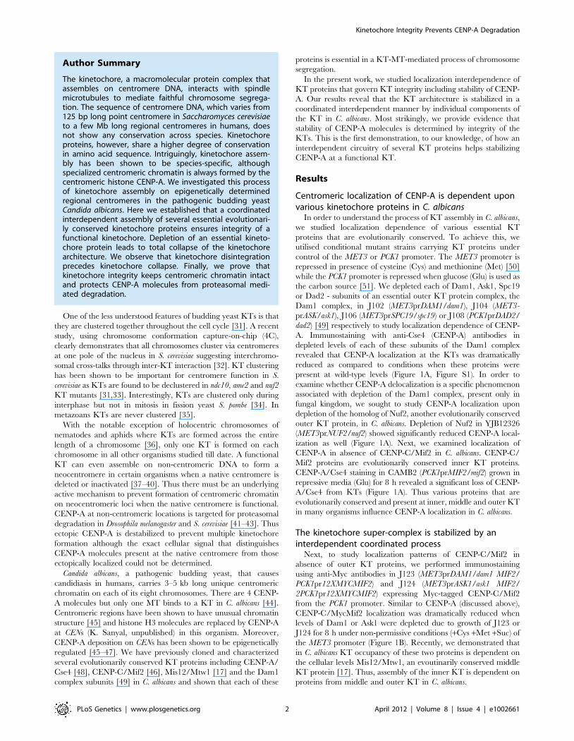
Author Summary
The kinetochore, a macromolecular protein complex thatassembles on centromere DNA, interacts with spindlemicrotubules to mediate faithful chromosome segrega-tion. The sequence of centromere DNA, which varies from125 bp long point centromere in Saccharomyces cerevisiaeto a few Mb long regional centromeres in humans, doesnot show any conservation across species. Kinetochoreproteins, however, share a higher degree of conservationin amino acid sequence. Intriguingly, kinetochore assem-bly has been shown to be species-specific, althoughspecialized centromeric chromatin is always formed by thecentromeric histone CENP-A. We investigated this processof kinetochore assembly on epigenetically determinedregional centromeres in the pathogenic budding yeastCandida albicans. Here we established that a coordinatedinterdependent assembly of several essential evolutionari-ly conserved kinetochore proteins ensures integrity of afunctional kinetochore. Depletion of an essential kineto-chore protein leads to total collapse of the kinetochorearchitecture. We observe that kinetochore disintegrationprecedes kinetochore collapse. Finally, we prove thatkinetochore integrity keeps centromeric chromatin intactand protects CENP-A molecules from proteasomal medi-ated degradation.
Kinetochore Integrity Prevents CENP-A Degradation
PLoS Genetics | www.plosgenetics.org 2 April 2012 | Volume 8 | Issue 4 | e1002661
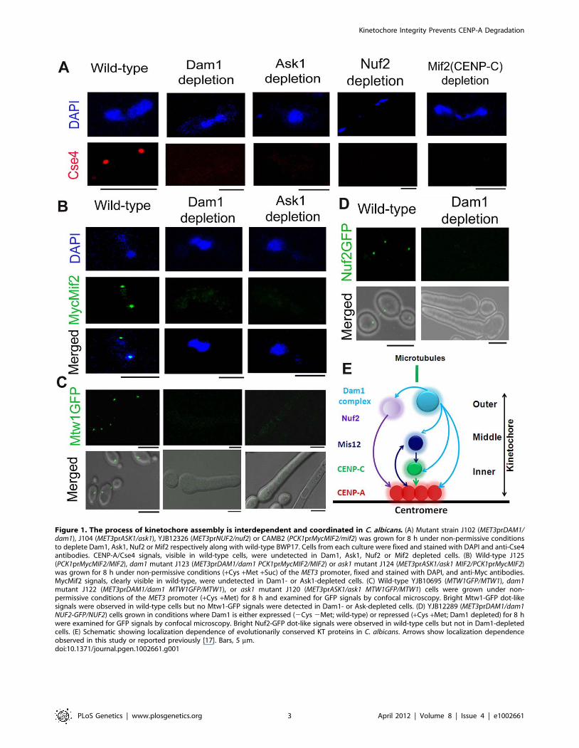
Figure 1. The process of kinetochore assembly is interdependent and coordinated in C. albicans. (A) Mutant strain J102 (MET3prDAM1/dam1), J104 (MET3prASK1/ask1), YJB12326 (MET3prNUF2/nuf2) or CAMB2 (PCK1prMycMIF2/mif2) was grown for 8 h under non-permissive conditionsto deplete Dam1, Ask1, Nuf2 or Mif2 respectively along with wild-type BWP17. Cells from each culture were fixed and stained with DAPI and anti-Cse4antibodies. CENP-A/Cse4 signals, visible in wild-type cells, were undetected in Dam1, Ask1, Nuf2 or Mif2 depleted cells. (B) Wild-type J125(PCK1prMycMIF2/MIF2), dam1 mutant J123 (MET3prDAM1/dam1 PCK1prMycMIF2/MIF2) or ask1 mutant J124 (MET3prASK1/ask1 MIF2/PCK1prMycMIF2)was grown for 8 h under non-permissive conditions (+Cys +Met +Suc) of the MET3 promoter, fixed and stained with DAPI, and anti-Myc antibodies.MycMif2 signals, clearly visible in wild-type, were undetected in Dam1- or Ask1-depleted cells. (C) Wild-type YJB10695 (MTW1GFP/MTW1), dam1mutant J122 (MET3prDAM1/dam1 MTW1GFP/MTW1), or ask1 mutant J120 (MET3prASK1/ask1 MTW1GFP/MTW1) cells were grown under non-permissive conditions of the MET3 promoter (+Cys +Met) for 8 h and examined for GFP signals by confocal microscopy. Bright Mtw1-GFP dot-likesignals were observed in wild-type cells but no Mtw1-GFP signals were detected in Dam1- or Ask-depleted cells. (D) YJB12289 (MET3prDAM1/dam1NUF2-GFP/NUF2) cells grown in conditions where Dam1 is either expressed (2Cys 2Met; wild-type) or repressed (+Cys +Met; Dam1 depleted) for 8 hwere examined for GFP signals by confocal microscopy. Bright Nuf2-GFP dot-like signals were observed in wild-type cells but not in Dam1-depletedcells. (E) Schematic showing localization dependence of evolutionarily conserved KT proteins in C. albicans. Arrows show localization dependenceobserved in this study or reported previously [17]. Bars, 5 mm.doi:10.1371/journal.pgen.1002661.g001
Kinetochore Integrity Prevents CENP-A Degradation
PLoS Genetics | www.plosgenetics.org 3 April 2012 | Volume 8 | Issue 4 | e1002661

These results prompted us to further investigate the role of the
Dam1 complex on the occupancy of a middle KT protein. KT
localized Mtw1-GFP signals that were visible in wild-type conditions
were absent when Ask1 or Dam1 was repressed for 8 h in J122
(MET3prDAM1/dam1 MTW1GFP/MTW1) or J120 (MET3prASK1/
ask1 MTW1GFP/MTW1) (Figure 1C). Together we conclude that
integrity of the middle KT is also determined by outer KT proteins.
Having established localization dependence of various domains of a
KT on each other, we further examined how localization of one
protein depends on another at the outer KT. As compared to wild-
type, Nuf2-GFP localization at the KT was reduced dramatically
when Dam1 was depleted in YJB12289 (MET3prDAM1/dam1
NUF2GFP/NUF2) (Figure 1D). These results suggest that integrity
of different domains of a KT is interdependent and assembly of
various components of a KT is highly coordinated (Figure 1E).
Integrity of a kinetochore remains unaffected upondisruption of the mitotic spindle
Unlike most organisms including fission yeast and humans, KTs
are attached to spindle MTs throughout the cell cycle in S. cerevisiae
except for a brief period of 2–3 minutes during centromere
replication [52,53]. A similar KT-MT interaction at all stages of
the cell cycle is evidenced in C. albicans as well [17]. Various
proteins from C. albicans discussed above have been shown to be
essential in KT-MT mediated process of chromosome segregation
as severe spindle defects were observed upon depletion of each of
these proteins [17,46,48,49]. In this study, we examined whether
or not reduced KT localization of various KT proteins was due to
impairment of the mitotic spindle caused by depletion of each of
these proteins. To test this possibility, we disrupted the mitotic
spindle in 10118 (CSE4:GFP:CSE4/cse4) expressing GFP-tagged
Cse4 by treating cells with a spindle depolymerizing drug
nocodazole (NOC). Tubulin staining of these NOC treated cells
exhibited disruption of the mitotic spindle structure as expected
(Figure 2A). However, no significant change in the intensity of
Cse4-GFP signals was observed between NOC treated (mean
intensity value = 225628 a.u.) and untreated (mean intensity
value = 227632 a.u.) cells of 10118 (Figure 2B, Figure S2). A
similar experiment to compare Mtw1-GFP levels in NOC treated
and untreated cells of YJB10695 (MTW1GFP/MTW1) exhibited
no significant difference (Figure 2C) as well. Localization of Dad2,
a subunit of the Dam1 complex, is unaltered in presence or
absence of NOC [49]. Together, these results showed that
localization of various components of a KT is independent of
integrity of the mitotic spindle. CENP-A/Cse4 ChIP analysis
further indicated that spindle integrity does not have significant
effect on the stability of centromeric chromatin (Figure 2D).
Partial depletion of several kinetochore proteins leads todisintegration of the clustered kinetochores
In order to understand how absence of a KT protein leads to
collapse of an entire KT, we examined the KT structure at
Figure 2. Stability of the kinetochore structure is independent of structural integrity of the mitotic spindle. A) Wild-type BWP17 cells,untreated (DMSO) or treated with nocodazole (+NOC), were fixed and stained with DAPI, and anti-tubulin antibodies. As compared to untreated cells(upper panels), NOC treated cells (lower panels) exhibited a significant loss of tubulin staining indicating a compromised structure of the mitoticspindle as expected by NOC treatment. (B) CENP-A/Cse4-GFP signals were analyzed in absence (DMSO) or presence of NOC (+NOC) in 10118(CSE4:GFP:CSE4/cse4) cells. (C) Similarly, Mtw1-GFP signals were examined in absence (DMSO) or presence of NOC in YJB10695 (MTW1GFP/MTW1) cells.No significant change in intensity of either Cse4-GFP or Mtw1-GFP signals in NOC treated cells. (D) PCR analysis on Cse4 ChIP-DNA obtained fromNOC treated BWP17 cells. Primers from CENR (CaChrR: 1747812–1748023), CEN1 (CaChr1: 1565723–1565967), CEN7 (CaChr7: 427369–427560) or 17 kbaway from CEN7 (CaChr7: 444584–444875) (non-CEN) were used for PCR analysis. T, total DNA; +, IP DNA with anti-Cse4 antibodies and 2, beads onlycontrol without antibodies. Bars, 5 mm.doi:10.1371/journal.pgen.1002661.g002
Kinetochore Integrity Prevents CENP-A Degradation
PLoS Genetics | www.plosgenetics.org 4 April 2012 | Volume 8 | Issue 4 | e1002661
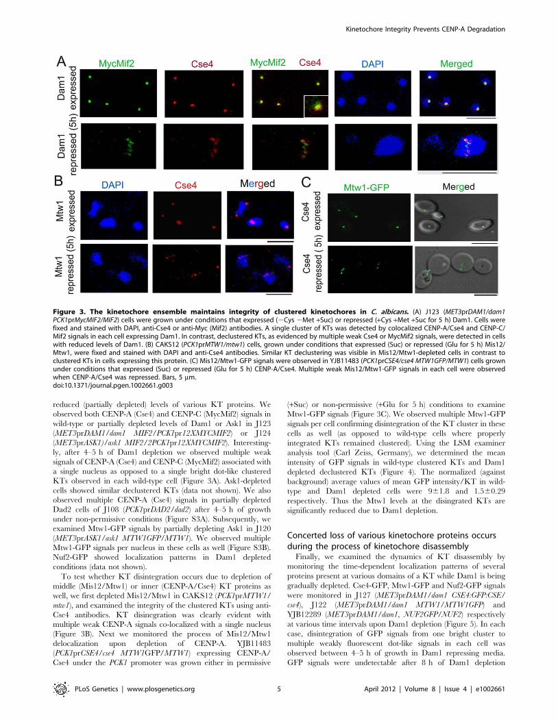
reduced (partially depleted) levels of various KT proteins. We
observed both CENP-A (Cse4) and CENP-C (MycMif2) signals in
wild-type or partially depleted levels of Dam1 or Ask1 in J123
(MET3prDAM1/dam1 MIF2/PCK1pr12XMYCMIF2) or J124
(MET3prASK1)/ask1 MIF2/2PCK1pr12XMYCMIF2). Interesting-
ly, after 4–5 h of Dam1 depletion we observed multiple weak
signals of CENP-A (Cse4) and CENP-C (MycMif2) associated with
a single nucleus as opposed to a single bright dot-like clustered
KTs observed in each wild-type cell (Figure 3A). Ask1-depleted
cells showed similar declustered KTs (data not shown). We also
observed multiple CENP-A (Cse4) signals in partially depleted
Dad2 cells of J108 (PCK1prDAD2/dad2) after 4–5 h of growth
under non-permissive conditions (Figure S3A). Subsequently, we
examined Mtw1-GFP signals by partially depleting Ask1 in J120
(MET3prASK1/ask1 MTW1GFP/MTW1). We observed multiple
Mtw1-GFP signals per nucleus in these cells as well (Figure S3B).
Nuf2-GFP showed localization patterns in Dam1 depleted
conditions (data not shown).
To test whether KT disintegration occurs due to depletion of
middle (Mis12/Mtw1) or inner (CENP-A/Cse4) KT proteins as
well, we first depleted Mis12/Mtw1 in CAKS12 (PCK1prMTW1/
mtw1), and examined the integrity of the clustered KTs using anti-
Cse4 antibodies. KT disinegration was clearly evident with
multiple weak CENP-A signals co-localized with a single nucleus
(Figure 3B). Next we monitored the process of Mis12/Mtw1
delocalization upon depletion of CENP-A. YJB11483
(PCK1prCSE4/cse4 MTW1GFP/MTW1) expressing CENP-A/
Cse4 under the PCK1 promoter was grown either in permissive
(+Suc) or non-permissive (+Glu for 5 h) conditions to examine
Mtw1-GFP signals (Figure 3C). We observed multiple Mtw1-GFP
signals per cell confirming disintegration of the KT cluster in these
cells as well (as opposed to wild-type cells where properly
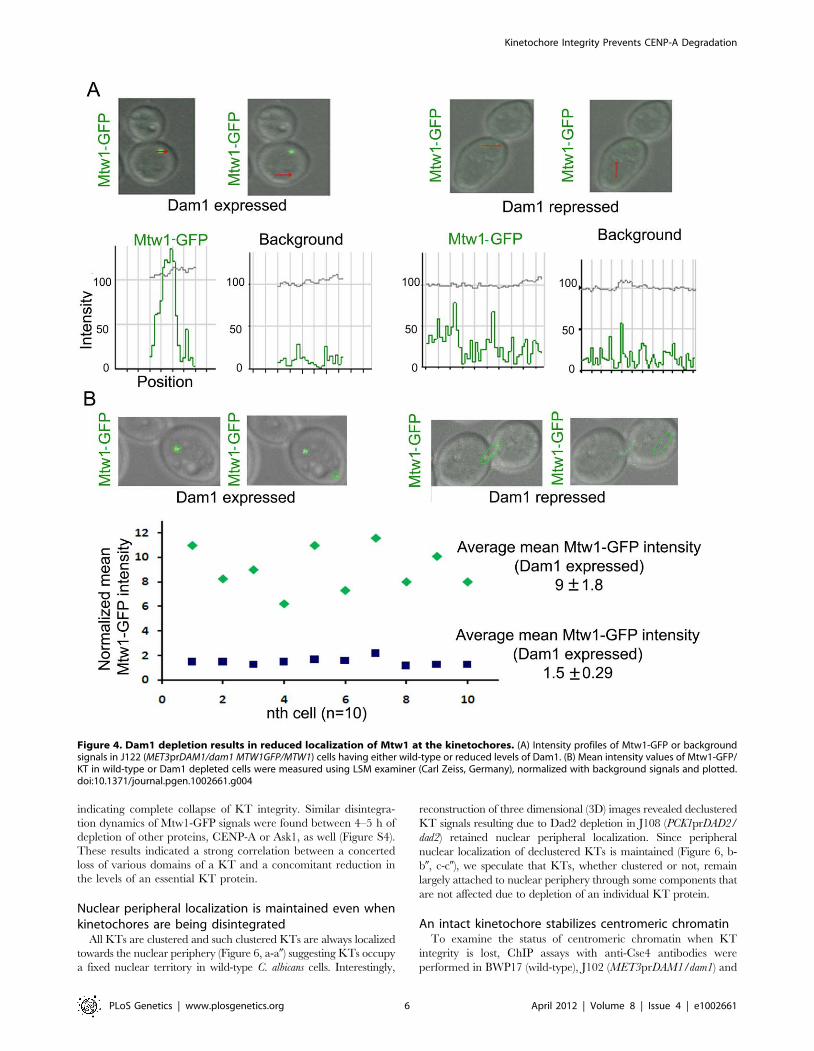
integrated KTs remained clustered). Using the LSM examiner
analysis tool (Carl Zeiss, Germany), we determined the mean
intensity of GFP signals in wild-type clustered KTs and Dam1
depleted declustered KTs (Figure 4). The normalized (against
background) average values of mean GFP intensity/KT in wild-
type and Dam1 depleted cells were 961.8 and 1.560.29
respectively. Thus the Mtw1 levels at the disingrated KTs are
significantly reduced due to Dam1 depletion.
Concerted loss of various kinetochore proteins occursduring the process of kinetochore disassembly
Finally, we examined the dynamics of KT disassembly by
monitoring the time-dependent localization patterns of several
proteins present at various domains of a KT while Dam1 is being
gradually depleted. Cse4-GFP, Mtw1-GFP and Nuf2-GFP signals
were monitored in J127 (MET3prDAM1/dam1 CSE4:GFP:CSE/
cse4), J122 (MET3prDAM1/dam1 MTW1/MTW1GFP) and
YJB12289 (MET3prDAM1/dam1, NUF2GFP/NUF2) respectively
at various time intervals upon Dam1 depletion (Figure 5). In each
case, disintegration of GFP signals from one bright cluster to
multiple weakly fluorescent dot-like signals in each cell was
observed between 4–5 h of growth in Dam1 repressing media.
GFP signals were undetectable after 8 h of Dam1 depletion
Figure 3. The kinetochore ensemble maintains integrity of clustered kinetochores in C. albicans. (A) J123 (MET3prDAM1/dam1PCK1prMycMIF2/MIF2) cells were grown under conditions that expressed (2Cys 2Met +Suc) or repressed (+Cys +Met +Suc for 5 h) Dam1. Cells werefixed and stained with DAPI, anti-Cse4 or anti-Myc (Mif2) antibodies. A single cluster of KTs was detected by colocalized CENP-A/Cse4 and CENP-C/Mif2 signals in each cell expressing Dam1. In contrast, declustered KTs, as evidenced by multiple weak Cse4 or MycMif2 signals, were detected in cellswith reduced levels of Dam1. (B) CAKS12 (PCK1prMTW1/mtw1) cells, grown under conditions that expressed (Suc) or repressed (Glu for 5 h) Mis12/Mtw1, were fixed and stained with DAPI and anti-Cse4 antibodies. Similar KT declustering was visible in Mis12/Mtw1-depleted cells in contrast toclustered KTs in cells expressing this protein. (C) Mis12/Mtw1-GFP signals were observed in YJB11483 (PCK1prCSE4/cse4 MTW1GFP/MTW1) cells grownunder conditions that expressed (Suc) or repressed (Glu for 5 h) CENP-A/Cse4. Multiple weak Mis12/Mtw1-GFP signals in each cell were observedwhen CENP-A/Cse4 was repressed. Bars, 5 mm.doi:10.1371/journal.pgen.1002661.g003
Kinetochore Integrity Prevents CENP-A Degradation
PLoS Genetics | www.plosgenetics.org 5 April 2012 | Volume 8 | Issue 4 | e1002661
indicating complete collapse of KT integrity. Similar disintegra-
tion dynamics of Mtw1-GFP signals were found between 4–5 h of
depletion of other proteins, CENP-A or Ask1, as well (Figure S4).
These results indicated a strong correlation between a concerted
loss of various domains of a KT and a concomitant reduction in
the levels of an essential KT protein.
Nuclear peripheral localization is maintained even whenkinetochores are being disintegrated
All KTs are clustered and such clustered KTs are always localized
towards the nuclear periphery (Figure 6, a-a0) suggesting KTs occupy
a fixed nuclear territory in wild-type C. albicans cells. Interestingly,
reconstruction of three dimensional (3D) images revealed declustered
KT signals resulting due to Dad2 depletion in J108 (PCK1prDAD2/
dad2) retained nuclear peripheral localization. Since peripheral
nuclear localization of declustered KTs is maintained (Figure 6, b-
b0, c-c0), we speculate that KTs, whether clustered or not, remain
largely attached to nuclear periphery through some components that
are not affected due to depletion of an individual KT protein.
An intact kinetochore stabilizes centromeric chromatinTo examine the status of centromeric chromatin when KT
integrity is lost, ChIP assays with anti-Cse4 antibodies were
performed in BWP17 (wild-type), J102 (MET3prDAM1/dam1) and
Figure 4. Dam1 depletion results in reduced localization of Mtw1 at the kinetochores. (A) Intensity profiles of Mtw1-GFP or backgroundsignals in J122 (MET3prDAM1/dam1 MTW1GFP/MTW1) cells having either wild-type or reduced levels of Dam1. (B) Mean intensity values of Mtw1-GFP/KT in wild-type or Dam1 depleted cells were measured using LSM examiner (Carl Zeiss, Germany), normalized with background signals and plotted.doi:10.1371/journal.pgen.1002661.g004
Kinetochore Integrity Prevents CENP-A Degradation
PLoS Genetics | www.plosgenetics.org 6 April 2012 | Volume 8 | Issue 4 | e1002661

J108 (PCK1prDAD2/dad2) strains grown in non-permissive media
for 8 h. These experiments revealed a drastic decrease in CENP-A
binding to CENs in Dam1 or Dad2 depleted cells as compared to
wild-type cells confirming that integrity of CENP-A-bound
centromeric chromatin is highly affected in these mutants
(Figure 7A). Since the localization of CENP-C/Mif2 was also
affected when Dam1 was depleted we tested centromere
occupancy of CENP-C/Mif2 in J125 (MIF2/PCK1pr12XMYC-
MIF2) or depleted levels of Dam1 in J123 (MET3prDAM1)/dam1
MIF2/PCK1pr12XMYCMIF2) by ChIP assays with anti-MycMif2
antibodies (Figure 7B). The ChIP-PCR analysis confirmed that
CENP-C/Mif2 occupancy at centromeres was also dramatically
reduced in Dam1 depleted cells as compared to wild-type cells. We
have recently shown that, Mtw1/Mis12 is required for inner
kinetochore assembly including localization of CENP-A and
CENP-C [17]. Together these results confirmed that Dam1 is
required for integrity of centromeric chromatin in C. albicans.
CENP-A is unstable in absence of an essential KT proteinSince centromeric chromatin is disintegrated when various KT
proteins are depleted, we examined protein levels of CENP-A in
these conditions to find out the fate of CENP-A molecules that are
Figure 5. Disintegration of the kinetochore cluster precedes kinetochore collapse. Levels of GFP-tagged inner (Cse4), middle (Mtw1) orouter (Nuf2) KT proteins under gradual repression of Dam1 were monitored at indicated time after shift of strains J127 (MET3prDAM1/dam1CSE4:GFP:CSE4/cse4), J122 (MET3prDAM1/dam1 MTW1GFP/MTW1) and YJB12289 (MET3prDAM1/dam1 NUF2GFP/NUF2) to non-permissive medium forthe MET3 promoter. In each case GFP (left) and GFP+DIC (right) images were shown. Bars, 5 mm.doi:10.1371/journal.pgen.1002661.g005
Figure 6. Nuclear peripheral localization is maintained indisintegrated declustered kinetochores. J108 (PCK1prDAD2/dad2) cells grown for 5 h under non-permissive conditions of thePCK1 promoter were fixed and stained with DAPI and anti-Cse4antibodies. Images representing views from different rotational angles(a-a0, b-b9, c-c0) are shown in each case. Each wild-type cell exhibited asingle bright dot-like signal of CENP-A/Cse4 at the periphery of theDAPI-stained nucleus (a-a0). Each Dad2-depleted cell, on the other hand,exhibited multiple weak CENP-A/Cse4 signals colocalized with a DAPIstained nucleus (b-b0 and c-c0). Similar to a single intact clustered KTs inthe nucleus of each wild-type cell, multiple disintegrated declustered KTsignals in the nucleus of each mutant cell were also localized at thenuclear periphery.doi:10.1371/journal.pgen.1002661.g006
Figure 7. The Dam1 complex maintains inner kinetochoreassembly including integrity of centromeric chromatin. (A) PCRanalysis on Cse4 ChIP DNA obtained from wild-type BWP17, Dam1-depleted J102 (MET3prDAM1/dam1) or Dad2-depleted J108(PCK1prDAD2/dad2) cells grown for 8 h in non-permissive conditions.Primers from CENR (CaChrR: 1747812–1748023), CEN1 (CaChr1:1565723–1565967), CEN7 (CaChr7: 427369–427560) or 17 kb away fromCEN7 (CaChr7: 444584–444875) (non-CEN) were used for PCR analysis. T,total DNA; +, IP DNA with anti-Cse4 antibodies and 2, beads onlycontrol without antibodies. CENP-A/Cse4 recruitment was found to behighly reduced at centromeres in Dam1- or Dad2-depleted cells ascompared to wild-type. (B) MycMif2 ChIP-PCR analysis with DNAobtained from J123 (MET3prDAM1/dam1 PCK1prMycMIF2/MIF2) eitherexpressing Dam1 (WT) or depleted of it. Same primer pairs from CENR,CEN1, CEN7 or non-centromeric region LEU2 (non CEN) were used forPCR analysis. T, total DNA; +, IP DNA with anti-Myc antibodies and 2,beads only control without antibodies.doi:10.1371/journal.pgen.1002661.g007
Kinetochore Integrity Prevents CENP-A Degradation
PLoS Genetics | www.plosgenetics.org 7 April 2012 | Volume 8 | Issue 4 | e1002661
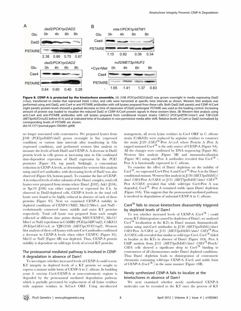
no longer associated with centromeres. We prepared lysates from
J108 (PCK1prDAD2/dad2) grown overnight in Suc (expressed
condition) or various time intervals after transferring in Glu
(repressed condition), and performed western blot analysis to
measure the levels of both Dad2 and CENP-A. A decrease in Dad2
protein levels in cells grown at increasing time in Glu confirmed
time-dependent repression of Dad2 expression by the PCK1
promoter (Figure 8A, top panel). Strikingly, a concomitant
reduction in CENP-A levels, as determined by western blot analysis
using anti-Cse4 antibodies, with decreasing levels of Dad2 was also
observed (Figure 8A, bottom panel). To examine the fate of CENP-
A in reduced levels of other subunits of the Dam1 complex, total cell
lysates were prepared from strains where Dam1 (J102), Ask1 (J104),
or Spc19 (J106) was either expressed or repressed for 8 h. As
observed in Dad2-depleted cells, CENP-A levels in the total cell
lysate were found to be highly reduced in absence of each of these
proteins (Figure S5). Next we examined CENP-A stability in
depleted conditions of CENP-C/Mif2, Mis12/Mtw1, and Nuf2 -
evolutionarily conserved inner, middle and outer KT proteins
respectively. Total cell lysate was prepared from each sample
collected at different time points during Mif2/CENP-C, Mis12/
Mtw1 or Nuf2 reprefssion in CAMB2 (PCK1prMIF/mif2), CAKS12
(PCK1prCSE4/cse4) or YJB12326 (MET3prNUF2/nuf2). Western
blot analysis of these cell lysates with anti-Cse4 antibodies confirmed
a decrease in CENP-A levels when either CENP-C (Figure S5),
Mis12 or Nuf2 (Figure 8B) was depleted. Thus, CENP-A protein
stability is dependent on wild-type levels of several KT proteins.
The proteasomal mediated pathway is involved in CENP-A degradation in absence of Dam1
To investigate whether increased levels of CENP-A could rescue
KT integrity in depleted levels of a KT protein we sought to
express a mutant stable form of CENP-A in C. albicans. In budding
yeast S. cerevisiae Cse4/CENP-A at non-centromeric regions is
degraded by the proteasomal mediated degradation pathway
which is partially prevented by replacement of all lysine residues
with arginine residues in ScCse4 ORF. Using site-directed
mutagenesis, all seven lysine residues in Cse4 ORF in C. albicans
strain (CAKS2b) were replaced by arginine residues to construct
the strain J129 (CSE47R-Prot A/cse4) where Protein A (Prot A)
tagged mutated Cse47R is the only source of CENP-A (Figure 9A).
All the changes were confirmed by DNA sequencing (Figure S6).
Western blot analysis (Figure 9B) and immunolocalization
(Figure 9C) using anti-Prot A antibodies revealed that Cse47R -
Prot A is functionally expressed in C. albicans.
To examine the effect of Dam1 depletion on the stability of
Cse47R, we expressed Cse4-Prot A and Cse47R-Prot A in the Dam1
conditional mutant. Western blot analysis in J130 (MET3prDAM1)/
dam1 CSE4-Prot A/CSE4) or J131 (MET3prDAM1/dam1 CSE47R-
Prot A/CSE4) revealed that while wild-type Cse4-Prot A was
degraded, Cse47R -Prot A remained stable upon Dam1 depletion
(Figure 10A). This suggests that the proteasomal mediated pathway
is involved in degradation of unbound CENP-A in C. albicans.
Cse47R fails to rescue kinetochore disassembly triggeredby depleted levels of Dam1
To test whether increased levels of CENP-A (Cse47R ) could
rescue KT disintegration caused by depletion of Dam1, we analysed
Cse47R localization at the KTs in Dam1 mutant. Immunolocali-
zation using anti-Cse4 antibodies in J130 (MET3prDAM1)/dam1
CSE4-Prot A/CSE4) or J131 (MET3prDAM1/dam1 CSE47R-Prot
A/CSE4) cells revealed that similar to wild-type Cse4, Cse47R failed
to localize at the KTs in absence of Dam1 (Figure 10A). Prot A
ChIP analysis from J131 (MET3prDAM1/dam1 CSE47R-ProtA/
CSE4) cells showed a significant drop in Cse47R binding to
centromeres of all chromosomes under Dam1 depleted conditions.
Thus Dam1 depletion leads to disintegration of centromeric
chromatin containing wild-type CENP-A (Cse4) and stable form
of CENP-A (Cse47R ) in the same manner (Figure 10B).
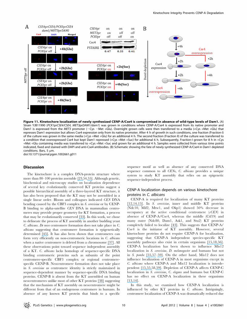
Newly synthesized CENP-A fails to localize at thekinetochores in absence of Dam1
We next examined whether newly synthesized CENP-A
molecules can be recruited to the KT once the process of KT
Figure 8. CENP-A is protected by the kinetochore ensemble. (A) J108 (PCK1prDAD2/dad2) was grown overnight in media expressing Dad2(+Suc), transferred to media that repressed Dad2 (+Glu), and cells were harvested at specific time intervals as shown. Western blot analysis wasperformed using anti-Dad2, anti-Cse4 or anti-PSTAIRE antibodies with cell lysates prepared from these cells. Both Dad2 (left panels) and CENP-A/Cse4(right panels) protein levels showed a gradual decrease as time of repression of Dad2 prolonged. PSTAIRE was used as the loading control. Increasingamount of protein was loaded to visualize the reduced Dad2 or CENP-A/Cse4 protein signals in these western blots. (B) Western blot analysis usinganti-Cse4 and anti-PSTAIRE antibodies with cell lysates prepared from conditional mutant strains CAKS12 (PCK1prMTW1/mtw1) and YJB12326(MET3prNUF2/nuf2) before (0 h) and at indicated time of incubation in non-permissive media after shift. Relative levels of Cse4 or Dad2 normalized bycorresponding levels of PSTAIRE are shown.doi:10.1371/journal.pgen.1002661.g008
Kinetochore Integrity Prevents CENP-A Degradation
PLoS Genetics | www.plosgenetics.org 8 April 2012 | Volume 8 | Issue 4 | e1002661

disassembly is initiated under Dam1 depleted conditions. We
utilized YJB11990 (PCK1prCSE4/CSE4 MET3prDAM1/dam1) in
which CSE4 and DAM1 are placed under control of the PCK1 and
MET3 promoters respectively. We studied the fate of newly
synthesized CENP-A molecules by inducing expression of CENP-
A/Cse4 (+Suc) while Dam1 is kept in repressed state (+Cys +Met).
To achieve this, YJB11990 was grown in Dam1 repressible
conditions till the point where (a) KTs starts to decluster (4 h) or
(b) KTs are disintegrated (8 h) (Figure 11A). These cells were then
transferred to media that represses the MET3 promoter (+Cys
+Met to keep Dam1 depleted) but induces the PCK1 promoter
(+Suc to overexpress Cse4) and grown for an additional 4 h.
Expression of new CENP-A/Cse4 molecules in such conditions
was confirmed by western blot analysis (Figure 11A). Indirect
immunolocalization using anti-Cse4 antibodies revealed that
newly synthesized CENP-A/Cse4 molecules expressed from the
induced PCK1 promoter (Figure 11A) could not be recruited at
KTs under Dam1 repressible conditions (Figure 11B).
Figure 10. Depletion of Dam1 leads to degradation of CENP-A/Cse4 through the ubiquitin-mediated proteasomal degradationpathway. (A) Lysates were prepared from strains J130 (MET3prDAM1)/dam1 CSE4-Prot A/CSE4) and J131 (MET3prDAM1/dam1 CSE47R-Prot A/CSE4)expressing wild-type Cse4 -Prot A (left) or mutant Cse47R -Prot A (right) grown under conditions where Dam1 is either expressed (2Cys 2Met) orrepressed (+Cys +Met). Western blot analysis was performed with these lysates using anti-Prot A or anti-PSTAIRE antibodies. Indirectimmunolocalization with anti-Cse4 antibodies revealed that recruitment of either wild-type Cse4 or mutant Cse47R at the kinetochore iscompromised in absence of Dam1. (B) Prot A (Cse4) ChIP assays in strain J131 (MET3prDAM1/dam1 CSE47R-Prot A/CSE4) confirmed that stable mutatedCse47R is unable to maintain centromeric chromatin in absence of Dam1. CEN, centromeres; T, Total input DNA, Prot-A, immunoprecipitated DNAwith anti-Prot A antibodies, 2, beads only control. Bars, 5 mm.doi:10.1371/journal.pgen.1002661.g010
Figure 9. Cse47R -Prot A is functional in C. albicans. (A) Each of the 7 lysine (K) residues of Cse4 ORF shown in schematic has been mutated toarginine (R) residues by site-directed mutagenesis. (B) Lysates from strains expressing wild-type J128 (CSE4-Prot A/cse4) and J129 (CSE47R-Prot A/cse4)Cse4-Prot-A or Cse47R–Prot-A were subjected to western blot analysis with anti-Prot-A or anti-PSTAIRE antibodies. (C) Indirect immunolocalization byanti-Prot A (Cse4) antibodies in J129 (CSE47R-Prot A/cse4) exhibited normal KT localization patterns of Cse47R. Bar, 5 mm.doi:10.1371/journal.pgen.1002661.g009
Kinetochore Integrity Prevents CENP-A Degradation
PLoS Genetics | www.plosgenetics.org 9 April 2012 | Volume 8 | Issue 4 | e1002661
Discussion
The kinetochore is a complex DNA-protein structure where
more than 80–100 proteins assemble [23,54,55]. Although genetic,
biochemical and microscopy studies on localization dependence
of several key evolutionarily conserved KT proteins suggest a
possible hierarchical assembly of a three-layered KT structure, it
has also been proposed that the KT may not be assembled in a
single linear order. Bloom and colleagues indicated CEN DNA
bending caused by the CBF3 complex in S. cerevisiae or by CENP-
B binding to alpha-satellite CEN DNA in mammalian centro-
meres may provide proper geometry for KT formation, a process
that may be evolutionarily conserved [33]. In this work, we chose
to delineate the process of KT assembly in another budding yeast
C. albicans. De novo centromere formation does not take place in C.
albicans suggesting that centromere formation is epigenetically
determined [45]. It has also been shown that centromeres can
form very efficiently on non-centromeric locations in C. albicans
when a native centromere is deleted from a chromosome [37]. All
these observations point toward sequence independent assembly
of a KT. C. albicans lacks homologs of sequence-specific DNA
binding centromeric proteins such as subunits of the point
centromere-specific CBF3 complex or regional centromere-
specific CENP-B. Neocentromere formation does not take place
in S. cerevisiae as centromere identity is strictly maintained in
sequence-dependent manner by sequence-specific DNA binding
proteins. CENP-B is absent from the KT assembled on human
neocentromeres unlike most of other KT proteins [38] suggesting
that the mechanism of KT assembly on neocentromere might be
different from that of an endogenous centromere in humans. In
absence of any known KT protein that binds to a specific
sequence motif as well as absence of any conserved DNA
sequence common to all CENs, C. albicans provides a unique
system to study KT assembly that relies on an epigenetic
sequence-independent process.
CENP-A localization depends on various kinetochoreproteins in C. albicans
CENP-A is required for localization of many KT proteins
[12,14,15]. In S. cerevisiae, inner and middle KT proteins
(Ndc10, Mif2, Mtw1, and Okp1) showed 50% decrease in
occupancy at the active conditional centromere (cCEN) in
absence of CENP-A/Cse4, whereas the middle (Ctf19) and
many outer (Ndc80, Dam1, Ask1, and Stu2) KT proteins
completely failed to localize [43]. This suggests that CENP-A/
Cse4 is the initiator of KT assembly. However, several
kinetochore proteins do not require CENP-A for localization,
suggesting that CENP-A independent species-specific KT
assembly pathways also exist in certain organisms [15,18,56].
CENP-A localization has been shown to influence Mis12
localization in S. cerevisiae, D. melanogaster and humans but not
in S. pombe [55,57–59]. On the other hand, Mis12 does not
influence localization of CENP-A in most organisms except in
C. albicans where CENP-A and Mis12 localization is interde-
pendent [15,55,58,59]. Depletion of CENP-A affects CENP-C
localization in S. cerevisiae, C. elegans and humans but CENP-C
has no effect on CENP-A localization in these organisms
[13,14].
In this study, we examined how CENP-A localization is
influenced by other KT proteins in C. albicans. Intriguingly,
centromere localization of CENP-A was dramatically reduced due
Figure 11. Kinetochore localization of newly synthesized CENP-A/Cse4 is compromised in absence of wild-type levels of Dam1. (A)Strain YJB11990 (PCK1prCSE4/CSE4, MET3prDAM1/dam1) was grown in conditions where CENP-A/Cse4 is expressed from its native promoter andDam1 is expressed from the MET3 promoter (2Cys 2Met +Glu). Overnight grown cells were then transferred to a media (+Cys +Met +Glu) thatrepresses Dam1 expression but allows Cse4 expression only from its native promoter. After 4 h of growth in such conditions, one fraction (Fraction-I)of the culture was grown in the same media (+Cys +Met +Glu) for an additional 4 h. The second fraction (Fraction II) of the culture was transferred toa condition that overexpressed Cse4 but kept Dam1 repressed (+Cys +Met +Suc) for additional 6 h. Subsequently, Fraction-I grown for 8 h in +Cys+Met +Glu containing media was transferred to +Cys +Met +Suc and grown for an additional 4 h. Samples were collected from various time pointsindicated, fixed and stained with DAPI and anti-Cse4 antibodies. (B) Schematic showing the fate of newly synthesized CENP-A/Cse4 in Dam1 depletedconditions. Bars, 5 mm.doi:10.1371/journal.pgen.1002661.g011
Kinetochore Integrity Prevents CENP-A Degradation
PLoS Genetics | www.plosgenetics.org 10 April 2012 | Volume 8 | Issue 4 | e1002661

to depletion of inner (Mif2/CENP-C) or outer (Dam1 complex
and Nuf2) KT proteins. Recently, we showed that a middle KT
protein Mis12/Mtw1 homolog C. albicans influences assembly of
two inner KT proteins CENP-A and CENP-C [17]. Thus
localization dependence of CENP-A on various KT proteins
varies from species to species.
An interdependent coordinated protein circuitrystabilizes the kinetochore structure
Since it is unusual and striking that outer KT proteins influence
localization of CENP-A, we further investigated localization
dependence of various other proteins to determine the sequence
of events that lead to KT formation on unique short regional
centromeres of C. albicans. An unprecedented observation of
collapse of the KT architecture in absence of an essential protein
from a KT in C. albicans raises an important question. How do
KTs assemble in C. albicans? KT disassembly due to depletion of
various proteins suggests that KT assembly is probably not a step-
wise process in C. albicans. We propose that KT proteins of various
sub-complexes assemble in a unique interdependent concerted
manner to form and stabilize the macromolecular KT architecture
in C. albicans Our results thus indicate that the KT in C. albicans
may not even have a layered structure, unlike the one observed in
humans.
Stability of the kinetochore structure is independent ofstructural integrity of the mitotic spindle
Since all proteins we analyzed in this study have been previously
shown to be essential for the proper dynamics of a mitotic spindle
in C. albicans [17,48,49] we investigated whether or not an intact
mitotic spindle is required for maintaining integrity of KTs.
Localization of CENP-A or Mis12 was found to be unaffected in
presence of a MT-depolymerizing drug nocodazole. In NOC
treated cells tubulin staining showed two weak dot-like signals
probably representing SPBs after duplication. Cse4 and Mtw1
GFP signals in NOC treated cells are also seen as two dots situated
close to each other. Thus we conclude that stabilization of the KT
structure is independent of structural integrity of the mitotic
spindle.
The kinetochore ensemble maintains integrity of thekinetochore cluster in C. albicans
Like S. cerevisiae, KT-MT interaction is established early during
S phase in C. albicans. Live cell imaging by time-lapse microscopy
in our previous study revealed that all centromeres are clustered
together throughout the cell cycle in C. albicans [17]. Moreover,
similar localization patterns of these clustered centromeres at the
nuclear periphery were observed both in S. cerevisiae and C. albicans.
In absence of a metaphase plate in budding yeasts [60], clustered
centromeres may provide a platform for MT attachment and
synchronous separation of sister chromatids during anaphase.
Conditional KT mutants provided us an opportunity to follow the
process of KT disassembly in a time dependent manner during
gradual depletion of various KT proteins in C. albicans. Our results
clearly indicate that disintegration of the KT cluster is a common
intermediate step before KT collapse and each component is
required to keep all the KTs clustered in C. albicans. Chromosome
confirmation capture (3C) assay to study interaction among
centromeres of different chromosomes in S. cerevisiae revealed that
crosslinking frequencies between different centromeres is signifi-
cantly higher than other chromosomal sites except telomeric
regions [61]. A recent study, where chromosome confirmation
capture on chip (4C) and massively parallel sequencing were used
to globally capture inter and intrachromosomal interactions in S.
cerevisiae, demonstrates centromeres as the chromosome landmarks
that mediate interchromosomal interaction [32]. The clustering of
centromeres that marked the primary point of engagement
between different chromosomes was the most striking feature of
the inter-chromosomal contacts. In the light of these observations
in S. cerevisiae and our results in this study, we speculate that
interchromosomal interaction at the centromeres may facilitate
stabilization of the kinetochore ensemble as depletion of various
KT proteins leads to disintegration of clustered centromeres in
both species.
An intact kinetochore protects CENP-A from degradationChIP analysis and immunolocalization studies confirmed that
CENP-A occupancy is severely impaired when an essential KT
protein is depleted from C. albicans cells. We predicted that
disintegration of centromeric chromatin in absence of KT proteins
may expose free CENP-A molecules for cellular degradation.
Indeed, western blot analysis confirmed that cellular CENP-A
protein levels were drastically reduced in KT mutants tested. Since
integrity of centromeric chromatin is also dependent upon
individual KT proteins which in turn help in maintaining overall
KT integrity, it is tempting to speculate that establishment of
centromeric chromatin and assembly of KTs start together in a
coordinated way in C. albicans.
The proteasomal mediated pathway of CENP-Adegradation is evolutionarily conserved
The ubiquitin mediated degradation pathway is one of the
major pathways that degrade CENP-A/Cse4 in S. cerevisiae and
CENP-A/CID in Drosophila. In Dam1 depleted strain where wild-
type Cse4 is unstable, the mutant Cse47R showed higher stability
suggesting a similar CENP-A degradation pathway is active in C.
albicans. Thus, although the process of CENP-A recruitment on
point and regional centromeres may differ but the mechanism that
regulates CENP-A stability to prevent ectopic KT formation
(especially in C. albicans where efficiency of neocentromere
formation is remarkably high) seem to be conserved among
organisms carrying point and regional centromeres.
Dam1 is required for CENP-A recruitment at thekinetochores
Wild-type CENP-A is unstable when the KT ensemble is
disintegrated and centromeric chromatin is disrupted in absence of
an essential KT protein. It is possible that reduction in CENP-A
levels is the cause not the consequence of KT disassembly under
such conditions. We wondered whether expression of a stable form
of CENP-A (Cse47R) can prevent disintegration of the KT
structure in absence of Dam1. We examined stable mutant
CENP-A, Cse47R localization at the KT in Dam1 depleted cells
and found that increasing the stability of CENP-A (confirmed by
western blot analysis) does not prevent CENP-A delocalization at
the KT in absence of Dam1. The Cse47R failed to maintain
integrity of centromeric chromatin as well. Next we sought to
provide new CENP-A molecules using an inducible promoter
(PCK1) to examine if newly synthesized CENP-A can be recruited
to the KTs in absence of Dam1. However, these newly synthesized
CENP-A molecules (detected by western blot analysis) failed to
rescue KT integrity further confirming that an individual KT
protein is absolutely essential for protecting the centromeric bound
CENP-A by maintaining the integrity of the KT ensemble that is
laid on the foundation of CENP-A associated centromeric
chromatin.
Kinetochore Integrity Prevents CENP-A Degradation
PLoS Genetics | www.plosgenetics.org 11 April 2012 | Volume 8 | Issue 4 | e1002661
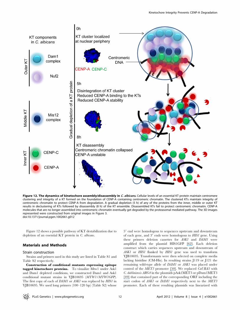
Figure 12 shows a possible pathway of KT destabilization due to
depletion of an essential KT protein in C. albicans.
Materials and Methods
Strain constructionStrains and primers used in this study are listed in Table S1 and
Table S2 respectively.
Construction of conditional mutants expressing epitope
tagged kinetochore proteins. To visualize Mtw1 under Ask1
and Dam1 depleted conditions, we constructed Dam1 and Ask1
conditional mutant strains in YJB10695 (MTW1/MTW1GFP).
The first copy of each of DAM1 or ASK1 was replaced by HIS1 in
YJB10695. We used long primers (100–120 bp) (Table S2) whose
59 end were homologous to sequences upstream and downstream
of each gene, and 39 ends were homologous to HIS1 gene. Using
these primers deletion cassettes for ASK1 and DAM1 were
amplified from the plasmid HIS1GFP [62]. Each deletion
construct which carries sequences upstream and downstream of
ASK1 or HIS1 flanked by HIS1 gene was used to transform
YJB10695. Transformants were then selected on complete media
lacking histidine (CM-His). In resulting strains J119 or J121 the
remaining wild-type allele of DAM1 or ASK1 was placed under
control of the MET3 promoter [50]. We replaced CaURA3 with
C. dubliniensis ARG4 in the plasmids pAsk1MET3 or pDam1MET3
[49] that contained part of the corresponding ORF including the
start codon of ASK1 or DAM1 respectively next to the MET3
promoter. Each of these resulting plasmids was linearized with
Figure 12. The dynamics of kinetochore assembly/disassembly in C. albicans. Cellular levels of an essential KT protein maintain centromereclustering and integrity of a KT formed on the foundation of CENP-A containing centromeric chromatin. The clustered KTs maintain integrity ofcentromeric chromatin to protect CENP-A from degradation. A gradual depletion (5 h) of any of the proteins from the inner, middle or outer KTresults in declustering of KTs followed by disassembly (8 h) of the KT ensemble. Disassembled KTs fail to protect centromeric chromatin. CENP-Amolecules that are no longer assembled into centromeric chromatin eventually get degraded by the proteasomal mediated pathway. The 3D imagesrepresented were constructed from original images in Figure 3.doi:10.1371/journal.pgen.1002661.g012
Kinetochore Integrity Prevents CENP-A Degradation
PLoS Genetics | www.plosgenetics.org 12 April 2012 | Volume 8 | Issue 4 | e1002661
ClaI to transform J119 or J121 and transformants were selected
on media lacking arginine (CM-Arg). Resulting conditional Ask1
and Dam1 mutant strains were named J120 and J122
respectively.
To visualize Mif2 signals in Ask1 or Dam1 depleted condi-
tions, we constructed strains expressing Myc-tagged Mif2 in
Dam1 or Ask1 conditional mutant strains. A cassette containing
PCK1prMycMIF2 was amplified from strain CAMB2 [46] using
primers Mif2PckMycPst1-F and Mif2pck1SacII-R and subse-
quently cloned into PstI and SacII sites of pSF2A vector [63] that
contained the nourseothricin (NAT) marker. The resulting
plasmid was linearized using HpaI and introduced into J102
(Dam1 conditional mutant) or J104 (Ask1 conditional mutant)
strains to get J123 (dam1/MET3DAM1, MIF2/PCK1prMycMIF2)
or J124 (ask1/MET3ASK1, MIF2/PCK1prMycMIF2).
Construction of strains expressing Cse4-Prot A and
Cse47R-Prot A. The CSE4 ORF was cloned into SacII and
SpeI sites of pBluescript to construct pBSCSE4. All seven lysine
residues were changed into arginine residues in the plasmid
pBSCSE4 using a site-directed mutagenesis kit (Stratagene, Cat
# 200522) to construct pBSCSE47R. The TAP cassette
(containing URA3 and TAP that contains Prot A) and CSE4
downstream sequences were cloned as NcoI/KpnI and KpnI/SacI
fragments respectively into the corresponding sites of pUC19 to
construct pCSE4DSTAP. The CSE47R fragment was amplified
from pBSCSE47R, digested with SalI and NcoI and cloned into
corresponding sites of pCSE4DSTAP to yield pCSE47RTAP.
Similarly the CSE4 fragment was cloned into SalI and NcoI sites
of pCSE4DSTAP to construct pCSE4TAP. Both pCSE4TAP
and pCSE47RTAP were linearized using XhoI and these
linearized fragments were used to transform CAKS2b (CSE4/
cse4::hisG ) to give rise to J128 (CSE4-TAP(URA3)/cse4::hisG )
and J129 (CSE47R-TAP(URA3)/cse4::hisG) respectively. Sub-
sequently, J126 (dam1::HIS1 MET3prDAM1(ARG4)/dam1::
HIS1) was transformed with XhoI-digested pCSE4TAP and
pCSE47RTAP separately to give rise to J130 (dam1::HIS1
MET3prDAM1(ARG4)/dam1::HIS1 CSE4-TAP(URA3)/CSE4)
and J131 (dam1::HIS1 MET3prDAM1(ARG4)/dam1::HIS1 CSE47R-
TAP(URA3)/CSE4) respectively.
Media and growth conditionsConditional mutant strains of Dam1 (J102, J121, J122, J127,
J130, J131, YJB11990 and YJB12289), Ask1 (J104, J120, and
J124), Spc19 (J106) and Nuf2 (YJB12326) that carry DAM1, ASK1
SPC19 and NUF2 respectively under control of the MET3
promoter were grown in YPDU (1% yeast extract, 2% peptone,
3% glucose and 0.01% uridine) as permissive media and YPDU+5 mM cysteine (+Cys)+5 mM methionine (+Met) as non-permis-
sive media. Conditional mutant strains of Mif2 (CAMB2, J123,
J124 and J125), Dad2 (J108), Cse4 (CAKS3b and YJB11483) and
Mtw1 (CAKS12) that carry MIF2, DAD2, CSE4 and MTW1
respectively under control of the PCK1 promoter were grown in
YPSU (1% Yeast Extract, 2% Peptone, 2% Succinate and 0.01%
Uridine) as permissive media and YPDU as non-permissive media.
All C. albicans strains were grown at 30uC.
GFP imagingC. albicans conditional mutants with GFP tagged KT proteins
grown overnight in inducing media were transferred to repressible
media at initial OD600 - 0.150. Cells were harvested at various
time intervals after growth in repressible media. Harvested cells
were resuspeneded in 50% glycerol and subsequently imaged
using a confocal microscope (Zeiss LSM 510 META). Images were
further processed by Adobe Photoshop.
Nocodazole treatmentFor nocodazole treatment, cells were grown overnight in
YPDU, reinoculated in YPDU with an initial OD600 = 0.2.
Nocodazole (Sigma, Cat # M1404) was added at a concentration
of 20 mg/ml when OD600 = 0.4 (1 generation) was achieved. Cells
were grown for an additional 4 h before harvesting for
immunolocalization and ChIP assays.
Chromatin immunoprecipitation (ChIP) assayChIP assays were performed using a protocol described
previously [49]. An exponentially growing culture of C. albicans
strain was fixed with 1% formaldehyde for 15 min. The reaction
was quenched for 5 min at room temperature using glycine to a
final concentration of 125 mM. Cells were washed and suspended
in resuspension buffer (0.2 mM Tris-HCl pH, 9.4, 10 mM DTT).
Resuspended cells were incubated at 30uC for 15 min on a shaker
at 180 rpm. Cells were washed and resuspended in spheroplasting
buffer (1.2 M Sorbitol, 20 mM Na-HEPES, pH 7.5). Spheroplast-
ing (95%) was performed using lyticase (Sigma, Cat # L2524) at
30uC at low speed. Spheroplasting was stopped by adding ice-cold
postspheroplasting buffer (1.2 M Sorbitol, 1 mM MgCl2, 20 mM
Na-PIPES, pH 6.8). Spheroplasts were subsequently washed with
ice-cold 16 PBS, Buffer I (0.25% TritonX-100, 10 mM EDTA,
0.5 mM EGTA, 10 mM Na-HEPES, pH 6.5), Buffer II (200 mM
NaCl, 1 mM EDTA, 0.5 mM EGTA, 10 mM Na-HEPES) and
finally resuspended in extraction buffer (140 mM NaCl, 1 mM
EDTA, 50 mM K-HEPES, 0.1% sodium deoxycholate, 1%
Triton X-100, pH 7.5) with protease inhibitor cocktail (Sigma)
at a concentration of 100 ml/100 ml starting culture. Next, sonica-
tion was performed to get sheared chromatin fragments of an
average size of 300–700 bp by SONICS Vibra cell sonicator. The
soluble fraction of sheared chromatin was obtained by centrifuging
the sonicated solution at 13000 rpm for 15 min at 4uC.
Total DNA (T). About 1/10th of total soluble chromatin was
processed separately as total DNA (whole cell lysate). Equal
volume of elution buffer I (50 mM Tris?HCl, pH 8/10 mM
EDTA/1% SDS) was added to the chromatin solution separated
for Total DNA, and incubated at 65uC overnight to reverse
crosslinking. TE (16, 10 mM Tris?HCl, pH 8, 1 mM EDTA,) was
added to the starting material to make final SDS concentration
0.5%. Sample was treated with RNase A (Sigma # R4875) and
Proteinase K (NEB Cat # P8102). The DNA was extracted with
an equal volume of phenol/chloroform/isoamyl alcohol (25:24:1)
in the presence of 0.4 M LiCl and precipitated with ethanol for
15 min at room temperature. It was spun at 16,0006g for 20 min
at room temperature. The DNA pellet was washed with 70%
ethanol and resuspended in TE.
Immunoprecipitated material (IP). Rest of the soluble
chromatin solution was diluted 5.7-fold with IP dilution buffer
(167 mM NaCl, 1.1 mM EDTA, 1.1% Triton X-100, 167 mM
Tris–HCl, pH 8.0) and divided equally in two tubes. Rabbit anti-
CaCse4p antibody was added to a final concentration of 4 mg/ml
in one tube (+Ab) and no antibody (2Ab) was added to the other.
The tubes were slowly rotated overnight at 4uC. A slurry of
Protein A-Sepharose beads (50 ml per ml of 50% slurry in TE)
(Sigma) was added and the tube was again rotated overnight at
4uC. Next beads were sequentially washed twice in 12 ml of
extraction buffer, and once each in 12 ml of extraction
buffer+500 mM NaCl, LiCl wash buffer (10 mM Tris-HCl,
pH 8, 250 mM LiCl, 1 mM Igepal CA-630, 0.5% sodium
deoxycholate/1 mM EDTA) and TE. Beads were subjected to
elution of IP complexes in elution buffer I (1/10 volume of IP
dilution buffer) at 65uC for 15 min, and then centrifuged at
4000 rpm for 5 min. Supernatant was collected and beads were
Kinetochore Integrity Prevents CENP-A Degradation
PLoS Genetics | www.plosgenetics.org 13 April 2012 | Volume 8 | Issue 4 | e1002661

washed a second time with elution buffer II (10 mM Tris-HCl,
pH 8/1 mM EDTA/0.67% SDS) for 5 min at 65uC and the
supernatant was collected as above. Pooled eluates were incubated
overnight at 65uC to reverse crosslinking. To purify DNA, the
SDS concentration was diluted to 0.5% with TE, and the reaction
was treated with RNase A, followed by Proteinase K. The 4 M
LiCl was added to a final concentration of 0.4 M and the DNA
was extracted with an equal volume of phenol/chloroform/
isoamyl alcohol (25:24:1). Aqueous layer was precipitated in 100%
ethanol overnight at 220uC. DNA was recovered by
centrifugation at 16,0006 g for 45 min at 4uC, washed with
70% ethanol, spun for 15 min, and the DNA pellet was dried for
30 min. The recovered DNA was resuspended in TE.
ChIP assays with anti-Myc antibodies in J123 (MycMIF2) or anti-
Prot-A antibodies were performed using the same protocol except
for the following modification. J123 cells were crosslinked with 1%
formaldehyde for 45 min. The protein enrichment on a specific
DNA sequence was determined by specific PCR primers (Table S2).
Subcellular immunolocalizationIndirect immunofluoroscence was performed using protocol
described previously [49]. Asynchronous and exponentially
growing culture was fixed using 1 ml 37% formaldehyde per
10 ml culture for 1 h. Fixed cells were washed and resuspended in
0.1 M Phosphate buffer (pH 6.4). Next, 70–80% spheroplasting
was achieved using lyticase (Sigma) and b-mercaptoethanol.
Spheroplasts were pelleted down gently at low speed and
resuspended in PBS. Teflon coated slide was incubated with
polylysine (1 mg/ml) for 5 min, washed with water and dried.
Next 15 ml of fixed cells were placed onto each well and incubated
for 5 min. Cells attached to the slides were fixed in ice cold
methanol (220uC) for 6 min and ice-cold acetone (220uC) for
30 seconds. Blocking was performed with 2% skim milk in PBS for
30 min. Subsequently cells were incubated with primary antibod-
ies for 1 h and washed with PBS four times. Subsequently,
secondary antibodies were added onto each well and incubated
1 h in a dark humid chamber. Finally the slide was washed four
times with phosphate buffered saline (PBS). DAPI solution (50 ng/
ml in 70% glycerol) was added on to each well and coverslip and
slide were sealed together. Co-immunolocalization experiments
were performed using the same protocol.
Image analysisImages were captured using Carl Zeiss confocal laser scanning
microscope (LSM 510 META) using LSM 5 Image Examiner.
Three dimentional (3D) images were generated using LSM 3D
rendering software (Figure 6 and Figure 12) (Carl Zeiss, Germany).
Images were rotated in 3D and snapshots were taken from three
different rotational angles (a-a0, b-b9, c-c0) in Figure 6. Images
were susequently processed in Adobe Photoshop.
Western blot analysisWild-type or conditional mutant strains were grown under
inducing and repressed conditions for 8 h. Protein extracts were
made by disrupting the cells in RIPA buffer (300 mM NaCl, 50 mM
Tris-HCl pH 8.0, 5 mM EDTA pH 8.0, 0.5% Triton-X) using glass
beads (Sigma cat # G8772). The lysate were subjected to electro-
phoresis using 12% SDS PAGE and transferred to nitrocellulose
membrane for 1 h at 20 V by semi-dry method. Proper transfer was
checked by Ponceau S staining. Membranes were blocked with 5%
skim milk for 1 h followed by incubation with primary antibodies in
5% skim milk overnight at 4uC. Membranes were washed five times
with PBS+0.05% Tween and incubated with secondary antibodies
in 5% skim milk for 2 h. Membranes were washed five times with
16 PBS+0.05% Tween and developed by VersaDoc (Bio-Rad) or
exposed to X-ray films. Quantification of the western blots was
performed using the Quantity one software (Bio-Rad).
AntibodiesPrimary antibodies used for immunolocalization studies were as
follows- affinity purified rabbit anti-Dad2 antibodies-1:50 dilution,
affinity purified rabbit anti-CENP-A antibodies - 1:500 dilution
[48], mouse anti-Myc -1:50 dilution (Calbiochem, Cat # OP10L),
rabbit anti-Prot A- 1:1500 (Sigma Cat # P2921), rat anti-tubulin
(Invitrogen, Cat # YOL1/34) - 1:100 dilution. The fluorescent
secondary antibodies for immunolocalization were obtained from
Invitrogen and used at dilution 1:500 for Alexa Fluor goat anti-
rabbit IgG 568 (Cat #A11011) , 1:100 for Alexa Fluor goat anti-
rat IgG 488 (Cat # A11006) and 1:500 for Alexa Fluor anti -
mouse 488 (Cat # A11001).
Primary antibodies used for western blot analysis were rabbit
anti-CENP-A (1:500), mouse anti-PSTAIRE (1:2000, Sigma Cat
# P7962), rabbit anti-Prot A (1: 5000, Sigma Cat # P2921) and
rabbit anti-Dad2 (1:500, unpurified sera) antibodies. Secondary
antibodies used were anti-rabbit HRP conjugated (1:2000,
Bangalore Genei Cat # 105499), anti-mouse HRP conjugated
(1:2000, Bangalore Genei, Cat # HP06).
Supporting Information
Figure S1 Depletion of an outer kinetochore protein leads to
reduced levels of CENP-A at the kinetochore in C. albicans. Wild-
type, and conditional mutant J106 (MET3prSPC19/spc19) or J108
(PCK1prDAD2/dad2) were grown for 8 h under non-permissive
conditions to deplete Spc19 or Dad2 respectively. These cells were
fixed and stained with DAPI and anti-Cse4 antibodies. CENP-A/
Cse4 signals, visible in wild-type cells, were undetected in Spc19 or
Dad2 depleted cells. Bars, 5 mm.
(JPG)
Figure S2 CENP-A levels at kinetochores are independent of
spindle integrity. Intensity of Cse4-GFP/KT was measured in
untreated (DMSO) or nocodazole (NOC) treated 10118
(CSE4:GFP:CSE4/cse4) cells and plotted. No significant change in
GFP intensity was observed between NOC treated cells and
untreated cells.
(JPG)
Figure S3 The kinetochore cluster is disintegrated in absence of
an essential kinetochore protein. (A) Dad2-depleted J108
(PCK1prDAD2/dad2) cells grown for 5 h under non-permissive
conditions were fixed and stained with DAPI and anti-Cse4
antibodies. These Dad2-depleted cells exhibited multiple weak
Cse4 signals per nucleus suggesting that clustered KTs were in the
process of disintegration. (B) Parent YJB10695 (MTW1GFP/
MTW1) or ask1 conditional mutant J120 (MET3prASK1/ask1
MTW1GFP/MTW1) cells grown under non-permissive conditions
of the MET3 promoter (+Cys +Met) for 5 h exhibited clustered or
declustered Mtw1GFP signals in presence or absence of Ask1
respectively. Bars, 5 mm.
(JPG)
Figure S4 Kinetochore disintegration precedes kinetochore
collapse. Levels of GFP-tagged Mtw1 (a middle KT protein)
under gradual repression of CENP-A/Cse4 (an inner KT protein;
upper panels) or Ask1 (an outer KT protein; lower panels) was
monitored at indicated time after shift to non-permissive medium
for CENP-A/Cse4 or Ask1 expression. In each case GFP (left) and
GFP+DIC (right) images are shown. Bar, 5 mm.
(JPG)
Kinetochore Integrity Prevents CENP-A Degradation
PLoS Genetics | www.plosgenetics.org 14 April 2012 | Volume 8 | Issue 4 | e1002661
Figure S5 CENP-A is unstable in absence of an essential
kinetochore protein. (A) J102 (MET3prDAM1/dam1) or J104
(MET3prASK1/ask1) expressing Dam1 or Ask1 from the
MET3 promoter and CAMB2 (PCK1prMIF2/mif2) or J106
(MET3prSPC19/spc19) expressing Mif2 or Spc19 from the PCK1
promoter were grown overnight in inducing media and then
transferred to repressing media for 8 h. Western blot analysis was
performed using anti-Cse4 and anti-PSTAIRE antibodies with cell
lysates prepared from 0 h and 8 h of growth in repressing media.
CENP-A/Cse4 (right panels) protein levels showed a significant
decrease when each of Dam1, Ask1, CENP-C/Mif2 or Spc19 was
depleted.
(JPG)
Figure S6 Site-directed mutagenesis to create CENP-A/Cse4
mutant Cse47R in C. albicans. All seven lysine residues were
changed to arginine residues in CSE4 ORF using the site-directed
mutagenesis kit (Stratagene). Incorporation of all the changes was
confirmed by sequencing of relevant regions from wild-type J128
(CSE4-TAP(URA3)/cse4::hisG, top panel) or J129 (CSE47R-TA-
P(URA3)/ cse4::hisG, bottom panel) strain. Changes K189-R189
and K196- R196 were confirmed by the reverse primer.
(JPG)
Table S1 C. albicans strains used in this study.
(DOC)
Table S2 Primers used in this study.
(DOC)
Acknowledgments
We thank the confocal imaging facility of the Molecular Genetics and
Biology Unit, JNCASR, and the Berman Lab, University of Minnesota, for
providing us C. albicans strains.
Author Contributions
Conceived and designed the experiments: JT KS. Performed the
experiments: JT. Analyzed the data: JT KS. Contributed reagents/
materials/analysis tools: JT KS. Wrote the paper: JT KS.
References
1. Allshire RC, Karpen GH (2008) Epigenetic regulation of centromeric
chromatin: old dogs, new tricks? Nat Rev Genet 9: 923–937.
2. Clarke L, Carbon J (1980) ISOLATION OF A YEAST CENTROMERE ANDCONSTRUCTION OF FUNCTIONAL SMALL CIRCULAR CHROMO-
SOMES. Nature 287: 504–509.
3. Clarke L (1990) Centromeres of budding and fission yeasts. Trends Genet 6:
150–154.
4. Fishel B, Amstutz H, Baum M, Carbon J, Clarke L (1988) Structuralorganization and functional analysis of centromeric DNA in the fission yeast
Schizosaccharomyces pombe. Mol Cell Biol 8: 754–763.
5. Sun X, Wahlstrom J, Karpen G (1997) Molecular structure of a functionalDrosophila centromere. Cell 91: 1007–1019.
6. Camahort R, Li B, Florens L, Swanson SK, Washburn MP, et al. (2007) Scm3 is
essential to recruit the histone h3 variant cse4 to centromeres and to maintain afunctional kinetochore. Mol Cell 26: 853–865.
7. Camahort R, Shivaraju M, Mattingly M, Li B, Nakanishi S, et al. (2009) Cse4 is
part of an octameric nucleosome in budding yeast. Mol Cell 35: 794–805.
8. Dimitriadis EK, Weber C, Gill RK, Diekmann S, Dalal Y (2010) Tetramericorganization of vertebrate centromeric nucleosomes. Proc Natl Acad Sci U S A
107: 20317–20322.
9. Mizuguchi G, Xiao H, Wisniewski J, Smith MM, Wu C (2007) Nonhistone
Scm3 and histones CenH3-H4 assemble the core of centromere-specificnucleosomes. Cell 129: 1153–1164.
10. Foltz DR, Jansen LE, Bailey AO, Yates JR, Bassett EA, et al. (2009)
Centromere-specific assembly of CENP-a nucleosomes is mediated by HJURP.Cell 137: 472–484.
11. Ekwall K (2007) Epigenetic control of centromere behavior. Annu Rev Genet
41: 63–81.
12. Blower MD, Karpen GH (2001) The role of Drosophila CID in kinetochoreformation, cell-cycle progression and heterochromatin interactions. Nat Cell
Biol 3: 730–739.
13. Desai A, Rybina S, Muller-Reichert T, Shevchenko A, Hyman A, et al. (2003)KNL-1 directs assembly of the microtubule-binding interface of the kinetochore
in C. elegans. Genes Dev 17: 2421–2435.
14. Oegema K, Desai A, Rybina S, Kirkham M, Hyman AA (2001) Functional
analysis of kinetochore assembly in Caenorhabditis elegans. J Cell Biol 153:1209–1226.
15. Goshima G, Kiyomitsu T, Yoda K, Yanagida M (2003) Human centromere
chromatin protein hMis12, essential for equal segregation, is independent ofCENP-A loading pathway. J Cell Biol 160: 25–39.
16. Hajra S, Ghosh SK, Jayaram M (2006) The centromere-specific histone variant
Cse4p (CENP-A) is essential for functional chromatin architecture at the yeast 2-microm circle partitioning locus and promotes equal plasmid segregation. J Cell
Biol 174: 779–790.
17. Roy B, Burrack LS, Lone MA, Berman J, Sanyal K (2011) CaMtw1, a memberof the evolutionarily conserved Mis12 kinetochore protein family, is required for
efficient inner kinetochore assembly in the pathogenic yeast Candida albicans.
Mol Microbiol.
18. Hayashi T, Fujita Y, Iwasaki O, Adachi Y, Takahashi K, et al. (2004) Mis16 andMis18 are required for CENP-A loading and histone deacetylation at
centromeres. Cell 118: 715–729.
19. Takahashi K, Takayama Y, Masuda F, Kobayashi Y, Saitoh S (2005) Twodistinct pathways responsible for the loading of CENP-A to centromeres in the
fission yeast cell cycle. Philos Trans R Soc Lond B Biol Sci 360: 595–606;discussion 606–597.
20. Okada M, Cheeseman IM, Hori T, Okawa K, McLeod IX, et al. (2006) The
CENP-H-I complex is required for the efficient incorporation of newly
synthesized CENP-A into centromeres. Nat Cell Biol 8: 446–457.
21. Cleveland DW, Mao Y, Sullivan KF (2003) Centromeres and kinetochores: from
epigenetics to mitotic checkpoint signaling. Cell 112: 407–421.
22. Lechner J, Carbon J (1991) A 240 kd multisubunit protein complex, CBF3, is a
major component of the budding yeast centromere. Cell 64: 717–725.
23. De Wulf P, McAinsh AD, Sorger PK (2003) Hierarchical assembly of the
budding yeast kinetochore from multiple subcomplexes. Genes Dev 17:
2902–2921.
24. Cheeseman IM, Desai A (2008) Molecular architecture of the kinetochore-
microtubule interface. Nat Rev Mol Cell Biol 9: 33–46.
25. McAinsh AD, Tytell JD, Sorger PK (2003) Structure, function, and regulation of
budding yeast kinetochores. Annu Rev Cell Dev Biol 19: 519–539.
26. Welburn JP, Grishchuk EL, Backer CB, Wilson-Kubalek EM, Yates JR, et al.
(2009) The human kinetochore Ska1 complex facilitates microtubule depoly-
merization-coupled motility. Dev Cell 16: 374–385.
27. Bharadwaj R, Qi W, Yu H (2004) Identification of two novel components of the
human NDC80 kinetochore complex. J Biol Chem 279: 13076–13085.
28. Cheeseman IM, Brew C, Wolyniak M, Desai A, Anderson S, et al. (2001)
Implication of a novel multiprotein Dam1p complex in outer kinetochore
function. J Cell Biol 155: 1137–1145.
29. Enquist-Newman M, Cheeseman IM, Van Goor D, Drubin DG, Meluh PB, et
al. (2001) Dad1p, third component of the Duo1p/Dam1p complex involved in
kinetochore function and mitotic spindle integrity. Mol Biol Cell 12:
2601–2613.
30. Janke C, Ortiz J, Tanaka TU, Lechner J, Schiebel E (2002) Four new subunits of
the Dam1-Duo1 complex reveal novel functions in sister kinetochore
biorientation. Embo Journal 21: 181–193.
31. Jin QW, Fuchs J, Loidl J (2000) Centromere clustering is a major determinant of
yeast interphase nuclear organization. J Cell Sci 113(Pt 11): 1903–1912.
32. Duan Z, Andronescu M, Schutz K, McIlwain S, Kim YJ, et al. (2010) A three-
dimensional model of the yeast genome. Nature 465: 363–367.
33. Anderson M, Haase J, Yeh E, Bloom K (2009) Function and assembly of DNA
looping, clustering, and microtubule attachment complexes within a eukaryotic
kinetochore. Mol Biol Cell 20: 4131–4139.
34. Funabiki H, Hagan I, Uzawa S, Yanagida M (1993) Cell cycle-dependent
specific positioning and clustering of centromeres and telomeres in fission yeast.
J Cell Biol 121: 961–976.
35. Brenner S, Pepper D, Berns MW, Tan E, Brinkley BR (1981) Kinetochore
structure, duplication, and distribution in mammalian cells: analysis by human
autoantibodies from scleroderma patients. J Cell Biol 91: 95–102.
36. Monen J, Maddox PS, Hyndman F, Oegema K, Desai A (2005) Differential role
of CENP-A in the segregation of holocentric C. elegans chromosomes during
meiosis and mitosis. Nat Cell Biol 7: 1248–1255.
37. Ketel C, Wang HS, McClellan M, Bouchonville K, Selmecki A, et al. (2009)
Neocentromeres form efficiently at multiple possible loci in Candida albicans.
PLoS Genet 5: e1000400. doi:10.1371/journal.pgen.1000400.
38. Marshall OJ, Chueh AC, Wong LH, Choo KH (2008) Neocentromeres: new
insights into centromere structure, disease development, and karyotype
evolution. Am J Hum Genet 82: 261–282.
39. Ishii K, Ogiyama Y, Chikashige Y, Soejima S, Masuda F, et al. (2008)
Heterochromatin integrity affects chromosome reorganization after centromere
dysfunction. Science 321: 1088–1091.
Kinetochore Integrity Prevents CENP-A Degradation
PLoS Genetics | www.plosgenetics.org 15 April 2012 | Volume 8 | Issue 4 | e1002661

40. Williams BC, Murphy TD, Goldberg ML, Karpen GH (1998) Neocentromere
activity of structurally acentric mini-chromosomes in Drosophila. Nat Genet 18:30–37.
41. Moreno-Moreno O, Torras-Llort M, Azorın F (2006) Proteolysis restricts
localization of CID, the centromere-specific histone H3 variant of Drosophila, tocentromeres. Nucleic Acids Res 34: 6247–6255.
42. Collins KA, Furuyama S, Biggins S (2004) Proteolysis contributes to theexclusive centromere localization of the yeast Cse4/CENP-A histone H3
variant. Curr Biol 14: 1968–1972.
43. Collins KA, Castillo AR, Tatsutani SY, Biggins S (2005) De novo kinetochoreassembly requires the centromeric histone H3 variant. Mol Biol Cell 16:
5649–5660.44. Joglekar AP, Bouck D, Finley K, Liu X, Wan Y, et al. (2008) Molecular
architecture of the kinetochore-microtubule attachment site is conservedbetween point and regional centromeres. J Cell Biol 181: 587–594.
45. Baum M, Sanyal K, Mishra PK, Thaler N, Carbon J (2006) Formation of
functional centromeric chromatin is specified epigenetically in Candida albicans.Proc Natl Acad Sci U S A 103: 14877–14882.
46. Sanyal K, Baum M, Carbon J (2004) Centromeric DNA sequences in thepathogenic yeast Candida albicans are all different and unique. Proc Natl Acad
Sci U S A 101: 11374–11379.
47. Padmanabhan S, Thakur J, Siddharthan R, Sanyal K (2008) Rapid evolution ofCse4p-rich centromeric DNA sequences in closely related pathogenic yeasts,
Candida albicans and Candida dubliniensis. Proc Natl Acad Sci U S A 105:19797–19802.
48. Sanyal K, Carbon J (2002) The CENP-A homolog CaCse4p in the pathogenicyeast Candida albicans is a centromere protein essential for chromosome
transmission. Proc Natl Acad Sci U S A 99: 12969–12974.
49. Thakur J, Sanyal K (2011) The essentiality of the fungus-specific Dam1 complexis correlated with a one-kinetochore-one-microtubule interaction present
throughout the cell cycle, independent of the nature of a centromere. EukaryotCell 10: 1295–1305.
50. Care RS, Trevethick J, Binley KM, Sudbery PE (1999) The MET3 promoter:
a new tool for Candida albicans molecular genetics. Mol Microbiol 34:792–798.
51. Leuker CE, Sonneborn A, Delbruck S, Ernst JF (1997) Sequence and promoter
regulation of the PCK1 gene encoding phosphoenolpyruvate carboxykinase of
the fungal pathogen Candida albicans. Gene 192: 235–240.
52. Tanaka TU (2010) Kinetochore-microtubule interactions: steps towards bi-
orientation. EMBO J 29: 4070–4082.
53. Tanaka K, Kitamura E, Tanaka TU (2010) Live-cell analysis of kinetochore-
microtubule interaction in budding yeast. Methods 51: 206–213.
54. Liu X, McLeod I, Anderson S, Yates JR, He X (2005) Molecular analysis of
kinetochore architecture in fission yeast. EMBO J 24: 2919–2930.
55. Liu ST, Rattner JB, Jablonski SA, Yen TJ (2006) Mapping the assembly
pathways that specify formation of the trilaminar kinetochore plates in human
cells. J Cell Biol 175: 41–53.
56. Regnier V, Vagnarelli P, Fukagawa T, Zerjal T, Burns E, et al. (2005) CENP-A
is required for accurate chromosome segregation and sustained kinetochore
association of BubR1. Mol Cell Biol 25: 3967–3981.
57. Westermann S, Cheeseman IM, Anderson S, Yates JR, Drubin DG, et al. (2003)
Architecture of the budding yeast kinetochore reveals a conserved molecular
core. J Cell Biol 163: 215–222.
58. Przewloka MR, Zhang W, Costa P, Archambault V, D’Avino PP, et al. (2007)
Molecular analysis of core kinetochore composition and assembly in Drosophila
melanogaster. PLoS ONE 2: e478. doi:10.1371/journal.pone.0000478.
59. Takahashi K, Chen ES, Yanagida M (2000) Requirement of Mis6 centromere
connector for localizing a CENP-A-like protein in fission yeast. Science 288:
2215–2219.
60. Straight AF, Marshall WF, Sedat JW, Murray AW (1997) Mitosis in living
budding yeast: anaphase A but no metaphase plate. Science 277: 574–578.
61. Dekker J, Rippe K, Dekker M, Kleckner N (2002) Capturing chromosome
conformation. Science 295: 1306–1311.
62. Gerami-Nejad M, Berman J, Gale CA (2001) Cassettes for PCR-mediated
construction of green, yellow, and cyan fluorescent protein fusions in Candida
albicans. Yeast 18: 859–864.
63. Reuss O, Vik A, Kolter R, Morschhauser J (2004) The SAT1 flipper, an
optimized tool for gene disruption in Candida albicans. Gene 341: 119–127.
Kinetochore Integrity Prevents CENP-A Degradation
PLoS Genetics | www.plosgenetics.org 16 April 2012 | Volume 8 | Issue 4 | e1002661