Metaphase Arrest with Centromere Separation in polo Mutants of Drosophila
13
The Rockefeller University Press, 0021-9525/2001/05/663/13 $5.00 The Journal of Cell Biology, Volume 153, Number 4, May 14, 2001 663–675 http://www.jcb.org/cgi/content/full/153/4/663 663 Metaphase Arrest with Centromere Separation in polo Mutants of Drosophila Mary M. Donaldson,* ‡ Álvaro A.M. Tavares,* Hiroyuki Ohkura,* Peter Deak,* ‡ and David M. Glover* ‡ *Cancer Research Campaign Cell Cycle Genetics Research Group, Department of Anatomy and Physiology, University of Dundee, Dundee DD1 4HN, Scotland; and ‡ Department of Genetics, University of Cambridge, Cambridge CB2 3EH, United Kingdom Abstract. The Drosophila gene polo encodes a con- served protein kinase known to be required to organize spindle poles and for cytokinesis. Here we report two strongly hypomorphic mutations of polo that arrest cells of the larval brain at a point in metaphase when the majority of sister kinetochores have separated by between 20–50% of the total spindle length in intact cells. In contrast, analysis of sister chromatid separation in squashed preparations of cells indicates that some 83% of sisters remain attached. This suggests the sepa- ration seen in intact cells requires the tension produced by a functional spindle. The point of arrest corresponds to the spindle integrity checkpoint; Bub1 protein and the 3F3/2 epitope are present on the separated kineto- chores and the arrest is suppressed by a bub1 mutation. The mutant mitotic spindles are anastral and have as- sembled upon centrosomes that are associated with Centrosomin and the abnormal spindle protein (Asp), but neither with g-tubulin nor CP190. We discuss roles for Polo kinase in recruiting centrosomal proteins and in regulating progression through the metaphase–ana- phase checkpoint. Key words: mitosis • spindle checkpoint • Drosophila • Polo kinase • cell cycle Introduction The Polo-like protein kinases (Plks) 1 are a conserved fam- ily of enzymes that play a variety of roles in the passage of cells through M phase (for reviews, see Glover et al., 1998; Nigg, 1998). They are named after the Drosophila polo gene originally identified through a recessive maternal ef- fect lethal mutation. Flies homozygous for the original polo 1 allele can develop to adulthood due to the weakly hypomorphic nature of this mutation and the provision of wild-type protein from their heterozygous mothers. How- ever, they do exhibit spindle pole defects at several devel- opmental stages including multiply branched spindles in syncytial polo 1 -derived embryos, and spindles with broad poles and a low frequency of circular mitotic figures in lar- val neuroblasts (Sunkel and Glover, 1988; Llamazares et al., 1991). Requirements for polo during male meiosis are also evident from the chromosome nondisjunction and failures in cytokinesis seen during meiosis of polo 1 testes (Sunkel and Glover, 1988; Carmena et al., 1998; Herrmann et al., 1998). A more recent examination of meiosis in polo 1 eggs and at the onset of their zygotic development indicates abnormalities in microtubule organizing centres in the meiotic spindle, the sperm aster, and the astral arrays of microtubules associated with the polar bodies (Riparbelli et al., 2000). These roles of Polo kinase are echoed in other organisms, suggesting conserved function. Disruption of the fission yeast counterpart, plo1, leads to formation of monopolar spindles (Ohkura et al., 1995). Interfering with enzyme ac- tivity using antibodies can also lead to the formation of monopolar spindles in Xenopus, or human cells (Lane and Nigg, 1996; Qian et al., 1998). In human cells, this appeared to be linked to a failure of the centrosome to increase its microtubule nucleating activity upon mitotic entry. In- deed, a direct role for polo in mitotic entry is suggested by the finding that Xenopus Polo-like kinase, Plx1, copurifies with and can activate cdc25, and may thus play a role in the positive feedback loop that operates during p34 cdc2 ac- tivation at the G2-M transition (Kumagai and Dunphy, 1996; Abrieu et al., 1998; Qian et al., 1998). A requirement for the Plks to promote the onset of cy- tokinesis also appears to have been conserved from the yeasts to the metazoans. Disruption of the fission yeast plo1 leads also to the formation of multinucleate cells in which neither an actin ring nor a septum has been formed. Overexpression of plo1 1 in fission yeast, on the other Address correspondence to D.M. Glover, Department of Genetics, University of Cambridge, Downing St., Cambridge CB2 3EH, United Kingdom. Tel.: 44-1223-333988. Fax: 44-1223-333968. E-mail: [email protected] A.A.M. Tavares’ present address is Department of Chemical Engineer- ing, Instituto Superior Técnico, Lisboa 1049, and Cell Division Group, In- stituto Gulbenkian de Ciencia, Oeiras 2781, Portugal. H. Ohkura’s present address is The Wellcome Trust Centre for Cell Bi- ology, Institute of Cell and Molecular Biology, University of Edinburgh, Edinburgh EH9 3JR, Scotland. 1 Abbreviations used in this paper: APC, anaphase-promoting complex; Asp, abnormal spindle protein; g-TuRC, g-tubulin ring complex; plk, Polo-like protein kinase. on November 12, 2013 jcb.rupress.org Downloaded from Published May 7, 2001
-
Upload
independent -
Category
Documents
-
view
2 -
download
0
Transcript of Metaphase Arrest with Centromere Separation in polo Mutants of Drosophila

The Rockefeller University Press, 0021-9525/2001/05/663/13 $5.00The Journal of Cell Biology, Volume 153, Number 4, May 14, 2001 663–675http://www.jcb.org/cgi/content/full/153/4/663 663
Metaphase Arrest with Centromere Separation in
polo
Mutants of
Drosophila
Mary M. Donaldson,*
‡
Álvaro A.M. Tavares,* Hiroyuki Ohkura,* Peter Deak,*
‡
and David M. Glover*
‡
*Cancer Research Campaign Cell Cycle Genetics Research Group, Department of Anatomy and Physiology, University of Dundee,
Dundee DD1 4HN, Scotland; and
‡
Department of Genetics, University of Cambridge, Cambridge CB2 3EH, United Kingdom
Abstract.
The
Drosophila
gene
polo
encodes a con-served protein kinase known to be required to organizespindle poles and for cytokinesis. Here we report twostrongly hypomorphic mutations of
polo
that arrestcells of the larval brain at a point in metaphase whenthe majority of sister kinetochores have separated bybetween 20–50% of the total spindle length in intactcells. In contrast, analysis of sister chromatid separationin squashed preparations of cells indicates that some83% of sisters remain attached. This suggests the sepa-ration seen in intact cells requires the tension producedby a functional spindle. The point of arrest correspondsto the spindle integrity checkpoint; Bub1 protein and
the 3F3/2 epitope are present on the separated kineto-chores and the arrest is suppressed by a
bub1
mutation.The mutant mitotic spindles are anastral and have as-sembled upon centrosomes that are associated withCentrosomin and the abnormal spindle protein (Asp),
but neither with
g
-tubulin nor CP190. We discuss rolesfor Polo kinase in recruiting centrosomal proteins andin regulating progression through the metaphase–ana-phase checkpoint.
Key words: mitosis • spindle checkpoint •
Drosophila
• Polo kinase • cell cycle
Introduction
The Polo-like protein kinases (Plks)
1
are a conserved fam-ily of enzymes that play a variety of roles in the passage ofcells through M phase (for reviews, see Glover et al., 1998;
Nigg, 1998). They are named after the
Drosophila polo
gene originally identified through a recessive maternal ef-fect lethal mutation. Flies homozygous for the original
polo
1
allele can develop to adulthood due to the weaklyhypomorphic nature of this mutation and the provision ofwild-type protein from their heterozygous mothers. How-ever, they do exhibit spindle pole defects at several devel-opmental stages including multiply branched spindles insyncytial
polo
1
-derived
embryos, and spindles with broadpoles and a low frequency of circular mitotic figures in lar-val neuroblasts (Sunkel and Glover, 1988; Llamazares et al.,1991). Requirements for
polo
during male meiosis are alsoevident from the chromosome nondisjunction and failures
in cytokinesis seen during meiosis of
polo
1
testes (Sunkel andGlover, 1988; Carmena et al., 1998; Herrmann et al., 1998). Amore recent examination of meiosis in
polo
1
eggs and at theonset of their zygotic development indicates abnormalities inmicrotubule organizing centres in the meiotic spindle, thesperm aster, and the astral arrays of microtubules associatedwith the polar bodies (Riparbelli et al., 2000).
These roles of Polo kinase are echoed in other organisms,suggesting conserved function. Disruption of the fission
yeast counterpart,
plo1
, leads to formation of monopolarspindles (Ohkura et al., 1995). Interfering with enzyme ac-tivity using antibodies can also lead to the formation ofmonopolar spindles in
Xenopus
, or human cells (Lane andNigg, 1996; Qian et al., 1998). In human cells, this appearedto be linked to a failure of the centrosome to increase itsmicrotubule nucleating activity upon mitotic entry. In-
deed, a direct role for
polo
in mitotic entry is suggested bythe finding that
Xenopus
Polo-like kinase, Plx1, copurifieswith and can activate cdc25, and may thus play a role in
the positive feedback loop that operates during p34
cdc2
ac-tivation at the G2-M transition (Kumagai and Dunphy,1996; Abrieu et al., 1998; Qian et al., 1998).
A requirement for the Plks to promote the onset of cy-tokinesis also appears to have been conserved from theyeasts to the metazoans. Disruption of the fission yeast
plo1
leads also to the formation of multinucleate cells inwhich neither an actin ring nor a septum has been formed.
Overexpression of
plo1
1
in fission yeast, on the other
Address correspondence to D.M. Glover, Department of Genetics,University of Cambridge, Downing St., Cambridge CB2 3EH, UnitedKingdom. Tel.: 44-1223-333988. Fax: 44-1223-333968. E-mail:[email protected]
A.A.M. Tavares’ present address is Department of Chemical Engineer-ing, Instituto Superior Técnico, Lisboa 1049, and Cell Division Group, In-stituto Gulbenkian de Ciencia, Oeiras 2781, Portugal.
H. Ohkura’s present address is The Wellcome Trust Centre for Cell Bi-ology, Institute of Cell and Molecular Biology, University of Edinburgh,Edinburgh EH9 3JR, Scotland.
1
Abbreviations used in this paper:
APC, anaphase-promoting complex;Asp, abnormal spindle protein;
g
-TuRC,
g
-tubulin ring complex; plk,Polo-like protein kinase.
on Novem
ber 12, 2013jcb.rupress.org
Dow
nloaded from
Published May 7, 2001
The Journal of Cell Biology, Volume 153, 2001 664
hand, leads to the formation of multiple septa at any stageof the cell cycle, indicating the potential of the enzyme toovercome the dependence of this process upon the com-pletion of mitosis (Ohkura et al., 1995). Expression of anactivated form of mammalian Plk in budding yeast hasalso been found to drive the formation of multiple septa,suggesting this is a conserved property of the enzyme (Leeand Erikson, 1997). In
Drosophila
, the requirement of
polo
for cytokinesis appears to be linked to the need to re-organize the central region of the spindle in late M phase(Herrmann et al., 1998). In budding yeast, Cdc5p is alsoshown to play a role in regulating cytokinesis, and thepolo-box, a conserved motif in the noncatalytic domain ofPlks, is required for this (Song and Lee, 2001).
There is also evidence that the polo-like kinases can ac-tivate certain functions of the anaphase-promoting com-plex (APC), an E3 ubiquitin-protein ligase that directs thedegradation of anaphase inhibitors, Pds1p in buddingyeast (Cohen-Fix et al., 1996) and Cut2p in fission yeast(Funabiki et al., 1996), and the mitotic cyclins (for reviews,see Townsley and Ruderman, 1998; Yanagida, 1998).Descombes and Nigg (1998) showed that the addition ofcatalytically inactive polo-like kinase to colony-stimulat-ing factor (CSF)-arrested extracts of
Xenopus
eggs blocksthe Ca
2
1
-triggered destruction of cyclin B and inactivationof p34
cdc2
and also prevented the destruction of exogenousAPC-dependent substrates. Furthermore, M phase exitwould not take place in this system after immunodepletionof Plx1, but could be restored by the addition of catalyti-cally active enzyme. The mouse polo-like kinase will alsophosphorylate and activate the bacterially expressed APCcomponents Cdc16 and Cdc27 in vitro (Kotani et al.,1998). The mitotic cyclin Clb2p is not degraded in mutantsfor the budding yeast plk, Cdc5p (Shirayama et al., 1998),whereas overexpression of Cdc5p increased APC activityand decreased the levels of Clb2p, an outcome not ob-tained with a “kinase-dead” mutant (Charles et al., 1998).
Until now, a requirement for
polo
in regulating APC in
Drosophila
has not been apparent from the phenotypes ofthe alleles that have been studied.
polo
1
mutants, for exam-ple, are able to progress through development, as a func-tion of the weakly hypomorphic protein is sufficientlysupplemented by maternally provided wild-type protein.Consequently, it has been difficult to assess fully the func-tions of Polo kinase in
polo
1
somatic cells which are capableof progression through multiple cell cycles. In this paper,we describe the mitotic phenotype of two strongly hypo-morphic alleles,
polo
9
and
polo
10
, which block the prolifera-tion of diploid tissues. Cells of these mutants appear poisedto initiate anaphase and sister kinetochores are pulledapart. Chromatids undergoing partial separation can re-main attached through their telomeres and do not migrateto the spindle poles. We discuss potential roles for Polo ki-nase in regulating specific functions of checkpoint proteinsand the APC at the metaphase–anaphase transition.
Materials and Methods
Immunoblotting
Extracts of larval brains
were prepared for immunoblotting according toScaerou et al. (1999). 6 brains were used per lane for wild-type, 7 for
polo
1
,
and 12 per lane for
polo
9/10
. This ensured equal protein loading, which wastested by probing with the C4 mouse monoclonal antibody to actin. ThePolo and
g
-tubulin proteins were identified by probing with MA81 rabbitanti-Polo and GTU88 mouse anti-
g
-tubulin antibodies.
Giemsa-stained Preparations of the Larval Central Nervous System
Third-instar larvae were washed and dissected in PBS. The associatedimaginal discs were removed and the brains were incubated in 45% aceticacid for 30 s, then in 60% acetic acid for 3 min. The brains were thensquashed hard between a poly-lysine–coated slide (Sigma-Aldrich) and asiliconized coverslip. The slides were frozen in liquid nitrogen and thecoverslip flipped off with a scalpel blade. The preparations were then de-hydrated in 75 and 100% ethanol for 5 min each before being left to airdry. The preparations were then stained in Giemsa and mounted in DPX.Preparations were observed at 60 and 100
3
objective on a compound mi-croscope. In experiments where colchicine was used, the dissected brainswere incubated with 10
2
5
M colchicine, in Schneiders tissue culture me-dium (Trueman and Bate, 1988) containing FCS, for 30 min, 1 h, 2 h, or4 h, before being incubated in 45% acetic acid.
Immunostaining
Immunostaining of whole mount preparations of third-instar larval brainswas carried out by first dissecting the larvae in PBS and then fixing in3.7% formaldehyde for 20 min. Brains were washed in PBS, then perme-abilized in 0.3% Tween 20 in PBS for 10 min. The tissue was “blocked” bya 40-min incubation in PBS containing 10% FCS before being incubatedovernight with the primary antibodies in PBS containing 0.1% Tween 20(PBST) containing 2.5
m
g/ml RNase, at 4
8
C. Brains were then washed inPBST and incubated with a 10
2
2
dilution of secondary antibodies in PBSTcontaining 10% FCS for between 2 and 4 h. Brains were finally washed inPBST before being incubated in propidium iodide to stain DNA. Thepreparation was mounted in Vectashield.
Immunostaining with the 3F3/2 antibody was performed according toBousbaa et al. (1997), except that no taxol was used, and the staining of
a
-tubulin carried out simultaneously by adding YL1/2 into the primary an-tibody incubation, and the appropriate suitable secondary antibody intothe subsequent incubation.
Microscopy was performed using a Nikon Optiphot compound micro-scope with a Bio-Rad Laboratories MRC 1024 Scanning Confocal attach-ment. Images were captured using Lasersharp software and manipulatedin Adobe Photoshop.
Results
Strong polo Hypomorphs Show Mitotic Arrest in Larval Brain Cells
We wished to isolate strong hypomorphic alleles of
polo
inorder to examine their effect upon mitosis in somatic cells.As P-elements generally insert either into the 5
9
regula-tory regions or coding sequences to downregulate or inac-tivate their target genes, we sought to identify such allelesin a collection of mutants generated by P-element–medi-ated mutagenesis (Deak et al., 1997). We discovered twonew
polo
alleles,
polo
9
and
polo
10
, that have their lethalphase during late third larval instar development and haverespective mitotic indices that are elevated about 11- and8-fold above cells of wild-type larval brains (Fig. 1 and Ta-ble I). This is considerably higher than
polo
1
that has a mi-totic index about fourfold greater than wild-type in this tis-sue.
polo
1
mutants produce comparable levels of proteinto wild-type, but the kinase is thought to be defective dueto a failure of an activating phosphorylation (Tavares etal., 1996). In contrast, the amounts of Polo kinase in the
polo
9
and
polo
10
mutants are barely detectable (Fig. 2 B).This is a consequence of the insertion of P-elements in thetwo lines into the noncoding regions of the first exon of
on Novem
ber 12, 2013jcb.rupress.org
Dow
nloaded from
Published May 7, 2001

Donaldson et al.
Metaphase Arrest in polo Mutants of Drosophila
665
Figure 1. Mitotic figures from wild-type, polo9, and polo10 brains. (A) A wild-type metaphase figure. (B) A wild-type anaphase figure.(C) Hypercondensed mitotic chromosomes from polo9. The arrow points to a pair of separated sister chromatids. (D) polo10 cell show-ing separated sisters (arrow) that could be at early anaphase. (E) An anaphase (arrow) and metaphase (arrowhead) figure from polo10.(F) A tetraploid polo10 cell in which several sister chromatids appear separated throughout their length, and yet joined at their telo-meres (arrow). (G) A polyploid polo9 cell in which chromosomes form long chains attached at their telomeres (arrows). (H) Bar graphshowing the proportion of cells at prophase and metaphase (Pro. & Met.) or anaphase and telophase (Ana. & Tel.) in wild-type (wt),polo1, polo9, and polo10 brains. (I) Wild-type cell from a larval brain treated for 4 h with colchicine. (J) polo10 cell after 30 min colchicinetreatment. The arrow marks separated sisters. (K) polo10 cell after 2 h colchicine treatment.
on Novem
ber 12, 2013jcb.rupress.org
Dow
nloaded from
Published May 7, 2001
The Journal of Cell Biology, Volume 153, 2001 666
the
polo
gene at a position that affects either one (
polo
10
)or both (
polo
9
) of the two
polo
transcription units (Fig. 2A). The levels of enzyme in
polo
9
are less than in
polo
10
,consistent with the sites of insertion, and they account forthe relative severity of the mutant phenotype indicated bythe mitotic indices.
Examination of mitotic chromosomes in squashed prep-arations of the central nervous systems from
polo
9
or
polo
10
larvae indicated several characteristic features.First, by comparison with wild-type mitotic cells (Fig. 1, Aand B), chromosomes are much more highly condensed,indicating that the cells have been delayed in mitosis forsome time (Fig. 1, C, E, and F). The majority of cells ap-pear arrested in a metaphase-like state in that most sisterchromatids (83%) are still joined at their centromeres, andthe proportion of figures in which chromosomes are wellseparated at anaphase is relatively low, although in abso-lute numbers it is comparable to that in wild-type cells(Fig. 1 H and Table I). A proportion of cells are polyploid(Fig. 1, F and G, and Table I), suggesting that they havebeen able to escape the metaphase block and undergo afurther cell cycle in the absence of chromosome segrega-tion or cytokinesis. A low proportion of circular mitoticfigures could be seen, as reported previously for
polo
13
. In
z
40% of cells, at least one set of the sister chromatids ap-pear to have prematurely separated (arrows in Fig. 1, Cand D; see also the legend to Table I). In addition, chro-mosomes frequently appeared to be connected throughtheir telomeric regions. Fig. 1, F and G, show polyploidcells in which many chromosomes are linked in this way(arrows). This phenotype strongly resembles that de-scribed previously in the mutant
UbcD1
gene that encodesa class I ubiquitin-conjugating (E2) enzyme (Cenci et al.,1997). The mitotic index of the
polo
9
and
polo
10
brains didnot change significantly after colchicine treatment, sug-gesting that the population of cells in this tissue could notrespond further to disruption of spindle integrity (Fig. 1, Jand K). A similar frequency of sister chromatid separation
was observed independently of whether the preparationhad been made in the presence or absence of colchicine.
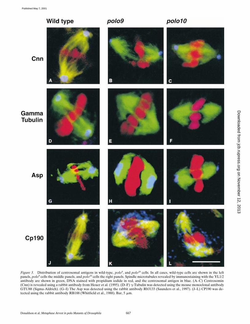
polo Differentially Affects Centrosomal Components
Defects in spindle poles and other microtubule organizingcentres have been reported previously for
polo
1
. However,the
polo9/10 mutants have allowed us to examine for thefirst time the effects of greatly diminished levels of thePolo kinase on the spindles and centrosomes of somaticcells. Examination of whole mount preparations of polo9/10
larval brains stained with antibodies to a-tubulin to revealthe mitotic spindle confirms the results seen with Giemsa-stained preparations. It reveals a very high mitotic indexwith the majority of cells arrested in a metaphase-likestate. The arrested cells all have bipolar spindles with ro-bust arrays of spindle microtubules, but lacking asters attheir poles (Figs. 3–6). Consistent with the previously ob-served failure of the CP190 antigen to assemble onto cen-trosomes in syncytial polo1-derived embryos, CP190 doesnot concentrate in the centrosomes of polo9/10 neuroblasts,but is scattered throughout the spindle with some ten-dency to accumulate around the condensed chromosomes(Fig. 3, K and L). g-Tubulin is also missing from the spindlepoles (Fig. 3, E and F). The protein cannot be seen inthese particular micrographs, although other preparationsshow weak staining, suggesting it is dispersed throughoutthe mutant cells. This interpretation is supported by thedetection of undiminished levels of g-tubulin by Westernblotting of extracts of these mutant brains (Fig. 2). In con-trast, both centrosomin (Fig. 3, B and C) and abnormalspindle protein (Asp) (Fig. 3, H and I) are both present atthe spindle poles in the mutant cells, indicating that Polokinase function is not required for their localization.
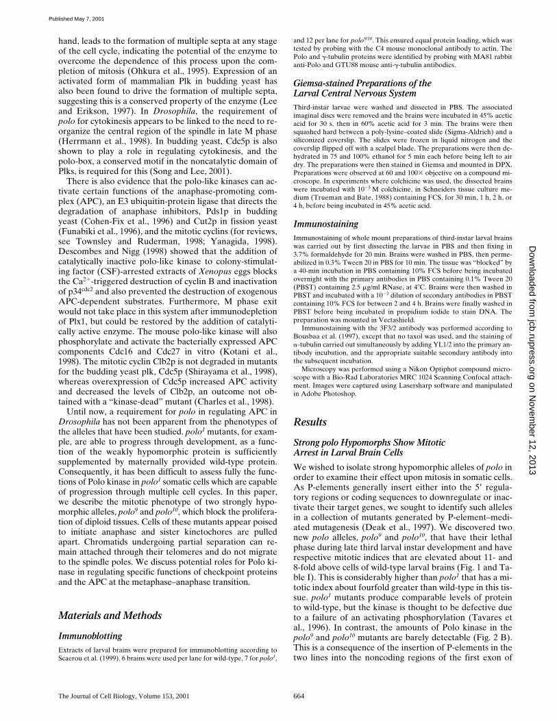
Table I. Analysis of the Mitotic Phenotype of polo Alleles in the Larval Central Nervous System
Cellsscored*
Prophaseand
metaphase‡
Circularmitoticfigures
Anaphaseand
telophase
Mitotic cells:aneuploid or polyploid§
Mitoticindexi M/A¶
n % % % %
Wild-type 2,521 2.8 0 0.5 1.2 3.3 5.8polo1 1,793 10.4 0.7 1.0 2.3 11.9 11.6polo9 1,616 32.9 1.4 0.7 16.1 35.6 51.4polo10 1,723 22.9 1.2 1.0 11.3 25.0 26.1polo10 bub1 1,766 3.1 0.5 0.4 49.2 3.7 8.3
In addition to the above quantitation, numbers of pairs of sister chromatids showingseparation were also counted in polo10 cells in the presence and absence of colchicine.Of 272 chromatid pairs scored in untreated polo10 cells, 17.2% were separated,compared with 17.0% of 176 chromatid pairs scored in colchicine-treated preparations.40% of untreated polo10 cells had at least one separated pair of sister chromatids. Thisfigure was similar (36.6%) after colchicine treatment.*Total number of cells scored in 10 fields (603 objective) from 5 brains for each ge-notype.‡Cells were scored as being metaphase if chromosomes were closely associated and ifthey contain some sister chromatids that appeared to be still attached.§Note, this is percentage of mitotic cells and not total cells.iThe mitotic index is scored as the number of mitotic cells as a percentage of age of to-tal cell number.¶Metaphase/anaphase ratio.
Figure 2. Sites of P-element insertions in polo9 and polo10 andrelative expression levels of their gene products. (A) Schematicdiagram of insertion sites in which P-elements are indicated bythe filled red triangles above the linear map at positions 234 and2176 nucleotides upstream of the initiator ATG codon of thePolo protein. Two major starts for the initiation of transcriptionare indicated by the horizontal black triangles at positions 2130and 2210 nucleotides. Exons are indicated by the open box andthe first intron by the kinked line. (B) Western blots to comparethe levels of Polo kinase and g-tubulin in wild-type (wt), polo1,polo9, and polo10 larval brains (see Materials and Methods).
on Novem
ber 12, 2013jcb.rupress.org
Dow
nloaded from
Published May 7, 2001
Donaldson et al. Metaphase Arrest in polo Mutants of Drosophila 667
Figure 3. Distribution of centrosomal antigens in wild-type, polo9, and polo10 cells. In all cases, wild-type cells are shown in the leftpanels, polo9 cells the middle panels, and polo10 cells the right panels. Spindle microtubules revealed by immunostaining with the YL1/2antibody are shown in green, DNA stained with propidium iodide in red, and the centrosomal antigen in blue. (A–C) Centrosomin(Cnn) is revealed using a rabbit antibody from Heuer et al. (1995). (D–F) g-Tubulin was detected using the mouse monoclonal antibodyGTU88 (Sigma-Aldrich). (G–I) The Asp was detected using the rabbit antibody Rb3133 (Saunders et al., 1997). (J–L) CP190 was de-tected using the rabbit antibody RB188 (Whitfield et al., 1988). Bar, 5 mm.
on Novem
ber 12, 2013jcb.rupress.org
Dow
nloaded from
Published May 7, 2001
The Journal of Cell Biology, Volume 153, 2001 668
Sister Chromatids Have Undergone Partial Separation in Strong polo Hypomorphs
It appeared from the above immunostaining experimentsthat the condensed chromatin had a broader distributionabout the central part of the spindle in the polo9 or polo10
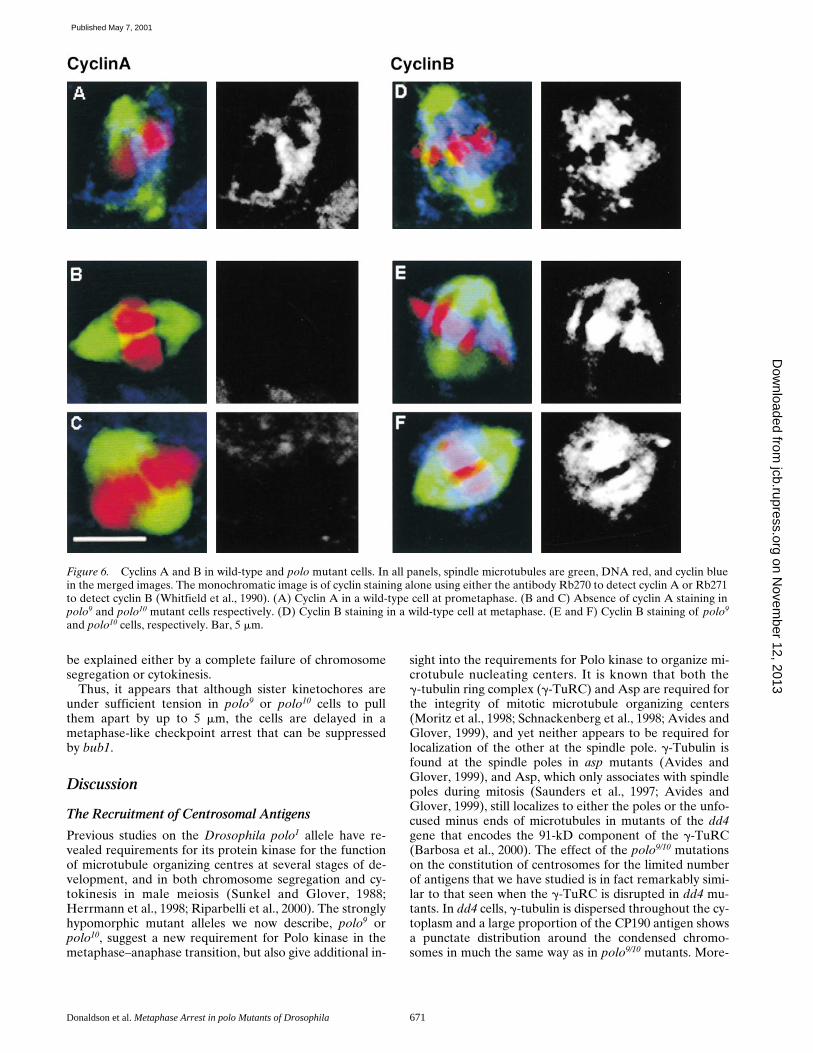
mutant cells than in wild-type cells at metaphase. We werecurious to know whether this reflected any separation ofthe centromeric regions of the chromatids, so we localizedthe product of the gene proliferation disrupter (prod) inpolo9 and polo10 cells. This protein localizes principally tothe centromeric regions of chromosomes 2 and 3 (Torok etal., 1997). In wild-type cells at metaphase in which chro-mosomes are fully aligned on the metaphase plate, fourpunctate regions of Prod staining can be seen correspond-ing to the second and third chromosome pairs in which thesister centromeric regions are still tightly adjoined (Fig. 4A). In polo9 (Fig. 4 B) or polo10 cells (Fig. 4 C), we fre-quently observed eight dots in two lines of four separatedby 2–5 mm in the central region of the 10-mm long spindle.Approximately 80% of pairs of centromeric regionsshowed separation in these whole mount preparations ofbrains (see the legend to Fig. 4). This appearance suggeststhat movement of the centromeric regions of the chromo-somes towards the spindle poles has been initiated andthat the sister kinetochores are being pulled apart. Thebroad distribution of the chromosomes in these prepara-tions contrasts with their highly condensed appearance inthe squashed preparations. This may indicate that thechromatin is highly stretched when associated with the in-tact mitotic spindle. This broad distribution of chromatinin whole mount preparations is in contrast to compact con-densed chromosomes in the metaphase arrest in fizzy (Fig.4, D–F), a gene required to activate the anaphase promot-ing complex for degradation of both cyclin A and cyclin B(for a review, see Hershko, 1999). The distribution of Prodin fizzy cells indicates that the sister centromeres have notseparated; only four dots of staining are seen and these arenot aligned but randomly positioned within the area occu-pied by the condensed chromosomes.
polo9 and polo10 Cells Bear the Signature of Checkpoint Arrest
The segregation of sister chromatids into daughter cells atanaphase is normally prevented until a stable bipolar at-tachment is made between the kinetochores and the spin-dle microtubules. This process is monitored by the spindleintegrity checkpoint which requires the function of a con-served complex of proteins originally identified in buddingyeast as the products of the genes BUB1, BUB3 (Hoyt etal., 1991), MAD1, MAD2, and MAD3 (Li and Murray,1991). In metazoan cells this complex localizes to unat-tached kinetochores to inhibit the activation of the APC(for a review, see Amon, 1999). Mutations in the Drosoph-ila counterpart of one of these proteins, Bub1, indicatethat it is required for the fidelity of chromosome segrega-tion (Basu et al., 1999). The Bub1 protein associates withkinetochores as wild-type cells from the larval central ner-vous system progress to metaphase (Fig. 5 A), and isabruptly lost at anaphase (Fig. 5 B). We found metaphase-like arrested polo9 or polo10 cells in which 16 dots of Bub1staining could be seen corresponding to the separated sis-
ter kinetochores (Fig. 5, C and D). Thus, the kinetochoresappeared to have undergone alignment and the first stagesof separation and yet Bub1 remained associated with them.
A phosphoepitope recognized by the 3F3/2 monoclonalantibody is also found at kinetochores that are reportedlynot under tension (Gorbsky and Ricketts, 1993). It hasbeen thought that when bipolar attachments are made,and tension at the kinetochore is established, the epitopebecomes dephosphorylated and so it is no longer recog-nized by the 3F3/2 antibody. In Drosophila wild-type neu-roblasts, the 3F3/2 epitope can be seen weakly at the ki-netochores and at the spindle poles at metaphase (Fig. 5 E)and is lost from the chromosomes at anaphase (Fig. 5 F).Interestingly, spindle pole staining of 3F3/2 is not seen inthe polo9 and polo10 mutants, probably reflecting the dis-ruptions of the centrosome discussed above (Fig. 5, G andH). However, staining remains often as two separate linesof eight dots corresponding to the sister kinetochores ofthe four pairs of chromosomes (Fig. 5, G and H). Thus,this tension-sensing checkpoint epitope is present at ki-netochores in the mutant cells even though one might ex-pect the kinetochores to be under tension as they arepulled toward the spindle poles.
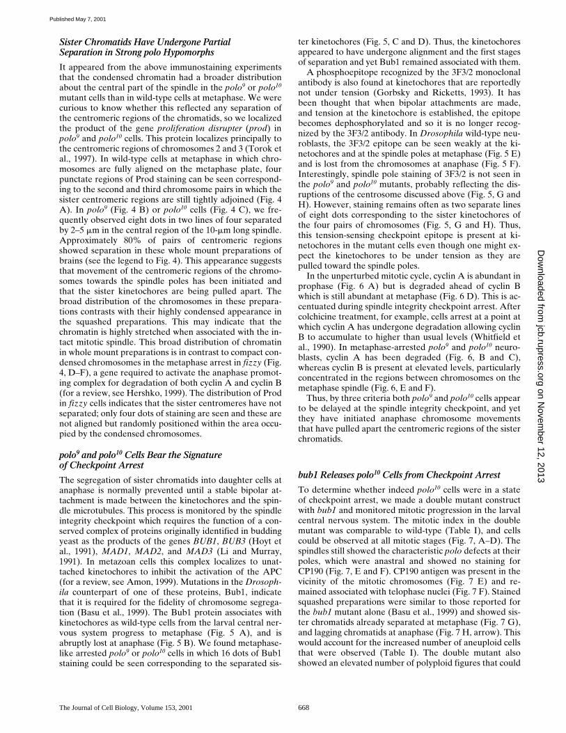
In the unperturbed mitotic cycle, cyclin A is abundant inprophase (Fig. 6 A) but is degraded ahead of cyclin Bwhich is still abundant at metaphase (Fig. 6 D). This is ac-centuated during spindle integrity checkpoint arrest. Aftercolchicine treatment, for example, cells arrest at a point atwhich cyclin A has undergone degradation allowing cyclinB to accumulate to higher than usual levels (Whitfield etal., 1990). In metaphase-arrested polo9 and polo10 neuro-blasts, cyclin A has been degraded (Fig. 6, B and C),whereas cyclin B is present at elevated levels, particularlyconcentrated in the regions between chromosomes on themetaphase spindle (Fig. 6, E and F).
Thus, by three criteria both polo9 and polo10 cells appearto be delayed at the spindle integrity checkpoint, and yetthey have initiated anaphase chromosome movementsthat have pulled apart the centromeric regions of the sisterchromatids.
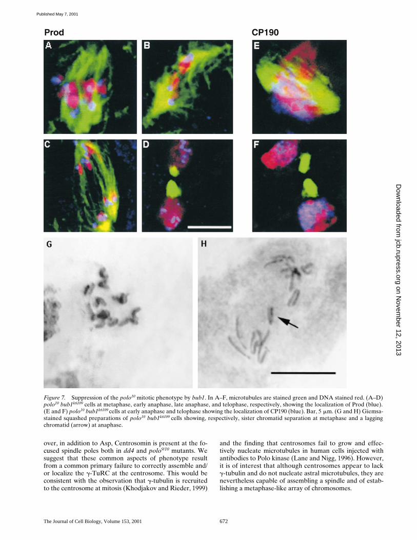
bub1 Releases polo10 Cells from Checkpoint Arrest
To determine whether indeed polo10 cells were in a stateof checkpoint arrest, we made a double mutant constructwith bub1 and monitored mitotic progression in the larvalcentral nervous system. The mitotic index in the doublemutant was comparable to wild-type (Table I), and cellscould be observed at all mitotic stages (Fig. 7, A–D). Thespindles still showed the characteristic polo defects at theirpoles, which were anastral and showed no staining forCP190 (Fig. 7, E and F). CP190 antigen was present in thevicinity of the mitotic chromosomes (Fig. 7 E) and re-mained associated with telophase nuclei (Fig. 7 F). Stainedsquashed preparations were similar to those reported forthe bub1 mutant alone (Basu et al., 1999) and showed sis-ter chromatids already separated at metaphase (Fig. 7 G),and lagging chromatids at anaphase (Fig. 7 H, arrow). Thiswould account for the increased number of aneuploid cellsthat were observed (Table I). The double mutant alsoshowed an elevated number of polyploid figures that could
on Novem
ber 12, 2013jcb.rupress.org
Dow
nloaded from
Published May 7, 2001

Donaldson et al. Metaphase Arrest in polo Mutants of Drosophila 669
Figure 4. Localization of Prod in wild-type (A), polo (B and C), and fizzy (D–F) cells. Spindle microtubules stained with the rat mono-clonal antibody YL1/2 are stained green. DNA is stained red. Prod (blue) was detected using a rabbit antibody (Torok et al., 1997).Merged images are shown in the top panels with the separated channel for Prod staining below. Bar, 5 mm. 79.3% of 213 clear pairs ofsister centromeric regions were scored as having separated in immunostained polo9 cells. A similar frequency of separation (78.6% of112 pairs) was observed in polo10 cells. Centromere separation was not observed by anti-Prod staining in the fizzyx4 mutant.
on Novem
ber 12, 2013jcb.rupress.org
Dow
nloaded from
Published May 7, 2001

The Journal of Cell Biology, Volume 153, 2001 670
Figure 5. Association of Bub1 and the 3F3/2epitope with the mitotic apparatus in wild-type andpolo mutant cells. In all panels, spindle microtubulesare stained green and DNA stained red. Bub1 (A–F)or 3F3/2 (G–L) staining are shown in blue. (A andB) Wild-type cells at metaphase and anaphase, re-spectively. (C and D) polo9 cells showing Bub1staining with the rabbit antibody Rb666 (gift of C.Sunkel, University of Porto, Porto, Portugal) on theseparated kinetochores. (E and F) Wild-type cells atmetaphase and anaphase, respectively, stained withthe 3F3/2 mouse monoclonal antibody (gift of G.Gorbsky, University of Oklahoma, Oklahoma City,Oklahoma). Note the presence of the 3F3/2 epitopeat centrosomes. (G and H) polo10 cells showing 3F3/2staining on the separated kinetochores, and absentfrom the spindle poles. Bar, 5 mm.
on Novem
ber 12, 2013jcb.rupress.org
Dow
nloaded from
Published May 7, 2001
Donaldson et al. Metaphase Arrest in polo Mutants of Drosophila 671
be explained either by a complete failure of chromosomesegregation or cytokinesis.
Thus, it appears that although sister kinetochores areunder sufficient tension in polo9 or polo10 cells to pullthem apart by up to 5 mm, the cells are delayed in ametaphase-like checkpoint arrest that can be suppressedby bub1.
Discussion
The Recruitment of Centrosomal Antigens
Previous studies on the Drosophila polo1 allele have re-vealed requirements for its protein kinase for the functionof microtubule organizing centres at several stages of de-velopment, and in both chromosome segregation and cy-tokinesis in male meiosis (Sunkel and Glover, 1988;Herrmann et al., 1998; Riparbelli et al., 2000). The stronglyhypomorphic mutant alleles we now describe, polo9 orpolo10, suggest a new requirement for Polo kinase in themetaphase–anaphase transition, but also give additional in-
sight into the requirements for Polo kinase to organize mi-crotubule nucleating centers. It is known that both theg-tubulin ring complex (g-TuRC) and Asp are required forthe integrity of mitotic microtubule organizing centers(Moritz et al., 1998; Schnackenberg et al., 1998; Avides andGlover, 1999), and yet neither appears to be required forlocalization of the other at the spindle pole. g-Tubulin isfound at the spindle poles in asp mutants (Avides andGlover, 1999), and Asp, which only associates with spindlepoles during mitosis (Saunders et al., 1997; Avides andGlover, 1999), still localizes to either the poles or the unfo-cused minus ends of microtubules in mutants of the dd4gene that encodes the 91-kD component of the g-TuRC(Barbosa et al., 2000). The effect of the polo9/10 mutationson the constitution of centrosomes for the limited numberof antigens that we have studied is in fact remarkably simi-lar to that seen when the g-TuRC is disrupted in dd4 mu-tants. In dd4 cells, g-tubulin is dispersed throughout the cy-toplasm and a large proportion of the CP190 antigen showsa punctate distribution around the condensed chromo-somes in much the same way as in polo9/10 mutants. More-
Figure 6. Cyclins A and B in wild-type and polo mutant cells. In all panels, spindle microtubules are green, DNA red, and cyclin bluein the merged images. The monochromatic image is of cyclin staining alone using either the antibody Rb270 to detect cyclin A or Rb271to detect cyclin B (Whitfield et al., 1990). (A) Cyclin A in a wild-type cell at prometaphase. (B and C) Absence of cyclin A staining inpolo9 and polo10 mutant cells respectively. (D) Cyclin B staining in a wild-type cell at metaphase. (E and F) Cyclin B staining of polo9
and polo10 cells, respectively. Bar, 5 mm.
on Novem
ber 12, 2013jcb.rupress.org
Dow
nloaded from
Published May 7, 2001
The Journal of Cell Biology, Volume 153, 2001 672
over, in addition to Asp, Centrosomin is present at the fo-cused spindle poles both in dd4 and polo9/10 mutants. Wesuggest that these common aspects of phenotype resultfrom a common primary failure to correctly assemble and/or localize the g-TuRC at the centrosome. This would beconsistent with the observation that g-tubulin is recruitedto the centrosome at mitosis (Khodjakov and Rieder, 1999)
and the finding that centrosomes fail to grow and effec-tively nucleate microtubules in human cells injected withantibodies to Polo kinase (Lane and Nigg, 1996). However,it is of interest that although centrosomes appear to lackg-tubulin and do not nucleate astral microtubules, they arenevertheless capable of assembling a spindle and of estab-lishing a metaphase-like array of chromosomes.
Figure 7. Suppression of the polo10 mitotic phenotype by bub1. In A–F, microtubules are stained green and DNA stained red. (A–D)polo10 bub1k6109 cells at metaphase, early anaphase, late anaphase, and telophase, respectively, showing the localization of Prod (blue).(E and F) polo10 bub1k6109 cells at early anaphase and telophase showing the localization of CP190 (blue). Bar, 5 mm. (G and H) Giemsa-stained squashed preparations of polo10 bub1k6109 cells showing, respectively, sister chromatid separation at metaphase and a laggingchromatid (arrow) at anaphase.
on Novem
ber 12, 2013jcb.rupress.org
Dow
nloaded from
Published May 7, 2001
Donaldson et al. Metaphase Arrest in polo Mutants of Drosophila 673
Metaphase or Anaphase Arrest in polo Cells?
If plks play an essential role in the activation of cdk1, anarrest at the G2/M transition in polo9 and polo10 mighthave been expected. As this is not observed, it suggests ei-ther that this function is not required at this stage ofDrosophila development, or it is redundant, or it can befulfilled by the little amount of detectable Polo protein.The phenotype of polo9/10 is more typical of cells unable topass beyond the metaphase–anaphase checkpoint, as theyhave low levels of cyclin A and high levels of cyclin B. It isgenerally believed that the inhibitory signal of this check-point arises from unaligned chromosomes and requires acomplex containing the Bub1 and 3 and Mad1, 2, and 3proteins that associates with unattached kinetochores.Mad3 binds to and inactivates Cdc20p-fizzy, a protein thatdirects the APC to a specific set of substrates. In yeast, oneof the Cdc20-APC substrates is Pds1, whose proteolysis re-leases the separin Esp1 that in turn catalyses the degrada-tion of Scc1p resulting in sister chromatid separation (for areview, see Nasmyth et al., 2000). In the presence of theBub1 complex of checkpoint proteins, the APC cannot beactivated and cells become arrested in metaphase (for areview, see Gardner and Burke, 2000). polo9/10 cells haveBub1 and the 3F3 epitope at their kinetochores, suggestingthey are held in a state of checkpoint arrest by the Bub1complex. This is confirmed by our observation that bub1mutation suppresses the mitotic phenotype of polo10 andso releases the metaphase-like arrest.
However, polo9/10 cells exhibit a paradox that sets themapart from other metaphase arrest mutants of Drosophila.Their chromosomes appear to have undergone alignmentand anaphase movements activated in that sister kineto-chores appear to be not only attached through microtu-bules to the poles but also pulled apart by about 2–5 mm.This itself might have been expected to relieve the check-point arrest, and usually the 3F3 epitope is lost fromchromosomes under tension. In contrast, Giemsa-stainedsquashed preparations reveal that only z17% of sisterchromatids remain separated in squashed preparations ofpolo9/10 brain cells made in the presence or absence ofcolchicine. This suggests that the majority of sister chro-matids are still held together, explaining their failure tomove to the spindle poles. In addition, some chromosomesappear connected in chains through telomeric linkages.Chaining of chromosomes in this way has been describedpreviously in the mutants for the UbcD1 gene that en-codes a class I ubiquitin-conjugating (E2) enzyme (Cenciet al., 1997). This observation suggests that Polo kinasemay in part play a role in regulating the proteolytic systemresponsible for breaking such linkages.
Centromere separation seen in intact polo9/10 cells mustbe accounted for by pole-directed forces of motor proteinson the chromatids. Although several explanations are pos-sible to resolve the paradox of the polo phenotype, the sim-plest is that Polo kinase is actively required to relieve thefunction of the Bub1 checkpoint complex in the progres-sion through mitosis. This would have some parallels withthe finding that in budding yeast, CDC5 is essential to es-cape the metaphase arrest imposed by DNA damage sur-veillance mechanisms (Toczyski et al., 1997). This hypothe-sis implies that the polo arrest point lies on the cusp of the
metaphase–anaphase transition. This is at a later stage thanthe requirement for the fizzy gene product function. Infizzy mutants, neither A- nor B-type cyclins are degradedand as we show here, there is no separation of centromericregions as seen in polo mitotic arrest. This incidentally con-trasts with mutants for CDC20, the fizzy homologue ofbudding yeast where centromeric regions are observed tobe separated (Tanaka et al., 2000). However, the transientseparation of sister centromeres while chromosome armsstill show cohesion does appear to reflect a general aspectof normal progression through metaphase in wild-typebudding yeast (Goshima and Yanagida, 2000; He et al.,2000; Tanaka et al., 2000). If similar events occur in meta-zoan cells, this aspect of normal progression throughmetaphase might be accentuated in polo cells that appearto have initiated anaphase by the criterion of the apparentpoleward tension imposed at the centromeres, and yet arenot released from “checkpoint” arrest.
Mitotic Exit
Polo kinase may also be required to promote completeseparation of sister chromatids and for those specific as-pects of APC activity that mediate cyclin B degradation.The latter would have some similarities to budding yeastwhere mutants in the plk gene CDC5 are reported to showno effect on Cdc20-APC function and so permit Pds1p tobe degraded, in the absence of degradation of the mitoticcyclin Clb2p (Shirayama et al., 1998). Moreover, overex-pression of Cdc5p results in proteolysis of Clb2p but notPds1p, suggesting that the CDC5 plk promotes Hct1(Schwab et al., 1997; Visintin et al., 1997; Charles et al.,1998) (fizzy related)/APC functions to regulate cyclin Blevels. In fact, the phenotype of polo9/10 mutants resemblesthat of mutants expressing nondegradable cyclin B in sev-eral respects. Expression of stable cyclin B (lacking its de-struction box) from a GAL4 responsive promoter inDrosophila larval neuroblasts results in a mitotic arrest inwhich the greater proportion of cells arrest in anaphase(Rimmington et al., 1994). In embryonic cells, the pheno-types resulting from expression of stable forms of cyclinsA, B1, and B3 support a sequential requirement for thethree proteins: stable cyclin A leads to a metaphase delay,stable cyclin B an early anaphase arrest, and stable cyclinB3 a late anaphase arrest (Sigrist et al., 1995). Sister chro-matids clearly separate in those cells expressing stable cy-clin B as indicated by in situ hybridization with a dodeca-satellite probe to identify the centromeric region of thethird chromosomes. A role for polo in promoting those as-pects of APC activity that mediate cyclin B degradationwould be consistent with the observations that mammalianPlk can phosphorylate and activate the APC in vitro (Ko-tani et al., 1998). However, the ability of the polo10 bub1double mutant to progress through mitosis would suggestthat this APC function can also be activated at some levelin the absence of polo function.
In budding yeast, there is now considerable evidencethat Hct1p can be activated by the Cdc14p protein phos-phatase (Alexandru et al., 1999; Fraschini et al., 1999;Gardner and Burke, 2000). Cdc14p appears itself to be ac-tivated downstream of the GTP-bound active form ofTem1p. This pathway is held in check in response to spin-
on Novem
ber 12, 2013jcb.rupress.org
Dow
nloaded from
Published May 7, 2001
The Journal of Cell Biology, Volume 153, 2001 674
dle damage by a two component GTPase activating pro-tein (GAP) formed between Bub2p and the Bfa1/Byr4protein that promotes formation of the GDP-bound stateof Tem1p which does not favor Cdc14 activation. TheCDC5 polo-like kinase appears to regulate the phosphory-lation of Bfa1/Byr4 in this process (Lee et al., 2001). In fis-sion yeast, the equivalent pathway regulates the onset ofthe septation process and appears to be under the controlof Plo1 kinase (Tanaka et al., 2001). It is of considerableinterest to know the extent to which the pathway might beconserved and whether an analogous process regulates theonset of cytokinesis in metazoans. The requirement forpolo for cytokinesis in Drosophila is obscured by earliermitotic defects seen in somatic cells with all alleles. Theexception is male meiosis, where it seems that spindlecheckpoint pathways do not lead to metaphase arrest.However, cells from the polo9 bub1 double mutant do at-tain high levels of polyploidy, suggestive of a failure in cy-tokinesis once the metaphase arrest is bypassed. It will bea future challenge to determine the extent to which thismitotic exit network has been conserved in metazoans andits relationship to the regulation of the onset of anaphase.
We thank Daryl Henderson for his comments on the manuscript.This work was supported by grants from the Cancer Research Cam-
paign, the Biotechnology and Biological Sciences Research Council, andthe European Union.
Submitted: 31 August 2000Revised: 16 February 2001Accepted: 19 March 2001
References
Abrieu, A., T. Brassac, S. Galas, D. Fisher, J.-C. Labbe, M. Doree. 1998. ThePolo-like kinase Plx is a component of the MPF amplification loop at the G2/M-phase transition of the cell cycle in Xenopus. J. Cell Sci. 111:1751–1757.
Alexandru, G., W. Zachariae, A. Schleiffer, and K. Nasmyth. 1999. Sister chro-matid separation and chromosome re-duplication are regulated by differentmechanisms in response to spindle damage. EMBO (Eur. Mol. Biol. Organ.)J. 10:2707–2721.
Amon, A. 1999. The spindle checkpoint. Curr. Opin. Genes Dev. 9:69–75.Avides, M., and D.M. Glover. 1999. Abnormal spindle protein Asp, and the in-
tegrity of mitotic centrosomal microtubule organizing centres. Science. 283:1733–1735.
Barbosa, V., R. Yamamoto, A.T.C. Carpenter, D.S. Henderson, and D.M.Glover. 2000. Mutation of a Drosophila gamma tubulin ring complex subunitencoded by discs degenerate-4 differentially disrupts centrosomal protein lo-calization. Genes Dev. 14:3126–3139.
Basu, J., H. Bousbaa, E. Logarinho, Z. Li, B.C. Williams, C. Lopes, C.E.Sunkel, and M.L. Goldberg. 1999. Mutations in the essential spindle check-point gene bub1 cause chromosome missegregation and fail to block apopto-sis in Drosophila. J. Cell Biol. 146:13–28.
Bousbaa, H., L. Correia, G. Gorbsky, and C.E. Sunkel. 1997. Mitotic phospho-epitopes are expressed in Kc cells, neuroblasts and isolated chromosomes inDrosophila melanogaster. J. Cell Sci. 110:1979–1988.
Carmena, M., M.G. Riparbelli, G. Minestrini, A. Tavares, R. Adams, G. Cal-laini, and D.M. Glover. 1998. Drosophila polo kinase is required for cytoki-nesis. J. Cell Biol. 143:659–671.
Cenci, G., R.B. Rawson, G. Belloni, D.H. Castrillon, M. Tudor, R. Petrucci,and M.L. Goldberg. 1997. UbcD1, a Drosophila ubiquitin-conjugating en-zyme required for proper telomere behavior. Genes Dev. 11:863–875.
Charles, J.F., S.L. Jaspersen, R.L. Tinker-Kulberg, L. Hwang, A. Szidon, andD.O. Morgan. 1998. The polo-related kinase Cdc5 activates and is destroyedby the mitotic cyclin destruction machinery in S. cerevisiae. Curr. Biol.8:497–507.
Cohen-Fix, O., J.M. Peters, M.W. Kirshner, and D. Koshland. 1996. Anaphaseinitiation in Saccharomyces cerevisiae is controlled be the APC-dependentdegradation of the anaphase inhibitor Pds1p. Genes Dev. 10:3081–3093.
Deak, P., M.M. Omar, R.D. Saunders, M. Pal, O. Komonyi, J. Szidonya, P. Ma-roy, Y. Zhang, M. Ashburner, P. Benos, et al. 1997. P-Element insertion al-leles of essential genes on the third chromosome of Drosophila melano-gaster: correlation of physical and cytogenic maps in chromosomal region86E to 87F. Genes Dev. 147:1697–1722.
Descombes, P., and E.A. Nigg. 1998. The polo-like kinase Plx1 is required for
M phase exit and destruction of mitotic regulators in Xenopus egg extracts.EMBO (Eur. Mol. Biol. Organ.) J. 17:1328–1335.
Fraschini, R., E. Formenti, G. Lucchini, and S. Piatti. 1999. Budding yeast Bub2is localized at spindle pole bodies and activates the mitotic checkpoint via adifferent pathway from Mad2. J. Cell Biol. 145:979–991.
Funabiki, H., K. Kumada, and M. Yanagida. 1996. Fission yeast Cut1 and Cut2are essential for sister chromatid separation, concentrate along themetaphase spindle and form large complexes. EMBO (Eur. Mol. Biol. Or-gan.) J. 15:6617–6628.
Gardner, R.D., and D.J. Burke. 2000. The spindle checkpoint: two transitions,two pathways. Trends Cell Biol. 10:154–158.
Glover, D.M., I.M. Hagan, and A.A.M. Tavares. 1998. Polo-like kinases: a teamthat plays throughout mitosis. Genes Dev. 12:3777–3787.
Gorbsky, G., and W.A. Ricketts. 1993. Differential expression of a phospho-epitope at the kinetochores of moving chromosomes. J. Cell Biol. 122:1311–1321.
Goshima, G., and M. Yanagida. 2000. Establishing biorientation occurs withprecocious separation of the sister kinetochores, but not the arms, in theearly spindle of budding yeast. Cell. 100:619–633.
He, X.W., S. Asthana, and P. Sorger. 2000. Transient sister chromatid separa-tion and elastic deformation of chromosomes during mitosis in buddingyeast. Cell. 101:763–775.
Herrmann, S., I. Amorim, and C.E. Sunkel. 1998. The POLO kinase is requiredat multiple stages during spermatogenesis in Drosophila melanogaster.Chromosoma. 107:440–451.
Hershko, A. 1999. Mechanisms and regulation of the degradation of cyclin B.Philos. Trans. R. Soc. Lond. B. Biol. Sci. 354:1571–1575.
Heuer, J.G., K. Li, and T.C. Kaufman. 1995. The Drosophila homeotic targetgene centrosomin (cnn) encodes a novel centrosomal protein with leucinezippers and maps to a genomic region required for midgut morphogenesis.Development. 121:3861–3876.
Hoyt, M.A., L. Totis, and B.T. Roberts. 1991. S. cerevisiae genes required forcell cycle arrest in response to loss of microtubule function. Cell. 66:507–517.
Khodjakov, A., and C.L. Rieder. 1999. The sudden recruitment of gamma-tubulin to the centrosome at the onset of mitosis, and its dynamic exchangethroughout the cell cycle, do not require microtubules. J. Cell Biol. 146:585–596.
Kotani, S., S. Tugendreich, M. Fujii, P.M. Jorgensen, N. Watanabe, C. Hoog, P.Hieter, and K. Todokoro. 1998. PKA and MPF-activated polo-like kinaseregulate anaphase-promoting complex activity and mitosis progression. Mol.Cell. 1:371–380.
Kumagai, A., and W.G. Dunphy. 1996. Purification and molecular cloning ofPlx1, a Cdc25-regulatory kinase from Xenopus egg extracts. Science. 273:1377–1380.
Lane, H.A., and E.A. Nigg. 1996. Antibody microinjection reveals an essentialrole for human polo-like kinase 1 (Plk1) in the functional maturation of mi-totic centrosomes. J. Cell Biol. 135:1701–1713.
Lee, K.S., and R.L. Erikson. 1997. Plk in a functional homolog of Saccharomy-ces cerevisiae Cdc5, and elevated Plk activity induces multiple septationstructures. Mol. Cell. Biol. 17:3408–3417.
Lee, S.E., L.M. Frenz, S. Jensen, A.L. Johnson, D. Fesquet, and L.H. Johnston.2001. The Bub2-dependent mitotic pathway in yeast acts every cell cycle andregulates cytokinesis. J. Cell Sci. In press.
Li, R., and A.W. Murray. 1991. Feedback control of mitosis in budding yeast.Cell. 66:519–531.
Llamazares, S., A. Moreira, A. Tavares, C. Girdham, B.A. Spruce, C. Gonzalez,R.E. Karess, D.M. Glover, and C.E. Sunkel. 1991. polo encodes a protein ki-nase homolog required for mitosis in Drosophila. Genes Dev. 5:2153–2165.
Moritz, M., Y. Zheng, B.M. Alberts, and K. Oegema. 1998. Recruitment of thegamma tubulin ring complex to Drosophila salt-stripped centrosome scaf-folds. J. Cell Biol. 142:775–786.
Nasmyth, K., L.M. Peters, and F. Uhlmann. 2000. Splitting the chromosome:cutting the ties that bind sister chromatids. Science. 288:1379–1384.
Nigg, E.A. 1998. Polo-like kinases: positive regulators of cell division from startto finish. Curr. Opin. Cell Biol. 10:776–783.
Ohkura, H., I.M. Hagan, and D.M. Glover. 1995. The conserved Schizosaccha-romyces pombe kinase plo1, required to form a bipolar spindle, the actinring and septum, can drive septation formation in G1 and G2 cells. GenesDev. 9:1059–1073.
Qian, Y.-W., E. Erikson, C. Li, and J.L. Maller. 1998. Activated polo-like ki-nase Plx1 is required at multiple points during mitosis in Xenopus laevis.Mol. Cell. Biol. 18:4262–4271.
Rimmington, G., B. Dalby, and D.M. Glover. 1994. Expression of N-terminallytruncated cyclin B in the Drosophila larval brain leads to mitotic delay atlate anaphase. J. Cell Science. 107:2729–2738.
Riparbelli, M.G., G. Callaini, and D.M. Glover. 2000. Failure of pronuclear mi-gration and repeated divisions of polar body nuclei associated with MTOCdefects in polo eggs of Drosophila. J. Cell Sci. 113:3341–3350.
Saunders, R., M. Avides, T. Howard, C. Gonzalez, and D.M. Glover. 1997. TheDrosophila gene abnormal spindle encodes a novel microtubule-associatedprotein that associates with the polar regions of the mitotic spindle. J. CellBiol. 137:881–890.
Scaerou, F., I. Aguilera, R. Saunders, N. Kane, L. Blottiere, and R. Karess.1999. The rough deal protein is a new kinetochore component required foraccurate chromosome segregation in Drosophila. J. Cell Sci. 12:3757–3768.
on Novem
ber 12, 2013jcb.rupress.org
Dow
nloaded from
Published May 7, 2001
Donaldson et al. Metaphase Arrest in polo Mutants of Drosophila 675
Schnackenberg, B.J., A. Khodjakov, C.L. Rieder, and R.E. Palazzo. 1998. Thedisassembly and reassembly of functional centrosomes in vitro. Proc. Natl.Acad. Sci. USA. 95:9295–9300.
Schwab, M., A.S. Lutum, and W. Seufert. 1997. Yeast Hct1 is a regulator ofClb2 cyclin proteolysis. Cell. 90:683–693.
Shirayama, M., W. Zachariae, R. Ciosk, and K. Nasmyth. 1998. The polo-likekinase Cdc5p and the WD-repeat protein Cdc20p/fizzy are regulators andsubstrates of the anaphase promoting complex in Saccharomyces cerevisisae.EMBO (Eur. Mol. Biol. Organ.) J. 17:1336–1349.
Sigrist, S.J., H. Jacobs, R. Stratmann, and C.F. Lehner. 1995. Exit from mitosisis regulated by Drosophila fizzy and the sequential destruction of cyclins A,B and B3. EMBO (Eur. Mol. Biol. Organ.) J. 14:4827–4838.
Song, S., and K.S. Lee. 2001. A novel function of Saccharomyces cerevisiaeCDC5 in cytokinesis. J. Cell Biol. 152:451–470.
Sunkel, C.E., and D.M. Glover. 1988. polo, a mitotic mutant of Drosophila dis-playing abnormal spindle poles. J. Cell Sci. 89:25–38.
Tanaka, T., J. Fuchs, J. Loidl, and K. Nasmyth. 2000. Cohesin ensures bipolarattachment of microtubules to sister centromeres and resists their preco-cious separation Nat. Cell Biol. 2:492–499.
Tanaka, K., J. Petersen, F. MacIver, D.P. Mulvihill, D.M. Glover, and I.M. Hagan.2001. The role of Plo1 kinase in mitotic commitment and septation inSchizosaccharomyces pombe. EMBO (Eur. Mol. Biol. Organ.) J. 20:1259–1270.
Tavares, A., D.M. Glover, and C.E. Sunkel. 1996. The conserved mitotic kinasepolo is regulated by phosphorylation and has preferred microtubule associ-ated substrates in Drosophila embryo extracts. EMBO (Eur. Mol. Biol. Or-
gan.) J. 15:4873–4883.Toczyski, D.P., D.J. Galgoczy, and L.H. Hartwell. 1997. CDC5 and CKII con-
trol adaptation to the yeast DNA damage checkpoint. Cell. 90:1097–1106.Torok, T., P.D. Harvie, M. Buratovich, and P.J. Bryant. 1997. The product of
proliferation disrupter is concentrated at centromeres and required for mi-totic chromosome condensation and cell proliferation in Drosophila. GenesDev. 11:213–225.
Townsley, F.M., and J.V. Ruderman. 1998. Proteolytic ratchets that controlprogression through mitosis. Trends Cell Biol. 8:238–244.
Trueman, J., and M. Bate. 1988. Spatial and temporal patterns of neurogenesisin the central nervous system of Drosophila melanogaster. Dev. Biol. 125:145–157.
Visintin, R., S. Prinz, and A. Amon. 1997. CDC20 and CDH1: a family of sub-strate-specific activators of APC-dependent proteolysis. Science 278:460–463.
Whitfield, W.G.F., S.E. Millar, H. Saumweber, M. Frasch, and D.M. Glover.1988. Cloning of a gene encoding an antigen associated with the centrosomein Drosophila. J. Cell Sci. 89:467–480.
Whitfield, W.G.F., C. Gonzalez, G. Maldanado-Codina, and D.M. Glover.1990. The A- and B-type cyclins of Drosophila are accumulated and de-stroyed in temporally distinct events that define separable phases of the G2-Mtransition. EMBO (Eur. Mol. Biol. Organ.) J. 9:2563–2572.
Yanagida, M. 1998. Fission yeast cut mutations revisited: control of anaphase.Trends Cell Biol. 8:144–149.
on Novem
ber 12, 2013jcb.rupress.org
Dow
nloaded from
Published May 7, 2001