Chromosome 17 Centromere Duplication and Responsiveness to Anthracycline-Based Neoadjuvant...
7
Chromosome 17 Centromere Duplication and Responsiveness to Anthracycline-Based Neoadjuvant Chemotherapy in Breast Cancer 1,2 Ariadna Tibau* , † , Laura López-Vilaró †, ‡ , Maitane Pérez-Olabarria † , Tania Vázquez † , Cristina Pons ‡ , Ignasi Gich § , Carmen Alonso*, Belén Ojeda*, Teresa Ramón y Cajal*, Enrique Lerma †, ‡,¶ , Agustí Barnadas* , †,¶,3 and Daniel Escuin †,3 * Department of Medical Oncology, Hospital de la Santa Creu i Sant Pau, Barcelona, Spain; † Institut d'Investigacions Biomèdiques Sant Pau, Barcelona, Spain; ‡ Department of Pathology, Hospital de la Santa Creu i Sant Pau, Barcelona, Spain; § Department of Epidemiology, Hospital de la Santa Creu i Sant Pau, Barcelona, Spain; ¶ Universitat Autònoma de Barcelona, Bellaterra, Cerdanyola del Vallès, Spain Abstract Human epidermal growth factor receptor 2 (HER2) and topoisomerase II alpha (TOP2A) genes have been proposed as predictive biomarkers of sensitivity to anthracycline chemotherapy. Recently, chromosome 17 centromere enumeration probe (CEP17) duplication has also been associated with increased responsiveness to anthracyclines. However, reports are conflicting and none of these tumor markers can yet be considered a clinically reliable predictor of response to anthracyclines. We studied the association of TOP2A gene alterations, HER2 gene amplification, and CEP17 duplication with response to anthracycline-based neoadjuvant chemotherapy in 140 patients with operable or locally advanced breast cancer. HER2 was tested by fluorescence in situ hybridization and TOP2A and CEP17 by chromogenic in situ hybridization. Thirteen patients (9.3%) achieved pathologic complete response (pCR). HER2 amplification was present in 24 (17.5%) of the tumors. TOP2A amplification occurred in seven tumors (5.1%). CEP17 duplication was detected in 13 patients (9.5%). CEP17 duplication correlated with a higher rate of pCR [odds ratio (OR) 6.55, 95% confidence interval (95% CI) 1.25-34.29, P = .026], and analysis of TOP2A amplification showed a trend bordering on statistical significance (OR 6.97, 95% CI 0.96-50.12, P = .054). TOP2A amplification and CEP17 duplication combined were strongly associated with pCR (OR 6.71, 95% CI 1.66-27.01, P = .007). HER2 amplification did not correlate with pCR. Our results suggest that CEP17 duplication predicts pCR to primary anthracycline-based chemotherapy. CEP17 duplication, TOP2A amplifications, and HER2 amplifications were not associated with prognosis. Neoplasia (2014) 16, 861–867 www.neoplasia.com Volume 16 Number 10 October 2014 pp. 861–867 861 Abbreviations: CEP17, chromosome 17 centromere enumeration probe; CI, confidence interval; CISH, chromogenic in situ hybridization; DFS, disease-free survival; EC-D, epirubicin (90 mg/m 2 ) and cyclophosphamide (600 mg/m 2 ) followed by docetaxel (100 mg/ m 2 ); ER, estrogen receptor; FEC75, fluorouracil (600 mg/m 2 ), epirubicin (75 mg/m 2 ), and cyclophosphamide (600 mg/m 2 ); FISH, fluorescence in situ hybridization; HR, hazard ratio; HER2, human epidermal growth factor receptor 2; OR, odds ratio; OS, overall survival; pCR, pathologic complete response; PR, progesterone receptor; TOP2A, topoisomerase II alpha Address all Correspondence to: Agustí Barnadas, MD or Daniel Escuin, PhD Department of Medical Oncology, Hospital de la Santa Creu i Sant Pau, Sant Antoni Maria Claret 167, 08025 Barcelona, Spain. E-mail: [email protected], [email protected] 1 This article refers to supplementary material, which is designated by Supplementary Table 1 and is available online at www.neoplasia.com. 2 This study was supported by a grant from the Red Temática de Investigación Cooperativa de Cáncer (RTICC; RD12/0036/0076) to all authors, by grants from the Spanish Society of Clinical Oncology (SEOM; 2009 and 2012), the Molt Il lustríssima Administració (MIA) Foundation (2009), and Hospital de la Santa Creu i Sant Pau to A.T., by a grant from the Instituto de Salud Carlos III (ISCIII, PI10/0307) to D.E., and by a Pfizer award to A.B. The study sponsors had no involvement in the experimental work, data analysis, or manuscript preparation. Conflict of interests: The authors declare that they have no conflict of interest. 3 Contributed equally as senior authors. Received 30 June 2014; Revised 15 August 2014; Accepted 20 August 2014 © 2014 Neoplasia Press, Inc. Published by Elsevier Inc. This is an open access article under the CC BY-NC-ND license (http://creativecommons.org/licenses/ by-nc-nd/3.0/). 1476-5586/14 http://dx.doi.org/10.1016/j.neo.2014.08.012
-
Upload
independent -
Category
Documents
-
view
0 -
download
0
Transcript of Chromosome 17 Centromere Duplication and Responsiveness to Anthracycline-Based Neoadjuvant...

www.neoplasia.com
Volume 16 Number 10 October 2014 pp. 861–867 861
Abbreviatiointerval; Cepirubicin (m2); ER, escyclophospHER2, humpathologicAddress allMedical O08025 Barc1This articTable 1 an2This studCooperativ
Chromosome 17 CentromereDuplication and Responsiveness toAnthracycline-Based NeoadjuvantChemotherapy in Breast Cancer1,2
ns: CEP17, chromosome 17 centromere enumeration probe; CI, confidenceISH, chromogenic in situ hybridization; DFS, disease-free survival; EC-D,90mg/m2) and cyclophosphamide (600mg/m2) followed by docetaxel (100mg/trogen receptor; FEC75, fluorouracil (600 mg/m2), epirubicin (75 mg/m2), andhamide (600 mg/m2); FISH, fluorescence in situ hybridization; HR, hazard ratio;an epidermal growth factor receptor 2;OR, odds ratio;OS, overall survival; pCR,
complete response; PR, progesterone receptor; TOP2A, topoisomerase II alphaCorrespondence to: Agustí Barnadas, MD orDaniel Escuin, PhDDepartment ofncology, Hospital de la Santa Creu i Sant Pau, Sant Antoni Maria Claret 167,elona, Spain. E-mail: [email protected], [email protected] refers to supplementary material, which is designated by Supplementaryd is available online at www.neoplasia.com.y was supported by a grant from the Red Temática de Investigacióna de Cáncer (RTICC; RD12/0036/0076) to all authors, by grants from the
Ariadna Tibau*,†, Laura López-Vilaró†,‡,Maitane Pérez-Olabarria†, Tania Vázquez†,Cristina Pons‡, Ignasi Gich§,Carmen Alonso*, Belén Ojeda*,Teresa Ramón y Cajal*, Enrique Lerma†,‡, ¶,Agustí Barnadas*,†,¶,3 and Daniel Escuin†,3
*Department of Medical Oncology, Hospital de la SantaCreu i Sant Pau, Barcelona, Spain; †Institut d'InvestigacionsBiomèdiques Sant Pau, Barcelona, Spain; ‡Department ofPathology, Hospital de la Santa Creu i Sant Pau, Barcelona,Spain; §Department of Epidemiology, Hospital de la SantaCreu i Sant Pau, Barcelona, Spain; ¶Universitat Autònoma deBarcelona, Bellaterra, Cerdanyola del Vallès, Spain
AbstractHuman epidermal growth factor receptor 2 (HER2) and topoisomerase II alpha (TOP2A) genes have been proposedas predictive biomarkers of sensitivity to anthracycline chemotherapy. Recently, chromosome 17 centromereenumeration probe (CEP17) duplication has also been associated with increased responsiveness toanthracyclines. However, reports are conflicting and none of these tumor markers can yet be considered aclinically reliable predictor of response to anthracyclines. We studied the association of TOP2A gene alterations,HER2 gene amplification, and CEP17 duplication with response to anthracycline-based neoadjuvant chemotherapyin 140 patients with operable or locally advanced breast cancer. HER2 was tested by fluorescence in situhybridization and TOP2A and CEP17 by chromogenic in situ hybridization. Thirteen patients (9.3%) achievedpathologic complete response (pCR). HER2 amplification was present in 24 (17.5%) of the tumors. TOP2Aamplification occurred in seven tumors (5.1%). CEP17 duplication was detected in 13 patients (9.5%). CEP17duplication correlated with a higher rate of pCR [odds ratio (OR) 6.55, 95% confidence interval (95% CI) 1.25-34.29,P = .026], and analysis of TOP2A amplification showed a trend bordering on statistical significance (OR 6.97, 95%CI 0.96-50.12, P= .054). TOP2A amplification and CEP17 duplication combined were strongly associated with pCR(OR 6.71, 95% CI 1.66-27.01, P = .007). HER2 amplification did not correlate with pCR. Our results suggest thatCEP17 duplication predicts pCR to primary anthracycline-based chemotherapy. CEP17 duplication, TOP2Aamplifications, and HER2 amplifications were not associated with prognosis.
Neoplasia (2014) 16, 861–867
Spanish Society of Clinical Oncology (SEOM; 2009 and 2012), the Molt Il lustríssimaAdministració (MIA) Foundation (2009), and Hospital de la Santa Creu i Sant Pau toA.T., by a grant from the Instituto de Salud Carlos III (ISCIII, PI10/0307) to D.E.,and by a Pfizer award to A.B. The study sponsors had no involvement in theexperimental work, data analysis, or manuscript preparation. Conflict of interests: Theauthors declare that they have no conflict of interest.3Contributed equally as senior authors.Received 30 June 2014; Revised 15 August 2014; Accepted 20 August 2014
© 2014 Neoplasia Press, Inc. Published by Elsevier Inc. This is an open accessarticle under the CC BY-NC-ND license (http://creativecommons.org/licenses/by-nc-nd/3.0/).1476-5586/14http://dx.doi.org/10.1016/j.neo.2014.08.012

862 CEP17 Duplication and Responsiveness in Breast Cancer Tibau et al. Neoplasia Vol. 16, No. 10, 2014
IntroductionPredicting response to anthracycline-based therapy is a central challengein patients with breast cancer. Several randomized studies have shown thatanthracycline-based adjuvant therapy produces amodest improvement insurvival in patients with early-stage breast cancer [1]. In addition, it hasbeen shown that incorporation of taxanes further improves pathologiccomplete response (pCR) in the neoadjuvant setting [2–7]. However, therisk of serious side effects must be considered and predictive factors areneeded to help clinicians select themost appropriate drug for each patient.To date, no specific biomarkers have been identified to predict tumorresponse to anthracycline-based chemotherapy.
The human epidermal growth factor receptor 2 (HER2) and topoisome-rase II alpha (TOP2A) genes have been proposed as markers of sensitivityto anthracycline chemotherapy. Although several studies have reported anassociation between HER2 amplification with anthracycline sensitivity[8–13], only two of these were statistically significant [10,13]. In vitro andin vivo studies indicate that HER2 positivity alone does not alteranthracycline sensitivity [14] and the underlying mechanism remainselusive. The TOP2A gene, located at 17q12-q21, close to the HER2gene, encodes TOP2A, a key enzyme in DNA replication and themolecular target of anthracyclines [15,16]. When HER2 is amplified,genes situated around 17q21, such as TOP2A gene, may be either co-amplified or deleted [17]. Because of this physical proximity, someresearchers have proposed that the link between HER2-positive diseaseand anthracycline sensitivity is the presence of TOP2A alterations(amplifications and deletions) rather than HER2 amplifications [18,19].However, the results of these studies have not always been consistent [20].
Given the location of HER2 and TOP2A on chromosome 17, recentreports have focused on the predictive role of other molecular alterationslocalized within the 17q21 region, including alterations in key genes suchas HER1-3 [21], p53 [22], and BRCA1 [23], and variations in the copynumber of subchromosomal regions, including the chromosome 17centromere (CEP17) duplication [21]. On the basis of recent data fromarray comparative genomic hybridization, CEP17 duplication is definedas increased copy number of CEP17 [21] rather than polysomies of thewhole chromosome 17 [24,25]. CEP17 duplication has been described asa marker of genomic instability [26] and has received a great deal ofattention as a potential predictor of anthracycline benefit [21,27].
We hypothesized that CEP17 duplication is associated with a higherresponse to neoadjuvant chemotherapy. We explored the association ofCEP17 duplication and TOP2A alterations with response to anthracy-cline-based neoadjuvant therapy. We studied a cohort of patientswith early or locally advanced breast cancer treated with anthracycline-based primary chemotherapy, and we chose pCR as a surrogate markerof chemosensitivity.
Materials and Methods
Ethics StatementThis study was conducted according to the Declaration of Helsinki
principles, with approval from the Clinical Research Ethics Committeeat Institut d'Investigacions Biomèdiques Sant Pau. Written informedconsent was obtained from all patients.
Study Design and PatientsWe retrospectively studied 140 consecutive patients with stage II or
III breast cancer who received anthracycline-based neoadjuvantchemotherapy in our hospital between 1993 and 2010. All patientshad confirmed diagnosis based on histopathology of biopsy and none
of them had prior treatment with surgery, chemotherapy, or radiation.Our study included patients with HER2-positive carcinomas treatedbefore the approval of trastuzumab in 2006. The study excludedpatients with bilateral or inflammatory tumors. Fifty-five patientsreceived neoadjuvant treatment with anthracyclines alone [FEC75:fluorouracil (600 mg/m2), epirubicin (75 mg/m2), and cyclophospha-mide (600 mg/m2) given every 3 weeks for four to six cycles, n = 40, orFAC60: fluorouracil (500 mg/m2), doxorubicin (60 mg/m2), andcyclophosphamide (500 mg/m2) given every 3 weeks for four to sixcycles, n = 15] between 1993 and 2002. Eighty-five patients receivedneoadjuvant treatmentwith anthracyclines in combination with taxanes[EC-D: epirubicin (90 mg/m2) and cyclophosphamide (600 mg/m2)given every 3 weeks for four cycles followed by docetaxel (100 mg/m2)every 3 weeks for four cycles] from 2003 to 2010. Patients were stagedaccording to the tumor-node-metastasis (TNM) system. Clinicalresponse was assessed by palpation, breast ultrasound, mammography,and/or magnetic resonance imaging before systemic therapy and beforecurative surgery. Clinical responses were evaluated according to theresponse evaluation criteria in solid tumors (RECIST) criteria [28] everytwo cycles. Patients treated with FEC75 or FAC60with partial responsereceived surgical treatment after four cycles of chemotherapy and twoadditional cycles of FEC75 or FAC60 were administered after surgery.Patients treated with EC-D underwent breast surgery after completionof chemotherapy. Patients with positive hormone receptor tumorsreceived radiotherapy and endocrine therapy. The extent of residualdisease was measured in the surgical specimen. The primary endpointfor this study was pCR defined as the absence of invasive cancer in thebreast and axillary lymph nodes at the time of definitive surgery. Thesecondary endpoints for the study were disease-free survival (DFS) andoverall survival (OS). Patients were followed up according to the breastcancer guidelines.
Tumor Samples and Tissue MicroarraysWe analyzed 140 representative formalin-fixed, paraffin-embedded
tumor core biopsies obtained before neoadjuvant treatment. Paraffinblocks were stored at room temperature. Samples were identified onlyby an identification number assigned to each patient. A stainedsection of each tumor sample was prepared to confirm the diagnosisand to identify representative tumor areas. Tissue microarrays wereprepared from formalin-fixed, paraffin-embedded tissue taken fromthree representative areas of each tumor. Serial 5-μm sections wereobtained for immunohistochemical, fluorescence in situ hybridization(FISH), and chromogenic in situ hybridization (CISH) analyses.
ImmunohistochemistryPrediluted antibodies for estrogen receptor (ER; clone EP1),
progesterone receptor (PR; clone 636) and Ki67 (clone MIB-I) wereobtained from Dako (Glostrup, Denmark). Sections were processedin a PT Module using Dako high pH buffer (Dako) fordeparaffinization and antigen retrieval. Sections for the Ki-67 studywere processed with Dako low pH buffer. All immunohistochemicalstains were performed in an Autostainer Link using the EnVisionmethod (Dako). HER2 overexpression was analyzed using theHercepTest assay (Dako). Tumors were classified as ER or PRpositive when at least 1% of the tumor cells showed staining in thenuclei cells [29]. HER2 was considered overexpressed when auniform intense (3+) membrane staining was present in N30% ofinvasive tumor cells [30]. The percentage of Ki67-stained nuclei wasevaluated independently of the intensity and its positivity cutoff value

Table 1. Basic Patient and Tumor Characteristics.
Variable n (%)
N 140Age, years Mean ± SD 53.7 ± 13.5
Median (range) 51 (25.5-86.1)Menopausal status Premenopausal 65 (46.4)
Postmenopausal 75 (53.6)Tumor stage II 50 (35.7)
III 90 (64.3)Tumor status T2 39 (27.9)
T3 42 (30.0)T4 59 (42.1)
Node status N0 47 (33.6)N1 63 (45.0)N2 26 (18.6)N3 4 (2.9)
Tumor grade 1 11 (7.9)2 63 (45.0)3 66 (47.1)
ER Positive 87 (62.1)Negative 53 (37.9)
PR Positive 68 (48.6)Negative 72 (51.4)
Ki67 status b20% 74 (42.9)≥20% 60 (52.9)Missing data 6 (4.3)
HER2 Normal 113 (81)Amplification 24 (17)Missing data 3 (2)
TOP2A Normal 124 (89)Amplification 7 (5)Deletion 6 (4)Missing data 3 (2)
Neoadjuvant therapy FEC75/FAC60 55 (39.3)EC-D 85 (60.7)
Surgical treatment Mastectomy 88 (62.9)Lumpectomy 52 (37.1)
Pathologic response Complete 13 (9.3%)Residual disease 127 (90.7)
Neoplasia Vol. 16, No. 10, 2014 CEP17 Duplication and Responsiveness in Breast Cancer Tibau et al. 863
was ≥20% [31]. Two pathologists independently evaluated allimmunostainings, and discordant results were reviewed to reachan agreement.
In situ Hybridization AnalysesHER2 gene status was confirmed for all patients by FISH.
TOP2A gene status was evaluated by CISH and FISH in equivocalcases. A good correlation of the results for TOP2A obtained byFISH or CISH has been previously reported [32]. We used theHER2 FISH pharmDX (Dako) and the TOP2A FISH pharmDX(Dako) assays, respectively. The CISH assay was performed usingDako dual color assay (Dako). All tests were performed followingthe manufacturer's recommendations. Fluorescence signals wereevaluated using an Olympus BX51 fluorescent microscope andappropriate filter sets. The assessment of FISH results for TOP2Aand HER2 was performed in ≥60 nuclei per case. TOP2A genestatus was assessed by CISH in ≥30 tumor cells per case. HER2gene amplification was defined when the ratio HER2/CEP17 was≥2 in accordance with the manufacturer’s recommendations.TOP2A amplification was considered when the ratio TOP2A/CEP17 was ≥2 and TOP2A deletion was considered when the ratioTOP2A/CEP17 was b0.8. We use the term CEP17 duplicationthroughout the text as defined previously [21]. The cutoff value forCEP17 duplication was determined as N1.86 observed CEP signalsper cell [21,26].
Statistical AnalysesDFS was defined as the time from initiation of treatment to date
of first relapse (local, regional, contralateral or metastatic), secondprimary cancer, or death resulting from any cause (whicheveroccurred first). OS was defined as the time from sample collection todeath resulting from any cause. Patients lost to follow-up werecensored at the last contact. Quantitative variables between groupswere compared using the Student’s t test, and categorical variableswere compared using the Chi-square or Fisher exact test. Theunivariable association between variables and pCR was assessedusing the Chi-square test. The multivariable logistic regressionmodel for pCR was adjusted for age at diagnosis (b50 vs ≥50 years),tumor stage (T2 vs T3 vs T4), nodal stage (negative vs positive),histologic grade (1 or 2 vs 3), neoadjuvant chemotherapy (FEC75/FAC60 vs EC-D), ER (negative vs positive), PR (negative vspositive), HER2 (normal vs amplified), TOP2A status (normal vsamplified), and CEP17 status (normal vs duplicated). Kaplan-Meierand log-rank analyses were used to compare DFS and OS. The Coxproportional hazard model with a single covariate was used to obtainthe hazard ratios (HRs) for relapse or death and associated 95%confidence interval (95% CI). HER2 (normal vs amplified),TOP2A status (normal vs amplified), and CEP17 status (normalvs duplicated) as individual variables were entered in a final Coxmodel adjusted for traditional prognostic factors including age atdiagnosis (b50 vs ≥50 years), nodal stage (negative vs positive),tumor size (T2 vs T3 vs T4), histologic grade (1 or 2 vs 3),neoadjuvant chemotherapy (FEC75/FAC60 vs EC-D), ER (nega-tive vs positive), PR (negative vs positive), and Ki67 (low vs high).The Cox proportional hazard model was used in an exploratoryanalysis of tumors to analyze tumors with either TOP2Aamplification or CEP17 duplication; we refer to these tumorsthroughout the text as tumors with combined TOP2A or CEP17duplication. A two-sided P value ≤ .05 was considered significant.
Statistical analyses were performed using the SPSS 19.0 statisticalsoftware package (SPSS, Chicago, IL).
Patient selection, assay performance, and data analysis concurred tothe reporting recommendations for tumor marker prognostic studies(REMARK) guidelines [33].
Results
Patient CharacteristicsPatient and tumor characteristics are summarized in Table 1. A
total of 101 of the 140 patients had T3 or T4 tumors (72.1%), and93 had lymph node–positive tumors (66.4%); these factors explainthe observed low rate of conservative treatment (37.1%). A pCR afterneoadjuvant chemotherapy was achieved in 13 patients (9.3%). Fourpatients (7.3%) achieved pCR after FEC75/FAC60 and nine(10.6%) patients after EC-D treatment. No significant differencesin pCR were observed according to type of neoadjuvant treatment ortumor stage (Supplementary Table 1).
Association ofCEP17DuplicationwithClinicopathologic VariablesTable 2 shows the associations of CEP17 status with patient
characteristics and tumor-related variables. Status of HER2, TOP2A,and CEP17 duplication was assessable in 137 (97.9%) tumors.HER2amplification was found in 24 samples (17.5%), TOP2A wasamplified in 7 (5.1%) of the tumors, TOP2A deletion was found in6 samples (4.4%), and CEP17 duplication was detected in 13 (9.5%)samples. Tumors with combined TOP2A amplification and CEP17
Table 2. Correlation between Clinicopathologic Characteristics and CEP17 Status.
Variable n (%) CEP17 Status P *
Normal Duplicated
CEP17 137 124 (90.5) 13 (9.5)Age, years ≤50 65 (47.5) 61 (49) 4 (30.8) .206
N50 72 (52.5) 63 (51) 9 (69.2)Menopausal status Premenopausal 63 (46) 59 (47) 4 (30.8) .247
Postmenopausal 74 (54) 65 (53) 9 (69.2)Tumor stage II 49 (35.8) 44 (33.5) 5 (38.5) .830
III 88 (64.2) 80 (64.5) 8 (61.5)Tumor status T2 38 (27.7) 33 (27) 5 (38.6) .606
T3 42 (30.7) 38 (30) 4 (30.7)T4 57 (41.6) 53 (43) 4 (30.7)
Node status N0 46 (33.5) 42 (34) 4 (31) .538N1 62 (45.5) 54 (44) 8 (61.5)N2 25 (18) 24 (19) 1 (7.5)N3 4 (3) 4 (3) 0 (0)
Tumor grade 1 11 (8) 11 (9) 0 (0) .4992 62 (45) 55 (44) 7 (54)3 64 (47) 58 (47) 6 (46)
ER Positive 86 (62.8) 77(62) 9 (69.2) .613Negative 51 (37.2) 47 (38) 4 (30.8)
PR Positive 68 (49.6) 62 (50) 6 (46) .792Negative 69 (50.4) 62 (50) 7 (54)
Ki67 status b20% 73 (53.3) 66 (53) 7 (54) .937≥20% 60 (50.4) 54 (43.5) 6 (46)Missing data 4 (2.9) 4 (3) 0 (0)
HER2 Normal 112 (82) 103 (83) 9 (69) .166Amplification 23 (17) 19 (15) 4 (31)Missing data 2 (1) 2 (2) 0 (0)
TOP2A Normal 124 (91) 111 (89.5) 13 (100) .220Amplification 7 (5) 7 (5.5) 0 (0)Deletion 6 (4) 6 (5) 0 (0)
Neoadjuvant therapy FEC75/FAC60 84 (61.3) 76 (61) 8 (61.5) .986EC-D 53 (38.7) 48 (39) 5 (38.5)
Surgical treatment Mastectomy 86 (62.9) 78 (62.9) 8 (61.5) .923Lumpectomy 51 (37.1) 46 (37.1) 5 (38.5)
Pathologic response Complete 12 (8.7) 9 (7.3) 3 (23) .055Residual disease 125 (91.3) 115 (92.7) 10 (77)
* P values were calculated using the Chi-square test.
864 CEP17 Duplication and Responsiveness in Breast Cancer Tibau et al. Neoplasia Vol. 16, No. 10, 2014
duplication alterations were present in 20 (14.6%) of the tumors.CEP17 duplication was not associated with HER2 gene amplification(P = .166), high grade (P = .499), ER negativity (P = .613), or a highKi67 (P = .937). Tumors with HER2 amplification had TOP2Aamplification in three cases (42.9%, P = .062), TOP2A deletionin three cases (50%, P = .028), and CEP17 aneusomy in two cases(40% P = .164). HER2 amplifications were significantly associatedwith TOP2A alterations (amplifications and deletions; P = .003) andwith CEP17 aneusomy (P = .048; data not shown).
Table 3. Multivariable Analysis of Predictive Factors for pCR.
Variable Score pCR
OR (95% CI) P *
ER Positive 1Negative 7.12 (1.69-30.03) .007
TOP2A Normal 1Amplification 6.97 (0.96-50.12) .054
CEP17 Normal 1Duplication 6.55 (1.25-34.29) .026
HER2 Normal 1Amplification 0.804 (0.18-3.597) .755
TOP2A/CEP17 † Normal 1Altered 6.71 (1.66-27.01) .007
* Logistic regression models adjusted for known prognostic factors (age, nodal status, tumor size, tumorgrade, and hormone receptor status) and neoadjuvant therapy.
† Tumors with combined TOP2A amplification and CEP17 duplication.
Association of Clinicopathologic Parameters with pCRIn the multivariable analysis, CEP17 duplication associated
with a high percentage of pCR [odds ratio (OR) 6.55, 95% CI1.25-34.29, P = .026] and TOP2A gene amplification showed abenefit with borderline significance (OR 6.97, 95% CI 0.96-50.12, P = .054; Table 3). When TOP2A amplifications andCEP17 duplication were combined, we observed a significantassociation with pCR (OR 6.71, 95% CI 1.66-27.01, P = .007;Table 3). We did not find a significant difference in the benefit oftreatment with anthracyclines when assessing deletion (P = .429) orcombined TOP2A amplifications and deletions (P = .089) inunivariable analysis. HER2 amplifications showed no directassociation with pCR (P = .144). When pCR was assessedaccording to treatment received (Supplementary Table 1),CEP17 duplications were the only factor associated with betterresponse to FEC70/FAC65 in both univariable (P = .004) andmultivariable analyses (OR 14, 95% CI 1.427-137.324, P = .025).Meanwhile, the multivariable analysis showed that ER status (OR12.68, 95% CI 1.427-137.324, P = .011) and TOP2A/CEP17(OR 6.31, 95% CI 1.981-81.186, P = .007) were associated withbetter response to EC-D (data not shown).
Prognostic Associations with DFS and OSThe univariable analysis showed that CEP17 duplication, HER2
amplification, and TOP2A amplification were not significantlyassociated with DFS or OS (Table 4). However, when TOP2Aamplification and CEP17 duplication were combined, Kaplan-Meierand log-rank analyses showed a significant association with betterDFS (P = .026) and a borderline non-significant trend with OS (P =.054; Figure 1). The univariable analysis showed a similar associationof the combination of TOP2A amplification and CEP17 duplicationwith DFS (HR 4.34, 95% CI 1.05-17.92, P = .042) and OS (HR3.70, 95% CI 0.89-15.34, P = .071; Table 4). However, afteradjustment for the classic prognostic factors, age, and neoadjuvanttherapy, the multivariable analysis showed that CEP17 duplication,TOP2A amplification, HER2 amplification, and the combination ofTOP2A amplifications and CEP17 duplication failed to show anassociation with DFS or OS (Table 5).
DiscussionThe main finding in this study is that CEP17 duplication associateswith pCR to anthracycline-based neoadjuvant therapy in patientswith breast cancer. To our knowledge, this is the first study to analyzethe association of CEP17 duplication with response to neoadjuvant
Table 4. Biomarker Analysis and Relation with DFS and OS.
Variable Score n DFS OS
HR (95% CI) P HR (95% CI) P *
CEP17 Normal 124 1 1Duplicated 13 2.64 (0.64-10.9) .179 2.42 (0.58-10.02) .223
HER2 Normal 113 1 1Amplified 24 0.97 (0.45-2.08) .960 0.96 (0.44-2.06) .907
TOP2A Non-amplified 130 1 1Amplified 7 21.9 (0.92-5275) .269 21.73 (0.04-11545) .336
Ki67 ≥20% 60 1 1b20% 74 1.89 (1.04-3.46) .036 1.91 (1.02-3.58) .044
TOP2A/CEP17 † Normal 117 1 1Altered 20 4.35 (1.05-17.93) .042 3.71 (0.89-15.34) .071
* Univariable Cox proportional hazard models based on 136 patients, 44 recurrences, and 40 deaths.† Tumors with combined TOP2A amplification and CEP17 duplication.
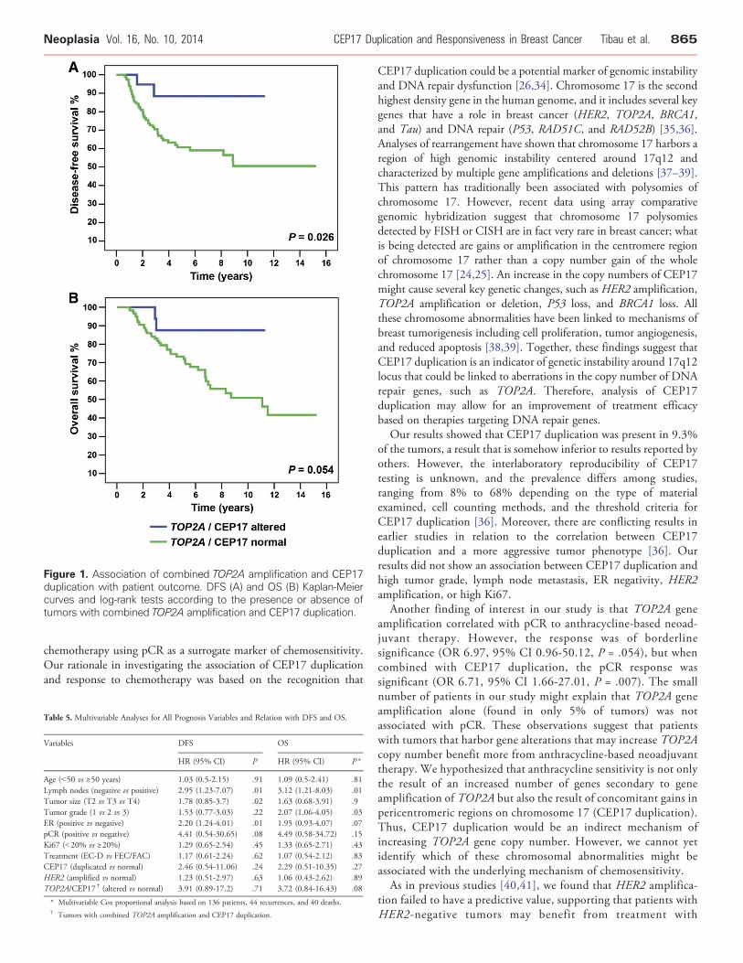
Figure 1. Association of combined TOP2A amplification and CEP17duplication with patient outcome. DFS (A) and OS (B) Kaplan-Meiercurves and log-rank tests according to the presence or absence oftumors with combined TOP2A amplification and CEP17 duplication.
Neoplasia Vol. 16, No. 10, 2014 CEP17 Duplication and Responsiveness in Breast Cancer Tibau et al. 865
chemotherapy using pCR as a surrogate marker of chemosensitivity.Our rationale in investigating the association of CEP17 duplicationand response to chemotherapy was based on the recognition that
Table 5. Multivariable Analyses for All Prognosis Variables and Relation with DFS and OS.
Variables DFS OS
HR (95% CI) P HR (95% CI) P *
Age (b50 vs ≥50 years) 1.03 (0.5-2.15) .91 1.09 (0.5-2.41) .81Lymph nodes (negative vs positive) 2.95 (1.23-7.07) .01 3.12 (1.21-8.03) .01Tumor size (T2 vs T3 vs T4) 1.78 (0.85-3.7) .02 1.63 (0.68-3.91) .9Tumor grade (1 vs 2 vs 3) 1.53 (0.77-3.03) .22 2.07 (1.06-4.05) .03ER (positive vs negative) 2.20 (1.24-4.01) .01 1.95 (0.93-4.07) .07pCR (positive vs negative) 4.41 (0.54-30.65) .08 4.49 (0.58-34.72) .15Ki67 (b20% vs ≥20%) 1.29 (0.65-2.54) .45 1.33 (0.65-2.71) .43Treatment (EC-D vs FEC/FAC) 1.17 (0.61-2.24) .62 1.07 (0.54-2.12) .83CEP17 (duplicated vs normal) 2.46 (0.54-11.06) .24 2.29 (0.51-10.35) .27HER2 (amplified vs normal) 1.23 (0.51-2.97) .63 1.06 (0.43-2.62) .89TOP2A/CEP17 † (altered vs normal) 3.91 (0.89-17.2) .71 3.72 (0.84-16.43) .08
* Multivariable Cox proportional analysis based on 136 patients, 44 recurrences, and 40 deaths.† Tumors with combined TOP2A amplification and CEP17 duplication.
CEP17 duplication could be a potential marker of genomic instabilityand DNA repair dysfunction [26,34]. Chromosome 17 is the secondhighest density gene in the human genome, and it includes several keygenes that have a role in breast cancer (HER2, TOP2A, BRCA1,and Tau) and DNA repair (P53, RAD51C, and RAD52B) [35,36].Analyses of rearrangement have shown that chromosome 17 harbors aregion of high genomic instability centered around 17q12 andcharacterized by multiple gene amplifications and deletions [37–39].This pattern has traditionally been associated with polysomies ofchromosome 17. However, recent data using array comparativegenomic hybridization suggest that chromosome 17 polysomiesdetected by FISH or CISH are in fact very rare in breast cancer; whatis being detected are gains or amplification in the centromere regionof chromosome 17 rather than a copy number gain of the wholechromosome 17 [24,25]. An increase in the copy numbers of CEP17might cause several key genetic changes, such as HER2 amplification,TOP2A amplification or deletion, P53 loss, and BRCA1 loss. Allthese chromosome abnormalities have been linked to mechanisms ofbreast tumorigenesis including cell proliferation, tumor angiogenesis,and reduced apoptosis [38,39]. Together, these findings suggest thatCEP17 duplication is an indicator of genetic instability around 17q12locus that could be linked to aberrations in the copy number of DNArepair genes, such as TOP2A. Therefore, analysis of CEP17duplication may allow for an improvement of treatment efficacybased on therapies targeting DNA repair genes.
Our results showed that CEP17 duplication was present in 9.3%of the tumors, a result that is somehow inferior to results reported byothers. However, the interlaboratory reproducibility of CEP17testing is unknown, and the prevalence differs among studies,ranging from 8% to 68% depending on the type of materialexamined, cell counting methods, and the threshold criteria forCEP17 duplication [36]. Moreover, there are conflicting results inearlier studies in relation to the correlation between CEP17duplication and a more aggressive tumor phenotype [36]. Ourresults did not show an association between CEP17 duplication andhigh tumor grade, lymph node metastasis, ER negativity, HER2amplification, or high Ki67.
Another finding of interest in our study is that TOP2A geneamplification correlated with pCR to anthracycline-based neoad-juvant therapy. However, the response was of borderlinesignificance (OR 6.97, 95% CI 0.96-50.12, P = .054), but whencombined with CEP17 duplication, the pCR response wassignificant (OR 6.71, 95% CI 1.66-27.01, P = .007). The smallnumber of patients in our study might explain that TOP2A geneamplification alone (found in only 5% of tumors) was notassociated with pCR. These observations suggest that patientswith tumors that harbor gene alterations that may increase TOP2Acopy number benefit more from anthracycline-based neoadjuvanttherapy. We hypothesized that anthracycline sensitivity is not onlythe result of an increased number of genes secondary to geneamplification of TOP2A but also the result of concomitant gains inpericentromeric regions on chromosome 17 (CEP17 duplication).Thus, CEP17 duplication would be an indirect mechanism ofincreasing TOP2A gene copy number. However, we cannot yetidentify which of these chromosomal abnormalities might beassociated with the underlying mechanism of chemosensitivity.
As in previous studies [40,41], we found that HER2 amplifica-tion failed to have a predictive value, supporting that patients withHER2-negative tumors may benefit from treatment with

866 CEP17 Duplication and Responsiveness in Breast Cancer Tibau et al. Neoplasia Vol. 16, No. 10, 2014
anthracyclines. We also observed that patients with TOP2A normaltumors might have some additional benefit from treatment withanthracyclines. Our results suggest that other mechanisms, such asthe immune response, tumor-stromal interactions, and CEP17duplication, may participate in the increased sensitivity toanthracyclines in these patients [42–45]. Therefore, the use ofanthracyclines should not be limited to patients with HER2amplification or TOP2A amplification.
We were unable to identify any statistically significant relationshipbetween CEP17 duplication and patient outcome. Our results areconsistent with two previous studies [21,27] that analyzed prognosticand predictive associations of CEP17 duplication in early-stagebreast cancer patients treated with adjuvant chemotherapy regimenscontaining anthracyclines and non-anthracyclines. Bartlett et al. [21]assessed the role of CEP17 duplication in 1931 tumors from 2391women enrolled onto the UK National Epirubicin Adjuvant Trial(NEAT/BR9601). Interestingly, although this study also failed tofind a significant association of CEP17 duplication with patientprognosis (DFS HR 1.12, 95% CI 0.92-1.36, P = .24, OS HR 1.17,95% CI 0.95-1.44, P = .13), it showed that the benefit on survivalfrom the addition of epirubicin was confined to tumors with CEP17duplication. Similar results were obtained by Pritchard et al. [27] inthe randomized controlled mammary 5 (MA.5) adjuvant trial. Theresults did not identify a significant association of CEP17 with risk ofrecurrence (HR 1.15, 95% CI 0.86-1.54, P = .34) or risk of death(HR 1.39, 95% CI 0.90-2.15, P = .14) in multivariable analysis.However, in patients whose tumors exhibit CEP17 duplication andwho received anthracyclines, there was a significant benefit forrecurrence-free survival (HR 0.59, 95% CI 0.37-0.92, P = .02) andOS (HR 0.61, 95% CI 0.37-1.01, P = .05) in univariable analysis.This association disappeared after adjustment for other variables.
The potential limitations of our study are the restricted sample sizeand the retrospective design, which could bias patient selection. Inaddition, the absence of a study group receiving non-anthracycline-based chemotherapy meant we were unable to distinguish whether thepCR rate was attributed to higher sensitivity to anthracyclines or tochemotherapy with anthracyclines, taxanes, cyclophosphamide, and/or5-fluorouracil. These results are therefore preliminary and requireconfirmation in larger prospective studies specifically designed tovalidate the predictive role of these biomarkers and to compare regimenswith and without anthracyclines.
In conclusion, our results suggest that CEP17 duplication may be auseful biomarker of sensitivity to neoadjuvant anthracycline-basedchemotherapy. A better understanding of CEP17 abnormalities mighthelp to select patients who benefit most from anthracycline-basedchemotherapy regimens and to identify patients in whom toxicityfrom these drugs could be avoided.
Supplementary data to this article can be found online at http://dx.doi.org/10.1016/j.neo.2014.08.012.
AcknowledgmentsThe authors thank Carolyn Newey for manuscript editing.
References
[1] Early Breast Cancer Trialists' Collaborative Group (EBCTCG) (2005). Effectsof chemotherapy and hormonal therapy for early breast cancer on recurrenceand 15-year survival: an overview of the randomised trials. Lancet 365,1687–1717.
[2] Gogas H and Fountzilas G (2003). The role of taxanes as a component ofneoadjuvant chemotherapy for breast cancer. Ann Oncol 14, 667–674.
[3] Estevez LG and Gradishar WJ (2004). Evidence-based use of neoadjuvanttaxane in operable and inoperable breast cancer. Clin Cancer Res 10,3249–3261.
[4] Trudeau M, Sinclair SE, and Clemons M (2005). Neoadjuvant taxanes in thetreatment of non-metastatic breast cancer: a systematic review. Cancer Treat Rev31, 283–302.
[5] Schott AF and Hayes DF (2012). Defining the benefits of neoadjuvantchemotherapy for breast cancer. J Clin Oncol 30, 1747–1749.
[6] Thompson AM and Moulder-Thompson SL (2012). Neoadjuvant treatment ofbreast cancer. Ann Oncol 23(Suppl 10), x231–x236.
[7] Peto R, Davies C, and Godwin J, et al (2012). Comparisons between differentpolychemotherapy regimens for early breast cancer: meta-analyses of long-termoutcome among 100,000 women in 123 randomised trials. Lancet 379,432–444.
[8] Di Leo A, Gancberg D, and Larsimont D, et al (2002). HER-2 amplification andtopoisomerase IIalpha gene aberrations as predictive markers in node-positivebreast cancer patients randomly treated either with an anthracycline-basedtherapy or with cyclophosphamide, methotrexate, and 5-fluorouracil. ClinCancer Res 8, 1107–1116.
[9] Di Leo A, Larsimont D, and Gancberg D, et al (2001). HER-2 and topo-isomerase IIalpha as predictive markers in a population of node-positive breastcancer patients randomly treated with adjuvant CMF or epirubicin pluscyclophosphamide. Ann Oncol 12, 1081–1089.
[10] Pritchard KI, Shepherd LE, andO'Malley FP, et al (2006). HER2 and responsivenessof breast cancer to adjuvant chemotherapy. N Engl J Med 354, 2103–2111.
[11] Muss HB, Thor AD, and Berry DA, et al (1994). c-erbB-2 expression andresponse to adjuvant therapy in women with node-positive early breast cancer.N Engl J Med 330, 1260–1266.
[12] Paik S, Bryant J, and Tan-Chiu E, et al (2000). HER2 and choice of adjuvantchemotherapy for invasive breast cancer: National Surgical Adjuvant Breastand Bowel Project Protocol B-15. J Natl Cancer Inst 92, 1991–1998.
[13] Paik S, Bryant J, and Park C, et al (1998). erbB-2 and response to doxorubicin inpatients with axillary lymph node-positive, hormone receptor-negative breastcancer. J Natl Cancer Inst 90, 1361–1370.
[14] Pegram MD, Finn RS, and Arzoo K, et al (1997). The effect of HER-2/neuoverexpression on chemotherapeutic drug sensitivity in human breast and ovariancancer cells. Oncogene 15, 537–547.
[15] Jarvinen TA, Tanner M, and Rantanen V, et al (2000). Amplification anddeletion of topoisomerase IIalpha associate with ErbB-2 amplification and affectsensitivity to topoisomerase II inhibitor doxorubicin in breast cancer. Am J Pathol156, 839–847.
[16] Cummings J and Smyth JF (1993). DNA topoisomerase I and II as targets forrational design of new anticancer drugs. Ann Oncol 4, 533–543.
[17] Jarvinen TA, Tanner M, and Barlund M, et al (1999). Characterization oftopoisomerase II alpha gene amplification and deletion in breast cancer. GenesChromosomes Cancer 26, 142–150.
[18] Knoop AS, Knudsen H, and Balslev E, et al (2005). Retrospective analysis oftopoisomerase IIa amplifications and deletions as predictive markers in primarybreast cancer patients randomly assigned to cyclophosphamide, methotrexate,and fluorouracil or cyclophosphamide, epirubicin, and fluorouracil: DanishBreast Cancer Cooperative Group. J Clin Oncol 23, 7483–7490.
[19] O'Malley FP, Chia S, and Tu D, et al (2009). Topoisomerase II alpha andresponsiveness of breast cancer to adjuvant chemotherapy. J Natl Cancer Inst 101,644–650.
[20] Di Leo A, Desmedt C, and Bartlett JM, et al (2011). HER2 and TOP2A aspredictive markers for anthracycline-containing chemotherapy regimens asadjuvant treatment of breast cancer: a meta-analysis of individual patient data.Lancet Oncol 12, 1134–1142.
[21] Bartlett JM, Munro AF, and Dunn JA, et al (2010). Predictive markers ofanthracycline benefit: a prospectively planned analysis of the UKNational Epirubicin Adjuvant Trial (NEAT/BR9601). Lancet Oncol 11,266–274.
[22] Bidard FC, Matthieu MC, and Chollet P, et al (2008). p53 status and efficacy ofprimary anthracyclines/alkylating agent-based regimen according to breast cancermolecular classes. Ann Oncol 19, 1261–1265.
[23] Fedier A, Steiner RA, and Schwarz VA, et al (2003). The effect of loss of Brca1 onthe sensitivity to anticancer agents in p53-deficient cells. Int J Oncol 22,1169–1173.

Neoplasia Vol. 16, No. 10, 2014 CEP17 Duplication and Responsiveness in Breast Cancer Tibau et al. 867
[24] Marchiò C, Lambros MB, and Gugliotta P, et al (2009). Does chromosome 17centromere copy number predict polysomy in breast cancer? A fluorescence insitu hybridization and microarray-based CGH analysis. J Pathol 219, 16–24.
[25] Yeh IT, Martin MA, and Robetorye RS, et al (2009). Clinical validation of anarray CGH test for HER2 status in breast cancer reveals that polysomy 17 is arare event. Mod Pathol 22, 1169–1175.
[26] Watters AD, Going JJ, and Cooke TG, et al (2003). Chromosome 17 aneusomyis associated with poor prognostic factors in invasive breast carcinoma. BreastCancer Res Treat 77, 109–114.
[27] Pritchard KI, Munro A, and O'Malley FP, et al (2012). Chromosome 17centromere (CEP17) duplication as a predictor of anthracycline response:evidence from the NCIC Clinical Trials Group (NCIC CTG)MA.5 Trial. BreastCancer Res Treat 131, 541–551.
[28] Jaffe CC (2006). Measures of response: RECIST, WHO, and new alternatives.J Clin Oncol 24, 3245–3251.
[29] Hammond ME, Hayes DF, and Dowsett M, et al (2010). American Society ofClinical Oncology/College of American Pathologists guideline recommendationsfor immunohistochemical testing of estrogen and progesterone receptors in breastcancer. J Clin Oncol 28, 2784–2795.
[30] Wolff AC, Hammond ME, and Schwartz JN, et al (2007). American Society ofClinical Oncology/College of American Pathologists guideline recommendationsfor human epidermal growth factor receptor 2 testing in breast cancer. ArchPathol Lab Med 131, 18–43.
[31] Goldhirsch A, Winer EP, and Coates AS, et al (2013). Personalizing thetreatment of women with early breast cancer: highlights of the St GallenInternational Expert Consensus on the Primary Therapy of Early Breast Cancer2013. Ann Oncol 24, 2206–2223.
[32] Hannemann J, Kristel P, and van Tinteren H, et al (2006). Molecular subtypes ofbreast cancer and amplification of topoisomerase II alpha: predictive role in doseintensive adjuvant chemotherapy. Br J Cancer 95, 1334–1341.
[33] McShane LM, Altman DG, and Sauerbrei W, et al (2006). REportingrecommendations for tumor MARKer prognostic studies (REMARK). BreastCancer Res Treat 100, 229–235.
[34] Corzo C, Bellosillo B, and Corominas JM, et al (2007). Does polysomy ofchromosome 17 have a role in ERBB2 and topoisomerase IIalpha expression?
Gene, mRNA and protein expression: a comprehensive analysis. Tumour Biol 28,221–228.
[35] Zody MC, Garber M, and Adams DJ, et al (2006). DNA sequence of humanchromosome 17 and analysis of rearrangement in the human lineage.Nature 440,1045–1049.
[36] Reinholz MM, Bruzek AK, and Visscher DW, et al (2009). Breast cancer andaneusomy 17: implications for carcinogenesis and therapeutic response. LancetOncol 10, 267–277.
[37] Orsetti B, Nugoli M, and Cervera N, et al (2004). Genomic and expressionprofiling of chromosome 17 in breast cancer reveals complex patterns ofalterations and novel candidate genes. Cancer Res 64, 6453–6460.
[38] Chin SF, Wang Y, and Thorne NP, et al (2007). Using array-comparativegenomic hybridization to define molecular portraits of primary breast cancers.Oncogene 26, 1959–1970.
[39] Fridlyand J, Snijders AM, and Ylstra B, et al (2006). Breast tumor copy numberaberration phenotypes and genomic instability. BMC Cancer 6, 96.
[40] Bartlett JM, Munro A, and Cameron DA, et al (2008). Type 1 receptor tyrosinekinase profiles identify patients with enhanced benefit from anthracyclines inthe BR9601 adjuvant breast cancer chemotherapy trial. J Clin Oncol 26,5027–5035.
[41] Cardoso F, Durbecq V, and Larsimont D, et al (2004). Correlation betweencomplete response to anthracycline-based chemotherapy and topoisomeraseII-alpha gene amplification and protein overexpression in locally advanced/metastatic breast cancer. Int J Oncol 24, 201–209.
[42] Desmedt C, Di Leo A, and de Azambuja E, et al (2011). Multifactorial approachto predicting resistance to anthracyclines. J Clin Oncol 29, 1578–1586.
[43] Farmer P, Bonnefoi H, and Anderle P, et al (2009). A stroma-related genesignature predicts resistance to neoadjuvant chemotherapy in breast cancer. NatMed 15, 68–74.
[44] Ejlertsen B, Jensen MB, and Nielsen KV, et al (2010). HER2, TOP2A, andTIMP-1 and responsiveness to adjuvant anthracycline-containing chemotherapyin high-risk breast cancer patients. J Clin Oncol 28, 984–990.
[45] West NR, Milne K, and Truong PT, et al (2011). Tumor-infiltratinglymphocytes predict response to anthracycline-based chemotherapy in estrogenreceptor-negative breast cancer. Breast Cancer Res 13, R126.