Accurate identification of centromere locations in yeast genomes using Hi-C
9
Nucleic Acids Research, 2015 1 doi: 10.1093/nar/gkv424 Accurate identification of centromere locations in yeast genomes using Hi-C Nelle Varoquaux 1,2,3 , Ivan Liachko 4 , Ferhat Ay 4 , Joshua N. Burton 4 , Jay Shendure 4 , Maitreya J. Dunham 4 , Jean-Philippe Vert 1,2,3 and William S. Noble 4,5,* 1 Mines ParisTech, PSL-Research University, CBIO-Centre for Computational Biology, 35 rue St Honor´ e 77300 Fontainebleau, France, 2 Institut Curie, Paris, F-75248, France, 3 U900, INSERM, Paris, F-75248, France, 4 Department of Genome Sciences, University of Washington, 3720 15th Ave NE, Seattle, WA 98195, USA and 5 Department of Computer Science and Engineering, University of Washington, 185 Stevens Way, Seattle, WA98195, USA Received December 19, 2014; Revised March 25, 2015; Accepted April 17, 2015 ABSTRACT Centromeres are essential for proper chromosome segregation. Despite extensive research, centromere locations in yeast genomes remain difficult to in- fer, and in most species they are still unknown. Re- cently, the chromatin conformation capture assay, Hi- C, has been re-purposed for diverse applications, in- cluding de novo genome assembly, deconvolution of metagenomic samples and inference of centromere locations. We describe a method, Centurion, that jointly infers the locations of all centromeres in a single genome from Hi-C data by exploiting the cen- tromeres’ tendency to cluster in three-dimensional space. We first demonstrate the accuracy of Centu- rion in identifying known centromere locations from high coverage Hi-C data of budding yeast and a hu- man malaria parasite. We then use Centurion to in- fer centromere locations in 14 yeast species. Across all microbes that we consider, Centurion predicts 89% of centromeres within 5 kb of their known lo- cations. We also demonstrate the robustness of the approach in datasets with low sequencing depth. Fi- nally, we predict centromere coordinates for six yeast species that currently lack centromere annotations. These results show that Centurion can be used for centromere identification for diverse species of yeast and possibly other microorganisms. INTRODUCTION Centromeres are chromosomal regions whose function en- ables faithful chromosome segregation via formation of the kinetochore (1). These elements are also key regulators of genome stability (2) and replication timing (3,4). In animal and plant genomes, centromeres are large heterochromatic zones, but many yeast species have point centromeres, which are sequence elements as small as 125 bp (5). The relative simplicity of yeast centromeres has allowed their functional dissection, and the abundance of sequenced yeast species has shed light on the evolution of centromeric elements across hundreds of millions of years of evolution (6). The Hemiascomycete yeasts comprise a highly important taxon of model organisms in genetics and genomics (7,8), and some are crucial in biotechnology applications such as recombinant protein expression (9). Most yeast plasmid ex- pression systems are dependent on locating and identifying yeast centromeres because they confer the property of stable segregation to episomal plasmids (10). However, efforts to annotate yeast centromeres are hindered by the extraordi- nary diversity among species (11). Mapping centromeres in diverse species has been attempted, usually through phylo- genetic tools (6,12) or chromatin immunoprecipitation (13). However, both approaches have drawbacks, the former due to the divergence of underlying functional motifs and the latter due to non-specific signal. A method of mapping cen- tromeres that does not rely on evolutionary predictions or rare protein–DNA interactions would therefore be useful for identifying centromeres in novel species. These new cen- tromere sequences could then be used, for example, to build new plasmid-based strain engineering tools in species im- portant for research and biotechnology. Chromosome conformation capture tools such as Hi-C and related protocols use proximity ligation and massively parallel sequencing to probe the three-dimensional (3D) ar- chitecture of chromosomes within the genome (14–16). Hi- C and related techniques create a contact map, consisting of a matrix of genome-wide interaction counts between pairs of loci. Contact maps have recently been shown to con- tain long-range contiguity information: Hi-C has been used in the scaffolding of de novo genome assemblies (17,18), molecular haplotyping (19) and metagenomic deconvolu- tion (20,21). These methods have also paved the way for a more systematic analysis of genome architecture, including long-range gene regulation and chromatin architecture (22– * To whom correspondence should be addressed. Tel: +1 206 221 4973; Fax: +1 206 685 7301; Email: [email protected] C The Author(s) 2015. Published by Oxford University Press on behalf of Nucleic Acids Research. This is an Open Access article distributed under the terms of the Creative Commons Attribution License (http://creativecommons.org/licenses/by/4.0/), which permits unrestricted reuse, distribution, and reproduction in any medium, provided the original work is properly cited. Nucleic Acids Research Advance Access published May 4, 2015 by guest on April 12, 2016 http://nar.oxfordjournals.org/ Downloaded from
Transcript of Accurate identification of centromere locations in yeast genomes using Hi-C

Nucleic Acids Research, 2015 1doi: 10.1093/nar/gkv424
Accurate identification of centromere locations inyeast genomes using Hi-CNelle Varoquaux1,2,3, Ivan Liachko4, Ferhat Ay4, Joshua N. Burton4, Jay Shendure4,Maitreya J. Dunham4, Jean-Philippe Vert1,2,3 and William S. Noble4,5,*
1Mines ParisTech, PSL-Research University, CBIO-Centre for Computational Biology, 35 rue St Honore 77300Fontainebleau, France, 2Institut Curie, Paris, F-75248, France, 3U900, INSERM, Paris, F-75248, France, 4Departmentof Genome Sciences, University of Washington, 3720 15th Ave NE, Seattle, WA 98195, USA and 5Department ofComputer Science and Engineering, University of Washington, 185 Stevens Way, Seattle, WA 98195, USA
Received December 19, 2014; Revised March 25, 2015; Accepted April 17, 2015
ABSTRACT
Centromeres are essential for proper chromosomesegregation. Despite extensive research, centromerelocations in yeast genomes remain difficult to in-fer, and in most species they are still unknown. Re-cently, the chromatin conformation capture assay, Hi-C, has been re-purposed for diverse applications, in-cluding de novo genome assembly, deconvolution ofmetagenomic samples and inference of centromerelocations. We describe a method, Centurion, thatjointly infers the locations of all centromeres in asingle genome from Hi-C data by exploiting the cen-tromeres’ tendency to cluster in three-dimensionalspace. We first demonstrate the accuracy of Centu-rion in identifying known centromere locations fromhigh coverage Hi-C data of budding yeast and a hu-man malaria parasite. We then use Centurion to in-fer centromere locations in 14 yeast species. Acrossall microbes that we consider, Centurion predicts89% of centromeres within 5 kb of their known lo-cations. We also demonstrate the robustness of theapproach in datasets with low sequencing depth. Fi-nally, we predict centromere coordinates for six yeastspecies that currently lack centromere annotations.These results show that Centurion can be used forcentromere identification for diverse species of yeastand possibly other microorganisms.
INTRODUCTION
Centromeres are chromosomal regions whose function en-ables faithful chromosome segregation via formation of thekinetochore (1). These elements are also key regulators ofgenome stability (2) and replication timing (3,4). In animaland plant genomes, centromeres are large heterochromaticzones, but many yeast species have point centromeres, which
are sequence elements as small as 125 bp (5). The relativesimplicity of yeast centromeres has allowed their functionaldissection, and the abundance of sequenced yeast specieshas shed light on the evolution of centromeric elementsacross hundreds of millions of years of evolution (6).
The Hemiascomycete yeasts comprise a highly importanttaxon of model organisms in genetics and genomics (7,8),and some are crucial in biotechnology applications such asrecombinant protein expression (9). Most yeast plasmid ex-pression systems are dependent on locating and identifyingyeast centromeres because they confer the property of stablesegregation to episomal plasmids (10). However, efforts toannotate yeast centromeres are hindered by the extraordi-nary diversity among species (11). Mapping centromeres indiverse species has been attempted, usually through phylo-genetic tools (6,12) or chromatin immunoprecipitation (13).However, both approaches have drawbacks, the former dueto the divergence of underlying functional motifs and thelatter due to non-specific signal. A method of mapping cen-tromeres that does not rely on evolutionary predictions orrare protein–DNA interactions would therefore be usefulfor identifying centromeres in novel species. These new cen-tromere sequences could then be used, for example, to buildnew plasmid-based strain engineering tools in species im-portant for research and biotechnology.
Chromosome conformation capture tools such as Hi-Cand related protocols use proximity ligation and massivelyparallel sequencing to probe the three-dimensional (3D) ar-chitecture of chromosomes within the genome (14–16). Hi-C and related techniques create a contact map, consisting ofa matrix of genome-wide interaction counts between pairsof loci. Contact maps have recently been shown to con-tain long-range contiguity information: Hi-C has been usedin the scaffolding of de novo genome assemblies (17,18),molecular haplotyping (19) and metagenomic deconvolu-tion (20,21). These methods have also paved the way for amore systematic analysis of genome architecture, includinglong-range gene regulation and chromatin architecture (22–
*To whom correspondence should be addressed. Tel: +1 206 221 4973; Fax: +1 206 685 7301; Email: [email protected]
C© The Author(s) 2015. Published by Oxford University Press on behalf of Nucleic Acids Research.This is an Open Access article distributed under the terms of the Creative Commons Attribution License (http://creativecommons.org/licenses/by/4.0/), whichpermits unrestricted reuse, distribution, and reproduction in any medium, provided the original work is properly cited.
Nucleic Acids Research Advance Access published May 4, 2015 by guest on A
pril 12, 2016http://nar.oxfordjournals.org/
Dow
nloaded from

2 Nucleic Acids Research, 2015
24). These advances raise the possibility that contact mapsmight be used to determine the location of subchromoso-mal genomic structures such as centromeres and nucleoli.
A recent study attempted to map centromere locationsusing Hi-C contact probability maps (25). This approachexploits the strong architectural features of yeast genomesto determine centromere positions and rDNA clustersin Saccharomyces cerevisiae, Naumovozyma castellii, Nu-raishia capsulata and Debaryomyces hansenii. In yeasts, cen-tromeres are tethered by microtubules to the spindle polebody, leading to centromere clustering (24). Similar cluster-ing is also present in other organisms, such as the parasitePlasmodium falciparum and the plant Arabidopsis thaliana(26,27). The clustering of elements creates a distinct peakof interactions between chromosomes in the trans Hi-C ma-trix, and an X-shape in the cis-elements of the interchromo-somal contact counts Pearson correlation matrix. Marie-Nelly et al. (25) exploit this X-shape structure in trans con-tact counts correlation matrices to first detect a 40 kb win-dow containing each centromere.
In a subsequent step, they carve out 40 kb-by-40 kb win-dows of contact counts for each pair of centromeres and re-fine the prediction by fitting a Gaussian on the sum of transelements of these windows, a procedure similar to thoseused for single molecule localization or high-resolution mi-croscopy (28). However, this method has several limitations.First, the procedure relies on the correct pre-localization ofcandidate centromeres. This step fails when other sequencesalso colocalize (for instance, rDNA sequences). Second, thelast step of the procedure collapses the data of several transinteraction windows into a 1D profile and calls the differ-ent centromeres independently from each other, thus poten-tially losing some valuable information.
Here we propose a novel method, Centurion, that jointlycalls all centromeres in a genome-wide Hi-C contact map.The key idea is that a joint optimization can effectively ex-ploit the clustering of centromeres in 3D. We first compareour method to the one described by Marie-Nelly et al. onfour publicly available high-resolution Hi-C contact maps(S. cerevisiae (29) and three stages of P. falciparum (26)).This comparison demonstrates that Centurion infers cen-tromere positions more accurately than the previously pub-lished method. We then apply our method to Hi-C datafrom 14 diverse yeast species (20), yielding high-resolutioncentromere location predictions for each chromosome ineach species. For the eight species that already have cen-tromere annotations available, our predictions match veryclosely with the existing calls. For species with as-yet un-characterized centromeres, our predictions will serve as thebasis for targeted experimental validation and could be usedto create new plasmid tools in these yeasts. Our results sug-gest that Centurion has great potential to identify the cen-tromere locations of many yeasts for which standard tech-niques have failed to date. Furthermore, we demonstratethat Centurion works well even with very limited sequenc-ing depth Hi-C libraries generated from pooled samples,making it a practical as well as powerful tool to use onsingle microorganisms and metagenomic mixtures. Centu-rion is freely available as open source software at http://cbio.ensmp.fr/centurion.
MATERIALS AND METHODS
Single organism Hi-C data
We use Hi-C data gathered in two previous studies: anasynchronous budding yeast (S. cerevisiae) sample (16)and three different stages of the human malaria para-site P. falciparum (26). For the budding yeast Hi-C data,we download and use the files HindIII + MspI (intraand inter) from http://noble.gs.washington.edu/proj/yeast-architecture/sup.html. For the three stages of P. falciparum,we download and use the Hi-C raw contact counts at10 kb resolution from GEO archive (Accession codes:GSM1215592, GSM1215593, GSM1215594).
Metagenomic Hi-C data
For Hi-C data from metagenomic samples we use the twosynthetic mixtures (M-Y, M-3D) generated in (20). We alsoperform additional sequencing of the M-3D sample usingtwo restriction enzymes that cut more frequently than the6-bp cutters HindIII and NcoI used in the original publica-tion. We perform these additional Hi-C experiments exactlyas described in (20) with the exception that we use Sau3AI (a4-bp cutter that recognizes ‘GATC’) and AflIII (a 6-bp cut-ter that recognizes ‘ACRYGT’) to fragment the DNA. Wethen combine the reads from these two libraries (Sau3AIand AflIII) to produce Hi-C contact maps.
We process the Hi-C libraries from these metagenomicsamples in a similar fashion to the Hi-C data from the abovementioned single organism samples, with the exception oftwo differences. First, we map the reads to a meta-referencegenome that concatenates the reference genomes of all theorganisms in the corresponding sample. This mapping strat-egy discards contacts which cannot be uniquely assigned toa single organism, thereby reducing contamination betweencontact maps. Second, because of the longer read lengthsfor the metagenomic libraries compared to single organ-isms (80–101 bp versus 20–50 bp), we post-process the non-mapped reads that contain a cleavage site for the restrictionenzyme used for the library generation, as previously de-scribed (30). This post-processing increases the number ofinformative contacts extracted from the metagenomic Hi-C libraries by 5–15% depending on the read length and thecleavage site frequency. The resulting set of informative con-tacts are processed further at appropriate resolution, as de-scribed below.
Assembling the K. wickerhamii genome
Two input genome assemblies are used for creating the newK. wickerhamii reference genome. The first is the publiclyavailable K. wickerhamii reference genome originally se-quenced by Baker et al. (31), and the second is the K. wick-erhamii associated cluster from Burton et al. (20). These as-semblies are merged with CISA (32) and then merged us-ing the mate-pair library from (20) using the ‘scaffold’ com-mand from IDBA (33). Hi-C reads are then aligned to thisassembly, and the seven scaffolds containing the seven K.wickerhamii centromeres are identified. Lastly, this assem-bly is run through Lachesis (17), with a restriction that theseven centromere-containing scaffolds could not be merged.
by guest on April 12, 2016
http://nar.oxfordjournals.org/D
ownloaded from

Nucleic Acids Research, 2015 3
Data normalization
Hi-C contact counts are subject to many biases (GC-content, mappability, etc.) (34). To correct for technical bi-ases, we apply to the raw contact counts an iterative correc-tion and eigenvector decomposition method proposed byImakaev et al. (35), based on the assumption that all locishould interact equally. We then rescale the resulting matrixsuch that the average normalized contact count is equal tothe average raw contact counts.
Centromere calling
We segment the full genome into N windows of similarlength (N = 611 for S. cerevisiae at 20 kb) and summarizethe Hi-C data by the contact count matrix C ∈ R
N×N, whereCij is the normalized number of physical interactions cap-tured between loci in windows i and j. For each windowi ∈ [1, N] we denote by B(i) ∈ [1, L] the chromosome towhich window i belongs, L being the total number of chro-mosomes (L = 16 for S. cerevisiae). We also denote by xithe genomic coordinate of the center of the i-th window.Our objective is to infer the genomic coordinates p = (p1,. . . , pL) of the centromeres of the L chromosomes. Moreprecisely, centromeres usually consist of a sequence with alength ranging from several hundred base pairs for pointcentromeres to several thousand base pairs for regional cen-tromeres. In this work, we infer the mean position of thesesequences.
Our main assumption is that, because centromeres colo-calize in the nucleus, we expect loci near centromeres in dif-ferent chromosomes to be enriched in Hi-C contacts. Tocapture this enrichment, we model the contact counts be-tween windows i and j of different chromosomes k and lby a 2-D Gaussian function centered on the correspondingcentromeres pk and pl:
a exp(
− (xi − pk)2 + (xj − pl )2)2σ 2
)+ b ,
with parameters a, b and � ≥ 0. Then, denoting by D the setof pairs of windows (i, j) from different chromosomes withnon-zero counts, we jointly estimate the parameters (a, b, �)and the positions of the L centromeres by a least-squares fitof the Hi-C count data, namely, by minimizing in a, b, � ≥0 and p = (p1, . . . , pL) the following objective function:
∑(i, j )∈D
[Ci j − a exp
(− (xi − pB(i ))2 + (xj − pB( j ))2)
2σ 2
)− b
]2
. (1)
Note that in this optimization, the position of each cen-tromere is constrained to be on its corresponding chromo-some. Note also that for each non-zero entry of the con-tact count matrix, we only fit the Gaussian centered on thecorresponding pair of loci. Thus, when the centromeres areclose to a chromosome boundary, we only fit a truncatedGaussian.
Initializing the optimization problem
Because the optimization problem (1) is non convex, the lo-cal minimum found by the algorithm depends on the initial-ization of the parameters, in particular of the centromeres’
positions. We therefore need a heuristic to initialize cen-tromere positions. Because centromeres tend to interact intrans with other centromeres, a simple heuristic is to choosethe position on each chromosome at the center of the win-dow with the largest total number of trans contact counts.However, we found that this heuristic was often not suffi-cient, because other loci besides centromeres, such as telom-eres or rDNA clusters, can exhibit large numbers of transinteractions. We therefore implemented another heuristicto generate other good initializations and to explore morelocal minima. In short, on each chromosome we detect afew local maxima (typically, two per chromosome) of asmoothed trans contact counts curve. We then initialize theoptimization by combining each choice of centromere lo-cation among the candidates on each chromosome. If timeconstraints do not allow us to test all such initializations(with two choices on 14 chromosomes, this corresponds to214 = 16 384 different initializations), then we can furtherreduce the exploration of local minima by starting from thebest candidate on each chromosome (i.e. with the largestnumber of trans contact counts), optimizing the objectivefunction from this initialization, and then moving to other’nearby’ local minima of the objective function by chang-ing centromere initialization to another candidate one cen-tromere at a time, until no nearby local minimum is betterthan the one we have converged to.
A Python implementation of the proposed method isavailable at http://cbio.ensmp.fr/centurion.
Measuring the performance
To measure the performance of the centromere position pre-diction on datasets for which we have the ground truth, wecompute the distance in base pairs between the predictionpred and the segment (b, e) as follows:
max ((b − pred)+, (pred − e)+)
where (u)+ is u if u ≥ 0 , 0 otherwise.
RESULTS
Validating the method on S. cerevisiae and P. falciparum
To evaluate the accuracy of our centromere predictionmethod (Figure 1), we first applied it to two organismswith known centromere coordinates and available Hi-Cdata. The first one is the widely studied budding yeast S.cerevisiae. The genome of S. cerevisiae has 16 chromo-somes and thus 16 centromeres, all of which colocalizenear the spindle pole body (36). All 32 telomeres of S.cerevisiae tether to the nuclear envelope. The repetitiveribosomal DNA of S. cerevisiae occurs on chromosomeXII and is bundled into the nucleolus at the oppositeside of the nucleus from the spindle pole body (37).These organizational principles constrain the chromo-somes to fold into a distinct configuration, known as theRabl configuration, which resembles a water lily shape(38). The contacts between centromeres in S. cerevisiaechromosomes are known to result in a strong enrich-ment of centromere-to-centromere Hi-C links (29). Wesought to evaluate Centurion’s ability to pinpoint the exact
by guest on April 12, 2016
http://nar.oxfordjournals.org/D
ownloaded from

4 Nucleic Acids Research, 2015
4. Selecting candidate centromeres.
5. Jointly optimizing centromere coordinates over all chromosomes.
6a. Measuring accuracy (centromeres known)
6b. Calling de-novo centromeres (centromeres unknown)
2. Normalizing and smoothing the contact maps.
3. Finding peaks in marginalized trans contact counts.
1. Mapping and filtering of Hi-C data.
2a �
1b
�
1a �
2b�
16b �
16
a �
……………..…
candidate set = {{1a, 2b, …, 16a}, {1b, 2b, 16a}, ...}
..…
f(cij - fit1)=5 f(cij - fit1)=1 � f(cij - fitn)=3
Figure 1. Outline of Centurion’s computational workflow. 1. Paired-end Hi-C reads are mapped and filtered to produce genome-wide contact maps (seeMaterials and Methods). 2. Contact maps are normalized to correct for technical and experimental biases (35). 3. Peaks in marginalized trans contactcounts are identified as candidate centromere locations. 4. If necessary, a heuristic reduces the number of centromere candidates that will be used toinitialize the joint optimization. 5. A joint optimization procedure finds the best set of centromere coordinates, one per chromosome, minimizing thesquared distance between the 2D Gaussian fits and the observed trans contact counts. 6. For organisms with known centromere locations, the accuracy ofpredicted centromere locations is evaluated; otherwise, the method provides de novo centromere calls.
centromere locations directly from a Hi-C contact map (39).
Using 40 kb-resolution Hi-C contact maps from Duanet al. (29) (Figure 2A and B), Centurion predicts centromerecoordinates with an average deviation of 11 kb from theknown coordinates. Notably, Centurion’s Gaussian fittingprocedure allows the centromere calls to achieve finer reso-lution than is provided by the input contact maps. Using 20kb resolution contact maps, the average deviation drops to9 kb. Furthermore, we observed that normalizing the con-tact maps (35) yields substantially improved results, reduc-ing the average deviation to 2.5 kb for both the 20 kb and 40kb resolution. We investigated the differences in the predic-tion accuracy of our method among the 16 different chro-mosomes. While our predictions were within 1 kb of theknown centromere coordinates for the chromosomes V, VI,IX, XIII and XV (59 bp, 235 bp, 111 bp, 289 bp and 163 bpaway, respectively), they were >5 kb away for chromosomes
III, VII and XII (5011 bp, 5327 bp and 6457 bp away, re-spectively). While the cause of this fluctuation of accuracyis not yet known, chromosomes III and XII house the onlymajor blocks of heterochromatin in this genome other thantelomeres (the silent mating loci and rDNA, respectively),suggesting that linked heterochromatinized loci may inter-fere with accurate centromere prediction.
We then applied our method to a second species, themalaria parasite P. falciparum, which is responsible for themost virulent form of malaria (40). We recently used Hi-Cto provide a global picture of the genome architecture of P.falciparum at three stages (ring, schizont and trophozoite)throughout its erythrocytic life cycle in human blood (26).Centromere coordinates for P. falciparum were only identi-fied systematically relatively recently (41). We applied Cen-turion to the contact maps of each of these three stages at 10kb, 20 kb and 40 kb resolutions (Supplementary Figure S3).As with S. cerevisiae, we observe some variation in the ac-
by guest on April 12, 2016
http://nar.oxfordjournals.org/D
ownloaded from
Nucleic Acids Research, 2015 5
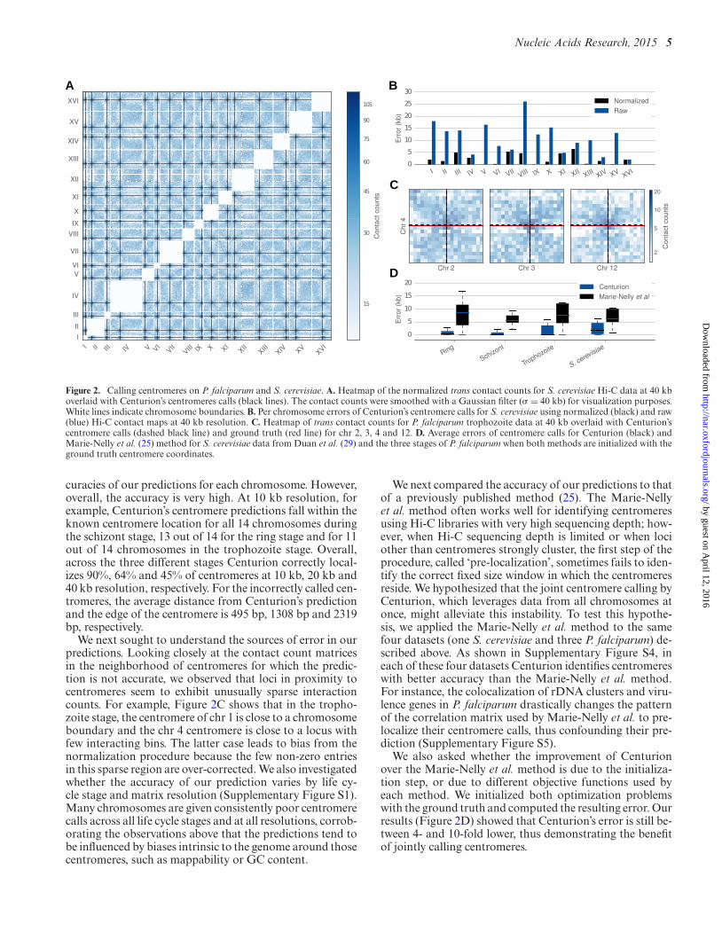
Figure 2. Calling centromeres on P. falciparum and S. cerevisiae. A. Heatmap of the normalized trans contact counts for S. cerevisiae Hi-C data at 40 kboverlaid with Centurion’s centromeres calls (black lines). The contact counts were smoothed with a Gaussian filter (� = 40 kb) for visualization purposes.White lines indicate chromosome boundaries. B. Per chromosome errors of Centurion’s centromere calls for S. cerevisiae using normalized (black) and raw(blue) Hi-C contact maps at 40 kb resolution. C. Heatmap of trans contact counts for P. falciparum trophozoite data at 40 kb overlaid with Centurion’scentromere calls (dashed black line) and ground truth (red line) for chr 2, 3, 4 and 12. D. Average errors of centromere calls for Centurion (black) andMarie-Nelly et al. (25) method for S. cerevisiae data from Duan et al. (29) and the three stages of P. falciparum when both methods are initialized with theground truth centromere coordinates.
curacies of our predictions for each chromosome. However,overall, the accuracy is very high. At 10 kb resolution, forexample, Centurion’s centromere predictions fall within theknown centromere location for all 14 chromosomes duringthe schizont stage, 13 out of 14 for the ring stage and for 11out of 14 chromosomes in the trophozoite stage. Overall,across the three different stages Centurion correctly local-izes 90%, 64% and 45% of centromeres at 10 kb, 20 kb and40 kb resolution, respectively. For the incorrectly called cen-tromeres, the average distance from Centurion’s predictionand the edge of the centromere is 495 bp, 1308 bp and 2319bp, respectively.
We next sought to understand the sources of error in ourpredictions. Looking closely at the contact count matricesin the neighborhood of centromeres for which the predic-tion is not accurate, we observed that loci in proximity tocentromeres seem to exhibit unusually sparse interactioncounts. For example, Figure 2C shows that in the tropho-zoite stage, the centromere of chr 1 is close to a chromosomeboundary and the chr 4 centromere is close to a locus withfew interacting bins. The latter case leads to bias from thenormalization procedure because the few non-zero entriesin this sparse region are over-corrected. We also investigatedwhether the accuracy of our prediction varies by life cy-cle stage and matrix resolution (Supplementary Figure S1).Many chromosomes are given consistently poor centromerecalls across all life cycle stages and at all resolutions, corrob-orating the observations above that the predictions tend tobe influenced by biases intrinsic to the genome around thosecentromeres, such as mappability or GC content.
We next compared the accuracy of our predictions to thatof a previously published method (25). The Marie-Nellyet al. method often works well for identifying centromeresusing Hi-C libraries with very high sequencing depth; how-ever, when Hi-C sequencing depth is limited or when lociother than centromeres strongly cluster, the first step of theprocedure, called ‘pre-localization’, sometimes fails to iden-tify the correct fixed size window in which the centromeresreside. We hypothesized that the joint centromere calling byCenturion, which leverages data from all chromosomes atonce, might alleviate this instability. To test this hypothe-sis, we applied the Marie-Nelly et al. method to the samefour datasets (one S. cerevisiae and three P. falciparum) de-scribed above. As shown in Supplementary Figure S4, ineach of these four datasets Centurion identifies centromereswith better accuracy than the Marie-Nelly et al. method.For instance, the colocalization of rDNA clusters and viru-lence genes in P. falciparum drastically changes the patternof the correlation matrix used by Marie-Nelly et al. to pre-localize their centromere calls, thus confounding their pre-diction (Supplementary Figure S5).
We also asked whether the improvement of Centurionover the Marie-Nelly et al. method is due to the initializa-tion step, or due to different objective functions used byeach method. We initialized both optimization problemswith the ground truth and computed the resulting error. Ourresults (Figure 2D) showed that Centurion’s error is still be-tween 4- and 10-fold lower, thus demonstrating the benefitof jointly calling centromeres.
by guest on April 12, 2016
http://nar.oxfordjournals.org/D
ownloaded from

6 Nucleic Acids Research, 2015
10 20 30 40 50 60 70 80 90
Percentage of kept reads
100
101
102
103
104
Ave
rage
varia
nce
(bp) Ring
SchizontTrophozoiteS. cerevisiae
Figure 3. Impact of Hi-C library sequencing depth on the stability of thecentromere calls. Average variance of the results of Centurion on 500 gen-erated datasets obtained by downsampling the raw contact counts to thedesired coverage.
Resolution, sequencing depth and prediction accuracy
To assess the stability of our predictions, we simulated 500bootstrapped datasets of S. cerevisiae and of each stage ofP. falciparum with an expected total number of reads equalto the contact count matrices. These bootstrapped sampleswere obtained by drawing a contact count for each pairof loci i and j from a Poisson distribution of intensity cij.We then ran the optimization process on the bootstrappeddatasets, starting with initial values randomly placed within40 kb of the centromere calls from our optimization in Sup-plementary Tables S1–S4. Our results show that the opti-mization is very stable (average variance of 25 bp for ring, 6bp for schizont and 12 bp for trophozoite), suggesting thatthe stochastic sampling of the sequencing procedure doesnot significantly affect centromere predictions.
We then sought to investigate the extent to which the ma-trix resolution and sequencing depth affect the accuracy ofCenturion’s predictions. As already seen in SupplementaryFigures S2 and S3, different species give different results:for S. cerevisiae, increasing the matrix resolution to 10 kbresults in lowered accuracy of centromere calls, while in P.falciparum the call quality improves slightly. We speculatedthat our ability to call centromeres in a given species at agiven resolution may depend on the choice of restriction en-zyme, the sequencing depth and the resolution of the con-tact map.
We next evaluated the effect of depth of sequence cover-age on the quality of our centromere predictions. We gener-ated 500 low-coverage datasets by randomly downsamplingthe raw contact counts. We then ran the optimization pro-cess on these downsampled datasets, initializing with per-turbed calls as before. We observe that the low-coveragecentromere calls remain highly stable and accurate. As illus-trated in Figure 3, results across all datasets only begin todegrade when downsampling to <10% of the total numberof reads, which corresponds to <1 count per bin on average.Centurion is thus applicable to call centromeres at low costor for low-abundance species in metagenomic samples.
Centromere calls on a metagenomic dataset
We next sought to call centromeres in several species simul-taneously by combining Centurion with metagenomic Hi-C libraries. We previously (20) generated two Hi-C datasetsfrom synthetic mixtures: one containing 16 yeast strains (in-
cluding four strains of S. cerevisiae), and one containinga mixture of 8 yeasts and 10 prokaryotic species. The twosamples contain a total of 19 yeast species, some of whichare much better characterized than others: centromere po-sitions are already known for eight species (K. lactis, L.kluyveri, L. thermotolerans, S. cerevisiae, S. kudriavzevii, S.mikatae, S. pombe, S. rouxii) and partially for one more (S.bayanus) (12,42–44).
We aligned the reads from the metagenomic Hi-Cdatasets to these yeast species’ reference genomes (see Ma-terials and Methods). The quality of the individual speciesdatasets differ greatly because the organisms vary in abun-dance in the metagenomic samples, and because many se-quences are shared nearly identically between organisms,making the number of uniquely mappable reads for eachorganism range between 109 k for one of the S. cerevisiaestrains and 26 M for the bacteria V. fischeri. Consequently,the sparsity of the matrices is variable (Supplementary Ta-bles S5 and S6). Furthermore, some contact count matri-ces include at least one interaction count for >99% of allpossible locus pairs, whereas other matrices are below 5%.Similarly, in the 40 kb matrices, the average number of inter-chromosomal contact counts per bin varies from <0.004 to>200. In particular, the matrices for the four S. cerevisiaestrains are very sparse: the reference genomes of the fourstrains are very similar to one another; thus, we are not ableto map reads uniquely. We therefore discarded those strainsfrom our analysis, as well as organisms with incomplete ref-erence genomes. We applied Centurion to the remaining 14yeasts (E. gossypii, K. lactis, K. wickerhamii, L. kluyveri, L.waltii, S. bayanus, S. kudriavzevii, S. mikatae, S. paradoxus,S. stipitis, P. pastoris, L. thermotolerans, S. pombe, S. rouxii)on both 20 kb and 40 kb contact maps.
Across these 14 species Centurion performs well, both onhigh-coverage datasets (K. lactis, L. kluyveri, S. bayanus)and low-coverage datasets (S. mikatae), at 20 kb and 40 kb,finding centromeres at an average deviation from the groundtruth of 10 kbp (Figure 4B and Supplementary Figure S6).Given this success with yeasts with known centromere po-sitions, we next made de novo centromere calls for the othersix yeast species present in the metagenomic samples. Theseregions, visualized in Supplementary Figures S15–S20, arestrong candidates for experimental validation by other ap-proaches. One feature that is shared by centromeres acrossall studied fungi is that they reside in regions of early repli-cation timing (3,4). Thus if our centromere calls lie in re-gions of advanced replication timing in a species for whichreplication timing has been profiled but centromeres havenot yet been identified, this data could be used to assess thevalidity of our predictions. Accordingly, we overlaid the po-sitions of our centromere calls in P. pastoris, where repli-cation has been recently profiled (45). In all four chromo-somes, P. pastoris centromere predictions lay in regions ofearly replication timing (Supplementary Figure S21), lend-ing support to our predictions.
The effect of the choice of restriction enzyme
In addition to the resolution of our contact matrices, theunderlying resolution of the Hi-C data itself may limit theaccuracy of our predictions. Hi-C reads can only occur near
by guest on April 12, 2016
http://nar.oxfordjournals.org/D
ownloaded from

Nucleic Acids Research, 2015 7
I II III IV V VIVII
I
II
III
IV
V
VI
VII
1.5
3.0
4.5
6.0
7.5
9.0
10 .5
Con
tact
coun
ts
K. lactis
L. kluyveri
L. thermotolerans
S. kudriavzevii
S. bayanus
S. mikatae
S. pombe
S. rouxii0
5
10
15
20
25
Err
or(k
b)
A
B
Figure 4. Centromere calling on a metagenomic sample. A. Heatmap of thetrans contact counts for K. wickerhamii overlaid with de novo centromerecalls (black lines). The contact counts were smoothed with a Gaussian filter(� = 40 kb) for visualization purposes. White lines indicate chromosomeboundaries. B. Box plots indicating the error (in kb) for each chromosomein Centurion’s centromere calls for eight yeasts with known centromerecoordinates from the combined metagenomic Hi-C samples M-3D and M-Y of (20) on the 20 kb contact count matrices.
the recognition site of the restriction enzyme used in the Hi-C assay; indeed, the best resolution we can hope to achieveis a matrix in which each corresponds to one restriction en-zyme fragment. Some restriction enzymes cut much morefrequently than others. Thus, we speculated that a Hi-Cexperiment using enzymes that cut more frequently mightyield more accurate results than an experiment using lessfrequently cutting enzymes.
To address this question, we compare the accuracy ofcentromere calling from two Hi-C libraries created from asingle metagenomic sample using different combinations ofrestriction enzymes. The first library was created using thetwo 6 bp-cutters, HindIII and NcoI. The second library usesSau3AI, which has a 4 bp recognition site, and AflIII, whichhas a 6 bp recognition site with two degenerate sites, mak-ing it effectively a 5 bp cutter. Digestion with HindIII/NcoIyields a total of 8324 restriction fragments, whereas diges-tion with Sau3AI/AflIII yields 42359 restriction fragments.We corrected for the difference in Hi-C sequencing depthbetween Sau3AI/AflIII and the NcoI/HindIII libraries bygenerating downsampled datasets with an equal number ofreads from each sequencing library. We then normalizedthe datasets and applied Centurion. The sample includes
three species for which we possess the ground truth cen-tromere locations, only one of which (L. thermotolerans)had enough reads in both the Ncol/HindIII (63 000 reads)and the pooled Sau3AI/AflIII (55 000 reads) datasets tocorrectly call the centromeres. The error on the downsam-pled Sau3AI/AflIII datasets (8 kbp) was on average halfas large as the error on the the Ncol/HindIII datasets (16kbp). Thus, we conclude that using a restriction enzymewith more frequent cutting sites enables more precise cen-tromere calls at fine scales.
DISCUSSION
While centromeres are a fundamental element in the biologyof genomes, their identification in diverse species has provendifficult due to sequence divergence and limitations of avail-able tools. In this work, we have developed a novel method,Centurion, that uses centromere colocalization and the pat-tern it creates in Hi-C contact maps to jointly call cen-tromeres for all chromosomes of an organism. We first es-tablished the feasibility of this approach by demonstratingthat Centurion accurately calls regional centromeres on theparasite P. falciparum and the yeast S. cerevisiae as wellas point centromeres on several other yeasts with knowncentromere coordinates. For the species with high depthHi-C sequencing, Centurion often identified centromereswithin 1 kb of the actual coordinates (41 times out of 58for three stages of P. falciparum and S. cerevisiae data).We then used Centurion to infer centromeres of multipleyeast species (eight with known, six with unknown cen-tromere coordinates) from two metagenomic Hi-C samples.Our results showed that Centurion still accurately identifiescentromere coordinates from samples with only limited se-quencing depth. Thus, Centurion can be used to accuratelyand efficiently identify centromere locations in yeast species.
The task of centromere identification from Hi-C data hasbeen attempted recently by others (25). Centurion offersa few key differences compared to the previous approach.The first difference is in the pre-localization of candidatecentromeres. Marie-Nelly et al.’s method uses only the cisPearson correlation information independently per chro-mosome to identify the initial candidates. However, the pat-tern created by centromeres in the Pearson correlation ma-trix can be very similar to the patterns generated by othergenomic elements such as rDNA coding regions or by spe-cific gene clusters (e.g. virulence genes in P. falciparum).Because Marie-Nelly et al.’s method restricts the furthersearch for the best centromere coordinate to only the can-didates from the pre-localization step, an inaccurate can-didate (e.g. an rDNA region instead of a centromere) willprevent the method from finding the correct centromere lo-cation. Centurion, on the other hand, utilizes trans con-tact information jointly across all chromosomes for its pre-localization step. Furthermore, Centurion allows multiplecandidates per chromosome during the second step of theoptimization, thereby leaving room for correcting potentialerrors in the pre-localization step. The second differencebetween the two methods is in how they use the subma-trices that correspond to trans contact maps flanking thepairs of candidate centromeres from the pre-localizationstep. For an organism with N chromosomes, Marie-Nelly
by guest on April 12, 2016
http://nar.oxfordjournals.org/D
ownloaded from

8 Nucleic Acids Research, 2015
et al.’s method carves out the N-1 trans submatrices for eachchromosome, sums these N-1 matrices and then collapsesthe sum into a 1D vector of row/column sums. Then, inde-pendently for each chromosome, the method fits a Gaussianto this 1D vector, and the resulting peak corresponds to thepredicted centromere location. In this procedure, both thesummation of N-1 matrices and the collapsing of the result-ing matrix into a 1D vector of sums result in loss of impor-tant information embedded in 2D maps. Furthermore, per-forming the Gaussian fit separately for each chromosomedoes not fully take into account the joint colocalization ofthe other N-1 centromeres. To address these issues, Cen-turion infers a 2D Gaussian fit that best explains the ob-served trans contact counts, jointly optimizing these 2D fitsfor all pairs of centromeres. Both of these improvements inthe pre-localization and the optimization steps allow Centu-rion to perform better specifically for the cases with limitedsequencing depth.
Our approach could be improved in several respects.First, better modeling of zero contact counts may improveinference for organisms with many repeated sequences inthe pericentromeric regions, or datasets with low sequenc-ing depth. Second, one could model contact counts as aGaussian distribution centered on the pairs of centromerelocations. Maximizing the log likelihood of such a modelmight yield improved performance. Last, as described here,our method requires reference genomes for the metage-nomic samples. It would be possible to first build referencegenomes directly from the Hi-C data, using methods likeLachesis (17) or GRAAL (46), and then infer centromerelocations using the inferred references. However, the inher-ent structure of Hi-C contact counts for organisms withcolocalizing centromeres will likely present a challenge forthese methods because pericentromeric sequences on differ-ent chromosomes are likely to appear to be adjacent to oneanother.
Finally, our new centromere predictions have practicalapplications. Autonomously replicating plasmids and arti-ficial chromosomes are useful tools for research and strainengineering (9). Identification of centromeres in new specieswill facilitate building such constructs over an expandedspecies range. P. pastoris, for example, is a common indus-trial chassis (47), but existing plasmid tools in the specieshave elevated loss rates (48) that could be stabilized by ad-dition of a centromere. Many of our centromere calls wereaccurate to <1 kb, making experimental validation pos-sible. Sequencing data is available from the Short ReadArchive at http://www.ncbi.nlm.nih.gov/Traces/sra/?study=SRP057812.
SUPPLEMENTARY DATA
Supplementary Data are available at NAR Online.
ACKNOWLEDGEMENTS
We thank Celia Payen for providing the yeast centromereannotations, Stefan van der Walt for advice on peak de-tection algorithms, Fabrice Varoquaux for help on under-standing the specificity of A. thaliana genome and ChloeAzencott for helpful comments on the manuscript.
FUNDING
European Research Council [SMAC-ERC-280032 to J.-P.V., N.V.]; European Commission [HEALTH-F5-2012-305626 to J.-P.V., N.V.]; French National Research Agency[ ANR-11-BINF-0001 to J.-P.V., N.V.]; National Insti-tute of Health/National Human Genome Research In-stitute [HG006283 to J.S., T32HG000035 to J.N.B.]; Na-tional Institute of Health/National Institute of GeneralMedical Sciences [P41 GM103533 to I.L., M.J.D., W.S.N.;R01AI106775 to F.A., W.S.N.]; National Science Founda-tion [1243710 to I.L., M.J.D.]. M.J.D. is a Rita Allen Foun-dation Scholar and a Senior Fellow in the Genetic Networksprogram at the Canadian Institute for Advanced Research.Funding for open access charge: An NIH grant will be thesource of funding for the publication charges.Conflict of interest statement. None declared.
REFERENCES1. Bloom,K.S. (2014) Centromeric heterochromatin: the primordial
segregation machine. Annu. Rev. Genet., 48, 457–484.2. Feng,W., Bachant,J., Collingwood,D., Raghuraman,M.K. and
Brewer,B.J. (2009) Centromere replication timing determines differentforms of genomic instability in Saccharomyces cerevisiae checkpointmutants during replication stress. Genetics, 183, 1249–1260.
3. Koren,A., Tsai,H.J., Tirosh,I., Burrack,L.S., Barkai,N. and Berman,J.(2010) Epigenetically-inherited centromere and neocentromere DNAreplicates earliest in S-phase. PLoS Genet., 6, e1001068.
4. Pohl,T.J., Brewer,B.J. and Raghuraman,M.K. (2012) Functionalcentromeres determine the activation time of pericentric origins ofDNA replication in Saccharomyces cerevisiae. PLoS Genet., 8,e1002677.
5. Cottarel,G., Shero,J.H., Hieter,P. and Hegemann,J.H. (1989) A125-base-pair CEN6 DNA fragment is sufficient for complete meioticand mitotic centromere functions in Saccharomyces cerevisiae. Mol.Cell. Biol., 9, 3342–3349.
6. Gordon,J.L., Byrne,K.P. and Wolfe,K.H. (2011) Mechanisms ofchromosome number evolution in yeast. PLoS Genet., 7, e1002190.
7. Dujon,B. (2010) Yeast evolutionary genomics. Nat. Rev. Genet., 11,512–524.
8. Hittinger,C.T. (2013) Saccharomyces diversity and evolution: abudding model genus. Trends Genet., 29, 309–317.
9. Boer,E., Steinborn,G., Kunze,G. and Gellissen,G. (2007) Yeastexpression platforms. Appl. Microbiol. Biotechnol., 77, 513–523.
10. Murray,A.W. and Szostak,J.W. (1983) Pedigree analysis of plasmidsegregation in yeast. Cell, 34, 961–970.
11. Malik,H.S. and Henikoff,S. (2009) Major evolutionary transitions incentromere complexity. Cell, 138, 1067–1082.
12. The Genolevures Consortium, Souciet,J.L., Dujon,B., Gaillardin,C.,Johnston,M., Baret,P.V., Cliften,P., Sherman,D.J., Weissenbach,J.,Westhof,E. et al. (2009) Comparative genomics of protoploidSaccharomycetaceae. Genome Res., 19, 1696–1709.
13. Lefrancois,P., Euskirchen,G.M., Auerbach,R.K., Rozowsky,J.,Gibson,T., Yellman,C.M., Gerstein,M. and Snyder,M. (2009)Efficient yeast ChIP-Seq using multiplex short-read DNAsequencing. BMC Genomics, 10, 37.
14. Lieberman-Aiden,E., van Berkum,N.L., Williams,L., Imakaev,M.,Ragoczy,T., Telling,A., Amit,I., Lajoie,B.R., Sabo,P.J.,Dorschner,M.O. et al. (2009) Comprehensive mapping of long-rangeinteractions reveals folding principles of the human genome. Science,326, 289–293.
15. Kalhor,R., Tjong,H., Jayathilaka,N., Alber,F. and Chen,L. (2011)Genome architectures revealed by tethered chromosomeconformation capture and population-based modeling. Nat.Biotechnol., 30, 90–98.
16. Duan,Z., Andronescu,M., Schutz,K., McIlwain,S., Kim,Y.J., Lee,C.,Shendure,J., Fields,S., Blau,C.A. and Noble,W.S. (2010) Athree-dimensional model of the yeast genome. Nature, 465, 363–367.
17. Burton,J.N., Adey,A., Patwardhan,R.P., Qiu,R., Kitzman,J.O. andShendure,J. (2013) Chromosome-scale scaffolding of de novo genome
by guest on April 12, 2016
http://nar.oxfordjournals.org/D
ownloaded from

Nucleic Acids Research, 2015 9
assemblies based on chromatin interactions. Nat. Biotechnol., 31,1119–1125.
18. Kaplan,N. and Dekker,J. (2013) High-throughput genome scaffoldingfrom in vivo DNA interaction frequency. Nat. Biotechnol., 31,1143–1147.
19. Selvaraj,S., J,R.D., Bansal,V. and Ren,B. (2013) Whole-genomehaplotype reconstruction using proximity-ligation and shotgunsequencing. Nat. Biotechnol., 31, 1111–1118.
20. Burton,J.N., Liachko,I., Dunham,M.J. and Shendure,J. (2014)Species-level deconvolution of metagenome assemblies withHi-C-based contact probability maps. G3 (Bethesda), 4, 1339–1346.
21. Beitel,C.W., Froenicke,L., Lang,J.M., Korf,I.F., Michelmore,R.W.,Eisen,J.A. and Darling,A.E. (2014) Strain- and plasmid-leveldeconvolution of a synthetic metagenome by sequencing proximityligation products. PeerJ, 2, e415.
22. Nora,E.P., Lajoie,B.R., Schulz,E.G., Giorgetti,L., Okamoto,I.,Servant,N., Piolot,T., van Berkum,N.L., Meisig,J., Sedat,J. et al.(2012) Spatial partitioning of the regulatory landscape of theX-inactivation centre. Nature, 485, 381–385.
23. Dixon,J.R., Selvaraj,S., Yue,F., Kim,A., Li,Y., Shen,Y., Hu,M.,Liu,J.S. and Ren,B. (2012) Topological domains in mammaliangenomes identified by analysis of chromatin interactions. Nature, 485,376–380.
24. Mizuguchi,T., Fudenberg,G., Mehta,S., Belton,J.M., Taneja,N.,Folco,H.D., FitzGerald,P., Dekker,J., Mirny,L., Barrowman,J. et al.(2014) Cohesin-dependent globules and heterochromatin shape 3Dgenome architecture in S. pombe. Nature, 516, 432–435.
25. Marie-Nelly,H., Marbouty,M., Cournac,A., Liti,G., Fischer,G.,Zimmer,C. and Koszul,R. (2014) Filling annotation gaps in yeastgenomes using genome-wide contact maps. Bioinformatics, 30,2105–2113.
26. Ay,F., Bunnik,E.M., Varoquaux,N., Bol,S.M., Prudhomme,J.,Vert,J.-P., Noble,W.S. and Le Roch,K.G. (2014) Three-dimensionalmodeling of the P. falciparum genome during the erythrocytic cyclereveals a strong connection between genome architecture and geneexpression. Genome Res., 24, 974–988.
27. Feng,S., Cokus,S.J., Schubert,V., Zhai,J., Pellegrini,M. andJacobsen,S.E. (2014) Genome-wide Hi-C analyses in wild-type andmutants reveal high-resolution chromatin interactions in Arabidopsis.Mol. Cell, 55, 694–707.
28. Ober,R.J., Ram,S. and Ward,E.S. (2004) Localization accuracy insingle-molecule microscopy. Biophys. J., 86, 1185–1200.
29. Duan,Z., Andronescu,M., Schutz,K., Lee,C., Shendure,J., Fields,S.,Noble,W.S. and Anthony Blau,C. (2012) A genome-wide 3C-methodfor characterizing the three-dimensional architectures of genomes.Methods, 58, 277–288.
30. Ay,F., Vu,T.H., Zeitz,M.J., Varoquaux,N., Carette,J.E., Vert,J.,Hoffman,A.R. and Noble,W.S. (2015) Identifying multi-locuschromatin contacts in human cells using tethered multiple 3C. BMCGenomics, 16, 121.
31. Baker,C.R., Tuch,B.B. and Johnson,A.D. (2011) ExtensiveDNA-binding specificity divergence of a conserved transcriptionregulator. Proc. Natl. Acad. Sci. U.S.A., 108, 7493–7498.
32. Lin,S.H. and Liao,Y.C. (2013) CISA: contig integrator for sequenceassembly of bacterial genomes. PLoS ONE, 8, e60843.
33. Peng,Y., Leung,H.C., Yiu,S.M. and Chin,F.Y. (2012) IDBA-UD: a denovo assembler for single-cell and metagenomic sequencing data withhighly uneven depth. Bioinformatics, 28, 1420–1428.
34. Yaffe,E. and Tanay,A. (2011) Probabilistic modeling of Hi-C contactmaps eliminates systematic biases to characterize globalchromosomal architecture. Nat. Genet., 43, 1059–1065.
35. Imakaev,M., Fudenberg,G., McCord,R.P., Naumova,N.,Goloborodko,A., Lajoie,B.R., Dekker,J. and Mirny,L.A. (2012)Iterative correction of Hi-C data reveals hallmarks of chromosomeorganization. Nat. Methods, 9, 999–1003.
36. Jin,F., Li,Y., Dixon,J.R., Selvaraj,S., Ye,Z., Lee,A.Y., Yen,C.A.,Schmitt,A.D., Espinoza,C.A. and Ren,B. (2013) A high-resolutionmap of the three-dimensional chromatin interactome in human cells.Nature, 503, 290–294.
37. Venema,J. and Tollervey,D. (1999) Ribosome synthesis inSaccharomyces cerevisiae. Annu. Rev. Genet., 33, 261–311.
38. Zimmer,C. and Fabre,E. (2011) Principles of chromosomalorganization: lessons from yeast. J. Cell Biol., 192, 723–733.
39. Gotta,M., Laroche,T., Formenton,A., Maillet,L., Scherthan,H. andGasser,S.M. (1996) The clustering of telomeres and colocalizationwith Rap1, Sir3, and Sir4 proteins in wild-type Saccharomycescerevisiae. J. Cell Biol., 134, 1349–1363.
40. World Health Organization. (2012) World Malaria Report. http://www.who.int/malaria/publications/world malaria report 2012/en/.
41. Hoeijmakers,W.A., Flueck,C., Francoijs,K.J., Smits,A.H., Wetzel,J.,Volz,J.C., Cowman,A.F., Voss,T., Stunnenberg,H.G. and Bartfai,R.(2012) Plasmodium falciparum centromeres display a uniqueepigenetic makeup and cluster prior to and during schizogony. CellMicrobiol., 14, 1391–1401.
42. Scannell,D.R., Zill,O.A., Rokas,A., Payen,C., Dunham,M.J.,Eisen,M.B., Rine,J., Johnston,M. and Hittinger,C.T. (2011) Theawesome power of yeast evolutionary genetics: new genomesequences and strain resources for the Saccharomyces sensu strictogenus. G3 (Bethesda), 1, 11–25.
43. McDowall,M.D., Harris,M.A., Lock,A., Rutherford,K.,Staines,D.M., Bahler,J., Kersey,P.J., Oliver,S.G. and Wood,V. (2014)PomBase 2015: updates to the fission yeast database. Nucleic AcidsRes., 43, D656–D661.
44. Dujon,B., Sherman,D., Fischer,G., Durrens,P., Casaregola,S.,Lafontaine,I., De Montigny,J., Marck,C., Neuveglise,C., Talla,E.et al. (2004) Genome evolution in yeasts. Nature, 430, 35–44.
45. Liachko,I., Youngblood,R.A., Tsui,K., Bubb,K.L., Queitsch,C.,Raghuraman,M.K., Nislow,C., Brewer,B.J. and Dunham,M.J. (2014)GC-rich DNA elements enable replication origin activity in themethylotrophic yeast Pichia pastoris. PLoS Genet., 10, e1004169.
46. Marie-Nelly,H., Marbouty,M., Cournac,A., Flot,J.-F., Liti,G.,Parodi,D.P., Syan,S., Guillen,N., Margeot,A., Zimmer,C. et al. (2014)High-quality genome (re)assembly using chromosomal contact data.Nat. Commun., 5, 5695.
47. Cregg,J.M., Tolstorukov,I., Kusari,A., Sunga,J., Madden,K. andChappell,T. (2009) Expression in the yeast Pichia pastoris. Meth.Enzymol., 463, 169–189.
48. Liachko,I. and Dunham,M.J. (2014) An autonomously replicatingsequence for use in a wide range of budding yeasts. FEMS Yeast Res.,14, 364–367.
by guest on April 12, 2016
http://nar.oxfordjournals.org/D
ownloaded from