Speciation studies of mono- and binuclear Pd(II) complexes involving mixed nitrogen-sulfur donor...
16
Workflow: Annotated pdf, CrossRef and tracked changes PROOF COVER SHEET Author(s): Mohamed M. Shoukry Article title: Speciation studies of mono- and binuclear Pd(II) complexes involving mixed nitrogen-sulfur donor ligand and 4,4′-bipiperidine as a linker Article no: GCOO 1043909 Enclosures: 1) Query sheet 2) Article proofs Dear Author, 1. Please check these proofs carefully. It is the responsibility of the corresponding author to check these and approve or amend them. A second proof is not normally provided. Taylor & Francis cannot be held responsible for uncorrected errors, even if introduced during the production process. Once your corrections have been added to the article, it will be considered ready for publication. Please limit changes at this stage to the correction of errors. You should not make trivial changes, improve prose style, add new material, or delete existing material at this stage. You may be charged if your corrections are excessive (we would not expect corrections to exceed 30 changes). For detailed guidance on how to check your proofs, please paste this address into a new browser window: http://journalauthors.tandf.co.uk/production/checkingproofs.asp Your PDF proof file has been enabled so that you can comment on the proof directly using Adobe Acrobat. If you wish to do this, please save the file to your hard disk first. For further information on marking corrections using Acrobat, please paste this address into a new browser window: http:// journalauthors.tandf.co.uk/production/acrobat.asp 2. Please review the table of contributors below and confirm that the first and last names are structured correctly and that the authors are listed in the correct order of contribution. This check is to ensure that your name will appear correctly online and when the article is indexed. Sequence Prefix Given name(s) Surname Suffix 1 2 Mohamed M. Sameya M.T. Shoukry Ezzat
-
Upload
independent -
Category
Documents
-
view
0 -
download
0
Transcript of Speciation studies of mono- and binuclear Pd(II) complexes involving mixed nitrogen-sulfur donor...

Workflow: Annotated pdf, CrossRef and tracked changes
PROOF COVER SHEETAuthor(s): Mohamed M. Shoukry
Article title: Speciation studies of mono- and binuclear Pd(II) complexes involving mixed nitrogen-sulfur
donor ligand and 4,4′-bipiperidine as a linker
Article no: GCOO 1043909
Enclosures: 1) Query sheet
2) Article proofs
Dear Author,
1. Please check these proofs carefully. It is the responsibility of the corresponding author to check theseand approve or amend them. A second proof is not normally provided. Taylor & Francis cannot be heldresponsible for uncorrected errors, even if introduced during the production process. Once yourcorrections have been added to the article, it will be considered ready for publication.
Please limit changes at this stage to the correction of errors. You should not make trivial changes,improve prose style, add new material, or delete existing material at this stage. You may be charged ifyour corrections are excessive (we would not expect corrections to exceed 30 changes).
For detailed guidance on how to check your proofs, please paste this address into a new browserwindow: http://journalauthors.tandf.co.uk/production/checkingproofs.asp
Your PDF proof file has been enabled so that you can comment on the proof directly using AdobeAcrobat. If you wish to do this, please save the file to your hard disk first. For further information onmarking corrections using Acrobat, please paste this address into a new browser window: http://journalauthors.tandf.co.uk/production/acrobat.asp
2. Please review the table of contributors below and confirm that the first and last names arestructured correctly and that the authors are listed in the correct order of contribution. This checkis to ensure that your name will appear correctly online and when the article is indexed.
Sequence Prefix Given name(s) Surname Suffix
12
Mohamed M.Sameya M.T.
ShoukryEzzat

Queries are marked in the margins of the proofs, and you can also click the hyperlinks below.Content changes made during copy-editing are shown as tracked changes. Inserted text is in red font andrevisions have a red indicatorå. Changes can also be viewed using the list comments function. Tocorrect the proofs, you should insert or delete text following the instructions below, but do not addcomments to the existing tracked changes.
AUTHOR QUERIES
General points:
1. Permissions: You have warranted that you have secured the necessary written permission from theappropriate copyright owner for the reproduction of any text, illustration, or other material in yourarticle. Please see http://journalauthors.tandf.co.uk/permissions/usingThirdPartyMaterial.asp.
2. Third-party content: If there is third-party content in your article, please check that the rightsholderdetails for re-use are shown correctly.
3. Affiliation: The corresponding author is responsible for ensuring that address and email details arecorrect for all the co-authors. Affiliations given in the article should be the affiliation at the time theresearch was conducted. Please see http://journalauthors.tandf.co.uk/preparation/writing.asp.
4. Funding: Was your research for this article funded by a funding agency? If so, please insert ‘Thiswork was supported by <insert the name of the funding agency in full>’, followed by the grantnumber in square brackets ‘[grant number xxxx]’.
5. Supplemental data and underlying research materials: Do you wish to include the location of theunderlying research materials (e.g. data, samples or models) for your article? If so, please insert thissentence before the reference section: ‘The underlying research materials for this article can beaccessed at <full link>/ description of location [author to complete]’. If your article includessupplemental data, the link will also be provided in this paragraph. See <http://journalauthors.tandf.co.uk/preparation/multimedia.asp> for further explanation of supplemental dataand underlying research materials.
6. The CrossRef database (www.crossref.org/) has been used to validate the references. Changesresulting from mismatches are tracked in red font.
AQ1 Please provide an institutional e-mail address, if available, to be included in the arti-cle, as per journal style.
AQ2 Would you consider changing the title “Speciation studies of mono- and binuclearPd(II) complexes involving mixed nitrogen-sulfur donor ligand and 4,4′-bipiperidineas a linker ” to “Speciation studies of mono- and binuclear Pd(II) complexes involv-ing mixed nitrogen–sulfur donor ligand and 4,4′-bipiperidine as a linker”? Pleasesuggest.
AQ3 The disclosure statement has been inserted. Please correct if this is inaccurate.
AQ4 The CrossRef database (www.crossref.org/) has been used to validate the references.Mismatches between the original manuscript and CrossRef are tracked in red font.Please provide a revision if the change is incorrect. Do not comment on correctchanges.
AQ5 Please provide missing volume for Refs. [12, 21–23].

How to make corrections to your proofs using Adobe Acrobat/Reader
Taylor & Francis offers you a choice of options to help you make corrections to your proofs. Your PDFproof file has been enabled so that you can mark up the proof directly using Adobe Acrobat/Reader. Thisis the simplest and best way for you to ensure that your corrections will be incorporated. If you wish todo this, please follow these instructions:
1. Save the file to your hard disk.
2. Check which version of Adobe Acrobat/Reader you have on your computer. You can do this by click-ing on the “Help” tab, and then “About”.
If Adobe Reader is not installed, you can get the latest version free from http://get.adobe.com/reader/.
3. If you have Adobe Acrobat/Reader 10 or a later version, click on the “Comment” link at the right-hand side to view the Comments pane.
4. You can then select any text and mark it up for deletion or replacement, or insert new text as needed.Please note that these will clearly be displayed in the Comments pane and secondary annotation is notneeded to draw attention to your corrections. If you need to include new sections of text, it is alsopossible to add a comment to the proofs. To do this, use the Sticky Note tool in the task bar. Pleasealso see our FAQs here: http://journalauthors.tandf.co.uk/production/index.asp.
5. Make sure that you save the file when you close the document before uploading it to CATS using the“Upload File” button on the online correction form. If you have more than one file, please zip themtogether and then upload the zip file.
If you prefer, you can make your corrections using the CATS online correction form.
Troubleshooting
Acrobat help: http://helpx.adobe.com/acrobat.htmlReader help: http://helpx.adobe.com/reader.html
Please note that full user guides for earlier versions of these programs are available from the Adobe Helppages by clicking on the link “Previous versions” under the “Help and tutorials” heading from the rele-vant link above. Commenting functionality is available from Adobe Reader 8.0 onwards and from AdobeAcrobat 7.0 onwards.
Firefox users: Firefox’s inbuilt PDF Viewer is set to the default; please see the following for instructionson how to use this and download the PDF to your hard drive: http://support.mozilla.org/en-US/kb/view-pdf-files-firefox-without-downloading-them#w_using-a-pdf-reader-plugin

Speciation studies of mono- and binuclear Pd(II) complexesinvolving mixed nitrogen-sulfur donor ligand and
4,4′-bipiperidine as a linker
5MOHAMED M. SHOUKRY* and SAMEYA M.T. EZZAT
Faculty of Science, Department of Chemistry, University of Cairo, Cairo, Egypt
(Received 3 February 2015; accepted 26 March 2015)
0
0.5
1
1.5
450350250
Wavenumber (nm)
Abs
orba
nce
B
D
A
C
10Speciation of mono- and binuclear Pd(II) complexes involving mixed nitrogen–sulfur donor ligandis investigated; 4,4′-bipiperidine is used as a linker.
Pd(MME)Cl2 complex, where MME = methionine methyl ester, was synthesized and characterizedby elemental analysis and spectroscopic techniques. [Pd(MME)(H2O)2]
2+ interacts with some DNAconstituents giving 1 : 1 and 1 : 2 complexes. The binuclear complexes having 4,4′-bipiperidine as a
15linker and involving [Pd(MME)(H2O)2]2+ and DNA constituents were investigated. The results show
formation of [(H2O)(MME)Pd(Bip)Pd(MME)(H2O)]4+. Inosine, uracil, and thymine interact with the
previously mentioned complex by substitution of the two coordinated water molecules. Formationconstants of all possible mono- and binuclear complexes were determined and their speciationdiagrams were evaluated.
20Keywords: Palladium(II) complexes; Methionine methyl ester; 4,4′-bipiperidine; DNA constituents;Binuclear complexes; Equilibrium constants
1. Introduction
Since Rosenberg [1] discovered the antitumor activity of cis-diamminedichloroplatinum(II),significant contributions were made to produce new platinum-containing compounds to
AQ2
AQ1
*Corresponding author. Email: [email protected] to Prof. Rudi van Eldik on the occasion of his 70th birthday
© 2015 Taylor & Francis
Journal of Coordination Chemistry, 2015http://dx.doi.org/10.1080/00958972.2015.1043909
GCOO 1043909 CE: KR QA: RB7 May 2015 Coll: QC:Initial
Authorquery:
Inserted Text
Would you consider changing the title “Speciation studies of mono- and binuclear Pd(II) complexes involving mixed nitrogen-sulfur donor ligand and 4,4′-bipiperidine as a linker ” to “Speciation studies of mono- and binuclear Pd(II) complexes involving mixed nitrogen–sulfur donor ligand and 4,4′-bipiperidine as a linker”? Please suggest.
Authorquery:
Inserted Text
Please provide an institutional e-mail address, if available, to be included in the article, as per journal style.
Deleted text:
Inserted Text
-

5 overcome the physiological disadvantages, such as several side effects, drug resistance, andlimitation in the field of application. Most of the developed and studied platinum complexesinclude nitrogen-containing monodentate, bidentate, or tridentate ligands [2–8]. The ulti-mate aim of the modifications of the parent drug is to make related analogs that produce adifferent spectrum of DNA lesions and so circumvent the problem of resistance to cisplatin
10 [2, 9]. Therefore, nonclassical platinum derivatives were also investigated, which mayviolate the classical structure–activity relationship [2]. Complexes are known that includesulfur-containing ligands and show antitumor activity [10]. An example is [Pt(CH3SCH2CH2SCH3)Cl2], Pt(dt), a sulfur analog of the well-studied [Pt(H2NCH2CH2NH2)Cl2], Pt(en) complex [10]. Baltić et al. examined the antitumor activity of Pt(dt) against the
15 human breast cancer cell line and found the complex to inhibit the growth of MCF-7 cellsin a dose and time-dependent manner [10]. Another example is dichloro(2-methylth-iomethylpyridine)platinum(II), Pt(mtp)Cl2, a complex with nitrogen as well as sulfurdonors. In the literature, different synthetic pathways are described [11–13] and the biden-tate mixed N,S-complex was found to be a promising cytostatic agent [14, 15]. In addition
20 to new mononuclear complexes with improved properties in terms of toxicity or cross-resis-tance to cisplatin, a new class of binuclear Pt(II) complexes has been developed by Farrell’sgroup [16–19]. The apparent advantage of these complexes is the high charge (+4), com-pared to the neutral mononuclear complexes, resulting in good solubility, efficient electro-static interaction with polyanionic DNA (the major target of platinating agents) and fast
25 uptake [20].Palladium(II) complexes show discrete antitumor activity in vitro compared to the
platinum-based drugs because of their extremely high liability in biological fluids. We havea long-standing interest in equilibria of complex-formation reactions of (diamine)PdCl2[21–27] and dinuclear palladium(II) [28, 29] complexes with bio-relevant ligands. It will be
30 of interest to extend these investigations and study the mono- and binuclear complexes withN,S-donor ligands. In this investigation, a chelated methionine methyl (MME) ester wasused. It has nitrogen and sulfur sites for chelation. The present investigation describes thesynthesis and characterization of a Pd(II) complex with MME. The interaction of[Pd(MME)(H2O)2]
2+ with DNA constituents and 4,4′-bipiperidine was studied. Also, the35 binuclear complexes involving Pd(MME)2+ and 4,4′-bipiperidine linking two Pd(MME)2+
species were investigated.
2. Experimental
2.1. Materials
K2PdCl4, MME, 4,4′-bipiperidine·2HCl (Bip), and cysteine·HCl were obtained from40 Aldrich. The DNA constituents (inosine, inosine-5′-monophosphate (IMP), adenosine-5′-
monophosphate, thymine, thymidine, uracil, and uridine) were provided by Sigma ChemicalCo. For equilibrium studies, Pd(MME)Cl2 was converted into the diaqua complex bytreating it with two equivalents of AgNO3 as described before [23]. The ligands in the formof hydrochlorides were converted into the corresponding hydronitrates. The nucleotides
45 were prepared in the protonated form with standard HNO3 solution. All solutions were pre-pared in deionized water. The structural formulas of the investigated ligands are given inscheme 1.
2 M.M. Shoukry and S.M.T. Ezzat
GCOO 1043909 CE: KR QA: RB7 May 2015 Coll: QC:Initial
Deleted text:
Inserted Text
-
Deleted text:
Inserted Text
-
Deleted text:
Inserted Text
-
Deleted text:
Inserted Text
as

2.2. Synthesis
Pd(MME)Cl2 was prepared by dissolving K2PdCl4 (2.82 mmol) in 10 mL water with stir-5ring. The clear solution of [PdCl4]
2− was filtered and MME (2.82 mmol) dissolved in10 mL H2O was added dropwise to the stirred solution. The pH was adjusted to 2–3 byaddition of HCl and/or NaOH. A yellowish-brown precipitate of Pd(MME)Cl2 was formedand stirred for a further 30 min at 50 °C. After filtering off the precipitate, it was thoroughlywashed with H2O, ethanol and diethyl ether. A yellow powder was obtained. Anal. Calcd
10for C6H13NSO2PdCl2 (F. Wt. = 340.380: C, 21.16; H, 3.81; N, 4.11; S, 9.40. Found: C,21.2; H, 4.0; N, 3.7, S, 9.1%).
2.3. Equipment and potentiometric analysis
Potentiometric titrations were performed with a Metrohm 686 titroprocessor equipped witha 665 Dosimat. The titroprocessor and electrode were calibrated with standard buffer solu-
15tions, prepared according to NBS specification [30]. All titrations were carried out at 25.0
P
P
O O
O
O
O
OH OH
N
N
N
NH
O
O
OH OH
OHN
N
N
NH
O
N
NN
N
NH 2
O O
O
O
O
OH OH
NH
O
NH
O
OCH 3
OH
ON
NH
O OH
OCH 3
O NH
NHO
OH OH
OHN
NH
O
O
NH 2
OCH 3S
OMeH
Inosine
971
3
Inosine-5´-monophosphate
971
3
6
971
3
Adenosine-5´-monophosphate
Uracil
132
4
Thymidine
123 4
123 4
Thymi
1
32
4
Uridine
Methionine methyl ester
Scheme 1. Structural formulas of ligands.
Mono- and binuclear Pd(II) complexes 3
GCOO 1043909 CE: KR QA: RB7 May 2015 Coll: QC:Initial
± 0.1 °C in purified nitrogen using a titration vessel described previously [31]. Elementalanalysis was done by CHNS Automatic Analyzer, Vario ElIII-Elementar.
The acid dissociation constants of the ligands were determined by titrating 0.05 mmolsamples of each with standard NaOH solutions. Ligands were converted into their proto-
5 nated form with standard HNO3 solutions. The acid dissociation constants of the coordi-nated waters in [Pd(MME)(H2O)2]
2+ were determined by titrating 0.05 mmol of complexwith standard 0.05 M NaOH solution. The formation constants of the complexes weredetermined by titrating solution mixtures of [Pd(MME)(H2O)2]
2+ (0.05 mmol) and theligand in the concentration ratio of 1 : 2 (Pd : ligand) for the DNA constituents. The forma-
10 tion constants of the binuclear complexes of Bip were determined by titrating solution mix-ture of 0.062 mmol of [Pd(MME)(H2O)2]
2+ and Bip in concentration ratio of 2 : 1 (Pd :Bip). The formation constants of the binuclear DNA complexes were determined by titrat-ing solution mixtures of 0.062 mmol of [Pd(MME)(H2O)2]
2+, Bip and DNA constituent inconcentration ratio of 2 : 1 : 2 (Pd : Bip : DNA constituent). The titrated solution mixtures
15 each had a volume of 40 mL and the titrations were carried out at 25 °C and 0.1 M ionicstrength (adjusted with NaNO3). A standard 0.05 M NaOH solution was used as titrant. ThepH meter readings were converted to hydrogen ion concentration by titrating a standardHNO3 solution (0.01 M), the ionic strength of which was adjusted to 0.1 M with NaNO3,with standard NaOH (0.05 M) at 25 °C. The pH values from 2 to 12 were plotted against p
20 [H] values. The relationship pH − p[H] = 0.05 was observed. The species formed werecharacterized by the general equilibrium
pMþ qLþ rH�ðMÞpðLÞqðHÞrfor which the formation constants are given by
bpqr ¼½ðMÞpðLÞqðHÞr�½M�p½L�q½H]r
25 where M, L, and H stand for [Pd(MME)(H2O)2]2+ ion, Bip or DNA constituent, and proton,
respectively. In case of binuclear complex with DNA constituent M, L, and H stand for[(Pd(MME))2(Bip)(H2O)2]
4+, DNA constituent and proton, respectively. The calculationswere performed using the computer program MINIQUAD-75 [32]. The stoichiometry andstability constants of the complexes formed were determined by trying various possible
30 composition models for the systems studied. The model selected was that which gave thebest statistical fit and was chemically consistent with the magnitudes of various residuals,as described elsewhere [32]. Tables 1–3 list the stability constants together with their stan-dard deviations and the sum of the squares of the residuals derived from the MINIQUADoutput. The concentration distribution diagrams were obtained with the program SPECIES
35 [33] under the experimental condition used.
2.4. Spectrophotometric measurements
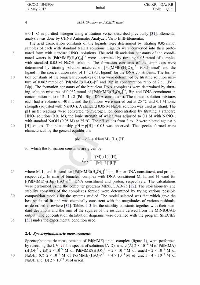
Spectrophotometric measurements of Pd(MME)-uracil complex (figure 1), were performedby recording the UV–visible spectra of solutions (A-D), where (A) 2 × 10⁻⁴ M of Pd(MMA)(H2O)2
2+; (B) 2 × 10⁻⁴ M of Pd(MME)(H2O)22+ + 2 × 10⁻⁴ M of uracil + 2 × 10⁻⁴ M of
40 NaOH, (C) 2 × 10⁻⁴ M of Pd(MME)(H2O)22+ + 4 × 10⁻⁴ M of uracil + 4 × 10⁻⁴ M of
NaOH and (D) 2 × 10⁻⁴ M of uracil.
4 M.M. Shoukry and S.M.T. Ezzat
GCOO 1043909 CE: KR QA: RB7 May 2015 Coll: QC:Initial
Deleted text:
Inserted Text
,
Deleted text:
Inserted Text
-
Deleted text:
Inserted Text
V

Spectrophotometric measurements of Pd(MME)-Bip complex were performed by record-ing the UV–visible spectra of solutions (A-D), where (A) 2 × 10⁻⁴ M of Pd(MME)(H2O)2
2+; (B) 2 × 10⁻⁴ M of Pd(MME)(H2O)22+ + 1 × 10⁻⁴ M of Bip + 2 × 10⁻⁴ M of
5NaOH, (C) 2 × 10⁻⁴ M of Pd(MME)(H2O)22+ + 1 × 10⁻⁴ M of Bip + 4 × 10⁻⁴ M of NaOH
and (D) 1 × 10⁻⁴ M of Bip. The spectral measurements of the binuclear complex of uracil,taken as an example for DNA, were performed by recording the spectra of solutions (E–H)(E) 2 × 10⁻⁴ M of [Pd(MME)(H2O)2]
2+, 1 × 10⁻⁴ M of Bip and 2 × 10⁻⁴ M of NaOH; (F)2 × 10⁻⁴ M of [Pd(MME)(H2O)2]
2+, 1 × 10⁻⁴ M of Bip, 1 × 10⁻⁴ M of uracil and103 × 10−4 M of NaOH; (G) 2 × 10⁻⁴ M of [Pd(MMA)(H2O)2]
2+, 1 × 10⁻⁴ M of Bip,2 × 10−4 M of uracil and 4 × 10⁻⁴ M of NaOH; and (H) 2 × 10⁻⁴ M of uracil. Under theseprevailing experimental conditions and after neutralization of the hydrogen ions released,associated with complex formation, it is supposed that the complexes have been completely
Table 1. Formation constants for complexes of [Pd(MME)(H2O)2]2+ with DNA constituents at 25 °C and
0.1 M ionic strength.
System M L Ha log βb Pd(MME) pKac
Pd(MME)-OH 1 0 −1 −5.29 (0.01) 5.291 0 −2 −12.96 (0.05) 7.672 0 −1 −2.46 (0.07) 8.43
Inosine 0 1 1 8.43 (0.01)1 1 0 7.98 (0.02) 4.011 2 0 12.63 (0.03)1 1 1 11.99 (0.03)
Inosine-5′-monophosphate0 1 1 8.95 (0.02) 8.950 1 2 15.27 (0.03) 6.320 1 3 17.10 (0.01)1 1 0 9.34 (0.01)1 2 0 13.44 (0.01)1 1 1 16.12 (0.01) 6.781 1 2 20.16 (0.02) 4.04
Uracil0 1 1 9.28 (0.01) 9.281 1 0 8.74 (0.01)1 2 0 15.32 (0.02)
Uridine0 1 1 9.01 (0.01) 9.011 1 0 8.55 (0.02)1 2 0 14.61 (0.03)
Thymidine0 1 1 9.55 (0.02) 9.551 1 0 9.04 (0.006)1 2 0 15.62 (0.01)
Thymine0 1 1 9.58 (0.01) 9.581 1 0 9.12 (0.004)1 2 0 15.78 (0.008)
Adenosine-5′-monophosphate 0 1 1 6.39 (0.02) 6.39
0 1 2 11.01 (0.03) 4.621 1 0 9.11 (0.06)1 1 1 13.21 (0.1) 4.11 1 2 15.72 (0.1) 2.51
aM, L, and H are the stoichiometric coefficients corresponding to Pd(MME), DNA units, and H+, respectively.blog β of Pd(MME)-DNA units. Standard deviations are given in parentheses; sum of square of residuals are less than5e−7.cThe pKa of the protonated species (log β111 − log β110).
Mono- and binuclear Pd(II) complexes 5
GCOO 1043909 CE: KR QA: RB7 May 2015 Coll: QC:Initial
Deleted text:
Inserted Text
-
Deleted text:
Inserted Text
V
formed. In each mixture the volume was brought to 10 mL by addition of deionized water5 and ionic strength is kept constant at 0.1 M NaNO3.
3. Results and discussion
3.1. Characterization of the solid complexes
The analytical data indicates that the complex is of 1 : 1 stoichiometry, Pd(MME)Cl2. TheIR spectrum of the Pd(MME)Cl2 complex exhibits bands at 3300–3400 cm−1, attributed to
10 stretching vibrations of NH2. The complex exhibits bands for (NH2) bending at 1465 and1562 cm−1 and bands for the stretching vibration corresponding to Pd–N at 480 and523 cm−1. The 1H NMR spectrum of the complex is in accordance with previously reporteddata [34].
3.2. Acid–base equilibria of the ligands
15 The acid dissociation constants of the ligands were determined in a solution of constantionic strength of 0.1 M (NaNO3) at 25 °C. The results obtained are in good agreement withliterature data [35].
3.3. Hydrolysis of [Pd(MME)(H2O)2]2+
[Pd(MME)(H2O)2]2+ may undergo hydrolysis. Its acid–base chemistry was characterized by
20 fitting the potentiometric data to various acid–base models. The best-fit model wasconsistent with the formation of three species: 10–1, 10–2, and 20–1, as given in reactions1–3. Trials were made to fit the potentiometric data assuming formation of the monohy-droxo-bridged dimer, 20–2, but this resulted in a very poor fit to the data. The dimericspecies 20–2 were detected by Nagy et al. [36] for a similar system. The formation of the
0
0.5
1
1.5
450350250
Wavenumber (nm)
Abs
orba
nce B
D
A
C
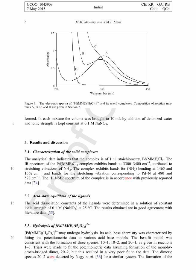
Figure 1. The electronic spectra of [Pd(MME)(H2O)2]2+ and its uracil complexes. Composition of solution mix-
tures A, B, C, and D are given in Section 2.
6 M.M. Shoukry and S.M.T. Ezzat
GCOO 1043909 CE: KR QA: RB7 May 2015 Coll: QC:Initial
Deleted text:
Inserted Text
as

5dihydroxo-bridged dimer (20–2), found for most Pd-diimine complexes, is not favored inthe case of the Pd-MME complex. This may be accounted for on the basis that the stronglabilization effect of the S-donor will cause the dimeric form (20–2) to be strained andconsequently energetically not favored [23].
½PdðMMEÞðH2OÞ2�2þ100
�pKa1 ½PdðMMEÞðH2OÞðOHÞ� þ Hþ
10�1(1)
½PdðMMEÞðH2OÞðOHÞ�þ10�1
�pKa2 ½PdðMMEÞðOHÞ2� þ Hþ
10�2(2)
10
½PdðMMEÞðH2OÞ2�2þ100
þ ½PdðMMEÞðH2OÞðOHÞ�þ10�1
�logKdimer
½ðH2OÞPdðMMEÞðOHÞPdðMMEÞðH2OÞ�3þ20�1
þH2O(3)
The pKa1 and pKa2 values for [Pd(MME)(H2O)2]2+ are 5.29 and 7.67, respectively. The
equilibrium constant for the dimerization reaction three can be calculated by equation 4 as152.83.
log Kdimer ¼ log b20�1 � log b10�1 (4)
3.4. Complexes of DNA constituents
Inosine and nucleotides such as inosine-5΄-monophosphate and adenosine-5′-monophos-20phate form protonated complexes, in addition to the formation of 1 : 1 and 1 : 2 complexes.
Inosine forms a monoprotonated complex (111). The pKa value of the protonated inosinecomplex is 4.01. This value corresponds to N1H. The lowering of this value with respect tothat of free inosine (pKa = 8.43) is due to acidification upon complex formation [37]. Ino-sine-5′-monophosphate forms mono- and diprotonated complexes. The pKa values of the
25protonated species of the IMP complex (112) are 4.04 (log β112 − log β111) and 6.78 (logβ111 – log β110).
The former pKa value corresponds to N1H and the second pKa value to –PO2(OH). TheN1H group was acidified upon complex formation by 4.91 pKa units. Acidification of theN1H group upon complex formation is consistent with previous reports for IMP complex
30[37, 38]. The phosphate group was not acidified upon complex formation since it is farfrom the coordination center. IMP complex is more stable than that of inosine, explained onthe basis of different Coulombic forces operating between the ions resulting from the nega-tively charged phosphate. Hydrogen bonding between phosphate and exocyclic amine isalso thought to contribute to the increased stability. Such hydrogen bonding was reported
35previously for similar system [39, 40].The pyrimidines uracil, uridine, thymine, and thymidine have basic nitrogen donors (N3)
in the measurable pH range [41] and as a consequence form 1 : 1 and 1 : 2 complexes withPd(MMA)2+ species. As a result of the high pKa values of pyrimidines (pKa > 9), complexformation predominates above pH 8.5. The thymine complex is more stable than that of
Mono- and binuclear Pd(II) complexes 7
GCOO 1043909 CE: KR QA: RB7 May 2015 Coll: QC:Initial
Deleted text:
Inserted Text
3

5 uracil, probably due to the higher basicity of the N3 site of thymine resulting from theinductive effect of the extra electron-donating methyl group.
The spectra given in figure 1 shows that the band at 353 nm corresponding to [Pd(MME)(H2O)2]
2+ (A) undergoes a blue shift to a band at 345 nm for [Pd(MME)(uracil-H)]+,species 110 (B). This band is further shifted to a shoulder at 336 nm for [Pd(MME)(uracil-
10 H)2], species 120 (C). The band appears as shoulders due to the large absorption ofuracil (D).
2
4
6
8
10
12
3210
pH - Pd(MME)-Bipiperidine o Calculated
Volume of NaOH added,ml
Figure 2. Potentiometric titration curves of [Pd(MME)(H2O)2]2+-bipiperidine system.
OH 2
Pd
Pd
NH NH 2
OH 2
Pd NH NH
OH 2
Pd Pd
OH 2
NH NH
(MME)
(MME)(H 2O) 22
(111)
+
(110)
(210)
+2
+2
+2 +2
(MME)
(MME) (MME)
Scheme 2. Complex formation equilibria of Pd(MME)-Bip complexes.
8 M.M. Shoukry and S.M.T. Ezzat
GCOO 1043909 CE: KR QA: RB7 May 2015 Coll: QC:Initial

3.5. Complex formation equilibria of binuclear Pd(MME)2+ complex involving 4,4′-bipiperidine and some selected DNA constituents
The titration curve of mixture of Pd(MME)(H2O)2]2+ and 4,4΄-bipiperidine in ratio (2 : 1)
5(figure 2), shows a sharp inflection at a = 1 (a is number of mole of base added per mole of4,4΄-bipiperidine), corresponding to complete formation of [(H2O)(MME)Pd(Bip)Pd(MME)(H2O)]
4+ with formation constant, log β210 = 20.04 (scheme 2, table 2).Beyond a = 1, the binuclear complex is subjected to hydrolysis. In this region, the titra-
tion data are fitted considering the formation of the hydrolyzed species with stoichiometric10coefficients 10–1 and 10–2 as given in scheme 3.
The speciation diagram of Pd(MME)-bipiperidine system is given in figure 3. The binu-clear complex, [(H2O)(MME)Pd(Bip)Pd(MME)(H2O)]
4+ (210), starts to form at low pHand on increasing pH, its concentration increases to the predominant species up to pH 7.2and reaches a maximum concentration of 87.5% at pH 5.2.
Table 2. Formation constants for mixed ligand complexes of Pd(MME)(H2O)2]2+ with
bipiperidine at 25 °C and 0.1 M ionic strength.
System M L Ha log βb pKac
Bipiperidine 0 1 1 10.96 (0.01) 10.960 1 2 21.12 (0.02) 10.161 1 0 13.33 (0.08)1 1 1 19.98 (0.03) 6.652 1 0 20.06 (0.10)
aM, L, and H are the stoichiometric coefficients corresponding to Pd(MME), bipiperidine and H+,respectively.blog β of Pd(MME)-Bip. Standard deviations are given in parentheses; sum of square of residuals areless than 5e−7.cThe pKa of the ligand or the protonated complex.
OH 2
Pd Pd
OH 2
NH NH
OH
Pd Pd
OH 2
NH NH
OH
Pd Pd
OH
NH NH
(MME) (MME)
-H +
(MME) (MME)
-H +
(MME) (MME)
(100)
(10-1)
(10-2)
+ +
+ +2
+2+2
Scheme 3. Acid–base equilibria of [(H2O)(MME)Pd(Bip)Pd(MME)(H2O)]4+.
Mono- and binuclear Pd(II) complexes 9
GCOO 1043909 CE: KR QA: RB7 May 2015 Coll: QC:Initial
Deleted text:
Inserted Text
a i
Deleted text:
Inserted Text
s

5 The complex formation between [(H2O)(MME)Pd(Bip)Pd(MME)(H2O)]4+ and inosine,
taken as an example of a DNA constituent, showed the formation of 1 : 1 and 1 : 2complexes, as given in scheme 4. The stability constant of the DNA complexes is thymi-ne > inosine > uracil (table 3).
The speciation diagram of [(H2O)Pd(MME)(Bip)Pd(MME)(H2O)]4+-inosine complex is
10 given in figure 4. The 1 : 1 complex starts to form at pH 2 and on increasing pH, its con-centration increases, reaching a relative amount of 94% at pH 5.6. The 1 : 2 complex attainsa maximum formation degree of 89% at pH 10.1. The hydrolyzed species are formed afterpH 10.0. From the biological point of view, the DNA complex predominates in thephysiological pH range and the reaction of the binuclear complex with DNA is quite
15 feasible.
100
10-1
10-2
20-2
110
111
210
0
20
40
60
80
100
4 6 8 10 12
pH
% S
peci
es
Figure 3. Concentration distribution of various species as a function of pH in the [Pd(MME)(H2O)2]2+-bip-
iperidine system.
OH2
Pd Pd
OH 2
NH NH
OH2
Pd PdNH NH
Pd PdNH NH
(MME) (MME)
(100)
Ino
(MME) (MME)
(110)
Ino
Ino
(MME) (MME)
(120)
InoIno
+2 +2
+ +
++
Scheme 4. Complex formation equilibria of [(H2O)(MME)-Pd-(Bip)-Pd(MME)(H2O)]4+-inosine complex.
10 M.M. Shoukry and S.M.T. Ezzat
GCOO 1043909 CE: KR QA: RB7 May 2015 Coll: QC:Initial
Deleted text:
Inserted Text
s

Spectral bands of Pd(MME)(H2O)22+ and its 4,4′-bipiperidine complex are quite different
in the position of the maximum wavelength and molar absorptivity. The spectrum of[Pd(MME)(H2O)2]
2+ (mixture A) shows an absorption maximum at 353 nm. The spectrumobtained for [(H2O)(MME)Pd(Bip)Pd(MME)(H2O)]
4+ (mixture B), exhibits a band at5339 nm, which further shifts to 315 nm by addition of extra 2 × 10⁻⁴ M of NaOH for
formation of [(OH)(MME)Pd(Bip)Pd(MME)(OH)]2+ (mixture C). There is no UV absorp-tion for free Bip in this region (mixture D).
Spectra of Pd(MME)(H2O)22+ complexes with 4,4΄-bipiperidine and uracil are scanned.
The spectrum obtained for [(H2O)(MME)Pd(Bip)Pd(MME)(H2O)]4+ (mixture E) shows a
Table 3. Formation constants for binuclear complexes of [(H2O)(MME)Pd(Bip)Pd(MME)(H2O)]4+ and someDNA constituents at 25 °C and 0.1 M ionic strength.
System M L Ha log βb pKac
[(H2O)(MME)Pd(Bip)Pd(MME)(H2O)]4+
1 0 −1 −8.04 (0.07) 8.041 0 −2 −16.87 (0.05) 8.83
Inosine0 1 1 8.80 (0.02) 8.801 1 0 7.89 (0.01)1 2 0 12.84 (0.02)
Uracil0 1 1 9.18 (0.01) 9.181 1 0 6.62 (0.09)1 2 0 11.95 (0.1)
Thymine0 1 1 9.65 (0.01) 9.651 1 0 8.66 (0.02)1 2 0 15.59 (0.03)
aM, L, and H are the stoichiometric coefficients corresponding to (MME)Pd(Bip)Pd(MME), DNA units, and H+, respectively.blog β of (MME)Pd(Bip)Pd(MME)-DNA constituent complex. Standard deviations are given in parentheses; sum of square ofresiduals are less than 5e−7.cThe pKa of the ligand or the protonated complex.
100
10-2
120110
0
20
40
60
80
100
2 4 6 8 10 12
pH
% S
peci
es
Figure 4. Concentration distribution of various species as a function of pH in the Pd(MME)-bipiperidine-inosinesystem.
Mono- and binuclear Pd(II) complexes 11
GCOO 1043909 CE: KR QA: RB7 May 2015 Coll: QC:Initial

5 band at 339 nm. The spectrum obtained for [(H2O)(MME)Pd(Bip)Pd(MME)(uracil)]3+
(mixture F) and [(uracil)(MME)Pd(Bip)Pd(MME)(uracil)]2+ (mixture G) exhibit shouldersat 332 nm and 315 nm, respectively. The spectral band shifts are taken as evidence for binu-clear complex formation, supporting the potentiometric results. Further investigation on thebinuclear complex formation may need further studies as mono- and polynuclear NMR
10 measurements.
4. Conclusion
The present investigation describes complex formation equilibria of Pd(MME)(H2O)22+
with some selected DNA constituents and 4,4′-bipiperidine. The results indicate formationof binuclear complex and the reaction with DNA constituents is feasible. The data support
15 the biological significance of the di- and trinuclear platinum(II) complexes having potentantitumor activity [42]. In the present study, [Pd(MME)(H2O)2]
2+ does not form the dihy-droxo-bridged dimer (20–2) as reported for most Pd-diimine complexes. This may beexplained on the basis that strong labilization of the S-donor will cause the dimeric form(20–2) to be strained and consequently energetically not favored. It is interesting to com-
20 pare the results of the present study with those of Pd(II) complexes involving N,N-donors.The stability constant of Pd(MME)-DNA complex is higher than those of Pd(II) complexeswith N,N-dimethylethylenediamine [43], This same effect arises in the binuclear complexes.This may be due to the hydrogen bonding between the bound DNA constituents and theester group of the bound MME.
25 Disclosure statement
No potential conflict of interest was reported by the authors.
References
[1] B. Rosenberg, L. Vancamp, J.E. Trosko, V.H. Mansour. Nature, 222, 385 (1969).[2] E. Wong, C.M. Giandomenico. Chem. Rev., 99, 2451 (1999).
30 [3] L.A.S. Costa, W.R. Rocha, W.R. De Almeida, H.F. Dos Santos. Chem. Phys. Lett., 387, 182 (2004).[4] N. Farrell. Comments Inorg. Chem., 16, 373 (1995).[5] V. Brabec, J. Kašpárková, O. Vrána, O. Nováková, J.W. Cox, Y. Qu, N. Farrell. Biochemistry, 38, 6781
(1999).[6] Y. Qu, N. Farrell. J. Am. Chem. Soc., 113, 4851 (1991).
35 [7] N. Farrell, Y. Qu. Inorg. Chem., 28, 3416 (1989).[8] N. Farrell, Y. Qu, L. Feng, B. Van Houten. Biochemistry, 29, 9522 (1990).[9] L.R. Kelland. Nat. Rev. Cancer, 7, 573 (2007).[10] G. Bogdanović, V. Kojić, T. Srdić, D. Jakimov, M.I. Djuran, Z.D. Bugarčić, M. Baltić, V.V. Baltić.
Met.-Based Drugs, 9, 33 (2002).40 [11] A. Baldo, G. Chessa, G. Marangoni, B. Pitteri. Polyhedron, 4, 1429 (1985).
[12] R.C. Jones, R.L. Madden, B.W. Skelton, V.-A. Tolhurst, A.H. White, A.M. Williams, A.J. Wilson, B.F. Yates.Eur. J. Inorg. Chem., 1048 (2005).
[13] R.C. Jones, B.W. Skelton, V.-A. Tolhurst, A.H. White, A.J. Wilson, A.J. Canty. Polyhedron, 26, 708 (2007).[14] J.G.H. Du Preez, Platinum(II) Complexes, Preparation and Use, 2005, document number: CA Patent
45 2547275, p. 38.[15] N. Summa, W. Schiessl, R. Puchta, N. van Eikema Hommes, R. van Eldik. Inorg. Chem., 45, 2948 (2006).
AQ3
AQ4
AQ5
12 M.M. Shoukry and S.M.T. Ezzat
GCOO 1043909 CE: KR QA: RB7 May 2015 Coll: QC:Initial
Authorquery:
Inserted Text
The disclosure statement has been inserted. Please correct if this is inaccurate.
Authorquery:
Inserted Text
The CrossRef database (www.crossref.org/) has been used to validate the references. Mismatches between the original manuscript and CrossRef are tracked in red font. Please provide a revision if the change is incorrect. Do not comment on correct changes.
Authorquery:
Inserted Text
Please provide missing volume for Refs. [12, 21–23].
Deleted text:
Inserted Text
van Camp
Deleted text:
Inserted Text
Kasparkova
Deleted text:
Inserted Text
Vrana
Deleted text:
Inserted Text
Novakova
Deleted text:
Inserted Text
Burgarčić
Deleted text:
Inserted Text
van Eikema
[16] M.E. Oehlsen, Y. Qu, N. Farrell. Inorg. Chem., 42, 5498 (2003).[17] M.E. Oehlsen, A. Hegmans, Y. Qu, N. Farrell. J. Biol. Inorg. Chem., 10, 433 (2005).[18] N.P. Farrell, S.G. De Almeida, K.A. Skov. J. Am. Chem. Soc., 110, 5018 (1988).
5[19] N. Farrell, Y. Qu, L. Feng, B. Van Houten. Biochemistry, 29, 9522 (1990).[20] Q. Liu, Y. Qu, R. Van Antwerpen, N. Farrell. Biochemistry, 45, 4248 (2006).[21] M.R. Shehata, M.M. Shoukry, R. Van Eldik. Eur. J. Inorg. Chem., 3912 (2009).[22] T. Soldatovic, M.M. Shoukry, R. Puchta, Z.D. Bugarcic, R. Van Eldik. Eur. J. Inorg. Chem., 2261 (2009).[23] M.R. Shehata, M.M. Shoukry, F.M. Nasr, R. van Eldik. Dalton Trans., 779 (2008).
10[24] M.M. Shoukry, R. Van Eldik. J. Chem. Soc., Dalton Trans., 2673 (1996).[25] M.M.A. Mohamed, M.M. Shoukry. Polyhedron, 20, 343 (2001).[26] A.A. El-Sherif, M.M. Shoukry, R. Van Eldik. J. Chem. Soc., Dalton Trans., 1425 (2003).[27] T. Rau, M.M. Shoukry, R. van Eldik. Inorg. Chem., 36, 1454 (1997).[28] M.M.A. Mohamed, M.M. Shoukry. J. Sol. Chem., 40, 2023 (2011).
15[29] M.M.A. Mohamed, M.M. Shoukry. J. Coord. Chem., 64, 2667 (2011).[30] R.G. Bates. Determination of pH: Theory and Practice, 2nd Edn, Wiley, New York (1975).[31] M.M. Shoukry, W.M. Hosny, M.M. Khalil. Transition Met. Chem., 20, 252 (1995).[32] P. Gans, A. Sabatini, A. Vacca. Inorg. Chim. Acta, 18, 237 (1976).[33] L. Pettit. Personal Communication, University of Leeds (1993).
20[34] M. Calaf, A. Caubet, V. Moreno, M. Font-Bardia, X. Solans. J. Inorg. Biochem., 59, 63 (1995).[35] D.D. Perrin. Stability Constants of Metal Ion Complexes: Part B, Organic Ligands, Pergamon Press, Oxford,
(1979).[36] Z. Nagy, I. Sovago. J. Chem. Soc., Dalton Trans., 2467 (2001).[37] H. Sigel, S.S. Massoud, N.A. Corfu. J. Am. Chem. Soc., 116, 2958 (1994).
25[38] B.P. Operschall, E.M. Bianchi, R. Griesser, H. Sigel. J. Coord. Chem., 62, 23 (2009).[39] D. Kiser, F.P. Intini, Y. Xu, G. Natile, L.G. Marzilli. Inorg. Chem., 33, 4149 (1994).[40] S.O. Ano, F.P. Intini, G. Natile, L.G. Marzilli. J. Am. Chem. Soc., 119, 8570 (1977).[41] S. Ganguly, K.K. Kundu. Can. J. Chem., 72, 1120 (1994).[42] N. Farrell. Met. Ions Biol. Syst., 42, 251 (2004).
30[43] M.R. Shehata, M.M. Shoukry, S. Ali. J. Coord. Chem., 65, 1311 (2012).
Mono- and binuclear Pd(II) complexes 13
GCOO 1043909 CE: KR QA: RB7 May 2015 Coll: QC:Initial
Deleted text:
Inserted Text
Almeida
Deleted text:
Inserted Text
Sabarini
Deleted text:
Inserted Text
Bardia


![Mono and binuclear complexes involving [Pd(N,N-dimethylethylenediamine)(H2O)2], 4,4′-bipiperidine and DNA constituents](https://static.fdokumen.com/doc/165x107/631fd774d85b325bc2095ade/mono-and-binuclear-complexes-involving-pdnn-dimethylethylenediamineh2o2.jpg)