A chromatin binding site in the tail domain of nuclear lamins that interacts with core histones
12
A Chromatin Binding Site in the Tail Domain of Nuclear Lamins That Interacts with Core Histones Hideo Taniura, Charles Glass, and Larry Gerace Departments of Cell and Molecular Biology, The Scripps Research Institute, La Jolla, California 92037 Abstract. Interaction of chromatin with the nuclear en- velope and lamina is thought to help determine higher order chromosome organization in the interphase nu- cleus. Previous studies have shown that nuclear lamins bind chromatin directly. Here we have localized a chro- matin binding site to the carboxyl-terminal tail domains of both A- and B-type mammalian lamins, and have characterized the biochemical properties of this binding in detail. Recombinant glutathione-S-transferase fusion proteins containing the tail domains of mammalian lamins C, BI, and B 2 were analyzed for their ability to associate with rat liver chromatin fragments immobi- lized on microtiter plate wells. We found that all three lamin tails specifically bind to chromatin with apparent Kds of 120-300 nM. By examining a series of deletion mutants, we have mapped the chromatin binding region of the lamin C tail to amino acids 396--430, a segment immediately adjacent to the rod domain. Furthermore, by analysis of chromatin subfractions, we found that core histones constitute the principal chromatin bind- ing component for the lamin C tail. Through coopera- tivity, this lamin-histone interaction could be involved in specifying the high avidity attachment of chromatin to the nuclear envelope in vivo. T HE nuclear lamina is a filamentous protein mesh- work that lines the nucleoplasmic surface of the nu- clear envelope (NE) 1 (reviewed by Gerace and Burke, 1988; McKeon, 1991; Nigg, 1992; Georgatos et al., 1994). The lamina is thought to provide a structural frame- work for the NE and an anchoring site at the nuclear pe- riphery for interphase chromosomes, and therefore could play a major role in interphase nuclear organization. The lamina consists of a polymeric assembly of nuclear lamins, members of the intermediate filament (IF) protein super- family (see McKeon, 1991; Nigg, 1992), as well as a num- ber of less abundant lamina-associated polypeptides (dis- cussed by Gerace and Foisner, 1994). Vertebrate lamins are classified as A- or B-subtypes based on their sequence and biochemical properties. B-type lamins (lamins B1 and B2) are present in somatic cells throughout development, while A-type lamins (lamins A and C) are expressed only during or after terminal differentiation in most cells. Mam- Address correspondence to Larry Gerace, Depts. of Cell and Molecular Biology, The Scripps Research Institute, 10666 North Torrey Pines Rd., La Jolla, CA 92037. Tel.: (619) 554-8514. Fax: (619) 554-6253. Dr. Taniura's present address is Division of Regulation of Macromo- lecular Function, Institute for Protein Research, Osaka University, Osaka, Japan 565. Dr. Glass' present address is R. W. Johnson, PRI, Dept. of Biology, The Scripps Research Institute, La Jolla, CA 92037. 1. Abbreviations used in this paper. GST, glutathione-S-transferase; IF, in- termediate filament; NE, nuclear envelope; NLS, nuclear localization se- quence. malian lamins A and C appear to be alternative splice products of the lamin A gene. They are identical for the first 566 amino acids, and contain unique carboxyl termini of 98 residues and 6 residues, respectively. By contrast, the two mammalian B-type lamins of somatic cells are the products of separate genes (Hoger et al., 1990). Additional alternative splice variants of the lamin A and lamin B 2 genes have been characterized in mammalian germ cells (Furukawa and Hotta, 1993; Furukawa et al., 1994). Like other IF proteins, lamins consist of a central a-helP cal rod domain flanked by an NHE-terminal "head" do- main and a COOH-terminal "tail" domain (see McKeon, 1991; Nigg, 1992; Heins and Aebi, 1994). The head domain of vertebrate lamins is usually ~30 amino acids, while the tail domain is typically ~180-275 residues. The basic lamin protomer is a homotypic dimer formed by parallel, unstag- gered association of the rod domain to form a two- stranded coiled-coil a-helix. This subunit, which forms a rod-like structure with a length of ~50 nm, engages in both head-to-tail and lateral interactions to form lamin fil- aments and related structures with an axial periodicity of ~25 nm (Aebi et al., 1986; Gieffers and Krohne, 1991; Heitlinger et al., 1991; Moir et al., 1991). Thus, discrete re- gions of the lamin molecules (e.g., tail domains) are dis- played at repeating intervals along lamin filaments. In vitro assembly studies with mutant lamins have shown that both the rod domain and head and tail domains are in- volved in filament formation (Giefers and Krohne, 1991; Moir et al., 1991; Heitlinger et al., 1992). The major functions of the lamina are likely to involve © The Rockefeller University Press, 0021-9525/95/I0/33/12 $2.00 The Journal of Cell Biology, Volume 131, Number 1, October 1995 33M4 33 on October 26, 2014 jcb.rupress.org Downloaded from Published October 1, 1995
Transcript of A chromatin binding site in the tail domain of nuclear lamins that interacts with core histones

A Chromatin Binding Site in the Tail Domain of Nuclear Lamins That Interacts with Core Histones Hideo Taniura, Charles Glass, and L a r r y Gerace
Departments of Cell and Molecular Biology, The Scripps Research Institute, La Jolla, California 92037
Abstract. Interaction of chromatin with the nuclear en- velope and lamina is thought to help determine higher order chromosome organization in the interphase nu- cleus. Previous studies have shown that nuclear lamins bind chromatin directly. Here we have localized a chro- matin binding site to the carboxyl-terminal tail domains of both A- and B-type mammalian lamins, and have characterized the biochemical properties of this binding in detail. Recombinant glutathione-S-transferase fusion proteins containing the tail domains of mammalian lamins C, BI, and B 2 were analyzed for their ability to associate with rat liver chromatin fragments immobi-
lized on microtiter plate wells. We found that all three lamin tails specifically bind to chromatin with apparent Kds of 120-300 nM. By examining a series of deletion mutants, we have mapped the chromatin binding region of the lamin C tail to amino acids 396--430, a segment immediately adjacent to the rod domain. Furthermore, by analysis of chromatin subfractions, we found that core histones constitute the principal chromatin bind- ing component for the lamin C tail. Through coopera- tivity, this lamin-histone interaction could be involved in specifying the high avidity attachment of chromatin to the nuclear envelope in vivo.
T HE nuclear lamina is a filamentous protein mesh- work that lines the nucleoplasmic surface of the nu- clear envelope (NE) 1 (reviewed by Gerace and
Burke, 1988; McKeon, 1991; Nigg, 1992; Georgatos et al., 1994). The lamina is thought to provide a structural frame- work for the NE and an anchoring site at the nuclear pe- riphery for interphase chromosomes, and therefore could play a major role in interphase nuclear organization. The lamina consists of a polymeric assembly of nuclear lamins, members of the intermediate filament (IF) protein super- family (see McKeon, 1991; Nigg, 1992), as well as a num- ber of less abundant lamina-associated polypeptides (dis- cussed by Gerace and Foisner, 1994). Vertebrate lamins are classified as A- or B-subtypes based on their sequence and biochemical properties. B-type lamins (lamins B1 and B2) are present in somatic cells throughout development, while A-type lamins (lamins A and C) are expressed only during or after terminal differentiation in most cells. Mam-
Address correspondence to Larry Gerace, Depts. of Cell and Molecular Biology, The Scripps Research Institute, 10666 North Torrey Pines Rd., La Jolla, CA 92037. Tel.: (619) 554-8514. Fax: (619) 554-6253.
Dr. Taniura's present address is Division of Regulation of Macromo- lecular Function, Institute for Protein Research, Osaka University, Osaka, Japan 565.
Dr. Glass' present address is R. W. Johnson, PRI, Dept. of Biology, The Scripps Research Institute, La Jolla, CA 92037.
1. Abbreviat ions used in this paper. GST, glutathione-S-transferase; IF, in- termediate filament; NE, nuclear envelope; NLS, nuclear localization se- quence.
malian lamins A and C appear to be alternative splice products of the lamin A gene. They are identical for the first 566 amino acids, and contain unique carboxyl termini of 98 residues and 6 residues, respectively. By contrast, the two mammalian B-type lamins of somatic cells are the products of separate genes (Hoger et al., 1990). Additional alternative splice variants of the lamin A and lamin B 2
genes have been characterized in mammalian germ cells (Furukawa and Hotta, 1993; Furukawa et al., 1994).
Like other IF proteins, lamins consist of a central a-helP cal rod domain flanked by an NHE-terminal "head" do- main and a COOH-terminal "tail" domain (see McKeon, 1991; Nigg, 1992; Heins and Aebi, 1994). The head domain of vertebrate lamins is usually ~30 amino acids, while the tail domain is typically ~180-275 residues. The basic lamin protomer is a homotypic dimer formed by parallel, unstag- gered association of the rod domain to form a two- stranded coiled-coil a-helix. This subunit, which forms a rod-like structure with a length of ~50 nm, engages in both head-to-tail and lateral interactions to form lamin fil- aments and related structures with an axial periodicity of ~25 nm (Aebi et al., 1986; Gieffers and Krohne, 1991; Heitlinger et al., 1991; Moir et al., 1991). Thus, discrete re- gions of the lamin molecules (e.g., tail domains) are dis- played at repeating intervals along lamin filaments. In vitro assembly studies with mutant lamins have shown that both the rod domain and head and tail domains are in- volved in filament formation (Giefers and Krohne, 1991; Moir et al., 1991; Heitlinger et al., 1992).
The major functions of the lamina are likely to involve
© The Rockefeller University Press, 0021-9525/95/I0/33/12 $2.00 The Journal of Cell Biology, Volume 131, Number 1, October 1995 33M4 33
on October 26, 2014
jcb.rupress.orgD
ownloaded from
Published October 1, 1995
interactions with the inner nuclear membrane and chro- matin, and these associations are beginning to be charac- terized in detail. Attachment of the lamina to the inner membrane is thought to involve lamin-binding integral membrane proteins (reviewed by Gerace and Foisner, 1994), and COOH-terminal isoprenylation of certain lamins (reviewed by McKeon, 1991; Nigg, 1992). A close association of the lamina with chromatin is suggested by structural studies (see Belmont et al., 1993 and references therein), and may preferentially involve discrete chromo- somal regions (Hochstrasser and Sedat, 1987). Since re- cent in vitro binding studies have demonstrated specific in- teractions of lamins with various chromatin substrates (Burke, 1990; Glass and Gerace, 1990; Hoger et al., 1991; Yuan et al., 1991; Glass et al., 1993), the lamina-chromatin interaction is likely to involve lamins themselves. In addi- tion, chromatin-binding integral membrane proteins of the lamina such as LAP2 (Foisner and Gerace, 1993; Fu- rukawa etal., 1995) may participate in this association.
We previously found that the e~-helical rod domain of lamins A/C contains a specific chromatin binding site, as measured by in vitro association of the recombinant rod domain with mitotic chromosome surfaces (Glass et al., 1993). In this study, we have demonstrated that a second chromatin binding region occurs in the tail domains of both A- and B-type tamins. Using solid phase binding to chromatin isolated from rat liver nuclei, we found that the tail domains of lamins C, B1, and B2 all bind specifically to isolated chromatin fragments, with apparent KdS of 120- 300 nM. Furthermore, we mapped the chromatin binding region of the lamin C tail to an ~30-amino acid region flanking the COOH-terminal end of the rod domain, and showed that core histones are the major chromatin com- ponent responsible for the binding of this region. These re- sults are discussed in relationship to the role of the lamina in higher order chromatin structure and the attachment of specific chromosomal regions to the NE in vivo.
Materials and Methods
Expression of Fusion Proteins and Recombinant Lamin Fragments in Escherichia coli cDNA clones for human lamins A and C were provided by the laboratory of Dr. G. Blobel (Rockefeller University, NY), and cDNA clones for mouse lamins B1 and B2 were obtained from Dr. G. Krohne (University of Wtirtzburg, Germany). Lamin fragment sequences were amplified by PCR, using synthetic oligonucleotide primers flanked by sequences for re- striction enzyme sites for subcloning into the expression plasmid pGEX2T (Pharmacia LKB Biotechnology, Piscataway, N J).
Polypeptides were expressed in the E. coil strain BL21 (Novageu, Inc., Madison, WI). Single colonies from transformed bacteria were grown to an A~00 of 0.5-0.7 at which point expression was induced by addition of 2 mM IPTG. Bacterial growth was continued another 2 h. Purification of GST fusion proteins was performed as described by Smith and Corcoran (1990). Bacterial cell pellets from 500-ml cultures were resuspended in 25 ml PBS (10 mM sodium phosphate, pH 7.4, 140 mM NaC1) containing 0.2 mM PMSF. Resuspended cells were sonicated with a Branson Sonifier for five 30-s periods on ice. Triton X-100 was added to 1%, and the solution was spun at 12,000 g for 10 min. The supernatant was incubated with 1.25 ml glutathione-agarose beads for 30 min at room temperature. Beads were then washed three times with PBS and fusion proteins were eluted by three sequential incubations with 5 ml of 50 mM Tris, pH 8.0, 200 mM NaCI, 5 mM reduced glutathione. Purified fusion proteins were stored at -20°C.
Isolation and Fractionation of Chromatin Chromatin fragments were isolated from rat liver nuclei as described by Noll et al. (1975) and Zentgraf and Franke (1984). Rat liver nuclei, iso- lated as described (Gerace et al., 1982), were resuspended at 0.4 Azr0 U/ml in 0.34 M sucrose, 60 mM KC1, 15 mM NaCl, 0.15 mM spermine, 0.5 mM spermidine, 15 mM Tris, pH 7.5, 15 mM 2-mercaptoethanol. The suspen- sion was brought to 1 mM CaCl~ and digested with micrococcal nuclease (15 unit/ml) for 30 s at 37°C. Digestion was terminated by chilling on ice and addition of 0.I M EDTA, pH 7.0, to a final concentration of 0.01 M. The nuclei were then centrifuged for 5 min at 4,000 g, and lysed by suspen- sion in 0.2 mM EDTA, pH 7.0. Next, 0.5-ml samples were loaded on top of a 10-50% linear sucrose gradient containing 10 mM Tris 7.4, 100 mM NaCl, 1 mM EDTA, 0.2 mM PMSF. Centrifugation was performed at 4°C in a SW41 rotor (Beckman Instrs., Inc., Fullerton, CA) for 90 min at 40,000 rpm. Peak fractions with an absorbance at 260 nm were pooled and stored at -80°C.
For fractionation of chromatin, chromatin fragments were made 0.6 M NaCl and loaded on 10-50% linear sucrose gradients in 10 mM Tris, pH 7.4, 0.6 M NaCl, 1 mM EDTA, and 0.2 mM PMSF. Fractions at the top of the gradient were pooled to yield a histone HI-enriched fraction, and frac- tions sedimenting with the major nucleic acid peak were pooled to yield a core histone/DNA fraction. For isolation of core histones, chromatin frag- ments were applied to a hydroxylapatite column in 0.1 M potassium phos- phate, pH 7.0, 0.1 M NaCL After washing with the same buffer containing 0.6 M NaCI to remove H1 and other loosely bound proteins, core histones were eluted by the same buffer containing 2 M NaC1, pooled and stored at -80°C.
Solid-phase Binding Assay Recombinant polypeptides were labeled with 125I using the lodogen re- agent (Pierce Chem. Co., Rockford, IL) according to the manufacturer's instructions. After a 10-min labeling period, polypeptides were separated from free iodide by dialysis into the binding buffer containing 0.2 mM PMSF without BSA. Typical labeling yielded specific activities of 0.5-1.0 x 10⁶ cpm/0.g.
Lamin-chromatin binding was determined by a solid-phase binding as- say originally described by Nachman and Leung (1982). Chromatin or chromatin subfractions (200 ml) at 10 mg/ml were immobilized in microti- ter wells (Immulon 4; Dynatech Labs., Inc., Chantilly, VA) by incubating overnight at 4°C in coating buffer (10 mM Tris, pH 7.5,100 mM N aC1, and 1 mM EDTA). HI-depleted chromatin and purified core histones (see above) were dialyzed from solutions containing high salt into coating buffer before adsorption to microtiter wells. Before ligand addition, the wells were blocked for 2 h with a binding buffer (20 mM Hopes, pH 7.2, 150 mM NaC1, 5 mM MgCI, and 30 mg/ml BSA), and washed once with the same buffer. Radiolabeled lamin fusion proteins were added in the ab- sence or presence of unlabeled competitors and incubated for 2 h at room temperature. After three washes with binding buffer, the radioactivity in each well was measured by gamma counting. Nonspecific background was taken as the amount of t25I-labeled ligand that remained bound per well when 2 ~g/ml radiolabeled ligand was incubated with a 50-fold excess of nonradioactive competitor. This is a suitable measure of nonspecific background, since a very similar level of background binding as defined in this manner was obtained for the 125f-labeled GST-lamin C tail and 125I-labeled GST-lamin C tail deletion mutants that were found to be defi- cient in chromosome binding (Fig. 6). Typical reactions yielded 4,000- 6,000 cprrdwetl specifically bound after incubation with 2 p.g/ml radiola- beled GST-lamin taii constructs.
To examine possible binding of lamins to isolated DNA by a solid phase assay, a modified procedure was used to attach the DNA to micro- titer wells, which involved adsorption of lightly biotinylated DNA to streptavidin-coated plates. This is because purified DNA did not signifi- cantly bind to microtiter wells under the conditions where purified pro- teins and protein-DNA complexes were efficiently adsorbed (see above). Purified DNA in water was lightly coupled with biotin by mixing with Photoprobe biotin (Vector Labs., Inc., Burlingame, CA) and illuminating for 10 min on ice as recommended by the manufacturer. Free biotin was then separated from the DNA by two extractions with isobutanol, and the DNA was precipitated with ethanol. The biotinylated DNA was then dis- solved in water and adsorbed to the surface of Reacti-Bind streptavidin coated polystyrene s~rip plates (Pierce). Approximately 200 ng DNA per well was bound. Binding of radiolabeled lamin fusion proteins was then analyzed as above.
Individual binding experiments shown in this paper were carried out at
The Journal of Cell Biology, Volume 13 l, 1995 34
on October 26, 2014
jcb.rupress.orgD
ownloaded from
Published October 1, 1995

Other Procedures
Protein concentrations were determined with a Bio-Rad protein assay kit. SDS-PAGE was performed by the method of Laemmli (1970).
Results
A
Analysis of Binding of the Lamin Tail Domain to Chromatin with a Solid Phase Assay
We developed a quantitative binding assay to investigate a possible interaction between the tail domains of nuclear lamins and chromatin. Previous studies of the lamin-chro- matin interaction have involved intact lamins and lamin fragments containing the a-helical rod domain (Burke, 1990; Glass and Gerace, 1990; Hoger et al., 1991; Yuan et al., 1991; Glass et al., 1993). Since these proteins are insol- uble under conditions of physiological salt and pH due to the strong tendency of the rod to self-associate, they are difficult to use for quantitative binding studies. Moreover, they generally require the use of buffers containing dena- turants (e.g. urea) for isolation, and their capacity for com- plete in vitro renaturation after these treatments is un-
known. By contrast, we found that the lamin tail domains can be expressed as soluble fusion proteins in E. coli and purified under nondenaturing conditions. Since the tail do- mains do not strongly self associate, it was feasible to use recombinant fusion proteins containing the lamin tail do- mains for saturation binding analysis with isolated chro- matin fragments. We immobilized the chromatin frag- ments on the surfaces of microtiter wells to analyze binding using a solid phase assay, because the strong ten- dency of chromatin to aggregate in physiological buffers (van Holde, 1989) made it difficult to carry out reproduc- ible binding assays in solution. By contrast, the solid phase assay was highly reproducible (see Materials and M e t h - ods) and permitted the rapid analysis of a large number of samples.
Interphase chromatin used for these binding studies was prepared by micrococcal nuclease digestion of isolated rat liver nuclei followed by sucrose gradient centrifugation of the nuclease-released material (Fig. 1 A). The peak gradi- ent fractions that were pooled and used for the binding studies (fractions 8-10) contained ~6--10-kb fragments of DNA, corresponding to 30-50 nucleosomes (Fig. 1 B). The overwhelmingly major proteins in these fractions were his- tone H1 and core histones (Fig. 1 C). Tail domains of lamins were expressed in E. coli as glutathione-S-trans- ferase (GST) fusion proteins, which were purified from soluble bacterial extracts using glutathione affinity col- umns. The recombinant proteins that we analyzed were
3.0"
E g 2.0 tO Od
1.0
0.0 0 5 10 15 20
fraction number
least three times, and the different repetitions yielded very similar results. Data from a single experiment is shown. Data points were taken in dupli- cates, and presented as the mean with standard deviation. To calculate ap- parent KdS for the interaction of fusion protein with chromatin, data was expressed in linearizing plots for single site competitive interactions (Hulme and Birdsall, 1992).
i
25
Figure 1. Preparation of inter- phase chromatin and recombi- nant fusion proteins containing the lamin tail domains. (A) Su- crose gradient sedimentation of chromatin fragments re- leased from rat liver nuclei by micrococcal nuclease digestion. Chromatin was sedimented on a 10-50% sucrose gradient, and DNA was measured by absor- bance at 260 nm. (B) Agarose gel electrophoresis of DNA present in different chromatin fractions. DNA from the peak region of the sucrose gradient (fractions 6-13) was analyzed by electrophoresis on a 0.3% agarose gel. (C) SDS-gel elec- trophoresis of proteins present in different chromatin fractions. Aliquots of sucrose gradient fractions 6-13 were analyzed on a 15 % SDS-polyacrylamide gel, and proteins were visual- ized by Coomassie blue stain- ing. (D) SDS-gel electrophore- sis of purified recombinant GST-lamin tail fusions and GST. About 3 ~g of each protein was analyzed on a 12% SDS-poly- acrylamide gel. Lane 1, GST- lamin C tail (389-572); lane 2, GST-lamin B1 tail (391-588); lane 3, GST-lamin B2 tail (382-596); lane 4, GST.
Taniura et al. Chromatin Binding of Lamin Tail Domain 35
on October 26, 2014
jcb.rupress.orgD
ownloaded from
Published October 1, 1995
GST-lamin C tail (residues 389-572), GST-lamin B1 tail (residues 391-588), GST-lamin B2 tail (residues 382-596) and GST alone. The GST and GST-lamin C tail obtained in this fashion were mainly intact and migrated as single major bands at the expected mobilities, while the G S T - lamin B1 tail and GST-lamin B 2 tail preparations con- tained intact fusion protein and some faster migrating deg- radation products (Fig. 1 D).
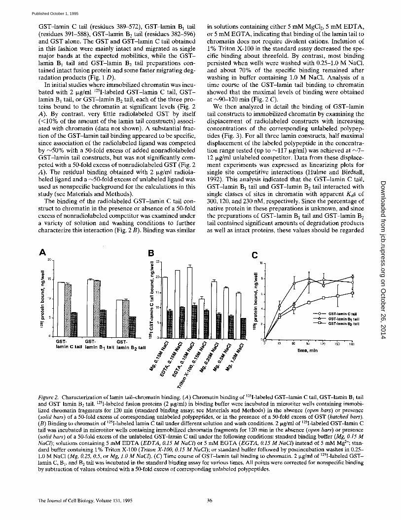
In initial studies where immobilized chromatin was incu- bated with 2 txg/ml 125I-labeled GST-lamin C tail, G S T - lamin B 1 tail, or GST-lamin B E tail, each of the three pro- teins bound to the chromatin at significant levels (Fig. 2 A). By contrast, very little radiolabeled GST by itself (<10% of the amount of the lamin tail constructs) associ- ated with chromatin (data not shown). A substantial frac- tion of the GST-lamin tail binding appeared to be specific, since association of the radiolabeled ligand was competed by ~ 5 0 % with a 50-fold excess of added nonradiolabeled GST-lamin tail constructs, but was not significantly com- peted with a 50-fold excess of nonradiolabeled GST (Fig. 2 A). The residual binding obtained with 2 txg/ml radiola- beled ligand and a ~50-fold excess of unlabeled ligand was used as nonspecific background for the calculations in this study (see Materials and Methods).
The binding of the radiolabeled GST-lamin C tail con- struct to chromatin in the presence or absence of a 50-fold excess of nonradiolabeled competitor was examined under a variety of solution and washing conditions to further characterize this interaction (Fig. 2 B). Binding was similar
in solutions containing either 5 mM MgC12, 5 mM E D T A , or 5 mM E G T A , indicating that binding of the lamin tail to chromatin does not require divalent cations. Inclusion of 1% Triton X-100 in the standard assay decreased the spe- cific binding about threefold. By contrast, most binding persisted when wells were washed with 0.25-1.0 M NaC1, and about 70% of the specific binding remained after washing in buffer containing 1.0 M NaC1. Analysis of a time course of the GST-lamin tail binding to chromatin showed that the maximal levels of binding were obtained at ~90-120 min (Fig. 2 C).
We then analyzed in detail the binding of GST-lamin tail constructs to immobilized chromatin by examining the displacement of radiolabeled constructs with increasing concentrations of the corresponding unlabeled polypep- tides (Fig. 3). For all three lamin constructs, half maximal displacement of the labeled polypeptide in the concentra- tion range tested (up to ~117 Ixg/ml) was achieved at ~ 7 - 12 txg/ml unlabeled competitor. Data from these displace- ment experiments was expressed as linearizing plots for single site competitive interactions (Hulme and Birdsall, 1992). This analysis indicated that the GST-lamin C tail, GST-lamin B1 tail and GST-lamin B2 tail interacted with single classes of sites in chromatin with apparent KoS of 300, 120, and 230 nM, respectively. Since the percentage of native protein in these preparations is unknown, and since the preparations of GST-lamin B1 tail and GST-lamin B2 tail contained significant amounts of degradation products as well as intact proteins, these values should be regarded
A 2 O .
o -~ 1S.
10. ,D
0
~ .
GST- GST- GST- lamln C tall lamln B 1 tal l lamln B 2 tall
B ~.~ 25-
~Z0-
0
o c ~ lO.
o
f 0
.e .e .e
_~=
o.oO
C 10-
4 -
II
/ / ' ~ ~ GST-lamln B 1 tall Z- / ~ GST-lamln B 2 tall
0c ' £ ' 6b " 9; " ~0 " 1;0 ' 1£ time, mln
Figure 2. Characterization of lamin tail-chromatin binding. (A) Chromatin binding of 125I-labeled GST-lamin C tail, GST-lamin B] tail and GST-lamin B2 tail. 125I-labeled fusion proteins (2 Ixg/ml) in binding buffer were incubated in microtiter wells containing immobi- lized chromatin fragments for 120 min (standard binding assay; see Materials and Methods) in the absence (open bars) or presence (solid bars) of a 50-fold excess of corresponding unlabeled polypeptides, or in the presence of a 50-fold excess of GST (hatched bars). (B) Binding to chromatin of 125I-labeled lamin C tail under different solution and wash conditions. 2 Ixg/ml of 125I-labeled GST-lamin C tail was incubated in microtiter wells containing immobilized chromatin fragments for 120 min in the absence (open bars) or presence (solid bars) of a 50-fold excess of the unlabeled GST-lamin C tail under the following conditions: standard binding buffer (Mg, 0.15 M NaCl); solutions containing 5 mM EDTA (EDTA, 0.15 M NaCI) or 5 mM EGTA (EGTA, 0.15 M NaCl) instead of 5 mM Mg2÷; stan- dard buffer containing 1% Triton X-100 (Triton X-IO0, 0.15 M NaCl); or standard buffer followed by postincubation washes in 0.25- 1.0 M NaC1 (Mg, 0.25, 0.5, or Mg, 1.0 M NaCI). (C) Time course of GST-lamin tail binding to chromatin. 2 Ixg/ml of 125I-labeled GST- lamin C, B1, and B2 tail was incubated in the standard binding assay for various times. All points were corrected for nonspecific binding by subtraction of values obtained with a 50-fold excess of corresponding unlabeled polypeptides.
The Journal of Cell Biology, Volume 131, 1995 36
on October 26, 2014
jcb.rupress.orgD
ownloaded from
Published October 1, 1995

A 1 0 0
9O
70~ r~)-
III 3 0 -
E zo.
0-
K~=3OOnM B 1 aom ~,
°:'.1 . . . . \ E
E =
unlabeled GST-lamln C tall, I~ /ml
C K.d= 12OHM 100 [
011" t " llz 80
.~. _ ~ ,0i " ~ . ,D
4 0 .
3 0 .
10 .
0 .
unlabeled GST-lamln B 1 tail, pg/ml
Kd:230nM =,o
• z b " ~b "" 6b ' . b " l d 0 '
unlabeled GST-lamln B 2 tail, pg/ml
Figure 3. Displacement of leSI-labeled GST-lamin C, B1, and B2 tails from chromatin by the corresponding unlabeled polypeptides, and calculation of dissociation constants, 2 ~g/ml of ~25I-labeled GST-lamin C tail, GST-lamin B 1 tail or GST-lamin Be tail (A-C, respec- tively) were incubated in the standard binding assay in the presence of various concentrations of the corresponding unlabeled GST- polypeptides. Binding at each point was corrected for nonspecific background by subtraction of values obtained with 117 ~g/ml unla- beled competitor (see Fig. 2). Insets: Data for specific binding was analyzed by plotting (1-RL/RLo)/A vs. 1-RL/RLo, where RL is the amount of radioactive fusion protein bound to chromatin at cold competitor concentration A, and RLo is the amount of radioactive fu- sion protein bound in the absence of unlabeled competitor. The slope given by this plot equals -1/K d (Hulme and Birdsall, 1992).
as approximate. It is not possible to calculate the amount of tamin bound to chromatin at saturation with this solid phase assay, because the fraction of sites within the immo- bilized chromatin fragments that was active for binding is unknown.
We tried to express the lamin A (389-664) tail as a GST fusion protein in E. coli, but found that it was extensively proteolyzed under all conditions tested, especially within the A-specific region. However, we successfully expressed largely undegraded lamin A tail without a fusion partner in insect cells using the baculovirus expression system, and also expressed undegraded lamin C tail without a fusion partner in E. coli using a pET vector. These lamin frag- ments were purified by ion exchange chromatography in the presence of 8 M urea, and were used for binding stud- ies after removal of the urea. In binding displacement ex- periments, the curves for the lamin A tail and the lamin C tail were very similar to each other. When the data was ex- pressed in linearizing plots for single site competitive in- teractions (see Fig. 3 legend), KdS of ~400 nM were calcu- lated for both the lamin A tail and lamin C tail (data not shown). The higher apparent Kd for the chromatin binding of the lamin C tail isolated in urea, compared with that for the GST-lamin C construct isolated under nondenaturing conditions, could be explained by incomplete renaturation of the lamin C tail after urea treatment. If the lamin A tail and lamin C tail refold to an active chromatin binding con- formation to the same extent, this would indicate that the unique 98 and 6 residue extensions present on lamin A and lamin C, respectively, do not differentially influence chromatin binding.
Mapping the Chromatin-binding Region of the Lamin C Tail
To define the specific region of the lamin C tail involved in chromatin binding, we expressed as GST fusion proteins a series of deletion mutants of the lamin C tail lacking por-
tions of either the amino- or carboxyl-terminal ends (Fig. 4 A). These were purified as largely intact fusion proteins (Fig. 4 B) and were evaluated for their ability to displace a radiolabeled GST fusion protein containing the full-length lamin C tail (38%572) from chromatin, in comparison to the displacement by the unlabeled GST-lamin C tail itself (Fig. 4, C and D). The carboxyl-terminal deletions lamin C (389-480) and lamin C (389-430) had full displacement ac- tivity, whereas lamin C (389-416) was almost completely inactive (Fig. 4 B). The amino-terminal deletion lamin C (396-572) resulted in complete displacement, while the amino-terminal deletions lamin C (401-572) and lamin C (415-572) exhibited only partial displacement of binding. A final deletion, lamin C (431-572), had no displacement activity (Fig. 4 C). Together, the results with these amino- and carboxyl-terminal deletion mutants suggest that at least two regions between residues 396-430 of the lamin C tail are important for chromatin binding.
The nuclear localization sequence (NLS) of lamins A/C is present in the tail domain at amino acids 417-422 (KKRKLE). To determine whether the NLS is important for the chromatin binding activity, we prepared several lamin C tail constructs, including amino-terminal tail dele- tions, from which residues 417-422 were deleted (ANLS). These were GST-lamin C (389-572 ANLS), GST-lamin C (396--572 ANLS), and GST-lamin C (401-572 ANLS) (Fig. 5, A and B). Lamin C (389-572 ANLS) and lamin C (396- 572 ANLS) both exhibited only partial displacement of the binding (Fig. 5 C). Combining the amino-terminal de- letion of lamin C (401-572), which by itself had lost some displacement activity, with the ANLS mutant, which also was partially inactive, yielded the construct lamin C (401- 572 ANLS) that lost almost all ability to displace the intact lamin C tail from chromatin (Fig. 5 C). These results to- gether indicate that residues 396-401 and 417-422 (the NLS) both have important roles in the chromatin binding activity of the lamin C tail, and that the combined effects of these regions are additive•
Taniura et al. Chromatin Binding of Lamin Tail Domain 37
on October 26, 2014
jcb.rupress.orgD
ownloaded from
Published October 1, 1995

A construct
389 572 + +
389 480 + +
389 430 + +
389 416
396 572 + +
401 572 +
415 572 +
431, 572
C
c
c
E I::
el E
1(I)
9 0 .
80.
7'0.
60.
5 0 .
40.
:30.
~0.
10 ,
O-
" zb " ~b " eb - eb 1 6 o 1 ~ o 1 ~ o
u n l a b e l e d c o m p e t i t o r , I*g/ml
D ItD t
"0
.o
E ~
E 4o
E
10
Jt I
1 -- - -0- ' - - 389-572 5 ~ 396-512
415-572 ~ JL 430-57Z
zb 4b ~ ~ " 1do " do
un labe led compet i to r , ~ / m l
Figure 4. Displacement of 125I-labeled GST-lamin C tail from chromatin by GST fusion proteins containing amino- or carboxyl-termi- nal deletions of lamin C tail. (A) Schematic diagram of the deletions analyzed. Terminal amino acids are numbered, and their displace- ment activity is summarized (competition). (B) Electrophoresis of purified recombinant GST fusion proteins on a 12% SDS gel. Proteins were visualized by staining with Coomassie blue. Lane numbers correspond to the number of each construct designated in A. (C-D) Binding of t25I-labeled GST-|amin C tail (2 p.g/ml) to chromatin was measured in the presence of various concentrations of each unla- beled recombinant polypeptide. Binding at each point was corrected for nonspecific background by subtraction of the value obtained with 122 txg/ml unlabeled GST-lamin C tail (see Fig. 2). (C) Displacement of the binding with constructs containing carboxyl-terminal deletions. (D) Displacement of the binding with constructs containing amino-terminal deletions.
We further prepared several bidirectional (NH2- and COOH-terminal) deletions to test in the binding displace- ment assay (Figs. 5, A and B). Lamin C (425--480), lamin C (415-480), and lamin C (411-480) were inactive in displac- ing the lamin C tail from chromatin (Fig. 5 D). By contrast, lamin C (396-480) and even lamin C (396--430) were suffi- cient for complete displacement of binding (Fig. 5 D). These data are consistent with the results of the amino- and carboxyl-terminal deletions (above), and further dem- onstrate that the minimal region required for complete displacement of chromatin binding by the lamin C tail in the context of these fusion proteins is residues 396-430.
To complement the results of the binding displacement experiments discussed above, we directly investigated the chromatin binding of several of the deletion mutants (Fig. 6). We radiolabeled GST fusion proteins containing lamin C (389-572), lamin C (389-430), lamin C (415-572), and lamin C (389-572 ANLS), and examined association of
these proteins with chromatin in the absence and presence of an excess of each unlabeled polypeptide to correct for nonspecific background (see Fig. 6 legend). The construct containing lamin C (389-430) showed a very similar level of specific chromatin binding compared to the full-length lamin C tail (389-572). By contrast, lamin C (415-572) and lamin C (389-572 ANLS), which were only partially active in competing for chromatin binding by the lamin C tail, were significantly reduced in their specific chromatin- binding activity. These results extend the binding compe- tition studies presented above (Figs. 4 and 5) and verify that a short segment near the amino terminus of the lamin C tail contains the chromatin binding site of this lamin domain.
Identification of Core Histones as the Chromatin Binding Site for Lamin C
To identify the specific chromatin component that pro-
The Journal of Cell Biology, Volume 131, 1995 38
on October 26, 2014
jcb.rupress.orgD
ownloaded from
Published October 1, 1995

A construct
389 1
389 416 423 2
396 416423 3
401 416 423 4
425 480 5
415 480 6
411 480 7
396 48O 8
396 430 9
572
572
572
572
comoetlUon
4.4.
4-
4-
+4.
4.+
C
c "¢1 _c JQ
E
E ¢U E
100~
9 0 .
aO°
7 0 .
60
SO
4O
3O
29.
10 .
O-
o zb ~ ~ m l m 1 ~ 1 ~
unlabeled competitor, I~g/ml
389-572 389.5'r2,B,43
396-572~b'4.S
4ol .57"~N.S
431-572
D
90
I~ 80 . ¢:
t-
t=
i:: I 0
I=
! 70 . 1 "-- -0--- 389-572
6O. ~t\ 5 ~ 425-480 \ \k . + , , + 50.
~ x \ r ~ 4.4m
+
10-
O .
• , • i • i • ! • •
o 20 4o 60 80 I ~ +,~o
unlabeled competitor, i~J/ml
Figure 5. Displacement of 125I-labeled GST-lamin C tail from chromatin by GST fusion proteins containing bidirectional and internal deletions of lamin C tail. (A) Schematic diagram of the deletions analyzed. Terminal amino acids are numbered, and their displacement activity is summarized (competition). (B) Electrophoresis of purified recombinant GST fusion proteins on a 12% SDS gel. Proteins were visualized by staining with Coomassie blue. Lane numbers correspond to the number of each construct designated in A. (C-D) Binding of 12SI-labeled GST-lamin C tail (2 ixg/ml) to chromatin was measured in the presence of various concentrations of each unlabeled re- combinant polypeptide. Binding at each point was corrected for nonspecific background by subtraction of the value obtained with 122 i~g/ml unlabeled GST-lamin C tail (see Fig. 2). (C) Displacement of the binding with NLS and amino-terminal combined deletions. (D) Displacement of the binding by various bidirectional deletions.
vides the binding site for the lamin C tail (Fig. 7), we prepared several chromatin subfractions. A histone HI - enriched fraction (which also contained nonhistone chro- mosomal proteins) and a second fraction consisting of core histones bound to D N A were obtained by treatment of chromatin fragments with 0.6 M salt and sucrose gradient sedimentation (Fig. 7 A, lanes 1 and 2, respectively). A third fraction consisting of purified core histones was ob- tained by binding chromatin fragments to a hydroxylapa- tite column followed by salt elution (Fig. 7 A, lane 3; see Materials and Methods). These fractions were separately adsorbed to microtiter wells and analyzed in a binding dis- placement assay with the radiolabeled GST-lamin C tail (389-572), where the unlabeled competitors were GST fu- sions containing lamin C (389-572), lamin C (389--430). and lamin C (431-572)• It should be noted that no at- tempts were made to normalize the amount of protein bound to the microtiter wells with the three histone frac- tions, so the amount of ligand bound to the three fractions cannot be directly compared. Also, raw binding data un-
corrected for nonspecific background is presented for all of the fractions analyzed (Figs. 7, B-D), since a priori we did not know which fraction displayed specific binding.
The radiolabeled lamin C tail construct bound to all three of the histone subfractions (Figs. 7, B and D). Most of the binding to the histone HI-enriched fraction (Fig. 7 B) appeared to be nonspecific, as it was only weakly dis- placed by lamin C (389-572) and lamin C (389--430). How- ever, since even weak binding displacement was not ob- tained with lamin C (431-572), which lacks the chromatin binding site, it is possible that the histone HI-enriched fraction contains a specific chromatin binding component (possibly low amounts of contaminating core histones; see below). The high nonspecific background in this case pre- cludes clear-cut conclusions. By contrast, binding of the radiolabeled lamin C tail to the core histone/DNA fraction (Fig. 7 C) and the purified core histone fraction (Fig. 7 D) was strongly displaced by lamin C (389-572) and lamin C (389--430). The degree of binding displacement in these cases was similar to that obtained when binding to intact
Taniura et al. Chromatin Binding of Lamin Tail Domain 39
on October 26, 2014
jcb.rupress.orgD
ownloaded from
Published October 1, 1995
8
C ,q
o 4 i -
"3
K z
0 389-572 389-430 415-572 389-572ANLS
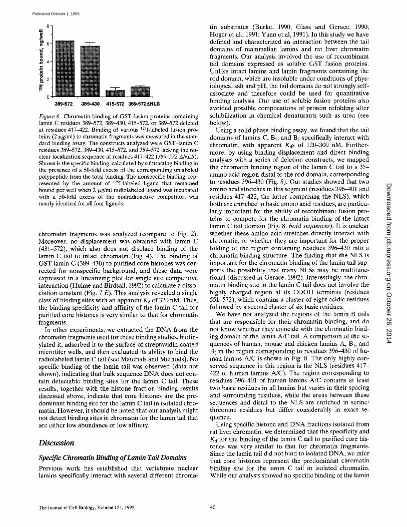
Figure 6. Chromatin binding of GST fusion proteins containing lamin C residues 389-572, 389-430, 415-572, or 389-572 deleted at residues 417-422. Binding of various 125I-labeled fusion pro- teins (2 ~g/ml) to chromatin fragments was measured in the stan- dard binding assay. The constructs analyzed were GST-lamin C residues 389-572, 389-430, 415-572, and 389-572 lacking the nu- clear localization sequence at residues 417-422 (389-572 ANLS). Shown is the specific binding, calculated by subtracting binding in the presence of a 50-fold excess of the corresponding unlabeled polypeptide from the total binding. The nonspecific binding, rep- resented by the amount of 125I-labeled ligand that remained bound per well when 2 ixg/ml radiolabeled ligand was incubated with a 50-fold excess of the nonradioactive competitor, was nearly identical for all four ligands.
chromatin fragments was analyzed (compare to Fig. 2). Moreover, no displacement was obtained with lamin C (431-572), which also does not displace binding of the lamin C tail to intact chromatin (Fig. 4). The binding of GST-lamin C (389-430) to purified core histones was cor- rected for nonspecific background, and these data were expressed in a linearizing plot for single site competitive interaction (Hulme and Birdsall, 1992) to calculate a disso- ciation constant (Fig. 7 E). This analysis revealed a single class of binding sites with an apparent Kd of 320 nM. Thus, the binding specificity and affinity of the lamin C tail for purified core histones is very similar to that for chromatin fragments.
In other experiments, we extracted the DNA from the chromatin fragments used for these binding studies, biotin- ylated it, adsorbed it to the surface of streptavidin-coated microtiter wells, and then evaluated its ability to bind the radiolabeled lamin C tail (see Materials and Methods). No specific binding of the lamin tail was observed (data not shown), indicating that bulk sequence DNA does not con- tain detectable binding sites for the lamin C tail. These results, together with the histone fraction binding results discussed above, indicate that core histones are the pre- dominant binding site for the lamin C tail in isolated chro- matin. However, it should be noted that our analysis might not detect binding sites in chromatin for the lamin tail that are either low abundance or low affinity.
Discussion
Specific Chromatin Binding of Lamin Tail Domains
Previous work has established that vertebrate nuclear lamins specifically interact with several different chroma-
tin substrates (Burke, 1990; Glass and Gerace, 1990; Hoger et al., 1991; Yuan et al, 1991). In this study we have defined and characterized an interaction between the tail domains of mammalian lamins and rat liver chromatin fragments. Our analysis involved the use of recombinant tail domains expressed as soluble GST fusion proteins. Unlike intact lamins and lamin fragments containing the rod domain, which are insoluble under conditions of phys- iological salt and pH, the tail domains do not strongly self- associate and therefore could be used for quantitative binding analysis. Our use of soluble fusion proteins also avoided possible complications of protein refolding after solubilization in chemical denaturants such as urea (see below).
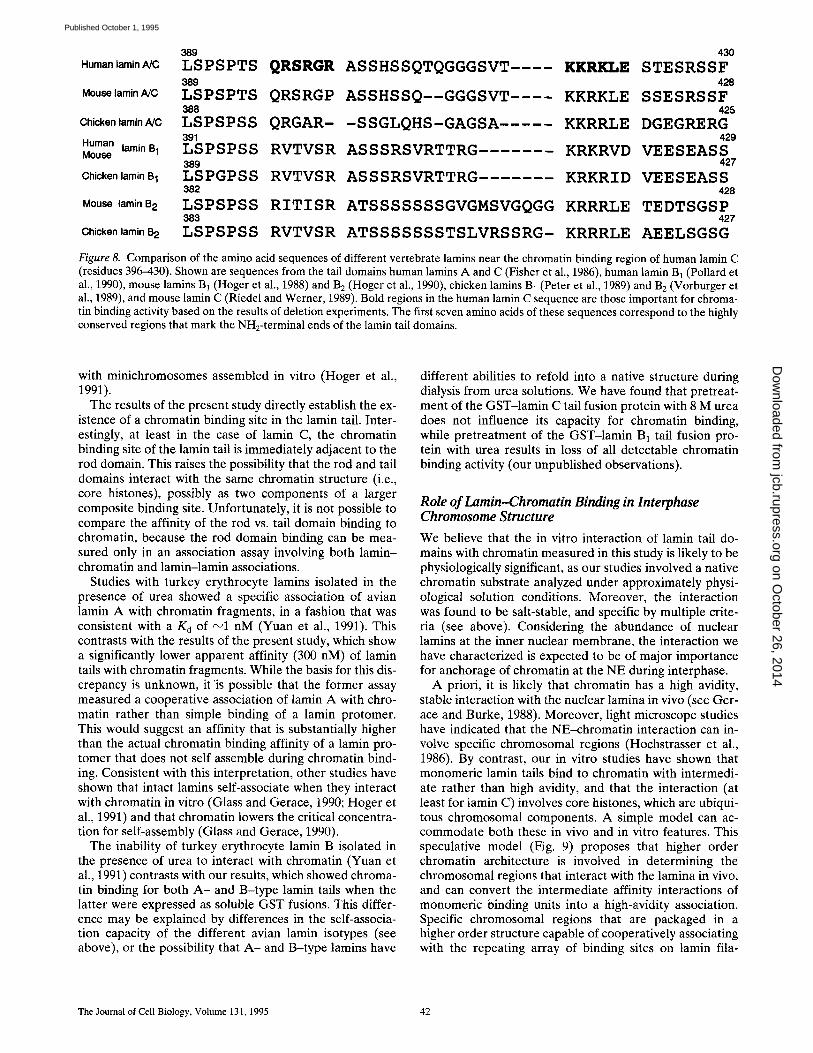
Using a solid phase binding assay, we found that the tail domains of lamins C, B1, and B2 specifically interact with chromatin, with apparent KdS of 120-300 nM. Further- more, by using binding displacement and direct binding analyses with a series of deletion constructs, we mapped the chromatin binding region of the lamin C tail to a 35- amino acid region distal to the rod domain, corresponding to residues 396-430 (Fig. 8). Our studies showed that two amino acid stretches in this segment (residues 396-401 and residues 417-422, the latter comprising the NLS), which both are enriched in basic amino acid residues, are particu- larly important for the ability of recombinant fusion pro- teins to compete for the chromatin binding of the intact lamin C tail domain (Fig. 8, bold sequences). It is unclear whether these amino acid stretches directly interact with chromatin, or whether they are important for the proper folding of the region containing residues 396-430 into a chromatin-binding structure. The finding that the NLS is important for the chromatin binding of the lamin tail sup- ports the possibility that many NLSs may be multifunc- tional (discussed in Gerace, 1992). Interestingly, the chro- matin binding site in the lamin C tail does not involve the highly charged region at its COOH terminus (residues 551-572), which contains a cluster of eight acidic residues followed by a second cluster of six basic residues.
We have not analyzed the regions of the lamin B tails that are responsible for their chromatin binding, and do not know whether they coincide with the chromatin bind- ing domain of the lamin A/C tail. A comparison of the se- quences of human, mouse and chicken lamins A, B1, and B2 in the region corresponding to residues 396-430 of hu- man lamins A/C is shown in Fig. 8. The only highly con- served sequence in this region is the NLS (residues 417- 422 of human lamins A/C). The region corresponding to residues 396-401 of human lamins A/C contains at least two basic residues in all lamins but varies in their spacing and surrounding residues, while the areas between these sequences and distal to the NLS are enriched in serine/ threonine residues but differ considerably in exact se- quence.
Using specific histone and DNA fractions isolated from rat liver chromatin, we determined that the specificity and Kd for the binding of the lamin C tail to purified core his- tones was very similar to that for chromatin fragments. Since the lamin tail did not bind to isolated DNA, we infer that core histones represent the predominant chromatin binding site for the lamin C tail in isolated chromatin. While our analysis showed no specific binding of the lamin
The Journal of Cell Biology, Volume 131, 1995 40
on October 26, 2014
jcb.rupress.orgD
ownloaded from
Published October 1, 1995

B k~
lB.
-i
c el E
O 22
[ o
C
4
tZ, ~).
c ~ 3~430 -t,- e, ~ ,~1~r2
---o--385-430 ~u 6 ~ 389-b~ 431-572 ~/) 389-572 f~ 4,
2, fro 40 60 BO 100 I~ 140
unlabeled competitors, pg/rnl unlabeled competitors, po/ml
D 16 A --
- r - - 14
t2
10
4 "t ~ 431-572 2 t ~ 389-572
1 0 20 40 60 80 100 120 140
unlabeled competitors, Pg/ml
E Kd=320nM 0,07
0 ` 0 6 ~
O,O$ ~ .
~:, 0.03
. . . . . .
0`0 02. 0.4 0`1; 0.8 1.0
1-RI./RL 0 1.2
Figure 7. Displacement of 125I-labeled GST-lamin C tail from various chromatin subfractions by GST fusion proteins containing various lamin C tail regions. (A) Protein composition of chromatin subfractions. The histone HI-enriched fraction, core histone-DNA fraction and purified core histones were isolated as described in Materials and Methods. Proteins were analyzed by electrophoresis on a 15% SDS-get and staining with Coomassie blue. Lane 1, histone Hi-enriched fraction; lane 2, core histone-DNA fraction; lane 3, purified core histones, (B-D) Chromatin subfractions were immobilized on microtiter wells in coating buffer, and incubated with 125I-labeled GST-lamin C tail (389-572) at a concentration of 2 I~g/ml in the presence of various concentrations of unlabeled GST-lamin C residues 389-572, 389-430, or 431-572. Shown are uncorrected binding data. (B) Displacement of 125I-labeled GST-lamin C tail from the histone Hi-enriched fraction by unlabeled fusion proteins. (C) Displacement of 125I-labeled GST-lamin C tail from the core histone-DNA frac- tion by unlabeled fusion proteins. (D) Displacement of 125I-labeled GST-lamin C tail from purified core histones by unlabeled fusion proteins. (E) Determination of a dissociation constant for the binding of 125I-labeled GST-lamin C (389--430) to purified core histones. The data for each point obtained with 125I-labeled GST-lamin C tail (389-430) in D was corrected for nonspecific background by sub- tracting the binding obtained in the presence of 125 ~Lg/ml unlabeled GST-lamin C tail. These data were used to plot (1-RL/RLo)/A vs. 1-RL/RLo to determine the K d (see Fig. 3).
C tail to bulk double-stranded DNA, we cannot exclude an interaction with a specific D N A sequence class that is not sufficiently enriched in bulk sequence D N A to be de- tectable with our assay. We note that lamins and other proteins containing coiled-coiled a-helices, including cyto- plasmic intermediate filament proteins, have been found to interact in vitro with single stranded D N A (Shoeman and Traub, 1990; Luderus et al., 1994), as well as with A/T- rich S A R / M A R D N A sequences (Luderus et al,, 1992), which have a propensity to form single-stranded regions (Kohwi-Shigematsu and Kohwi, 1990). Whether single- stranded S A R / M A R sequences occur in vivo is unknown. However, an interaction of S A R / M A R sequences with the nuclear lamina was not observed in studies involving an approximately physiological buffer with divalent cations (Izaurralde et al,, 1988).
Relationship to Other Investigations of the Lamin-Chromatin Interaction
In a previous study where chromatin association was as- sayed by dialysis of solubilized recombinant lamin con- structs into a solution of physiological pH and salt in the presence of mitotic chromosomes, we showed that the rod domain of nuclear lamins A/C by itself specifically inter- acts with chromatin (Glass et al., 1993). However, since as- sociation of the rod domain with chromatin occurred with significantly lower efficiency than seen for constructs con- taining both rod and tail domains (Glass et al., 1993), it was possible that the lamin tail also contributed to chro- matin binding. This notion was consistent with studies showing that certain deletions in the tail domains of Xeno- pus lamin A and L]~ compromised their ability to associate
Taniura et al. Chromatin Binding of Lamin Tail Domain 41
on October 26, 2014
jcb.rupress.orgD
ownloaded from
Published October 1, 1995
389 430
HumanlaminA/C L S P S P T S QRSRGR A S S H S S Q T Q G G G S V T . . . . KK:RZ~E S T E S R S S F 389 428
MouselaminA/C LSPSPTS QRSRGP ASSHSSQ--GGGSVT .... KKRKLE SSESRSSF 388 425
ChickenlaminA/C LSPSPSS QRGAR- - S S G L Q H S - G A G S A KKRRLE DGEGRNRG 391 429
Human lamin B I LSPSPSS RVTVSR ASSSRSVRTTRG KRKRVD VEESEASS Mouse 389 427
ChickenlaminB1 LSPGPSS RVTVSR ASSSRSVRTTRG KRKRID VEESEASS 382 428
Mouse laminB 2 LSPSPSS RITISR ATSSSSSSSGVGMSVGQGG KRRRLE TEDTSGSP 383 427
Chicken lamina 2 LSPSPSS RVTVSR ATSSSSSSSTSLVRSSRG- KRRRLE AEELSGSG
Figure 8. Comparison of the amino acid sequences of different vertebrate lamins near the chromatin binding region of human lamin C (residues 396-430). Shown are sequences from the tail domains human lamins A and C (Fisher et al., 1986), human lamin B1 (Pollard et al., 1990), mouse lamins B1 (Hoger et al., 1988) and B 2 (Hoger et al., 1990), chicken lamins B1 (Peter et al., 1989) and B 2 (Vorburger et al., 1989), and mouse lamin C (Riedel and Werner, 1989). Bold regions in the human lamin C sequence are those important for chroma- tin binding activity based on the results of deletion experiments. The first seven amino acids of these sequences correspond to the highly conserved regions that mark the NHE-terminal ends of the lamin tail domains.
with minichromosomes assembled in vitro (Hoger et al., 1991).
The results of the present study directly establish the ex- istence of a chromatin binding site in the lamin tail. Inter- estingly, at least in the case of lamin C, the chromatin binding site of the lamin tail is immediately adjacent to the rod domain. This raises the possibility that the rod and tail domains interact with the same chromatin structure (i.e., core histones), possibly as two components of a larger composite binding site. Unfortunately, it is not possible to compare the affinity of the rod vs. tail domain binding to chromatin, because the rod domain binding can be mea- sured only in an association assay involving both lamin- chromatin and lamin-lamin associations.
Studies with turkey erythrocyte lamins isolated in the presence of urea showed a specific association of avian lamin A with chromatin fragments, in a fashion that was consistent with a Ko of ~1 nM (Yuan et al., 1991). This contrasts with the results of the present study, which show a significant]y lower apparent affinity (300 nM) of lamin tails with chromatin fragments. While the basis for this dis- crepancy is unknown, it "is possible that the former assay measured a cooperative association of lamin A with chro- matin rather than simple binding of a lamin protomer. This would suggest an affinity that is substantially higher than the actual chromatin binding affinity of a lamin pro- tomer that does not self assemble during chromatin bind- ing. Consistent with this interpretation, other studies have shown that intact lamins self-associate when they interact with chromatin in vitro (Glass and Gerace, 1990; Hoger et al., 1991) and that chromatin lowers the critical concentra- tion for self-assembly (Glass and Gerace, 1990).
The inability of turkey erythrocyte lamin B isolated in the presence of urea to interact with chromatin (Yuan et al., 1991) contrasts with our results, which showed chroma- tin binding for both A - and B-type lamin tails when the latter were expressed as soluble GST fusions. This differ- ence may be explained by differences in the self-associa- tion capacity of the different avian lamin isotypes (see above), or the possibility that A - and B-type lamins have
different abilities to refold into a native structure during dialysis from urea solutions. We have found that pretreat- ment of the GST-lamin C tail fusion protein with 8 M urea does not influence its capacity for chromatin binding, while pretreatment of the GST-lamin B1 tail fusion pro- tein with urea results in loss of all detectable chromatin binding activity (our unpublished observations).
Role of Lamin-Chromatin Binding in Interphase Chromosome Structure
We believe that the in vitro interaction of lamin tail do- mains with chromatin measured in this study is likely to be physiologically significant, as our studies involved a native chromatin substrate analyzed under approximately physi- ological solution conditions. Moreover, the interaction was found to be salt-stable, and specific by multiple crite- ria (see above). Considering the abundance of nuclear lamins at the inner nuclear membrane, the interaction we have characterized is expected to be of major importance for anchorage of chromatin at the NE during interphase.
A priori, it is likely that chromatin has a high avidity, stable interaction with the nuclear lamina in vivo (see Get- ace and Burke, 1988). Moreover, light microscope studies have indicated that the NE-chromatin interaction can in- volve specific chromosomal regions (Hochstrasser et al., 1986). By contrast, our in vitro studies have shown that monomeric lamin tails bind to chromatin with intermedi- ate rather than high avidity, and that the interaction (at least for tamin C) involves core histones, which are ubiqui- tous chromosomal components. A simple model can ac- commodate both these in vivo and in vitro features. This speculative model (Fig. 9) proposes that higher order chromatin architecture is involved in determining the chromosomal regions that interact with the lamina in vivo, and can convert the intermediate affinity interactions of monomeric binding units into a high-avidity association. Specific chromosomal regions that are packaged in a higher order structure capable of cooperatively associating with the repeating array of binding sites on lamin fila-
The Journal of Cell Biology, Volume 131, 1995 42
on October 26, 2014
jcb.rupress.orgD
ownloaded from
Published October 1, 1995
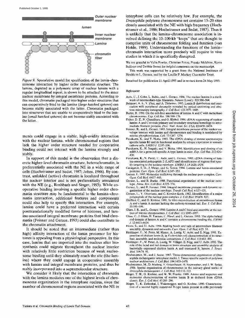
Figure 9. Speculative model for specification of the lamin-chro- mosome interaction by higher order chromatin structure. The lamina, depicted as a polymeric array of nuclear lamins with a regular longitudinal repeat, is shown to be attached to the inner nuclear membrane by integral membrane proteins. According to this model, chromatin packaged into higher order structures that can cooperatively bind to the lamina (large hatched spheres) can become stably associated with the latter. Chromatin packaged into structures that are unable to cooperatively bind to the lam- ina (small hatched spheres) do not become stably associated with the latter.
ments could engage in a stable, high-avidity interaction with the nuclear lamina, while chromosomal regions that lack the higher order structure needed for cooperative binding could not interact with the lamina strongly and stably.
In support of this model is the observation that a dis- crete higher-level chromatin structure, heterochromatin, is preferentially associated with the NE in a wide range of cells (Hochstrasser and Sedat, 1987; Johns, 1988). By con- trast, unfolded (active) chromatin is localized throughout the nuclear interior and is not preferentially associated with the NE (e.g., Roshbash and Singer, 1993). While co- operative binding involving a specific higher order chro- matin structure may be fundamental to the lamina-chro- matin interaction, additional features and components could also help to specify this interaction. For example, lamins could have a preferred interaction with certain posttranslationally modified forms of histones, and lam- ina-associated integral membrane proteins that bind chro- matin (Foisner and Gerace, 1993) could also contribute to the chromatin attachment.
It should be noted that an intermediate (rather than high) affinity interaction of the lamin protomer for his- tones is appealing from a physiological perspective. In this case, lamins that are imported into the nucleus after bio- synthesis could migrate throughout the nuclear interior with relatively little restriction because of weak nucleo- some binding until they ultimately reach the site (the lam- ina) where they could engage in cooperative assembly with lamins and integral membrane proteins, and thus be stably incorporated into a supramolecular structure.
We consider it likely that the interaction of chromatin with the lamina mediates one of the highest levels of chro- mosome organization in the interphase nucleus, since the number of chromosomal regions associated with the NE in
interphase cells can be relatively low. For example, the Drosophila polytene chromosome set contains 15-20 sites closely associated with the NE with high frequency (Hoch- strasser et al., 1986; Hochstrasser and Sedat, 1987). Thus it is unlikely that the lamina-chromosome association is in- volved defining the 10-100-kb "loops" that are thought to comprise units of chromosome folding and function (van Holde, 1989). Understanding the functions of the lamin- chromatin interaction more precisely will require in vivo studies in which it is specifically disrupted.
We are grateful to Velia Fowler, Christian Fritze, Frauke Melchior, Kevin Sullivan and Debbie Sweet for helpful comments on the manuscript.
This work was supported by a grant from the National Institutes of Health to L. Gerace, and by the Lucille P. Markey Charitable Trust.
Received for publication t 1 April 1995 and in revised form 24 May 1995.
References
Aebi, U., J. Cohn, L. Buhle, and L. Gerace. 1986. The nuclear lamina is a mesh- work of intermediate-type filaments. Nature (Lond.). 323:560-564.
Belmont, A. S., Y. Zhai, and A. Thilenius. 1993. Lamin B distribution and asso- ciation with peripheral chromatin revealed by optical sectioning and elec- tron microscopy tomography. J. Cell Biol. 123:1671-1685.
Burke, B, 1990. On the cell-free association of [amins A and C with metaphase chromosomes. Exp. Cell Res, 186:169-176.
Fisher, D, Z., N. Chaudhary, and G. Blobel. 1986. cDNA sequencing of nuclear lamins A and C reveals primary and secondary structural homology to inter- mediate filament proteins. Proc, Natl. Acad. Sci. USA. 83:6450~454,
Foisner, R., and L. Gerace. 1993. Integral membrane proteins of the nuclear en- velope interact with lamins and chromosomes and binding is modulated by mitotic phosphorylation. Cell. 73:1267-1279.
Furukawa, K., and Y. Hotta. 1993. The cDNA cloning of mouse spermatoeyte specific lamin B3 and its functional analysis by ectopic expression in somatic culture cells. E M B O J. 12:97-106.
Furukawa, K., H. Inagaki, and Y. Holta. 1994. Identification and cloning of an mRNA for a germ cell-specific A-type lamin in mice. Exp. Cell Res. 212:426- 430.
Furukawa, K., N. Pant6, U. Aebi, and L. Gerace. 1995. cDNA cloning of lam- ina-associated polypeptide 2 (LAP2) and identification of regions that spec- ify targeting to the nuclear envelope. E M B O J. 14:1626-1636.
Georgatos, S. D., J. Meier, and G. Simos. 1994. Lamins and lamin-associated proteins. Curr. Opin. Cell BioL 6:347-353.
Gerace, L. 1992. Molecular trafficking through the nuclear pore complex. Curr. Opin. Cell Biol. 4:637~645.
Gerace, L., and B. Burke. 1988. Functional organization of the nuclear enve- lope. Annu. Rev. Cell Biol. 4:335-374.
Gerace, L, and R. Foisner. 1994. Integral membrane proteins and dynamic or- ganization of the nuclear envelope. Trends Cell Biol. 4:127-131.
Gerace, L., Y. Ottaviano, and C. Kondor-Koch. 1982. Identification of a major polypeptide of the nuclear pore complex. ,L Cell Biol. 95:826-837.
Gieffers, C., and G. Krohne. 1991. In vitro reconstitution of recombinant lamin A and a lamin A mutant lacking the carboxy-terminal tail. Eur. J. Cell BioL 55:191-199.
Glass, J. R., and L. Gerace. 1990. Lamins A and C bind and assemble at the sur- face of mitotic chromosomes. Z Cell Biol. 111:1047-1057.
Glass, C., J. Glass, H. Taniura, C. Hasel, and L. Gerace. 1993. The alpha helical rod domain of lamins A and C contains a chromosome binding site. E M B O Z 12:4413-4424.
Heins, S., and U. Aebi. 1994. Making heads and tails of intermediate filament assembly, dynamics and networks. Curt Opin. Cell Biol. 6:25-33.
Heitlinger, E., M. Peter, M. Haner, A. Lustig, U. Aebi, and E. Nigg, 1991. Ex- pression of chicken lamin B2 in Escherichia coil: characterization of its struc- ture, assembly, and molecular interactions. J. Cell Biol. 113:485-495.
Heitlinger, E., M. Peter, A. Lustig, W. Villiger, E. Nigg, and U. Aebi. 1992. The role of the head and tail domain in lamin structure and assembly: analysis of bacterially expressed chicken lamin A and truncated B2 lamins. Z Struct. Biol. 108:74-91.
Hochstrasser, M., and J. Sedat. 1987. Three-dimensional organization of Dro- sophila melanogaster interphase nuclei. I. Tissue-specific aspects of polytene nuclear architecture. J. Cell Biol. 104:1455-1470.
Hochstrasser, M., D. Mathog, Y. Gruenbaum, H. Saumweber, and J. W. Sedat. 1986. Spatial organization of chromosomes in the salivary gland nuclei of Drosophila melanogaster. ,L Cell Biol. 102:112-123.
Hoger, T. H., G. Krohne, and W. W. Franke. 1988. Amino acid sequence and molecular characterization of murine lamin B as deduced from cDNA clones. Eur. J. Cell Biol. 47:283-290,
Hoger, T., K. Zatloukal, I. Waizenegger, and G. Krohne. 1990. Characteriza- tion of a second highly conserved B-type lamin present in cells previously
Taniura et al. Chromatin Binding of Lamin Tail Domain 43
on October 26, 2014
jcb.rupress.orgD
ownloaded from
Published October 1, 1995
thought to contain only a single B-type lamin. Chromosoma. 99:379-390. Hoger, T., G. Krohne, and J. Kleinschmidt. 1991. Interaction ofXenopus lamins
A and Lll with chromatin in vitro mediated by a sequence element in the carboxyterminal domain. Exp. Cell Res. 197:280-289.
Hulme, E. C., and N. J. M. Birdsall. 1992. In Receptor Ligand Interactions. A Practical Approach. Oxford University Press, NY. 63-176.
Izaurralde, E., J. Mirkovitch, and U. K. Laemmli. 1988. Interaction of DNA with nuclear scaffolds in vitro. J. Mol. Biol. 200:111-125.
Johns, B. 1988. The biology of heterochromatin. In Heterochromatin: Molecu- lar and Structural Aspects. R. S. Verman, editor. Cambridge University Press, NY. 1-147.
Kohwi-Shigematsu, T., and Y. Kohwi. 1990. Torsional stress stabilizes extended base unpairing in suppressor sites flanking immunoglobulin heavy chain en- hancer. Biochemistry. 29:9551-9560.
Laemmli, U. K. 1970. Cleavage of structural proteins during assembly at the head of the bacteriophage T4. Nature (Lond.). 227:680-682.
Luderus, M., A. de Graaf, E. Mattia, J. Blaauwen, M. Grande, L. Jong, and R. yon Driel. 1992. Binding of matrix attachment regions to lamin-B1. Cell. 70: 949-959.
Luderus, M., J. der Blaauwen, O. Smit, D. Compton, and R. von Driel. 1994. Binding of matrix attachment regions to lamin polymers involves single- stranded regions and the minor groove. Mol. Cell Biol. 14:6297~305.
McKeon, F. 1991. Nuclear lamin proteins: domains required for nuclear target- ing, assembly, and cell-cycle-regulated dynamics. Curr. Opin. Cell Biol. 3:82- 86.
McKeon, F. D., M. W. Kirshner, and D. Caput. 1986. Homologies in both pri- mary and secondary structure between nuclear envelope and intermediate filament proteins. Nature (Lond.). 319:463-468.
Moir, R. D., A.D. Donaldson, and M. Stewart. 1991. Expression in Escherichia coli of human lamins A and C: influence of head and tail domains on assem- bly properties and paracrystal formation. Z Cell Sci. 99:363-372.
Nachman, L. K., and K. Leung. 1982. Complex formation of platelet membrane glycoproteins IIb and IIIa with fibrinogen. J. Clin. Invest. 69:263-269.
Nigg, E. 1992. Assembly-disassembly of the nuclear lamina. Curr. Opin. Cell Biol. 4:105-109.
Noll, M., J. O. Thomas, and R. D. Kornberg. 1975. Preparation of native chro- matin and damage caused by shearing. Science (Wash. DC). 187:1203-1206.
Peter, M., G. T. Kitten, C. F. Lehner, K. Vorburger, S. M. Bailer, F. G. Maridor, and E. A. Nigg. 1989. Cloning and sequencing of cDNA clones encoding chicken lamins A and B 1 and comparison of the primary structures of verte- brate A- and B-type lamins. J. Mol. Biol. 208:393--404.
Pollard, K. M., E. K. L. Chan, B. Grant, K. F. Sullivan, E. M. Tan, and C. A. Glass. 1990. In vitro posttranslational modification of lamin B cloned from a human T-cell line. Mol. Cell Biol. 10(5):2164-2175.
Riedel, W., and D. Werner. 1989. Nucleotide sequence of the full-length mouse lamin C cDNA and its deduced amino-acid sequence. Biochimica et Bio- physica Acta. 1008:119-122.
Roshbash, M., and R. H. Singer. 1993. RNA travel: tracks from DNA to cyto- plasm. Cell. 75:399-401.
Shoeman, R., and P. Traub. 1990. The in vitro DNA-binding properties of puri- fied nuclear lamin proteins and vimentin. J. Biol. Chem. 265:9055-9061.
Smith, D., and L. Cnrcoran. 1990. Current Protocols in Molecular Biology. Greene Publishing Associates, NY. 16.7.1-16.7.8.
van Holde, K. E. 1989. Chromatin. Springer Verlag, NY. Vorburger, K., C. F. Lehner, G. T. Kitten, H. M. Eppenberger, and E. A. Nigg.
1989. A 2nd higher vertebrate B-type lamin cDNA sequence determination and in vitro processing of chicken lamin-B2. J. Mol. Biol. 208:405-415.
Yuan, J., G. Sirens, G. Blobel, and S. D. Georgatos. 1991. Binding of lamin A to polynucleosomes. J. Biol. Chem. 266:9211-9215.
Zentgraf, H., and W. W. Franke. 1984. Differences of supranucleosomal organi- zation in different kinds of chromatin: cell type-specific globular subunits containing different numbers of nucleosomes. J. Cell Biol. 99:272-286.
The Journal of Cell Biology, Volume 131, 1995 44
on October 26, 2014
jcb.rupress.orgD
ownloaded from
Published October 1, 1995