Hyperthermia: A Potential Game-Changer in the Management ...

Histone Deacetylase Inhibitors Sensitize Lung CancerCells to Hyperthermia: Involvement of Ku70/SirT-1 inThermo-ProtectionMohamed K. Hassan1,2*, Hidemichi Watari1*, Alaa-eldin Salah-eldin3¤a, Ahmed S. Sultan4¤b,
Zainab Mohamed1, Yoichiro Fujioka5, Yusuke Ohba5, Noriaki Sakuragi1
1 Department of Obstetrics and Gynecology, Hokkaido University, Graduate School of Medicine, Sapporo, Japan, 2 Biotechnology program, Department of Zoology,
Faculty of Science, Port Said University, Port Said, Egypt, 3 Department of Zoology, Aswan Faculty of Science, Aswan University, Aswan, Egypt, 4 Department of Oncology,
Lombardi Comprehensive Cancer Center, Georgetown University Medical Center, Washington, DC, United States of America, 5 Department of Cell Physiology, Hokkaido
University, Graduate School of Medicine, Sapporo, Japan
Abstract
This study describes the sensitization mechanism to thermal stress by histone deacetylase inhibitors (HDACIs) in lung cancercells and shows that Ku70, based on its acetylation status, mediates the protection of lung cancer from hyperthermia(42.5uC, 1-6 hrs). Ku70 regulates apoptosis by sequestering pro-apoptotic Bax. However, its role in thermal stress is not fullyunderstood. The findings showed that, pre-treating lung cancer cells with HDACIs, nicotinamide (NM) or Trichostatin A (TsA)or both significantly enhanced hyperthermia-induced Bax-dependent apoptosis in PC-10 cells. We found that hyperthermiainduces SirT-1, Sirtuin, upregulation but not HDAC6 or SirT-3, therefore transfection with dominant negative SirT-1 (Y/H) alsoeliminated the protection and resulted in more cell death by hyperthermia, in H1299 cells through Bax activation.Hyperthermia alone primed lung cancer cells to apoptosis without prominent death. After hyperthermia Bax wasupregulated, Bcl-2 was downregulated, the Bax/Bcl-2 ratio was inversed and Bax/Bcl-2 heterodimer was dissociated.Although hyperthermia did not affect total Ku70 expression level, it stimulated Ku70 deacetylation, which in turn could bindmore Bax in the PC-10 cells. These findings suggest an escape mechanism from hyperthermia-induced Bax activation. Toverify the role of Ku70 in this protection mechanism, Ku70 was silenced by siRNA. Ku70 silencing significantly sensitized thelung cancer cells to hyperthermia. The Ku70 KD cells underwent cytotoxic G1 arrest and caspase-dependant apoptosis whencompared to scrambled transfectants which showed only G2/M cytostatic arrest in the cell lines investigated, suggesting anadditional cell cycle-dependent, novel, role of Ku70 in protection from hyperthermia. Taken together, our data show a Ku70-dependent protection mechanism from hyperthermia. Targeting Ku70 and/or its acetylation during hyperthermia mayrepresent a promising therapeutic approach for lung cancer.
Citation: Hassan MK, Watari H, Salah-eldin A-e, Sultan AS, Mohamed Z, et al. (2014) Histone Deacetylase Inhibitors Sensitize Lung Cancer Cells to Hyperthermia:Involvement of Ku70/SirT-1 in Thermo-Protection. PLoS ONE 9(4): e94213. doi:10.1371/journal.pone.0094213
Editor: Sue Cotterill St. Georges, University of London, United Kingdom
Received August 30, 2013; Accepted March 14, 2014; Published April 11, 2014
Copyright: � 2014 Hassan et al. This is an open-access article distributed under the terms of the Creative Commons Attribution License, which permitsunrestricted use, distribution, and reproduction in any medium, provided the original author and source are credited.
Funding: This study was supported in part by a grant-in-aid HW for Scientific Research (C 22591844 and 20659255) from the Ministry of Education, Culture,Sports, Science and Technology of Japan. The funders had no role in study design, data collection and analysis, decision to publish, or preparation of themanuscript.
Competing Interests: The authors have declared that no competing interests exist.
* E-mail: [email protected] (MKH); [email protected] (HW)
¤a Current address: Medical Laboratories Department, College of Science in AlZulfi, Majmaah University, Majmaah, Saudi Arabia¤b Current address: Biochemistry Dept., Faculty of Science, Alexandria University, Alexandria, Egypt
Introduction
A long-standing research interest has been targeted the specific
mechanisms responsible for the development of cancer cell
resistance to different therapies. Targeting these mechanisms
may enhance the specific destruction of cancer cells. Hyperther-
mia is a modality used in the clinical setting, for the treatment of
many cancers; it is usually used in combination with radiotherapy
and/or chemotherapy [1,2]. However, a significant obstacle to the
effectiveness of hyperthermia is the development of cellular
resistance, which blocks apoptotic signaling and enhances cell
survival [3,4]. This resistance causes limitation of apoptosis after
hyperthermia [5,6]. Thus, the identification of the mechanisms
responsible for the development of thermo-resistance in cancer
cells, might help improve specific targeting to enhance cellular
sensitivity treatment outcomes to hyperthermia. Resistance to
apoptosis is a common characteristic of cancer cells [3,7].
Apoptosis is induced by, extrinsic and intrinsic pathways [8].
Binding of ligands to a death receptor activates the extrinsic
pathway; the intrinsic pathway is activated by cell stress, such as
DNA damage. The Bcl-2 protein family regulates the intrinsic
pathway; it influences the permeability of the outer mitochondrial
membrane [9]. Members of the Bcl-2 family are divided into
proapoptotic proteins such as Bax, Bak, and Bok, and anti-
apoptotic proteins including Bcl-2, Bcl-xL, Bcl-w, and Mcl-1 [10–
13].
Accumulation of Bcl-2 and Bcl-xL can protect cells from
apoptosis, promote cell survival and accelerate tumor growth by
sequestering pro-apoptotic Bax. Ku70 is another anti-apoptotic
molecule; it naturally binds Bax, sequestering it from activation or
PLOS ONE | www.plosone.org 1 April 2014 | Volume 9 | Issue 4 | e94213

mitochondrial translocation in unstressed cells [14,15]. Ku70 is
one of the components of the Ku70/Ku80 heterodimer that is
involved in DNA damage repair [16]. Acetylation of two critical
lysines, on the carboxyl terminus of Ku70, regulates the binding/
dissociation to Bax and this affects the subsequent sensitivity of the
cell to apoptotic stimuli [14]. Only deacetylated Ku70 can bind to
Bax. High expression of Ku70 in cancer cells would enhance DNA
repair ability and reduce Bax-mediated apoptosis; therefore, Ku70
might play a role in treatment resistance. The apoptosis-related
activity of Ku70 is independent of its role in DNA repair [17]. The
Ku70 acetylation/deacetylation cycle is regulated by histone acetyl
transferases and histone deacetylases (HDACs). Ku70 is a target of
some members of class I/II HDAC and class III HDAC [18,19].
The HDAC family of proteins is divided into two categories: zinc-
dependent enzymes (HDAC1-11), subdivided into class I and class
II which are inhibited by Trichostatin A (TSA) and NAD+-
dependent enzymes (class III; SIRT1-7) which is inhibited by
nicotinimide (NAM). More precisely, SirT-1, a member of the
class III HDACs, plays a crucial role in Ku70 deacetylation, which
enhances the protection of cells from Bax during caloric restriction
[19]. The majority of cancer cells over-express SirT1 [20]. Thus,
targeting the Ku70-dependent protection from apoptosis, by
HDAC inhibitors that inhibit SirT-1, could be an effective strategy
for sensitizing cancer cells to different therapies. In this study, our
model is that lung cancer cells are significantly killed by
hyperthermia when pretreated with HDACIs compared with
hyperthermia only. SirT-1 and its target, Ku70, are central to the
mechanism by which lung cancer cells can escape thermal-induced
death. Changes in the activity of Bax, Ku70 acetylation and the
cell cycle were studied during exposure to hyperthermia. In
addition, the efficiency of sequence specific targeting of Ku70,
using siRNA, were also studied with regard to sensitizing lung
cancer cells to hyperthermia. Ku70 appears to play a crucial role
in the protection of cells from hyperthermia probably by
sequestering up-regulated Bax.
Materials and Methods
Cell linesTwo human, non-small cell lung carcinoma cell lines: PC-10
[21], and H1299, were purchased (American Type Culture
Collection, ATCC) and cultured in RPMI 1640 medium (Gibco,
Grand Island, NY, USA) containing 10% fetal bovine serum
(Sigma, St. Louis, MO, USA) (complete medium) in a humidified
atmosphere of 5% CO2 at 37uC. The cultured medium was
replaced by fresh complete medium every three days.
Antibodies and reagentsAnti-human Bax rabbit polyclonal antibody (pAb) and anti-
human Ku70 mouse monoclonal antibody (mAb) were purchased
from BD Bioscience (Erembodegem, Belgium); another anti-
human Ku70 mouse mAb for immunoprecipitation, anti-human
HDAC-6 and anti-human SirT-3 rabbit (pAb) were purchased
from Abcam (Cambridge, UK); anti-human Ku80 mouse mAb
from Signal Transduction (USA), anti human SirT-1 and anti pan-
K (Acetylated lysine) from Upstate (Upstate Biotechnology, Lake
Placid, NY, USA); anti human Bcl-2 mouse mAb was purchased
from DAKO (Glostrup, Denmark). Detection by immunoblotting
was carried out with anti-mouse or anti-rabbit secondary HRP-
conjugated Antibodies (Dako) diluted at 1: 2,000. Immunofluo-
rescence staining was performed using anti-rabbit FITC-conju-
gated secondary Antibodies (Dako) diluted at 1: 50. Histone
deacetylase inhibitors (HDACIs), Nicotinamide (NAM) and
Trichostatin (TsA) were purchased from Wako Chemicals, Japan.
HDACI treatment optimizationEach HDACI used was screened for its sub-lethal dose in a pilot
experiment. Cell viability was evaluated using the MTT assay,
with or without DMSO only, and the addition of different doses of
each of the HDACIs; 300 nM of TsA and 20 mM of NAM were
chosen as non-toxic doses and further used in the subsequent
experiments.
Heat treatmentThe cells attached to the bottom of the culture dishes, were pre-
culture for 48 h, and were then incubated in a humidified
atmosphere of 5% CO2 at either 37.0uC (control) or 42.5uC for 1–
9 h.
Flow cytometry, cell cycle analysis and Annexin Vstaining
After exposure to hyperthermia, the cells were re-incubated at
37uC for 0 h, 24 h, or 48 h. Then, the cell cycle phases were
analyzed. Briefly, the cells were fixed with 70% ethanol at 4uCovernight. After washing with Ca2+-Mg2+-free Dulbecco’s PBS,
the cells were treated with 0.1 mg/ml RNase (Type I-A; Sigma, St
Louis, MO, USA) and then stained with 100 mg/ml propidium
iodide (PI; Sigma), in the dark, at room temperature for 20 min.
After passing through a 40 nm nylon mesh, the samples were kept
on ice until measurements. The data obtained, using the FACS
calibrator, were used to analyze the cell cycle phase proportions
with ModFit software. A cell fraction of DNA, below the sub-G0/
G1 peak, indicated apoptotic cells; DNA histograms were used for
their estimation.
For Annexin V staining, the cells were directly stained with PI
and Annexin-V Flous (Roche) for 10 min and then washed with
incubation buffer. The cells were identified with a FACS calibrator
after setting the voltage using non-treated stained control cells.
The cells were analyzed using Cell Quest software and classified
into four different stages: unstained living cells, early age apoptotic
cells stained only with Annexin-V, middle age apoptotic cells
doubly stained, and late age apoptotic and necrotic cells stained
with PI only.
ImmunoprecipitationThe cells (56106/dish) were washed with cold PBS, lysed on ice
in RIPA lysis buffer (50 mM Tris, pH 7, 150 mM NaCl, 0.5%
sodium deoxycholate and .1% NP-40)(NP-40; Nacalai Teque,
Kyoto, Japan) or CHAPS lysis buffer (150 mM NaCl, 10 mM
HEPES, pH 7.4, 1% CHAPS) [22] supplemented with a protease
inhibitor cocktail (Sigma) for 1 h, and then centrifuged at
15,000 rpm for 10 min. The supernatant was mixed with protein
A-Sepharose (for pAb) or protein G-Sepharose (for mAb)
(Amersham Pharmacia Biotech, Piscataway, NJ, USA), pre-
swelled in PBS and pre-coated with the desired antibody against
Bax, Ku70, Acetylated lysine or SirT-1 by gently shaking for 1 h at
4uC and centrifuged for 1 min at 3000 rpm. After washing with
lysis buffer, the immunocomplex was fractionated by SDS-PAGE
(10–12% gels) and then underwent Western blot analysis.
Western Blot AnalysisAfter SDS-PAGE, the proteins were transferred to a PVDF
membrane (Amersham, Buckinghamshire, UK). The membrane
was blocked at 4uC overnight with blocking buffer. The
membrane was incubated for 1 h at room temperature with the
desired Ab. After washing three times with TPBS, the membranes
were incubated for 1 h with secondary Ab, at room temperature,
followed by three washes with TPBS. The membrane was
Ku70/SirT-1 Mediates Protection from Hyperthermia
PLOS ONE | www.plosone.org 2 April 2014 | Volume 9 | Issue 4 | e94213

developed using ECL reagents (Amersham). The chemilumines-
cence was visualized with a polaroid camera (Amersham
Pharmacia) and quantified using densitometry.
siRNA design and transfectionsiRNA oligomers against Ku70 mRNA (Ku70-siRNA) and a
control sequence that did not match any gene sequence (Cont-
siRNA) were either purchased as a validated one (Ambion, USA;
Ku70-siRNA-2 and cont-siRNA-2, respectively) or designed by
the investigators and then synthesized by Ambion according to the
following sequence: Ku-siRNA, 5_UUCUCUUGGUAA-
CUUUCCCdTdT_3 (Ku70-siRNA-1) and 3_dTdTAAGA-
GAACCAUUGAAAGGG_5; Cont-siRNA, 5_GCG CGC UUU
GUA GGA UUC GdTdT_3 and 3_dTdTCGCGCG AAA CAU
CCU AAG C_5 (cont-siRNA-1). This sequence targeting was
validated [23]. siRNA oligomers against Bax mRNA (Bax-si) was
purchased from Qiagene (Cat No. SI04948202). The Bax-si, the
Ku70-siRNAs or cont-siRNAs were transfected into the lung
cancer cells (105 cells/60-mm dish) using SiPORT Neofex (Ambion;
USA) to a final concentration of 200 nM, two times. One day after
the last transfection, the cells were trypsinized and plated onto
60 mm dishes (50.000 cells per dish) in triplicate. After cell
attachment, the cells were exposed to hyperthermia at the
indicated time intervals according to the experimental design.
Each experiment was repeated at least three independent times for
reproducibility and statistical calculation.
Statistical evaluationStatistical analyses were performed using Minitab Release
(Ver.12). Data are expressed as the mean 6 S.E.M. One way
analysis of variance (ANOVA) was used to assess the statistical
significance between means. Differences between means were
considered significant at p-values less than 0.05.
Results
HDACIs significantly facilitated cell death withhyperthermia
As a fact, SirT-1, a human deacetylase, was specifically targeted
by small molecules known as HDACIs, for example NAM.
Moreover, Ku70 deacetylation was inhibited by inhibitor mole-
cules that target the class I/II HDAC; for example TsA. We first
pre-treated the PC-10 cells with different HDACIs, NAM
(20 mM), TsA (300 nM) or both, for 4 h right before exposure
to hyperthermia. This combined treatment significantly increased
apoptosis, more than two folds, compared with hyperthermia
treatment alone, as observed by phase contrast microscopy
(Figure. 1A) and Annexin V staining (Figure. 1B). The Bax
expression pattern was analyzed in whole cell lysates from treated
and untreated PC-10 cells. Bax expression (21 kDa) was increased
by hyperthermia. Interestingly, HDACIs (NAM and TSA)
increased the production of 18 kDa Bax, which is known to be a
N-terminus truncated form of Bax. Given that Bax activation is
induced by either the N-terminus exposure by conformational
change (reversible change) or N-terminal truncation (irreversible
change), these findings suggest that sustainable enhanced apoptosis
with hyperthermia and HDACIs is Bax-dependent and this
apoptosis may depend on upregulation of Bax or the release of
Bax from antiapoptotic protein(s) to promote apoptosis
(Figure. 1C). Importantly, the treatment of lung cancer cells with
HDACIs, only at selected doses, had no appreciable toxic effect.
Similar effects of HDACIs were observed in the H1299 cells
(Figure. S1 and S2). Unexpectedly, the triple combination of
NAM, TsA and hyperthermia was less effective in H1299 than
dual combination. Although the exact molecular mechanism of
this phenomenon remains obscure, one possible reason is that the
triple combination may enhance the division of the surviving cells
escaped the challenge of triple treatment, which can yield the
production of new daughter cells within the 48 hours post
treatment (before annexin V staining detection) and cause the
reduction of the annexin V staining percentage finally. To verify
that Bax plays the major role in hyperthermia-induced apoptosis
after targeting Ku70 deacetylation, Bax was targeted by specific
siRNA in PC-10 cells (see materials and methods). Bax was
amenable to siRNA transfection (Bax-si) when compared with
control scrambled oligo siRNA (cont-si) as detected by western blot
analysis (Figure. 1D; upper panel). Again, when Bax-knocked
down PC-10 cells were treated with hyperthermia for 6 h in the
presence of HDACIs, the hyperthermia-induced cell death was
significantly inhibited (Figure. 1D; lower panel). Meanwhile, cell
death was not completely blocked under hyperthermia treatment
alone (Figure. 1D; lower panel and Figure. S3). This result may
indicate the involvement of other pro-apoptotic molecules in the
limited cell death induced by hyperthermia.
Effect of hyperthermia on expression of apoptosis relatedproteins in PC-10 cells
To investigate the effect of hyperthermia on Bax and its major
binding molecules, some of the apoptosis-related proteins (Bax,
Bcl-2, Ku70 and Ku80) were studied by western blot analysis in a
representative cell line, PC-10. Because most of the housekeeping
genes are responsive to hyperthermia, thus Ponceau S staining was
used to show the loading control. Hyperthermia (42.5uC) for 1–9 h
induced quantitative changes in Bax and Bcl-2 expression, with no
observable changes in Bcl-xL, Ku70 and Ku80 (Figure. 2A) Bax
expression was slightly up-regulated while Bcl-2 expression was
down-regulated in PC-10 cells. The Bax/Bcl-2 ratio gradually
increased after hyperthermia treatment (Figure. 2B, upper panel).
Western blot analysis of Bax and Bcl-2, with different loading
amounts of protein (20 mg, 40 mg and 60 mg/lane), after 0 h and
6 h hyperthermia treatment confirmed an increased Bax/Bc-l2
ratio (Figure. 2B, lower panel) as verified denstimetrically (using
Scion Image software). Similar observations were also obtained in
H1299 cells (Figure. S4). Hence, we had much interest to answer
the question why cancer cells, with wild-type Bax, which was
upregulated, did not show prominent apoptosis after hyperthermia
unless DHACIs are added. Noticeably, by immunocytochemistry
Bax and Bcl-2 showed mainly cytosolic localization in the cell lines
studied (Figure. S5). Therefore, we moved to study Bax
dimerization.
Disturbance of Bax heterodimerization by hyperthermiaIn unstressed cells, Bax heterodimerizes with many, anti-
apoptotic, partner molecules; it homodimerizes under stress to
induce apoptosis. To study the effect of hyperthermia on Bax
dimerization, Bax was immunoprecipitated from the PC-10 cell
lysate after 6 h exposure to hyperthermia. Figure.3A, shows
decreased Bax/Bcl-2 heterodimer formation; the Bax/Bcl-xL
heterodimer was not affected by hyperthermia, according to the
results of the Western blot analysis. Of note, was that the amount
of Ku70 bound to Bax increased with increased exposure time to
hyperthermia. The Ku70 immunoprecipitation confirmed en-
hanced Bax/Ku70 binding after 6 h exposure to hyperthermia,
while Ku70/Ku80 binding showed no change (Figure. 3B).
Similar observation was detected when CHAPS buffer was used
for the immunoprecipitation experiments (Figure. S6)
Ku70/SirT-1 Mediates Protection from Hyperthermia
PLOS ONE | www.plosone.org 3 April 2014 | Volume 9 | Issue 4 | e94213

Hyperthermia reduced Ku70 acetylation in the PC-10cells
Acetylation of either K539 or K542 at the Ku70 C-terminal
linker is sufficient to completely block Ku70 suppression of Bax-
mediated apoptosis [14]. The effects of hyperthermia on the Ku70
acetylation status were therefore investigated by probing the blot
of Ku70 immunoprecipitant with antiacetylated lysine Ab
(Figure. 4A); the results show a significant reduction in Ku70
acetylation after 6 h exposure to hyperthermia in PC-10 cells. In
addition, the amount of Ku70 detected, in the acetylated protein
immunoprecipitant, decreased in a time-dependent manner
(Figure. 4B).
SirT-1 mediates Ku70-dependent cytoprotection fromhyperthermia
It is well known that Ku70 acetylation is specifically reversed by
SirT-1, a human deacetylase, under mild stress (such as, caloric
restriction); Under such conditions, Ku70 sequesters more Bax
Figure 1. A. Phase contrast photographs of PC-10 cells, two days after treatment with HDACIs then- hyperthermia for 6 h. The image showed limitednumber of recovered cells as well as the rounded and floating apoptotic cells in the colonies pre-treated with HDACIs then hyperthermia. B. Pre-treatment of PC-10 cells with HDACIs significantly increased hyperthermia-induced apoptosis. Comparative cytograms show Annexin V staining afterhyperthermia in PC-10 cells pre-treated with nictotinamide (20 mM), Trichostatin A (300 nM) or both (upper panel). Summary of average annexin Vresults is concluded (lower panel). C. Bax expression levels, by Western analysis, in PC-10 cells after hyperthermia (6 h) pre-treated with differentHDACIs. Lower band (18 KDa) indicates truncated, active, Bax only increased when cells treated with combination of HDACIs and hyperthermia. D. Arepresentative western blot shows the amendment of Bax for the specific siRNA used. PC-10 cells were either transfected with Bax-si or cont-si twice.Actin immunoblot was used as a loading control (upper panel). Significant reduction of annexin V staining after hyperthermia and HDACIs in Bax KDcells compared with control(s) indicating that Bax is the key proapoptotic player in the double treatment-induced apoptosis (lower panel). Each datapoint represents the mean of three experiments; bars denote SD; ** indicates difference from control transfectant at P,0.01.doi:10.1371/journal.pone.0094213.g001
Ku70/SirT-1 Mediates Protection from Hyperthermia
PLOS ONE | www.plosone.org 4 April 2014 | Volume 9 | Issue 4 | e94213

Figure 2. Hyperthermia affects expression levels of different apoptosis-related proteins. A.Whole cell extracts were prepared at indicatedperiods after 42.5uC hyperthermia and Bax, Ku70, Ku80 and Bcl-2 were analyzed by immunoblotting. Slight increase in Bax only after 6 and 9 hhyperthermia and limited Bax activation after 9 h are observable. Hyperthermia reduced Bcl-2 while Bcl-xL and Ku70 had not been clearly affected.Anaysis was performed in the same blot so each protein worked as a loading control for the other. A representative Ponceau S staining of themembrane was shown to verify the normalization because most of the basic house-keeping genes (e.g: actin and tubulin) are responding tohyperthermia. B. Quantitative analyses of Bcl-2 and Bax protein expression during hyperthermia. Each band was quantified densitimetrically. Arepresentative set of Bax/Bcl-2 ratio is shown. Western analysis with different loading amount of PC-10 cells lysates after 6 h hyperthermia verifies thechange in the Bax/Bcl-2 ratio, while Bcl-xL expression was used as a loading control from same blot.doi:10.1371/journal.pone.0094213.g002
Figure 3. Hyperthermia modulates Bax association with Ku70 and Bcl-2. A. PC-10 cells were incubated at 42.5uC for 0, 1, 3 and 6 h. Bax wasco-immunoprecipitated from 2 mg total protein and Bcl-xL, Bcl-2 and Ku70 were detected in the immunoprecipitant by western analysis.Hyperthermia induces Bax up-regulation and Bax dissociation from Bcl2 and enhances association between Bax and Ku70, while no effect on the Bax/Bcl-xL association. In contrast, Ku70 was co-immunoprecipitated from similar cell lysates. Bax and Ku80 are shown in the immunoprecipitant. B. Afterhyperthermia, total Ku70 levels showed no changes, but association between Ku70 and Bax was enhanced.doi:10.1371/journal.pone.0094213.g003
Ku70/SirT-1 Mediates Protection from Hyperthermia
PLOS ONE | www.plosone.org 5 April 2014 | Volume 9 | Issue 4 | e94213

and protects cells from Bax-mediated apoptosis [19]. To
investigate whether acetylation inhibition, with exposure to
hyperthermia, was due to activation of SirT-1, we analyzed the
SirT-1 expression after exposure to hyperthermia (0–6 h). Western
blot analysis revealed that SirT-1 expression was induced by
hyperthermia in PC-10 cells (Figure. 4C). Further, SirT-1 was
immunoprecipitated from PC-10 whole cell lysates and then
subjected to Western blotting analysis. The amount of Ku70
immunoprecipitated with SirT-1 was enhanced by exposure to
hyperthermia (Figure. 4C). Similarly, the amount of SirT-1
immunoprecipitated with Ku70 was also enhanced by exposure
to hyperthermia (0–6 h) confirming that Ku70/SirT-1 binding
was enhanced by hyperthermia in PC-10 cells (Figure.4D). We
speculated that up-regulated SirT-1, under conditions of hyper-
thermia, binds to Ku70 and changes it from acetylated to
deacetylated form, which allows Ku70 to sequester more Bax,
either liberated from Bcl2 or newly induced under hyperthermia,
to inhibit hyperthermia-induced apoptosis finally. These results
explain, at least in part, why HDACIs, such as NAM, can enhance
apoptosis by hyperthermia, probably by targeting some HDACs
like SirT-1. Notably, other histone deacetylases including HDAC6
and SirT-3, did not show significant changes in the expression
profile after exposure to hyperthermia (Figure. S7). To confirm the
above results, H1299 cells (with relatively high transfection
efficiency, .50% as determined by Beta gal transfection) were
transiently transfected either with wild-type SirT-1 or dominant
negative H363Y/SirT-1. Hyperthermia-induced apoptosis was
significantly enhanced in the H363Y/SirT-1-transfected cells
compared to control vector transfectants (Figure. 5A; P,0.01).
Importantly, the wild-type SirT-1-transfected cells showed slight
but significant (P,0.05) protection from hyperthermia in the
tested cells, indicating that the anti-apoptotic effect of the
exogenous SirT-1 is significant but limited. This was likely
because the endogenous SirT-1 had triggered most of the
spontaneous protection from hyerthermia as indicated by the
DNA content experiments (Figure. 5A). Similar results were
obtained from the Annexin V staining; H363Y/SirT-1 transfec-
tion into H1299 cells significantly enhanced apoptosis after
exposure to hyperthermia compared to the empty vector
transfectant (Figure. 5B). Notably, neither wild-type SirT-1 nor
Figure 4. Hyperthermia treatment reduced Ku70 acetylation. A. Ku70 was immunoprecipitated from equal protein amounts (3 mg) of PC-10cell lysat after hyperthermia at indicated time. Ku70 and acetyl Lysine were detected in the immunoprecipitant. Total Ku70 showed no change after6 h hyperthermia (in put) but acetylated Ku70 was decreased. B. All acetylated proteins were immunoprecipitated, fractionated, blotted and Ku70was detected in the blot. Bands indicated acetylated Ku70 became fainter with increasing hyperthermia time. C. Hyperthermia enhanced both Ku70expression and Ku70/SirT-1 binding. SirT-1 was immunoprecipitated from PC-10 after hyperthermia (0–6 h). Blot indicates SirT-1 up-regulation (in put;lower panel) and enhanced binding to Ku70, in the same blot, was up-regulated (upper panels). D. Similar hyperthermia treatment in H1299. TotalKu70 was precipitated. Ku70 and SirT-1 were detected in the immunoprecipitant by Western analysis. SirT-1/Ku70 binding was increased, indicatingan enhanced Ku70 deacetylation in lung cancer cells by hyperthermia.doi:10.1371/journal.pone.0094213.g004
Ku70/SirT-1 Mediates Protection from Hyperthermia
PLOS ONE | www.plosone.org 6 April 2014 | Volume 9 | Issue 4 | e94213

dominant negative H363Y/SirT-1 transfection individually
showed appreciable changes in the cell cycle.
Targeting Ku70 by siRNA enhanced hyperthermia-induced cell death
To examine whether Ku70 was a key mediator in the
aforementioned protection of the cells from hyperthermia, Ku70
mRNA was targeted by sequence specific Ku70-siRNA-1,-2.
Ku70 mRNA was amenable to Ku7-siRNA-1 (custom design) and
subsequently, the level of protein expression was significantly
reduced (dose; 200 nM) in PC-10 cells (Figure. 6A). The control
siRNA (cont-siRNA-1) transfection did not affect Ku70 expres-
sion. In addition, Ku70-siRNA-2 transfection (see materials and
methods) was confirmed to efficiently knockdown Ku70
(Figure. 6A, lower panel). Next, both the Ku70 knockdown (KD)
and control cells were challenged with exposure to hyperthermia.
The Ku70 KD PC-10 cells showed enhanced cell death after
hyperthermia exposure in a time-dependent manner (Figure. 6B,
C) compared to the cont-siRNA transfectant, two days after
treatment, as indicated by the FACS analysis (using Ku70-siRNA-
1). Similar results were obtained with the use of H1299 cells (using
Ku70-siRNA-2 and cont-siRNA-2 (Figure. S8)). These results
suggest that Ku70 mediates cytoprotection from hyperthermia
exposure and likely plays a key role in hyperthermia-induced
apoptosis.
Figure 5. Dominant negative SirT-1 (H363Y) and HDACIs enhanced hyperthermia-induced cell death. A. SirT-1 is involved in thecytoprotection from hyperthermia. Western blot shows the over-expressed SirT-1. FACS analysis of H1299 cells after transient transfection either withwide type SirT-1, dominant negative SirT-1 or FLAG expression vector. H363Y/SirT-1 significantly enhanced hyperthermia-induced cell death whilewide type SirT-1 had no significant effect (upper panel). B. Results of annexin V staining of H1299 cells, from similar experiment, confirming thatH363Y/SirT-1-induced cell death is apoptosis (lower panel).doi:10.1371/journal.pone.0094213.g005
Ku70/SirT-1 Mediates Protection from Hyperthermia
PLOS ONE | www.plosone.org 7 April 2014 | Volume 9 | Issue 4 | e94213
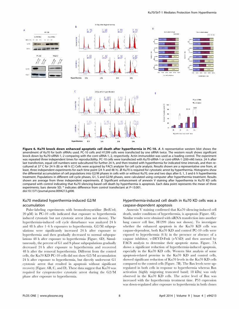
Ku70 mediated hyperthermia-induced G2/Maccumulation
Pulse-labeling experiments with bromodeoxyuridine (BrdUrd;
20 mM) in PC-10 cells indicated that exposure to hyperthermia
induced cytostatic but not cytotoxic arrest (data not shown). The
hyperthermia-induced cell cycle disturbance was analyzed 24 h
and 48 h after 1–6 h exposures to hyperthermia. G2/M subpop-
ulations were significantly increased 24 h after exposure to
hyperthermia and then gradually decreased to normal subpopu-
lations 48 h after exposure to hyperthermia (Figure. 6D). Simul-
taneously, the percent of G1 and S phase subpopulations gradually
decreased 24 h after exposure to hyperthermia and recovered
48 h after the removal hyperthermia. Different from the control
cells, the Ku70 KD PC-10 cells did not show G2/M accumulation
24 h after exposure to hyperthermia, but directly underwent G1
cytotoxic arrest that resulted in apoptosis without significant
recovery (Figure. 6B, C, and D). These data suggest that Ku70 was
required for cytoprotective cytostatic arrest during the G2/M
phase after exposure to hyperthermia.
Hyperthermia-induced cell death in Ku70 KD cells was acaspase-dependent apoptosis
Annexin V staining confirmed that Ku70 silencing-induced cell
death, under conditions of hyperthermia, is apoptosis (Figure. 6E).
Similar results were obtained with siRNA transfection into another
lung cancer cell line, H1299 (data not shown). To investigate
whether the enhanced apoptosis in the Ku70 KD cells was
caspase-dependent, both Ku70 KD and control PC-10 cells were
exposed to hyperthermia (6 h) in the presence or absence of a
caspase inhibitor, v-DEVD-Fmk (z-VAD) and then assessed by
FACS analysis to determine their apoptotic status. Figure. 7A
shows a significant reduction of hyperthermia-induced apoptosis,
especially in the Ku70 KD cells. Western blot analysis of some
apoptosis-related proteins in the Ku70 KD and control cells,
showed significant reduction of Ku70 levels in the Ku70 KD cells
compared to the control cells (Figure. 7B). The Bax levels were up-
regulated in both cells in response to hyperthermia whereas Bax
activation (highly migrating truncated band; 18 kDa) was only
observed in the Ku70 KD cells. The active level of Bax was
increased with the hyperthermia treatment time. P53 expression
was down-regulated after exposure to hyperthermia in both clones
Figure 6. Ku70 knock down enhanced apoptotic cell death after hyperthermia in PC-10. A. A representative western blot shows theamendment of Ku70 for both siRNAs used. PC-10 cells and H1299 cells were transfected by one siRNA twice. The western result shows significantknock down by Ku70-siRNA-1,-2 comparing with the cont-siRNA-1,-2, respectively. Actin immunoblot was used as a loading control. The experimentwas repeated three independent times for reproducibility. PC-10 cells were transfected with Ku70-siRNA-1 or cont-siRNA-1 (200-nM) twice. 24 h afterlast transfection, equal cell numbers were subcultured for further 24 h, and then treated with hyperthermia for indicated time intervals, and then re-cultured at 37uC for 24 h (B) or 48 h (C) Cells were acquired by FACS analyzer for cell cycle analysis. Results shown are a representative one from, atleast, three independent experiments for each time point (24 h and 48 h). D. Ku70 is required for cytostatic arrest by hyperthermia. Histograms showthe differential accumulation of cell populations into G2/M phases in cells with or without Ku70, one and two days after 0, 1, 3 and 6 h hyperthermiatreatment. Populations in different cell cycle phases, G1, S and G2/M phases, were calculated using computer after hyperthermia treatment. Resultsshown are average from three independent experiments. E. Significant enhancement of annexin V staining after hyperthermia in Ku70 KD cellscompared with control indicating that Ku70 silencing-based cell death by hyperthermia is apoptosis. Each data point represents the mean of threeexperiments; bars denote SD; * indicates difference from control transfectant at P,0.001.doi:10.1371/journal.pone.0094213.g006
Ku70/SirT-1 Mediates Protection from Hyperthermia
PLOS ONE | www.plosone.org 8 April 2014 | Volume 9 | Issue 4 | e94213

(Figure. 7B; far lower panels); these results suggest that Bax up-
regulation was independent of P53 as confirmed in H1299 cells
which express no functional P53. Figure. 7C concludes the
possible protection mechanism by Ku70 and interprets how
HDACIs disturb this mechanism.
Discussion
The results of this study suggest a candidate mechanism
responsible for resistance to hyperthermia-induced apoptosis in
lung cancer cells. As a fact, Bcl-2 heterodimerizes with Bax to
inhibit its apoptotic effects [24]. Thus, the Bax/Bcl-2 ratio reflects
apoptosis susceptibility [25]. However, Bcl-2 and Bax function
independently to regulate cell death [26]. Although hyperthermia
can activate some caspases [27], when hyperthermia is used
clinically for cancer treatment, hyperthermia-induced apoptosis
has very limited effects. This is consistent with our observation that
Bcl-2 was down-regulated while Bax was up-regulated, without
prominent Bax activation, in the cells studied. The Bax/Bcl-2 ratio
increased under conditions of hyperthermia.
It is well known that Ku70 plays a dual role in DNA double
strand break (DSBs) repair and in suppressing Bax-mediated
apoptosis, by interacting with Ku80 and Bax [28,29]. However, in
the absence of DNA breaks, it is not known whether Ku70 inhibits
apoptosis by associating only with Bax or by mediating other
pathway(s) that affect Bax. The recently reported cytoprotective
function of Ku70 is based on deacetylation [14,17,29], that
renders cancer cells more susceptible to DNA damaging agents or
to Bax activating factors and that are affected by targeting
acetylation. The results of this study showed that the total amount
of Ku70 did not significantly change; however, Bax/Ku70 binding
was increased with exposure to hyperthermia. The Ku70 binding
to Bax might be due to either increased Bax expression or Bax
liberated from Bcl-2. The acetylation status of Ku70 changed with
exposure to hyperthermia.
Ku70 acetylation, by both the I/II HDACs and class III/Sirtuin
deacetylases, including SirT-1, has been previously reported
[17,30]. The results of this study demonstrated that, SirT-1 was
directly up-regulated and interacted with Ku70, under conditions
of hyperthermia, resulting in deacetylation, and the subsequent
ability to sequester more Bax. This scenario might be one plausible
mechanism associated with the promotion of cell survival. SirT-1,
was reported to deacetylates specific lysine residues of many
substrate proteins including Ku70 [31]. SirT-1 was consistently
found to mediate survival with exposure to stress [32]. Ku70 is
acetylated by p300, PCAF, and CBP. This acetylation process
accelerates Bax-mediated apoptosis [14]. Ku70 deacetylation has
been shown to contribute to longevity under conditions of caloric
restriction [19]. Ku70 acts to sequester Bax from mitochondria
[14,15]. Here, we found that SirT-1 and Ku70 work together to
modulate thermo-sensitivity by regulating Ku70 acetylation. This
is consistent with the reports describing SirT-1 as a responder to
environmental stress [20,32]. The results of this study demon-
strated that the inhibition of Ku70 deacetylation, by specific
HDACIs, NAM or TsA, attenuated the protective role of SirT-1
from hyperthermia and enhanced hyperthermia-induced apopto-
sis.
Lung cancer cells were sensitive to Ku70 siRNA-based
inhibition. This inhibition interfered with the protective mecha-
nism against hyperthermia, and resulted in significant apoptosis.
The DNA content, annexin-V staining and morphological
changes observed, all confirmed induced apoptosis. Bak and/or
Bax activation is necessary for intrinsic apoptosis [33]. Bax
activation is essential and sufficient for mitochondrial permeabi-
lization and cytochrome C release [33,34]. The results of this study
demonstrated Bax activation after exposure to hyperthermia in
Ku70 KD cells; in addition, apoptosis was blocked by treatment
with the apoptosis inhibitor, z-VAD. The findings of this study
showed that heat stress-induced apoptosis takes several hours in
cells compared to minutes in isolated mitochondrial systems
[35,36]. The antiapoptotic proteins, Bcl-2 and Bcl-xL, can
sequester activator proteins and inhibit their ability to homo-
dimerize and regulate apoptosis. Ku70 can be added to the list of
antiapoptotic molecules that sequester Bax and inhibit its
activation after exposure to hyperthermia ex vivo. Hyperthermia
primed the signal for apoptosis by increasing the expression of
Bax. However, the direct activation of Bax by hyperthermia may
either require more exposure time or special conditions. Among
these conditions are the addition of HDACIs to culture media to
induce Bax/Ku70 flipping and Bax-based apoptosis. The combi-
nation (hyperthermia and HDACIs) treatment-induced apoptosis
was significantly inhibited in Bax KD cells, which indicates the
crucial role of Bax in this cell death. Rather than ‘‘death by
default,’’ the emerging view is that apoptosis requires Bax
activation, which can be achieved by targeting Ku70 deacetylation
with HDACIs or Ku70 knockdown during exposure to hyper-
thermia.
Human lung cancer cells are thermo-sensitive [36]. Hyperther-
mia may induce double strand DNA breaks [37]; however, only
limited cell death occurs with hyperthermia independent of DNA
breaks [38]. Although hyperthermia destroys some cells, by an
unknown mechanism, hyperthermia selectively induces apoptosis
during the S-phase in lung cancer cells [39]. This is probably
because the S-phase and M-phase are the most sensitive to the cell
death program [40].
Many studies, including this one, have shown that hyperthermia
results in temporary (cytostatic) arrest of most cancer cells in the
G2/M phase [41]. A majority of such cells re-enter the cell cycle
after removal of the thermal stress and limited apoptosis occurs.
The results of this study showed that Ku70 silencing inhibited the
cytostatic effects of hyperthermia and caused cytotoxic G1
accumulation. These findings suggest that Ku70 is essential for
G2/M accumulation and subsequent protection from hyperther-
mia. Yamamoto et al., [42], reported that cells are more
susceptible to a death signal during G2/M because of Bcl-2
phosphorylation, which lowers the apoptosis threshold. The results
of this study did not show Bcl-2 phosphorylation but rather only
down-regulation under conditions of hyperthermia; these findings
indicate that Bcl-2 does not play a role in protection from
hyperthermia ex vivo. Instead, Ku70 was mainly involved in the
protection scenario. Changing the acetylation status of cells may
influence chromatin condensation and hence, DNA-repair.
Deacetylation of a critical component of DNA repair machinery
such as Ku70 or Ku70 KD may affect DNA repair machinery;
however, again, cell death by hyperthermia had not been
attributed to DNA break [39]. Even though the Ku70 KD cells,
in this study, did not show abnormal cell cycle patterns without
stress. These findings indicate that Ku70 plays a key role in the cell
cycle progression that is essential for protection from hyperther-
mia. Thus, targeting Ku70, rather than inhibiting its deacetyla-
tion, may facilitate cell toxicity under conditions of hyperthermia,
by a mechanism that is associated with a cell cycle-dependent
disturbance.
Conclusion
In summary, the main finding of this study was the biphasic role
of Ku70 during thermal stress. This finding might have
Ku70/SirT-1 Mediates Protection from Hyperthermia
PLOS ONE | www.plosone.org 9 April 2014 | Volume 9 | Issue 4 | e94213

therapeutic relevance, with regard to the interplay between Ku70
acetylation and/or expression as a modulator of subsequent Bax
activation. The results of this study add to the understanding of
apoptosis under thermal stress as well as the identification of
potential targets to improve hyperthermia related treatment of
lung cancer; the targeting of Ku70 might have therapeutic
relevance in combination with siRNAs and/or specific HDACIs.
Figure 7. Enhanced cell death in Ku70 KD cells is caspase-dependent. A. Ku70 KD and control PC-10 cells were treated with hyperthermia(42.5uC) for 6 h in presence or absence of caspase inhibitor (Z-VAD; 50 nM). Two days later, cells were fixed and acquired by FACS analyzer. Cell deathby hyperthermia in both cell clones was regressed by caspase inhibitor indicating that hyperthermia kills Ku70 KD and control cancer cells throughcaspase-dependent apoptosis. B. Proteomic profile of some apoptosis-related proteins in Ku70 KD cells. Ku70 KD and control PC-10 cells treated withhyperthermia at indicated time, lysed, fractionated and blotted. Ku70, Bax and P53 were detected in the blots. Right panel, Ku70 clearly knockeddown. Bax shows extra band around 18 kDa indicating active Bax only in the Ku70 KD cell but not the control transfectant, left panel. The active bandincreased with increasing hyperthermia treatment time. P53 decreased by hyperthermia in both clones indicating that hyperthermia-induced Bax up-regulation and activation are P53 independent. C. Schematic presentation describes role of Ku70 in cellular protection from hyperthermia. Ku70, likeBcl-2 and Bcl-xL, restrains Bax from translocation into mitochondria in cells without stress. Under hyperthermia application, Bcl-2 is down-regulatedand some Bax become free. In addition, more Bax is overexpressed by hyperthermia. Despite Bax regulation, no Bax is activated because Ku70 bindwith this free Bax. Total Ku70 has change but acetylated Ku70 transformed into deacetylated due to SirT-1 activation. Addition of HDACIs duringhyperthermia sensitized lung cancer cells to hyperthermia. HDACIs inhibited SirT-1 function and subsequently increase the chance of Bax activationand translocation to mitochondria inducing apoptosis under hyperthermia.doi:10.1371/journal.pone.0094213.g007
Ku70/SirT-1 Mediates Protection from Hyperthermia
PLOS ONE | www.plosone.org 10 April 2014 | Volume 9 | Issue 4 | e94213

Supporting Information
Figure S1 Phase contrast photographs of H1299 cells two days
after treatment with HDACIs (NAM at 20 mM or TsA at 300 nM
final conc. or both for 4 h) then hyperthermia for 6 h. The image
showed limited number of recovered cells as well as the rounded
and floating apoptotic cells mainly in the colonies pre-treated with
HDACIs then hyperthermia. HDACIs themselves did not induce
significant cell death.
(TIF)
Figure S2 Hyperthermia-induced apoptosis was enhanced by
HDACis. Pre-treatment of H1299 cells with DHACIs (NAM at
20 mM or TsA at 300 nM final conc. for four hours) significantly
increased hyperthermia-induced apoptosis. Comparative cyto-
grams show Annexin V staining after HDACis and hyperthermia
in H1299 cells.
(TIF)
Figure S3 In PC-10 cells, Hyperthermia and HDACis combi-
nation-induced apoptosis was attenuated by Bax siRNA. PC-10
cells were either transfected with Bax siRNA or cont siRNA.
When these cells were pre-treated with HDACIs (NAM at 20 mM
or TsA at 300 nM final conc. for four hours) followed by
hyperthermia (6 h), the apoptotic outcome was significantly
decreased in the Bax KD cells compared with control ones.
Comparative cytograms show Annexin V staining after HDACis
and hyperthermia in Bax KD and control PC-10 cells.
(TIF)
Figure S4 Proteomic analysis of some apoptosis –related
proteins in H1299 cells. Whole cell extracts were prepared after
0 h or 6 h hyperthermia (42.5uC). Bax, Ku70 and Bcl-2 were
analyzed by immunoblotting. Six hours hyperthermia induced
slight increase in Bax expression level and reduced Bcl-2 while Bcl-
xL and Ku70 had not been affected. Analysis was performed in the
same blot so each protein worked as a loading control for the
other. A representative Coomassi Brilliant Blue (CBB) staining of
the membrane was shown to act as a loading control.
(TIF)
Figure S5 Representative images show localization of Bcl-2 and
Bax in lung cancer cell lines. Bax (green), Bcl-2 (green), and nuclei
(red) were stained. Bax localization: cytosol in PC-10 cells (a) and
in the cytoplasm and the nucleus in H1299 cells (b). Bcl-2 is
localized in the cytoplasm in all cells tested (c,d).
(TIF)
Figure S6 Hyperthermia modulates Bax association with Ku70
in CHAPS buffer. PC-10 cells were incubated at 42.5uC for 0, 1, 3
and 6 h. then lysed in CHAPS buffer. Ku70 was co-immunopre-
cipitated from 2 mg total protein and Bax was detected in the
immunoprecipitant by western analysis (upper panel). Total Ku70
levels showed no changes. Hyperthermia induced Bax up-
regulation and enhanced association between Bax and Ku70.
On the other hand, Bax was co-immunoprecipitated from similar
cell lysates. Ku70 was detected in the immunoprecipitant.
Again,after hyperthermia association between Ku70 and Bax
was enhanced.
(TIF)
Figure S7 Hyperthermia did not change expression of HDAC6
or SirT-3. PC-10 was treated with hyperthermia (0–6 h) and cells
were lysed and fractionated and blotted. Both HDAC-6 and SirT-
3 expression was evaluated by immunoblotiing analysis. Immu-
nodetection indicated that hyperthermia did not induce significant
changes in the expression of both proteins in PC-10.
(TIF)
Figure S8 Ku70 is required for cytostatic arrest by hyperther-
mia. H1299 cells were transfected with Ku70-siRNA-2 or cont-
siRNA-2 (100 nM) twice. 24 h after last transfection, equal cell
numbers were subcultured for further 24 h, and then treated with
hyperthermia for indicated time periods and then re-cultured at
37uC for 24 h (a) or 48 h (b) Cells were acquired by FACS
analyzer for cell cycle analysis. A representative results is shown at
each time point (24 h and 48 h).
(TIF)
Acknowledgments
Authors thank Prof. Shoichi Inoue and Dr. Takahiko Kobayashi for their
technical advice.
Author Contributions
Conceived and designed the experiments: MH HW. Performed the
experiments: MH AS.S ZM. Analyzed the data: MH AE.S YF.
Contributed reagents/materials/analysis tools: YO NS. Wrote the paper:
MH YO.
References
1. Wust P, Hildebrandt B, Sreenivasa G, Rau B, Gellermann J, et al. (2002)
Hyperthermia in combined treatment of cancer. Lancet Oncol 3: 487–97.2. Takahashi I, Emi Y, Hasuda S, Kakeji Y, Maehara Y, et al. (2002) Clinical
application of hyperthermia combined with anticancer drugs for the treatment ofsolid tumors. Surgery 131:578–84.
3. Mosser DD, Morimoto RI (2004) Molecular chaperones and the stress of
oncogenesis. Oncogene 23: 2907–18.4. Calderwood SK, Asea A (2002) Targeting HSP70-induced thermotolerance for
design of thermal sensitizers. Int J Hyperthermia 18:597–608.5. Li WX, Chen CH, Ling CC, Li GC (1996) Apoptosis in heat-induced cell killing.
The protective role of hsp-70 and the sensitization effect of the c-myc gene.
Radiat Res 145: 324–30.6. Moroi J, Kashiwagi S, Kim S, Urakawa M, Ito H, Yamaguchi K (1996)
Regional differences in apoptosis in murine gliosarcoma (T9) induced by mildhyperthermia. Int J Hyperthermia 12: 345–54.
7. Rossi A, Ciafre S, Balsamo M, Pierimarchi P, Santoro MG (2006) Targeting theHeat Shock Factor 1 by RNA Interference: A Potent Tool to Enhance
Hyperthermochemo-therapy Efficacy in Cervical Cancer. Cancer Res 66: 7678–
85.8. Chowdhury I, Tharakan B, Bhat GK (2006) Current concepts in apoptosis: The
physiological suicide program revisited. Cell Mol Biol Lett 11: 506–25.9. Reed JC (2006) Proapoptotic multidomain Bcl-2/Bax-family proteins: mecha-
nisms, physiological roles, and therapeutic opportunities. Cell Death Differ 13:
1378–86.
10. Cheng E, Wei M, Weiler S, Flavell R, Mak T, et al. (2001) BCL-2, BCL-XL
sequester BH3 domain-only molecules preventing BAX- and BAK-mediated
mitochondrial apoptosis. Mol Cell 8: 705–11
11. Wei MC, Zong WX, Cheng EH, Panoutsakopoulou V, Ross AJ, et al. (2001)
Proapoptotic BAX and BAK. a requisite gateway to mitochondrial dysfunction
and death. Science 292: 727–30
12. Evan GI, Vousden KH (2001) Proliferation, cell cycle and apoptosis in cancer.
Nature 411: 342–48.
13. Pelengaris S, Khan M, Evan GI (2002) Suppression of Myc-induced apoptosis in
beta cells exposes multiple oncogenic properties of Myc and triggers
carcinogenic progression. Cell 109: 321–34.
14. Cohen HY, Lavu S, Bitterman KJ, Hekking B, Imahiyerobo TA, et al. (2004)
Acetylation of the C terminus of Ku70 by CBP and PCAF controls Bax-
mediated apoptosis. Mol Cell13: 627–38.
15. Jose G, Gama V, Yoshida T, Sun W, Hayes P, et al. (2007) Bax inhibiting
peptide derived from Ku70 and cell penetrating penta peptides. Biochemical
Society Transactions, 35: 797–801.
16. Weterings E, Van Gent DC (2004) The mechanism of nonhomologous end-
joining: a synopsis of synapsis. DNA Repair 3: 1425–35.
17. Zhao HJ, Hosoi Y, Miyachi H, Ishii K, Yoshida M, et al. (200) A dependent
protein kinase activity correlates with Ku70 expression and radiation sensitivity
in esophageal cancer cell lines. Clin Cancer Res 6: 1073–78.
Ku70/SirT-1 Mediates Protection from Hyperthermia
PLOS ONE | www.plosone.org 11 April 2014 | Volume 9 | Issue 4 | e94213

18. Subramanian C, Opipari AW Jr, Bian X, Castle VP, Kwok RP (2005) Ku70
acetylation mediates neuroblastoma cell death induced by histone deacetylase
inhibitors. Proc Natl Acad Sci USA 102: 4842–4847.
19. Cohen HY, Miller C, Bitterman KJ, Wall NR, Hekking B, et al. (2004) Calorie
restriction promotes mammalian cell survival by inducing the SIRT1
deacetylase. Science 305: 390–92.
20. Lim chang-Su (2006) SIRT1: cellular senescence, cancer and organismal aging?
Med Hypo 67: 341–44.
21. Inoue S, Takaoka K, Endo T, Mizuno S, Ogawa Y, et al. (1997) In vitro
confirmation of newly established lung cancer cell lines using flow cytometry and
multicellular tumor spheroids. Lung Cancer 17: 85–101.
22. Yamaguchi H, Wang HG (2001) The protein kinase PKB/Akt regulates cell
survival and apoptosis by inhibiting Bax conformational change. Oncogene
20(53):7779–86.
23. Ayene IS, Ford LP, Koch CJ (2005) Ku protein targeting by Ku70 small
interfering RNA enhances human cancer cell response to topoisomerase II
inhibitor and radiation. Mol Cancer Ther 4: 529–36.
24. Clair EG, Anderson SJ, Oltavia ZN (1997) Bcl-2 counters apoptosis by Bax
heterodimerization-dependent and -independent mechanisms in the T-cell
lineage. J Biol Chem 272: 29347–55.
25. Oltvai ZN, Milliman CL, Korsmeyer SJ (1993) Bcl-2 heterodimerizes in vivo
with a conserved homolog, Bax, that accelerate programmed cell death. Cell 74:
609–19.
26. Knudson CM, Korsmeyer SJ (1997) Bcl-2 and Bax function independently to
regulate cell death. Nat Genet 1997; 16: 358–63.
27. Nijhuis A, Le S, Gac Poot A., Feijen J, Vermes I (2008) Bax-mediated
mitochondrial membrane permeabilization after heat treatment is caspase-2
dependent. Inter J Hyperther 24: 357–365
28. Mazumder S, Plesca D, Kinter M, Almasan A (2007) Interaction of a cyclin E
fragment with ku70 regulates bax-mediated apoptosis. Mol Cell Biol 27: 3511–
20.
29. Li Y, Gama V, Yoshida T, Yoshida T, Gomez JA, et al. (2007) Bax-inhibiting
peptide protects cells from polyglutamine toxicity caused by Ku70 acetylation.
Cell Death Differ 14: 2058–67.
30. Chen CS, Wang YC, Yang HC, Huang PH, Kulp SK, et al. (2007) Histone
deacetylase inhibitors sensitize prostate cancer cells to agents that produce DNAdouble-strand breaks by targeting Ku70 acetylation. Cancer Res 67: 5318–27.
31. Vaziri H, Dessain SK, Ng Eaton E, Imai SI, Frye RA, et al. (2001) hSIR2
(SIRT1) functions as a NAD-dependent p53 deacetylase. Cell 107: 149–59.32. Monteiro JP, Cano MI (2011) SIRT1 deacetylase activity and the maintenance
of protein homeostasis in response to stress: an overview. Protein Pept Lett.18(2):167–73.
33. Kuwana T, Mackey MR, Perkins G, Ellisman MH, Latterich M, et al.
(2002)Bid, Bax and lipids cooperate to form supramolecular openings in theouter mitochondrial membrane. Cell 111: 331–42.
34. Korsmeyer SJ, Wei MC, Saito M, Weiler S, Oh KJ, et al. (2007) Pro-apoptoticcascade activates BID, which oligomerizes BAK or BAX into pores that result in
the release of cytochrome c. Cell Death Differ 11: 66–73.35. Pagliari LJ, Kuwana T, Bonzon C, Newmeyer DD, Tu S, et al. (2005) The
multidomain proapoptotic molecules Bax and Bak are directly activated by heat.
Proc Nat Aca Sci 102: 17975–80.36. Van der Zee J (2002) Heating the patient: A promising approach? Ann Oncol
13: 1173–84.37. Takahashi A, Matsumoto H, Nagayama K, Kitano M, Hirose S, et al. (2004)
Evidence for the Involvement of Double-Strand Breaks in Heat-Induced Cell
Killing. Cancer Res 64: 8839–45.38. Kampinga HH, Laszlo A (2005) DNA Double Strand Breaks Do Not Play a
Role in Heat-Induced Cell Killing. Cancer Res 65: 10632–3.39. Dewey WC, Li XL, Wong RS (1990) Cell killing, chromosomal aberrations, and
division delay as thermal sensitivity is modified during the cell cycle. Radiat Res122: 268–74.
40. Higashikubo R, Holland JM, Roti JL (1989) Comparative effects of caffeine on
radiation-and heat-induced alterations in cell cycle progression. Radiat Res 119:246–60.
41. Zolzer F, Streffer C (2001) G2-phase delays after irradiation and/or heattreatment assessed by two-parameter flow cytometry. Radiat Res 155: 50–6.
42. Yamamoto K, Ichijo H, Korsmeyer SJ (1999) Bcl-2 is phosphorylated and
inactivated by an ASK1/Jun N-terminal protein kinase pathway normallyactivated at G2/M. Mol Cell Biol 19: 8469 –78.
Ku70/SirT-1 Mediates Protection from Hyperthermia
PLOS ONE | www.plosone.org 12 April 2014 | Volume 9 | Issue 4 | e94213
Copyright © 2022 FDOKUMEN