Bile Salts Differentially Sensitize Esophageal Squamous Cells to CD95 (Fas/Apo1 Receptor) Mediated...
14
Bile salts sensitize esophageal squamous cells to CD95 (Fas/ Apo-1 receptor) mediated apoptosis via a protein trafficking mechanism Sanjay Naran, Peter Abrams, Pierre Queiroz de Oliveira, and Steven J. Hughes Department of Surgery, Section of Gastrointestinal Surgery, University of Pittsburgh School of Medicine, Pittsburgh, PA 15261 Abstract Introduction—The role of non-acidic reflux contents on the pathophysiology of Barrett’s Esophagus remains poorly understood. We hypothesized that esophageal squamous epithelium differs from Barrett’s columnar epithelium in response to bile salts with respect to subsequent changes in the cell surface expression of CD95 (Fas/Apo-1) and sensitivity to CD95-mediated apoptosis. Methods—Immortalized esophageal squamous cells (HET-1A) and Barrett’s Esophagus cells (BAR-T), and esophageal adenocarcinoma cells (Flo-1) were treated with toxic and non-toxic bile salts at concentrations observed in gastroesophageal refluxate. CD95 cell-surface expression and apoptotic response to activating anti-CD95 antibody treatment was determined by FACScan analysis. Results—Bile salt exposure resulted in a dose-dependent increase in CD95 cell-surface expression in HET-1A cells, but not BAR-T or Flo-1 cells. This response occurred rapidly, within a time-frame inconsistent with de novo protein synthesis and was blocked by protein kinase C (PKC) inhibition. Surprisingly, PKC inhibition in Flo-1 cells resulted in an increase in CD95 cell surface expression. Following bile salt exposure, a corresponding increase in the induction of CD95-mediated apoptosis was observed in HET-1A cells; PKC inhibition sensitized Flo-1 cells to apoptosis. Conclusions—Our findings suggest that esophageal squamous cells are sensitized to CD95- mediated apoptosis following bile salt exposure. This response differs from that in columnar epithelial cells, and may offer a potential mechanism of selection pressure that contributes to the pathophysiology of Barrett’s Esophagus. Keywords Barrett’s Esophagus; Gastroesophageal Reflux Disease; Esophageal Adenocarcinoma © 2010 Elsevier Inc. All rights reserved. Correspondence: Steven J. Hughes, M.D., Associate Professor, University of Pittsburgh, 497 Scaife Hall, 3550 Terrace Street, Pittsburgh, PA 15261, [email protected]. Publisher's Disclaimer: This is a PDF file of an unedited manuscript that has been accepted for publication. As a service to our customers we are providing this early version of the manuscript. The manuscript will undergo copyediting, typesetting, and review of the resulting proof before it is published in its final citable form. Please note that during the production process errors may be discovered which could affect the content, and all legal disclaimers that apply to the journal pertain. NIH Public Access Author Manuscript J Surg Res. Author manuscript; available in PMC 2012 December 1. Published in final edited form as: J Surg Res. 2011 December ; 171(2): 504–509. doi:10.1016/j.jss.2010.05.001. NIH-PA Author Manuscript NIH-PA Author Manuscript NIH-PA Author Manuscript
Transcript of Bile Salts Differentially Sensitize Esophageal Squamous Cells to CD95 (Fas/Apo1 Receptor) Mediated...

Bile salts sensitize esophageal squamous cells to CD95 (Fas/Apo-1 receptor) mediated apoptosis via a protein traffickingmechanism
Sanjay Naran, Peter Abrams, Pierre Queiroz de Oliveira, and Steven J. HughesDepartment of Surgery, Section of Gastrointestinal Surgery, University of Pittsburgh School ofMedicine, Pittsburgh, PA 15261
AbstractIntroduction—The role of non-acidic reflux contents on the pathophysiology of Barrett’sEsophagus remains poorly understood. We hypothesized that esophageal squamous epitheliumdiffers from Barrett’s columnar epithelium in response to bile salts with respect to subsequentchanges in the cell surface expression of CD95 (Fas/Apo-1) and sensitivity to CD95-mediatedapoptosis.
Methods—Immortalized esophageal squamous cells (HET-1A) and Barrett’s Esophagus cells(BAR-T), and esophageal adenocarcinoma cells (Flo-1) were treated with toxic and non-toxic bilesalts at concentrations observed in gastroesophageal refluxate. CD95 cell-surface expression andapoptotic response to activating anti-CD95 antibody treatment was determined by FACScananalysis.
Results—Bile salt exposure resulted in a dose-dependent increase in CD95 cell-surfaceexpression in HET-1A cells, but not BAR-T or Flo-1 cells. This response occurred rapidly, withina time-frame inconsistent with de novo protein synthesis and was blocked by protein kinase C(PKC) inhibition. Surprisingly, PKC inhibition in Flo-1 cells resulted in an increase in CD95 cellsurface expression. Following bile salt exposure, a corresponding increase in the induction ofCD95-mediated apoptosis was observed in HET-1A cells; PKC inhibition sensitized Flo-1 cells toapoptosis.
Conclusions—Our findings suggest that esophageal squamous cells are sensitized to CD95-mediated apoptosis following bile salt exposure. This response differs from that in columnarepithelial cells, and may offer a potential mechanism of selection pressure that contributes to thepathophysiology of Barrett’s Esophagus.
KeywordsBarrett’s Esophagus; Gastroesophageal Reflux Disease; Esophageal Adenocarcinoma
© 2010 Elsevier Inc. All rights reserved.Correspondence: Steven J. Hughes, M.D., Associate Professor, University of Pittsburgh, 497 Scaife Hall, 3550 Terrace Street,Pittsburgh, PA 15261, [email protected]'s Disclaimer: This is a PDF file of an unedited manuscript that has been accepted for publication. As a service to ourcustomers we are providing this early version of the manuscript. The manuscript will undergo copyediting, typesetting, and review ofthe resulting proof before it is published in its final citable form. Please note that during the production process errors may bediscovered which could affect the content, and all legal disclaimers that apply to the journal pertain.
NIH Public AccessAuthor ManuscriptJ Surg Res. Author manuscript; available in PMC 2012 December 1.
Published in final edited form as:J Surg Res. 2011 December ; 171(2): 504–509. doi:10.1016/j.jss.2010.05.001.
NIH
-PA Author Manuscript
NIH
-PA Author Manuscript
NIH
-PA Author Manuscript

INTRODUCTIONBarrett’s esophagus (BE) is a metaplastic, columnar epithelium that replaces the normalstratified squamous epithelium of the lower esophagus (32), and is generally accepted to bea pre-malignant epithelium from which esophageal adenocarcinoma (EA) may arise.Significant effort to improve our understanding of the biology behind this metaplastictransformation is warranted, as an alarming increase in the incidence of EA is occurring inthe developed world, and EA has now replaced squamous cancer as the most commonhistological type of esophageal cancer in the United States (1). Gastroesophageal refluxdisease (GERD) and obesity have been identified as the major risks factors for thedevelopment of EA (32).
Extensive evidence has established that BE develops as a complication of GERD. The roleof acidic reflux into the esophagus in the pathogenesis of BE is widely accepted (31),however, more recent investigation has suggested bile salts may play an important role inthe pathophysiology of this metaplasia (29). While numerous studies have improved ourknowledge in this regard, the mechanism(s) and relative contribution of bile salts in thepathophysiology of BE remains poorly understood.
We have previously reported that CD95 (Fas/Apo-1) expression is altered in virtually allcases of EA, and that these changes occur early in the progression of BE to EA (13). CD95(Fas/Apo-1) is a transmembranous cell-surface receptor and member of the “death receptor”family that includes tumor necrosis factor receptors (TNFR1 and TNFR2), and DR4 andDR5 (24). CD95 transmits an apoptotic signal following binding by Fas ligand (FasL) (24).Consistent with this function, near universal resistance to Fas-mediated apoptosis has beenobserved in malignancy (25). Our previous work demonstrated that decreased cell-surfaceexpression of CD95 in EA is associated with resistance to CD95-mediated apoptosis. Thisdecreased cell-surface expression is not due to reduced total protein levels, rearrangementsin the gene or altered transcription, but, rather appears to be due to alterations in post-translational trafficking resulting in accumulation of CD95 within the cytoplasm (13).
Two reports in the literature, in combination with our previous work, led us to design thepresent study. First, in experiments investigating the pathophysiology of cholestaticinhibition of liver regeneration, hepatocytes were shown to traffic CD95 from cytoplasmicstores to the cell surface in response to exposure to bile salts through a protein kinase C(PKC) dependent mechanism (16). This response to bile salt exposure sensitized hepatocytesto CD95 mediated apoptosis, and established post-translational regulation of CD95 via PKCregulated trafficking of storage pools of CD95 to the cell surface. Second, CD95 and FasLinteractions were shown to be essential to the pathophysiology of pancreatic acinar to ductalmetaplasia that progresses to pancreas cancer following pancreatic duct ligation in a murinemodel (3). Thus, taken together with our previous work, these findings led us to speculatethat CD95 may play a role in the pathophysiology of BE. We hypothesized that exposure tobile salts would result in increased CD95 cell-surface expression in squamous esophagealepithelial cells, but not in BE or EA cells.
MATERIALS AND METHODSCell Lines and Culture
The Het-1A cell line is a SV-40-immortalized, non-neoplastic, normal squamous epithelialcell line that has been previously described (34). Het-1A cells were obtained from theAmerican Type Culture Collection (Manassas, VA) and maintained per the recommendedgrowing conditions in LHC-9 growth medium (BioSource; Camarillo, CA). The non-neoplastic Barrett’s esophagus cell line (BAR-T), created via hTERT-immortalization of a
Naran et al. Page 2
J Surg Res. Author manuscript; available in PMC 2012 December 1.
NIH
-PA Author Manuscript
NIH
-PA Author Manuscript
NIH
-PA Author Manuscript

human sample of Barrett’s esophagus, was provided by Drs. RF Souza and CP Morales andis also described elsewhere (15). BAR-T cells were maintained in co-culture with amitomycin C-treated Swiss 3T3 feeder layer. The feeder cells were removed with 0.02%EDTA (titrated with NaOH to pH 7.4) before sub-culturing pure BAR-T cells 48 hours priorto experiments. Cells were maintained in keratinocyte basal media (KBM-2) (Cambrex;Walkersville, MD) supplemented with 5% FBS (Atlanta Biologicals; Norcross, GA), 0.1 nMcholera toxin (Calbiochem; San Diego, CA), 100 U/ml penicillin-streptomycin (GIBCO-BRL; Gaithersburgh, MD), 70 µg/ml bovine pituitary extract (Hammond Cell Technologies;Windsor, CA), 400 ng/ml hydrocortisone, 20 µg/ml EGF, 10 mg/ml adenine, 5 µg/mlinsulin, and 5 µg/ml transferrin (all from Sigma-Aldrich Corp.; St Louis, MO). Anestablished human EA cell line (Flo-1) was also studied and maintained in culture aspreviously described (13). BJAB cells, a B-cell lymphoma cell line that is sensitive toCD95-mediated apoptosis was also maintained in RPMI with 5% FBS and utilized as apositive control.
TreatmentsStock solutions of bile salts (1.0 mM) were freshly prepared immediately prior to eachtreatment by dissolving guanochenodeoxycholate (GCDC) or taurodeoxycholate (TDC)obtained from Sigma-Aldrich Corp. (St. Louis, MO) in standard growth media. Thesolutions were titrated with 1.0 N NaOH to a 6.5 pH, and the solutions were protected fromsources of light to prevent degradation. Stock solutions were then added directly to 1 cc offresh, standard culture media in the culture well to produce final bile salt concentrationsranging from 50 to 250 µM. Treatment duration ranged from 5 to 60 minutes. Cellsmaintained in complete media served as controls.
For PKC inhibition experiments, cells were pretreated for 10 minutes by exposure to 1 µMof the PKC-inhibitor bisindolymaleimide (BIM) (BIM dissolved in DMSO and diluted 1:10in PBS to create a 1 mM stock solution) prior to exposure to 250 µM of bile salts asdescribed above for total treatment durations ranging from 15 to 120 minutes. Cells treatedwith vehicle alone served as additional controls. All presented data represent results from aminimum of 3 individual experiments preformed on separate days.
Quantification of Cell Surface Expression of CD95CD95 cell-surface expression was analyzed using flow cytometry as previously described(13). In brief, adherent cells were gently freed from culture plates using 10 mM EDTA(titrated with NaOH to pH 7.4) in phosphate buffered saline (PBS) to preserve theextracellular epitopes of membranous proteins, and then washed in a buffer consisting ofPBS containing 10−3 mM NaN3 and 0.5% normal rabbit serum. The cells were thenincubated on ice with a 1:1000 dilution of anti-Fas antibody (Apo-1, Kamiya BiomedicalCo.; Seattle, WA) for 30 minutes, washed in buffer (5 minutes × 3), and then incubated withan appropriate FITC-conjugated anti-mouse secondary antibody (BioSource; Camarillo, CA)at a 1:2500 dilution for 30 minutes at 4°C while being protected from exposure to light.Controls consisted of unstained cells, and cells treated with secondary antibody alone. Afterthree final washings in PBS containing 10−3 mM NaN3, CD95 expression was analyzedusing a Becton Dickinson FACSort™ flow cytometer (Becton Dickinson; San Jose, CA).Data is reported as mean fluorescence, and was analyzed using the WinMDI (The ScrippsResearch Institute; San Diego, CA) software program.
Assessment of CD95-Mediated ApoptosisCells were treated with 250 µM of bile salts with or without pretreatment with 1 µM BIM(PKC-inhibitor). Control cells were treated with vehicle alone, and BJAB cells served as apositive control in each experiment to confirm the efficient induction of CD95-mediated
Naran et al. Page 3
J Surg Res. Author manuscript; available in PMC 2012 December 1.
NIH
-PA Author Manuscript
NIH
-PA Author Manuscript
NIH
-PA Author Manuscript

apoptosis. Additional culture wells served to confirm by FACScan analysis the impact ofbile salt and BIM treatment on cell-surface expression of CD95. Thirty minutes aftertreatments, cells were exposed to apoptosis-inducing, activating anti-CD95 antibody (Apo-1,Kamiya Biomedical Co.; Seattle, WA) and recombinant protein G (rPG), or rPG alone as acontrol. Twenty-four hours after this exposure, the cells sensitivity to FasL inducedapoptosis was determined by FACScan analysis. Adherent cells were freed from cultureusing 0.1% trypsin, washed in PBS (pH 7.4) containing 10−3 mM NaN3 and pelleted bycentrifugation. Analysis of apoptosis was performed, detected and quantified using anannexin V-FITC and propidium iodide (PI) staining apoptosis detection kit (BDBiosciences; San Diego, CA) according to the manufacturer’s instructions. Analysis wasagain performed using a Becton Dickinson FACSort™ flow cytometer (Becton Dickinson;San Jose, CA). Quadrant analysis was performed using the WinMDI software program.
Statistical AnalysisComparisons between treatment groups were made using a two-tailed Student’s t test.p<0.05 was used to determine statistical significance.
RESULTSTreatment with Bile Salts Increases Surface Expression of CD95 in HET-1A Cells, but NotBAR-T or EA Cells
We hypothesized that bile salt treatment would result in increased cell-surface expression ofCD95 in esophageal squamous epithelial cells (Het-1A), but fail to increase cell-surfaceexpression of CD95 in the Barrett’s Esophagus cell line (BAR-T). Immortalized Esophagealsquamous cells (Het-1A) and Barrett’s Esophagus cells (BAR-T), and esophagealadenocarcinoma cells (Flo-1) were treated with Guanochenodeoxycholate (GCDC) orTaurodeoxycholate (TDC) at concentrations of 50, 100, and 250 µM and harvested at 5, 30or 60 minutes. Control cells in each group were treated with equivalent volume of vehiclealone (pH 6.5). Cell-surface expression of CD95 was then determined via flow cytometryFACScan, using a phycoerythrin-conjugated anti-CD95 antibody. An increase in the surfaceexpression of CD95 in Het-1A cells was observed following exposure to each of the two bilesalts (Figure 1). However, neither the EA cell line nor the BAR-T cell line demonstrated achange (increase or decrease) in the surface expression of CD95 following bile saltexposure. Thus, bile salts were shown to increase the surface expression of CD95 in normalsquamous epithelial cells, but not in esophageal adenocarcinoma or Barrett’s esophaguscells.
Increases in Surface Expression of CD95 in Normal Squamous Epithelial Cells FollowingBile Salt Treatment Occurs Rapidly in a Dose Dependent Manner
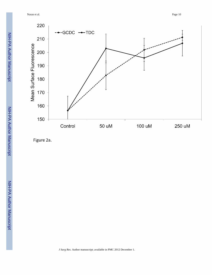
We next aimed to determine if CD95 cell-surface expression following exposure to bile saltswould respond in dose dependent fashion. Het-1A cell-surface CD95 expression wasobserved to be dose-dependent for GCDC, with a peak effect detected at a concentration of250 µM. For TDC, maximum effect was observed at a concentration of 50 µM, the lowestdose tested (Figure 2a). Surface expression of CD95 was not affected regardless of dose ineither Flo-1 or BAR-T cells (data not shown). Harvesting Het-1A cells at various time pointsrevealed this increase in the surface expression of CD95 began within 5 minutes followingtreatment and reached peak levels within 30 minutes (Figure 2b).
Naran et al. Page 4
J Surg Res. Author manuscript; available in PMC 2012 December 1.
NIH
-PA Author Manuscript
NIH
-PA Author Manuscript
NIH
-PA Author Manuscript

PKC Inhibition Blocks Bile-Salt Induced Increases in Fas Surface Expression in NormalSquamous Epithelial Cells, but Not in Adenocarcinoma Cells
Bile salts have been shown to induce trafficking of cytosolic pools of CD95 to the cellsurface via a PKC modulated pathway. As such, we hypothesized that by inhibiting PKCactivity, a bile salt-induced increase in the surface expression of CD95would be blocked.Esophageal squamous cells (Het-1A) and adenocarcinoma cells (Flo-1) were each treatedwith 250 µM of either GCDC, or TDC for a duration of 30, 60 or 120 minutes, withconcurrent exposure to the PKC-inhibitor Bisindolymaleimide (BIM). Control groups wereexposed to bile salts without concurrent exposure to BIM. Following treatment, cell surfaceexpression of CD95 was again determined via FACScan analysis, using phycoerythrin-conjugated anti-CD95 antibody. Within Het-1A cells, inhibition of PKC was shown to blockbile salt-induced increases in surface expression of CD95 (Figure 3a). Surprisingly, BIMtreatment appeared to have the opposite effect in Flo-1 cells following bile-salt exposure(Figure 3b); increasing the surface expression of CD95. Harvesting of Flo-1 cells at multipletime points following concurrent BIM and bile-salt treatment revealed this increase in thesurface expression of CD95 plateaued within 60 min(Figure 3b). Thus, PKC inhibition wasshown to block bile-salt induced increases in the cell surface expression of CD95 withinnormal squamous epithelial cells, but PKC inhibition increased cell-surface expression ofCD95 in an adenocarcinoma cell line.
Increased Surface Expression of CD95 Correlates with Increased Sensitivity to FasL-Induced Apoptosis
In order to confirm that an increase in CD95 expression correlates with an increase insensitivity to FasL-induced apopotisis, an Annexin V-Propidium Iodide apoptosis detectionkit was used to quantify cell viability in Het-1A and Flo-1 cells (Figure 4). We observed thatbile-salt treatment resulted in increased mean CD95 expression and to FasL-inducedapoptosis. PKC inhibition using pretreatment with BIM attenuated this response. In Flo-1cells, PKC inhibition via treatment with BIM was shown to exacerbate sensitivity to FasL-induced apoptosis.
DISCUSSIONWe have shown that esophageal squamous cells differ from metaplastic, esophagealcolumnar cells and adenocarcinoma cells in the response to exposure to bile salts withrespect to cell-surface expression of the apoptosis-inducing receptor CD95. Thesedifferences occur with treatment doses consistent with those present in gastro- andenterduodenal-refluxate of patients with GERD (8,9). We have further demonstrated thatthis response is dose dependent, and occurs in a time-frame consistent with a post-translational trafficking event that is dependent upon PKC activity. Finally, we demonstratedthat increased cell-surface expression of CD95 sensitized the cells to CD95-mediatedapoptosis.
Our findings support the possibility that an intrinsic difference in sensitivity to CD95-mediated apoptotic signals between squamous and enteric columnar epithelia in response tobile salt reflux may contribute to the pathophysiology of BE. This is consistent with thefindings of Leach, et al. that FasL/CD95 interactions are essential to pancreatic ductalmetaplasia that precedes invasive malignancy in a murine model of spontaneous pancreaticcancer following pancreatic duct ligation (3). However, this conclusion must be temperedgiven our dependence on immortalized cell lines grown in culture, and lack of in vivo data.Further study is certainly necessary before we can confidently conclude that this differencebetween esophageal squamous and BE epithelia clearly exists and contributes to the
Naran et al. Page 5
J Surg Res. Author manuscript; available in PMC 2012 December 1.
NIH
-PA Author Manuscript
NIH
-PA Author Manuscript
NIH
-PA Author Manuscript

development of columnar metaplasia of the esophagus. Current animal models of BEpreclude confirmation of our findings in vivo.
Our data further suggests that cell-surface expression of CD95 is regulated by post-translational trafficking of the protein modulated by PKC activity. Thus, our findings areconsistent with those observed in hepatocytes by Gores et al. (16). The role of PKC in post-translational trafficking of proteins, including delivery and retrieval of proteins to and fromthe cell surface is well established (19). We suspect that this response to bile salts is cell-type specific, and given normal physiology, that all columnar entercytes lack this responseto bile salt exposure. Our observations in the EA cell line Flo-1 may suggest that PKC-dependent endocytosis of CD95 contributes to the low level of CD95 surface expressionobserved in esophageal adenocarcinoma. As CD95 has been shown to contribute tochemotherapy-induced cell death (17,27) and PKC inhibition has been explored as therapyfor cancer (2,6,22), this observation suggests further investigation exploring the potential ofsynergistic effects between these therapies may be warranted.
Bile salts have been shown to result in a number of other events within esophageal epithelialcells. Conjugated bile salts and the inflammatory cytokines TNF-alpha and IL-1betaincrease CDX1 mRNA expression in vitro (26,30,33,35). CDX1, an important regulator ofnormal intestinal development (7,10) and deoxycholic acid up-regulates goblet-specific geneMUC2 expression in concert with CDX2 in human esophageal cells (12,18); thus bile saltexposure may also contribute to columnar differentiation (35). The bile acid receptor FXR issignificantly overexpressed in Barrett's esophagus compared to normal mucosa, esophagitisand esophageal adenocarcinoma. In addition, the induction of apoptosis by the FXRinhibitor guggulsterone in a Barrett's esophagus-derived cell line suggests that FXR maycontribute to the regulation of apoptosis in this epithelium (5). Finally, bile salt exposureincreases proliferation through PI-3K (14), and p38 and ERK MAPK pathways in BAR-Tcells (15). Thus, a number of mechanisms have been identified that could be driving anatural selection process that results in BE metaplasia following bile salt exposure.
Bile salts share significant molecular properties with hormones, and intracellular receptorsthat are activated by bile salt receptors have been identified (4,21). We intentionally limitedthese experiments to bile salt treatments at pH 6.5 to elucidate their effect in the context ofacid suppression therapy. At this pH, the bile slats are likely of neutral charge with access tothe cytoplasm (11,21,23). The ionic charge and subsequent cell permeability of thesecompounds is clearly dependent on pH, and further investigation into our observations atvarying pH is necessary to better understand how the observed effects may differ in moreacidic environments. Moreover, further elucidation is needed as to whether these effects aremediated by bile salt receptors or reflect changes in the lipid composition of the cellmembrane or intracellular organelles such as lipid rafts, known to harbor cytoplasmic poolsof CD95 (20,28).
In summary, TDC and GCDC exposure induces esophageal squamous cell sensitivity toCD95-mediated apoptosis via a post-translational mechanism dependent upon PKC.Barrett’s columnar epithelial cells lack this response, offering a potential mechanismcapable of contributing to the metaplastic transformation of the esophagus in response toGERD.
REFERENCES1. National Cancer Institute Surveillance, Epidemiology and End Results (SEER).
http://seer.cancer.gov/2. Ali AS, Ali S, El-Rayes BF, Philip PA, Sarkar FH. Exploitation of protein kinase C: a useful target
for cancer therapy. Cancer Treat Rev. 2009; 35:1–8. [PubMed: 18778896]
Naran et al. Page 6
J Surg Res. Author manuscript; available in PMC 2012 December 1.
NIH
-PA Author Manuscript
NIH
-PA Author Manuscript
NIH
-PA Author Manuscript

3. Crawford HC, Scoggins CR, Washington MK, Matrisian LM, Leach SD. Matrixmetalloproteinase-7 is expressed by pancreatic cancer precursors and regulates acinar-to-ductalmetaplasia in exocrine pancreas. J Clin Invest. 2002; 109:1437–1444. [PubMed: 12045257]
4. De Fabiani E, Mitro N, Godio C, Gilardi F, Caruso D, Crestani M. Bile acid signaling to thenucleus: finding new connections in the transcriptional regulation of metabolic pathways.Biochimie. 2004; 86:771–778. [PubMed: 15589685]
5. De Gottardi A, Dumonceau JM, Bruttin F, Vonlaufen A, Morard I, Spahr L, Rubbia-Brandt L,Frossard JL, Dinjens WN, Rabinovitch PS, Hadengue A. Expression of the bile acid receptor FXRin Barrett's esophagus and enhancement of apoptosis by guggulsterone in vitro. Mol Cancer. 2006;5:48. [PubMed: 17054793]
6. Fahrmann M. Targeting protein kinase C (PKC) in physiology and cancer of the gastric cell system.Curr Med Chem. 2008; 15:1175–1191. [PubMed: 18473812]
7. Freund JN, Domon-Dell C, Kedinger M, Duluc I. The Cdx-1 and Cdx-2 homeobox genes in theintestine. Biochem Cell Biol. 1998; 76:957–969. [PubMed: 10392709]
8. Gotley DC, Morgan AP, Ball D, Owen RW, Cooper MJ. Composition of gastro-oesophagealrefluxate. Gut. 1991; 32:1093–1099. [PubMed: 1955160]
9. Gotley DC, Morgan AP, Cooper MJ. Bile acid concentrations in the refluxate of patients with refluxoesophagitis. Br J Surg. 1988; 75:587–590. [PubMed: 3395829]
10. Guo RJ, Suh ER, Lynch JP. The role of Cdx proteins in intestinal development and cancer. CancerBiol Ther. 2004; 3:593–601. [PubMed: 15136761]
11. Hagenbuch B, Meier PJ. Sinusoidal (basolateral) bile salt uptake systems of hepatocytes. SeminLiver Dis. 1996; 16:129–136. [PubMed: 8781018]
12. Hu Y, Jones C, Gellersen O, Williams VA, Watson TJ, Peters JH. Pathogenesis of Barrettesophagus: deoxycholic acid up-regulates goblet-specific gene MUC2 in concert with CDX2 inhuman esophageal cells. Arch Surg. 2007; 142:540–544. discussion 544–545. [PubMed:17576890]
13. Hughes SJ, Nambu Y, Soldes OS, Hamstra D, Rehemtulla A, Iannettoni MD, Orringer MB, BeerDG. Fas/APO-1 (CD95) is not translocated to the cell membrane in esophageal adenocarcinoma.Cancer Res. 1997; 57:5571–5578. [PubMed: 9407969]
14. Jaiswal K, Tello V, Lopez-Guzman C, Nwariaku F, Anthony T, Sarosi GA Jr. Bile salt exposurecauses phosphatidyl-inositol-3-kinase-mediated proliferation in a Barrett's adenocarcinoma cellline. Surgery. 2004; 136:160–168. [PubMed: 15300175]
15. Jaiswal KR, Morales CP, Feagins LA, Gandia KG, Zhang X, Zhang HY, Hormi-Carver K, Shen Y,Elder F, Ramirez RD, Sarosi GA Jr, Spechler SJ, Souza RF. Characterization of telomerase-immortalized, non-neoplastic, human Barrett's cell line (BAR-T). Dis Esophagus. 2007; 20:256–264. [PubMed: 17509124]
16. Jones BA, Rao YP, Stravitz RT, Gores GJ. Bile salt-induced apoptosis of hepatocytes involvesactivation of protein kinase C. Am J Physiol. 1997; 272:G1109–G1115. [PubMed: 9176220]
17. Kaufmann SH, Earnshaw WC. Induction of apoptosis by cancer chemotherapy. Exp Cell Res.2000; 256:42–49. [PubMed: 10739650]
18. Kazumori H, Ishihara S, Rumi MA, Kadowaki Y, Kinoshita Y. Bile acids directly augment caudalrelated homeobox gene Cdx2 expression in oesophageal keratinocytes in Barrett's epithelium. Gut.2006; 55:16–25. [PubMed: 16118348]
19. Koivunen J, Aaltonen V, Peltonen J. Protein kinase C (PKC) family in cancer progression. CancerLett. 2006; 235:1–10. [PubMed: 15907369]
20. Lacour S, Hammann A, Grazide S, Lagadic-Gossmann D, Athias A, Sergent O, Laurent G,Gambert P, Solary E, Dimanche-Boitrel MT. Cisplatin-induced CD95 redistribution intomembrane lipid rafts of HT29 human colon cancer cells. Cancer Res. 2004; 64:3593–3598.[PubMed: 15150117]
21. Lefebvre P, Cariou B, Lien F, Kuipers F, Staels B. Role of bile acids and bile acid receptors inmetabolic regulation. Physiol Rev. 2009; 89:147–191. [PubMed: 19126757]
22. Mackay HJ, Twelves CJ. Targeting the protein kinase C family: are we there yet? Nat Rev Cancer.2007; 7:554–562. [PubMed: 17585335]
Naran et al. Page 7
J Surg Res. Author manuscript; available in PMC 2012 December 1.
NIH
-PA Author Manuscript
NIH
-PA Author Manuscript
NIH
-PA Author Manuscript
23. Meier PJ, Stieger B. Bile salt transporters. Annu Rev Physiol. 2002; 64:635–661. [PubMed:11826283]
24. Nagata S, Golstein P. The Fas death factor. Science. 1995; 267:1449–1456. [PubMed: 7533326]25. Owen-Schaub L, Chan H, Cusack JC, Roth J, Hill LL. Fas and Fas ligand interactions in malignant
disease. Int J Oncol. 2000; 17:5–12. [PubMed: 10853011]26. Park MJ, Kim KH, Kim HY, Kim K, Cheong J. Bile acid induces expression of COX-2 through the
homeodomain transcription factor CDX1 and orphan nuclear receptor SHP in human gastriccancer cells. Carcinogenesis. 2008; 29:2385–2393. [PubMed: 18775915]
27. Petak I, Houghton JA. Shared pathways: death receptors and cytotoxic drugs in cancer therapy.Pathol Oncol Res. 2001; 7:95–106. [PubMed: 11458271]
28. Simons K, Toomre D. Lipid rafts and signal transduction. Nat Rev Mol Cell Biol. 2000; 1:31–39.[PubMed: 11413487]
29. Sital RR, Kusters JG, De Rooij FW, Kuipers EJ, Siersema PD. Bile acids and Barrett's oesophagus:a sine qua non or coincidence? Scand J Gastroenterol Suppl. 2006:11–17. [PubMed: 16782617]
30. Souza RF, Krishnan K, Spechler SJ. Acid, bile, and CDX: the ABCs of making Barrett'smetaplasia. Am J Physiol Gastrointest Liver Physiol. 2008; 295:G211–G218. [PubMed:18556417]
31. Souza RF, Shewmake K, Terada LS, Spechler SJ. Acid exposure activates the mitogen-activatedprotein kinase pathways in Barrett's esophagus. Gastroenterology. 2002; 122:299–307. [PubMed:11832445]
32. Spechler SJ, Goyal RK. Barrett's esophagus. N Engl J Med. 1986; 315:362–371. [PubMed:2874485]
33. Stairs DB, Nakagawa H, Klein-Szanto A, Mitchell SD, Silberg DG, Tobias JW, Lynch JP, RustgiAK. Cdx1 and c-Myc foster the initiation of transdifferentiation of the normal esophagealsquamous epithelium toward Barrett's esophagus. PLoS ONE. 2008; 3:e3534. [PubMed:18953412]
34. Stoner GD, Kaighn ME, Reddel RR, Resau JH, Bowman D, Naito Z, Matsukura N, You M, GalatiAJ, Harris CC. Establishment and characterization of SV40 T-antigen immortalized humanesophageal epithelial cells. Cancer Res. 1991; 51:365–371. [PubMed: 1703038]
35. Wong NA, Wilding J, Bartlett S, Liu Y, Warren BF, Piris J, Maynard N, Marshall R, Bodmer WF.CDX1 is an important molecular mediator of Barrett's metaplasia. Proc Natl Acad Sci U S A.2005; 102:7565–7570. [PubMed: 15894614]
Naran et al. Page 8
J Surg Res. Author manuscript; available in PMC 2012 December 1.
NIH
-PA Author Manuscript
NIH
-PA Author Manuscript
NIH
-PA Author Manuscript

Figure 1. FACScan analysis of CD95 cell-surface expression in Het-1A, BAR-T and Flo-1 cellsfollowing guanochenodeoxycholate (GCDC) and taurodeoxycholate (TDC) bile salt exposureBoth bile salts were observed to cause an increase in the cell-surface expression of CD95 inthe squamous epithelial cell line Het-1A (GCDC p = 0.01, TDC p = 0.004). In contrast toHet-1A cell line, cell-surface expression of CD95 did not change following treatment ineither the immortalized Barrett's esophagus cell line BAR-T (p = 0.08) or esophagealadenocarcinoma cell line Flo-1 (p = 0.15).
Naran et al. Page 9
J Surg Res. Author manuscript; available in PMC 2012 December 1.
NIH
-PA Author Manuscript
NIH
-PA Author Manuscript
NIH
-PA Author Manuscript
Naran et al. Page 10
J Surg Res. Author manuscript; available in PMC 2012 December 1.
NIH
-PA Author Manuscript
NIH
-PA Author Manuscript
NIH
-PA Author Manuscript
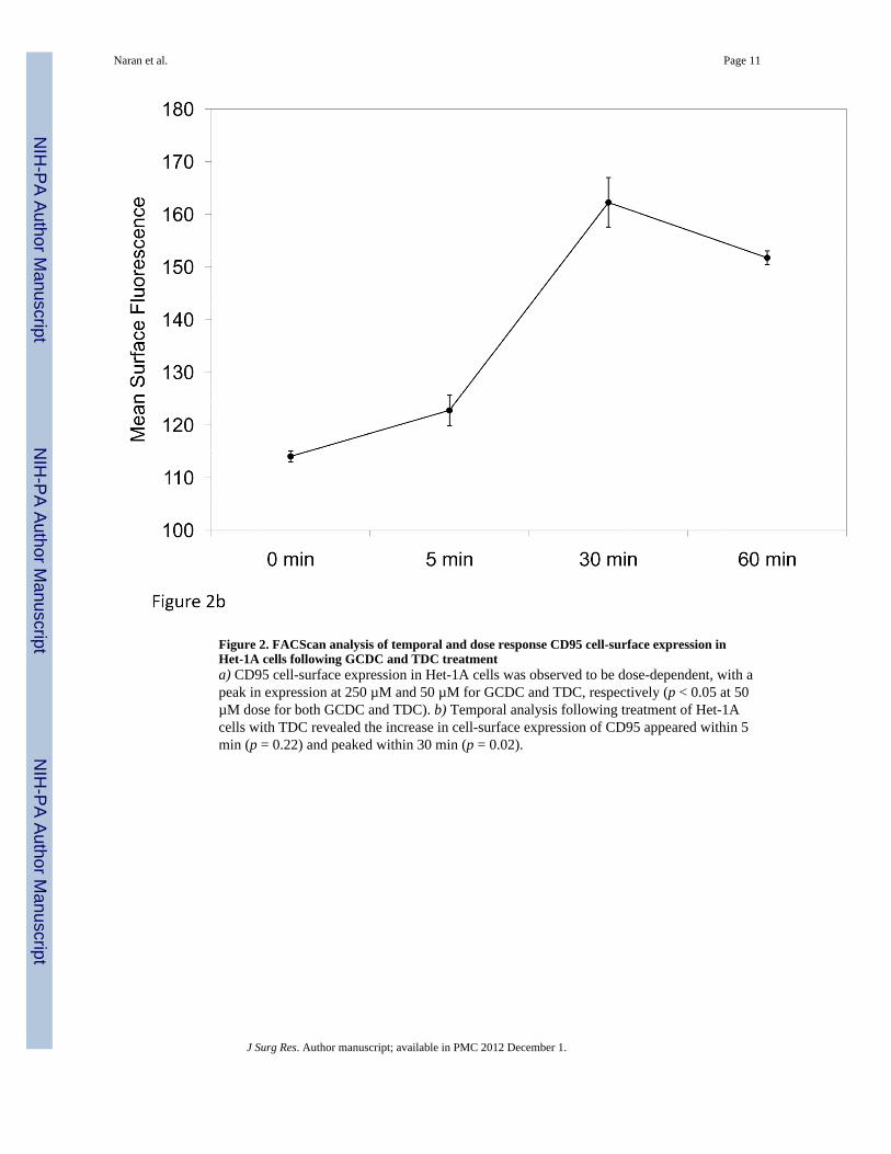
Figure 2. FACScan analysis of temporal and dose response CD95 cell-surface expression inHet-1A cells following GCDC and TDC treatmenta) CD95 cell-surface expression in Het-1A cells was observed to be dose-dependent, with apeak in expression at 250 µM and 50 µM for GCDC and TDC, respectively (p < 0.05 at 50µM dose for both GCDC and TDC). b) Temporal analysis following treatment of Het-1Acells with TDC revealed the increase in cell-surface expression of CD95 appeared within 5min (p = 0.22) and peaked within 30 min (p = 0.02).
Naran et al. Page 11
J Surg Res. Author manuscript; available in PMC 2012 December 1.
NIH
-PA Author Manuscript
NIH
-PA Author Manuscript
NIH
-PA Author Manuscript

Naran et al. Page 12
J Surg Res. Author manuscript; available in PMC 2012 December 1.
NIH
-PA Author Manuscript
NIH
-PA Author Manuscript
NIH
-PA Author Manuscript

Figure 3. FACScan analysis of CD95 cell-surface expression in Het-1A and Flo-1 cells followingbile salt exposure with concurrent PKC inhibitiona) Inhibition of PKC activity with BIM blocked bile salt-induced increases in the surfaceexpression of CD95 in Het-1A cells (control/BIM p = 0.38, control/GCDC p = 0.01, GCDC/BIM p = 0.006). b) Surprisingly, BIM treatment resulted in a significant increase in cell-surface expression of CD95 in Flo-1 cells (p =0.007 at 30 min)
Naran et al. Page 13
J Surg Res. Author manuscript; available in PMC 2012 December 1.
NIH
-PA Author Manuscript
NIH
-PA Author Manuscript
NIH
-PA Author Manuscript

Figure 4. Annexin V/propidium iodide staining analysis of apoptosis/necrosis in Het-1A andFlo-1 cells following bile salt and BIM treatmentFlow cytometric analysis of apoptotic subpopulations with a combination of annexin V-FITC and propidium iodide staining revealed that bile salt treatment of Het-1A cells resultedin an increase in percent apoptosis (p = 0.02), whereas BIM treatment was shown to blockany increase in FasL-induced apoptosis (p = 0.28). We also observed that both bile saltsalone and concurrent BIM treatment increase apoptosis in Flo-1 cells (p = 0.007, p = 0.03respectively).
Naran et al. Page 14
J Surg Res. Author manuscript; available in PMC 2012 December 1.
NIH
-PA Author Manuscript
NIH
-PA Author Manuscript
NIH
-PA Author Manuscript