The effect of omega-3 FAs on tumour angiogenesis and their therapeutic potential
10
Review The effect of omega-3 FAs on tumour angiogenesis and their therapeutic potential Laura Spencer * , Christopher Mann, Matthew Metcalfe, M’Balu Webb, Cristina Pollard, Daniel Spencer, David Berry, William Steward, Ashley Dennison Department of HPB and Pancreatic Surgery, Leicester General Hospital, Gwendolen Road, Leicester LE5 4PW, United Kingdom ARTICLE INFO Article history: Received 31 January 2009 Received in revised form 10 April 2009 Accepted 24 April 2009 Available online 1 June 2009 Keywords: Omega-3 fatty acids Tumour angiogenesis Angiogenesis inhibitor ABSTRACT Omega-3 fatty acid (omega-3 FA) consumption has long been associated with a lower inci- dence of colon, breast and prostate cancers in many human populations. Human trials have demonstrated omega-3 FA to have profound anti-inflammatory effects in those with cancer. In vitro and small animal studies have yielded a strong body of evidence establish- ing omega-3 FA as having anti-inflammatory, anti-apoptotic, anti-proliferative and anti- angiogenic effects. This review explores the evidence and the mechanisms by which omega-3 FA may act as angiogenesis inhibitors and identifies opportunities for original research trialling omega-3 FAs as anti-cancer agents in humans. The conclusions drawn from this review suggest that omega-3 FAs in particular eicosapentaenoic acid (EPA) and docosahexaenoic acid (DHA) found principally in oily fish have potent anti-angiogenic effects inhibiting production of many important angiogenic mediators namely; Vascular Endothelial Growth Factor (VEGF), Platelet-Derived Growth Factor (PDGF), Platelet-Derived Endothelial Cell Growth Factor (PDECGF), cyclo-oxygenase 2 (COX-2), prostaglandin-E2 (PGE2), nitric oxide, Nuclear Factor Kappa Beta (NFKB), matrix metalloproteinases and beta-catenin. Ó 2009 Elsevier Ltd. All rights reserved. 1. Introduction 1.1. Tumour angiogenesis Angiogenesis is the formation of new blood vessels. This pro- cess can be physiological and examples include the develop- ment of blood vessels in utero, or pathological. Pathological angiogenesis includes diabetic retinopathy, and the develop- ment of tumours both benign and malignant. In 1971 Folkman hypothesised that tumour growth is dependent on angiogenesis 1 and subsequently experimental work demonstrated that for a tumour to grow beyond a size of 1–2 mm 3 a substantial new blood supply must develop to support the increasing metabolic requirements. 2–4 The mechanisms of angiogenesis have been under investi- gation since 1931 when Clark and Clark observed real-time capillary growth, and are still not fully understood. 5 However, it is known that inflammation, hypoxia and mechanical forces such as sheer stress, stretching and exercise may acti- vate endothelial cells or cause release of growth factors or cytokines which become involved in a process known as ablu- minal sprouting – the conventional mechanism in which a new blood supply grows from an existing vessel. Fig. 1 dem- onstrates the phases of sprouting angiogenesis. 0959-8049/$ - see front matter Ó 2009 Elsevier Ltd. All rights reserved. doi:10.1016/j.ejca.2009.04.026 * Corresponding author: Tel./fax: +44 0116 2588244, mobile: +44 07779 332496. E-mail address: [email protected] (L. Spencer). EUROPEAN JOURNAL OF CANCER 45 (2009) 2077 – 2086 available at www.sciencedirect.com journal homepage: www.ejconline.com
-
Upload
independent -
Category
Documents
-
view
0 -
download
0
Transcript of The effect of omega-3 FAs on tumour angiogenesis and their therapeutic potential

E U R O P E A N J O U R N A L O F C A N C E R 4 5 ( 2 0 0 9 ) 2 0 7 7 – 2 0 8 6
. sc iencedi rec t . com
ava i lab le a t wwwjournal homepage: www.ejconl ine.com
Review
The effect of omega-3 FAs on tumour angiogenesisand their therapeutic potential
Laura Spencer*, Christopher Mann, Matthew Metcalfe, M’Balu Webb, Cristina Pollard,Daniel Spencer, David Berry, William Steward, Ashley Dennison
Department of HPB and Pancreatic Surgery, Leicester General Hospital, Gwendolen Road, Leicester LE5 4PW, United Kingdom
A R T I C L E I N F O
Article history:
Received 31 January 2009
Received in revised form 10 April
2009
Accepted 24 April 2009
Available online 1 June 2009
Keywords:
Omega-3 fatty acids
Tumour angiogenesis
Angiogenesis inhibitor
0959-8049/$ - see front matter � 2009 Elsevidoi:10.1016/j.ejca.2009.04.026
* Corresponding author: Tel./fax: +44 0116 25E-mail address: [email protected]
A B S T R A C T
Omega-3 fatty acid (omega-3 FA) consumption has long been associated with a lower inci-
dence of colon, breast and prostate cancers in many human populations. Human trials
have demonstrated omega-3 FA to have profound anti-inflammatory effects in those with
cancer. In vitro and small animal studies have yielded a strong body of evidence establish-
ing omega-3 FA as having anti-inflammatory, anti-apoptotic, anti-proliferative and anti-
angiogenic effects. This review explores the evidence and the mechanisms by which
omega-3 FA may act as angiogenesis inhibitors and identifies opportunities for original
research trialling omega-3 FAs as anti-cancer agents in humans. The conclusions drawn
from this review suggest that omega-3 FAs in particular eicosapentaenoic acid (EPA) and
docosahexaenoic acid (DHA) found principally in oily fish have potent anti-angiogenic
effects inhibiting production of many important angiogenic mediators namely; Vascular
Endothelial Growth Factor (VEGF), Platelet-Derived Growth Factor (PDGF), Platelet-Derived
Endothelial Cell Growth Factor (PDECGF), cyclo-oxygenase 2 (COX-2), prostaglandin-E2
(PGE2), nitric oxide, Nuclear Factor Kappa Beta (NFKB), matrix metalloproteinases and
beta-catenin.
� 2009 Elsevier Ltd. All rights reserved.
1. Introduction
1.1. Tumour angiogenesis
Angiogenesis is the formation of new blood vessels. This pro-
cess can be physiological and examples include the develop-
ment of blood vessels in utero, or pathological. Pathological
angiogenesis includes diabetic retinopathy, and the develop-
ment of tumours both benign and malignant.
In 1971 Folkman hypothesised that tumour growth is
dependent on angiogenesis1 and subsequently experimental
work demonstrated that for a tumour to grow beyond a size
er Ltd. All rights reserved
88244, mobile: +44 07779(L. Spencer).
of 1–2 mm3 a substantial new blood supply must develop to
support the increasing metabolic requirements.2–4
The mechanisms of angiogenesis have been under investi-
gation since 1931 when Clark and Clark observed real-time
capillary growth, and are still not fully understood.5 However,
it is known that inflammation, hypoxia and mechanical
forces such as sheer stress, stretching and exercise may acti-
vate endothelial cells or cause release of growth factors or
cytokines which become involved in a process known as ablu-
minal sprouting – the conventional mechanism in which a
new blood supply grows from an existing vessel. Fig. 1 dem-
onstrates the phases of sprouting angiogenesis.
.
332496.

Fig. 1 – Phases of sprouting angiogenesis.64 The process of abluminal sprouting is initiated by activation of endothelial cells
by growth factors, mechanical or inflammatory stimuli. Permeability (1) across the endothelial cell layer increases, and this is
followed by proliferation (2). Proteolysis (3) of basement membrane components (controlled mainly by MMPs) enables the
sprouting of the endothelial cell into the interstitial space. Continued coordination of cell adhesion and cytoskeletal
remodelling components provide directional migration of the sprouting (4) endothelial cells. Proliferation remains greatest at
the stalk of the growing sprout. Eventually, the new sprout forms a lumen (5) by the process of intracellular vacuolar fusion or
by the stabilisation of several cells around a central lumen. A new lateral branch will be formed when the sprout
anastamoses with a pre-existing capillary. Alternatively, circulating EPCs (A) may contribute to the sprout process, adhering
(B) to the activated endothelial cell, extravasating (C) through the endothelial cell layer, and clustering (D) within the
interstitium. Some of these EPCs will integrate (E) into the sprout and will comprise a portion of the newly formed capillary
while others may remain as perivascular cells.
2078 E U R O P E A N J O U R N A L O F C A N C E R 4 5 ( 2 0 0 9 ) 2 0 7 7 – 2 0 8 6
Many molecules such as growth factors and cytokines
have both stimulatory and inhibitory roles within sprout-
ing angiogenesis, the most investigated compound being
Vascular Endothelial Growth Factor (VEGF) which is
known to be a potent stimulator of angiogenesis.6 Fig. 2
displays the main mediators involved in the angiogenic
cascade.
Therapeutic manipulation of tumour angiogenesis is un-
der intense investigation and the search for chemo-preventa-
Stimuli Inflammation Exercise Hypoxia Mechanical
Growth Factors HGF EGF VEGF FGF2 PIGF
Signal Mediators PG, NO Rho-GTPases MAPK
InhiEndAngTIMTNFTumVaso
Key Inhibition
Stimulation
Fig. 2 – The main mediators invol
tive agents in the form of angiogenesis inhibitors is an
exciting new avenue in cancer prevention.7
This quest for angiogenesis inhibitors is not confined to
conventional chemo-preventative compounds but extends
to substances found in foodstuffs which have long been asso-
ciated with lower rates of cancer in populations who consume
high levels of foods containing these compounds. Examples
include; zinc, polyphenols (EGCG) found in green tea and
Omega-3 fatty acid (omega-3 FA) principally from oily fish.8
Cellular Effectors COX Bcl-2 eNOS MMPs CyclinD1 c-met αvβ3, άvβ5
Transcription Regulators ets1, egr1 β-catenin HIF1α
Angiogenesis Proliferation Migration Proteolysis
bitors ostatin iostatin Ps αstatin hibin
ved in the angiogenic cascade.

E U R O P E A N J O U R N A L O F C A N C E R 4 5 ( 2 0 0 9 ) 2 0 7 7 – 2 0 8 6 2079
1.2. Omega-3 FA
Omega-3 FAs (n – 3) are long-chain polyunsaturated fatty
acids with the first double bond 3 carbons from the methyl
end of the chain. Omega-6 (n – 6) fatty acids have a similar
structure with the first double bond 6 carbons from the
methyl end of the chain. Humans are unable to desaturate
the n – 3 or n – 6 double bond and as such this makes both
compounds ‘essential fatty acids’ obtained only from dietary
sources.
Omega-6 fatty acid is consumed as linoleic acid or arachi-
donic acid found in meats, and vegetable oils (safflower, corn
and soybean oil). The principal dietary source of omega-3 FA
is from oily cold-water fish namely eicosapentaenoic acid
(EPA) and docosahexaenoic acid (DHA).
Both omega-3 and omega-6 fatty acids are used as sub-
strates for the production of eicosanoids that are a class of
compounds including prostaglandins (PGs), thromboxanes
and leukotrienes intimately involved in immunomodulation,
inflammation and tumour formation. Eicosanoids produced
using omega-6 fatty acids (arachidonic acid) as a substrate
stimulate inflammation and tumour angiogenesis, whereas
eicosanoids produced from omega-3 fatty acids, EPA and
DHA are anti-inflammatory and do not stimulate angiogene-
sis.9,10 Fig. 3 illustrates the basic metabolism of omega-3
and omega-6 fatty acids.
The focus of this review is on the role of these omega-3 FAs
as angiogenesis inhibitors and their potential for use as natu-
ral chemo-preventative agents at all stages of the angiogenic
cascade is examined. Table 1 summarises the evidence for
this review.
1.3. Omega-3 FA and VEGF
Vascular Endothelial Growth Factor (VEGF) is a heparin-bind-
ing homodimeric glycoprotein with a molecular weight of
Fig. 3 – Simplified metabolism of omega-3 and omega-6 fatty aci
FAs give rise to generally antiinflammatory, anti-proliferative me
pro-proliferative mediators. COX = cyclo-oxygenase, LOX = lipox
45 kDa11 and a cysteine knot motif shared by other growth
factors such as Platelet-Derived Growth Factor (PDGF).12 The
VEGF family comprises five molecules such as VEGF-A, B, C,
D and Placenta Growth Factor (PIGF). Each molecule has
numerous isoforms of which VEGF-165 was reported to be
the most abundant and mitogenic isoform of VEGF-A.6
VEGF is a principle factor involved in almost every stage of
sprouting angiogenesis, it increases vascular permeability,13
induces endothelial cell proliferation and migration and pro-
motes endothelial cell survival.14
Numerous studies have demonstrated that VEGF or its
receptors are up-regulated in many human cancers,15–22 and
omega-3 fatty acids have been shown by a variety of different
studies to suppress VEGF production.
1.4. In vitro studies
Human umbilical vein endothelial cells (HUVECs) treated with
conjugated EPA showed less VEGF-stimulated tube formation
during sprouting angiogenesis than controls, VEGF-stimu-
lated migration of HUVEC was suppressed and certain matrix
metalloproteinases (MMPs) associated with endothelial cell
migration were diminished in HUVECs treated with conju-
gated EPA.23
A shark oil-olive oil blend inhibited VEGF binding to its
receptors (flk-1 and flk-2).24
Pre-treating bovine aortic endothelial cells (BAE cells) with
docosapentanoic acid (DPA) (an elongated metabolite of EPA)
suppressed endothelial cell tube-forming activity induced by
VEGF. DPA pre-treatment also suppressed the migratory activ-
ity of BAE cells and VEGF receptor-2 expression both in plastic
dish and in collagen gel cultures.25
A study investigating the effect of EPA on VEGF-induced
endothelial cell proliferation using bovine carotid artery
endothelial cells (BCE cells) showed that BCE cells treated
with 0.5 lg/ml EPA for 48 h displayed a dose-dependent sup-
ds. Basic metabolism of omega-3 and omega-6 FA. Omega-3
diators, omega-6 family gives rise to more proinflammatory
ygenase.

Table 1 – Summary of the observed effects of omega-3 FAs on various mediators of the angiogenic cascade.
Mediator Effect of omega-3 FA
VEGF in vitro Omega-3 FAs suppress VEGF-stimulated endothelial cell proliferation, migration and tube formation
during sprouting angiogenesis23,25,26
Decrease expression of the VEGF receptors flk-1 and flk-2 and inhibit binding of VEGF to its receptors24,26
Acting upstream inhibit critical mediators in the PGE2-induced signalling pathway which leads to
augmented VEGF expression in colon cancer cell lines27
In vivo Nude mice with omega-3 pre-supplemented diets undergoing implantation of human colorectal and
breast carcinomas demonstrated a decreased tumour microvessel density and tumour volume compared
to controls.28,29 Amounts of VEGF, PGE2 and COX-2 expressed in these tumours were also decreased28
Flank implantation of fibrosarcomas in Fischer 344 mice also demonstrated a decreased tumour cell
volume and decreased amounts of VEGF-alpha mRNA in those with EPA supplemented diets.30
Human Volunteers assigned a Mediterranean diet with a high omega-3 FA content compared to volunteers
consuming an ordinary Swedish diet demonstrated an increased omega-3:omega-6 ratio by 45% with
circulating levels of VEGF falling by 13%31
PDGF in vitro Inhibit production of PDGF-like protein from vascular endothelial cells40
Inhibit vascular smooth muscle proliferation by modulating various steps of the PDGF signal
transduction pathway43
Ex vivo Quiescent human mononuclear cells from humans with a pre-supplemented omega-3 rich diet
expressed reduced amounts of PDGF genes41
Human High dose oral fish oil supplementation of human volunteers yielded a rise in cellular omega-3 levels and
significantly decreased levels of PGDF-A and PDGF-B mRNA expression when compared with those on a
control diet42
PD-ECGF Little information available. One study demonstrated no change in PD-ECGF gene expression on
quiescent human mononuclear cells after prior dietary supplementation with omega-3 FA42
FGF Little information available. Inhibitory effect of omega-3 FA on FGF-induced angiogenesis not
observed.62,63
HGF, EGF Currently no studies investigating the effect of omega-3 FA on HGF or EGF. Opportunity for original
research
Nitric oxide in vitro Inhibit nitric oxide-dependent angiogenesis in a variety of ways:
Inhibit NO production and inducible nitric oxide synthase (iNOS) expression in murine macrophages.79–82
Down-regulate iNOS COX-2 and TNF-alpha genes by blocking NFKB and MAP-kinase activation78
In vivo A small animal model with endogenously high levels of omega-3 FA demonstrated that the incidence and
growth rate of experimentally induced colon tumours were decreased alongside the levels of iNOS and
NFKB84
COX-2 in vivo Several small animal models have identified that omega-3 FA enriched diets have inhibitory effects on
COX-2 and prostaglandin production27,95
Synergistic inhibitory effects on the growth of experimentally induced tumours of cells from varying
human cancer cell lines treated with omega-3 FAs and COX-2 inhibitors have recently been
demonstrated96–98
MMPs and Beta-catenin in vitro Matrix metalloproteinase 2 and 9 mRNA production is reduced by omega-3 FA.23 Beta-catenin has also
been shown to be reduced by treatment with omega-3 FA101
2080 E U R O P E A N J O U R N A L O F C A N C E R 4 5 ( 2 0 0 9 ) 2 0 7 7 – 2 0 8 6
pression to VEGF-induced endothelial cell proliferation. This
effect was not observed with BCE cells treated with arachi-
donic acid or DHA. Flk-1 expression was also inhibited in a
dose-dependent fashion in EPA-treated BCE cells.26
EPA and DHA inhibited ERK-1 and 2 phosphorylation and
HIF-alpha protein over-expression (critical steps in the Prosta-
glandin-E2 (PGE2)-induced signalling pathway leading to aug-
mented expression of VEGF in colon cancer cells). EPA showed
greater efficacy than DHA in vitro.27
1.5. In vivo studies
Omega-3 enriched diets decreased the amount of microves-
sels developing in HT-29 cell human colorectal tumours im-
planted in nude mice. The amount of VEGF, cyclo-
oxygenase 2 (COX-2) and PGE2 expressed in the tumours
was also decreased.27 Experiments in which breast carcino-
mas were implanted into nude mice that were then fed
with diets high in EPA or DHA and compared to controls
indicated that both tumour microvessel density counts
and levels of VEGF measured in the resected tumours were
significantly lower in the animals receiving these omega-3
FAs.28,29
Fischer 344 rats (200–250 g) underwent flank implantation
of the methylcholanthrene-induced fibrosarcoma and were
assigned to diets supplemented with corn oil, normal saline
or EPA. After resection of the tumour rats with the EPA sup-
plemented diet had a significantly decreased tumour volume
E U R O P E A N J O U R N A L O F C A N C E R 4 5 ( 2 0 0 9 ) 2 0 7 7 – 2 0 8 6 2081
and levels of VEGF-alpha mRNA were also significantly dimin-
ished in this group.30
A study investigating the effects of a diet high in omega-3
FA (Mediterranean diet) on healthy volunteers found that
after 6 weeks the omega-3:omega-6 ratio had increased in
those on the Mediterranean diet and levels of circulating
VEGF had subsequently decreased.31
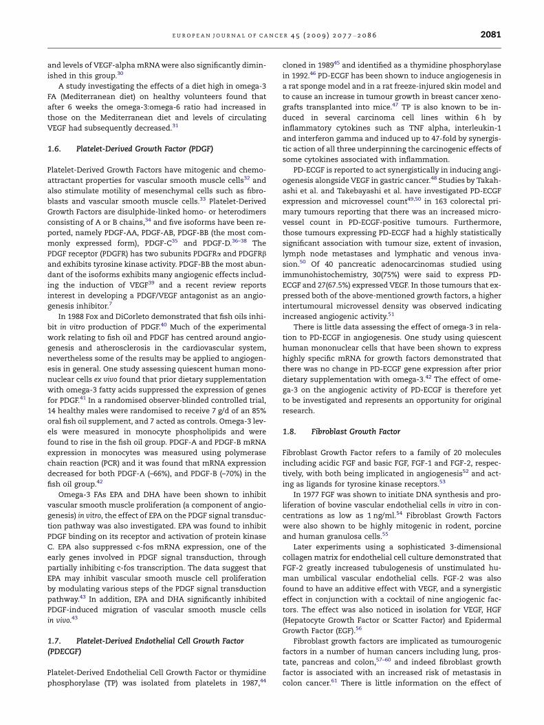
1.6. Platelet-Derived Growth Factor (PDGF)
Platelet-Derived Growth Factors have mitogenic and chemo-
attractant properties for vascular smooth muscle cells32 and
also stimulate motility of mesenchymal cells such as fibro-
blasts and vascular smooth muscle cells.33 Platelet-Derived
Growth Factors are disulphide-linked homo- or heterodimers
consisting of A or B chains,34 and five isoforms have been re-
ported, namely PDGF-AA, PDGF-AB, PDGF-BB (the most com-
monly expressed form), PDGF-C35 and PDGF-D.36–38 The
PDGF receptor (PDGFR) has two subunits PDGFRa and PDGFRb
and exhibits tyrosine kinase activity. PDGF-BB the most abun-
dant of the isoforms exhibits many angiogenic effects includ-
ing the induction of VEGF39 and a recent review reports
interest in developing a PDGF/VEGF antagonist as an angio-
genesis inhibitor.7
In 1988 Fox and DiCorleto demonstrated that fish oils inhi-
bit in vitro production of PDGF.40 Much of the experimental
work relating to fish oil and PDGF has centred around angio-
genesis and atherosclerosis in the cardiovascular system,
nevertheless some of the results may be applied to angiogen-
esis in general. One study assessing quiescent human mono-
nuclear cells ex vivo found that prior dietary supplementation
with omega-3 fatty acids suppressed the expression of genes
for PDGF.41 In a randomised observer-blinded controlled trial,
14 healthy males were randomised to receive 7 g/d of an 85%
oral fish oil supplement, and 7 acted as controls. Omega-3 lev-
els were measured in monocyte phospholipids and were
found to rise in the fish oil group. PDGF-A and PDGF-B mRNA
expression in monocytes was measured using polymerase
chain reaction (PCR) and it was found that mRNA expression
decreased for both PDGF-A (–66%), and PDGF-B (–70%) in the
fish oil group.42
Omega-3 FAs EPA and DHA have been shown to inhibit
vascular smooth muscle proliferation (a component of angio-
genesis) in vitro, the effect of EPA on the PDGF signal transduc-
tion pathway was also investigated. EPA was found to inhibit
PDGF binding on its receptor and activation of protein kinase
C. EPA also suppressed c-fos mRNA expression, one of the
early genes involved in PDGF signal transduction, through
partially inhibiting c-fos transcription. The data suggest that
EPA may inhibit vascular smooth muscle cell proliferation
by modulating various steps of the PDGF signal transduction
pathway.43 In addition, EPA and DHA significantly inhibited
PDGF-induced migration of vascular smooth muscle cells
in vivo.43
1.7. Platelet-Derived Endothelial Cell Growth Factor(PDECGF)
Platelet-Derived Endothelial Cell Growth Factor or thymidine
phosphorylase (TP) was isolated from platelets in 1987,44
cloned in 198945 and identified as a thymidine phosphorylase
in 1992.46 PD-ECGF has been shown to induce angiogenesis in
a rat sponge model and in a rat freeze-injured skin model and
to cause an increase in tumour growth in breast cancer xeno-
grafts transplanted into mice.47 TP is also known to be in-
duced in several carcinoma cell lines within 6 h by
inflammatory cytokines such as TNF alpha, interleukin-1
and interferon gamma and induced up to 47-fold by synergis-
tic action of all three underpinning the carcinogenic effects of
some cytokines associated with inflammation.
PD-ECGF is reported to act synergistically in inducing angi-
ogenesis alongside VEGF in gastric cancer.48 Studies by Takah-
ashi et al. and Takebayashi et al. have investigated PD-ECGF
expression and microvessel count49,50 in 163 colorectal pri-
mary tumours reporting that there was an increased micro-
vessel count in PD-ECGF-positive tumours. Furthermore,
those tumours expressing PD-ECGF had a highly statistically
significant association with tumour size, extent of invasion,
lymph node metastases and lymphatic and venous inva-
sion.50 Of 40 pancreatic adenocarcinomas studied using
immunohistochemistry, 30(75%) were said to express PD-
ECGF and 27(67.5%) expressed VEGF. In those tumours that ex-
pressed both of the above-mentioned growth factors, a higher
intertumoural microvessel density was observed indicating
increased angiogenic activity.51
There is little data assessing the effect of omega-3 in rela-
tion to PD-ECGF in angiogenesis. One study using quiescent
human mononuclear cells that have been shown to express
highly specific mRNA for growth factors demonstrated that
there was no change in PD-ECGF gene expression after prior
dietary supplementation with omega-3.42 The effect of ome-
ga-3 on the angiogenic activity of PD-ECGF is therefore yet
to be investigated and represents an opportunity for original
research.
1.8. Fibroblast Growth Factor
Fibroblast Growth Factor refers to a family of 20 molecules
including acidic FGF and basic FGF, FGF-1 and FGF-2, respec-
tively, with both being implicated in angiogenesis52 and act-
ing as ligands for tyrosine kinase receptors.53
In 1977 FGF was shown to initiate DNA synthesis and pro-
liferation of bovine vascular endothelial cells in vitro in con-
centrations as low as 1 ng/ml.54 Fibroblast Growth Factors
were also shown to be highly mitogenic in rodent, porcine
and human granulosa cells.55
Later experiments using a sophisticated 3-dimensional
collagen matrix for endothelial cell culture demonstrated that
FGF-2 greatly increased tubulogenesis of unstimulated hu-
man umbilical vascular endothelial cells. FGF-2 was also
found to have an additive effect with VEGF, and a synergistic
effect in conjunction with a cocktail of nine angiogenic fac-
tors. The effect was also noticed in isolation for VEGF, HGF
(Hepatocyte Growth Factor or Scatter Factor) and Epidermal
Growth Factor (EGF).56
Fibroblast growth factors are implicated as tumourogenic
factors in a number of human cancers including lung, pros-
tate, pancreas and colon,57–60 and indeed fibroblast growth
factor is associated with an increased risk of metastasis in
colon cancer.61 There is little information on the effect of

2082 E U R O P E A N J O U R N A L O F C A N C E R 4 5 ( 2 0 0 9 ) 2 0 7 7 – 2 0 8 6
omega-3 on FGF but two in vitro studies suggest that omega-3
FAs do not have an inhibitory effect on FGF-induced angio-
genesis.62,63 Further investigations into the effect of
omega-3 FA on this potent angiogenic factor are required.
1.9. Hepatocyte Growth Factor
Hepatocyte Growth Factor/Scatter Factor is secreted from
mesenchymal derived cells as an inactive precursor which
is activated by urokinase or tissue plasminogen activator.
The receptor for HGF is found on endothelial cells and is
termed c-met.64 Partly through its own actions and also
through its ability to activate VEGF, HGF has been shown to
have a strong role in angiogenesis.65 As yet there are no stud-
ies investigating the effects of omega-3 on HGF.
1.10. Epidermal Growth Factor
Epidermal Growth Factor binds to Human Epidermal Growth
Factor Receptors 1–4 (HER1–4).66 Over-expression of HER2 in
cancer cells is associated with increased VEGF and angiogenic
activity via increases in protein synthesis of Hypoxia Induc-
ible Factor 1a (HIF 1a).67 EGF and HER receptors are associated
with the pathogenesis of a number of different cancers
including breast, colorectal and pancreatic carcinomas68–70
and with the promising results from the development of
HER receptor antagonists, for example, the anti-HER2 therapy
trastuzumab developed for metastatic breast cancer and the
development of EGFR antagonists for colorectal cancer.71,72
No studies have assessed the role of omega-3 fatty acids on
EGF or HER.
1.11. Nitric oxide
Nitric oxide, produced by nitric oxide synthases, has both
vasodilatory and pro-angiogenic effects. It promotes endothe-
lial cell survival, inhibits apoptosis and enhances endothelial
cell proliferation.73,74 Inducible nitric oxide synthase (iNOS)
and COX-2-dependent angiogenesis are modulated by VEGF
in human colorectal cancer75,76 and in turn VEGF-mediated
angiogenesis is also dependent on nitric oxide production.77
Fig. 4 illustrates a proposed pathway for increased VEGF pro-
duction in response to increased levels of iNOS and COX-2.76
Omega-3 FAs have been shown to inhibit NO-dependent angi-
ogenesis in a variety of ways.
Fig. 4 – Potential mechanism of iNOS and COX-2 pathways in s
colleagues.76 Experimentally stimulated HT29 colon cancer cells
inducible nitric oxide synthase (iNOS) and increase the producti
NO production which causes increased production of both VEG
production of PGE2, which also increases VEGF production.
The omega-3 fatty acid alpha-linolenic acid (ALA) has been
shown to down-regulate iNOS, COX-2 and TNF alpha gene
expression by blocking Nuclear Factor Kappa Beta (NFKB)
and MAPK activation in LPS-stimulated RAW 264.7 cells.78
Omega-3 FAs in particular DHA inhibit NO production and
iNOS expression in stimulated murine macrophages.79–82
Inducible NO and NFKB have been shown to be down-regu-
lated in human colorectal cancer cells treated with DHA.83 A
recent study using a fat-1 transgenic mouse model with
endogenously high levels of omega-3 FA demonstrated that
the incidence and growth rate of colon tumours (experimen-
tally induced by inflammation and carcinogens) was de-
creased as were the levels of iNOS and NFKB.84
1.12. COX-2 and PGE2
Cyclo-oxygenase 2 is an enzyme catalysing the conversion of
arachidonic acid (omega-6 fatty acid) into prostaglandins
such as PGE2. In general metabolites of omega-6 fatty acids
are associated with increased levels of inflammation and tu-
mour angiogenesis.9,10 Dating back to 1974 both in vitro and
in vivo studies have demonstrated a link between prostaglan-
dins and cancer in particular the E series prostaglandins.85,86
NSAIDs (COX-2 inhibitors) such as celecoxib have been shown
to significantly reduce tumour formation in animal models,
and significantly reduce colonic polyp burden by 30% in con-
trolled trials in those with Familial Adenomatous Polyposis
(FAP).87–89 A large nested case-controlled study found that
long-term NSAID/COX-2 inhibitor usage was associated with
a significantly decreased risk of developing colorectal
cancer.90
COX-2 is up-regulated in most human cancers75,91 and
PGE2 is produced in large amounts in colorectal tumours
and has been shown experimentally to induce the production
of pro-angiogenic factors in many cell types.92,93 A recent
study by Cianchi et al. revealed a stimulatory effect of nitric
oxide on COX-2 activity in human colorectal cancers76 fur-
thermore, this interaction is likely to yield a co-operative ef-
fect in promoting angiogenesis through a PGE2 increase in
VEGF production.94 Several small animal models have identi-
fied omega-3 fatty acid-enriched diets as having inhibitory ef-
fects on COX-2 and prostaglandin production in both plasma
and experimentally induced tumours. Rats fed with a corn-oil
diet (rich in omega-6) or a flaxseed oil diet (rich in omega-3)
were subject to chemical induction of colon tumours. Tumour
timulating VEGF production proposed by Cianchi and
are stimulated by LPS or EGF and in turn cause stimulation of
on of cyclooxygenase-2 (COX-2). iNOS production stimulates
F and COX-2. Increased COX-2 activation stimulates further
E U R O P E A N J O U R N A L O F C A N C E R 4 5 ( 2 0 0 9 ) 2 0 7 7 – 2 0 8 6 2083
incidence was decreased in the flaxseed oil (n – 3) group com-
pared to the corn oil group (n – 4) (29.4% versus 82.6%) and lev-
els of COX-1 and COX-2 were significantly reduced in the
flaxseed oil group.95 The effect of EPA and DHA on human
colorectal cancer cell lines both in vitro and in vivo upon tu-
mours transplanted into nude mice has also been investi-
gated. EPA and DHA reduced VEGF, COX-2 expression and
PGE2 levels in HT-29 cells cultured in vitro. EPA and DHA also
inhibited ERK-1 and -2 phosphorylation and HIF-1alpha pro-
tein over-expression, critical steps in the PGE2-induced sig-
nalling pathway leading to the augmented expression of
VEGF in colon cancer cells. EPA and DHA also reduced growth
of tumours obtained by inoculating HT-29 cells in nude mice,
microvessel formation and the levels of VEGF, COX-2 and
PGE2 expressed in tumours.27 Recent evidence reveals a syn-
ergistic inhibitory effect on the growth of experimentally in-
duced tumours or cells from varying human cancer cell
lines treated with omega-3 FA and COX-2 inhibitors.96–98 Hy-
poxia Inducible Factor (HIF) serves as a pro-angiogenic factor
acting upstream from VEGF. HIF 1a has been found in a num-
ber of human cancer cell lines and is associated with in vitro
tumour vascularisation.99 HIF 1a has been identified as a piv-
otal transcription factor linking the inflammatory and onco-
genic pathways via Nuclear Factor Kappa Beta, COX-2 and
PGE2 mechanisms.100
1.13. Matrix metalloproteinases
Matrix metalloproteinases are zinc-dependent proteases
which have a critical role in the proteolysis of the basement
membrane – a key phase in sprouting angiogenesis. Certain
MMPs produced by endothelial cell are also involved in capil-
lary sprouting.64 MMPs 2 and 9 mRNA production was shown
to be inhibited by conjugated EPA in a study investigating the
effect of conjugated EPA on VEGF-induced angiogenesis in hu-
man endothelial cells.23
1.14. Beta-catenin
The production of this transcriptional regulator in the angio-
genic cascade has been shown to be inhibited in colon cancer
cells treated with DHA.101 Several other proteins regulated by
the TCF-beta-catenin pathway and involved in regulation of
tumour growth and angiogenesis were also down-regulated
by DHA, including peroxisome proliferator-activated receptor
delta, membrane type 1 (MT1)-matrix metalloproteinase
(MMP), MMP-7 and VEGF.102
2. Conclusion
In 1863 Rudolf Virchow described the relationship between
inflammation and cancer when he observed leucocytes in
neoplastic tissue.8 Today it is accepted that chronic inflam-
mation is a predisposing factor for many human cancers such
as Barrett’s oesophagus and its association with adenocarci-
noma of the oesophagus.
Factors such as PGE2, nitric oxide, COX-2 and NFKB have
well-documented roles in both the inflammatory and angio-
genic cascades with significant cross-relation in both path-
ways and this review demonstrates the potential for omega-
3 FAs as anti-inflammatory and anti-angiogenic agents via
inhibition of these factors and others including VEGF and
PDGF.
With the development of safe parenteral preparations con-
taining significant amounts of omega-3 FAs, human trials
have demonstrated that parenteral administration of fish-oil
lipid emulsion leads to a significant and rapid increase in
EPA and DHA concentrations in plasma, platelet and leuco-
cyte membrane phospholipids (within hours)103–105 this was
previously not achievable with oral preparations. These trials
have also suggested that omega-3 FAs via their immunomod-
ulatory effects decrease the re-operation rate,106 the require-
ment for post-operative antibiotics,107 the rate of sepsis,108
the incidence of post-operative venous thromboembolism104
and the length of hospital stay,106,109 and lower the mortality
rate for surgical patients.108
This development of commercially available human par-
enteral infusions of omega-3 fatty acids offers perhaps the
greatest opportunity to date for modulating immune function
in the chemoprevention of cancer whilst patients experience
the myriad of other beneficial effects associated with paren-
teral omega-3 FAs. Randomised controlled trials assessing
the effects of parenteral omega-3 fatty acid administration
in human cancer patients are now awaited with interest.
Conflict of interest statement
Mr. A. Dennison, Miss. L. Spencer, Miss. M. Webb, Mr. C. Mann,
Mrs. C. Pollard and Mr. M. Metcalfe are investigators in a trial
using omega-3 FA as potential angiogenesis inhibitors in hu-
man hepatic colorectal metastases. This trial has received
funding from B. Braun Pharmaceutical Company. Mr. D. Berry,
Professor W. Steward and Mr. D. Spencer have no conflict of
interest declared.
Acknowledgements
We would like to thank Professor P.C. Calder, Professor of
Nutritional Immunology, Institute of Human Nutrition, Uni-
versity of Southampton School of Medicine for his sugges-
tions regarding this manuscript. We would also like to
thank Mrs. P. Divall, clinical librarian at Leicester General Hos-
pital for her help with literature searching and obtaining ori-
ginal manuscripts.
R E F E R E N C E S
1. Folkman J. Tumor angiogenesis: therapeutic implications.New Engl J Med 1971;285:1182–6.
2. Folkman J, Cole P, Zimmerman S. Tumor behavior in isolatedperfused organs: in vitro growth and metastases of biopsymaterial in rabbit thyroid and canine intestinal segment. AnnSurg 1966;164:491–502.
3. Folkman J. Biology of endothelial cells. Boston: Martinus-Nijhoff; 1984.

2084 E U R O P E A N J O U R N A L O F C A N C E R 4 5 ( 2 0 0 9 ) 2 0 7 7 – 2 0 8 6
4. Gimbrone Jr MA, Leapman SB, Cotran RS, Folkman J. Tumordormancy in vivo by prevention of neovascularization. J ExpMed 1972;136:261–76.
5. Clark ER, Clark EL. Microscopic observations on the growthof blood capillaries in the living mammal. Am J Anat1939;64:251–301.
6. Ferrara N. Vascular endothelial growth factor: basic scienceand clinical progress. Endocr Rev 2004;25:581–611.
7. Ferrara N, Kerbel RS. Angiogenesis as a therapeutic target.Nature 2005;438:967–74.
8. Philpott M, Ferguson LR. Immunonutrition and cancer. MutatRes 2004;551:29–42.
9. Hardman WE. Omega-3 FA to augment cancer therapy. J Nutr2002;132:3508S–12S.
10. Hardman WE. N – 3 fatty acids and cancer therapy. J Nutr2004;134:3427S–30S.
11. Ferrara N, Henzel WJ. Pituitary follicular cells secrete a novelheparin-binding growth factor specific for vascularendothelial cells. Biochem Biophys Res Commun1989;161:851–8.
12. Muller YA, Li B, Christinger HW, Wells JA, Cunningham BC,de Vos AM. Vascular endothelial growth factor: crystalstructure and functional mapping of the kinase domainreceptor binding site. Proc Natl Acad Sci USA 1997;94:7192–7.
13. Ellis LM, Takahashi Y, Liu W, Shaheen RM. Vascularendothelial growth factor in human colon cancer: biologyand therapeutic implications. Oncologist 2000;5(Suppl. 1):11–5.
14. Nor JE, Christensen J, Mooney DJ, Polverini PJ. Vascularendothelial growth factor (VEGF)-mediated angiogenesis isassociated with enhanced endothelial cell survival andinduction of bcl-2 expression. Am J Pathol 1999;154:375–84.
15. Takekoshi K, Isobe K, Yashiro T, et al. Expression of vascularendothelial growth factor (VEGF) and its cognate receptors inhuman pheochromocytomas. Life Sci 2004;74:863–71.
16. Yang CC, Chu KC, Yeh WM. The expression of vascularendothelial growth factor in transitional cell carcinoma ofurinary bladder is correlated with cancer progression. UrolOncol 2004;22:1–6.
17. Linderholm BK, Lindh B, Beckman L, et al. Prognosticcorrelation of basic fibroblast growth factor and vascularendothelial growth factor in 1307 primary breast cancers.Clin Breast Cancer 2003;4:340–7.
18. Loggini B, Boldrini L, Gisfredi S, et al. CD34 microvesseldensity and VEGF expression in basal and squamous cellcarcinoma. Pathol Res Pract 2003;199:705–12.
19. Wong C, Wellman TL, Lounsbury KM. VEGF and HIF-1alphaexpression are increased in advanced stages of epithelialovarian cancer. Gynecol Oncol 2003;91:513–7.
20. Giatromanolaki A, Koukourakis MI, Simopoulos C,Polychronidis A, Sivridis E. Vascular endothelial growthfactor (VEGF) expression in operable gallbladder carcinomas.Eur J Surg Oncol 2003;29:879–83.
21. Shiraishi A, Ishiwata T, Shoji T, Asano G. Expression of PCNA,basic fibroblast growth factor, FGF receptor and vascularendothelial growth factor in adenomas and carcinomas ofthe human colon. Acta Histochem Cytochem 1995;28:21–9.
22. Kimura H, Konishi K, Nukui T, et al. Prognostic significanceof expression of thymidine phosphorylase and vascularendothelial growth factor in human gastric carcinoma. J SurgOncol 2001;76:31–6.
23. Tsuzuki T, Shibata A, Kawakami Y, Nakagawa K, Miyazawa T.Conjugated eicosapentaenoic acid inhibits vascularendothelial growth factor-induced angiogenesis bysuppressing the migration of human umbilical veinendothelial cells. J Nutr 2007;137:641–6.
24. Yuan L, Yoshida M, Davis PF. Inhibition of pro-angiogenicfactors by a lipid-rich shark extract. J Med Food 2006;9:300–6.
25. Tsuji M, Murota SI, Morita I. Docosapentaenoic acid (22:5, n –3) suppressed tube-forming activity in endothelial cellsinduced by vascular endothelial growth factor. ProstaglandinsLeukot Essent Fatty Acids 2003;68:337–42.
26. Yang SP, Morita I, Murota SI. Eicosapentaenoic acidattenuates vascular endothelial growth factor-inducedproliferation via inhibiting flk-1 receptor expression inbovine carotid artery endothelial cells. J Cell Physiol1998;176:342–9.
27. Calviello G, Di Nicuolo F, Gragnoli S, et al. N – 3 PUFAs reduceVEGF expression in human colon cancer cells modulatingthe COX-2/PGE2 induced ERK-1 and -2 and HIF-1alphainduction pathway. Carcinogenesis 2004;25:2303–10.
28. Rose DP, Connolly JM. Antiangiogenicity of docosahexaenoicacid and its role in the suppression of breast cancer cellgrowth in nude mice. Int J Oncol 1999;15:1011–5.
29. Mukutmoni-Norris M, Hubbard NE, Erickson KL. Modulationof murine mammary tumor vasculature by dietary n – 3 fattyacids in fish oil. Cancer Lett 2000;150:101–9.
30. Tevar R, Jho DH, Babcock T, Helton WS, Espat NJ. Omega-3fatty acid supplementation reduces tumor growth andvascular endothelial growth factor expression in a model ofprogressive non-metastasizing malignancy. JPEN J ParenterEnteral Nutr 2002;26:285–9.
31. Ambring A, Johansson M, Axelsen M, Gan L, Strandvik B,Friberg P. Mediterranean-inspired diet lowers the ratio ofserum phospholipid n – 6 to n – 3 fatty acids, the number ofleukocytes and platelets, and vascular endothelial growthfactor in healthy subjects. Am J Clin Nutr 2006;83:575–81.
32. Ross R, Glomset J, Kariya B, Harker L. A platelet-dependentserum factor that stimulates the proliferation of arterialsmooth muscle cells in vitro. Proc Natl Acad Sci USA1974;71:1207–10.
33. Heldin CH, Westermark B. Mechanism of action and in vivorole of platelet-derived growth factor. Physiol Rev1999;79:1283–316.
34. Ostman A, Heldin CH. Involvement of platelet-derivedgrowth factor in disease: development of specificantagonists. Adv Cancer Res 2001;80:1–38.
35. Cao R, Brakenhielm E, Li X, et al. Angiogenesis stimulated byPDGF-CC, a novel member in the PDGF family, involvesactivation of PDGFR-alphaalpha and -alphabeta receptors.FASEB J 2002;16:1575–83.
36. Li X, Eriksson U. Novel PDGF family members: PDGFC andPDGF-D. Cytokine Growth Factor Rev 2003;14:91–8.
37. Bergsten E, Uutela M, Li X, et al. PDGF-D is a specific,protease-activated ligand for the PDGF beta-receptor. Nat CellBiol 2001;3:512–6.
38. LaRochelle WJ, Jeffers M, McDonald WF, et al. PDGF-D, a newprotease-activated growth factor. Nat Cell Biol 2001;3:517–21.
39. Crosby JR, Tappan KA, Seifert RA, Bowen-Pope DF. Chimeraanalysis reveals that fibroblasts and endothelial cells requireplatelet-derived growth factor receptorbeta expression forparticipation in reactive connective tissue formation inadults but not during development. Am J Pathol1999;154:1315–21.
40. Fox PL, DiCorleto PE. Fish oils inhibit endothelial cellproduction of platelet-derived growth factor-like protein.Science 1988;241:453–6.
41. Jendraschak E, Kaminski WE, Hessel F, Kiefl R, von SchackyC. Growth factor mRNA profiles in unstimulated humanmononuclear cells: identification of genes which areconstitutively and variably expressed. Biochem Biophys ResCommun 1993;196:25–31.
42. Kaminski WE, Jendraschak E, Kiefl R, von Schacky C. Dietaryomega-3 fatty acids lower levels of platelet-derived growthfactor mRNA in human mononuclear cells. Blood1993;81:1871–9.

E U R O P E A N J O U R N A L O F C A N C E R 4 5 ( 2 0 0 9 ) 2 0 7 7 – 2 0 8 6 2085
43. Terano T, Shiina T, Tamura Y. Eicosapentaenoic acidsuppressed the proliferation of vascular smooth muscle cellsthrough modulation of various steps of growth signals. Lipids1996;31(Suppl.):S301–4.
44. Miyazono K, Okabe T, Urabe A, Takaku F, Heldin CH.Purification and properties of an endothelial cell growthfactor from human platelets. J Biol Chem 1987;262:4098–103.
45. Ishikawa F, Miyazono K, Hellman U, et al. Identification ofangiogenic activity and the cloning and expression ofplatelet-derived endothelial cell growth factor. Nature1989;338:557–62.
46. Moghaddam A, Bicknell R. Expression of platelet-derivedendothelial cell growth factor in escherichia coli andconfirmation of its thymidine phosphorylase activity.Biochemistry 1992;31:12141–6.
47. Moghaddam A, Zhang HT, Fan TP, et al. Thymidinephosphorylase is angiogenic and promotes tumor growth.Proc Natl Acad Sci USA 1995;92:998–1002.
48. Takahashi Y, Bucana CD, Akagi Y, et al. Significance ofplatelet-derived endothelial cell growth factor in theangiogenesis of human gastric cancer. Clin Cancer Res1998;4:429–34.
49. Takahashi Y, Bucana CD, Liu W, et al. Platelet-derivedendothelial cell growth factor in human colon cancerangiogenesis: role of infiltrating cells. J Natl Cancer Inst1996;88:1146–51.
50. Takebayashi Y, Akiyama S, Akiba S, et al. Clinicopathologicand prognostic significance of an angiogenic factor,thymidine phosphorylase, in human colorectal carcinoma. JNatl Cancer Inst 1996;88:1110–7.
51. Ikeda N, Adachi M, Taki T, et al. Prognostic significance ofangiogenesis in human pancreatic cancer. Brit J Cancer1999;79:1553–63.
52. Auguste P, Javerzat S, Bikfalvi A. Regulation of vasculardevelopment by fibroblast growth factors. Cell Tissue Res2003;314:157–66.
53. Bikfalvi A, Klein S, Pintucci G, Rifkin DB. Biological roles offibroblast growth factor-2. Endocr Rev 1997;18:26–45.
54. Gospodarowicz D, Moran JS, Braun DL. Control ofproliferation of bovine vascular endothelial cells. J Cell Physiol1977;91:377–85.
55. Gospodarowicz D, Bialecki H. Fibroblast and epidermalgrowth factors are mitogenic agents for cultured granulosacells of rodent, porcine, and human origin. Endocrinology1979;104:757–64.
56. Lafleur MA, Handsley MM, Knauper V, Murphy G, EdwardsDR. Endothelial tubulogenesis within fibrin gels specificallyrequires the activity of membrane-type-matrixmetalloproteinases (MT-MMPs). J Cell Sci 2002;115:3427–38.
57. Pardo OE, Lesay A, Arcaro A, et al. Fibroblast growth factor 2-mediated translational control of IAPs blocks mitochondrialrelease of Smac/DIABLO and apoptosis in small cell lungcancer cells. Mol Cell Biol 2003;23:7600–10.
58. Udayakumar TS, Nagle RB, Bowden GT. Fibroblast growthfactor-1 transcriptionally induces membrane type-1 matrixmetalloproteinase expression in prostate carcinoma cellline. Prostate 2004;58:66–75.
59. Kuwahara K, Sasaki T, Kuwada Y, Murakami M, Yamasaki S,Chayama K. Expressions of angiogenic factors in pancreaticductal carcinoma: a correlative study with clinicopathologicparameters and patient survival. Pancreas 2003;26:344–9.
60. Netzer P, Domek M, Pai R, Halter F, Tarnawski A. Inhibition ofhuman colon cancer cell growth by antisenseoligodeoxynucleotides targeted at basic fibroblast growthfactor. Aliment Pharmacol Ther 2001;15:1673–9.
61. George ML, Tutton MG, Abulafi AM, Eccles SA, Swift RI.Plasma basic fibroblast growth factor levels in colorectal
cancer: a clinically useful assay? Clin Exp Metastasis2002;19:735–8.
62. Yang SP, Morita I, Murota SI. Eicosapentaenoic acidattenuates vascular endothelial growth factor-inducedproliferation via inhibiting flk-1 receptor expression inbovine carotid artery endothelial cells. J Cell Physiol1998;176:342–9.
63. Morita I. Regulation of angiogenesis-expression of VEGFreceptors. Hum Cell 1998;11:215–20.
64. Milkiewicz M, Ispanovic E, Doyle JL, Haas TL. Regulators ofangiogenesis and strategies for their therapeuticmanipulation. Int J Biochem Cell Biol 2006;38:333–57.
65. Sengupta S, Gherardi E, Sellers LA, Wood JM, Sasisekharan R,Fan TP. Hepatocyte growth factor/scatter factor can induceangiogenesis independently of vascular endothelial growthfactor. Arterioscler Thromb Vasc Biol 2003;23:69–75.
66. Barnes CJ, Kumar R. Epidermal growth factor receptor familytyrosine kinases as signal integrators and therapeutictargets. Cancer Metastasis Rev 2003;22:301–7.
67. Laughner E, Taghavi P, Chiles K, Mahon PC, Semenza GL.HER2 (neu) signaling increases the rate of hypoxia-induciblefactor 1alpha (HIF-1alpha) synthesis: novel mechanism forHIF-1-mediated vascular endothelial growth factorexpression. Mol Cell Biol 2001;21:3995–4004.
68. Murphy LC, Dotzlaw H, Wong MS, et al. Epidermal growthfactor: receptor and ligand expression in human breastcancer. Semin Cancer Biol 1990;1:305–15.
69. Cohen RB. Epidermal growth factor receptor as a therapeutictarget in colorectal cancer. Clin Colorectal Cancer2003;2:246–51.
70. Ozawa F, Friess H, Tempia-Caliera A, Kleeff J, Buchler MW.Growth factors and their receptors in pancreatic cancer.Teratog Carcinog Mutagen 2001;21:27–44.
71. Brand FX, Ravanel N, Gauchez AS, et al. Prospect for anti-her2 receptor therapy in breast cancer. Anticancer Res2006;26:715–22.
72. Ciardiello F. Epidermal growth factor receptor inhibitors incancer treatment. Future Oncol 2005;1:221–34.
73. Dimmeler S, Hermann C, Galle J, Zeiher AM. Upregulation ofsuperoxide dismutase and nitric oxide synthase mediatesthe apoptosis-suppressive effects of shear stress onendothelial cells. Arterioscler Thromb Vasc Biol 1999;19:656–64.
74. Rossig L, Fichtlscherer B, Breitschopf K, et al. Nitric oxideinhibits caspase-3 by S-nitrosation in vivo. J Biol Chem1999;274:6823–6.
75. Cianchi F, Cortesini C, Bechi P, et al. Up-regulation ofcyclooxygenase 2 gene expression correlates with tumorangiogenesis in human colorectal cancer. Gastroenterology2001;121:1339–47.
76. Cianchi F, Cortesini C, Fantappie O, et al. Cyclooxygenase-2activation mediates the proangiogenic effect of nitric oxideon colorectal cancer. Clin Cancer Res 2004;10:2694–704.
77. Ziche M, Morbidelli L, Choudhuri R, et al. Nitric oxidesynthase lies downstream from vascular endothelial growthfactor-induced but not basic fibroblast growth factor-induced angiogenesis. J Clin Invest 1997;99:2625–34.
78. Ren J, Chung SH. Anti-inflammatory effect of alpha-linolenicacid and its mode of action through the inhibition of nitricoxide production and inducible nitric oxide synthase geneexpression via NF-kappaB and mitogen-activated proteinkinase pathways. J Agric Food Chem 2007;55:5073–80.
79. Komatsu W, Ishihara K, Murata M, Saito H, Shinohara K.Docosahexaenoic acid suppresses nitric oxide productionand inducible nitric oxide synthase expression in interferon-gamma plus lipopolysaccharide-stimulated murinemacrophages by inhibiting the oxidative stress. Free Radic BiolMed 2003;34:1006–16.

2086 E U R O P E A N J O U R N A L O F C A N C E R 4 5 ( 2 0 0 9 ) 2 0 7 7 – 2 0 8 6
80. Jeyarajah DR, Kielar M, Penfield J, Lu CY. Docosahexaenoicacid, a component of fish oil, inhibits nitric oxide productionin vitro. J Surg Res 1999;83:147–50.
81. Ohata T, Fukuda K, Takahashi M, Sugimura T, WakabayashiK. Suppression of nitric oxide production inlipopolysaccharide-stimulated macrophage cells by omega 3polyunsaturated fatty acids. Jpn J Cancer Res 1997;88:234–7.
82. Boutard V, Fouqueray B, Philippe C, Perez J, Baud L. Fish oilsupplementation and essential fatty acid deficiency reducenitric oxide synthesis by rat macrophages. Kidney Int1994;46:1280–6.
83. Narayanan BA, Narayanan NK, Simi B, Reddy BS. Modulationof inducible nitric oxide synthase and relatedproinflammatory genes by the omega-3 fatty aciddocosahexaenoic acid in human colon cancer cells. CancerRes 2003;63:972–9.
84. Nowak J, Weylandt KH, Habbel P, et al. Colitis-associatedcolon tumorigenesis is suppressed in transgenic mice rich inendogenous n – 3 fatty acids. Carcinogenesis 2007;28:1991–5.
85. Stein-Werblowski R. Prostaglandins and cancer. Oncology1974;30:169–76.
86. Karmali RA. Review: prostaglandins and cancer.Prostaglandins Med 1980;5:11–28.
87. Williams CS, Luongo C, Radhika A, et al. Elevatedcyclooxygenase-2 levels in min mouse adenomas.Gastroenterology 1996;111:1134–40.
88. Steinbach G, Lynch PM, Phillips RK, et al. The effect ofcelecoxib, a cyclooxygenase-2 inhibitor, in familialadenomatous polyposis. New Engl J Med 2000;342:1946–52.
89. Gottlieb S. COX 2 inhibitors might be useful in cancerprevention. BMJ 1999;319:1155.
90. Vinogradova Y, Hippisley-Cox J, Coupland C, Logan RF. Riskof colorectal cancer in patients prescribed statins,nonsteroidal anti-inflammatory drugs, and cyclooxygenase-2 inhibitors: nested case–control study. Gastroenterology2007;133:393–402.
91. Hendrickse CW, Kelly RW, Radley S, Donovan IA, KeighleyMR, Neoptolemos JP. Lipid peroxidation and prostaglandinsin colorectal cancer. Brit J Surg 1994;81:1219–23.
92. Gullino PM. Prostaglandins and gangliosides of tumormicroenvironment: their role in angiogenesis. Acta Oncol1995;34:439–41.
93. Pai R, Szabo IL, Soreghan BA, Atay S, Kawanaka H, TarnawskiAS. PGE(2) stimulates VEGF expression in endothelial cellsvia ERK2/JNK1 signaling pathways. Biochem Biophys ResCommun 2001;286:923–8.
94. Majima M, Hayashi I, Muramatsu M, Katada J, Yamashina S,Katori M. Cyclo-oxygenase-2 enhances basic fibroblastgrowth factor-induced angiogenesis through induction ofvascular endothelial growth factor in rat sponge implants.Brit J Pharmacol 2000;130:641–9.
95. Bommareddy A, Arasada BL, Mathees DP, Dwivedi C.Chemopreventive effects of dietary flaxseed on colon tumordevelopment. Nutr Cancer 2006;54:216–22.
96. Reddy BS, Patlolla JM, Simi B, Wang SH, Rao CV. Prevention ofcolon cancer by low doses of celecoxib, a cyclooxygenaseinhibitor, administered in diet rich in omega-3polyunsaturated fatty acids. Cancer Res 2005;65:8022–7.
97. Narayanan NK, Narayanan BA, Reddy BS. A combination ofdocosahexaenoic acid and celecoxib prevents prostatecancer cell growth in vitro and is associated with modulationof nuclear factor-kappaB, and steroid hormone receptors. IntJ Oncol 2005;26:785–92.
98. Chiu LC, Tong KF, Ooi VE. Cytostatic and cytotoxic effects ofcyclooxygenase inhibitors and their synergy withdocosahexaenoic acid on the growth of human skinmelanoma A-375 cells. Biomed Pharmacother 2005;59(Suppl. 2):S293–7.
99. Garcea G, Lloyd TD, Gescher A, Dennison AR, Steward WP,Berry DP. Angiogenesis of gastrointestinal tumours and theirmetastases – a target for intervention? Eur J Cancer2004;40:1302–13.
100. Jung YJ, Isaacs JS, Lee S, Trepel J, Neckers L. IL-1beta-mediated up-regulation of HIF-1alpha via an NFkappaB/COX-2 pathway identifies HIF-1 as a critical link betweeninflammation and oncogenesis. FASEB J 2003;17:2115–7.
101. Narayanan BA, Narayanan NK, Desai D, Pittman B, Reddy BS.Effects of a combination of docosahexaenoic acid and 1,4-phenylene bis(methylene) selenocyanate on cyclooxygenase2, inducible nitric oxide synthase and beta-cateninpathways in colon cancer cells. Carcinogenesis2004;25:2443–9.
102. Calviello G, Resci F, Serini S, et al. Docosahexaenoic acidinduces proteasome-dependent degradation of beta-catenin, down-regulation of survivin and apoptosis inhuman colorectal cancer cells not expressing COX-2.Carcinogenesis 2007;28:1202–9.
103. Morlion BJ, Torwesten E, Lessire H, et al. The effect ofparenteral fish oil on leukocyte membrane fatty acidcomposition and leukotriene-synthesizing capacity inpatients with postoperative trauma. Metabolism1996;45:1208–13.
104. Roulet M, Frascarolo P, Pilet M, Chapuis G. Effects ofintravenously infused fish oil on platelet fatty acidphospholipid composition and on platelet function inpostoperative trauma. JPEN J Parenter Enteral Nutr1997;21:296–301.
105. Mayer K, Fegbeutel C, Hattar K, et al. Omega-3 vs. omega-6lipid emulsions exert differential influence on neutrophils inseptic shock patients: impact on plasma fatty acids and lipidmediator generation. Intensive Care Med 2003;29:1472–81.
106. Grecu I, Mirea L, Grintescu I. Parenteral fish oilsupplementation in patients with abdominal sepsis. ClinNutr 2003;22:S23.
107. Heller AR, Rossler S, Litz RJ, et al. Omega-3 fatty acidsimprove the diagnosis-related clinical outcome. Crit Care Med2006;34:972–9.
108. Tsekos E, Reuter C, Stehle P, Boeden G. Perioperativeadministration of parenteral fish oil supplements in aroutine clinical setting improves patient outcome aftermajor abdominal surgery. Clin Nutr 2004;23:325–30.
109. Weiss G, Meyer F, Matthies B, Pross M, Koenig W, Lippert H.Immunomodulation by perioperative administration of n – 3fatty acids. Brit J Nutr 2002;87:S89–94.