Generating Compact Classifier Systems Using a Simple Artificial Immune System
■ Cancer: Origin and Terminology
■ Malignant Transformation of Cells
■ Oncogenes and Cancer Induction
■ Tumors of the Immune System
■ Tumor Antigens
■ Immune Response to Tumors
■ Tumor Evasion of the Immune System
■ Cancer Immunotherapy
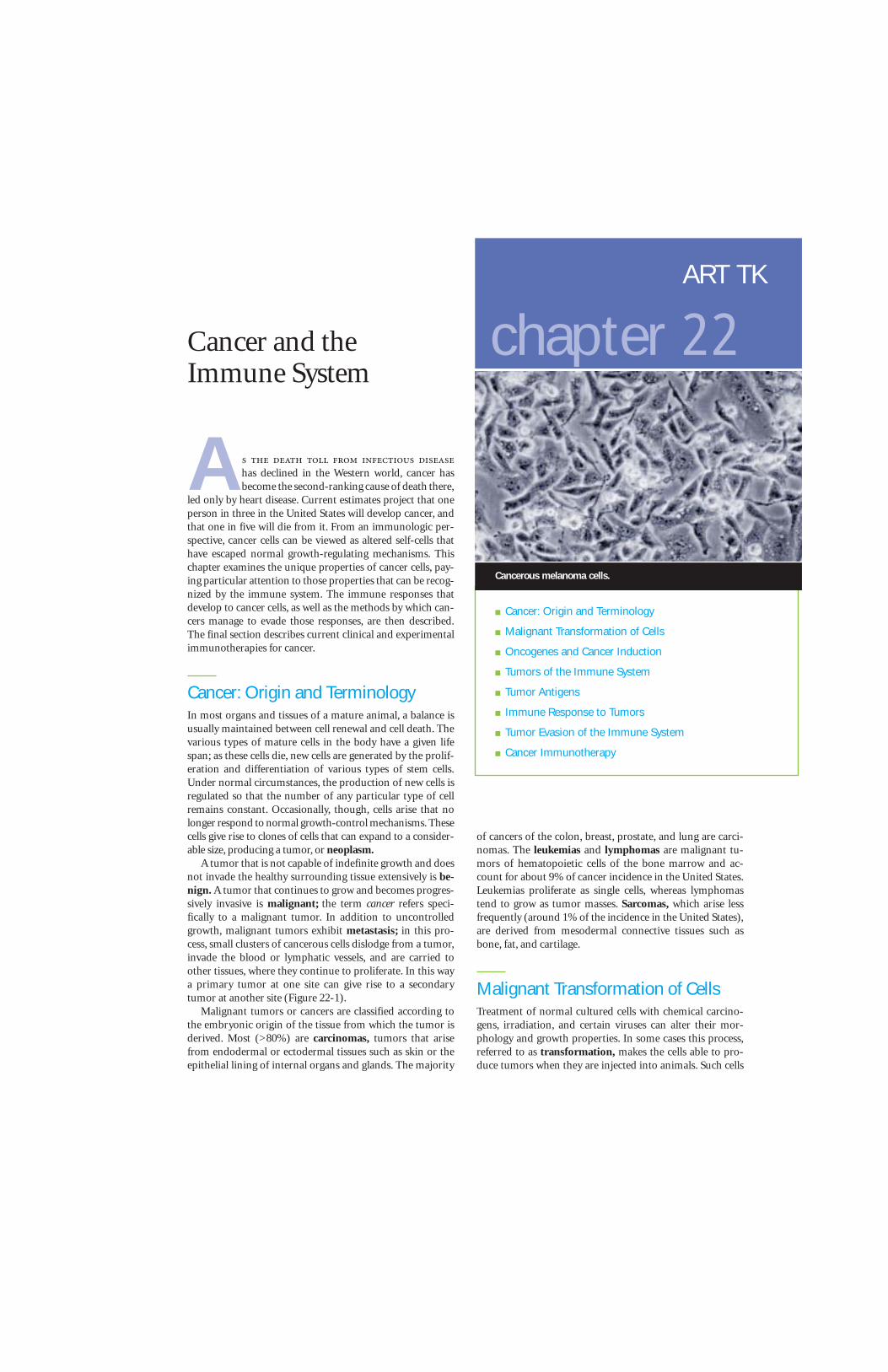
Cancerous melanoma cells.
Cancer and the Immune System
A
has declined in the Western world, cancer hasbecome the second-ranking cause of death there,
led only by heart disease. Current estimates project that oneperson in three in the United States will develop cancer, andthat one in five will die from it. From an immunologic per-spective, cancer cells can be viewed as altered self-cells thathave escaped normal growth-regulating mechanisms. Thischapter examines the unique properties of cancer cells, pay-ing particular attention to those properties that can be recog-nized by the immune system. The immune responses thatdevelop to cancer cells, as well as the methods by which can-cers manage to evade those responses, are then described.The final section describes current clinical and experimentalimmunotherapies for cancer.
Cancer: Origin and TerminologyIn most organs and tissues of a mature animal, a balance isusually maintained between cell renewal and cell death. Thevarious types of mature cells in the body have a given lifespan; as these cells die, new cells are generated by the prolif-eration and differentiation of various types of stem cells.Under normal circumstances, the production of new cells isregulated so that the number of any particular type of cellremains constant. Occasionally, though, cells arise that nolonger respond to normal growth-control mechanisms. Thesecells give rise to clones of cells that can expand to a consider-able size, producing a tumor, or neoplasm.
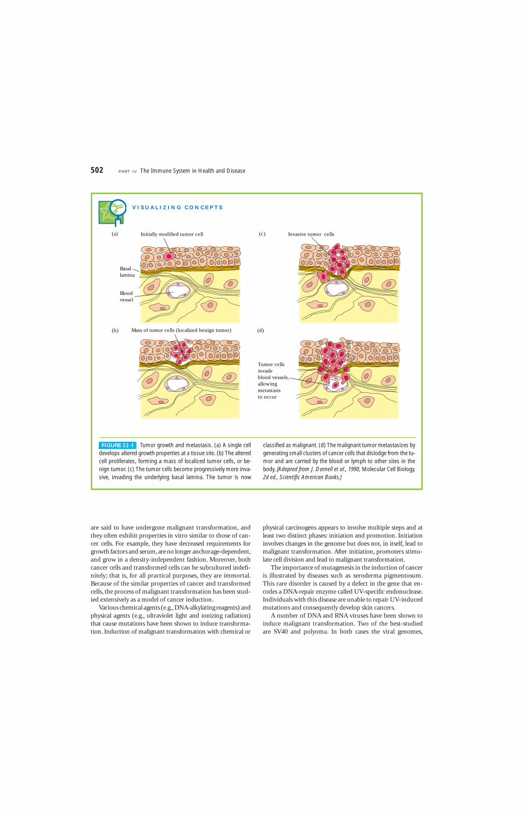
A tumor that is not capable of indefinite growth and doesnot invade the healthy surrounding tissue extensively is be-nign. A tumor that continues to grow and becomes progres-sively invasive is malignant; the term cancer refers speci-fically to a malignant tumor. In addition to uncontrolledgrowth, malignant tumors exhibit metastasis; in this pro-cess, small clusters of cancerous cells dislodge from a tumor,invade the blood or lymphatic vessels, and are carried toother tissues, where they continue to proliferate. In this waya primary tumor at one site can give rise to a secondarytumor at another site (Figure 22-1).
Malignant tumors or cancers are classified according tothe embryonic origin of the tissue from which the tumor isderived. Most (>80%) are carcinomas, tumors that arisefrom endodermal or ectodermal tissues such as skin or theepithelial lining of internal organs and glands. The majority
of cancers of the colon, breast, prostate, and lung are carci-nomas. The leukemias and lymphomas are malignant tu-mors of hematopoietic cells of the bone marrow and ac-count for about 9% of cancer incidence in the United States.Leukemias proliferate as single cells, whereas lymphomastend to grow as tumor masses. Sarcomas, which arise lessfrequently (around 1% of the incidence in the United States),are derived from mesodermal connective tissues such asbone, fat, and cartilage.
Malignant Transformation of CellsTreatment of normal cultured cells with chemical carcino-gens, irradiation, and certain viruses can alter their mor-phology and growth properties. In some cases this process,referred to as transformation, makes the cells able to pro-duce tumors when they are injected into animals. Such cells
chapter 22ART TK
are said to have undergone malignant transformation, andthey often exhibit properties in vitro similar to those of can-cer cells. For example, they have decreased requirements forgrowth factors and serum, are no longer anchorage-dependent,and grow in a density-independent fashion. Moreover, bothcancer cells and transformed cells can be subcultured indefi-nitely; that is, for all practical purposes, they are immortal.Because of the similar properties of cancer and transformedcells, the process of malignant transformation has been stud-ied extensively as a model of cancer induction.
Various chemical agents (e.g., DNA-alkylating reagents) andphysical agents (e.g., ultraviolet light and ionizing radiation)that cause mutations have been shown to induce transforma-tion. Induction of malignant transformation with chemical or
physical carcinogens appears to involve multiple steps and atleast two distinct phases: initiation and promotion. Initiationinvolves changes in the genome but does not, in itself, lead tomalignant transformation. After initiation, promoters stimu-late cell division and lead to malignant transformation.
The importance of mutagenesis in the induction of canceris illustrated by diseases such as xeroderma pigmentosum.This rare disorder is caused by a defect in the gene that en-codes a DNA-repair enzyme called UV-specific endonuclease.Individuals with this disease are unable to repair UV-inducedmutations and consequently develop skin cancers.
A number of DNA and RNA viruses have been shown toinduce malignant transformation. Two of the best-studiedare SV40 and polyoma. In both cases the viral genomes,
502 P A R T I V The Immune System in Health and Disease
V I S U A L I Z I N G C O N C E P T S
(a)
(b)
(c)
(d)
Initially modified tumor cell Invasive tumor cells
Mass of tumor cells (localized benign tumor)
Basallamina
Bloodvessel
Tumor cellsinvadeblood vessels,allowing metastasisto occur
FIGURE 22-1 Tumor growth and metastasis. (a) A single celldevelops altered growth properties at a tissue site. (b) The alteredcell proliferates, forming a mass of localized tumor cells, or be-nign tumor. (c) The tumor cells become progressively more inva-sive, invading the underlying basal lamina. The tumor is now
classified as malignant. (d) The malignant tumor metastasizes bygenerating small clusters of cancer cells that dislodge from the tu-mor and are carried by the blood or lymph to other sites in thebody. [Adapted from J. Darnell et al., 1990, Molecular Cell Biology,2d ed., Scientific American Books.]
which integrate randomly into the host chromosomal DNA,include several genes that are expressed early in the course ofviral replication. SV40 encodes two early proteins called largeT and little T, and polyoma encodes three early proteins calledlarge T, middle T, and little T. Each of these proteins plays arole in the malignant transformation of virus-infected cells.
Most RNA viruses replicate in the cytosol and do notinduce malignant transformation. The exceptions are retro-viruses, which transcribe their RNA into DNA by means of areverse-transcriptase enzyme and then integrate the tran-script into the host’s DNA. This process is similar in the cyto-pathic retroviruses such as HIV-1 and HIV-2 and in thetransforming retroviruses, which induce changes in the hostcell that lead to malignant transformation. In some cases,retrovirus-induced transformation is related to the presenceof oncogenes, or “cancer genes,” carried by the retrovirus.
One of the best-studied transforming retroviruses is theRous sarcoma virus. This virus carries an oncogene called v-src, which encodes a 60-kDa protein kinase (v-Src) that cat-alyzes the addition of phosphate to tyrosine residues on pro-teins. The first evidence that oncogenes alone could inducemalignant transformation came from studies of the v-src on-cogene from Rous sarcoma virus. When this oncogene wascloned and transfected into normal cells in culture, the cellsunderwent malignant transformation.
Oncogenes and Cancer InductionIn 1971, Howard Temin suggested that oncogenes might notbe unique to transforming viruses but might also be found innormal cells; indeed, he proposed that a virus might acquireoncogenes from the genome of an infected cell. He calledthese cellular genes proto-oncogenes, or cellular oncogenes(c-onc), to distinguish them from their viral counterparts (v-onc). In the mid-1970s, J. M. Bishop and H. E. Varmusidentified a DNA sequence in normal chicken cells that ishomologous to v-src from Rous sarcoma virus. This cellularoncogene was designated c-src. Since these early discoveries,numerous cellular oncogenes have been identified.
Sequence comparisons of viral and cellular oncogenesreveal that they are highly conserved in evolution. Althoughmost cellular oncogenes consist of a series of exons and in-trons, their viral counterparts consist of uninterrupted cod-ing sequences, suggesting that the virus might have acquiredthe oncogene through an intermediate RNA transcript fromwhich the intron sequences had been removed during RNAprocessing. The actual coding sequences of viral oncogenesand the corresponding proto-oncogenes exhibit a high de-gree of homology; in some cases, a single point mutation isall that distinguishes a viral oncogene from the correspond-ing proto-oncogene. It has now become apparent that most,if not all, oncogenes (both viral and cellular) are derived fromcellular genes that encode various growth-controlling pro-teins. In addition, the proteins encoded by a particular onco-
gene and its corresponding proto-oncogene appear to havevery similar functions. As described below, the conversion ofa proto-oncogene into an oncogene appears in many cases toaccompany a change in the level of expression of a normalgrowth-controlling protein.
Cancer-Associated Genes Have Many FunctionsHomeostasis in normal tissue is maintained by a highly reg-ulated process of cellular proliferation balanced by cell death.If there is an imbalance, either at the stage of cellular prolif-eration or at the stage of cell death, then a cancerous state willdevelop. Oncogenes and tumor suppressor genes have beenshown to play an important role in this process, by regulatingeither cellular proliferation or cell death. Cancer-associatedgenes can be divided into three categories that reflect thesedifferent activities, summarized in Table 22-1.
INDUCTION OF CELLULAR PROLIFERATION
One category of proto-oncogenes and their oncogenic coun-terparts encodes proteins that induce cellular proliferation.Some of these proteins function as growth factors or growth-factor receptors. Included among these are sis, which encodesa form of platelet-derived growth factor, and fms, erbB, andneu, which encode growth-factor receptors. In normal cells,the expression of growth factors and their receptors is care-fully regulated. Usually, one population of cells secretes agrowth factor that acts on another population of cells thatcarries the receptor for the factor, thus stimulating prolifera-tion of the second population. Inappropriate expression ofeither a growth factor or its receptor can result in uncon-trolled proliferation.
Other oncogenes in this category encode products thatfunction in signal-transduction pathways or as transcriptionfactors. The src and abl oncogenes encode tyrosine kinases,and the ras oncogene encodes a GTP-binding protein. Theproducts of these genes act as signal transducers. The myc,jun, and fos oncogenes encode transcription factors. Overac-tivity of any of these oncogenes may result in unregulatedproliferation.
INHIBITION OF CELLULAR PROLIFERATION
A second category of cancer-associated genes—called tumor-suppressor genes, or anti-oncogenes—encodes proteins thatinhibit excessive cell proliferation. Inactivation of these re-sults in unregulated proliferation. The prototype of this cate-gory of oncogenes is Rb, the retinoblastoma gene. Hereditaryretinoblastoma is a rare childhood cancer, in which tumorsdevelop from neural precursor cells in the immature retina.The affected child has inherited a mutated Rb allele; somaticinactivation of the remaining Rb allele leads to tumor growth.Probably the single most frequent genetic abnormality inhuman cancer is mutation in p53, which encodes a nuclearphosphoprotein. Over 90% of small-cell lung cancers and
Cancer and the Immune System C H A P T E R 22 503

over 50% of breast and colon cancers have been shown to beassociated with mutations in p53.
REGULATION OF PROGRAMMED CELL DEATH
A third category of cancer-associated genes regulates pro-grammed cell death. These genes encode proteins that eitherblock or induce apoptosis. Included in this category of onco-genes is bcl-2, an anti-apoptosis gene. This oncogene wasoriginally discovered because of its association with B-cell fol-licular lymphoma. Since its discovery, bcl-2 has been shown toplay an important role in regulating cell survival duringhematopoiesis and in the survival of selected B cells and T cells during maturation. Interestingly, the Epstein-Barrvirus contains a gene that has sequence homology to bcl-2and may act in a similar manner to suppress apoptosis.
Proto-Oncogenes Can Be Converted to Oncogenes
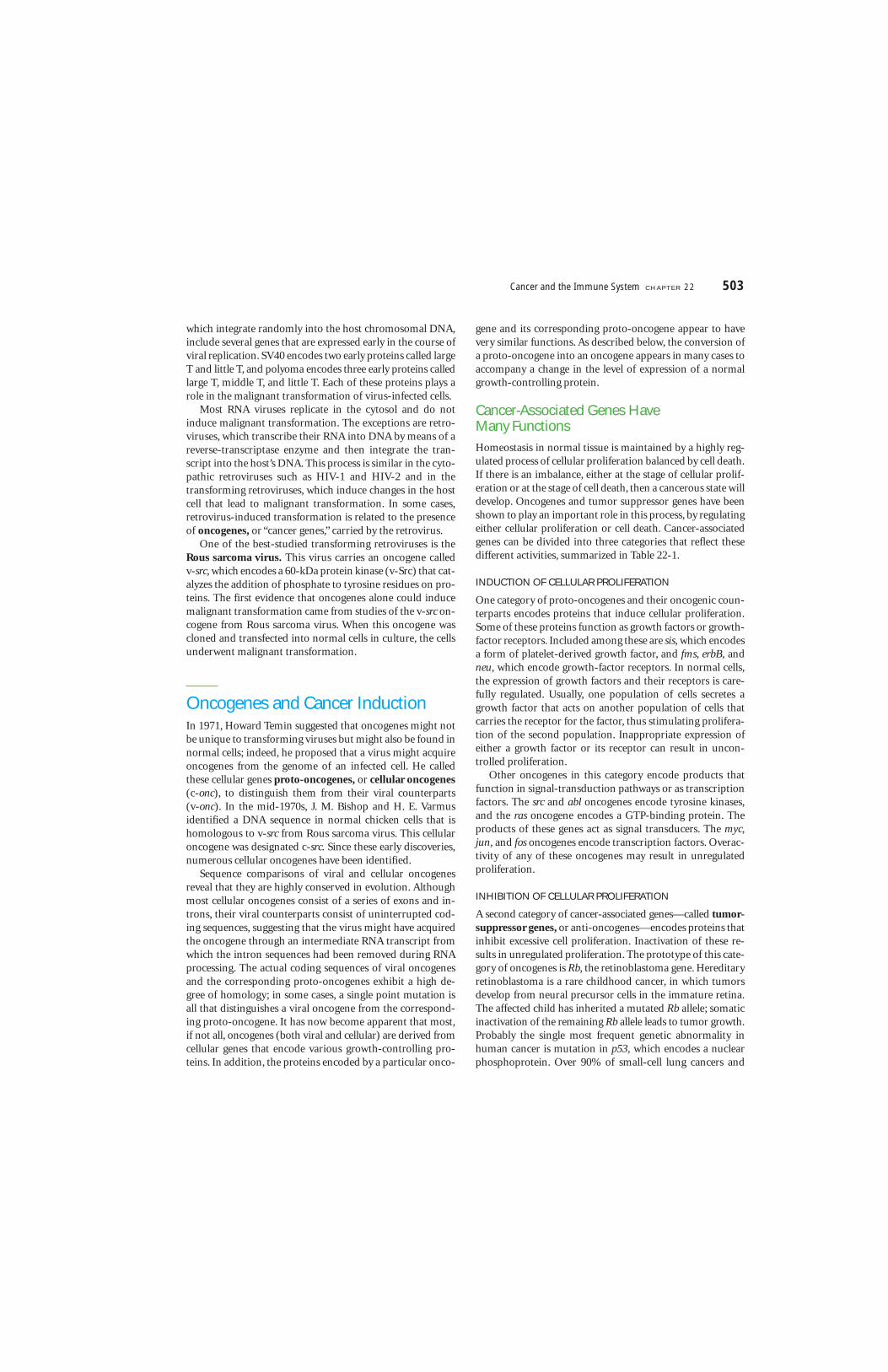
In 1972, R. J. Huebner and G. J. Todaro suggested that muta-tions or genetic rearrangements of proto-oncogenes by car-cinogens or viruses might alter the normally regulated functionof these genes, converting them into potent cancer-causingoncogenes (Figure 22-2). Considerable evidence supportingthis hypothesis accumulated in subsequent years. For example,some malignantly transformed cells contain multiple copies ofcellular oncogenes, resulting in increased production of onco-gene products. Such amplification of cellular oncogenes hasbeen observed in cells from various types of human cancers.Several groups have identified c-myc oncogenes in homo-geneously staining regions (HSRs) of chromosomes from can-
504 P A R T I V The Immune System in Health and Disease
TABLE 22-1 Functional classification of cancer-associated genes
Type/name Nature of gene product
CATEGORY I: GENES THAT INDUCE CELLULAR PROLIFERATION
Growth factorssis A form of platelet-derived growth factor (PDGF)
Growth-factor receptorsfms Receptor for colony-stimulating factor 1 (CSF-1)erbB Receptor for epidermal growth factor (EGF)neu Protein (HER2) related to EGF receptorerbA Receptor for thyroid hormone
Signal transducerssrc Tyrosine kinaseabl Tyrosine kinaseHa-ras GTP-binding protein with GTPase activityN-ras GTP-binding protein with GTPase activityK-ras GTP-binding protein with GTPase activity
Transcription factorsjun Component of transcription factor AP1fos Component of transcription factor AP1myc DNA-binding protein
CATEGORY II: TUMOR-SUPRESSOR GENES, INHIBITORS OF CELLULAR PROLIFERATION*
Rb Suppressor of retinoblastomap53 Nuclear phosphoprotein that inhibits formation of small-cell lung cander and colon cancersDCC Suppressor of colon carcinomaAPC Suppressor of adenomatous polyposisNF1 Suppressor of neurofibromatosisWT1 Suppressor of Wilm’s tumor
CATEGORY III: GENES THAT REGULATE PROGRAMMED CELL DEATH
bcl-2 Suppressor of apoptosis
* The activity of the normal products of the category II genes inhibits progression of the cell cycle. Loss of a gene or its inactivation by mutation in an indicated
tumor-suppressor gene is associated with development of the indicated cancers.
cer cells; these HSRs represent long tandem arrays of amplifiedgenes.
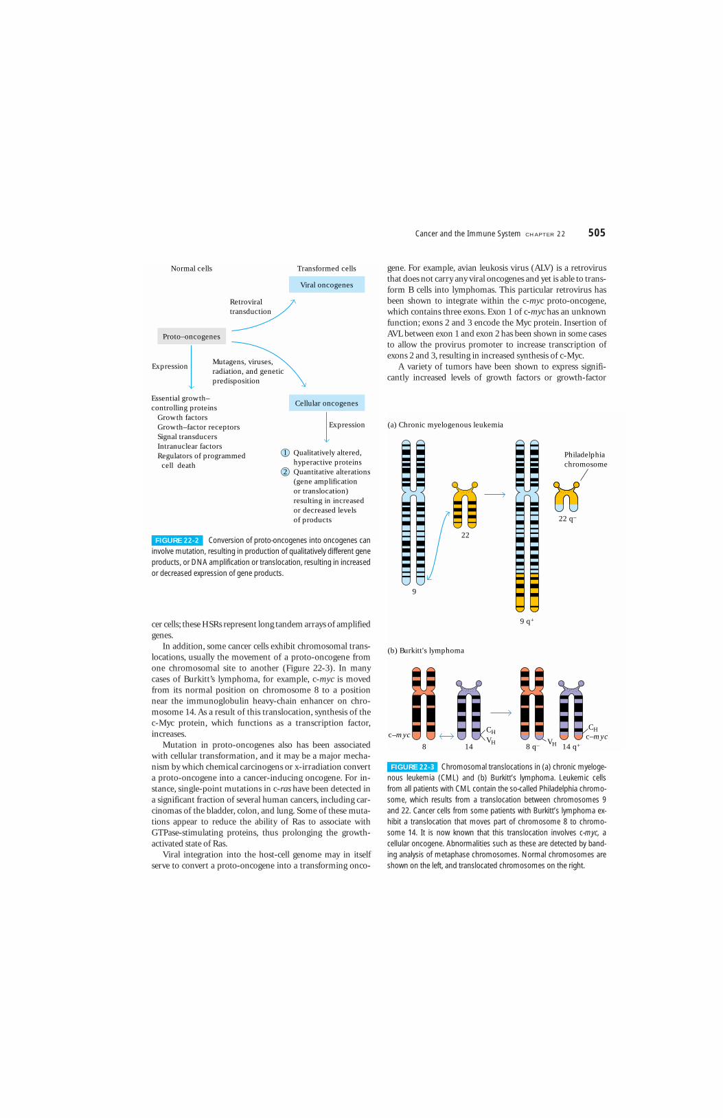
In addition, some cancer cells exhibit chromosomal trans-locations, usually the movement of a proto-oncogene fromone chromosomal site to another (Figure 22-3). In manycases of Burkitt’s lymphoma, for example, c-myc is movedfrom its normal position on chromosome 8 to a positionnear the immunoglobulin heavy-chain enhancer on chro-mosome 14. As a result of this translocation, synthesis of thec-Myc protein, which functions as a transcription factor,increases.
Mutation in proto-oncogenes also has been associatedwith cellular transformation, and it may be a major mecha-nism by which chemical carcinogens or x-irradiation converta proto-oncogene into a cancer-inducing oncogene. For in-stance, single-point mutations in c-ras have been detected ina significant fraction of several human cancers, including car-cinomas of the bladder, colon, and lung. Some of these muta-tions appear to reduce the ability of Ras to associate withGTPase-stimulating proteins, thus prolonging the growth-activated state of Ras.
Viral integration into the host-cell genome may in itselfserve to convert a proto-oncogene into a transforming onco-
gene. For example, avian leukosis virus (ALV) is a retrovirusthat does not carry any viral oncogenes and yet is able to trans-form B cells into lymphomas. This particular retrovirus hasbeen shown to integrate within the c-myc proto-oncogene,which contains three exons. Exon 1 of c-myc has an unknownfunction; exons 2 and 3 encode the Myc protein. Insertion ofAVL between exon 1 and exon 2 has been shown in some casesto allow the provirus promoter to increase transcription ofexons 2 and 3, resulting in increased synthesis of c-Myc.
A variety of tumors have been shown to express signifi-cantly increased levels of growth factors or growth-factor
Cancer and the Immune System C H A P T E R 22 505
Normal cells Transformed cells
Proto–oncogenes
Expression
Retroviraltransduction
Mutagens, viruses,radiation, and geneticpredisposition
Cellular oncogenes
Expression
Viral oncogenes
Essential growth–controlling proteins Growth factors Growth–factor receptors Signal transducers Intranuclear factors Regulators of programmed cell death
1 Qualitatively altered, hyperactive proteins2 Quantitative alterations (gene amplification or translocation) resulting in increased or decreased levels of products
FIGURE 22-2 Conversion of proto-oncogenes into oncogenes caninvolve mutation, resulting in production of qualitatively different geneproducts, or DNA amplification or translocation, resulting in increasedor decreased expression of gene products.
(a) Chronic myelogenous leukemia
9
22
Philadelphiachromosome
(b) Burkitt's lymphoma
8 14
9 q+
22 q–
CH
VH
CHc–myc c–myc
VH8 q– 14 q+
FIGURE 22-3 Chromosomal translocations in (a) chronic myeloge-nous leukemia (CML) and (b) Burkitt’s lymphoma. Leukemic cellsfrom all patients with CML contain the so-called Philadelphia chromo-some, which results from a translocation between chromosomes 9and 22. Cancer cells from some patients with Burkitt’s lymphoma ex-hibit a translocation that moves part of chromosome 8 to chromo-some 14. It is now known that this translocation involves c-myc, acellular oncogene. Abnormalities such as these are detected by band-ing analysis of metaphase chromosomes. Normal chromosomes areshown on the left, and translocated chromosomes on the right.
receptors. Expression of the receptor for epidermal growthfactor, which is encoded by c-erbB, has been shown to beamplified in many cancer cells. And in breast cancer, increasedsynthesis of the growth-factor receptor encoded by c-neu hasbeen linked with a poor prognosis.
The Induction of Cancer Is a MultistepProcessThe development from a normal cell to a cancerous cell isusually a multistep process of clonal evolution driven by aseries of somatic mutations that progressively convert the cellfrom normal growth to a precancerous state and finally acancerous state.
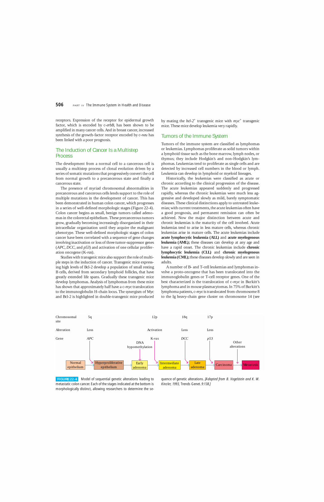
The presence of myriad chromosomal abnormalities inprecancerous and cancerous cells lends support to the role ofmultiple mutations in the development of cancer. This hasbeen demonstrated in human colon cancer, which progressesin a series of well-defined morphologic stages (Figure 22-4).Colon cancer begins as small, benign tumors called adeno-mas in the colorectal epithelium. These precancerous tumorsgrow, gradually becoming increasingly disorganized in theirintracellular organization until they acquire the malignantphenotype. These well-defined morphologic stages of coloncancer have been correlated with a sequence of gene changesinvolving inactivation or loss of three tumor-suppressor genes(APC, DCC, and p53) and activation of one cellular prolifer-ation oncogene (K-ras).
Studies with transgenic mice also support the role of multi-ple steps in the induction of cancer. Transgenic mice express-ing high levels of Bcl-2 develop a population of small resting B cells, derived from secondary lymphoid follicles, that havegreatly extended life spans. Gradually these transgenic micedevelop lymphomas. Analysis of lymphomas from these micehas shown that approximately half have a c-myc translocationto the immunoglobulin H-chain locus. The synergism of Mycand Bcl-2 is highlighted in double-transgenic mice produced
by mating the bcl-2+ transgenic mice with myc+ transgenicmice. These mice develop leukemia very rapidly.
Tumors of the Immune SystemTumors of the immune system are classified as lymphomasor leukemias. Lymphomas proliferate as solid tumors withina lymphoid tissue such as the bone marrow, lymph nodes, orthymus; they include Hodgkin’s and non-Hodgkin’s lym-phomas. Leukemias tend to proliferate as single cells and aredetected by increased cell numbers in the blood or lymph.Leukemia can develop in lymphoid or myeloid lineages.
Historically, the leukemias were classified as acute orchronic according to the clinical progression of the disease.The acute leukemias appeared suddenly and progressedrapidly, whereas the chronic leukemias were much less ag-gressive and developed slowly as mild, barely symptomaticdiseases. These clinical distinctions apply to untreated leuke-mias; with current treatments, the acute leukemias often havea good prognosis, and permanent remission can often beachieved. Now the major distinction between acute andchronic leukemias is the maturity of the cell involved. Acuteleukemias tend to arise in less mature cells, whereas chronicleukemias arise in mature cells. The acute leukemias includeacute lymphocytic leukemia (ALL) and acute myelogenousleukemia (AML); these diseases can develop at any age andhave a rapid onset. The chronic leukemias include chroniclymphocytic leukemia (CLL) and chronic myelogenousleukemia (CML); these diseases develop slowly and are seen inadults.
A number of B- and T-cell leukemias and lymphomas in-volve a proto-oncogene that has been translocated into theimmunoglobulin genes or T-cell receptor genes. One of thebest characterized is the translocation of c-myc in Burkitt’slymphoma and in mouse plasmacytomas. In 75% of Burkitt’slymphoma patients, c-myc is translocated from chromosome 8to the Ig heavy-chain gene cluster on chromosome 14 (see
506 P A R T I V The Immune System in Health and Disease
Chromosomalsite
Alteration
Gene
5q
Loss
APC
18q
Loss
DCC
12p
Activation
K-ras
17p
Loss
p53DNA
hypomethylation
Otheralterations
Normalepithelium
Hyperproliferativeepithelium
Earlyadenoma
Intermediateadenoma
Lateadenoma
Carcinoma Metastasis
FIGURE 22-4 Model of sequential genetic alterations leading tometastatic colon cancer. Each of the stages indicated at the bottom ismorphologically distinct, allowing researchers to determine the se-
quence of genetic alterations. [Adapted from B. Vogelstein and K. W.Kinzler, 1993, Trends Genet. 9:138.]

Figure 22-3b). In the remaining patients, c-myc remains onchromosome 8 and the � or � light-chain genes are translo-cated to a region 3� of c-myc. Kappa-gene translocationsfrom chromosome 2 to chromosome 8 occur 9% of the time,and �-gene translocations from chromosome 22 to chromo-some 8 occur 16% of the time.
Translocations of c-myc to the Ig heavy-chain gene clusteron chromosome 14 have been analyzed, and, in some cases,the entire c-myc gene is translocated head-to-head to a re-gion near the heavy-chain enhancer. In other cases, exons 1,2, and 3 or exons 2 and 3 of c-myc are translocated head-to-head to the S� or S� switch site (Figure 22-5). In each case,the translocation removes the myc coding exons from theregulatory mechanisms operating in chromosome 8 andplaces them in the immunoglobulin-gene region, a very ac-tive region that is expressed constitutively in these cells. Theconsequences of enhancer-mediated high levels of constitu-tive myc expression in lymphoid cells have been investigatedin transgenic mice. In one study, mice containing a transgeneconsisting of all three c-myc exons and the immunoglobulinheavy-chain enhancer were produced. Of 15 transgenic pupsborn, 13 developed lymphomas of the B-cell lineage within afew months of birth.
Tumor AntigensThe subdiscipline of tumor immunology involves the studyof antigens on tumor cells and the immune response to theseantigens. Two types of tumor antigens have been identifiedon tumor cells: tumor-specific transplantation antigens(TSTAs) and tumor-associated transplantation antigens
(TATAs). Tumor-specific antigens are unique to tumor cellsand do not occur on normal cells in the body. They mayresult from mutations in tumor cells that generate alteredcellular proteins; cytosolic processing of these proteinswould give rise to novel peptides that are presented with classI MHC molecules, inducing a cell-mediated response bytumor-specific CTLs (Figure 22-6). Tumor-associated anti-gens, which are not unique to tumor cells, may be proteinsthat are expressed on normal cells during fetal developmentwhen the immune system is immature and unable to respondbut that normally are not expressed in the adult. Reactivationof the embryonic genes that encode these proteins in tumorcells results in their expression on the fully differentiatedtumor cells. Tumor-associated antigens may also be proteinsthat are normally expressed at extremely low levels on normalcells but are expressed at much higher levels on tumor cells. Itis now clear that the tumor antigens recognized by human Tcells fall into one of four major categories:
■ Antigens encoded by genes exclusively expressed bytumors
■ Antigens encoded by variant forms of normal genes thathave been altered by mutation
■ Antigens normally expressed only at certain stages ofdifferentiation or only by certain differentiation lineages
■ Antigens that are overexpressed in particular tumors
Many tumor antigens are cellular proteins that give rise topeptides presented with MHC molecules; typically, these an-tigens have been identified by their ability to induce the pro-liferation of antigen-specific CTLs or helper T cells.
Cancer and the Immune System C H A P T E R 22 507
5′
JH
3′
Cµ exonsEnhancer
Switch regionD
JHVH
Promoter
Sµ
5′ 3′
Cµ exonsEnhancer
Sµ
123
c–myc exons
(a)
Rearranged Ig heavy–chaingene on chromosome 14
Translocated c–myc gene insome Burkitt's lymphomas
5′ 3′
Cµ exons
Sµ23
c–myc exons
(b)Translocated c–myc gene inother Burkitt's lymphomas
L
FIGURE 22-5 In many patients with Burkitt’s lymphoma, the c-mycgene is translocated to the immunoglobulin heavy-chain gene clusteron chromosome 14. In some cases, the entire c-myc gene is insertednear the heavy-chain enhancer (a), but in other cases, only the coding
exons (2 and 3) of c-myc are inserted at the S� switch site (b). Onlyexons 2 and 3 of c-myc are coding exons. Translocation may lead tooverexpression of c-Myc.

Some Antigens Are Tumor-SpecificTumor-specific antigens have been identified on tumors in-duced with chemical or physical carcinogens and on somevirally induced tumors. Demonstrating the presence of tumor-specific antigens on spontaneously occurring tumors is par-ticularly difficult because the immune response to such tu-mors eliminates all of the tumor cells bearing sufficientnumbers of the antigens and in this way selects for cells bear-ing low levels of the antigens.
CHEMICALLY OR PHYSICALLY INDUCED TUMOR ANTIGENS
Methylcholanthrene and ultraviolet light are two carcinogensthat have been used extensively to generate lines of tumor cells.When syngeneic animals are injected with killed cells from acarcinogen-induced tumor-cell line, the animals develop aspecific immunologic response that can protect against laterchallenge by live cells of the same line but not other tumor-celllines (Table 22-2). Even when the same chemical carcinogeninduces two separate tumors at different sites in the same ani-mal, the tumor antigens are distinct and the immune responseto one tumor does not protect against the other tumor.
The tumor-specific transplantation antigens of chemicallyinduced tumors have been difficult to characterize becausethey cannot be identified by induced antibodies but only bytheir T-cell–mediated rejection. One experimental approachthat has allowed identification of genes encoding some TSTAsis outlined in Figure 22-7. When a mouse tumorigenic cell line
(tum+), which gives rise to progressively growing tumors, istreated in vitro with a chemical mutagen, some cells aremutated so that they no longer are capable of growing into a
508 P A R T I V The Immune System in Health and Disease
Altered self-peptide
Mutation generates new peptidein class I MHC molecule (TSTA)
Oncofetalpeptide
Normal cell
Inappropriate expression ofembryonic gene (TATA)
Self-peptideSelf-peptide
Class I MHCClass I MHC
Overexpression ofnormal protein (TATA)
FIGURE 22-6 Different mechanisms generate tumor-specific transplantation antigens (TSTAs)and tumor-associated transplantation antigens (TATAs). The latter are more common.
TABLE 22-2Immune response to methyl-cholanthrene (MCA) or polyoma virus (PV)*
Transplanted Live tumor cells Tumor killed tumor cells for challenge growth
CHEMICALLY INDUCED
MCA-induced sarcoma A MCA-induced sarcoma A –
MCA-induced sarcoma A MCA-induced sarcoma B +
VIRALLY INDUCED
PV-induced sarcoma A PV-induced sarcoma A –
PV-induced sarcoma A PV-induced sarcoma B –
PV-induced sarcoma A SV40-induced sarcoma C +
*Tumors were induced either with MCA or PV, and killed cells from the induced
tumors were injected into syngeneic animals, which were then challenged with
live cells from the indicated tumor-cell lines. The absence of tumor growth after
live challenge indicates that the immune response induced by tumor antigens
on the killed cells provided protection against the live cells.

tumor in syngeneic mice. These mutant tumor cells are desig-nated as tum– variants. Most tum– variants have been shownto express TSTAs that are not expressed by the original tum+
tumor-cell line. When tum– cells are injected into syngeneicmice, the unique TSTAs that the tum– cells express are recog-nized by specific CTLs. The TSTA-specific CTLs destroy thetum– tumor cells, thus preventing tumor growth. To identify
the genes encoding the TSTAs that are expressed on a tum– cellline, a cosmid DNA library is prepared from the tum– cells.Genes from the tum– cells are transfected into the originaltum+ cells. The transfected tum+ cells are tested for the expres-sion of the tum– TSTAs by their ability to activate clonedCTLs specific for the tum– TSTA. A number of diverse TSTAshave been identified by this method.
Cancer and the Immune System C H A P T E R 22 509
Clone #1 (tum+)
No tumor growth
Clone #2 (tum–) Clone #3 (tum+)
TSTAs
Mutagenize and clone
Tum+ cells
Tumor growthTumor growth
Isolate and cloneCTLs from mouse
TSTA–specific CTLs
Incubate transfectedtum+ cells with TSTAspecific CTL clone
Prepare cDNAlibrary fromtum– cells
Transfecttum– geneinto tum+ cells
Tum– gene #1
Tum– gene #2
Tum– TSTA gene
Tumor growth
Tum+ cells donot expressTSTA
Transplant
Observefor lysis
No lysis (cells do not expressTSTA)
No lysis (cells do not expressTSTA)
Lysis (cells express TSTA)
FIGURE 22-7 One procedure for identifying genes encoding tumor-specific transplantation antigens (TSTAs). Most TSTAs can be detectedonly by the cell-mediated rejection they elicit. In the first part of this pro-cedure, a nontumorigenic (tum–) cell line is generated; this cell line ex-presses a TSTA that is recognized by syngeneic mice, which mount a
cell-mediated response against it. To isolate the gene encoding theTSTA, a cosmid gene library is prepared from the tum– cell line, thegenes are transfected into tumorigenic tum+ cells, and the transfectedcells are incubated with TSTA-specific CTLs.
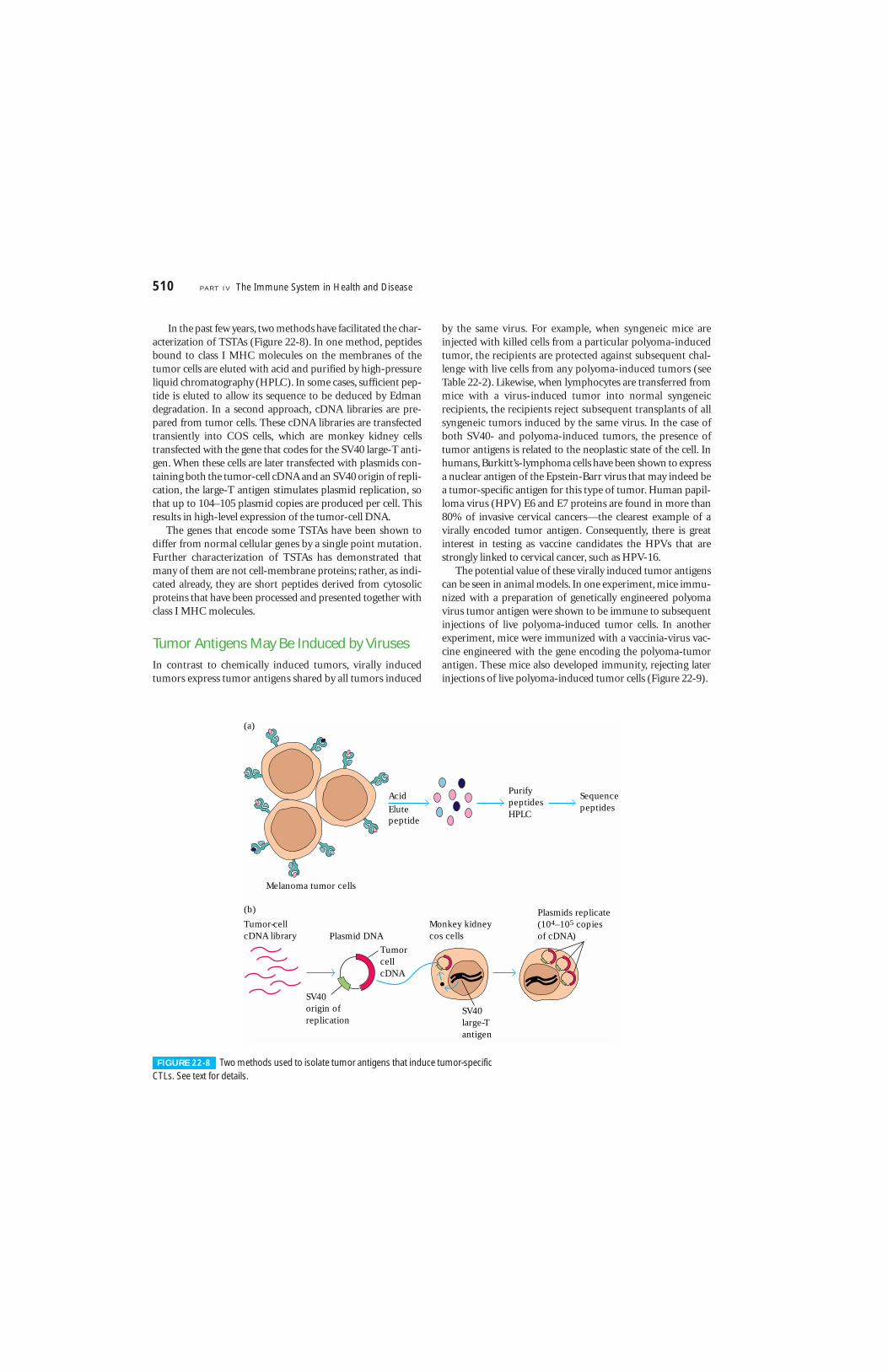
In the past few years, two methods have facilitated the char-acterization of TSTAs (Figure 22-8). In one method, peptidesbound to class I MHC molecules on the membranes of thetumor cells are eluted with acid and purified by high-pressureliquid chromatography (HPLC). In some cases, sufficient pep-tide is eluted to allow its sequence to be deduced by Edmandegradation. In a second approach, cDNA libraries are pre-pared from tumor cells. These cDNA libraries are transfectedtransiently into COS cells, which are monkey kidney cellstransfected with the gene that codes for the SV40 large-T anti-gen. When these cells are later transfected with plasmids con-taining both the tumor-cell cDNA and an SV40 origin of repli-cation, the large-T antigen stimulates plasmid replication, sothat up to 104–105 plasmid copies are produced per cell. Thisresults in high-level expression of the tumor-cell DNA.
The genes that encode some TSTAs have been shown todiffer from normal cellular genes by a single point mutation.Further characterization of TSTAs has demonstrated thatmany of them are not cell-membrane proteins; rather, as indi-cated already, they are short peptides derived from cytosolicproteins that have been processed and presented together withclass I MHC molecules.
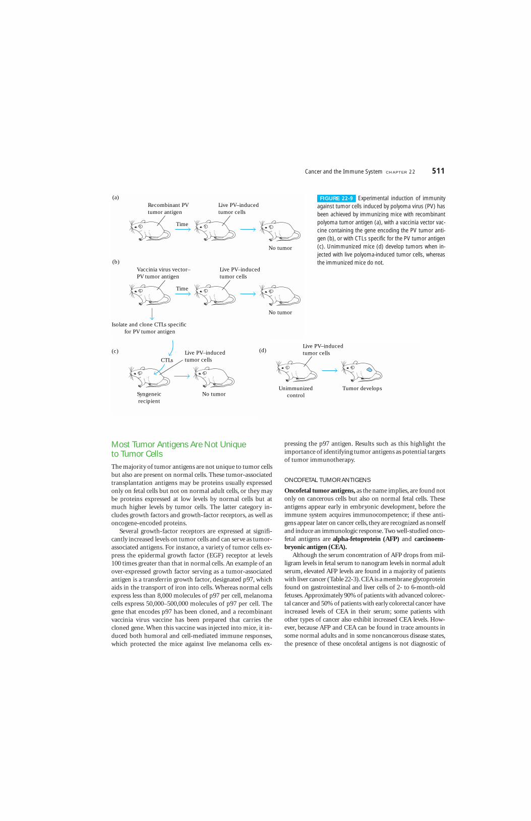
Tumor Antigens May Be Induced by VirusesIn contrast to chemically induced tumors, virally inducedtumors express tumor antigens shared by all tumors induced
by the same virus. For example, when syngeneic mice areinjected with killed cells from a particular polyoma-inducedtumor, the recipients are protected against subsequent chal-lenge with live cells from any polyoma-induced tumors (seeTable 22-2). Likewise, when lymphocytes are transferred frommice with a virus-induced tumor into normal syngeneicrecipients, the recipients reject subsequent transplants of allsyngeneic tumors induced by the same virus. In the case ofboth SV40- and polyoma-induced tumors, the presence oftumor antigens is related to the neoplastic state of the cell. Inhumans, Burkitt’s-lymphoma cells have been shown to expressa nuclear antigen of the Epstein-Barr virus that may indeed bea tumor-specific antigen for this type of tumor. Human papil-loma virus (HPV) E6 and E7 proteins are found in more than80% of invasive cervical cancers—the clearest example of avirally encoded tumor antigen. Consequently, there is greatinterest in testing as vaccine candidates the HPVs that arestrongly linked to cervical cancer, such as HPV-16.
The potential value of these virally induced tumor antigenscan be seen in animal models. In one experiment, mice immu-nized with a preparation of genetically engineered polyomavirus tumor antigen were shown to be immune to subsequentinjections of live polyoma-induced tumor cells. In anotherexperiment, mice were immunized with a vaccinia-virus vac-cine engineered with the gene encoding the polyoma-tumorantigen. These mice also developed immunity, rejecting laterinjections of live polyoma-induced tumor cells (Figure 22-9).
510 P A R T I V The Immune System in Health and Disease
Monkey kidneycos cells
(a)
Acid
Elutepeptide
PurifypeptidesHPLC
Melanoma tumor cells
Tumor-cellcDNA library
TumorcellcDNA
SV40origin ofreplication
Plasmid DNA
(b)
Sequencepeptides
SV40large-Tantigen
Plasmids replicate(104–105 copiesof cDNA)
FIGURE 22-8 Two methods used to isolate tumor antigens that induce tumor-specificCTLs. See text for details.
Most Tumor Antigens Are Not Unique to Tumor CellsThe majority of tumor antigens are not unique to tumor cellsbut also are present on normal cells. These tumor-associatedtransplantation antigens may be proteins usually expressedonly on fetal cells but not on normal adult cells, or they maybe proteins expressed at low levels by normal cells but atmuch higher levels by tumor cells. The latter category in-cludes growth factors and growth-factor receptors, as well asoncogene-encoded proteins.
Several growth-factor receptors are expressed at signifi-cantly increased levels on tumor cells and can serve as tumor-associated antigens. For instance, a variety of tumor cells ex-press the epidermal growth factor (EGF) receptor at levels100 times greater than that in normal cells. An example of anover-expressed growth factor serving as a tumor-associatedantigen is a transferrin growth factor, designated p97, whichaids in the transport of iron into cells. Whereas normal cellsexpress less than 8,000 molecules of p97 per cell, melanomacells express 50,000–500,000 molecules of p97 per cell. Thegene that encodes p97 has been cloned, and a recombinantvaccinia virus vaccine has been prepared that carries thecloned gene. When this vaccine was injected into mice, it in-duced both humoral and cell-mediated immune responses,which protected the mice against live melanoma cells ex-
pressing the p97 antigen. Results such as this highlight theimportance of identifying tumor antigens as potential targetsof tumor immunotherapy.
ONCOFETAL TUMOR ANTIGENS
Oncofetal tumor antigens, as the name implies, are found notonly on cancerous cells but also on normal fetal cells. Theseantigens appear early in embryonic development, before theimmune system acquires immunocompetence; if these anti-gens appear later on cancer cells, they are recognized as nonselfand induce an immunologic response. Two well-studied onco-fetal antigens are alpha-fetoprotein (AFP) and carcinoem-bryonic antigen (CEA).
Although the serum concentration of AFP drops from mil-ligram levels in fetal serum to nanogram levels in normal adultserum, elevated AFP levels are found in a majority of patientswith liver cancer (Table 22-3). CEA is a membrane glycoproteinfound on gastrointestinal and liver cells of 2- to 6-month-oldfetuses. Approximately 90% of patients with advanced colorec-tal cancer and 50% of patients with early colorectal cancer haveincreased levels of CEA in their serum; some patients withother types of cancer also exhibit increased CEA levels. How-ever, because AFP and CEA can be found in trace amounts insome normal adults and in some noncancerous disease states,the presence of these oncofetal antigens is not diagnostic of
Cancer and the Immune System C H A P T E R 22 511
Time
Recombinant PVtumor antigen
Live PV–inducedtumor cells
No tumor
(a)
Time
Vaccinia virus vector–PV tumor antigen
Live PV–inducedtumor cells
No tumor
(b)
Isolate and clone CTLs specific for PV tumor antigen
CTLs
(c)
Syngeneicrecipient
Live PV–inducedtumor cells
No tumor
(d)Live PV–inducedtumor cells
Unimmunizedcontrol
Tumor develops
FIGURE 22-9 Experimental induction of immunityagainst tumor cells induced by polyoma virus (PV) hasbeen achieved by immunizing mice with recombinantpolyoma tumor antigen (a), with a vaccinia vector vac-cine containing the gene encoding the PV tumor anti-gen (b), or with CTLs specific for the PV tumor antigen(c). Unimmunized mice (d) develop tumors when in-jected with live polyoma-induced tumor cells, whereasthe immunized mice do not.
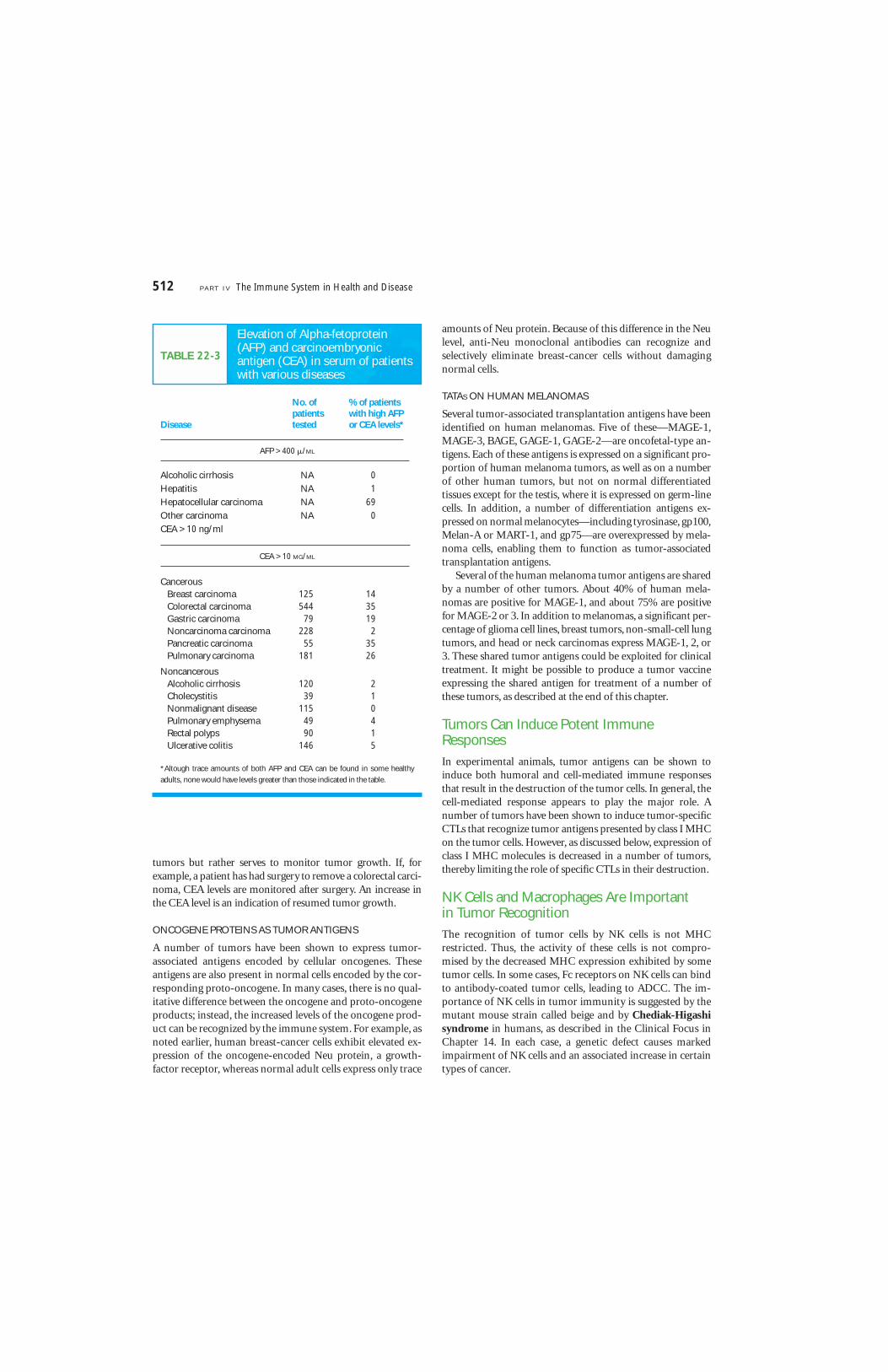
tumors but rather serves to monitor tumor growth. If, forexample, a patient has had surgery to remove a colorectal carci-noma, CEA levels are monitored after surgery. An increase inthe CEA level is an indication of resumed tumor growth.
ONCOGENE PROTEINS AS TUMOR ANTIGENS
A number of tumors have been shown to express tumor-associated antigens encoded by cellular oncogenes. Theseantigens are also present in normal cells encoded by the cor-responding proto-oncogene. In many cases, there is no qual-itative difference between the oncogene and proto-oncogeneproducts; instead, the increased levels of the oncogene prod-uct can be recognized by the immune system. For example, asnoted earlier, human breast-cancer cells exhibit elevated ex-pression of the oncogene-encoded Neu protein, a growth-factor receptor, whereas normal adult cells express only trace
amounts of Neu protein. Because of this difference in the Neulevel, anti-Neu monoclonal antibodies can recognize andselectively eliminate breast-cancer cells without damagingnormal cells.
TATAS ON HUMAN MELANOMAS
Several tumor-associated transplantation antigens have beenidentified on human melanomas. Five of these—MAGE-1,MAGE-3, BAGE, GAGE-1, GAGE-2—are oncofetal-type an-tigens. Each of these antigens is expressed on a significant pro-portion of human melanoma tumors, as well as on a numberof other human tumors, but not on normal differentiated tissues except for the testis, where it is expressed on germ-linecells. In addition, a number of differentiation antigens ex-pressed on normal melanocytes—including tyrosinase, gp100,Melan-A or MART-1, and gp75—are overexpressed by mela-noma cells, enabling them to function as tumor-associatedtransplantation antigens.
Several of the human melanoma tumor antigens are sharedby a number of other tumors. About 40% of human mela-nomas are positive for MAGE-1, and about 75% are positivefor MAGE-2 or 3. In addition to melanomas, a significant per-centage of glioma cell lines, breast tumors, non-small-cell lungtumors, and head or neck carcinomas express MAGE-1, 2, or3. These shared tumor antigens could be exploited for clinicaltreatment. It might be possible to produce a tumor vaccineexpressing the shared antigen for treatment of a number ofthese tumors, as described at the end of this chapter.
Tumors Can Induce Potent ImmuneResponses In experimental animals, tumor antigens can be shown toinduce both humoral and cell-mediated immune responsesthat result in the destruction of the tumor cells. In general, thecell-mediated response appears to play the major role. Anumber of tumors have been shown to induce tumor-specificCTLs that recognize tumor antigens presented by class I MHCon the tumor cells. However, as discussed below, expression ofclass I MHC molecules is decreased in a number of tumors,thereby limiting the role of specific CTLs in their destruction.
NK Cells and Macrophages Are Important in Tumor RecognitionThe recognition of tumor cells by NK cells is not MHCrestricted. Thus, the activity of these cells is not compro-mised by the decreased MHC expression exhibited by sometumor cells. In some cases, Fc receptors on NK cells can bindto antibody-coated tumor cells, leading to ADCC. The im-portance of NK cells in tumor immunity is suggested by themutant mouse strain called beige and by Chediak-Higashisyndrome in humans, as described in the Clinical Focus inChapter 14. In each case, a genetic defect causes markedimpairment of NK cells and an associated increase in certaintypes of cancer.
512 P A R T I V The Immune System in Health and Disease
TABLE 22-3
Elevation of Alpha-fetoprotein (AFP) and carcinoembryonic antigen (CEA) in serum of patientswith various diseases
No. of % of patientspatients with high AFP
Disease tested or CEA levels*
AFP > 400 �/ML
Alcoholic cirrhosis NA 0
Hepatitis NA 1
Hepatocellular carcinoma NA 69
Other carcinoma NA 0
CEA > 10 ng/ml
CEA > 10 MG/ML
CancerousBreast carcinoma 125 14Colorectal carcinoma 544 35Gastric carcinoma 79 19Noncarcinoma carcinoma 228 2Pancreatic carcinoma 55 35Pulmonary carcinoma 181 26
NoncancerousAlcoholic cirrhosis 120 2Cholecystitis 39 1Nonmalignant disease 115 0Pulmonary emphysema 49 4Rectal polyps 90 1Ulcerative colitis 146 5
*Altough trace amounts of both AFP and CEA can be found in some healthy
adults, none would have levels greater than those indicated in the table.

Cancer and the Immune System C H A P T E R 22 513
some of which are unique to melanoma(see Table 22-5). These observations areenhanced by the ability to create cDNAlibraries (see Chapter 23) from tumorcells. The cDNAs can be transfected intotarget cells expressing the appropriateMHC molecules and then used as targetsfor CTL-mediated killing. Once CTL reac-tivity is recognized, the transfected cDNAcan be isolated and identified as a poten-tial tumor antigen. The ability to isolategenes encoding tumor-associated anti-gens provides us with the opportunityto use these proteins as immunogensfor the induction of tumor-specificresponses. Additionally, the identifica-tion of tumor-associated proteins allowsus to identify peptides that elicit anti-tumor responses.
Over the past few years, several bio-tech companies have devised strategiesfor the development of vaccines againstmelanoma as well as other cancers. Thesestrategies have one thing in the com-mon; the induction of a cell-mediatedresponse to tumor-associated antigens.Antigens are derived from individualpatient tumors or established tumor celllines. The use of patient-derived tumorsis appealing for obvious reasons. Theresponse to that tumor should, in theory,be uniquely directed only at tumor anti-gens and not other, potentially allotypic,determinants. However, such individual-ized therapy could be very expensive andtime-consuming. In this scenario, thetumor would be biopsied or surgicallyremoved, placed into culture, and thenused as an immunogen. Establishing aprimary tumor in culture is not easy, evenfor melanoma, and the procedure cantake several weeks. The time factor, cou-pled with the realization that manytumors are not easy to grow in vitro,
places this into the category of “designertherapies” that may or may not be feasi-ble under the reality of managed healthcare today.
The use of established tumors as thesource of the immunogen is much moreaccessible in cost and practicality. Sam-ples from several tumors can be grown inculture and protein extracts prepared andfrozen, providing a source of immuno-gen for many patients. In addition toreduced costs, this strategy also allowscareful assessment of the immunogenic-ity of the tumor antigens found in thecultured cells. It is possible that sometumors may express higher levels oftumor-associated antigens and be moreimmunogenic than others. Indeed onebiotech company in California, CancerVax(www.CancerVax.com), has derived threecell lines that express high levels of over20 tumor-associated antigens. Addition-ally, these cells express MHC class I alle-les which are represented in the majorityof individuals in the population, meaningthat intracellular antigens will be present-ed properly. Cells are irradiated to renderthem incapable of cell division and usedas irradiated whole cells for immuniza-tion. The advantages of this approachlies in the ability to standardize the im-munogen as well as reducing the cost.
Antigen presentation is a critical fea-ture of any immunization strategy and oneway to enhance to immunization againsttumor antigens is to manipulate the fash-ion in which the antigen is presented.Professional antigen-presenting cells suchas dendritic cells are excellent candidates to employ in vaccination protocols. Sever-al companies have developed novel usesfor dendritic cells in cancer therapy. Den-dreon (www.Dendreon.com), a Seattle-based company, first isolates dendritic-cellprecursors from patient blood, then intro-duces the immunogen into the dendriticcells and returns the antigen-pulsed den-dritic cells to the bloodstream of the cancer patient. This company, throughgenomics-based drug discovery, has iden-tified tumor-associated antigens prevalenton a wide variety of cancers. Thus the
THE realization that the verte-brate immune system evolved to distin-guish self from non-self led to the notionthat our immune system could recog-nize a tumor as foreign. In fact, a majorresearch effort amongst cancer immu-nologists during the latter half of the20th century was the identification andcharacterization of tumor specific mole-cules, the so-called tumor antigens. Thisarea of research was met with skepti-cism. First of all, the existence of tumor-specific antigens was questionable; manyantigens were identified as tumor specif-ic only to find that other cells alsoexpressed these antigens. Secondly, ear-ly investigations in the field of tumorimmunology necessarily employed ani-mal models that may or may not be rele-vant to human cancers. However, withadvances in biotechnology, genomics,and proteomics, coupled with our in-creased understanding of the cellular in-teractions in the immune system, tumorimmunology now offers us the promiseof new drugs that will aid in the treat-ment of cancer. We now understand thattumor-associated antigens do exist andthat focusing the cellular arm of theimmune system toward the recognitionthese proteins is a rational approach tothe development of a cancer vaccine.
One of the best-studied tumor-immunity models is melanoma. Mela-noma has evolved as a model system forseveral reasons. First of all and paradox-ically, most human cancers are difficultto establish in tissue culture, making itdifficult to develop in vitro systems forexperimental manipulation. Melanomais relatively easy to adapt to tissue cul-ture, which has led to the identificationof several tumor-associated antigens,
C L I N I C A L F O C U S
Cancer Vaccines PromiseHope for the Future
(continued)
Numerous observations indicate that activated macro-phages also play a significant role in the immune response totumors. For example, macrophages are often observed tocluster around tumors, and their presence is often correlatedwith tumor regression. Like NK cells, macrophages are not
MHC restricted and express Fc receptors, enabling them tobind to antibody on tumor cells and mediate ADCC. Theantitumor activity of activated macrophages is probably me-diated by lytic enzymes and reactive oxygen and nitrogenintermediates. In addition, activated macrophages secrete a
514 P A R T I V The Immune System in Health and Disease
APCs, are internalized, and unexpected-ly, the antigen is processed and isthought to re-emerge as peptide/MHCclass I complexes on the APC, resultingin the priming of a CD8+ T-cell response.This would not be the predicted re-sponse, since the exogenous antigensare almost uniformly presented by classII MHC molecules. However, an impres-sive amount of experimental datademonstrates that HSPs isolated and purified from tumor tissue are a potentinducer of tumor-specific CTLs. Theseobservations have led to phase III clini-cal trials conducted by Antigenics (www.antigenics.com) of HSP/antigen com-
plexes as immunogens for kidney canceras well as melanoma. The mechanism bywhich HSP/antigen complexes bound toCD91 are delivered to the class I presen-tation machinery is not well understood,but it is clear that HSP/antigen complex-es, when presented to APCs, result in thevigorous activation of CD8+ T cells.
The promise for cancer vaccines ap-pears very bright. Genomics and pro-teomic methodologies provide noveltools for identifying tumor antigens.Additionally, there is a variety of ap-proaches available to engage the im-mune system to respond to tumor anti-gens. The past decade has seen a rapidincrease in the number of biotech com-panies directed at identifying cancer vac-cines, and the number of companies inphase II or phase III clinical trials invitesan air of optimism about this area of clin-ical research.
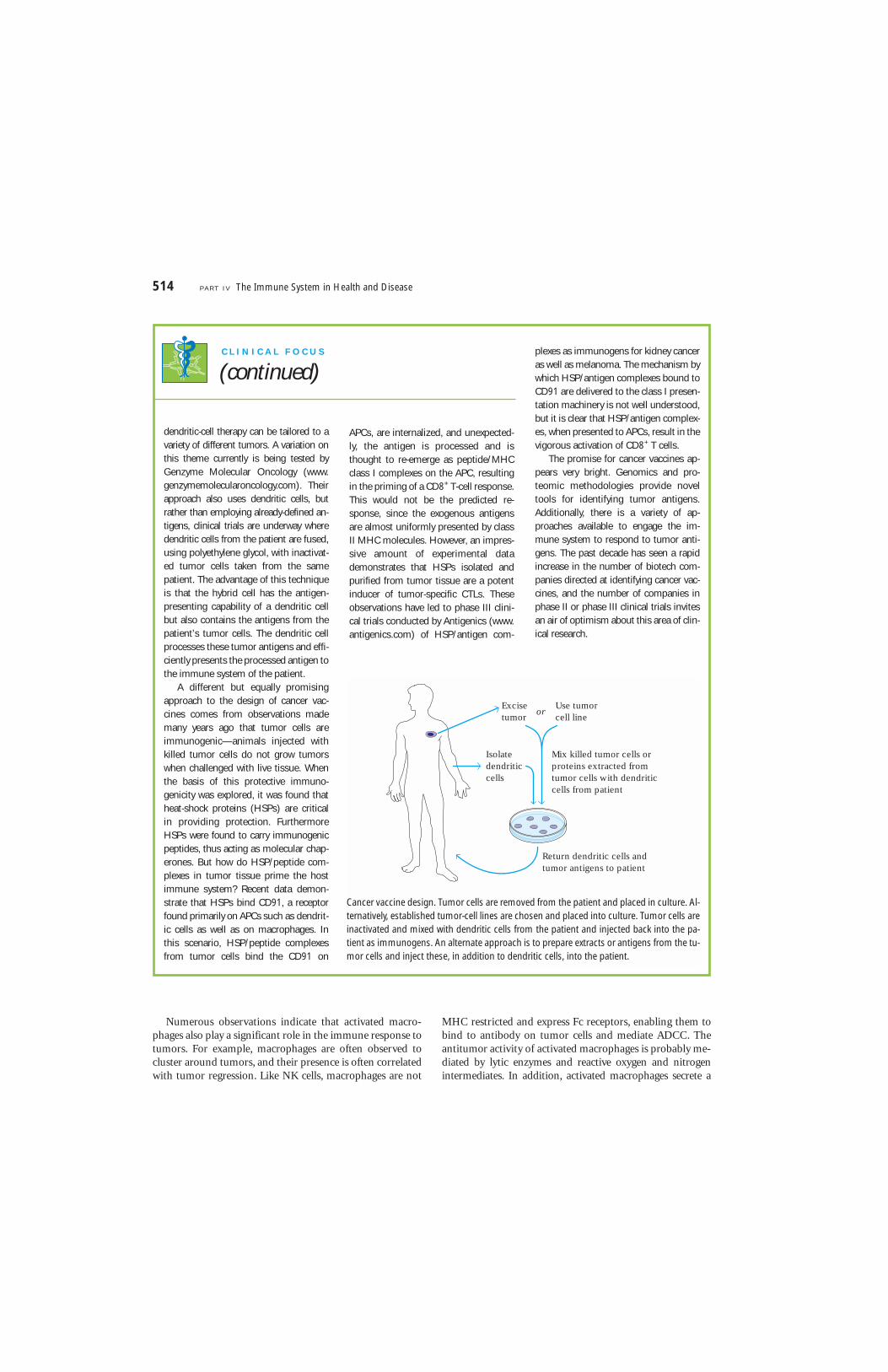
dendritic-cell therapy can be tailored to avariety of different tumors. A variation onthis theme currently is being tested byGenzyme Molecular Oncology (www.genzymemolecularoncology.com). Theirapproach also uses dendritic cells, butrather than employing already-defined an-tigens, clinical trials are underway wheredendritic cells from the patient are fused,using polyethylene glycol, with inactivat-ed tumor cells taken from the samepatient. The advantage of this techniqueis that the hybrid cell has the antigen-presenting capability of a dendritic cellbut also contains the antigens from thepatient’s tumor cells. The dendritic cellprocesses these tumor antigens and effi-ciently presents the processed antigen tothe immune system of the patient.
A different but equally promisingapproach to the design of cancer vac-cines comes from observations mademany years ago that tumor cells areimmunogenic—animals injected withkilled tumor cells do not grow tumorswhen challenged with live tissue. Whenthe basis of this protective immuno-genicity was explored, it was found thatheat-shock proteins (HSPs) are criticalin providing protection. FurthermoreHSPs were found to carry immunogenicpeptides, thus acting as molecular chap-erones. But how do HSP/peptide com-plexes in tumor tissue prime the hostimmune system? Recent data demon-strate that HSPs bind CD91, a receptorfound primarily on APCs such as dendrit-ic cells as well as on macrophages. Inthis scenario, HSP/peptide complexesfrom tumor cells bind the CD91 on
Excisetumor
Use tumorcell line
Isolatedendriticcells
Mix killed tumor cells orproteins extracted fromtumor cells with dendriticcells from patient
Return dendritic cells andtumor antigens to patient
or
Cancer vaccine design. Tumor cells are removed from the patient and placed in culture. Al-ternatively, established tumor-cell lines are chosen and placed into culture. Tumor cells areinactivated and mixed with dendritic cells from the patient and injected back into the pa-tient as immunogens. An alternate approach is to prepare extracts or antigens from the tu-mor cells and inject these, in addition to dendritic cells, into the patient.
C L I N I C A L F O C U S
(continued)

cytokine called tumor necrosis factor (TNF-�) that has po-tent antitumor activity. When TNF-� is injected into tumor-bearing animals, it has been found to induce hemorrhageand necrosis of the tumor.
IMMUNE SURVEILLANCE THEORY
The immune surveillance theory was first conceptualized inthe early 1900s by Paul Ehrlich. He suggested that cancer cellsfrequently arise in the body but are recognized as foreign andeliminated by the immune system. Some 50 years later, LewisThomas suggested that the cell-mediated branch of the im-mune system had evolved to patrol the body and eliminatecancer cells. According to these concepts, tumors arise only ifcancer cells are able to escape immune surveillance, either byreducing their expression of tumor antigens or by an impair-ment in the immune response to these cells.
Among the early observations that seemed to support theimmune surveillance theory was the increased incidence ofcancer in transplantation patients on immunosuppressivedrugs. Other findings, however, were difficult to reconcilewith this theory. Nude mice, for example, lack a thymus andconsequently lack functional T cells. According to the im-mune surveillance theory, these mice should show an in-crease in cancer, instead, nude mice are no more susceptibleto cancer than other mice. Furthermore, although individu-als on immunosuppressive drugs do show an increased inci-dence of cancers of the immune system, other common can-cers (e.g., lung, breast, and colon cancer) are not increased inthese individuals, contrary to what the theory predicts. Onepossible explanation for the selective increase in immune-system cancers is that the immunosuppressive agents them-selves may exert a direct carcinogenic effect on immune cells.
Experimental data concerning the effect of tumor-celldosage on the ability of the immune system to respond alsoare incompatible with the immune surveillance theory. Forexample, animals injected with very low or very high doses oftumor cells develop tumors, whereas those injected withintermediate doses do not. The mechanism by which a lowdose of tumor cells “sneaks through” is difficult to reconcilewith the immune surveillance theory. Finally, this theory as-sumes that cancer cells and normal cells exhibit qualitativeantigen differences. In fact, as stated earlier, many types oftumors do not express tumor-specific antigens, and any im-mune response that develops must be induced by quantitativedifferences in antigen expression by normal cells and tumorcells. However, tumors induced by viruses would be expectedto express some antigens encoded by the viral genome. Theseantigens are qualitatively different from those expressed bynormal tissues and would be expected to attract the attentionof the immune system. In fact, there are many examples of spe-cific immune responses to virally induced tumors.
Nevertheless, apart from tumors caused by viruses, thebasic concept of the immune surveillance theory—that malig-nant tumors arise only if the immune system is somehowimpaired or if the tumor cells lose their immunogenicity,enabling them to escape immune surveillance—at this time
remains unproved. In spite of this, it is clear that an immuneresponse can be generated to tumor cells, and therapeutic ap-proaches aimed at increasing that response may serve as a de-fense against malignant cells.
Tumor Evasion of the ImmuneSystemAlthough the immune system clearly can respond to tumorcells, the fact that so many individuals die each year fromcancer suggests that the immune response to tumor cells isoften ineffective. This section describes several mechanismsby which tumor cells appear to evade the immune system.
Anti-Tumor Antibodies Can Enhance Tumor GrowthFollowing the discovery that antibodies could be produced totumor-specific antigens, attempts were made to protect ani-mals against tumor growth by active immunization withtumor antigens or by passive immunization with antitumorantibodies. Much to the surprise of the researchers, these im-munizations did not protect against tumor growth; in manycases, they actually enhanced growth of the tumor.
The tumor-enhancing ability of immune sera subsequentlywas studied in cell-mediated lympholysis (CML) reactions invitro. Serum taken from animals with progressive tumorgrowth was found to block the CML reaction, whereas serumtaken from animals with regressing tumors had little or noblocking activity. K. E. and I. Hellstrom extended these find-ings by showing that children with progressive neuroblastomahad high levels of some kind of blocking factor in their seraand that children with regressive neuroblastoma did not havesuch factors. Since these first reports, blocking factors havebeen found to be associated with a number of human tumors.
In some cases, antitumor antibody itself acts as a blockingfactor. Presumably the antibody binds to tumor-specific anti-gens and masks the antigens from cytotoxic T cells. In manycases, the blocking factors are not antibodies alone but ratherantibodies complexed with tumor antigens. Although theseimmune complexes have been shown to block the CTL re-sponse, the mechanism of this inhibition is not known. Thecomplexes also may inhibit ADCC by binding to Fc receptorson NK cells or macrophages and blocking their activity.
Antibodies Can Modulate Tumor AntigensCertain tumor-specific antigens have been observed to dis-appear from the surface of tumor cells in the presence ofserum antibody and then to reappear after the antibody is nolonger present. This phenomenon, called antigenic modula-tion, is readily observed when leukemic T cells are injectedinto mice previously immunized with a leukemic T-cell anti-gen (TL antigen). These mice develop high titers of anti-TL
Cancer and the Immune System C H A P T E R 22 515
antibody, which binds to the TL antigen on the leukemic cellsand induces capping, endocytosis, and/or shedding of theantigen-antibody complex. As long as antibody is present,these leukemic T cells fail to display the TL antigen and thuscannot be eliminated.
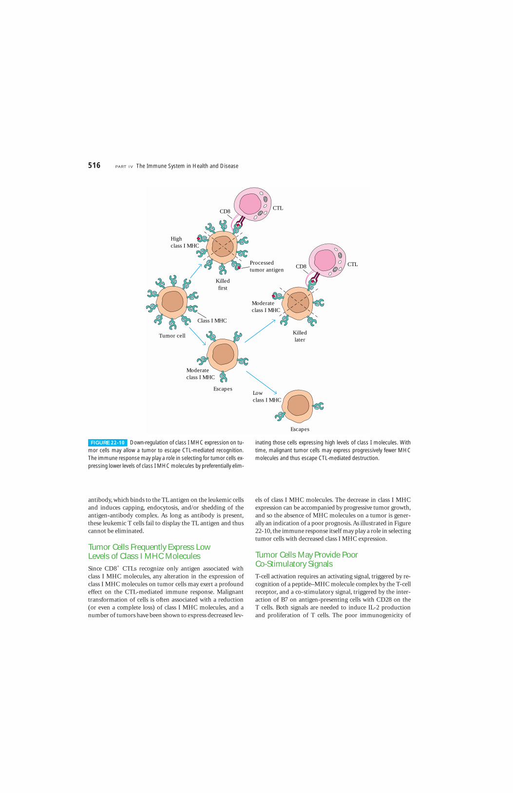
Tumor Cells Frequently Express Low Levels of Class I MHC MoleculesSince CD8+ CTLs recognize only antigen associated withclass I MHC molecules, any alteration in the expression ofclass I MHC molecules on tumor cells may exert a profoundeffect on the CTL-mediated immune response. Malignanttransformation of cells is often associated with a reduction(or even a complete loss) of class I MHC molecules, and anumber of tumors have been shown to express decreased lev-
els of class I MHC molecules. The decrease in class I MHCexpression can be accompanied by progressive tumor growth,and so the absence of MHC molecules on a tumor is gener-ally an indication of a poor prognosis. As illustrated in Figure22-10, the immune response itself may play a role in selectingtumor cells with decreased class I MHC expression.
Tumor Cells May Provide Poor Co-Stimulatory SignalsT-cell activation requires an activating signal, triggered by re-cognition of a peptide–MHC molecule complex by the T-cellreceptor, and a co-stimulatory signal, triggered by the inter-action of B7 on antigen-presenting cells with CD28 on the T cells. Both signals are needed to induce IL-2 productionand proliferation of T cells. The poor immunogenicity of
516 P A R T I V The Immune System in Health and Disease
Class I MHC
CTL
Killedfirst
Processedtumor antigen
Escapes
Tumor cell
Escapes
Killedlater
CTL
Moderateclass I MHC
Moderateclass I MHC
Lowclass I MHC
Highclass I MHC
CD8
CD8
FIGURE 22-10 Down-regulation of class I MHC expression on tu-mor cells may allow a tumor to escape CTL-mediated recognition.The immune response may play a role in selecting for tumor cells ex-pressing lower levels of class I MHC molecules by preferentially elim-
inating those cells expressing high levels of class I molecules. Withtime, malignant tumor cells may express progressively fewer MHCmolecules and thus escape CTL-mediated destruction.
many tumor cells may be due in large part to lack of the co-stimulatory molecules. Without sufficient numbers ofantigen-presenting cells in the immediate vicinity of a tumor,the T cells will receive only a partial activating signal, whichmay lead to clonal anergy.
Cancer ImmunotherapyAlthough various immune responses can be generated totumor cells, the response frequently is not sufficient to pre-vent tumor growth. One approach to cancer treatment is toaugment or supplement these natural defense mechanisms.Several types of cancer immunotherapy in current use orunder development are described in this concluding section.
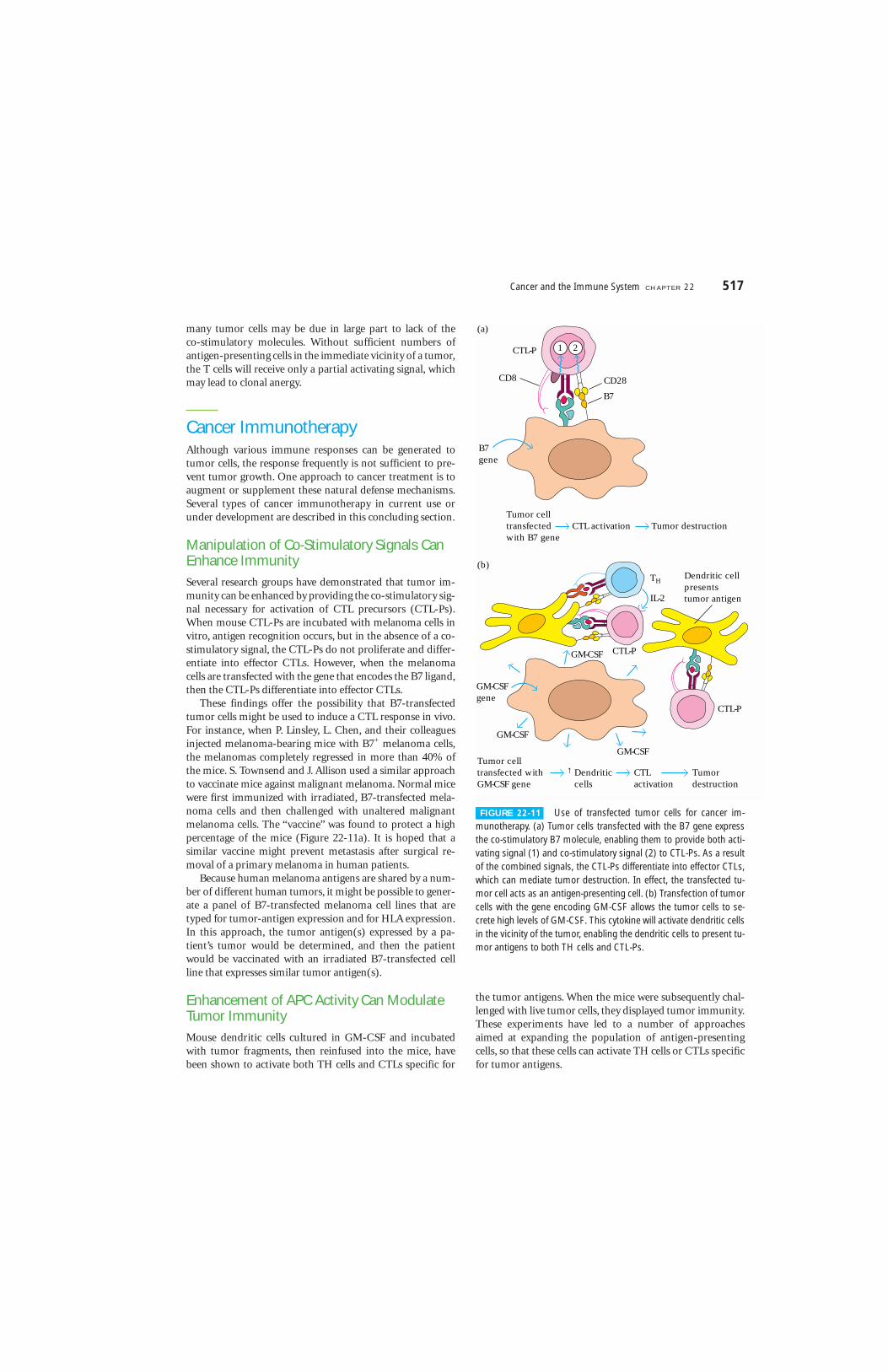
Manipulation of Co-Stimulatory Signals CanEnhance ImmunitySeveral research groups have demonstrated that tumor im-munity can be enhanced by providing the co-stimulatory sig-nal necessary for activation of CTL precursors (CTL-Ps).When mouse CTL-Ps are incubated with melanoma cells invitro, antigen recognition occurs, but in the absence of a co-stimulatory signal, the CTL-Ps do not proliferate and differ-entiate into effector CTLs. However, when the melanomacells are transfected with the gene that encodes the B7 ligand,then the CTL-Ps differentiate into effector CTLs.
These findings offer the possibility that B7-transfectedtumor cells might be used to induce a CTL response in vivo.For instance, when P. Linsley, L. Chen, and their colleaguesinjected melanoma-bearing mice with B7+ melanoma cells,the melanomas completely regressed in more than 40% ofthe mice. S. Townsend and J. Allison used a similar approachto vaccinate mice against malignant melanoma. Normal micewere first immunized with irradiated, B7-transfected mela-noma cells and then challenged with unaltered malignantmelanoma cells. The “vaccine” was found to protect a highpercentage of the mice (Figure 22-11a). It is hoped that asimilar vaccine might prevent metastasis after surgical re-moval of a primary melanoma in human patients.
Because human melanoma antigens are shared by a num-ber of different human tumors, it might be possible to gener-ate a panel of B7-transfected melanoma cell lines that aretyped for tumor-antigen expression and for HLA expression.In this approach, the tumor antigen(s) expressed by a pa-tient’s tumor would be determined, and then the patientwould be vaccinated with an irradiated B7-transfected cellline that expresses similar tumor antigen(s).
Enhancement of APC Activity Can ModulateTumor ImmunityMouse dendritic cells cultured in GM-CSF and incubatedwith tumor fragments, then reinfused into the mice, havebeen shown to activate both TH cells and CTLs specific for
the tumor antigens. When the mice were subsequently chal-lenged with live tumor cells, they displayed tumor immunity.These experiments have led to a number of approachesaimed at expanding the population of antigen-presentingcells, so that these cells can activate TH cells or CTLs specificfor tumor antigens.
Cancer and the Immune System C H A P T E R 22 517
(a)
CTL-P
CD8 CD28
B7gene
Tumor celltransfectedwith B7 gene
Dendritic cellpresentstumor antigen
CTL activation Tumor destruction
(b)
Tumor celltransfected withGM-CSF gene
Dendriticcells
CTLactivation
Tumordestruction
GM-CSFgene
GM-CSF
GM-CSF
GM-CSF
B7
CTL-P
CTL-P
TH
IL-2
1 2
↓
FIGURE 22-11 Use of transfected tumor cells for cancer im-munotherapy. (a) Tumor cells transfected with the B7 gene expressthe co-stimulatory B7 molecule, enabling them to provide both acti-vating signal (1) and co-stimulatory signal (2) to CTL-Ps. As a resultof the combined signals, the CTL-Ps differentiate into effector CTLs,which can mediate tumor destruction. In effect, the transfected tu-mor cell acts as an antigen-presenting cell. (b) Transfection of tumorcells with the gene encoding GM-CSF allows the tumor cells to se-crete high levels of GM-CSF. This cytokine will activate dendritic cellsin the vicinity of the tumor, enabling the dendritic cells to present tu-mor antigens to both TH cells and CTL-Ps.
One approach that has been tried is to transfect tumorcells with the gene encoding GM-CSF. These engineeredtumor cells, when reinfused into the patient, will secrete GM-CSF, enhancing the differentiation and activation of hostantigen-presenting cells, especially dendritic cells. As thesedendritic cells accumulate around the tumor cells, the GM-CSF secreted by the tumor cells will enhance the presentationof tumor antigens to TH cells and CTLs by the dendritic cells(Figure 22-11b).
Another way to expand the dendritic-cell population is toculture dendritic cells from peripheral-blood progenitor cellsin the presence of GM-CSF, TNF-�, and IL-4. These threecytokines induce the generation of large numbers of den-dritic cells. There is some hope that, if these dendritic cellsare pulsed with tumor fragments and then reintroduced intothe patient, they can activate TH and TC cells specific for thetumor antigens. Whether these hopes are justified will bedetermined by further investigation.
A number of adjuvants, including the attenuated strains ofMycobacterium bovis called bacillus Calmette-Guerin (BCG)and Corynebacterium parvuum, have been used to boost tumor immunity. These adjuvants activate macrophages, in-creasing their expression of various cytokines, class II MHCmolecules, and the B7 co-stimulatory molecule. These acti-vated macrophages are better activators of TH cells, resultingin generalized increases in both humoral and cell-mediatedresponses. Thus far, adjuvants have shown only modest thera-peutic results.
Cytokine Therapy Can Augment ImmuneResponses to TumorsThe isolation and cloning of the various cytokine genes hasfacilitated their large-scale production. A variety of experi-mental and clinical approaches have been developed to userecombinant cytokines, either singly or in combination, toaugment the immune response against cancer. Among thecytokines that have been evaluated in cancer immunother-apy are IFN-�, �, and �; IL-1, IL-2, IL-4, IL-5, and IL-12;GM-CSF; and TNF. Although these trials have producedoccasional encouraging results, many obstacles remain to thesuccessful use of this type of cancer immunotherapy.
The most notable obstacle is the complexity of the cytokinenetwork itself. This complexity makes it very difficult to knowprecisely how intervention with a given recombinant cytokinewill affect the production of other cytokines. And since somecytokines act antagonistically, it is possible that interventionwith a recombinant cytokine designed to enhance a particularbranch of the immune response may actually lead to suppres-sion. In addition, cytokine immunotherapy is plagued by thedifficulty of administering the cytokines locally. In some cases,systemic administration of high levels of a given cytokine hasbeen shown to lead to serious and even life-threatening con-sequences. Although the results of several experimental andclinical trials of cytokine therapy for cancer are discussed
here, it is important to keep in mind that this therapeuticapproach is still in its infancy.
INTERFERONS
Large quantities of purified recombinant preparations of theinterferons, IFN-�, IFN-�, and IFN-�, are now available,each of which has shown some promise in the treatment ofhuman cancer. To date, most of the clinical trials have in-volved IFN-�. Daily injections of recombinant IFN-� havebeen shown to induce partial or complete tumor regressionin some patients with hematologic malignancies such asleukemias, lymphomas, and myelomas and with solid tu-mors such as melanoma, Kaposi’s sarcoma, renal cancer, andbreast cancer.
Interferon-mediated antitumor activity may involve severalmechanisms. All three types of interferon have been shown toincrease class I MHC expression on tumor cells; IFN-� hasalso been shown to increase class II MHC expression onmacrophages. Given the evidence for decreased levels of classI MHC molecules on malignant tumors, the interferons mayact by restoring MHC expression, thereby increasing CTL ac-tivity against tumors. In addition, the interferons have beenshown to inhibit cell division of both normal and malignantlytransformed cells in vitro. It is possible that some of the anti-tumor effects of the interferons are related to this ability todirectly inhibit tumor-cell proliferation. Finally, IFN-� directlyor indirectly increases the activity of TC cells, macrophages,and NK cells, all of which play a role in the immune responseto tumor cells.
TUMOR NECROSIS FACTORS
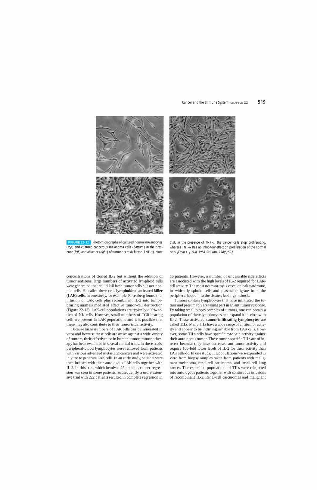
In some instances, the tumor necrosis factors TNF-� andTNF-� have been shown to exhibit direct antitumor activity,killing some tumor cells and reducing the rate of prolifera-tion of others while sparing normal cells (Figure 22-12). Inthe presence of TNF-� or TNF-�, a tumor undergoes visiblehemorrhagic necrosis and regression. TNF-� has also beenshown to inhibit tumor-induced vascularization (angiogene-sis) by damaging the vascular endothelial cells in the vicinityof a tumor, thereby decreasing the flow of blood and oxygenthat is necessary for progressive tumor growth.
IN VITRO–ACTIVATED LAK AND TIL CELLS
Animal studies have shown that lymphocytes can be acti-vated against tumor antigens in vitro by culturing them withx-irradiated tumor cells in the presence of IL-2 and addedtumor antigens. These activated lymphocytes mediate moreeffective tumor destruction than untreated lymphocyteswhen they are reinjected into the original tumor-bearing ani-mal. It is difficult, however, to activate in vitro enough lym-phocytes with antitumor specificity to be useful in cancertherapy.
While sensitizing lymphocytes to tumor antigens by thismethod, S. Rosenberg discovered that, in the presence of high
518 P A R T I V The Immune System in Health and Disease
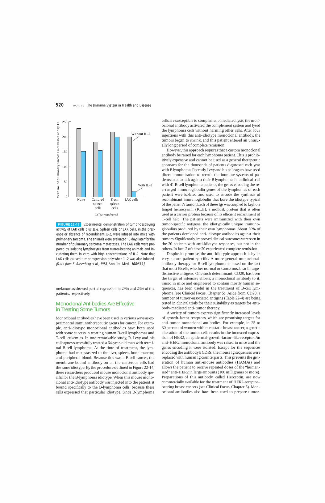
concentrations of cloned IL-2 but without the addition oftumor antigens, large numbers of activated lymphoid cellswere generated that could kill fresh tumor cells but not nor-mal cells. He called these cells lymphokine-activated killer(LAK) cells. In one study, for example, Rosenberg found thatinfusion of LAK cells plus recombinant IL-2 into tumor-bearing animals mediated effective tumor-cell destruction(Figure 22-13). LAK-cell populations are typically >90% ac-tivated NK cells. However, small numbers of TCR-bearingcells are present in LAK populations and it is possible thatthese may also contribute to their tumoricidal activity.
Because large numbers of LAK cells can be generated invitro and because these cells are active against a wide varietyof tumors, their effectiveness in human tumor immunother-apy has been evaluated in several clinical trials. In these trials,peripheral-blood lymphocytes were removed from patientswith various advanced metastatic cancers and were activatedin vitro to generate LAK cells. In an early study, patients werethen infused with their autologous LAK cells together withIL-2. In this trial, which involved 25 patients, cancer regres-sion was seen in some patients. Subsequently, a more exten-sive trial with 222 patients resulted in complete regression in
16 patients. However, a number of undesirable side effectsare associated with the high levels of IL-2 required for LAK-cell activity. The most noteworthy is vascular leak syndrome,in which lymphoid cells and plasma emigrate from theperipheral blood into the tissues, leading to shock.
Tumors contain lymphocytes that have infiltrated the tu-mor and presumably are taking part in an antitumor response.By taking small biopsy samples of tumors, one can obtain apopulation of these lymphocytes and expand it in vitro withIL-2. These activated tumor-infiltrating lymphocytes arecalled TILs. Many TILs have a wide range of antitumor activ-ity and appear to be indistinguishable from LAK cells. How-ever, some TILs cells have specific cytolytic activity againsttheir autologous tumor. These tumor-specific TILs are of in-terest because they have increased antitumor activity andrequire 100-fold lower levels of IL-2 for their activity thanLAK cells do. In one study, TIL populations were expanded invitro from biopsy samples taken from patients with malig-nant melanoma, renal-cell carcinoma, and small-cell lungcancer. The expanded populations of TILs were reinjectedinto autologous patients together with continuous infusionsof recombinant IL-2. Renal-cell carcinomas and malignant
Cancer and the Immune System C H A P T E R 22 519
FIGURE 22-12 Photomicrographs of cultured normal melanocytes(top) and cultured cancerous melanoma cells (bottom) in the pres-ence (left) and absence (right) of tumor necrosis factor (TNF-�). Note
that, in the presence of TNF-�, the cancer cells stop proliferating,whereas TNF-� has no inhibitory effect on proliferation of the normalcells. [From L. J. Old, 1988, Sci. Am. 258(5):59.]
melanomas showed partial regression in 29% and 23% of thepatients, respectively.
Monoclonal Antibodies Are Effective in Treating Some Tumors
Monoclonal antibodies have been used in various ways as ex-perimental immunotherapeutic agents for cancer. For exam-ple, anti-idiotype monoclonal antibodies have been usedwith some success in treating human B-cell lymphomas andT-cell leukemias. In one remarkable study, R. Levy and hiscolleagues successfully treated a 64-year-old man with termi-nal B-cell lymphoma. At the time of treatment, the lym-phoma had metastasized to the liver, spleen, bone marrow,and peripheral blood. Because this was a B-cell cancer, themembrane-bound antibody on all the cancerous cells hadthe same idiotype. By the procedure outlined in Figure 22-14,these researchers produced mouse monoclonal antibody spe-cific for the B-lymphoma idiotype. When this mouse mono-clonal anti-idiotype antibody was injected into the patient, itbound specifically to the B-lymphoma cells, because thesecells expressed that particular idiotype. Since B-lymphoma
cells are susceptible to complement-mediated lysis, the mon-oclonal antibody activated the complement system and lysedthe lymphoma cells without harming other cells. After fourinjections with this anti-idiotype monoclonal antibody, thetumors began to shrink, and this patient entered an unusu-ally long period of complete remission.
However, this approach requires that a custom monoclonalantibody be raised for each lymphoma patient. This is prohib-itively expensive and cannot be used as a general therapeuticapproach for the thousands of patients diagnosed each yearwith B lymphoma. Recently, Levy and his colleagues have useddirect immunization to recruit the immune systems of pa-tients to an attack against their B lymphoma. In a clinical trialwith 41 B-cell lymphoma patients, the genes encoding the re-arranged immunoglobulin genes of the lymphomas of eachpatient were isolated and used to encode the synthesis ofrecombinant immunoglobulin that bore the idiotype typicalof the patient’s tumor. Each of these Igs was coupled to keyholelimpet hemocyanin (KLH), a mollusk protein that is oftenused as a carrier protein because of its efficient recruitment ofT-cell help. The patients were immunized with their owntumor-specific antigens, the idiotypically unique immuno-globulins produced by their own lymphomas. About 50% ofthe patients developed anti-idiotype antibodies against theirtumors. Significantly, improved clinical outcomes were seen inthe 20 patients with anti-idiotype responses, but not in theothers. In fact, 2 of these 20 experienced complete remission.
Despite its promise, the anti-idiotypic approach is by itsvery nature patient-specific. A more general monoclonal-antibody therapy for B-cell lymphoma is based on the factthat most B cells, whether normal or cancerous, bear lineage-distinctive antigens. One such determinant, CD20, has beenthe target of intensive efforts; a monoclonal antibody to it,raised in mice and engineered to contain mostly human se-quences, has been useful in the treatment of B-cell lym-phoma (see Clinical Focus, Chapter 5). Aside from CD20, anumber of tumor-associated antigens (Table 22-4) are beingtested in clinical trials for their suitability as targets for anti-body-mediated anti-tumor therapy.
A variety of tumors express significantly increased levelsof growth-factor receptors, which are promising targets foranti-tumor monoclonal antibodies. For example, in 25 to 30 percent of women with metastatic breast cancer, a geneticalteration of the tumor cells results in the increased expres-sion of HER2, an epidermal-growth-factor–like receptor. Ananti-HER2 monoclonal antibody was raised in mice and thegenes encoding it were isolated. Except for the sequencesencoding the antibody’s CDRs, the mouse Ig sequences werereplaced with human Ig counterparts. This prevents the gen-eration of human anti-mouse antibodies (HAMAs) and allows the patient to receive repeated doses of the “human-ized” anti-HER2 in large amounts (100 milligrams or more).Preparations of this antibody, called Herceptin, are nowcommercially available for the treatment of HER2-receptor–bearing breast cancers (see Clinical Focus, Chapter 5). Mon-oclonal antibodies also have been used to prepare tumor-
520 P A R T I V The Immune System in Health and Disease
Cells transferred
0Mea
n n
o. o
f p
ulm
on
ary
sarc
om
a m
etas
tase
s at
day
13
None
150
200
With IL–2
250
100
50
Without IL–2
LAK cellsCulturedspleencells
Freshspleencells
FIGURE 22-13 Experimental demonstration of tumor-destroyingactivity of LAK cells plus IL-2. Spleen cells or LAK cells, in the pres-ence or absence of recombinant IL-2, were infused into mice withpulmonary sarcoma. The animals were evaluated 13 days later for thenumber of pulmonary sarcoma metastases. The LAK cells were pre-pared by isolating lymphocytes from tumor-bearing animals and in-cubating them in vitro with high concentrations of IL-2. Note thatLAK cells caused tumor regression only when IL-2 was also infused.[Data from S. Rosenberg et al., 1988, Ann. Int. Med., 108:853.]
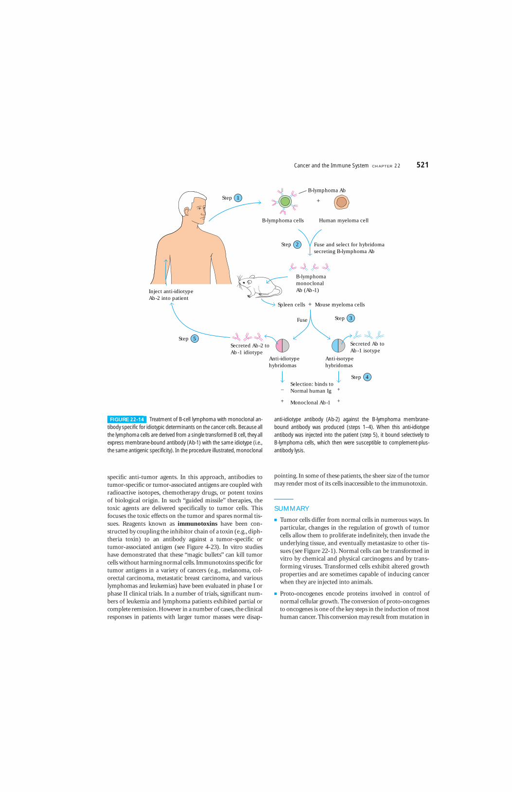
specific anti-tumor agents. In this approach, antibodies totumor-specific or tumor-associated antigens are coupled withradioactive isotopes, chemotherapy drugs, or potent toxinsof biological origin. In such “guided missile” therapies, thetoxic agents are delivered specifically to tumor cells. Thisfocuses the toxic effects on the tumor and spares normal tis-sues. Reagents known as immunotoxins have been con-structed by coupling the inhibitor chain of a toxin (e.g., diph-theria toxin) to an antibody against a tumor-specific ortumor-associated antigen (see Figure 4-23). In vitro studieshave demonstrated that these “magic bullets” can kill tumorcells without harming normal cells. Immunotoxins specific fortumor antigens in a variety of cancers (e.g., melanoma, col-orectal carcinoma, metastatic breast carcinoma, and variouslymphomas and leukemias) have been evaluated in phase I orphase II clinical trials. In a number of trials, significant num-bers of leukemia and lymphoma patients exhibited partial orcomplete remission. However in a number of cases, the clinicalresponses in patients with larger tumor masses were disap-
pointing. In some of these patients, the sheer size of the tumormay render most of its cells inaccessible to the immunotoxin.
SUMMARY
■ Tumor cells differ from normal cells in numerous ways. Inparticular, changes in the regulation of growth of tumorcells allow them to proliferate indefinitely, then invade theunderlying tissue, and eventually metastasize to other tis-sues (see Figure 22-1). Normal cells can be transformed invitro by chemical and physical carcinogens and by trans-forming viruses. Transformed cells exhibit altered growthproperties and are sometimes capable of inducing cancerwhen they are injected into animals.
■ Proto-oncogenes encode proteins involved in control ofnormal cellular growth. The conversion of proto-oncogenesto oncogenes is one of the key steps in the induction of mosthuman cancer. This conversion may result from mutation in
Cancer and the Immune System C H A P T E R 22 521
Fuse and select for hybridomasecreting B-lymphoma Ab
+
B-lymphoma cells Human myeloma cell
B-lymphoma Ab
Fuse
B-lymphomamonoclonalAb (Ab-1)
Spleen cells + Mouse myeloma cells
Inject anti-idiotypeAb-2 into patient
Secreted Ab-2 toAb-1 idiotype
Secreted Ab toAb-1 isotype
Anti-idiotypehybridomas
Anti-isotypehybridomas
–
+
+
+
Selection: binds toNormal human Ig
Monoclonal Ab-1
Step 5
Step 4
Step 3
Step 2
Step 1
FIGURE 22-14 Treatment of B-cell lymphoma with monoclonal an-tibody specific for idiotypic determinants on the cancer cells. Because allthe lymphoma cells are derived from a single transformed B cell, they allexpress membrane-bound antibody (Ab-1) with the same idiotype (i.e.,the same antigenic specificity). In the procedure illustrated, monoclonal
anti-idiotype antibody (Ab-2) against the B-lymphoma membrane-bound antibody was produced (steps 1–4). When this anti-idiotype antibody was injected into the patient (step 5), it bound selectively to B-lymphoma cells, which then were susceptible to complement-plus-antibody lysis.
an oncogene, its translocation, or its amplification (see Fig-ure 22-2).
■ A number of B- and T-cell leukemias and lymphomas areassociated with translocated proto-oncogenes. In its newsite, the translocated gene may come under the influenceof enhancers or promoters that cause its transcription athigher levels than usual (see Figure 22-5).
■ Tumor cells display tumor-specific antigens and the morecommon tumor-associated antigens. Among the latter areoncofetal antigens, (see Table 22-3) and increased levels ofnormal oncogene products (see Figure 22-6). In contrast totumor antigens induced by chemicals or radiation, virallyencoded tumor antigens are shared by all tumors induced bythe same virus.
■ The tumor antigens recognized by T cells fall into one offour major categories: antigens encoded by genes withtumor-specific expression; antigens encoded by variantforms of normal genes that have been altered by mutation;certain antigens normally expressed only at certain stagesof differentiation or differentiation lineages; antigens thatare overexpressed in particular tumors.
■ The use of a variety of genetic, biochemical, and immuno-logical approaches has allowed the identification of severaltumor-associated antigens (see Table 22-4). In many casesthe antigen is expressed on more than one type of tumor.
Common tumor antigens offer hope for the design ofbetter therapies, detection, and monitoring, and haveimportant implications for the possibility of anti-tumorimmunization.
■ The immune response to tumors includes CTL-mediatedlysis, NK-cell activity, macrophage-mediated tumor de-struction, and destruction mediated by ADCC. Several cyto-toxic factors, including TNF-� and TNF-�, help to mediatetumor-cell killing. Tumors may evade the immune responseby modulating their tumor antigens, by reducing theirexpression of class I MHC molecules, and by antibody-mediated or immune complex-mediated inhibition of CTLactivity.
■ Experimental cancer immunotherapy is exploring a vari-ety of approaches. Some of these are the enhancement ofthe co-stimulatory signal required for T-cell activation (seeFigure 22-11a), genetically engineering tumor cells tosecrete cytokines that may increase the intensity of theimmune response against them (see Figure 22-11b), thetherapeutic use of cytokines (see Figure 22-12), and waysof increasing the activity of antigen-presenting cells.
■ A number of encouraging clinical results have been ob-tained with therapy using monoclonal antibodies againsttumor-associated and (in a few cases) tumor-specific anti-gens (see Figure 22-14). Coupling of antibodies against
522 P A R T I V The Immune System in Health and Disease
TABLE 22-4Some tumor-associated antigens under examination as potential targets for monoclonal-antibody therapy
Tumor antigen Tumor type Target antigen
LYMPHOID CELL-SURFACE MARKERS
T-cell marker T-cell leukemia/lymphoma CD5
B-cell marker B-cell lymphoma CD20
Hematopoietic-cell marker Acute myeloblastic leukemia CD45
Anti-idiotype B-cell lymphoma Immunoglobulin
NONLYMPHOID TISSUE MARKERS
Cell-surface antigens
Carcinoembryonic antigen (CEA) Colon cancer (some others) Glycoprotein
MUC1 Breast cancer Glycoprotein
Gangliosides such as GD2 and GD3 Neuroectodermal tumors Glycolipids associated with neural tissue
Growth-factor receptors
Epidermal growth-factor receptor (EGFR) Some lung, head, neck, and breast tumors EGF-binding cell surface protein
HER2 (and EFG-like receptor) Breast and ovarian tumors Cell-surface EGF-binding protein
with homology to EGFR
SOURCE: Adapted from Scott and Welt, 1997, Curr. Opin. Immunol. 9:717.
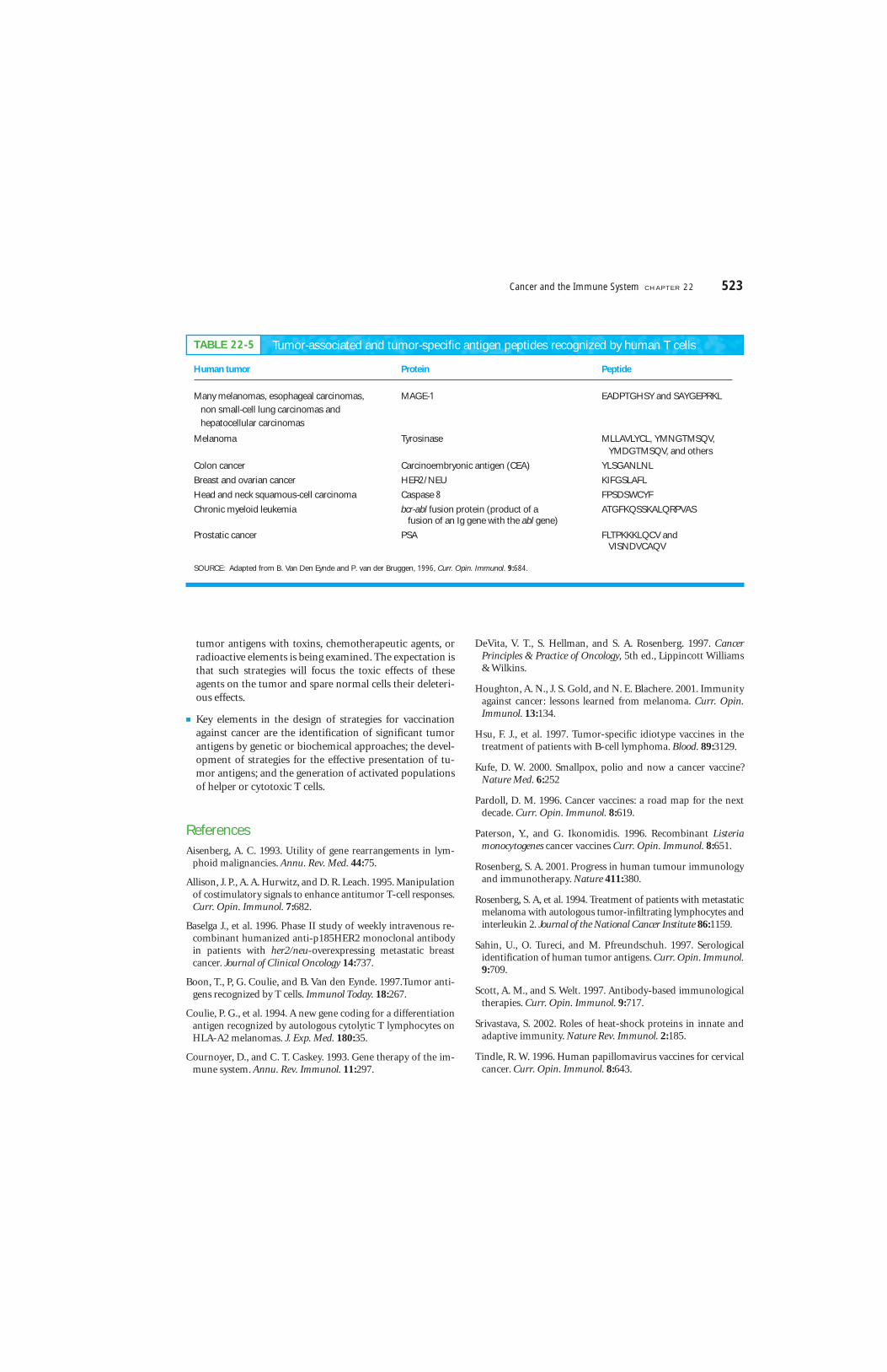
tumor antigens with toxins, chemotherapeutic agents, orradioactive elements is being examined. The expectation isthat such strategies will focus the toxic effects of theseagents on the tumor and spare normal cells their deleteri-ous effects.
■ Key elements in the design of strategies for vaccinationagainst cancer are the identification of significant tumorantigens by genetic or biochemical approaches; the devel-opment of strategies for the effective presentation of tu-mor antigens; and the generation of activated populationsof helper or cytotoxic T cells.
ReferencesAisenberg, A. C. 1993. Utility of gene rearrangements in lym-
phoid malignancies. Annu. Rev. Med. 44:75.
Allison, J. P., A. A. Hurwitz, and D. R. Leach. 1995. Manipulationof costimulatory signals to enhance antitumor T-cell responses.Curr. Opin. Immunol. 7:682.
Baselga J., et al. 1996. Phase II study of weekly intravenous re-combinant humanized anti-p185HER2 monoclonal antibodyin patients with her2/neu-overexpressing metastatic breastcancer. Journal of Clinical Oncology 14:737.
Boon, T., P, G. Coulie, and B. Van den Eynde. 1997.Tumor anti-gens recognized by T cells. Immunol Today. 18:267.
Coulie, P. G., et al. 1994. A new gene coding for a differentiationantigen recognized by autologous cytolytic T lymphocytes onHLA-A2 melanomas. J. Exp. Med. 180:35.
Cournoyer, D., and C. T. Caskey. 1993. Gene therapy of the im-mune system. Annu. Rev. Immunol. 11:297.
DeVita, V. T., S. Hellman, and S. A. Rosenberg. 1997. CancerPrinciples & Practice of Oncology, 5th ed., Lippincott Williams& Wilkins.
Houghton, A. N., J. S. Gold, and N. E. Blachere. 2001. Immunityagainst cancer: lessons learned from melanoma. Curr. Opin.Immunol. 13:134.
Hsu, F. J., et al. 1997. Tumor-specific idiotype vaccines in thetreatment of patients with B-cell lymphoma. Blood. 89:3129.
Kufe, D. W. 2000. Smallpox, polio and now a cancer vaccine?Nature Med. 6:252
Pardoll, D. M. 1996. Cancer vaccines: a road map for the nextdecade. Curr. Opin. Immunol. 8:619.
Paterson, Y., and G. Ikonomidis. 1996. Recombinant Listeriamonocytogenes cancer vaccines Curr. Opin. Immunol. 8:651.
Rosenberg, S. A. 2001. Progress in human tumour immunologyand immunotherapy. Nature 411:380.
Rosenberg, S. A, et al. 1994. Treatment of patients with metastaticmelanoma with autologous tumor-infiltrating lymphocytes andinterleukin 2. Journal of the National Cancer Institute 86:1159.
Sahin, U., O. Tureci, and M. Pfreundschuh. 1997. Serologicalidentification of human tumor antigens. Curr. Opin. Immunol.9:709.
Scott, A. M., and S. Welt. 1997. Antibody-based immunologicaltherapies. Curr. Opin. Immunol. 9:717.
Srivastava, S. 2002. Roles of heat-shock proteins in innate andadaptive immunity. Nature Rev. Immunol. 2:185.
Tindle, R. W. 1996. Human papillomavirus vaccines for cervicalcancer. Curr. Opin. Immunol. 8:643.
Cancer and the Immune System C H A P T E R 22 523
Some tumor-associated antigens under examination as potential targets for mono
Human tumor Protein Peptide
Many melanomas, esophageal carcinomas, MAGE-1 EADPTGHSY and SAYGEPRKL
non small-cell lung carcinomas and
hepatocellular carcinomas
Melanoma Tyrosinase MLLAVLYCL, YMNGTMSQV,YMDGTMSQV, and others
Colon cancer Carcinoembryonic antigen (CEA) YLSGANLNL
Breast and ovarian cancer HER2/NEU KIFGSLAFL
Head and neck squamous-cell carcinoma Caspase 8 FPSDSWCYF
Chronic myeloid leukemia bcr-abl fusion protein (product of a ATGFKQSSKALQRPVASfusion of an Ig gene with the abl gene)
Prostatic cancer PSA FLTPKKKLQCV andVISNDVCAQV
SOURCE: Adapted from B. Van Den Eynde and P. van der Bruggen, 1996, Curr. Opin. Immunol. 9:684.
TABLE 22-5 Tumor-associated and tumor-specific antigen peptides recognized by human T cells

Van Den Eynde, B., and P. van der Bruggen. 1996. T cell definedtumor antigens. Curr. Opin. Immunol. 9:684.
Weinberg, R. A. 1996. How cancer arises. Sci. Am. 275(3):62.
USEFUL WEB SITES
http://www.oncolink.upenn.edu/
Oncolink is a site that offers comprehensive informationabout many types of cancer. It is a good source of informationabout cancer research and advances in cancer therapy. The siteis regularly updated and it includes many useful links to otherresources.
http://www.cancer.org/index_4up.html
This is the Web site of the American Cancer Society. It containsa great deal of information on the incidence, treatment, pre-vention of cancer. The site also highlights significant achieve-ments in cancer research.
http://www.cytopathnet.org/
A good resource for information on the cytological examinationof tumors and on matters related to staining patterns that aretypical of the cell populations found in a number of cancers.
Study QuestionsCLINICAL FOCUS QUESTION You are an oncologist and wish totreat a patient with one of the newly available cancer vaccines,but the only tumor from this patient is preserved in formalde-hyde. Can you still use a vaccine? If so, what type of vaccine isavailable for your use? If you have a tumor sample containing liv-ing cells, are there other types of vaccines available?
1. Indicate whether each of the following statements is true orfalse. If you think a statement is false, explain why.
a. Hereditary retinoblastoma results from overexpressionof a cellular oncogene.
b. Translocation of c-myc gene is found in many patientswith Burkitt’s lymphoma.
c. Multiple copies of cellular oncogenes are sometimesobserved in cancer cells.
d. Viral integration into the cellular genome may convert aproto-oncogene into a transforming oncogene.
e. All oncogenic retroviruses carry viral oncogenes.
f. The immune response against a virus-induced tumor pro-tects against another tumor induced by the same virus.
g. LAK cells are tumor specific.
2. You are a clinical immunologist studying acute lymphoblas-tic leukemia (ALL). Leukemic cells from most patients with ALL have the morphology of lymphocytes but do not expresscell-surface markers characteristic of mature B or T cells. Youhave isolated cells from ALL patients that do not express mem-brane Ig but do react with monoclonal antibody against a nor-mal pre-B-cell marker (B-200). You therefore suspect that theseleukemic cells are pre-B cells. How would you use genetic analy-sis to confirm that the leukemic cells are committed to the B-celllineage?
3. In a recent experiment, melanoma cells were isolated frompatients with early or advanced stages of malignant melanoma.At the same time, T cells specific for tetanus-toxoid antigen wereisolated and cloned from each patient.
a. When early-stage melanoma cells were cultured togetherwith tetanus-toxoid antigen and the tetanus toxoid–specific T-cell clones, the T-cell clones were observed toproliferate. This proliferation was blocked by addition ofchloroquine or by addition of monoclonal antibody toHLA-DR. Proliferation was not blocked by addition ofmonoclonal antibody to HLA-A, -B, -DQ, or -DP. Whatmight these findings indicate about the early-stage mela-noma cells in this experimental system?
b. When the same experiment was repeated with advanced-stage melanoma cells, the tetanus-toxoid T-cell clonesfailed to proliferate in response to the tetanus-toxoidantigen. What might this indicate about advanced-stagemelanoma cells?
c. When early and advanced malignant melanoma cells werefixed with paraformaldehyde and incubated with pro-cessed tetanus toxoid, only the early-stage melanoma cellscould induce proliferation of the tetanus-toxoid-T-cellclones. What might this indicate about early-stage mela-noma cells?
d. How might you confirm your hypothesis experimentally?
4. What are three likely sources of tumor antigens?
5. Various cytokines have been evaluated for use in tumor im-munotherapy. Describe four mechanisms by which cytokinesmediate antitumor effects and the cytokines that induce each typeof effect.
6. Infusion of transfected melanoma cells into cancer patientsis a promising immunotherapy.
a. Which two genes have been transfected into melanomacells for this purpose? What is the rationale behind use ofeach of these genes?
b. Why might use of such transfected melanoma cells alsobe effective in treating other types of cancers?
524 P A R T I V The Immune System in Health and Disease
Copyright © 2022 FDOKUMEN