Antagonistic Control of Disease Resistance Protein Stability in the Plant Immune System
19
Antagonistic Control of Disease Resistance Protein Stability in the Plant Immune System Ben F. Holt III, 1 * Youssef Belkhadir, 1 * Jeffery L. Dangl 1,2,3,4 . Pathogen recognition by the plant immune system is governed by structurally related, polymorphic products of disease resistance (R) genes. RAR1 and/or SGT1b mediate the function of many R proteins. RAR1 controls preactivation R protein accumulation by an unknown mechanism. We demonstrate that Arabidopsis SGT1b has two distinct, genetically separable functions in the plant immune system: SGT1b antagonizes RAR1 to negatively regulate R pro- tein accumulation before infection, and SGT1b has a RAR1-independent function that regulates programmed cell death during infection. The balanced activities of RAR1 and SGT1, in concert with cytosolic HSP90, modulate preactivation R protein accumulation and signaling competence. Specificity in the Arabidopsis immune system relies on È125 polymorphic disease resistance (R) genes, many of which encode NB-LRR proteins containing nucleotide binding sites and leucine-rich repeats. NB-LRR proteins Brecognize[ pathogen proteins that can con- tribute to pathogen virulence in the absence of host recognition. When recognized by the plant, these are termed avirulence (Avr) pro- teins. Pathogens from various kingdoms trigger similar NB-LRR-mediated defense responses. Conserved plant proteins control NB-LRR sig- naling (1, 2). These include RAR1, SGT1, and cytosolic HSP90, each identified by reces- sive mutations and/or gene silencing in bar- ley, Arabidopsis, potato, tobacco, and tomato (3–8). RAR1 plays a generic role in maintaining preactivation NB-LRR protein levels (9–11) (see below). However, rar1 mutants suppress the resistance function of only a subset of 1 Department of Biology, 2 Curriculum in Genetics, 3 Department of Microbiology and Immunology, 4 Carolina Center for Genome Sciences, University of North Carolina, Chapel Hill, NC 27599, USA. *These authors contributed equally to this work. .To whom correspondence should be addressed. Department of Biology, 108 Coker Hall, University of North Carolina, CB# 3280, Chapel Hill, NC 27599– 3280, USA. E-mail: [email protected] Fig. 1. SGT1b antago- nizes RAR1 to control RPS5-mediated disease resistance. (A) Pseudo- monas syringae pv. to- mato ( Pto DC3000) carrying empty vec- tor (EV) or express- ing avrPphB (to trigger RPS5) or avrRpm1 (to trigger RPM1) was in- filtrated into leaves at È1 10 5 colony forming units (CFU)/ml. Photos of disease symp- toms were taken 5 days postinoculation (dpi). Plant lines, alternative alleles tested, extended protocols, and geno- typing are described in (30). (B) Plants (genotypes listed at bottom; mutant loci in red) were hand inoculated (bacterial strains listed above each panel) as in (A) and bacterial growth was assessed 3 dpi. Values are mean CFU/ml T 2 SE. (C) The upper half of each leaf was infiltrated as in (A) with 1 10 8 CFU/ml. At these higher inoculum levels [compare to (A)], HR is readily observed as tissue collapse before the onset of disease symptoms. For photographic purposes, we used trypan blue, which gives dark staining in regions of the leaf undergoing cell death (repre- sentative trypan leaves shown). Numbers of leaves scored as positive for HR out of the total examined for each genotype are listed below the trypan blue–stained leaves. (D) Plants were inoculated as in (A). In addition to Col-0 rar1-21 [rar1 allele used in (A)], we tested additional Arabidopsis ecotypes and rar1 alleles. As controls for mutant lines with reduced basal resistance, we inoculated enhanced disease susceptibility (eds1) mutants. R EPORTS www.sciencemag.org SCIENCE VOL 309 5 AUGUST 2005 929
Transcript of Antagonistic Control of Disease Resistance Protein Stability in the Plant Immune System
Antagonistic Control of DiseaseResistance Protein Stability
in the Plant Immune SystemBen F. Holt III,1* Youssef Belkhadir,1* Jeffery L. Dangl1,2,3,4.
Pathogen recognition by the plant immune system is governed by structurallyrelated, polymorphic products of disease resistance (R) genes. RAR1 and/orSGT1b mediate the function of many R proteins. RAR1 controls preactivationR protein accumulation by an unknown mechanism. We demonstrate thatArabidopsis SGT1b has two distinct, genetically separable functions in theplant immune system: SGT1b antagonizes RAR1 to negatively regulate R pro-tein accumulation before infection, and SGT1b has a RAR1-independent functionthat regulates programmed cell death during infection. The balanced activitiesof RAR1 and SGT1, in concert with cytosolic HSP90, modulate preactivation Rprotein accumulation and signaling competence.
Specificity in the Arabidopsis immune system
relies on È125 polymorphic disease resistance
(R) genes, many of which encode NB-LRR
proteins containing nucleotide binding sites
and leucine-rich repeats. NB-LRR proteins
Brecognize[ pathogen proteins that can con-
tribute to pathogen virulence in the absence
of host recognition. When recognized by the
plant, these are termed avirulence (Avr) pro-
teins. Pathogens from various kingdoms trigger
similar NB-LRR-mediated defense responses.
Conserved plant proteins control NB-LRR sig-
naling (1, 2). These include RAR1, SGT1, and
cytosolic HSP90, each identified by reces-
sive mutations and/or gene silencing in bar-
ley, Arabidopsis, potato, tobacco, and tomato
(3–8).
RAR1 plays a generic role in maintaining
preactivation NB-LRR protein levels (9–11)
(see below). However, rar1 mutants suppress
the resistance function of only a subset of
1Department of Biology, 2Curriculum in Genetics,3Department of Microbiology and Immunology,4Carolina Center for Genome Sciences, University ofNorth Carolina, Chapel Hill, NC 27599, USA.
*These authors contributed equally to this work..To whom correspondence should be addressed.Department of Biology, 108 Coker Hall, Universityof North Carolina, CB# 3280, Chapel Hill, NC 27599–3280, USA. E-mail: [email protected]
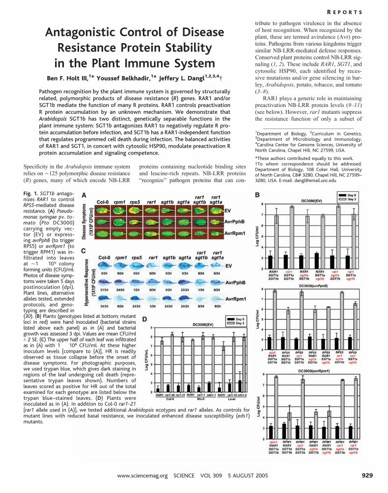
Fig. 1. SGT1b antago-nizes RAR1 to controlRPS5-mediated diseaseresistance. (A) Pseudo-monas syringae pv. to-mato (Pto DC3000)carrying empty vec-tor (EV) or express-ing avrPphB (to triggerRPS5) or avrRpm1 (totrigger RPM1) was in-filtrated into leavesat È1 � 105 colonyforming units (CFU)/ml.Photos of disease symp-toms were taken 5 dayspostinoculation (dpi).Plant lines, alternativealleles tested, extendedprotocols, and geno-typing are described in(30). (B) Plants (genotypes listed at bottom; mutantloci in red) were hand inoculated (bacterial strainslisted above each panel) as in (A) and bacterialgrowth was assessed 3 dpi. Values are mean CFU/mlT 2 SE. (C) The upper half of each leaf was infiltratedas in (A) with 1 � 108 CFU/ml. At these higherinoculum levels [compare to (A)], HR is readilyobserved as tissue collapse before the onset ofdisease symptoms. For photographic purposes,we used trypan blue, which gives dark staining inregions of the leaf undergoing cell death (repre-sentative trypan leaves shown). Numbers ofleaves scored as positive for HR out of the totalexamined for each genotype are listed below thetrypan blue–stained leaves. (D) Plants wereinoculated as in (A). In addition to Col-0 rar1-21[rar1 allele used in (A)], we tested additional Arabidopsis ecotypes and rar1 alleles. As controls formutant lines with reduced basal resistance, we inoculated enhanced disease susceptibility (eds1)mutants.
R E P O R T S
www.sciencemag.org SCIENCE VOL 309 5 AUGUST 2005 929

NB-LRR proteins. A Bthreshold model[ can
explain the discrepancy between genetic re-
quirements for RAR1 and its apparent biochem-
ical function (11). Thus, RAR1-Bindependent[NB-LRR proteins accumulate to relatively
high steady-state levels and remain above a
threshold required for efficient defense ac-
tivation even when destabilized in a rar1
background. In contrast, RAR1-Bdependent[NB-LRR proteins accumulate to relatively
low levels that fall below a critical thresh-
old in rar1 mutants. Consistent with the
semidominant nature of many R-mediated re-
sponses, the threshold model predicts that
NB-LRR proteins are quantitative, response-
limiting regulators. Cytosolic HSP90 is an ad-
ditional determinant of steady-state NB-LRR
protein accumulation (12). RAR1 likely collab-
orates with cytosolic HSP90 as a co-chaperone
maintaining signal-competent NB-LRR pro-
teins (13–16).
In yeast, SGT1 functions in kinetochore
and SCF ubiquitin-ligase assembly (17–19).
Arabidopsis has two SGT1 paralogs, SGT1a
and SGT1b (78% amino acid identity), but
only sgt1b mutations suppress NB-LRR func-
tion (7, 8, 20). RAR1, SGT1, and HSP90
interact in vivo, and RAR1 and SGT1 each in-
teract with subunits of the COP9 signalosome,
a likely proteasome lid complex (5, 14, 20).
Further, SGT1 interacts with SCF ubiquitin
ligase components, provoking speculation that
SGT1 mediates the degradation of negative
regulators of plant immune function (20). Con-
comitant losses of RAR1 and SGT1b additive-
ly impair function of the Arabidopsis NB-LRR
protein RPP5 (7), suggesting separable activ-
ities for these two genes. Accordingly, we de-
fine a RAR1-independent SGT1b function in
programmed cell death. Unexpectedly, how-
ever, our data also demonstrate that SGT1b
can negatively regulate NB-LRR protein ac-
cumulation, and that this activity is antago-
nized by both RAR1 and HSP90.
The Arabidopsis NB-LRR proteins RPM1,
RPS2, and RPS5 confer resistance to Pseudo-
monas syringae. Each is impaired in rar1
(10, 20, 21), but unaffected in sgt1a or sgt1b
(7, 22) (Fig. 1, A and B). Unexpectedly,
RPS5 function, but not RPM1 or RPS2 func-
tion, was recovered in rar1 sgt1b (Fig. 1, A
and B; RPS2 data not shown). None of the
rar1 mutant phenotypes were recovered in
rar1 sgt1a. Therefore, SGT1b mediates the
loss of RPS5 function in rar1, whereas SGT1a
and SGT1b may act redundantly in this process
for RPM1 and RPS2 (6).
NB-LRR activation often triggers a rapid
localized programmed cell death, called the
hypersensitive response (HR) (23). The HR
likely limits the growth of biotrophic fungi
and oomycetes (4, 21, 24, 25), although its
role in resistance to bacterial pathogens is un-
clear. RAR1 is required for RPS5-, RPM1-,
and RPS2-mediated HR (10). Of these, only
the RPS5-mediated HR additionally required
SGT1b (Fig. 1C; fig. S1A). Neither RPS5-,
RPM1-, nor RPS2-dependent HR were restored
in rar1 sgt1b. Using the oomycete parasite
Peronospora parasitica, we extended these find-
ings to two additional NB-LRR functions (RPP4
and RPP31; fig. S1, B to E). Thus, SGT1b
can control the HR in a RAR1-independent
manner. Further, NB-LRR–mediated disease
resistance and HR are genetically separable.
Notably, rar1 mutations in different ge-
netic backgrounds allowed enhanced growth
of the virulent bacterial strain P. syringae
(Pto) DC3000 (Fig. 1, B and D). These data
demonstrate a role for RAR1 in basal resist-
ance, an ostensibly R-independent response that
limits pathogen spread in susceptible plants
(1). This rar1 phenotype is also suppressed
in rar1 sgt1b, but not rar1 sgt1a (Fig. 1D).
Therefore, SGT1b also antagonizes RAR1 in
the control of basal resistance. Given that the
only known function for RAR1 is to promote
NB-LRR protein accumulation, then NB-LRR
proteins also are very likely to function in
basal resistance.
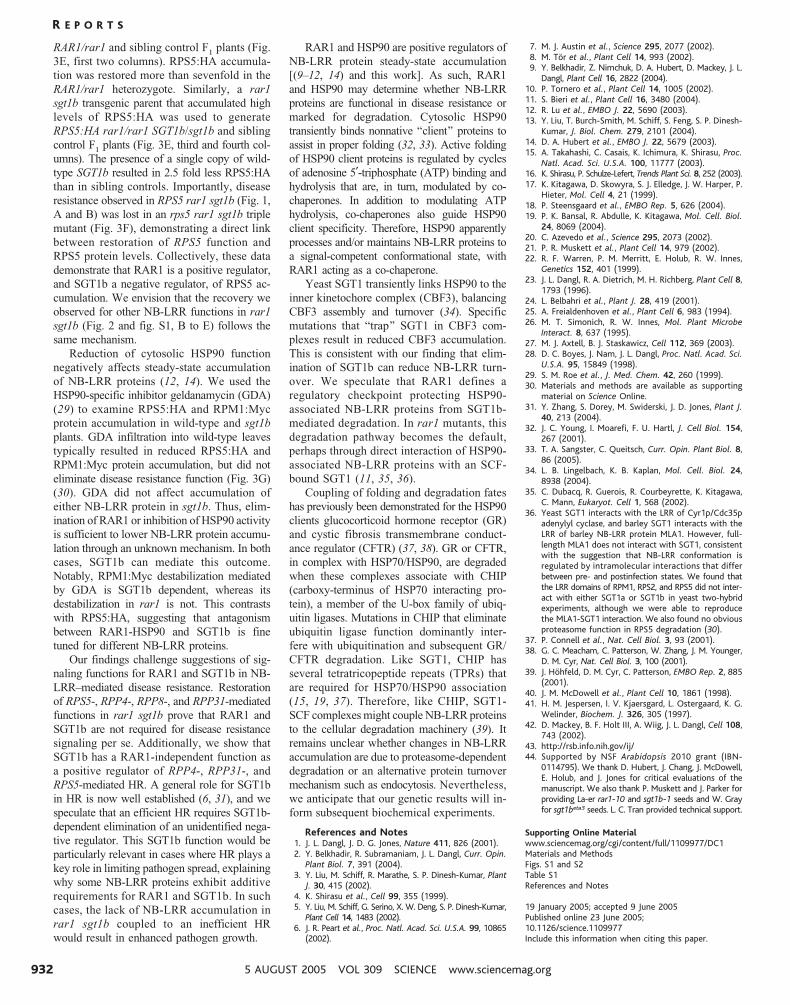
Requirements for RAR1 and SGT1b have
been defined for NB-LRR genes that confer
resistance to different isolates of the oo-
mycete parasite Peronospora parasitica (Pp)
(table S1). RPP8 was weakly impaired by
rar1, as indicated by low levels of asexual
parasite sporulation (Fig. 2, A and B). We bred
isogenic plants hemizygous for an RPP8 trans-
gene (RPP8/-) in each mutant background to
determine whether the small phenotypic ef-
fect of rar1 might depend on RPP8 dosage.
RPP8/j rar1 plants exhibited increased sus-
ceptibility as compared to homozygous con-
trols, supporting the threshold model (11).
RPP8/j rar1 sgt1b plants were completely
resistant, indicating that SGT1b mediates sus-
ceptibility in RPP8/j rar1. As with RPP4,
RPP31, and RPS5, these data are inconsistent
with the hypothesis that RAR1 and SGT1 act
additively in all NB-LRR–mediated disease
resistance responses.
Fig. 2. SGT1b antagonism of RAR1 is generalizable to several NB-LRR resistance specificities. (A)Seven- to 10-day-old cotyledons of rpp8 plants expressing a stable RPP8 transgene were inocu-lated with the asexual spores of Peronospora parasitica (Pp) isolate Emco5 (40). Representative,trypan blue–stained leaves are shown to illustrate cell death and Pp structures (hyphae, asexualsporangiophores). (B) Asexual sporangiophores were quantified 7 dpi on at least 50 cotyledons foreach of the indicated genetic backgrounds. The numbers below each tested genotype (key geno-types shown in red) represent mean sporangiophores/cotyledon (T 2 SE).
R E P O R T S
5 AUGUST 2005 VOL 309 SCIENCE www.sciencemag.org930
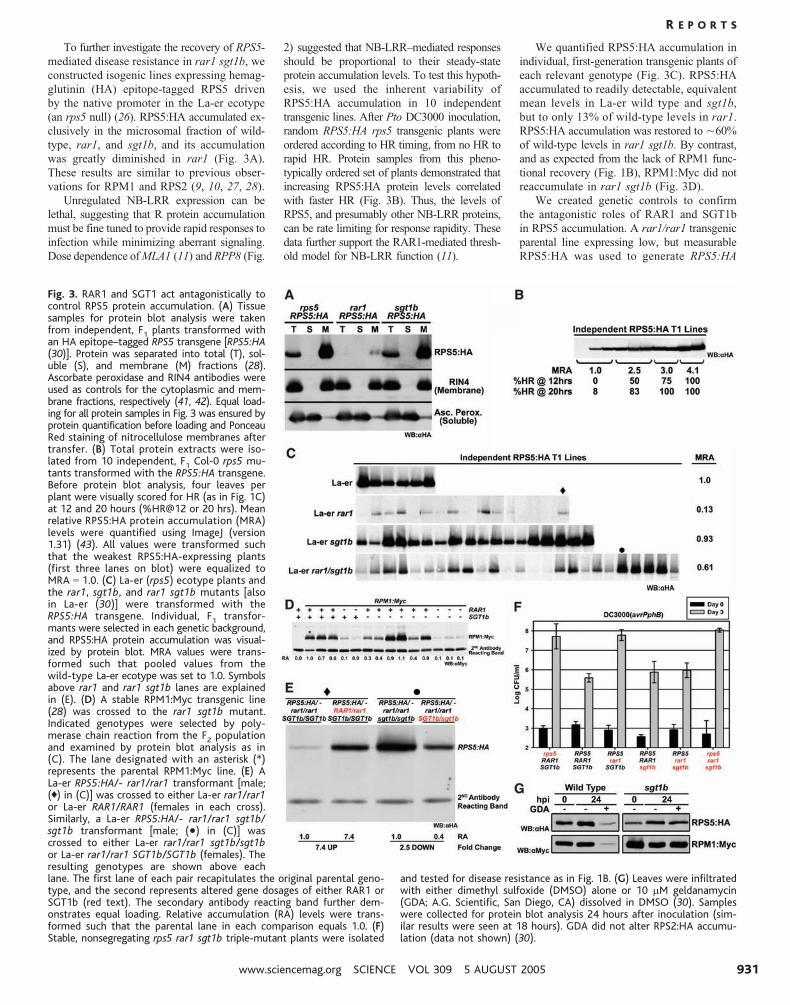
To further investigate the recovery of RPS5-
mediated disease resistance in rar1 sgt1b, we
constructed isogenic lines expressing hemag-
glutinin (HA) epitope-tagged RPS5 driven
by the native promoter in the La-er ecotype
(an rps5 null) (26). RPS5:HA accumulated ex-
clusively in the microsomal fraction of wild-
type, rar1, and sgt1b, and its accumulation
was greatly diminished in rar1 (Fig. 3A).
These results are similar to previous obser-
vations for RPM1 and RPS2 (9, 10, 27, 28).
Unregulated NB-LRR expression can be
lethal, suggesting that R protein accumulation
must be fine tuned to provide rapid responses to
infection while minimizing aberrant signaling.
Dose dependence of MLA1 (11) and RPP8 (Fig.
2) suggested that NB-LRR–mediated responses
should be proportional to their steady-state
protein accumulation levels. To test this hypoth-
esis, we used the inherent variability of
RPS5:HA accumulation in 10 independent
transgenic lines. After Pto DC3000 inoculation,
random RPS5:HA rps5 transgenic plants were
ordered according to HR timing, from no HR to
rapid HR. Protein samples from this pheno-
typically ordered set of plants demonstrated that
increasing RPS5:HA protein levels correlated
with faster HR (Fig. 3B). Thus, the levels of
RPS5, and presumably other NB-LRR proteins,
can be rate limiting for response rapidity. These
data further support the RAR1-mediated thresh-
old model for NB-LRR function (11).
We quantified RPS5:HA accumulation in
individual, first-generation transgenic plants of
each relevant genotype (Fig. 3C). RPS5:HA
accumulated to readily detectable, equivalent
mean levels in La-er wild type and sgt1b,
but to only 13% of wild-type levels in rar1.
RPS5:HA accumulation was restored to È60%
of wild-type levels in rar1 sgt1b. By contrast,
and as expected from the lack of RPM1 func-
tional recovery (Fig. 1B), RPM1:Myc did not
reaccumulate in rar1 sgt1b (Fig. 3D).
We created genetic controls to confirm
the antagonistic roles of RAR1 and SGT1b
in RPS5 accumulation. A rar1/rar1 transgenic
parental line expressing low, but measurable
RPS5:HA was used to generate RPS5:HA
Fig. 3. RAR1 and SGT1 act antagonistically tocontrol RPS5 protein accumulation. (A) Tissuesamples for protein blot analysis were takenfrom independent, F1 plants transformed withan HA epitope–tagged RPS5 transgene [RPS5:HA(30)]. Protein was separated into total (T), sol-uble (S), and membrane (M) fractions (28).Ascorbate peroxidase and RIN4 antibodies wereused as controls for the cytoplasmic and mem-brane fractions, respectively (41, 42). Equal load-ing for all protein samples in Fig. 3 was ensured byprotein quantification before loading and PonceauRed staining of nitrocellulose membranes aftertransfer. (B) Total protein extracts were iso-lated from 10 independent, F1 Col-0 rps5 mu-tants transformed with the RPS5:HA transgene.Before protein blot analysis, four leaves perplant were visually scored for HR (as in Fig. 1C)at 12 and 20 hours (%HR@12 or 20 hrs). Meanrelative RPS5:HA protein accumulation (MRA)levels were quantified using ImageJ (version1.31) (43). All values were transformed suchthat the weakest RPS5:HA-expressing plants(first three lanes on blot) were equalized toMRA 0 1.0. (C) La-er (rps5) ecotype plants andthe rar1, sgt1b, and rar1 sgt1b mutants [alsoin La-er (30)] were transformed with theRPS5:HA transgene. Individual, F1 transfor-mants were selected in each genetic background,and RPS5:HA protein accumulation was visual-ized by protein blot. MRA values were trans-formed such that pooled values from thewild-type La-er ecotype was set to 1.0. Symbolsabove rar1 and rar1 sgt1b lanes are explainedin (E). (D) A stable RPM1:Myc transgenic line(28) was crossed to the rar1 sgt1b mutant.Indicated genotypes were selected by poly-merase chain reaction from the F2 populationand examined by protein blot analysis as in(C). The lane designated with an asterisk (*)represents the parental RPM1:Myc line. (E) ALa-er RPS5:HA/- rar1/rar1 transformant [male;(0) in (C)] was crossed to either La-er rar1/rar1or La-er RAR1/RAR1 (females in each cross).Similarly, a La-er RPS5:HA/- rar1/rar1 sgt1b/sgt1b transformant [male; (&) in (C)] wascrossed to either La-er rar1/rar1 sgt1b/sgt1bor La-er rar1/rar1 SGT1b/SGT1b (females). Theresulting genotypes are shown above eachlane. The first lane of each pair recapitulates the original parental geno-type, and the second represents altered gene dosages of either RAR1 orSGT1b (red text). The secondary antibody reacting band further dem-onstrates equal loading. Relative accumulation (RA) levels were trans-formed such that the parental lane in each comparison equals 1.0. (F)Stable, nonsegregating rps5 rar1 sgt1b triple-mutant plants were isolated
and tested for disease resistance as in Fig. 1B. (G) Leaves were infiltratedwith either dimethyl sulfoxide (DMSO) alone or 10 mM geldanamycin(GDA; A.G. Scientific, San Diego, CA) dissolved in DMSO (30). Sampleswere collected for protein blot analysis 24 hours after inoculation (sim-ilar results were seen at 18 hours). GDA did not alter RPS2:HA accumu-lation (data not shown) (30).
R E P O R T S
www.sciencemag.org SCIENCE VOL 309 5 AUGUST 2005 931
RAR1/rar1 and sibling control F1
plants (Fig.
3E, first two columns). RPS5:HA accumula-
tion was restored more than sevenfold in the
RAR1/rar1 heterozygote. Similarly, a rar1
sgt1b transgenic parent that accumulated high
levels of RPS5:HA was used to generate
RPS5:HA rar1/rar1 SGT1b/sgt1b and sibling
control F1
plants (Fig. 3E, third and fourth col-
umns). The presence of a single copy of wild-
type SGT1b resulted in 2.5 fold less RPS5:HA
than in sibling controls. Importantly, disease
resistance observed in RPS5 rar1 sgt1b (Fig. 1,
A and B) was lost in an rps5 rar1 sgt1b triple
mutant (Fig. 3F), demonstrating a direct link
between restoration of RPS5 function and
RPS5 protein levels. Collectively, these data
demonstrate that RAR1 is a positive regulator,
and SGT1b a negative regulator, of RPS5 ac-
cumulation. We envision that the recovery we
observed for other NB-LRR functions in rar1
sgt1b (Fig. 2 and fig. S1, B to E) follows the
same mechanism.
Reduction of cytosolic HSP90 function
negatively affects steady-state accumulation
of NB-LRR proteins (12, 14). We used the
HSP90-specific inhibitor geldanamycin (GDA)
(29) to examine RPS5:HA and RPM1:Myc
protein accumulation in wild-type and sgt1b
plants. GDA infiltration into wild-type leaves
typically resulted in reduced RPS5:HA and
RPM1:Myc protein accumulation, but did not
eliminate disease resistance function (Fig. 3G)
(30). GDA did not affect accumulation of
either NB-LRR protein in sgt1b. Thus, elim-
ination of RAR1 or inhibition of HSP90 activity
is sufficient to lower NB-LRR protein accumu-
lation through an unknown mechanism. In both
cases, SGT1b can mediate this outcome.
Notably, RPM1:Myc destabilization mediated
by GDA is SGT1b dependent, whereas its
destabilization in rar1 is not. This contrasts
with RPS5:HA, suggesting that antagonism
between RAR1-HSP90 and SGT1b is fine
tuned for different NB-LRR proteins.
Our findings challenge suggestions of sig-
naling functions for RAR1 and SGT1b in NB-
LRR–mediated disease resistance. Restoration
of RPS5-, RPP4-, RPP8-, and RPP31-mediated
functions in rar1 sgt1b prove that RAR1 and
SGT1b are not required for disease resistance
signaling per se. Additionally, we show that
SGT1b has a RAR1-independent function as
a positive regulator of RPP4-, RPP31-, and
RPS5-mediated HR. A general role for SGT1b
in HR is now well established (6, 31), and we
speculate that an efficient HR requires SGT1b-
dependent elimination of an unidentified nega-
tive regulator. This SGT1b function would be
particularly relevant in cases where HR plays a
key role in limiting pathogen spread, explaining
why some NB-LRR proteins exhibit additive
requirements for RAR1 and SGT1b. In such
cases, the lack of NB-LRR accumulation in
rar1 sgt1b coupled to an inefficient HR
would result in enhanced pathogen growth.
RAR1 and HSP90 are positive regulators of
NB-LRR protein steady-state accumulation
E(9–12, 14) and this work^. As such, RAR1
and HSP90 may determine whether NB-LRR
proteins are functional in disease resistance or
marked for degradation. Cytosolic HSP90
transiently binds nonnative Bclient[ proteins to
assist in proper folding (32, 33). Active folding
of HSP90 client proteins is regulated by cycles
of adenosine 5¶-triphosphate (ATP) binding and
hydrolysis that are, in turn, modulated by co-
chaperones. In addition to modulating ATP
hydrolysis, co-chaperones also guide HSP90
client specificity. Therefore, HSP90 apparently
processes and/or maintains NB-LRR proteins to
a signal-competent conformational state, with
RAR1 acting as a co-chaperone.
Yeast SGT1 transiently links HSP90 to the
inner kinetochore complex (CBF3), balancing
CBF3 assembly and turnover (34). Specific
mutations that Btrap[ SGT1 in CBF3 com-
plexes result in reduced CBF3 accumulation.
This is consistent with our finding that elim-
ination of SGT1b can reduce NB-LRR turn-
over. We speculate that RAR1 defines a
regulatory checkpoint protecting HSP90-
associated NB-LRR proteins from SGT1b-
mediated degradation. In rar1 mutants, this
degradation pathway becomes the default,
perhaps through direct interaction of HSP90-
associated NB-LRR proteins with an SCF-
bound SGT1 (11, 35, 36).
Coupling of folding and degradation fates
has previously been demonstrated for the HSP90
clients glucocorticoid hormone receptor (GR)
and cystic fibrosis transmembrane conduct-
ance regulator (CFTR) (37, 38). GR or CFTR,
in complex with HSP70/HSP90, are degraded
when these complexes associate with CHIP
(carboxy-terminus of HSP70 interacting pro-
tein), a member of the U-box family of ubiq-
uitin ligases. Mutations in CHIP that eliminate
ubiquitin ligase function dominantly inter-
fere with ubiquitination and subsequent GR/
CFTR degradation. Like SGT1, CHIP has
several tetratricopeptide repeats (TPRs) that
are required for HSP70/HSP90 association
(15, 19, 37). Therefore, like CHIP, SGT1-
SCF complexes might couple NB-LRR proteins
to the cellular degradation machinery (39). It
remains unclear whether changes in NB-LRR
accumulation are due to proteasome-dependent
degradation or an alternative protein turnover
mechanism such as endocytosis. Nevertheless,
we anticipate that our genetic results will in-
form subsequent biochemical experiments.
References and Notes1. J. L. Dangl, J. D. G. Jones, Nature 411, 826 (2001).2. Y. Belkhadir, R. Subramaniam, J. L. Dangl, Curr. Opin.
Plant Biol. 7, 391 (2004).3. Y. Liu, M. Schiff, R. Marathe, S. P. Dinesh-Kumar, Plant
J. 30, 415 (2002).4. K. Shirasu et al., Cell 99, 355 (1999).5. Y. Liu, M. Schiff, G. Serino, X. W. Deng, S. P. Dinesh-Kumar,
Plant Cell 14, 1483 (2002).6. J. R. Peart et al., Proc. Natl. Acad. Sci. U.S.A. 99, 10865
(2002).
7. M. J. Austin et al., Science 295, 2077 (2002).8. M. Tor et al., Plant Cell 14, 993 (2002).9. Y. Belkhadir, Z. Nimchuk, D. A. Hubert, D. Mackey, J. L.
Dangl, Plant Cell 16, 2822 (2004).10. P. Tornero et al., Plant Cell 14, 1005 (2002).11. S. Bieri et al., Plant Cell 16, 3480 (2004).12. R. Lu et al., EMBO J. 22, 5690 (2003).13. Y. Liu, T. Burch-Smith, M. Schiff, S. Feng, S. P. Dinesh-
Kumar, J. Biol. Chem. 279, 2101 (2004).14. D. A. Hubert et al., EMBO J. 22, 5679 (2003).15. A. Takahashi, C. Casais, K. Ichimura, K. Shirasu, Proc.
Natl. Acad. Sci. U.S.A. 100, 11777 (2003).16. K. Shirasu, P. Schulze-Lefert, Trends Plant Sci. 8, 252 (2003).17. K. Kitagawa, D. Skowyra, S. J. Elledge, J. W. Harper, P.
Hieter, Mol. Cell 4, 21 (1999).18. P. Steensgaard et al., EMBO Rep. 5, 626 (2004).19. P. K. Bansal, R. Abdulle, K. Kitagawa, Mol. Cell. Biol.
24, 8069 (2004).20. C. Azevedo et al., Science 295, 2073 (2002).21. P. R. Muskett et al., Plant Cell 14, 979 (2002).22. R. F. Warren, P. M. Merritt, E. Holub, R. W. Innes,
Genetics 152, 401 (1999).23. J. L. Dangl, R. A. Dietrich, M. H. Richberg, Plant Cell 8,
1793 (1996).24. L. Belbahri et al., Plant J. 28, 419 (2001).25. A. Freialdenhoven et al., Plant Cell 6, 983 (1994).26. M. T. Simonich, R. W. Innes, Mol. Plant Microbe
Interact. 8, 637 (1995).27. M. J. Axtell, B. J. Staskawicz, Cell 112, 369 (2003).28. D. C. Boyes, J. Nam, J. L. Dangl, Proc. Natl. Acad. Sci.
U.S.A. 95, 15849 (1998).29. S. M. Roe et al., J. Med. Chem. 42, 260 (1999).30. Materials and methods are available as supporting
material on Science Online.31. Y. Zhang, S. Dorey, M. Swiderski, J. D. Jones, Plant J.
40, 213 (2004).32. J. C. Young, I. Moarefi, F. U. Hartl, J. Cell Biol. 154,
267 (2001).33. T. A. Sangster, C. Queitsch, Curr. Opin. Plant Biol. 8,
86 (2005).34. L. B. Lingelbach, K. B. Kaplan, Mol. Cell. Biol. 24,
8938 (2004).35. C. Dubacq, R. Guerois, R. Courbeyrette, K. Kitagawa,
C. Mann, Eukaryot. Cell 1, 568 (2002).36. Yeast SGT1 interacts with the LRR of Cyr1p/Cdc35p
adenylyl cyclase, and barley SGT1 interacts with theLRR of barley NB-LRR protein MLA1. However, full-length MLA1 does not interact with SGT1, consistentwith the suggestion that NB-LRR conformation isregulated by intramolecular interactions that differbetween pre- and postinfection states. We found thatthe LRR domains of RPM1, RPS2, and RPS5 did not inter-act with either SGT1a or SGT1b in yeast two-hybridexperiments, although we were able to reproducethe MLA1-SGT1 interaction. We also found no obviousproteasome function in RPS5 degradation (30).
37. P. Connell et al., Nat. Cell Biol. 3, 93 (2001).38. G. C. Meacham, C. Patterson, W. Zhang, J. M. Younger,
D. M. Cyr, Nat. Cell Biol. 3, 100 (2001).39. J. Hohfeld, D. M. Cyr, C. Patterson, EMBO Rep. 2, 885
(2001).40. J. M. McDowell et al., Plant Cell 10, 1861 (1998).41. H. M. Jespersen, I. V. Kjaersgard, L. Ostergaard, K. G.
Welinder, Biochem. J. 326, 305 (1997).42. D. Mackey, B. F. Holt III, A. Wiig, J. L. Dangl, Cell 108,
743 (2002).43. http://rsb.info.nih.gov/ij/44. Supported by NSF Arabidopsis 2010 grant (IBN-
0114795). We thank D. Hubert, J. Chang, J. McDowell,E. Holub, and J. Jones for critical evaluations of themanuscript. We also thank P. Muskett and J. Parker forproviding La-er rar1-10 and sgt1b-1 seeds and W. Grayfor sgt1beta3 seeds. L. C. Tran provided technical support.
Supporting Online Materialwww.sciencemag.org/cgi/content/full/1109977/DC1Materials and MethodsFigs. S1 and S2Table S1References and Notes
19 January 2005; accepted 9 June 2005Published online 23 June 2005;10.1126/science.1109977Include this information when citing this paper.
R E P O R T S
5 AUGUST 2005 VOL 309 SCIENCE www.sciencemag.org932
Supporting Online Material
Materials and Methods
Plant Cultivation, Transformation, Ecotypes and Mutants. Plants were grown on a
mixture of Promix (Premier Horticulture, Red Hill, PA), sand, and vermiculite in a 4:2:1
ratio, respectively. Plants were grown in growth chambers with 60% constant relative
humidity under 9 hours light at 24°C and 15 hours dark at 20°C. Agrobacterium (strain
GV3101) transformations and Basta (glufosinate-ammonium) selection of plants
expressing the BAR gene for resistance were performed as previously described (1, 2).
For Figures 1A-C, 2, and 3A-B,D,F-G we presented data for rar1-21 (3) and sgt1bedm1-1
(4) in the Col-0 ecotype. sgt1bedm1-1 is defined as a 7 gene deletion that includes SGT1b
and rar1-21 is a stop mutation in the CHORD I domain that may still make a truncated
protein (5). To demonstrate that our findings were not allele specific, we confirmed
several mutant phenotypes using alternative rar1 and sgt1b alleles. Loss of RPS5-
mediated HR in sgt1bedm1-1 was also observed in sgt1beta3 (a 1-bp deletion leading to
premature truncation in Col-0, (6), Supp. Figure 1A). La-er (RPP8) rar1-10 plants also
display a light susceptibility to Pp Emco5 that is suppressed in La-er rar1-10 sgt1b-1 (a
5 bp deletion and a single nucleotide substitution, respectively, resulting in premature
stop codons in both cases, (3, 7); Supp. Table 1). RPS5 loss of function in rar1-20, a
RAR1 deletion allele, is also restored in rar1-20 sgt1bedm1-1 (data not shown). Because
La-er does not have RPS5 (8), we performed the transgenic RPS5:HA quantification
18

(Figures 3C,E) with the La-er alleles rar1-10 and sgt1b-1. We obtained similar results
for RPS5:HA accumulation using Col-0 (RPS5) rar1-21 and sgt1bedm1-1 (data not
shown).
To confirm enhanced susceptibility to Pseudomonas syringae (Pto) DC3000 carrying an
empty vector (EV) in rar1-21 (Figure 1B, top panel), we tested rar1-1, rar1-10, and rar1-
20 in the ecotypes Ws-0, La-er, and Col-0, respectively (Figure 1D). As controls for
enhanced disease susceptibility, we used the eds1-1 (Ws-0) and eds1-2 (La-er)
mutants (9). The rar1 sgt1a double mutant in Figure 1 was generated with rar1-21 and
sgt1aKO (T-DNA insertion) alleles (courtesy of David Hubert, JLD). Genetic markers for
genotyping rar1-20, rar1-21, sgt1aKO, sgt1bedm1, and rps5-2 (Col-0 allele used for rps5)
are available upon request. Using TAIL PCR (10), the RPP8 transgene insertion was
mapped to an intergenic region of Chromosome IV, between the loci At4g33460 and
At4g33470 at ~nucleotide position 16101504 (TAIR Database; www.Arabidopsis.org).
Pathogen Strains and Isolates. For Figure 1A-D and Supplemental Figure 1A, Pto
DC3000(EV) or Pto DC3000(AvrPphB) (to trigger RPS5) or Pto DC3000(avrRpm1) (to
trigger RPM1) or Pto DC3000(avrRpt2) (to trigger RPS2, data not shown) were
resuspended in 10mM MgCl2 to ~1X10⁵ cfu/mL and syringe infiltrated into leaves of ~4
week old wild type and mutant plants (11). Bacterial growth assays were performed as
previously described (11). The HR tests in Figure 1C and Supplemental Figure 1A were
performed identically except the inoculum concentration was raised to 1X10⁸ cfu/mL.
19
The RPM1 HR was assessed 5 hours post inoculation, all other HR phenotypes were
examined at ~20 hours post inoculation.
Peronospora parasitica (Pp) propagation and inoculation was performed as previously
described (12). The Pp isolates Emco5, Emwa1, Noco2, and Cala1 were maintained on
the susceptible Arabidopsis ecotypes Col-0, Ws-0, Col-0, and La-er, respectively. The
susceptible ecotypes were as follows: The susceptible ecotypes were as follows:
Supplemental Figure 1B-D - Ws-0 (rpp4; (13)); Supplemental Figure 1E, Ws-0 (rpp31;
(14)). Figures 2A-B - Col-0 (rpp8; (15)); Supplemental Figure 2A - Col-0 (rpp5; (16));
Supplemental Figure 2B - La-er (rpp1a, rpp2a, rpp2b; (17, 18)); The resistant plant lines
were as follows: Supplemental Figure 1B-D - Col-0 (RPP4); Supplemental Figure 1E -
Col-0 (RPP31); Figure 2A-B - Col-0 transgenic for RPP8; Supplemental Figure 2A - La-
er (RPP5); Supplemental Figure 2B - Col-0 (RPP2A/B).
Trypan blue staining for cell death and the Pp structures was performed as previously
described (19). Pictures of trypan blue stained leaves following Pseudomonas
inoculations were done on a standard computer scanner. Pp inoculated, trypan blue
stained leaves were visualized by light microscope (Nikon Eclipse, Melville, NY).
Geldanamycin (GDA) Experiments. Accumulation of RPM1:Myc, RPS2:HA, and
RPS5:HA were examined by protein blot analysis following 10µM GDA (A.G. Scientific,
San Diego, CA) infiltration into leaves as described (20). We observed similar results 18
and 24 hours post GDA infiltration or when infiltrating 25µM GDA for RPM1:Myc, and
20

RPS5:HA. We did not observe reductions in RPS2 accumulation following GDA
treatment (data not shown) at 10µM, 25µM, or 50µM concentrations over this time
course. To test the disease resistance functions of RPS2, RPS5, and RPM1, we
performed several independent in planta bacterial growth assays by co-inoculating
bacteria and GDA as described (20). GDA treatment did not diminish disease resistance
in any case (data not shown). We often found that GDA treatment resulted in slightly
lower pathogen growth on susceptible plants when compared to DMSO (carrier) treated
control plants. Furthermore, GDA treated leaves exhibited visually obvious phytotoxic
symptoms (yellowing/chlorosis) 48-72 hours post inoculation, independent of bacterial
inoculation. This is problematic because 72 hours post-inoculation is the time point
when alterations in RPM1 and RPS5 functions are most readily quantifiable. Our
findings are inconsistent with previous demonstration of moderate GDA effects on
RPS2 function (20). Pleiotropic outcomes from HSP90 manipulation are documented
(21) and the effects of GDA might be variable depending on specific environmental
conditions. While GDA may give minor differences in bacterial growth under specific
environmental conditions, this inhibitor serves limited utility. Because plant HSP90
isoforms likely have overlapping functions (5), these assays will benefit greatly from the
future development of inducible silencing and/or isoform specific dominant negative
constructs.
Yeast Two-Hybrid Methods and DNA Manipulations. Directed interaction
experiments were performed in the yeast strain EGY48 as previously described (22).
The yeast "bait" and "prey" vectors pEG202 and pJG4-5, respectively, were modified for
21

compatibility with the GatewayTM cloning system (vectors courtesy of Hiro Kaminaka,
JLD; GatewayTM protocols available online at www.invitrogen.com; Invitrogen, Carlsbad,
CA; vector creation details are available on request). The newly created vectors,
pEG202gw and pJG4-5gw, were used as the final destination vectors for cloning the
LRR from RPM1, RPS2, and RPS5. Each LRR construct was started 10 amino acids
upstream of the presumptive LRR start and ended at the stop codon. The primers used
to clone each were as follows: RPM1 - RPM1 LRR F: CAC CAA TGA TGA CAG TGA
TGG TGA TGA TGC TGC and RPM1 LRR R: CTA AGA TGA GAG GCT CAC ATA
GAA AGA GCC; RPS2 - RPS2 LRR F: CAC CGT TGA GCC TAG CAT GGG ACA TAC
TGA AGC, RPS2 LRR R: TCA ATT TGG AAC AAA GCG CGG TAA ATA AC; RPS5 -
RPS5 LRR F: CAC CGC TGG TGT TGG GTT ACG TGA AGT ACC AAA, and RPS5
LRR R: TTA TGT TTC TCT CCA CCG CCA CCT GGA TG. CACC was added to the
forward (F) primer from each pair to facilitate cloning into the GatewayTM entry vector
pENTRTM/D-TOPO. Each clone was confirmed correct by sequencing and comparison
to the TAIR Database. LR ClonaseTM enzyme (Invitrogen) was then used to move each
clone from pENTRTM/D-TOPO to pEG202gw or pJG4-5gw. For the interaction tests, the
RPM1, RPS2, and RPS5 LRRs were in pEG202gw. SGT1a and SGT1b were in pJG4-
5gw (courtesy David Hubert, JLD). The cloned LRRs from MLA1 and MLA6 (in pEG202)
were used for positive and negative SGT1a/b interaction controls, respectively (courtesy
Qian-Hua Shen and Paul Shultze-Lefert). As expected, only MLA1 interacted with
SGT1a and SGT1b (22). Because no interaction was detected between the LRRs from
RPM1, RPS2, or RPS5 with either SGT1a or SGT1b, we confirmed that the proteins
22
were being made in yeast at comparable levels to both MLA1 and MLA6 (see Protein
Manipulations).
The following primers were used to clone RPS5 into pDONOR207 (Invitrogen): B1Half-
RPS5Prom: CAA AAA AGC AGG CTG GAG CCC CAT GAC CCA AAA AAT GGG,
B2Half-RPS5Stop: GAA AGC TGG GTC TGT TTC TCT CCA CCG CCA CCT G, B1
Full Site: GGG GAC AAG TTT GTA CAA AAA AGC AGG CTT C, and B2 Full Site: AGA
TTG GGG ACC ACT TTG TAC AAG AAA GCT GGG TC. To facilitate the BP ClonaseTM
reaction (Invitrogen, GatewayTM) required to clone RPS5 into pDONOR207 (courtesy of
Ian Small, URGV INRA, France), a two step PCR reaction was performed. In step one
the RPS5 target was amplified with a portion of the B1 and B2 sites (B1/2 Half primers
above), and in step two the B sites were completed with the B1/2 Full Site primers. The
final clone has 1,405 bp of RPS5 native promoter and the full RPS5 coding sequence
(sequence corresponds to Arabidopsis AGI nucleotide positions 4143604 to 4147675 on
Chromosome I). Sequencing of the entire RPS5 genomic clone revealed a single silent
nucleotide difference in the coding region compared to the TAIR Database.
pDONOR207/RPS5 was then combined with pGWB-BAR (Vector modified from pGWB-
14 (kindly provided by T. Nakagawa, Shimane University, Izumo, Japan) and pBAR1
(12) to provide in planta Basta selection; courtesy Hiro Kaminaka, BFH, JLD) in an LR
ClonaseTM reaction to create the binary vector pGWB-BAR/RPS5:HA. This vector
contains three consecutive HA epitopes in the correct translational frame at the C-
terminus of the construct. The final destination vector. pGWB-BAR/RPS5:HA, was
23
electroporated into the Agrobacterium strain GV3101 for transformation of appropriate
plant lines. Transformed plants were subsequently selected by Basta application.
Protein Manipulations. For the fractionation experiments (Figure 3A), tissue samples
were taken from multiple independent, first generation plants transformed with pGWB-
BAR/RPS5:HA. Samples from 10-15 plants were combined and protein was extracted
and separated into total, soluble, and membrane fractions by centrifugation in a sucrose
buffer (20mM Tris, pH 8.0, 0.33M Sucrose, 1mM EDTA, pH 8.0, 5µM DTT, 1X Sigma
Protease Inhibitors (Sigma, St. Louis, MO); (23)). Lanes (total, soluble, membrane)
were loaded in 1:1:1 cell equivalents corresponding to 50µg of total protein quantified
prior to fractionation. No accumulation of RPS5:HA was observed in the soluble fraction
of any genetic background following longer exposures. In additional experiments with
La-er(rps5) transformants, we observed the same distribution pattern for RPS5:HA
(data not shown). Equal loading of protein samples was insured by quantifying each
sample with Bio-Rad Laboratories (Hercules, CA) protein quantification buffer and
visually confirmed by Ponceau Red staining for each nitrocellulose membrane following
protein transfer. RPS5:HA was resolved and detected by standard SDS-PAGE protein
blotting on a 7.5% gel. Ascorbate peroxidase and RIN4 were resolved on 12% gels.
Proteins were transferred to nitrocellulose by standard methods. The ECL Plus Western
Blotting Detection System (Amersham Biosciences, Buckinghamshire, England) was
used for protein detection for these experiments and all others in this paper. Primary
antibody for RPS5:HA - high affinity anti-HA from rat (clone 3F10, Roche Applied
Biosciences, Indianapolis, IN); Secondary antibody – anti-Rat from goat conjugated to
24
horseradish peroxidase (Santa Cruz Biotechnology, Santa Cruz, CA). Proteins were
extracted identically in Figure 3C and E, except that they were subjected to a single
3,000 X gravity centrifugation for 5 minutes and the supernatant was quantified for
protein concentration. 150µg of total protein for each sample was then subjected to a
~20,000 X gravity centrifugation to concentrate the membrane fraction. This entire
membrane pellet was resuspended in 30µL sample buffer and loaded in a SDS-PAGE
gel. In Figure 3B, D and G proteins were extracted with standard lysis buffer (50mM
Tris, pH 8.0, 1% SDS, 1mM EDTA, 5µM DTT, 1X Sigma Protease Inhibitors). We found
that this buffer gave the most consistent extraction of RPM1:Myc and RPS5:HA. 50µg of
total protein/lane was loaded. Adobe Photoshop (version 7.0) was used to manipulate
all photographic images. In some instances rearrangements of lane order were made,
but all photographic adjustments, such as contrast or color, were uniform and performed
prior to these rearrangements.
Proteasome Assays. We performed two pharmaceutical assays to examine a role for
the proteasome in the reduced RPS5:HA accumulation in rar1. In the first, whole leaves
from La-er RPS5:HA and La-er rar1-10 RPS5:HA plants were infiltrated to complete
water soaking with the reversible proteasome inhibitor MG132 (100µM from 10mM
stock dissolved in DMSO; AG Scientific, San Diego, CA) or DMSO alone. Tissues
samples were collected at 0, 2, 4, 6, 8, and 24 hours post infiltration and examined by
protein blot analysis. La-er rar1-10 RPS5:HA plants infiltrated with the irreversible
proteasome inhibitor lactacystin (20µM from 2mM stock dissolved in DMSO; AG
Scientific) were also examined 24 hours post infiltration. No apparent change in
25
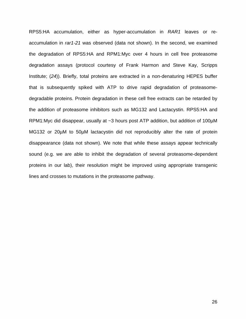
RPS5:HA accumulation, either as hyper-accumulation in RAR1 leaves or re-
accumulation in rar1-21 was observed (data not shown). In the second, we examined
the degradation of RPS5:HA and RPM1:Myc over 4 hours in cell free proteasome
degradation assays (protocol courtesy of Frank Harmon and Steve Kay, Scripps
Institute; (24)). Briefly, total proteins are extracted in a non-denaturing HEPES buffer
that is subsequently spiked with ATP to drive rapid degradation of proteasome-
degradable proteins. Protein degradation in these cell free extracts can be retarded by
the addition of proteasome inhibitors such as MG132 and Lactacystin. RPS5:HA and
RPM1:Myc did disappear, usually at ~3 hours post ATP addition, but addition of 100µM
MG132 or 20µM to 50µM lactacystin did not reproducibly alter the rate of protein
disappearance (data not shown). We note that while these assays appear technically
sound (e.g. we are able to inhibit the degradation of several proteasome-dependent
proteins in our lab), their resolution might be improved using appropriate transgenic
lines and crosses to mutations in the proteasome pathway.
26
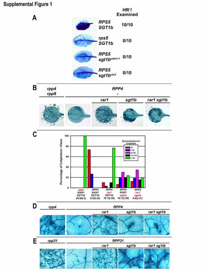
Supplemental Figures
Supplemental Figure 1. SGT1b is required for HR mediated by several disease
resistance specificities. (A) Two Arabidopsis sgt1b alleles are compromised for the
RPS5-mediated HR. This experiment was performed as in Figure 1D. sgt1bedm1 is a
complete deletion of SGT1b and sgt1beta3 is a 1 bp change resulting in a truncated
sgt1b protein. (B-E) The HR-promoting function of SGT1b in resistance to
Peronospora parasitica can be RAR1-independent. RPP4 activates relatively weak
disease resistance against Pp Emwa1 in cotyledons, (B-C), but is strong in adult leaves
(D). Both of these resistance phenotypes were nearly abolished in rar1, while sgt1b
plants exhibited modest levels of sporulation and distinctive trailing necrosis
phenotypes. Trailing necroses are thought to result from delayed or weakened HR (3,
12). RPP4 and RPP31 functions in rar1 sgt1b were phenotypically identical to sgt1b
single mutants. (B-C) These experiments were performed on cotyledons as described in
Figure 2 A-B, except the Pp isolate Emwa1 (13) was used to probe RPP4 function.
Double mutant rar1 sgt1b plants were indistinguishable from sgt1b single mutants in
both infected cotyledons and infected adult leaves. In particular, rar1 sgt1b plants
retained trailing necrosis phenotypes. Representative trypan blue stained leaves are
shown (B, D). (E) Col-0(rpp8) plants are susceptible to Pp isolate Emco5 as cotyledons.
Adult leaves (fourth pair and beyond) are generally fully resistant. The presumed R
gene(s) necessary for this resistance has been designated RPP31 (John McDowell,
pers. comm.). Note that the numerous densely stained structures in the first and third
panels (Ws-0 ecotype that does not exhibit adult resistance and Col-0 rar1,
27
respectively) are P. parasitica sexual reproductive structures called oospores (not HR
sites). Dense accumulations of oospores represent strong disease symptoms and are
easily differentiated from HR sites under higher magnification. The smaller, densely
stained sites in the second panel (Col-0) are typical HR sites.
Supplemental Figure 2. Peronospora parasitica resistance specificities are
variably impaired in rar1, sgt1b, and rar1 sgt1b. (A-B) These experiments were
performed on cotyledons as described in Figure 2A-B, except the Pp isolates Noco1
(13) and Cala1 (17) were used to probe RPP5 (A) and RPP2A/B (B) functions,
respectively. Cala1 resistance in Col-0 is controlled by two R genes (RPP2A and
RPP2B), but is represented in the figure as RPP2 for simplicity (17).
Supplemental Table 1. Summary of all genotype/pathogen combinations tested in
this study. NB-LRR resistance specificities that are impaired in rar1, but recovered in
rar1 sgt1 plants are shown in bold. NB-LRR functions that are additively impaired in rar1
sgt1b are shown in italics.
28

Supplemental Literature Cited 1. T. Altman, B. Damm, U. Halfter, L. Willmitzer, P.-C. Morris, in Methods in
Arabidopsis Research, C. Koncz, N.-H. Chua, J. Schell, Eds. (World Scientific Publishing Co., London, 1992), pp. 310-330.
2. N. Bechtold, J. Ellis, G. Pelletier, C. R. Acad. Sci., Paris 316, 1194 (1993). 3. P. R. Muskett et al., Plant Cell 14, 979 (2002). 4. M. Tör et al., Plant Cell 14, 993 (2002). 5. D. A. Hubert et al., Embo J 22, 5679 (2003). 6. W. M. Gray, P. R. Muskett, H. W. Chuang, J. E. Parker, Plant Cell 15, 1310
(2003). 7. M. J. Austin et al., Science 295, 2077 (2002). 8. M. T. Simonich, R. W. Innes, Molec. Plant-Microbe Interact. 8, 637 (1995). 9. J. E. Parker et al., Plant Cell 8, 2033 (1996). 10. Y. G. Liu, N. Mitsukawa, T. Oosumi, R. F. Whittier, Plant J 8, 457 (1995). 11. J. L. Dangl et al., in Methods in Arabidopsis Research, C. Koncz, N.-H. Chua, J.
Schell, Eds. (World Scientific, Singapore, 1992), pp. 393-418. 12. B. F. Holt III et al., Dev Cell 2, 807 (2002). 13. E. A. van der Biezen, C. T. Freddie, K. Kahn, J. E. Parker, J. D. Jones, Plant J
29, 439 (2002). 14. M. A. Torres, J. L. Dangl, J. D. G. Jones, Proc. Natl. Acad. Sci. USA 99, 523
(2002). 15. J. M. McDowell et al., Plant Cell 10, 1861 (1998). 16. J. E. Parker et al., Plant Cell 9, 879 (1997). 17. E. Sinapidou et al., Plant J 38, 898 (2004). 18. M. A. Botella et al., Plant Cell 10, 1847 (1998). 19. E. Koch, A. J. Slusarenko, Plant Cell 2, 437 (1990). 20. A. Takahashi, C. Casais, K. Ichimura, K. Shirasu, Proc Natl Acad Sci U S A 100,
11777 (2003). 21. T. A. Sangster, C. Queitsch, Curr Opin Plant Biol 8, 86 (2005). 22. S. Bieri et al., Plant Cell 16, 3480 (2004). 23. D. C. Boyes, J. Nam, J. L. Dangl, Proc. Natl. Acad. Sci., USA 95, 15849 (1998). 24. P. Mas, W. Y. Kim, D. E. Somers, S. A. Kay, Nature 426, 567 (2003).
29

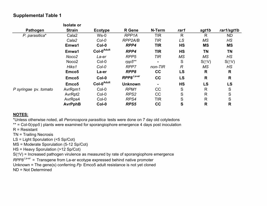
Supplemental Table 1
Isolate orPathogen Strain Ecotype R Gene N-Term rar1 sgt1b rar1/sgt1b
P. parasitica* Cala2 Ws-0 RPP1A TIR R R NDCala2 Col-0 RPP2A/B TIR LS MS HS
Emwa1 Col-0 RPP4 TIR HS MS MSEmwa1 Col-0Adult RPP4 TIR HS TN TNNoco2 La-er RPP5 TIR MS MS HSNoco2 Col-0 rpp5** - S S(↑V) S(↑V)Hiks1 Col-0 RPP7 non-TIR R MS HS
Emco5 La-er RPP8 CC LS R REmco5 Col-0 RPP8 La-er CC LS R REmco5 Col-0Adult Unknown - HS LS LS
P syringae pv. tomato AvrRpm1 Col-0 RPM1 CC S R SAvrRpt2 Col-0 RPS2 CC S R SAvrRps4 Col-0 RPS4 TIR S R SAvrPphB Col-0 RPS5 CC S R R
NOTES:*Unless otherwise noted, all Peronospora parasitica tests were done on 7 day old cotyledons** = Col-0(rpp5 ) plants were examined for sporangiophore emergence 4 days post inoculationR = ResistantTN = Trailing NecrosisLS = Light Sporulation (<5 Sp/Cot)MS = Moderate Sporulation (5-12 Sp/Cot)HS = Heavy Sporulation (>12 Sp/Cot)S(↑V) = Increased pathogen virulence as measured by rate of sporangiophore emergenceRPP8 La-er = Transgene from La-er ecotype expressed behind native promoterUnknown = The gene(s) conferring Pp Emco5 adult resistance is not yet clonedND = Not Determined