Withanolides, a new class of natural cholinesterase inhibitors with calcium antagonistic properties
12
Withanolides, a new class of natural cholinesterase inhibitors with calcium antagonistic properties q M. Iqbal Choudhary a, * , Sarfraz Ahmad Nawaz a , Zaheer-ul-Haq a , M. Arif Lodhi a , M. Nabeel Ghayur b , Saima Jalil a , Naheed Riaz a , Sammer Yousuf a , Abdul Malik a , Anwarul Hassan Gilani b , Atta-ur-Rahman a a Dr. Panjwani Center for Molecular Medicine and Drug Research, International Center for Chemical Sciences, University of Karachi, Karachi-75270, Pakistan b Department of Biological and Biomedical Sciences, The Aga Khan University, Karachi-74800, Pakistan Received 4 June 2005 Available online 29 June 2005 Dedicated to the memory of Prof. Abdus Salam (1926–1996), a nobel laureate (1976) Abstract The withanolides 1–3 and 4–5 isolated from Ajuga bracteosa and Withania somnifera, respectively, inhibited acetylcholinesterase (AChE, EC 3.1.1.7) and butyrylcholinesterase (BChE, EC 3.1.1.8) enzymes in a concentration-dependent fashion with IC 50 values ranging between 20.5 and 49.2 lM and 29.0 and 85.2 lM for AChE and BChE, respectively. Lineweaver–Burk as well as Dixon plots and their secondary replots indicated that compounds 1, 3, and 5 are the linear mixed-type inhibitors of AChE, while 2 and 4 are non-competitive inhibitors of AChE with K i values ranging between 20.0 and 45.0 lM. All compounds were found to be non-competitive inhibitors of BChE with K i values ranging between 27.7 and 90.6 lM. Molecular docking study revealed that all the ligands are completely buried inside the aromatic gorge of AChE, while compounds 1, 3, and 5 extend up to the catalytic triad. A comparison of the docking results showed that all ligands generally adopt the same binding mode and lie parallel to the surface of the gorge. The superposition of the docked structures demonstrated that the non-flexible skeleton of the ligands always penetrates the aromatic gorge through the six-membered ring A, allowing their simultaneous interaction with more than one subsite of the active center. The affinity of ligands with AChE was found to be the cumulative effects of number of hydrophobic contacts and hydrogen bonding. Furthermore, all compounds also displayed dose-dependent (0.005–1.0 mg/mL) spasmolytic and Ca 2+ antagonistic potentials in isolated rabbit jejunum preparations, compound 4 being the most active with an ED 50 value of 0.09 ± 0.001 mg/mL and 0.22 ± 0.01 lg/mL on spontaneous and K + -induced contractions, respectively. The cholinesterase inhib- itory potential along with calcium antagonistic ability and safe profile in human neutrophil viability assay could make compounds 1–5 possible drug candidates for further study to treat AlzheimerÕs disease and associated problems. Ó 2005 Elsevier Inc. All rights reserved. Keywords: Withanolides; Ajuga bracteosa; Withania somnifera; Acetylcholinesterase; Butyrylcholinesterase; Molecular docking studies; Enzyme inhibition; Calcium antagonist AChE (EC 3.1.1.7) is a key component of cholinergic brain synapses and neuromuscular junctions. The major biological role of the enzyme is the termination of im- pulse transmission by rapid hydrolysis of the cationic neurotransmitter acetylcholine [1]. According to the cholinergic hypothesis, memory impairments in patients with the senile dementia are due to a selective and irre- versible deficiency in the cholinergic functions in the brain [2]. This serves as the rationale for the use of AChE inhibitors for the symptomatic treatment of AD 0006-291X/$ - see front matter Ó 2005 Elsevier Inc. All rights reserved. doi:10.1016/j.bbrc.2005.06.086 q Abbreviations: AChE, acetylcholinesterase; BChE, butyrylcholin- esterase; PAS, peripheral anionic site; AD, AlzheimerÕs disease. * Corresponding author. Fax: +92 21 4819018, +92 21 4819019. E-mail address: [email protected] (M.I. Choudhary). www.elsevier.com/locate/ybbrc Biochemical and Biophysical Research Communications 334 (2005) 276–287 BBRC
-
Upload
independent -
Category
Documents
-
view
0 -
download
0
Transcript of Withanolides, a new class of natural cholinesterase inhibitors with calcium antagonistic properties

www.elsevier.com/locate/ybbrc
Biochemical and Biophysical Research Communications 334 (2005) 276–287
BBRC
Withanolides, a new class of natural cholinesterase inhibitorswith calcium antagonistic propertiesq
M. Iqbal Choudhary a,*, Sarfraz Ahmad Nawaz a, Zaheer-ul-Haq a, M. Arif Lodhi a,M. Nabeel Ghayur b, Saima Jalil a, Naheed Riaz a, Sammer Yousuf a, Abdul Malik a,
Anwarul Hassan Gilani b, Atta-ur-Rahman a
a Dr. Panjwani Center for Molecular Medicine and Drug Research, International Center for Chemical Sciences,
University of Karachi, Karachi-75270, Pakistanb Department of Biological and Biomedical Sciences, The Aga Khan University, Karachi-74800, Pakistan
Received 4 June 2005Available online 29 June 2005
Dedicated to the memory of Prof. Abdus Salam (1926–1996), a nobel laureate (1976)
Abstract
The withanolides 1–3 and 4–5 isolated from Ajuga bracteosa and Withania somnifera, respectively, inhibited acetylcholinesterase(AChE, EC 3.1.1.7) and butyrylcholinesterase (BChE, EC 3.1.1.8) enzymes in a concentration-dependent fashion with IC50 valuesranging between 20.5 and 49.2 lM and 29.0 and 85.2 lM for AChE and BChE, respectively. Lineweaver–Burk as well as Dixonplots and their secondary replots indicated that compounds 1, 3, and 5 are the linear mixed-type inhibitors of AChE, while 2
and 4 are non-competitive inhibitors of AChE with Ki values ranging between 20.0 and 45.0 lM. All compounds were found tobe non-competitive inhibitors of BChE with Ki values ranging between 27.7 and 90.6 lM. Molecular docking study revealed thatall the ligands are completely buried inside the aromatic gorge of AChE, while compounds 1, 3, and 5 extend up to the catalytictriad. A comparison of the docking results showed that all ligands generally adopt the same binding mode and lie parallel to thesurface of the gorge. The superposition of the docked structures demonstrated that the non-flexible skeleton of the ligands alwayspenetrates the aromatic gorge through the six-membered ring A, allowing their simultaneous interaction with more than one subsiteof the active center. The affinity of ligands with AChE was found to be the cumulative effects of number of hydrophobic contactsand hydrogen bonding. Furthermore, all compounds also displayed dose-dependent (0.005–1.0 mg/mL) spasmolytic and Ca2+
antagonistic potentials in isolated rabbit jejunum preparations, compound 4 being the most active with an ED50 value of0.09 ± 0.001 mg/mL and 0.22 ± 0.01 lg/mL on spontaneous and K+-induced contractions, respectively. The cholinesterase inhib-itory potential along with calcium antagonistic ability and safe profile in human neutrophil viability assay could make compounds1–5 possible drug candidates for further study to treat Alzheimer�s disease and associated problems.� 2005 Elsevier Inc. All rights reserved.
Keywords: Withanolides; Ajuga bracteosa; Withania somnifera; Acetylcholinesterase; Butyrylcholinesterase; Molecular docking studies; Enzymeinhibition; Calcium antagonist
AChE (EC 3.1.1.7) is a key component of cholinergicbrain synapses and neuromuscular junctions. The majorbiological role of the enzyme is the termination of im-
0006-291X/$ - see front matter � 2005 Elsevier Inc. All rights reserved.
doi:10.1016/j.bbrc.2005.06.086
q Abbreviations: AChE, acetylcholinesterase; BChE, butyrylcholin-esterase; PAS, peripheral anionic site; AD, Alzheimer�s disease.* Corresponding author. Fax: +92 21 4819018, +92 21 4819019.E-mail address: [email protected] (M.I. Choudhary).
pulse transmission by rapid hydrolysis of the cationicneurotransmitter acetylcholine [1]. According to thecholinergic hypothesis, memory impairments in patientswith the senile dementia are due to a selective and irre-versible deficiency in the cholinergic functions in thebrain [2]. This serves as the rationale for the use ofAChE inhibitors for the symptomatic treatment of AD
M.I. Choudhary et al. / Biochemical and Biophysical Research Communications 334 (2005) 276–287 277
in its early stages. Certain classes of intestinal spasmo-lytics are known to have a combination of Ca2+ antag-onist and AChE inhibitory activities. Histamine H1
blockers like promethazine and mepyramine, and H2
receptor antagonists such as cimetidine, oxmetidine,and ranitidine have been shown to possess AChE inhib-itory potentials [3,4]. The role of BChE (EC 3.1.1.8) innormal aging and brain diseases is still elusive. It hasbeen found that BChE is present in significantly higherquantities in Alzheimer�s plaques than that of normalage related non-demented brains [5].
The comprehensive study of the AChE/inhibitorcomplexes by X-ray crystallography has indicated anearly identical three-dimensional structure of the activesite, located 20 A from the protein surface at the bottomof a deep and narrow gorge [6]. The different positionsof the known inhibitors in the binding pocket suggestthat more than one clearly defined binding site existswhich are called esteratic and anionic subsites. Esteraticsubsite contains catalytic triad (Ser200, His440, andGlu327) [7] and oxyanion hole forming residues(Gly118, Gly119, and Ala201) [8]. The quaternaryammonium-binding locus (Trp84, Phe330, andGlu199) is responsible for binding of the quaternary tri-methyl ammonium tail group of ACh by cation–p inter-action [9]. The peripheral site, which is also calledperipheral anionic site (PAS), includes Tyr70, Asp72,Tyr121, Tyr334, and Trp279 residues [10,11]. Ligandoccupation of the peripheral anionic site may allosteri-cally change the conformation of the active center [11].Aromatic residues lining the gorge and residues, locatedat the outer rim of the gorge have been postulated to beinvolved in the initial binding and guiding of the sub-strate towards the active site [12].
The discovery of natural cholinesterase inhibitorshas been a very challenging area of drug developmentdue to the involvement of cholinesterases in Alzhei-mer�s disease and other related dementias. We havepreviously reported a number of new natural inhibi-tors of cholinesterases (AChE and BChE) isolatedfrom indigenous medicinal plants [13,14]. The steady-state inhibition kinetics, pharmacological profiles,SAR and 3D-QSAR, CoMFA and CoMSIA [15–17]studies have been conducted on a plenty of com-pounds. Continuing our program to introduce newdrug candidates of Alzheimer�s disease, we identifiedwithanolides 1–5, with efficacious cholinesterase inhib-itory potential.
The objectives of the current investigation were, first,to identify the cholinesterase inhibitors and then to ex-plore the possible binding modes of these compoundsin the active site of AChE by kinetics and moleculardocking studies. Second, to establish correlation be-tween AChE inhibition and Ca2+ antagonistic potentialof compounds 1–5 for their therapeutic relevance in ADand finally evaluation of cytotoxicity.
Experimental
The AChE and BChE inhibiting activities were measured by spec-trophotometric method developed by Ellman et al. [18]. Electric-eelAChE (EC 3.1.1.7), horse-serum BChE (EC 3.1.1.8), acetylthiocholineiodide, butyrylthiocholine chloride, 5,5 0-dithiobis[2-nitrobenzoic acid](DTNB), and galanthaminewere purchased fromSigma (St. Louis,MO,USA). All other chemicals were of analytical grade. Standard opera-tional assay protocol was the same as described previously [19].
The rate of the enzymatic reaction [18] was measured by the fol-lowing equation:
rate ðmol=L=minÞ ¼ change in absorbance=min
13; 600.
All the kinetic experiments were performed in 96-well microtiter-platesby using SpectraMAX 340.
Determination of kinetic parameters
The (IC50) concentration of test compounds that inhibited thehydrolysis of substrates (acetylthiocholine and butyrylthiocholine) by50% was determined by monitoring the effect of various concentrationsof the inhibitors in the assays on the inhibition values. The IC50
(inhibitor concentration that inhibits 50% activity of AChE and BChE)values were then calculated using the EZ-Fit Enzyme Kinetics program(Perrella Scientific, Amherst, USA). The interaction of compounds 1–5with AChE and BChE can be described by the following schemes:
where ES is the AChE–ATCh or BChE–BTCh complex and P is theproduct. Ki and aKi are the inhibition constants reflecting the interac-tions of inhibitors with the free AChE or BChE and the AChE-ATChor BChE-BTCh complexes, respectively.
Dissociation constant/inhibition constant (Ki) was determined bythe interpretation of Dixon plot [20]. Lineweaver–Burk plot [21] andtheir secondary replots using initial velocities were obtained over asubstrate concentration range between 0.1 and 0.4 mM for acetylthi-ocholine iodide (ATCh) and 0.05–0.2 mM for butyrylthiocholinechloride (BTCh). The dependency of Vmax/Km and Vmax on inhibitor[I] is given by:
V max=Km ¼ ðV max=KmÞK i
K i þ ½I�
� �) I ¼ K i
K i þ ½I� ) K i ¼ K i þ ½I�.
Non-linear regression equations were used to determine the values ofKi, Km, and Vmax in the Lineweaver–Burk plot and Dixon plots. TheKi value (dissociation constant/inhibition constant of AChE–inhibitoror BChE–inhibitor complex into free AChE or BChE and inhibitor)was determined graphically by Dixon plot and Lineweaver–Burk plots;first, 1/Vmaxapp was calculated at each intersection point of lines ofevery inhibitor concentration on y-axis of the Lineweaver–Burk plotand then replotted against various concentrations of inhibitor. Second,the slope of each line of inhibitor concentration on Lineweaver–Burkplot was plotted against inhibitor concentrations. Then, replottingslope versus various concentrations of inhibitor, Ki, was the intercepton x-axis.

278 M.I. Choudhary et al. / Biochemical and Biophysical Research Communications 334 (2005) 276–287
Statistical analysis
Graphs were plotted using GraFit program [22]. Values of thecorrelation coefficients, slopes, intercepts, and their standard errorswere obtained by the linear regression analysis using the same pro-gram. The correlation for all the lines of all graphs was found to be>0.99. Each point in the constructed graphs represents the mean ofthree experiments.
Molecular docking studies
The three-dimensional structures of ligands were constructed andoptimized using the SYBYL program [23]. Energy minimization wasperformed using the tripos force field with a distance gradient algorithmwith convergence criterion of 0.05 kcal/(mol A) and maximum 1000interactions, respectively. The FlexX [24] method was applied to dockligands with most of the default parameters, in the aromatic gorge ofAChE complexed with decamethonium (PDB id; 1ACL). A radius of6.5 A was used to define the active-site interaction points. FlexX soft-ware is a fast and flexible algorithm for docking small ligands in bindingsites of the enzymes, using an incremental construction algorithm thatactually builds the ligands in the binding site [23]. The software incor-porates protein–ligand interactions, placement of the ligand core, andrebuilding the complete ligand. Docking results were analyzed by VMD[25] and LIGPLOT [26]. All the computational studies were performedby using computer server on a dual processor 1.5 GHz Intel-based PCrunning the LINUX SUSE 8.2 (Kernel 2.4) operating system.
Spasmolytic and calcium antagonist activities
Antispasmodic activity of the compounds 1–5 was studied in iso-lated spontaneously contracting rabbit jejunum [27]. Rabbits from alocal breed of both sexes (1.5–2.0 kg) were obtained from the animalhouse of the Aga Khan Medical University (Karachi). Standardoperational protocol has been described previously [28].
Under these experimental conditions, the rabbit jejunum exhibitedspontaneous rhythmic contractions and therefore allowed the study ofthe relaxant (spasmolytic) activity directly without the use of an ago-nist. Calcium antagonist activity was confirmed by the ability of thecompounds 1–5 to relax the high K+ (80 mM)-induced contraction.
Cytotoxic evaluation
Isolation of human neutrophils. Heparinized fresh venous blood wasdrawn from healthy volunteers in a local blood bank and the neu-trophils were isolated by the method of Siddiqui et al. [29]. The iso-lation neutrophils have been described elsewhere [28].
Assay procedure. Isolated human neutrophils (1 · 107 cells/mL)were incubated first with test compounds for 30 min and then by theaddition of 0.25 mM WST-1 (Dojindo Laboratories, Kumamoto, Ja-pan) in water bath shaker at 37 �C [30]. After 3 h incubation, change inthe absorbance was measured at 450 nm in 96-well plate by usingSpectraMAX 340 (Molecular Devices, CA, USA). The OD is the meanof the five experimental replicates. The percentage (%) cell viability wascalculated by using the following formula:
percentage viability of cells ¼ fðOD test compound
� 100=OD controlÞ � 100g � 100.
Results and discussion
Withanolides 1–5, isolated from Ajuga bracteosa [31]and Withania somnifera [32], possess an erogstane skele-ton with lactone ring (Chart 1). The cholinesterase
inhibitory potential of these withanolides was measuredby using electric-eel (Torpedo californica) (1ACL)AChE. Oligomeric forms of electric-eel AChE are simi-lar to those of vertebrate�s nerve and muscle AChE [6].Moreover, results of studies on this enzyme can be cor-related with molecular modeling studies by the coordi-nates of eel AChE X-ray structure. Horse-serum BChEhas similarities with synaptic AChE in primary aminoacid sequence, deduced secondary structure, and ac-tive-site chemistry; the two enzymes also have overlap-ping specificities for substrates and inhibitors [33].
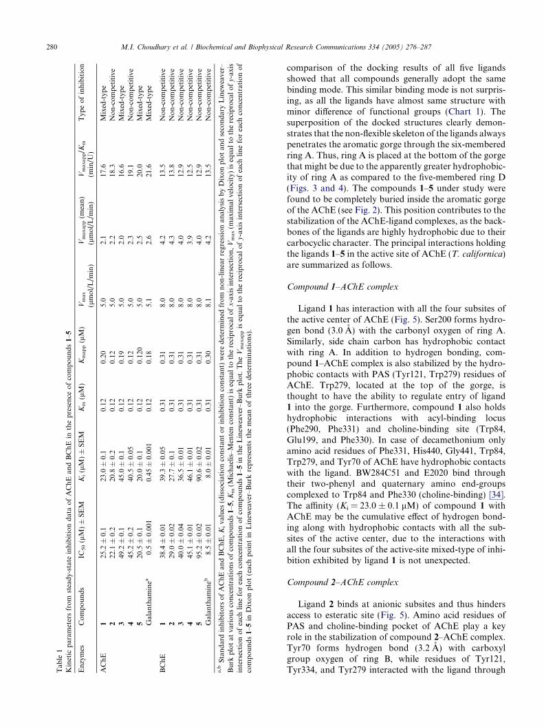
Compounds 1–5 inhibited acetylcholinesterase(AChE; EC 3.1.1.7) and butyrylcholinesterase (BChE,EC 3.1.1.8) enzymes in a concentration-dependent man-ner with Ki values ranging between 20.0 and 45.0 and27.7–90.6 lM for AChE and BChE, respectively. Ki val-ues were calculated by three different methods: first, theslopes of each line in the Lineweaver–Burk plot wereplotted against different concentrations of inhibitors;second, the 1/Vmaxapp was calculated by plotting differ-ent fixed concentrations of substrate (ATCh or BTCh)versus DV in the presence of different fixed concentra-tions of inhibitors in the respective assays of AChE orBChE. Then, Ki was calculated by plotting different con-centrations of inhibitor versus 1/Vmaxapp, Ki was theintercept on the x-axis. In the third method, Ki wasdirectly measured from Dixon plot as an intercept onx-axis. Determination of the inhibition type is importantin understanding the mechanism of inhibition and thesites of inhibitor binding. Lineweaver–Burk plot, Dixonplots, and their replots indicated that compounds 1, 3,and 5 have linear mixed-type of inhibition for AChEas in these cases there were decrease in Vmax values withthe increase of Km. Generally, this type of inhibition isthe combination of partial competitive and pure non-competitive type of inhibition. On the other hand, 2
and 4 exhibit pure non-competitive type of inhibitionfor AChE, as in these cases there were decreases in Vmax
without affecting the affinity (Km values) of the AChE orBChE towards the substrate (ATCh or BTCh), respec-tively. In other words, inhibitor and ATCh or BTCh bindrandomly and independently at the different sites ofAChE or BChE, respectively. It also indicated that theinhibition depends only on the concentration of inhibitorand dissociation constant (Ki). Similarly, all compounds1–5 were found to be the pure non-competitive inhibitorsof BChE. The Ki, Km, Kmaxapp, Vmax, Vmaxapp, Vmaxapp/Km, IC50 values and the type of inhibition have beenpresented in Table 1. The graphical analysis of steady-state inhibition data of compounds 1 and 2 for AChEhas been shown in Fig. 1. Compounds 5 (Ki =21.0 ± 0.1 lM) and 2 (Ki = 27.7 ± 0.1 lM) were foundto possess strong binding with AChE and BChE, respec-tively. Similarly, compounds 3 (Ki = 45.0 ± 0.1 lM) and5 (Ki = 90.6 ± 0.02 lM) displayed weak binding withAChE and BChE, respectively.

Chart 1. Structures of compounds 1–5.
M.I. Choudhary et al. / Biochemical and Biophysical Research Communications 334 (2005) 276–287 279
In order to predict the interactions of compounds 1–5in the aromatic gorge of AChE (T. californica), dockingpositions with the lowest energy were achieved, indicat-ing that the phase space has been sufficiently sampled.Docking protocol for each ligand was repeated manytimes and best docking positions with their respectiveminimum energies were consistently reproduced. Thesize and shape of this series of ligands supported agorge-spanning binding mode. Therefore, AChE co-crystallized with decamethonium was taken as a model,for comparison in order to monitor the performance ofour docking approach employed in this study.
The best ranking docking solutions showed thatAChE could accommodate compounds 1–5 ideally in-side the aromatic gorge. Comparison of the docking re-sults with aliphatic bis-quaternary inhibitors such as
decamethonium and/or aromatic ring containing inhib-itors such as BW284C51 [34] shows that compounds 1–5could not penetrate deep into the aromatic gorge likethese inhibitors rather remain close to the anionicsubsites. This might be due to the bulky skeleton ofcompounds 1–5 as compared to BW284C51 and deca-methonium known inhibitors of AChE. Compounds1–5 orient themselves along the active-site gorge in sucha way that their activity can be attributed only to differ-ent substituents at these ligands. Like DME999 [34],compounds 1–5 span the entire AChE surface (Fig. 2)with possibility of multiple-binding sites. The superim-position of the decamethonium ligand in the crystalstructure of the complex (PDB entry 1ACL) with our re-sults shows that decamethonium enters relatively deeperinto the aromatic gorge than our new inhibitors. A
Tab
le1
Kinetic
param
etersfrom
steady-stateinhibitiondataofAChE
andBChEin
thepresence
ofcompounds1–5
Enzymes
Compounds
IC50(lM)±
SEM
Ki(lM)±
SEM
Km(lM)
Kmapp(lM)
Vmax
(lmol/L/m
in)
Vmaxapp(m
ean)
(lmol/L/m
in)
Vmaxapp/K
m
(min/U
)Typ
eofinhibition
AChE
125
.2±
0.1
23.0
±0.1
0.12
0.20
5.0
2.1
17.6
Mixed-typ
e2
22.1
±0.2
20.8
±0.2
0.12
0.12
5.0
2.2
18.3
Non-competitive
349
.2±
0.1
45.0
±0.1
0.12
0.19
5.0
2.0
16.6
Mixed-typ
e4
45.2
±0.2
40.5
±0.05
0.12
0.12
5.0
2.3
19.1
Non-competitive
520
.5±
0.1
20.0
±0.1
0.12
0.12
05.0
2.5
20.0
Mixed-typ
eGalan
tham
inea
0.5±
0.00
10.45
±0.00
10.12
0.18
5.1
2.6
21.6
Mixed-typ
e
BChE
138
.4±
0.01
39.3
±0.05
0.31
0.31
8.0
4.2
13.5
Non-competitive
229
.0±
0.02
27.7
±0.1
0.31
0.31
8.0
4.3
13.8
Non-competitive
340
.0±
0.04
36.5
±0.01
0.31
0.31
8.0
4.0
12.9
Non-competitive
445
.1±
0.01
46.1
±0.01
0.31
0.31
8.0
3.9
12.5
Non-competitive
595
.2±
0.02
90.6
±0.02
0.31
0.31
8.0
4.0
12.9
Non-competitive
Galan
tham
ineb
8.5±
0.01
8.0±
0.01
0.31
0.30
8.1
4.2
13.5
Non-competitive
a,b
Standardinhibitors
ofAChEan
dBChE,Kivalues
(dissociationconstan
torinhibitionconstan
t)weredetermined
from
non-linearregressionan
alysisbyDixonplotan
dsecondaryLinew
eaver–
Burk
plotat
variousconcentrationsofcompounds1–5,Km(M
ichaelis–Mentenconstan
t)isequal
tothereciprocalofx-axisintersection,Vmax(m
axim
alvelocity)isequal
tothereciprocalofy-axis
intersectionofeach
lineforeach
concentrationofcompounds1–5
intheLinew
eaver–Burk
plot.TheVmaxappisequal
tothereciprocalofy-axisintersectionofeach
lineforeach
concentrationof
compounds1–5
inDixonplot(eachpointin
Linew
eaver–Burk
represents
themeanofthreedeterminations).
280 M.I. Choudhary et al. / Biochemical and Biophysical Research Communications 334 (2005) 276–287
comparison of the docking results of all five ligandsshowed that all compounds generally adopt the samebinding mode. This similar binding mode is not surpris-ing, as all the ligands have almost same structure withminor difference of functional groups (Chart 1). Thesuperposition of the docked structures clearly demon-strates that the non-flexible skeleton of the ligands alwayspenetrates the aromatic gorge through the six-memberedring A. Thus, ring A is placed at the bottom of the gorgethat might be due to the apparently greater hydrophobic-ity of ring A as compared to the five-membered ring D(Figs. 3 and 4). The compounds 1–5 under study werefound to be completely buried inside the aromatic gorgeof the AChE (see Fig. 2). This position contributes to thestabilization of the AChE-ligand complexes, as the back-bones of the ligands are highly hydrophobic due to theircarbocyclic character. The principal interactions holdingthe ligands 1–5 in the active site of AChE (T. californica)are summarized as follows.
Compound 1–AChE complex
Ligand 1 has interaction with all the four subsites ofthe active center of AChE (Fig. 5). Ser200 forms hydro-gen bond (3.0 A) with the carbonyl oxygen of ring A.Similarly, side chain carbon has hydrophobic contactwith ring A. In addition to hydrogen bonding, com-pound 1–AChE complex is also stabilized by the hydro-phobic contacts with PAS (Tyr121, Trp279) residues ofAChE. Trp279, located at the top of the gorge, isthought to have the ability to regulate entry of ligand1 into the gorge. Furthermore, compound 1 also holdshydrophobic interactions with acyl-binding locus(Phe290, Phe331) and choline-binding site (Trp84,Glu199, and Phe330). In case of decamethonium onlyamino acid residues of Phe331, His440, Gly441, Trp84,Trp279, and Tyr70 of AChE have hydrophobic contactswith the ligand. BW284C51 and E2020 bind throughtheir two-phenyl and quaternary amino end-groupscomplexed to Trp84 and Phe330 (choline-binding) [34].The affinity (Ki = 23.0 ± 0.1 lM) of compound 1 withAChE may be the cumulative effect of hydrogen bond-ing along with hydrophobic contacts with all the sub-sites of the active center, due to the interactions withall the four subsites of the active-site mixed-type of inhi-bition exhibited by ligand 1 is not unexpected.
Compound 2–AChE complex
Ligand 2 binds at anionic subsites and thus hindersaccess to esteratic site (Fig. 5). Amino acid residues ofPAS and choline-binding pocket of AChE play a keyrole in the stabilization of compound 2–AChE complex.Tyr70 forms hydrogen bond (3.2 A) with carboxylgroup oxygen of ring B, while residues of Tyr121,Tyr334, and Tyr279 interacted with the ligand through
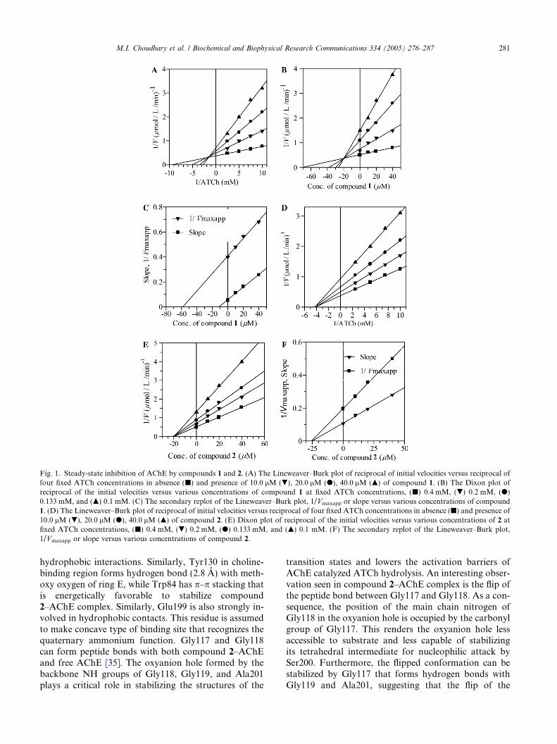
Fig. 1. Steady-state inhibition of AChE by compounds 1 and 2. (A) The Lineweaver–Burk plot of reciprocal of initial velocities versus reciprocal offour fixed ATCh concentrations in absence (j) and presence of 10.0 lM (.), 20.0 lM (d), 40.0 lM (m) of compound 1. (B) The Dixon plot ofreciprocal of the initial velocities versus various concentrations of compound 1 at fixed ATCh concentrations, (j) 0.4 mM, (.) 0.2 mM, (d)0.133 mM, and (m) 0.1 mM. (C) The secondary replot of the Lineweaver–Burk plot, 1/Vmaxapp or slope versus various concentrations of compound1. (D) The Lineweaver–Burk plot of reciprocal of initial velocities versus reciprocal of four fixed ATCh concentrations in absence (j) and presence of10.0 lM (.), 20.0 lM (d), 40.0 lM (m) of compound 2. (E) Dixon plot of reciprocal of the initial velocities versus various concentrations of 2 atfixed ATCh concentrations, (j) 0.4 mM, (.) 0.2 mM, (d) 0.133 mM, and (m) 0.1 mM. (F) The secondary replot of the Lineweaver–Burk plot,1/Vmaxapp or slope versus various concentrations of compound 2.
M.I. Choudhary et al. / Biochemical and Biophysical Research Communications 334 (2005) 276–287 281
hydrophobic interactions. Similarly, Tyr130 in choline-binding region forms hydrogen bond (2.8 A) with meth-oxy oxygen of ring E, while Trp84 has p–p stacking thatis energetically favorable to stabilize compound2–AChE complex. Similarly, Glu199 is also strongly in-volved in hydrophobic contacts. This residue is assumedto make concave type of binding site that recognizes thequaternary ammonium function. Gly117 and Gly118can form peptide bonds with both compound 2–AChEand free AChE [35]. The oxyanion hole formed by thebackbone NH groups of Gly118, Gly119, and Ala201plays a critical role in stabilizing the structures of the
transition states and lowers the activation barriers ofAChE catalyzed ATCh hydrolysis. An interesting obser-vation seen in compound 2–AChE complex is the flip ofthe peptide bond between Gly117 and Gly118. As a con-sequence, the position of the main chain nitrogen ofGly118 in the oxyanion hole is occupied by the carbonylgroup of Gly117. This renders the oxyanion hole lessaccessible to substrate and less capable of stabilizingits tetrahedral intermediate for nucleophilic attack bySer200. Furthermore, the flipped conformation can bestabilized by Gly117 that forms hydrogen bonds withGly119 and Ala201, suggesting that the flip of the
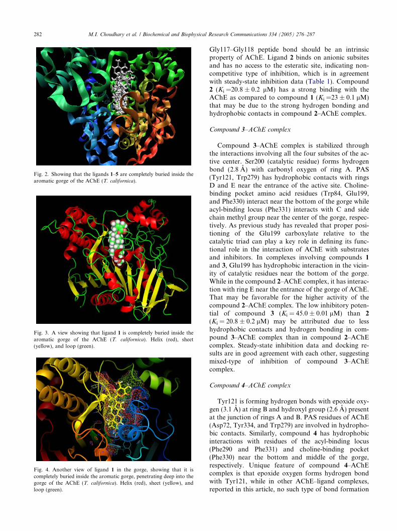
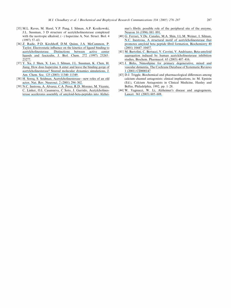
Fig. 3. A view showing that ligand 1 is completely buried inside thearomatic gorge of the AChE (T. californica). Helix (red), sheet(yellow), and loop (green).
Fig. 4. Another view of ligand 1 in the gorge, showing that it iscompletely buried inside the aromatic gorge, penetrating deep into thegorge of the AChE (T. californica). Helix (red), sheet (yellow), andloop (green).
Fig. 2. Showing that the ligands 1–5 are completely buried inside thearomatic gorge of the AChE (T. californica).
282 M.I. Choudhary et al. / Biochemical and Biophysical Research Communications 334 (2005) 276–287
Gly117–Gly118 peptide bond should be an intrinsicproperty of AChE. Ligand 2 binds on anionic subsitesand has no access to the esteratic site, indicating non-competitive type of inhibition, which is in agreementwith steady-state inhibition data (Table 1). Compound2 (Ki =20.8 ± 0.2 lM) has a strong binding with theAChE as compared to compound 1 (Ki =23 ± 0.1 lM)that may be due to the strong hydrogen bonding andhydrophobic contacts in compound 2–AChE complex.
Compound 3–AChE complex
Compound 3–AChE complex is stabilized throughthe interactions involving all the four subsites of the ac-tive center. Ser200 (catalytic residue) forms hydrogenbond (2.8 A) with carbonyl oxygen of ring A. PAS(Tyr121, Trp279) has hydrophobic contacts with ringsD and E near the entrance of the active site. Choline-binding pocket amino acid residues (Trp84, Glu199,and Phe330) interact near the bottom of the gorge whileacyl-binding locus (Phe331) interacts with C and sidechain methyl group near the center of the gorge, respec-tively. As previous study has revealed that proper posi-tioning of the Glu199 carboxylate relative to thecatalytic triad can play a key role in defining its func-tional role in the interaction of AChE with substratesand inhibitors. In complexes involving compounds 1
and 3, Glu199 has hydrophobic interaction in the vicin-ity of catalytic residues near the bottom of the gorge.While in the compound 2–AChE complex, it has interac-tion with ring E near the entrance of the gorge of AChE.That may be favorable for the higher activity of thecompound 2–AChE complex. The low inhibitory poten-tial of compound 3 (Ki = 45.0 ± 0.01 lM) than 2
(Ki = 20.8 ± 0.2 lM) may be attributed due to lesshydrophobic contacts and hydrogen bonding in com-pound 3–AChE complex than in compound 2–AChEcomplex. Steady-state inhibition data and docking re-sults are in good agreement with each other, suggestingmixed-type of inhibition of compound 3–AChEcomplex.
Compound 4–AChE complex
Tyr121 is forming hydrogen bonds with epoxide oxy-gen (3.1 A) at ring B and hydroxyl group (2.6 A) presentat the junction of rings A and B. PAS residues of AChE(Asp72, Tyr334, and Trp279) are involved in hydropho-bic contacts. Similarly, compound 4 has hydrophobicinteractions with residues of the acyl-binding locus(Phe290 and Phe331) and choline-binding pocket(Phe330) near the bottom and middle of the gorge,respectively. Unique feature of compound 4–AChEcomplex is that epoxide oxygen forms hydrogen bondwith Tyr121, while in other AChE–ligand complexes,reported in this article, no such type of bond formation

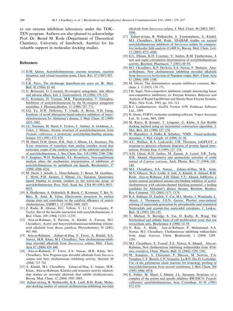
Fig. 5. 2D-Schematic representation of compounds 1–5 by LIGPLOTS, showing that hydrophobic contacts and hydrogen bonding are the principalinteractions, holding the ligand–receptor complexes in stable form.
M.I. Choudhary et al. / Biochemical and Biophysical Research Communications 334 (2005) 276–287 283
with epoixde has occurred. This may be due to the factthat in compound 4–AChE complex epoxide is presentat position C-6/C-7 of ring B, having a-orientation
and due to less operational steric hindrance it can formhydrogen bond easily, while in other AChE–inhibitorcomplexes it is b-oriented and cannot easily form
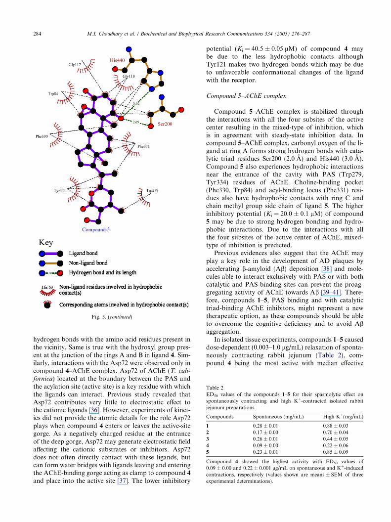
Table 2ED50 values of the compounds 1–5 for their spasmolytic effect onspontaneously contracting and high K+-contracted isolated rabbitjejunum preparations
Compounds Spontaneous (mg/mL) High K+(mg/mL)
1 0.28 ± 0.01 0.88 ± 0.032 0.17 ± 0.00 0.70 ± 0.043 0.26 ± 0.01 0.44 ± 0.054 0.09 ± 0.00 0.22 ± 0.065 0.23 ± 0.01 0.85 ± 0.09
Compound 4 showed the highest activity with ED50 values of0.09 ± 0.00 and 0.22 ± 0.001 lg/mL on spontaneous and K+-inducedcontractions, respectively (values shown are means ± SEM of threeexperimental determinations).
Fig. 5. (continued)
284 M.I. Choudhary et al. / Biochemical and Biophysical Research Communications 334 (2005) 276–287
hydrogen bonds with the amino acid residues present inthe vicinity. Same is true with the hydroxyl group pres-ent at the junction of the rings A and B in ligand 4. Sim-ilarly, interactions with the Asp72 were observed only incompound 4–AChE complex. Asp72 of AChE (T. cali-fornica) located at the boundary between the PAS andthe acylation site (active site) is a key residue with whichthe ligands can interact. Previous study revealed thatAsp72 contributes very little to electrostatic effect tothe cationic ligands [36]. However, experiments of kinet-ics did not provide the atomic details for the role Asp72plays when compound 4 enters or leaves the active-sitegorge. As a negatively charged residue at the entranceof the deep gorge, Asp72 may generate electrostatic fieldaffecting the cationic substrates or inhibitors. Asp72does not often directly contact with these ligands, butcan form water bridges with ligands leaving and enteringthe AChE-binding gorge acting as clamp to compound 4
and place into the active site [37]. The lower inhibitory
potential (Ki = 40.5 ± 0.05 lM) of compound 4 maybe due to the less hydrophobic contacts althoughTyr121 makes two hydrogen bonds which may be dueto unfavorable conformational changes of the ligandwith the receptor.
Compound 5–AChE complex
Compound 5–AChE complex is stabilized throughthe interactions with all the four subsites of the activecenter resulting in the mixed-type of inhibition, whichis in agreement with steady-state inhibition data. Incompound 5–AChE complex, carbonyl oxygen of the li-gand at ring A forms strong hydrogen bonds with cata-lytic triad residues Ser200 (2.0 A) and His440 (3.0 A).Compound 5 also experiences hydrophobic interactionsnear the entrance of the cavity with PAS (Trp279,Tyr334) residues of AChE. Choline-binding pocket(Phe330, Trp84) and acyl-binding locus (Phe331) resi-dues also have hydrophobic contacts with ring C andchain methyl group side chain of ligand 5. The higherinhibitory potential (Ki = 20.0 ± 0.1 lM) of compound5 may be due to strong hydrogen bonding and hydro-phobic interactions. Due to the interactions with allthe four subsites of the active center of AChE, mixed-type of inhibition is predicted.
Previous evidences also suggest that the AChE mayplay a key role in the development of AD plaques byaccelerating b-amyloid (Ab) deposition [38] and mole-cules able to interact exclusively with PAS or with bothcatalytic and PAS-binding sites can prevent the proag-gregating activity of AChE towards Ab [39–41]. There-fore, compounds 1–5, PAS binding and with catalytictriad-binding AChE inhibitors, might represent a newtherapeutic option, as these compounds should be ableto overcome the cognitive deficiency and to avoid Abaggregation.
In isolated tissue experiments, compounds 1–5 causeddose-dependent (0.003–1.0 lg/mL) relaxation of sponta-neously contracting rabbit jejunum (Table 2), com-pound 4 being the most active with median effective

Table 3Viability of human neutrophils (1 · 107 cells/mL) in the presence of200 lg/mL concentration of withanolides 1–5
Compounds Viability (%)
1 96.0 ± 2.92 95.5 ± 4.03 94.3 ± 3.64 94.2 ± 3.05 98.6 ± 2.0Galanthaminea 96 5 ± 2 5
a Positive control, mean ± SD of three experiments.
M.I. Choudhary et al. / Biochemical and Biophysical Research Communications 334 (2005) 276–287 285
dose (ED50) of 0.09 ± 0.001 lg/mL (mean ± SEM;n = 3). When tested for their possible mode of spasmo-lytic action, compounds 1–5 were also able to relax thehigh K+ (80 mM)-induced contraction. Compound 4
again showed the highest potential with an ED50 valueof 0.22 ± 0.01 lg/mL (mean ± SEM; n = 3), indicatingthe Ca2+ antagonistic potential. Verapamil, a standardcalcium antagonist, has exhibited a similar effect withan ED50 of 0.2 ± 0.04 lg/mL (mean ± SEM; n = 3). Itis a well-known fact that changes in the calcium homeo-stasis contribute towards aging, resulting in higher cor-tical functions as calcium ions maintain a nexusbetween membrane excitation and subsequent intracel-lular enzymatic responses [42]. Verapamil and nifedipinelike calcium antagonist have also been shown to be effec-tive in preventing old age dementia and Alzheimer�s dis-ease mainly due to the role played by calcium inmodulating brain functions [43,44]. The proclaimedhypothesis is to prevent calcium overload, particularlyin damaged neurons. The mechanism of cell loss inAD may involve an influx of calcium that causes neuro-nal dysfunction and/or neuronal death. By influencingneurotransmitter balance and preventing excessive ele-vation of intracellular neuronal calcium levels, com-pounds 1–5 might be expected to prolong cell survivaland improve cell function.
In order to evaluate the toxic effects of compounds1–5 on human neutrophils, a standard operational pro-tocol with galanthamine as positive control was devel-oped. Galanthamine is an AChE inhibitor and is usedas a drug for the treatment of Alzheimer�s disease. Theviability of human neutrophils (1 · 107 cells/mL) in thepresence of 200 lg/mL of compounds 1–5 has been pre-sented in Table 3. From the results (Table 3), it is clearthat compounds 1–5 have safe profile of human neutro-phil viability assay and possess non-toxic nature likethat of galanthamine.
Conclusions
This study was focused to identify the cholinesteraseinhibitors isolated from medicinally important plantsand to explore their possible binding modes in the active
site of AChE by kinetics and molecular docking studies.We discovered that withanolides 1, 3, and 5 were linearmixed-type of AChE inhibitors, while compounds 2 and4 were found to be non-competitive inhibitors of AChE.All 1–5 compounds were found to be non-competitiveinhibitors of BChE. Molecular docking study revealedthat all are completely buried in the aromatic gorge ofAChE, they are not as deep as decamethonium. Thatmight be due to the bulky skeleton of these ligands.They have almost similar binding mode, which wasnot unexpected as all the ligands have similar structurewith different functional groups. AChE–ligand complex-es in this class of compounds are mainly stabilizedthrough hydrophobic interactions and hydrogen bond-ing, especially with the amino acid residues of PAS ofAChE. As b-amyloid aggregating property of AChEduring the early stages of AD can be inhibited by non-competitive or mixed-type of inhibitors can preventthe proaggregating activity of AChE towards Ab.Therefore, compounds 1–5, PAS binding and/or withcatalytic triad-binding AChE inhibitors, might representa new therapeutic option, as these compounds should beable to overcome the cognitive deficiency and to avoidAb aggregation.
Therefore, it can be concluded that strong interac-tions of compounds 1–5 with the PAS or with bothPAS and catalytic triad residues of AChE indicate highinhibitory potential against AChE and consequentlyAChE-induced b-amyloid aggregation. Furthermore,compounds 1–5 also showed dose-dependent spasmolyt-ic and Ca2+ antagonistic activities in isolated rabbit jeju-num preparations. That can be helpful in preventing theexcessive elevation of intracellular neuronal calcium le-vel, and prolonging the cell survival and function. Thus,we can say that the Ca2+ antagonistic potential com-bined with cholinesterase inhibitory activities and safeprofile in human neutrophils viability assay could makecompounds 1–5 candidates for further study to treatAlzheimer�s disease and associated problems. Further-more, the observed binding modes of compounds 1–5in the active site of AChE explain the affinities of a seriesof withanolides and thus can provide a rational basis forthe structure-based drug design with improved pharma-cological properties.
Acknowledgments
The authors express their gratitude to the HigherEducation Commission (HEC) and Ministry of Scienceand Technology, Government of Pakistan, for providingthe financial support under the Pak-Kazakh ScientificCo-operation program. We are extremely grateful toProf. Dr. Rafat Ali Siddiqui (Methodist Research Insti-tute, University of Indianapolis, USA) for his usefulsuggestions and meaningful discussions during his visit
286 M.I. Choudhary et al. / Biochemical and Biophysical Research Communications 334 (2005) 276–287
to our enzyme inhibition laboratory under the TOK-TEN program. Authors are also pleased to acknowledgeProf. Dr. Bernd M. Rode (Department of TheoreticalChemistry, University of Innsbruck, Austria) for hisvaluable support in molecular docking studies.
References
[1] D.M. Quinn, Acetylcholinesterase: enzyme structure, reactiondynamics, and virtual transition states, Chem. Rev. 87 (1987) 955–979.
[2] E.K. Perry, The cholinergic hypothesis-ten years on, Br. Med.Bull. 42 (1986) 63–69.
[3] G. Bertaccini, G. Couruzzi, H2-receptor antagonists: side effectsand adverse effects, Ital. J. Gastroenterol. 16 (1984) 119–125.
[4] G. Kounenis, D. Voutsas, M.K. Papadopoulou, V. Elezoglou,Inhibition of acetylcholinesterase by the H2-receptor antagonistnizatidine, J. Pharmacobiodyn. 11 (1988) 767–771.
[5] S.Q. Yu, H.W. Holloway, T. Utsuki, A. Brossi, N.H. Greig,Synthesis of novel phenserine-based-selective inhibitors of butyr-ylcholinesterase for Alzheimer�s disease, J. Med. Chem. 42 (1999)1855–1861.
[6] J.L. Sussman, M. Harel, F. Frolow, C. Oefiner, A. Goldman, L.Toker, I. Silman, Atomic structure of acetylcholinesterase fromTorpedo californica: a prototypic acetylcholine-binding protein,Science 253 (1991) 872–879.
[7] M. Harel, D.M. Quinn, H.K. Nair, I. Silman, J.L. Sussman, TheX-ray structures of transition state analog complex reveal thatmolecular origin of the catalytic power of the substrate specificityof acetylcholinesterase, J. Am. Chem. Soc. 118 (1996) 2340–2346.
[8] T. Szegletes, W.D. Mallender, T.L. Rosenberry, Non-equilibriumanalysis alters the mechanistic interpretation of inhibition ofacetylcholinesterase by peripheral site ligands, Biochemistry 37(1998) 4206–4216.
[9] M. Harel, I. Schalk, L. Ehret-Sabatier, F. Bouet, M. Goeldner,C. Hirth, P.H. Axelsen, I. Silman, J.L. Sussman, Quaternaryligand binding to atomic residues in the active-site gorge ofacetylcholinesterase, Proc. Natl. Acad. Sci. USA 90 (1993) 9031–9035.
[10] A. Shafferman, A. Ordentlich, R. Barak, C. Kronman, T. Ber, N.Bino, R. Ariel, B. Velan, Electrostatic attraction by surfacecharge does not contribute to the catalytic efficiency of acetyl-cholinesterase, EMBO J. 13 (1994) 3448–3455.
[11] Z. Radic, R. Zduran, D.C. Vellom, Y. Li, C. Cervenasky, P.Taylor, Site of the faiculin interaction with acetylcholinesterase, J.Biol. Chem. 269 (1994) 11233–11239.
[12] Atta-ur-Rahman, S. Parveen, A. Khalid, A. Farooq, M.I.Choudhary, Acetyl and butytrylcholinesterase-inhibiting triterpe-noid alkaloids from Buxus papillosa, Phytochemistry 58 (2001)963–968.
[13] Atta-ur-Rahman, Zaheer-ul-Haq, F. Feroz, A. Khalid, S.A.Nawaz, M.R. Khan, M.I. Choudhary, New cholinesterase-inhib-iting steroidal alkaloids from Sarcococca saligna, Helv. Chem.Acta 87 (2004) 439–448.
[14] Atta-ur-Rahman, F. Feroz, S.A. Nawaz, M.R. Khan, M.I.Choudhary, New Pregnan-type steroidal alkaloids from Sarcocca
saligna and their cholinesterase inhibiting activity, Steroids 69(2004) 735–741.
[15] A. Khalid, M.I. Choudhary, Zaheer-ul-Haq, S. Anjum, M.R.Khan, Atta-ur-Rahman, Kinetics and structure–activity relation-ship studies on steroidal alkaloids that inhibit cholinesterases,Bioorg. Med. Chem. 12 (2004) 1995–2003.
[16] Zaheer-ul-haq, B. Wellenzohn, K.R. Liedl, B.M. Rode, Molec-ular-docking studies of natural cholinesterase-inhibiting steroidal
alkaloids from Sarcococa saligna, J. Med. Chem. 46 (2003) 5087–5090.
[17] Zaheer-ul-haq, B. Wellenzohn, S. Tonmunphean, A. Khalid,M.I. Choudhary, B.M. Rode, 3D-QSAR studies on naturalacetylcholinesterase inhibitors of Sarcococa saligna by compara-tive molecular field analysis (CoMFA), Bioorg. Med. Chem. Lett.13 (2003) 4375–4380.
[18] G.L. Ellman, K.D. Courtney, V. Andres, R.M. Featherstone, Anew and rapid colorimetric determination of acetylcholinesteraseactivity, Biochem. Pharmacol. 7 (1961) 88–95.
[19] M.I. Choudhary, K.P. Devkota, S.A. Nawaz, F. Shaheen, Atta-ur-Rahman, New cholinesterase inhibiting steroidal alkaloidsfrom Sarcococca hookeriana of Nepalese origin, Helv. Chem. Acta87 (2004) 1099–1108.
[20] M. Dixon, The determination enzyme inhibitors constant, Bio-chem. J. 5 (1953) 170–171.
[21] I.H. Segel, Non-competitive inhibition (simple intersecting linearnon-competitive inhibition), in: Enzyme Kinetics: Behavior andAnalysis of Rapid Equilibrium and Steady-State Enzyme Systems,Wiley, New York, 1993, pp. 101–112.
[22] R.J. Leatherbarrow, GraFit Version 4.09, Erithacus SoftwareLtd., 1999.
[23] K. Stains, SYBYL molecular modeling software. Tripos AssociateLtd., St. Louis, MO, 1999.
[24] M. Rarey, B. Kramer, T. Lengauer, G. Klebe, A fast flexibledocking method using an incremental construction algorithm, J.Mol. Biol. 261 (1996) 127–134.
[25] W. Humphrey, A. Dalke, K. Schulten, ‘‘VMD—Visual moleculardynamics, J. Mol. Graph. 14 (1996) 33–38.
[26] A.C. Wallace, R.A. Laskowski, J.M. Thornton, LIGPLOT: aprogram to generate schematic diagrams of protein–ligand inter-actions, Protein Eng. 8 (1995) 127–134.
[27] A.H. Gilani, K.H. Janbaz, M. Zaman, A. Lateef, S.R. Tariq,H.R. Ahmed, Hypotensive and spasmolytic activities of crudeextract of Cyperus scariosus, Arch. Pharm. Res. 17 (1994) 145–149.
[28] M.I. Choudhary, S.A. Nawaz, Zaheer-ul-Haq, M.K. Azim,M.N. Ghayur, M.A. Lodhi, S. Jalil, A. Khalid, A. Ahmed, B.M.Rode, Atta-ur-Rahman, A.H. Gilani, V.U. Ahmad, Juliflorine: apotent natural peripheral anionic-site-binding inhibitor of acetyl-cholinesterase with calcium-channel blocking potential, a leadingcandidate for Alzheimer�s disease therapy, Biochem. Biophys.Res. Commun. 332 (2005) 1171–1179.
[29] R.A. Siddiqui, D. English, Y. Cui, M.I. Martin, J. Wentlands, L.Akard, J. Thompson, J.G.N. Garcia, Phorbol ester-inducedpriming of superoxide generation by phosphatidic acid-stimulatedNeutrophils and granule-free neutrophil cytoplasts, J. Leukoc.Biol. 58 (1995) 189–195.
[30] V. Michael, N. Berridge, S. Tan, D. Kathy, R. Wang, Thebiochemical and cellular basis of cell proliferation assay that usetetrazolium salts, Biochemica 4 (1996) 14–19.
[31] N. Riaz, A. Malik, Aziz-ur-Rehman, P. Muhammad, S.A.Nawaz, M.I. Choudhary, Cholinesterase inhibiting withanolidesfrom Ajuga brateosa, Chem. Biodiversity 1 (2004) 1289–1295.
[32] M.I. Choudhary, S. Yousuf, S.A. Nawaz, S. Ahmed, Atta-ur-Rahman, New cholinesterase inhibiting withanolides from With-
ania somnifera, Chem. Pharm. Bull. 52 (2004) 1358–1361.[33] M. Arpagaus, A. Chatonnet, P. Masson, M. Newton, T.A.
Vaughan, C.F. Bartels, C.P. Nogueira, La.B.N. Du, O. Lockridge,Use of the polymerase chain reaction for homology probing ofbutyrylcholinesterase from several vertebrates, J. Biol. Chem. 266(1991) 6966–6974.
[34] E. Felder, M. Harel, I. Silman, J.L. Sussman, Structure of acomplex of the potent and specific inhibitor BW284C51 Torpedo
californica acetylcholinesterase, Acta Crystallogr. D 58 (1991)872–879.
M.I. Choudhary et al. / Biochemical and Biophysical Research Communications 334 (2005) 276–287 287
[35] M.L. Raves, M. Harel, Y.P. Pang, I. Silman, A.P. Kozikowski,J.L. Sussman, 3 D structure of acetylcholinesterase complexedwith the neotropic alkaloid, (�) huperzine A, Nat. Struct. Biol. 4(1997) 57–63.
[36] Z. Radic, P.D. Kirchhoff, D.M. Quinn, J.A. McCammon, P.Taylor, Electrostatic influence on the kinetics of ligand binding toacetylcholinesterase. Distinctions between active centerligands and fasciculin, J. Biol. Chem. 272 (1997) 23265–23277.
[37] Y. Xu, J. Shen, X. Luo, I. Silman, J.L. Sussman, K. Chen, H.Jiang, How does huperzine A enter and leave the binding gorge ofacetylcholinesterase? Steered molecular dynamics simulations, J.Am. Chem. Soc. 125 (2003) 11340–11349.
[38] H. Soreq, S. Seidman, Acetylcholinesterase—new roles of an oldactor, Nat. Rev. Neurosci. 2 (2001) 294–302.
[39] N.C. Instrosa, A. Alvarez, C.A. Perez, R.D. Moreno, M. Vicente,C. Linker, O.I. Casanueva, C Soto, J. Garrido, Acetylcholines-terase accelerates assembly of amyloid-beta-peptides into Alzhei-
mer�s fibrils: possible role of the peripheral site of the enzyme,Neuron 16 (1996) 881–891.
[40] G. Ferrari, V.De. Canales, M.A. Shin, I.L.M. Weiner, I. Silman,N.C. Inestrosa, A structural motif of acetylcholinesterase thatpromotes amyloid beta peptide fibril formation, Biochemistry 40(2001) 10447–10457.
[41] M. Bartolini, C. Bertucci, V. Cavrini, V. Andrisano, Beta-amyloidaggregation induced by human acetylcholinesterase inhibitionstudies, Biochem. Pharmacol. 65 (2003) 407–416.
[42] J. Birks, Nimodipine for primary degenerative, mixed andvascular dementia, The Cochrane Database of Systematic Reviews1 (2001) CD000147
[43] D.J. Triggle, Biochemical and pharmacological differences amongcalcium channel antagonists: clinical implications, in: M. Epstein(Ed.), Calcium Antagonists in Clinical Medicine, Hanley andBelfus, Philadelphia, 1992, pp. 1–28.
[44] W. Vagnucci, W. Li, Alzheimer�s disease and angiogenesis,Lancet. 361 (2003) 605–608.