Pharmacophoric features of nucleosidic HIV1RT inhibitors
7
Pharmacophoric Features of Nucleosidic HIV-1RT Inhibitors Arpita Yadav* and Sanjeev Kumar Singh Department of Chemistry, Institute of Engineering and Technology, CSJM University, Kanpur 208024, India Received 22 July 2002; accepted 9 December 2002 Abstract—A quantum pharmacological study on nucleosidic inhibitors of HIV-1RT has been performed. The main aim of this study is to discuss the pharmacophoric features (conformational and electrostatic) of nucleosidic inhibitors and compare them with normal substrate dNTP. Present study stresses on the need to refine nucleosidic drugs, as combination therapy to date is still one of the best remedies for AIDS. The results of ab initio HF calculations indicate very little effect of 3 0 substituent on ring puckering and suggest that potency regulation may be via very intricate phosphate-catalytic triad interactions. Our MESP maps also show charge complementarity between the drug and receptor. # 2003 Elsevier Science Ltd. All rights reserved. Introduction Acquired immuno deficiency syndrome (AIDS) is a deadly disease and hence considerable efforts have been made to find out effective drugs against this disease. 1,2 It is a viral disease that spreads rapidly by the replication of the virus. There are several stages in the replication of the virus catalyzed by various enzymes. One obvious way to stop the replication of the virus and in turn control the disease is to design inhibitors for one of these enzymes 3 referred to as the nucleosidic drugs. HIV-1Reverse Transcriptase (RT) is one such DNA enzyme involved in the transcription phase of replica- tion of the virus. The main function of this enzyme is to produce double stranded DNA from single strand RNA so that the genetic information can be integrated into host cell chromosome and the virus can replicate. The present study will focus on inhibitors for this enzyme. Although it is realized that resistance to currently available chemotherapeutics invariably emerges, there is a high medical need to develop more potent, safe and selective antiviral agents. In the present study we shall discuss pharmacophoric features of nucleosidic drugs, which is the first step towards designing more potent drugs. Nucleosidic drugs or inhibitors to be specific can inhibit a DNA enzyme like HIV-1RT by several mechanisms— the most common of all is the incorporation of the nucleosidic drug in the growing DNA chain. Apart from this the drug may show competitive inhibition by binding to the free enzyme; it may bind to the enzyme– DNA complex or it may even bind to the product- binding site. Examples of all these inhibitory mechan- isms are known 4 8 but the commonly used drugs which are modified nucleosides act by incorporation in the growing DNA chain. 9 Although they undoubtedly show inhibition of HIV-1RT they all lead to host toxicity because they can only be partially selective at best. To be able to develop more selective drugs, which can also overcome drug resistance problems, we must understand the drug’s pharmacophoric features and its interactions with the receptor. Some commonly clinically used nucleosidic drugs 10 16 that we have considered in this study are 2 0 3 0 dideoxy nucleosides, 2 0 3 0 didehydro dideoxy nucleosides, azidothy- midine (AZT) (Fig. 1). The characteristics of these drugs will be compared against the normal substrate for HIV- 1RT, that is dNTP deoxynucleotide triphosphate. (cf. Fig. 1) It is to be emphasized that although the nucleosidic drugs lead to host toxicity and drug resistance problems still, practically, one cannot avoid usage of nucleosidic drugs (combination therapy at best) as they are readily available in market, also their bioavailability is good and they are sure to work orally as well as intravenously. Recent theoretical studies have been more towards Monte Carlo simulations of non-nucleosidic inhibitors 0968-0896/03/$ - see front matter # 2003 Elsevier Science Ltd. All rights reserved. doi:10.1016/S0968-0896(03)00032-4 Bioorganic & Medicinal Chemistry 11 (2003) 1801–1807 *Corresponding author. Tel.: +91-512-591637; fax: +91-512-590007; e-mail: [email protected]
Transcript of Pharmacophoric features of nucleosidic HIV1RT inhibitors
Pharmacophoric Features of Nucleosidic HIV-1RT Inhibitors
Arpita Yadav* and Sanjeev Kumar Singh
Department of Chemistry, Institute of Engineering and Technology, CSJM University, Kanpur 208024, India
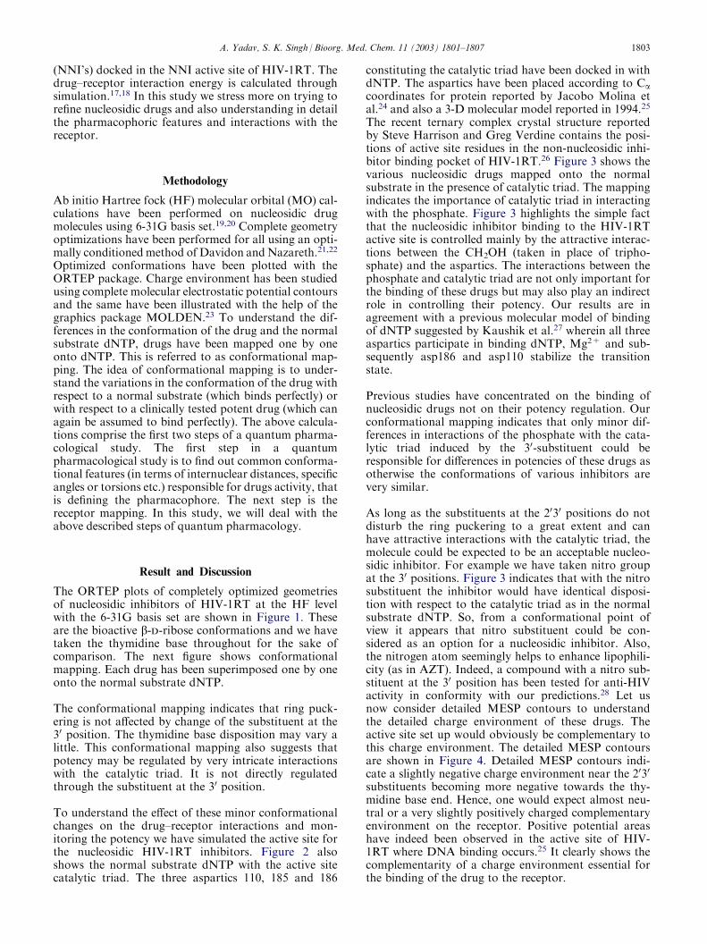
Received 22 July 2002; accepted 9 December 2002
Abstract—A quantum pharmacological study on nucleosidic inhibitors of HIV-1RT has been performed. The main aim of thisstudy is to discuss the pharmacophoric features (conformational and electrostatic) of nucleosidic inhibitors and compare them withnormal substrate dNTP. Present study stresses on the need to refine nucleosidic drugs, as combination therapy to date is still one ofthe best remedies for AIDS. The results of ab initio HF calculations indicate very little effect of 30 substituent on ring puckering andsuggest that potency regulation may be via very intricate phosphate-catalytic triad interactions. Our MESP maps also show chargecomplementarity between the drug and receptor.# 2003 Elsevier Science Ltd. All rights reserved.
Introduction
Acquired immuno deficiency syndrome (AIDS) is adeadly disease and hence considerable efforts have beenmade to find out effective drugs against this disease.1,2 Itis a viral disease that spreads rapidly by the replicationof the virus. There are several stages in the replication ofthe virus catalyzed by various enzymes. One obviousway to stop the replication of the virus and in turncontrol the disease is to design inhibitors for one ofthese enzymes3 referred to as the nucleosidic drugs.HIV-1Reverse Transcriptase (RT) is one such DNAenzyme involved in the transcription phase of replica-tion of the virus. The main function of this enzyme is toproduce double stranded DNA from single strand RNAso that the genetic information can be integrated intohost cell chromosome and the virus can replicate. Thepresent study will focus on inhibitors for this enzyme.Although it is realized that resistance to currentlyavailable chemotherapeutics invariably emerges, there isa high medical need to develop more potent, safe andselective antiviral agents. In the present study we shalldiscuss pharmacophoric features of nucleosidic drugs,which is the first step towards designing more potentdrugs.
Nucleosidic drugs or inhibitors to be specific can inhibita DNA enzyme like HIV-1RT by several mechanisms—
the most common of all is the incorporation of thenucleosidic drug in the growing DNA chain. Apartfrom this the drug may show competitive inhibition bybinding to the free enzyme; it may bind to the enzyme–DNA complex or it may even bind to the product-binding site. Examples of all these inhibitory mechan-isms are known4�8 but the commonly used drugs whichare modified nucleosides act by incorporation in thegrowing DNA chain.9 Although they undoubtedly showinhibition of HIV-1RT they all lead to host toxicitybecause they can only be partially selective at best.
To be able to develop more selective drugs, which canalso overcome drug resistance problems, we mustunderstand the drug’s pharmacophoric features and itsinteractions with the receptor.
Some commonly clinically used nucleosidic drugs10�16
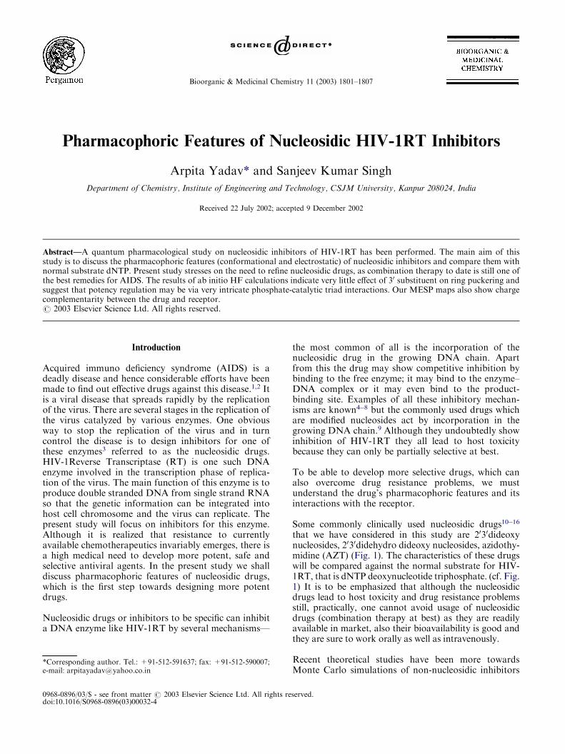
that we have considered in this study are 2030dideoxynucleosides, 2030didehydro dideoxy nucleosides, azidothy-midine (AZT) (Fig. 1). The characteristics of these drugswill be compared against the normal substrate for HIV-1RT, that is dNTP deoxynucleotide triphosphate. (cf. Fig.1) It is to be emphasized that although the nucleosidicdrugs lead to host toxicity and drug resistance problemsstill, practically, one cannot avoid usage of nucleosidicdrugs (combination therapy at best) as they are readilyavailable in market, also their bioavailability is good andthey are sure to work orally as well as intravenously.
Recent theoretical studies have been more towardsMonte Carlo simulations of non-nucleosidic inhibitors
0968-0896/03/$ - see front matter # 2003 Elsevier Science Ltd. All rights reserved.doi:10.1016/S0968-0896(03)00032-4
Bioorganic & Medicinal Chemistry 11 (2003) 1801–1807
*Corresponding author. Tel.: +91-512-591637; fax: +91-512-590007;e-mail: [email protected]
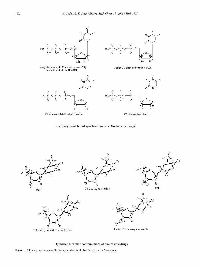
Figure 1. Clinically used nucleosidic drugs and their optimized bioactive conformations.
1802 A. Yadav, S. K. Singh / Bioorg. Med. Chem. 11 (2003) 1801–1807
(NNI’s) docked in the NNI active site of HIV-1RT. Thedrug–receptor interaction energy is calculated throughsimulation.17,18 In this study we stress more on trying torefine nucleosidic drugs and also understanding in detailthe pharmacophoric features and interactions with thereceptor.
Methodology
Ab initio Hartree fock (HF) molecular orbital (MO) cal-culations have been performed on nucleosidic drugmolecules using 6-31G basis set.19,20 Complete geometryoptimizations have been performed for all using an opti-mally conditioned method of Davidon andNazareth.21,22
Optimized conformations have been plotted with theORTEP package. Charge environment has been studiedusing complete molecular electrostatic potential contoursand the same have been illustrated with the help of thegraphics package MOLDEN.23 To understand the dif-ferences in the conformation of the drug and the normalsubstrate dNTP, drugs have been mapped one by oneonto dNTP. This is referred to as conformational map-ping. The idea of conformational mapping is to under-stand the variations in the conformation of the drug withrespect to a normal substrate (which binds perfectly) orwith respect to a clinically tested potent drug (which canagain be assumed to bind perfectly). The above calcula-tions comprise the first two steps of a quantum pharma-cological study. The first step in a quantumpharmacological study is to find out common conforma-tional features (in terms of internuclear distances, specificangles or torsions etc.) responsible for drugs activity, thatis defining the pharmacophore. The next step is thereceptor mapping. In this study, we will deal with theabove described steps of quantum pharmacology.
Result and Discussion
The ORTEP plots of completely optimized geometriesof nucleosidic inhibitors of HIV-1RT at the HF levelwith the 6-31G basis set are shown in Figure 1. Theseare the bioactive b-d-ribose conformations and we havetaken the thymidine base throughout for the sake ofcomparison. The next figure shows conformationalmapping. Each drug has been superimposed one by oneonto the normal substrate dNTP.
The conformational mapping indicates that ring puck-ering is not affected by change of the substituent at the30 position. The thymidine base disposition may vary alittle. This conformational mapping also suggests thatpotency may be regulated by very intricate interactionswith the catalytic triad. It is not directly regulatedthrough the substituent at the 30 position.
To understand the effect of these minor conformationalchanges on the drug–receptor interactions and mon-itoring the potency we have simulated the active site forthe nucleosidic HIV-1RT inhibitors. Figure 2 alsoshows the normal substrate dNTP with the active sitecatalytic triad. The three aspartics 110, 185 and 186
constituting the catalytic triad have been docked in withdNTP. The aspartics have been placed according to Cacoordinates for protein reported by Jacobo Molina etal.24 and also a 3-D molecular model reported in 1994.25
The recent ternary complex crystal structure reportedby Steve Harrison and Greg Verdine contains the posi-tions of active site residues in the non-nucleosidic inhi-bitor binding pocket of HIV-1RT.26 Figure 3 shows thevarious nucleosidic drugs mapped onto the normalsubstrate in the presence of catalytic triad. The mappingindicates the importance of catalytic triad in interactingwith the phosphate. Figure 3 highlights the simple factthat the nucleosidic inhibitor binding to the HIV-1RTactive site is controlled mainly by the attractive interac-tions between the CH2OH (taken in place of tripho-sphate) and the aspartics. The interactions between thephosphate and catalytic triad are not only important forthe binding of these drugs but may also play an indirectrole in controlling their potency. Our results are inagreement with a previous molecular model of bindingof dNTP suggested by Kaushik et al.27 wherein all threeaspartics participate in binding dNTP, Mg2+ and sub-sequently asp186 and asp110 stabilize the transitionstate.
Previous studies have concentrated on the binding ofnucleosidic drugs not on their potency regulation. Ourconformational mapping indicates that only minor dif-ferences in interactions of the phosphate with the cata-lytic triad induced by the 30-substituent could beresponsible for differences in potencies of these drugs asotherwise the conformations of various inhibitors arevery similar.
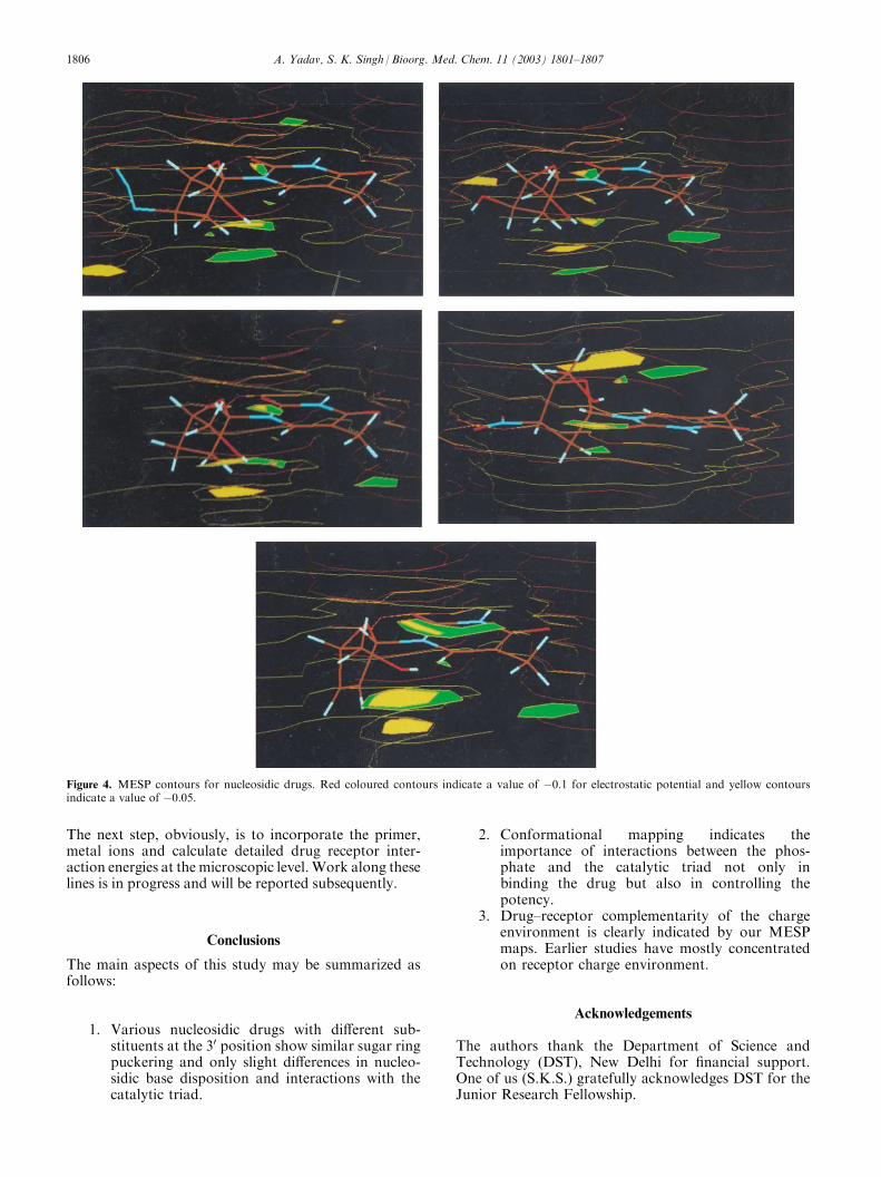
As long as the substituents at the 2030 positions do notdisturb the ring puckering to a great extent and canhave attractive interactions with the catalytic triad, themolecule could be expected to be an acceptable nucleo-sidic inhibitor. For example we have taken nitro groupat the 30 positions. Figure 3 indicates that with the nitrosubstituent the inhibitor would have identical disposi-tion with respect to the catalytic triad as in the normalsubstrate dNTP. So, from a conformational point ofview it appears that nitro substituent could be con-sidered as an option for a nucleosidic inhibitor. Also,the nitrogen atom seemingly helps to enhance lipophili-city (as in AZT). Indeed, a compound with a nitro sub-stituent at the 30 position has been tested for anti-HIVactivity in conformity with our predictions.28 Let usnow consider detailed MESP contours to understandthe detailed charge environment of these drugs. Theactive site set up would obviously be complementary tothis charge environment. The detailed MESP contoursare shown in Figure 4. Detailed MESP contours indi-cate a slightly negative charge environment near the 2030
substituents becoming more negative towards the thy-midine base end. Hence, one would expect almost neu-tral or a very slightly positively charged complementaryenvironment on the receptor. Positive potential areashave indeed been observed in the active site of HIV-1RT where DNA binding occurs.25 It clearly shows thecomplementarity of a charge environment essential forthe binding of the drug to the receptor.
A. Yadav, S. K. Singh / Bioorg. Med. Chem. 11 (2003) 1801–1807 1803
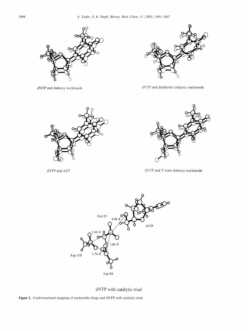
Figure 2. Conformational mapping of nucleosidic drugs and dNTP with catalytic triad.
1804 A. Yadav, S. K. Singh / Bioorg. Med. Chem. 11 (2003) 1801–1807

Figure 3. Conformational mapping of nucleosidic drugs in active site.
A. Yadav, S. K. Singh / Bioorg. Med. Chem. 11 (2003) 1801–1807 1805
The next step, obviously, is to incorporate the primer,metal ions and calculate detailed drug receptor inter-action energies at the microscopic level.Work along theselines is in progress and will be reported subsequently.
Conclusions
The main aspects of this study may be summarized asfollows:
1. Various nucleosidic drugs with different sub-stituents at the 30 position show similar sugar ringpuckering and only slight differences in nucleo-sidic base disposition and interactions with thecatalytic triad.
2. Conformational mapping indicates theimportance of interactions between the phos-phate and the catalytic triad not only inbinding the drug but also in controlling thepotency.
3. Drug–receptor complementarity of the chargeenvironment is clearly indicated by our MESPmaps. Earlier studies have mostly concentratedon receptor charge environment.
Acknowledgements
The authors thank the Department of Science andTechnology (DST), New Delhi for financial support.One of us (S.K.S.) gratefully acknowledges DST for theJunior Research Fellowship.
Figure 4. MESP contours for nucleosidic drugs. Red coloured contours indicate a value of �0.1 for electrostatic potential and yellow contoursindicate a value of �0.05.
1806 A. Yadav, S. K. Singh / Bioorg. Med. Chem. 11 (2003) 1801–1807
References and Notes
1. Wlodawer, A.; Erickson, J. Ann. Rev. Biochem. 1993, 62,543.2. Wanacott, A.; Cook, R.; Hayes, F. R.; Hann, M. M.; Jhoti,H.; McMeekin, P.; Mistry, A.; Murray-Rust, P.; Singh,O. M. P.; Weir, M. P. J. Med. Chem. 1993, 36, 3113.3. Cozzarelli, N. R. Ann. Rev. Biochem. 1977, 46, 641.4. Ma, Q. F.; Bathurst, I. C.; Barr, P. J.; Kenyon, G. L.J. Med. Chem. 1992, 35, 1938.5. Desjarlais, R. L.; Seibel, G. L.; Kuntz, I. D.; Furth, P. S.;Alverez, J. C.; Ortiz de Montellano, P. R.; De Camp, D. L.;Babe, L. M.; Craik, C. S. Proc. Natl. Acad. Sci. U.S.A. 1990,87, 6644.6. Oberg, B. Pharmacol. Ther. 1989, 40, 213.7. Fry, M.; Loeb, L. A. Animal Cell DNA Polymerases; CRC:Boca Raton, FL, 1986.8. Zimmer, C.; Wahnert, U. Prog. Biophys. Mol. Biol. 1986,47, 31.9. Weil, J.H. General Biochemistry. New Age International:New Delhi, 1997; pp 323, 326.10. Mutsuya, H.; Weinhold, J. K.; Furman, P. A.; St Clair,M. H.; Lehrman, S. N.; Gallo, R. C.; Bolognesi, D.; Barry,D. W.; Broder, S. Proc. Natl. Acad. Sci. U.S.A. 1985, 82,7096.11. Balzarini, J.; Pouwels, R.; Herdewijin, P.; Clcreq, E.De.;Cooney, D. A.; Kang, G. J.; Dalal, M.; Johns, D. G.; Broder,S. Biochem. Biophys. Res. Commun. 1986, 140, 735.12. Zimmermann, T. P.; Mahony, W. B.; Prus, K. L. J. Biol.Chem. 1987, 262, 5748.13. Cheng, Y. C.; Dutschman, G. E.; Bastow, K. S.; Sarn-gadharan, M. G.; Ting, R. Y. C. J. Biol. Chem. 1987, 262,2187.
14. Dahlberg, J. E.; Mitsuya, H.; Blam, S. B.; Broder, S.;Aaronson, S. A. Proc. Natl. Acad. Sci. U.S.A. 1987, 84, 2469.15. Mitsuya, H.; Broder, S. Proc. Natl. Acad. Sci. U.S.A.1986, 83, 1911.16. Balzarini, J.; Robins, M. J.; Zou, R.; Hardiwijin, P.;Clerck, E.De. Biochem. Biophys. Res. Commun. 1987, 145, 277.17. Silvestri, R.; Artico, M.; De Martino, G.; Novellino, E.;Greco, G.; Lavecchia, A.; Massa, S.; Giulia Loi, A.; Dor-atiotto, S.; La Colla, P. Bioorg. Med. Chem. 2000, 8, 2305.18. Rizzo, R.C.; Tirado-Rives, J.; Jorgenson, W.L. 2001, 44, 145.19. Ditchfield, R.; Hehre, W. J.; Pople, J. A. J. Chem. Phys.1971, 54, 724.20. Hehre, W. J.; Ditchfield, R.; Pople, J. A. J. Chem. Phys.1972, 56, 2257.21. Davidon, W. C.; Nazareth, L. Technical Memos 303 and306; Applied Mathematics Division, Argonne NationalLaboratories: Argonne, IL, 1977.22. Davidon, W. C. Math. Prog 1975, 9, 1.23. Schaftenaar, G. MOLDEN Version 3.6, University ofNijmegen, Toernooiveld 1, 6525 ED Nijmegen, The Nether-lands: CAOS/CAMM Center.24. Jacobo-molina, A.; Ding, J.; Nanni, R. G.; Clark, A. D.;Lu, X.; Tantillo, C.; Williams, R. L.; Kamer, G.; Ferris, A. L.;Clark, P.; Hizi, A.; Hughes, S. H.; Arnold, E. Proc. Natl.Acad. Sci. U.S.A. 1993, 90, 6320.25. Yadav, P. N. S.; Yadav, J. S.; Arnold, E.; Modak, M. J. J.Biol. Chem. 1994, 269, 716.26. Huang, H.; Chopra, R.; Verdine, G. L.; Harrison, S. C.Science 1998, 282, 1669.27. Kaushik, N.; Rege, N.; Yadav, P. N. S.; Sarafianos, S. G.;Modak, M. J.; Pandey, V. N. Biochemistry 1996, 35, 11536.28. Huang, J. J.; Ragouzeos, A.; Rideout, J. L. J. Heterocycl.Chem. 1995, 32, 691.
A. Yadav, S. K. Singh / Bioorg. Med. Chem. 11 (2003) 1801–1807 1807