
based inhibitors of serine proteases - University of Canterbury
250
THE DESIGN AND SYNTHESIS OF BASED INHIBITORS OF SERINE PROTEASES A thesis submitted in partial fulfilment of the requirements for the Degree of Doctor of Philosophy in Chemistry in the University of Canterbury by Michael John Brian Moore University of Canterbury Christchurch 1998
-
Upload
khangminh22 -
Category
Documents
-
view
1 -
download
0
Transcript of based inhibitors of serine proteases - University of Canterbury

THE DESIGN AND SYNTHESIS OF ~ECHANISM~ BASED INHIBITORS OF SERINE PROTEASES
A thesis
submitted in partial fulfilment
of the requirements for the Degree
of
Doctor of Philosophy in Chemistry
in the
University of Canterbury
by
Michael John Brian Moore ~
University of Canterbury
Christchurch
1998

HYSICAL CIENCES ~JBRAR'f
'HESIS
C1Jfj TABLE OF CONTENTS
ACKNOWLEDGEMENTS
ABSTRACT
ABBREVIATIONS
CHAPTERl INTRODUCTION
1.1 Serine Proteases and their Mechanism of Hydrolysis
1.2 Inhibition of Serine Proteases
1.3 Mechanism-Based Inactivation
CHAPTER 2 SUBSTITUTED N-[(ACYL AND ALKYL AND ARYL SULFONYL) OXY]IMIDES
1
4
10
16
32
2.1 Synthetic Strategy 33
2.2 2-Substituted Succinic Acids 39
2.3 3-Substituted N-hydroxysuccinimides 44
2.4 3-Substituted N-[(acyl and alkyl and aryl sulfonyl)oxyJsuccinirnides 52
2.5 3-Substituted N-[(acyl and methanesulfonyl)oxy]glutarimides 62
2.6 Summary 68
CHAPTER 3 (X-CHYMOTRYPSIN INHIBITION ASSAY 70
3.1 Mechanism for Inactivation of a-chymotrypsin by N-[(acyl and alkyl 71
and aryl sulfony l)oxy ]irnides
3.2 Inactivation Kinetics 77
3.3 Experimental Procedures 87

3.4 Assay Results 91
3.5 Structure-Activity Discussion 115
CHAPTER 4 ENANTIOPURE N-[(ALKYLSULFONYL) 126 OXY]IMIDES AND INHIBITION
4.1 Chirality and Biodiscrimination 127
4.2 Methods for Obtaining Chiral SuccinatesSuccinic Acid Derivatives 132
4.3 Synthesis and Inhibitory Activity of an Enantiopure 139
N -[(alkylsulfonyl) oxy]succinimide
CHAPTERS SUBSTITUTED N-[(4-HYDROXY METHYL) PHENYL]IMIDES: NOVEL INHIBITORS
5.1 Design and Proposed Mechanism of Inhibition
5.2 Synthesis and Assay
CHAPTER 6 EXPERIMENTAL
6.1 General
6.2 Chapter 2 Experimental
63 Chapter 4 Experimental
6.4 Chapter 5 Experimental
REFERENCES
APPENDIX
155
156
160
168
169
172
200
206
208
230

ACKNOWLEDGEMENTS
Thanks are offered to my supervIsor Dr. Andrew AbelL His exuberance and
enthusiasm were the catalyst for what has been an enjoyable time pursuing the content
of this magnum opus. His flair for fashion incorporated an anything will go with a
coffee coloured polo, rattle snake chemise, and caramel slacks feel that has since been
widely copied on the Parisian catwalks and other academic members of staff but only
had me convinced when the ensemble was displayed upon a pair of bata bullets.
This to match his invaluable guidance, encouragement and patience which were much
appreciated.
I would also like to thank other members of staff in the Chemistry Department for
assistance particularly Dr. John Blunt and Rewi Thompson for the provision of NMR
services; Bruce Clark for the upkeep of the mass spectrometer; and the organic
academic staff for helpful discussion. Professor Ward Robinson and Dr. Renuka
Kadirvelraj are acknowledged for their part in X-ray crystal structure determination.
The essence of my time spent at Canterbury has been the friendship existing between
students. Of particular note are the past and present members of the Abel] group and
those who have shared the laboratory experience generously extending their counsel
and resources.
To the family of friends on the outside who constantly berated me of the real world
and whose support has been thankless and finally to my family Mum, Dad, Bo and the
Cookie Marcus, and Steffi and Jesse:
'Res ipsa loquitur'

ABSTRACT
Serine proteases are involved in a number of physiological processes and have proved
to be a valuable therapeutic target in the treatment of disease states resulting when the
above processes move beyond homeostasis. The investigation of low molecular weight
compounds as irreversible and reversible inhibitors of serine proteases has been fueled
by the possibility of rational drug design and their use as meehanistic probes of enzyme
action. As introduced in Chapter one particular attention has been focused on
mechanism-based inactivators as these elicit clinically desirable specific, efficient and
irreversible inhibition. The synthesis and assay of funtionalised imide mechanism
based inhibitors of the serine protease a-chymotrypsin is the subject of this thesis.
N-[(Sulfonyl)oxy]succinimides of type 1.41 (L = S02R') are known mechanism
based inhibitors of a-chymotrypsin operating via a Lossen rearrangement that unmasks
an inactivating isocyanate species. Inhibitory activity has been found to be mediated by
the nature of the R substituent and the R' substituent of the L group. Structure activity
relationships were investigated by preparing a number of derivatives of type 1.41. The
design of the derivatives prepared focused on modulating the R' substituent to interact
with extended binding sites of a-chymotrypsin, a strategy that would enhance
inhibitory activity.
Chapter 2 describes the synthesis of 1.41 and 2.1. Retrosynthetic analysis identified a
route involving N-hydroxyimides to be favoured. A synthesis of 1.41 and 2.1 via this
key intermediate required reaction between hydroxylamine and succinic and glutaric
acid derivatives respectively. These derivatives were prepared using literature methods
employing Guareschi, Michael and malonate ester reactions. A systematic study of the
synthesis of succinic acids found the optimum route to involve the Stobbe condensation
however a short synthesis employing amide base alkylation of succinimide was
undertaken and this methodology may prove to be the ideal general route.
An aromatic series of derivatives, 1.4lf-h was therefore synthesised using the
methodology above as were "dimeric" inhibitors 1.41m and n capable of releasing two
equivalents of inactivating species during inhibition. Succinimides with 3-C phenyl
substituent rather than the benzyl substituent of the derivatives above, were prepared
and a series of N-[(sulfonyl)oxy]glutarimides 2.1a, c-e where the extent and type of
substitution were varied were prepared. N-[(Acyl)oxy]imides 1.41r-o and 2.1b (L = C(O)R') may also inhibit a-chymotrypsin and these too were investigated.
Chapter 3 discusses the assay of inhibitory activity of compounds of type 1.41 and
2.1 against a-chymotrypsin. All the synthesised derivatives, excepting a series of

N[(acyl)oxy]succinimides 1.41o-r, were found to be active to such a degree that all but
one of the active compounds could not be assayed using sampling techniques. These
potent inhibitors were then assayed using the progress curve method. Three compounds
1.41g and hand 2.1c were of such potency that the rate at which they inhibited a-
chymotrypsin could not be measured even with the progress curve method. All three of
these compounds possessed a benzyl substituent which was found to be a requiremnent
for the exhibition of mechanism-based inhibition in the succinimide series. Compounds
1.41g and 1.41h owed their potency to being able to interact favourably with the Sn'
subsites of a-chymotrypsin by containing aromatic substitution.
Chapter 4 discusses the use of Evan's oxazolidinone chemistry in the preparation of
chiral succinates from which an enantiopure inhibitor of type 1.41 was prepared.
Preliminary inhibition studies showed that (R)-l.4lf was less active than its racemate
indicating more activity resided in the (S)-enantiomer.
Chapter 5 discusses the design and synthesis of a potential novel imide mechanism
based inhibitor thought to act by unmasking a quinone imine methide in the active site
of a-chymotrypsin. Although the compound released reactivity on hydrolysis it was not
found to inhibit a-chymotrypsin significantly.

a-CT
Ac
AcC1
AC20
Ala
Ar
Arg
Bn
t-Bu
n-BuLi
t-BuOK
DCC
1,2-DCE
DMF
DMSO
EDAC
Et
Et3N
EtOH
h
His
HNE HOBt
HRMS
LDA
Me
MeOH
mesyl
min
p-NA
NMR
Ph
Phe
Pro
i-PrNEt
1'2
rt
Ser
ABBREVIATIONS
a-chymotrypsin
acetyl
acetyl chloride
acetic anhydride
alanine
aryl
arginine
Benzyl
tertiary butyl
n-butyllithium
potassium tertiary butoxide
N,N'-dicyclohexy lcarbodiimide
1,2-dichloroethane
N,N-dimethylformamide
dimethyl sulfoxide
1-(3-dimethylaminopropyl)-3-ethyl carbodimide
ethyl
triethylamine
ethanol
hour(s)
histidine
human neutrophil elastase
I-hydroxybenzotriazole
high resolution mass spectrometry
lithium di-isopropylamide
methyl
methanol
(methanesulfonyl)oxy
minute(s)
para-nitroanilide
nuclear magnetic resonance
phenyl
pheny 1 alan ine
proline
Hunigs base
correlation coefficient
room temperature
serine

CHAPTER!
INTRODUCTION

1 - Introduction 2 __ -L______ ______________________________________________________ __
Proteases constitute a large family of enzymes characterised by their function which
is the catalysis of the hydrolytic cleavage of peptide bonds in proteins and polypeptides
(Scheme 1.1).
R H ~ ,N"",
"",Way H 0
peptide bond
Protease .. +
Scheme 1.1. The protease catalysed hydrolysis of a peptide bond.
Proteases are involved in almost every aspect of life. The functions of proteases1
include hydrolysis of proteins and polypeptides for digestive and nutritional purposes,
release of peptide hormones and neuromodulators from inactive precursors, activation
of enzymes, for example clotting factors, and termination of biological responses by
degradation of the message-transmitting peptide.
Proteases have been classified into four classes each with their own distinct
mechanism.2,3 This classification is based on the most significant functional group
associated with the protease activity. Hence the serine, aspartic, cysteine proteases
derive their names from the most prominent catalytic amino acid residue in the active
site whereas the metallo proteases are named according to the associated prosthetic
metal ion. Table 1.1 lists examples of proteases in each mechanistic category.
The following section of this introduction deals with the mechanism of action of
serine proteases and the different approaches to inhibiting these enzymes.

1 - Introduction 3 --~---------------------------------------------------------------
Protease Serine
Metallo
Aspartic
Cysteine
Representative Enzymes Normal Function -thrombin, plasma kallikrein -blood coagulation
-trypsin, a-chymotrypsin, -digestion
pancreatic elastase
-enterokinase plasmin, -fibrinolysis, destruction of
plasminogen activator blood clots
-tissue kallikrein, post-protein
cleaving enzyme -hormone metabolism
-elastase, cathepsin G,
tryptases, mast cell chymases -phagocytosis
-acrosin -fertilization
-ATP dependent proteases -protein turnover
(intracellular)
-angiotensin converting -blood pressure regulation
enzyme, aminopeptidases,
renal dipeptidases
-collagenase
-carboxypeptidase
-macrophage elastase
-enkephalinases
-renin
-HN protease
-therrnolysin, pepsin
-tissue remodelling
-digestion
-peptide metabolism, blood
pressure regulation
-analgesic
-blood pressure regulation
-HIV replication
-digestion
-cathepsins B, H, L, calcium -protein turnover, bone
activated neutral proteases resorbtion
-papain -digestion
-picornavirus -viral replication
TABLE 1.1. Examples of Proteases, Subdivided into Mechanistic Categories

1 Introduction 4
1.1 SERINE PROTEASES AND THEIR MECHANISM OF PEPTIDE HYDROLYSIS
More than 130 serine proteases have been classified to date. 4 Their different
functions include digestion, processing of peptide prohormones, thrombolysis and
fibrinolysis, fertilization and blastocyst implantation. 5,6 Even though the physiological
functions of serine proteases are diverse, all employ a common catalytic mechanism. a
Chymotrypsin,7,8 a serine protease from the mammalian pancreas involved in digestion,
has been studied intensively and more information is available on its mode of catalysis
than for almost any other enzyme.9 Its active site has been well characterized by X-ray
crystallography and intermediates of the hydrolysis reaction it catalyses are known. It is
therefore representative of the mechanism of the whole class of serine proteases.
The proposed mechanism of peptide hydrolysis by a-chymotrypsin is illustrated in
Scheme 1.2. The first step in the hydrolysis is the complementary approach of the
enzyme and substrate that results in the non-covalent enzyme-substrate aggregate
known as the Michaelis-Menten complex (1.1). The driving force for its formation
depends on favourable binding interactions between the enzyme and substrate. The
structure of serine proteases remains quite conserved but differences in key structural
binding elements imparts specificity of action to a protease with regards to its substrate.
Topographical studies of a-chymotrypsin, for example, have shown the presence of a
hydrophobic pocket associated with the active-site which binds the amino acid a-C
substituent on the carboxyl side of the peptide bond which is to be hydrolysed (the so
called scissile bond). Hence a-chymotrypsin selectively cleaves peptides on the
carboxyl side of the amino acid residues; phenylalanine, tyrosine and tryptophan as
these have aromatic side chains able to interact favourably with this pocket.
The driving force for hydrolysis is the catalytic triad consisting of Ser-195, His-57
and Asp-102 (the numbers 102, 57 and 195 denote the position of the amino acid
residues Asp, His, and Ser, respectively in the polypeptide chain of the enzyme). His-57
and Asp-102 act as an electron sink and source in a charge relay system that enhances
the nucleophilicity of Ser-195 (Step I, Scheme 1.2) and the incoming water molecule
(Step II, Scheme 1.2). Catalysis is initiated by nucleophilic attack of the Ser-195 on the
susceptible carbonyl carbon of the scissile peptide bond (depicted by the arrow) of the
substrate to form a tetrahedral intermediate (1.2). The oxyanion of the tetrahedral
adduct is stabilised by hydrogen-bonding to the backbone NH groups of Gly-193 and
Ser-195 (see 1.2) which incorporate a binding element referred to as the oxyanion hole.
The proton on the serine hydroxyl group is ultimately transferred, via His-57, to the

I - Introduction 5 --~---------------------------------------------~----------
I
I
His-57
Ser-195
o
~N~ H p, H2N""'"
...... ''', " "H H ,
N' N
~ Ser-195 Gly-193
Michaelis Enzyme-substrate complex 1.1
Tetrahedral Intermediate 1.2
Acyl Enzyme 1.3a
Scheme 1.2 Proposed mechanism of substrate hydrolysis by a-chymotrypsin

1 - Introduction 6 --~---------------------------------------------------------
II
II
His-57 -----Ser-195 --=:::: J
,'N~ O u.""" '\-N,
-n •• H R rrr
VV'N~O-H rrrrr(..O" Asp-102
H 0 6 H2N""'"
rH H N 'N
'-J V Ser-195~Gly-193
Acyl Enzyme 1.3b
Tetrahedral Intermediate 1.4
Products and Enzyme 1.5
Scheme 1.2 (continued) Proposed mechanism of substrate hydrolysis by u
chymotrypsin

~~~~ __ ~~~~~______________ ________________________ 7
nitrogen atom of the eventual amine product, thereby facilitating collapse of the
tetrahedral intermediate to the acyl-enzyme ester (1.3a) and release of the C-terminal
peptide fragment. The acyl-enzyme 1.3b is hydrolysed by the catalytic addition of
water (Step II), facilitated by the charge relay system working in reverse via a second
tetrahedral intermediate (1.4), to afford the N-terminal carboxylic acid fragment of the
peptide, along with regenerated enzyme (1.5).
ENZYME-SUBSTRA TE SPECIFICITY
The active site of proteases consists of two domains: a catalytic site where the
covalent bond-forming and bond-breaking reactions take place, and an extended
binding domain where non-covalent interactions occur between the enzyme and the
amino acid residues of the substrate extending from either side of the scissile bond to be
cleaved. It is these binding sites which impart specificity of action to a protease with
regards to its substrate, as discussed above for a-chymotrypsin, thus enabling the
diversity of the physiological function that exists within a protease class. 10
The terminology of Schecter and Bergeril has been widely adopted to describe the
interactions between enzyme and substrate on binding. This nomenclature has been
applied to identify all the relevant interactions between the amino acid side chains of a
natural heptapeptide substrate and the specificity pockets of a-chymotrypsin (Figure
1.1). The amino acid residues of the substrate (or the inhibitor) are designated PI, P2,
etc, numbering from the carbonyl end of the scissile amide bond in the (left-hand)
direction of the amino terminal. The corresponding subsites of the enzyme are termed
Sl, S2, etc. The residues in the (right-hand) direction of the carboxy terminal from the
scissile bond are designated PI', P2', etc, and the corresponding subsites SI', S2', etc.
Binding of the substrate occurs through non-covalent interactions such as hydrogen
bonds and hydrophobic forces. The most important of these interactions is that between
the SI subsite of the enzyme and the PI residue of the substrate. This interaction is the
primary determinant of substrate specificity, and consequently the point of cleavage.
The specificities of a number of serine proteases is illustrated by their inhibition by a
number of natural peptide aldehydes chymostatin (1.6),12 elastatinal (1.7),13 leupeptin
(1.8).14
As discussed above, the size, shape and hydrophobic nature of the primary
specificity pocket S 1 of chymotrypsin-like enzymes is such that these enzymes cleave
peptides on the carboxyl side of aromatic residues: phenyalanine, tyrosine, tryptophan.
The PI residue of chymostatin is phenylalanine and hence it is a potent inhibitor of achymotrypsin (Ki = O.25IlM).15 Elastatinal1.7 (PI is alanine) and leupeptin 1.8 (PI is

~~.~~~l~~In~t~ro~d:u~~c~ti~on~ __________________________________________ ~8
Figure 1.1 The interaction of an idealised heptapeptide substrate with a serine protease
as described by Schecter and Berger terminology.
arginine) both lack a suitable bulky hydrophobic Pl residue and are therefore poor
inhibitors of a-chymotrypsin. The 1C50 value for both leupeptin A 1.8a and B 1.8b is
1100llM14 while elastatinall.7 does not inhibit a-chymotrypsin.13a
Elastases have shallow hydrophobic SI subsites which preferentially bind medium
sized aliphatic amino acids: isoleucine, valine and alanine. These enzymes are inhibited
by elastatinal1.7 which has a PI alanine residue. The more potent inhibition of porcine
pancreatic elastase by elastatinal 1.7 (Ki = O.24IlM) 15 is consistent with its smaller S 1
pocket16 relative to human leukocyte elastase (Ki = 50-80~LM for 1.7)15,17,18 which has
a preference for valine at its PI position.19 Since the Sl subsite of these enzymes is
quite small, enzyme substrate recognition is influenced, more so than other serine
proteases, by interactions between secondary binding sites e.g. S2-P2 and S2'-P2' as well
as the primary binding interaction Sl-Pl hence these enzymes have relatively long
extended active sites (S5'-S3).20,21
Trypsin has a negatively charged S 1 subsite due to the presence of the acidic amino
acid Asp-189 in its active site and hence it prefers substrates with a complementary
positive charge. Hence this enzyme is potently inhibited by leupeptin A 1.8a (ICso = 218JlM)15 and leupeptin B 1.8b (ICso = 211~)IS (PI for both is arginine). Elastatinal
1.7 and chymostatin 1.6 lack the required positively charged PI residue and are

consequently poor inhibitors as exemplified by chymostatin 1.6 which does not inhibit
trypsin. 12
1.6
1.8
1.8aR= CH3
1.8b R = CH3(CH2)2
1.7
:Figure 1.2 Natural peptide aldehyde reversible inhibitors of serine proteases

1 - Introduction 10
1.2 INHIBITION OF PROTEASE ENZYMES
The activity of proteases is strictly regulated by endogenous protease inhibitors.3,22
However, a number of disease states, e. g. emphysema, inflammation, tumor metastasis,
muscular dystrophy and hypertension, appear to be caused, to some extent, by
excessive proteolytic activity brought on by abnormally low levels of the endogenous
protease inhibitors.5,23 Serine proteases, in particular, have been implicated in
emphysema, adult respiratory distress syndrome, rheumatoid arthritis, pancreatitis,
inflammation and digestive disorders.5,24
The involvement of serine proteases ill a wide variety of physiological and
pathological processes, coupled with their well studied mechanism of action, has made
this class of enzyme an attractive target for the preparation of inhibitors. The rational
design of drugs to specifically target key metabolic pathways under enzymatic control
which have run amok, has been the main impetus for the development of enzyme
inhibitors. These inhibition studies have also revealed information regarding substrate
specificity and the catalytic mechanism of enzymes.
Active site directed competitive inhibitors are classically categorized as either
reversible or irreversible.25 There exists no definite time period for which an enzyme
has to remain inactivated for the inhibitor to be classified as either reversible or
irreversible. Therefore, there exists a continuum from inhibitors which form an enzyme
inactivator complex that is stable only for a short time before undergoing rapid
decomposition to those inhibitors that form an enzyme-in activator complex that is
resistant to denaturation or even acid hydrolysis. A general distinction between the two
exists in that reversible inhibitors closely resemble the normal substrate and act by
competing with it to form a stable, non-covalent inhibitor-enzyme complex whereas
irreversible inhibitors form a covalent or particularly strong inhibitor-enzyme
association. Inactivation refers to an irreversible inhibition process.
Most enzyme inhibitor drugs are non-covalent, reversible inhibitors. Optimum
activity of these requires maintenance of a high inhibitor concentration at the active
site. Because of drug metabolism and excretion, repetitive administration of the drug is
required. Inactivation has the distinct advantage over reversible inhibition in that a
prolonged effect is achieved translating into smaller and fewer doses. Conversely
reversible drugs are more manageable clinically in that if the enzyme is totally
inactivated a potentially dangerous period of days26 can pass before enzymic activity is
regained through gene-encoded synthesis of new amounts of enzyme.

11
REVERSIBLE INHIBITION
Peptides are a rational starting point in the design of inhibitors due to their high
specificity and affinity for a target enzyme. A number of oligopeptides, for example
1.9,27 have been shown to inhibit human neutrophil elastase (HNE) by binding in the
active site in such a manner that hydrolysis does not occur though the modes of action
are uncertain.
Boc-(V alh-NH-(CH2)11 CH3
1.9 Ki = O.21~M I HNE27
Peptides, however, have poor pharmalogical profiles due to poor oral absorption and
metabolic stability resulting in low in vivo potency. Peptidomimetics are small non
peptide ligands which retain the attractive potency and selectivity of the parent peptides
whilst displaying more favourable pharmacological properties. In contrast to non-serine
proteases, peptidomimetics have received little attention as inhibitors of serine
proteases. This may, in part, be attributed to the fact that reactions with the active site
serine provide an attractive inhibition strategy unavailable to other classes of enzyme.
Peptidoisoteres, a class of peptidomimetic, are based on replacement of the amide
bond with units that are functionally equivalent but due to the decreased peptide
character are more stable. Two peptidoisoteres, carbamates (e.g. 1.11)27 and carbazates
(e. g. 1.12)28 have been studied as inhibitors of human neutrophil elastase (RNE).
Carbamates ~re obtained from a peptide ester substrate, represented by structure 1.10,
by reversal of the positions of the a-nitrogen and the a-carbon atoms (compare 1.11
with 1.10) while replacement of the a-carbon with a nitrogen gives a carbazate
(compare 1.12 with 1.10).
1.10
Y N-N N s-l( 'N
MeO-Succ-(Ala)2-Pro-'/ i( ~~
o CH3

~ 0 ~ /'.. ~ ... N OPh
H3C02C N Val-Pro-N Y H H 0
29 1.12 1C50 = 0.28J.lM I HNE
12
Concurrent with the discovery of a number of natural peptide aldehydes (see Figure
1.2) as reversible inhibitors of serine proteases, synthetic aldehydes were reported as
inhibitors of the cysteine protease papain. 30 Subsequently, peptide aldehydes have been
developed as inhibitors of serine proteases. These react with Ser-195 to give a covalent
tetrahedral adduct that mimics the the second tetrahedral intermediate (1.4) involved in
serine protease catalysed peptide hydrolysis (Scheme 1.2). Optimization of the peptide
backbones to match the target enzymes' natural substrates has resulted in very potent
inhibitors as illustrated by the tetrapeptide 1.1331 whose carbobenzyloxy (Cbz)
substituent mimics the desmosine residues in elastin the natural substrate of human
leukocyte elastase.
While peptide aldehydes have been shown to be potent reversible inhibitors the
aldehyde functionality is relatively unstable. An approach taken to improve stability
and potency has been the replacement of the aldehydic proton with a group possessing
greater electron withdrawing ability,32 Examples of the resulting inhibitors are shown
in Table 1.2.
Trif1uoromethylketones,33-5 such as (1.15),36 have been found to be 10- to 100-fold
more potent inhibitors of serine proteases than the corresponding aldehydes (compare
1.15 and 1.13) and over 1000-fold more pbtent'than the methyl ketones (1.14)31 due to
increased hydrogen-binding with the oxyanion hole leading to a more stable enzyme
inhibitor adduct.
Replacement of one of the fluorine atoms of 1.15 to give a difluoromethylketone
decreases the electrophilicity of the scissile bond carbonyl and hence potency. However
synthetic methodology has developed so that the hydrogen atom of a simple
diflouoromethylketone can be replaced with an alkyl, keto, amide or ester group which
have improved electronic properties. The replacement may also be tailored to interact
with the S' subsites of the enzyme to give difluoromethylketone inhibitors which are
more potent than the parent trifluoromethyl e.g. 1.16 is seven fold more active than
1.15.
1 - Introduction 13
Compound Inhibitor Ki OlM)/HNE
number
1.13
Cbz-val-pro-~~H 0.041 31
H 0
1.14 8.031
~CH' Cbz-Val-Pro-N H 0
1.15 0.001636
~CF' Cbz-Val-Pro-N H 0
1.16
CbZ-Val-Pro-~CF"T~Ph 0.0002337
H 0 0
1.17
Cbz-val-Pro-~YOEt 0.000638
H 0
1.18
Cbz-val-pro-NNH2
0.001837
H 0
1.19
Cbz-Val-Pro-~\J~ 0.001637
H 0
1.20
CbZ-Val-pro-N)
0.000631
H 0
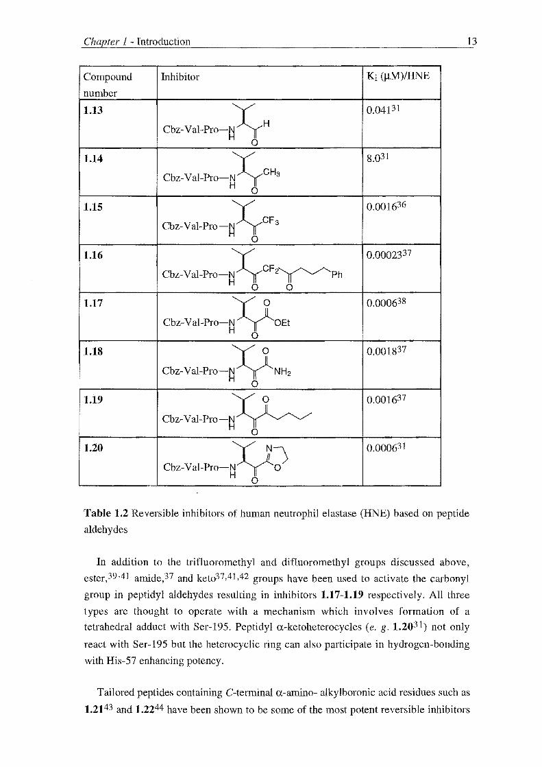
Table 1.2 Reversible inhibitors of human neutrophil elastase (HNE) based on peptide
aldehydes
In addition to the trifiuoromethyl and difluoromethyl groups discussed above,
ester,39-41 amide,37 and ket037,41,42 groups have been used to activate the carbonyl
group in peptidyl aldehydes resulting in inhibitors 1.17-1.19 respectively. All three
types are thought to operate with a mechanism which involves formation of a
tetrahedral adduct with Ser-195. Peptidyl a-ketoheterocycles (e. g. 1.2031 ) not only
react with Ser-195 but the heterocyclic ring can also participate in hydrogen-bonding
with His-57 enhancing potency.
Tailored peptides containing C-terminal a-amino- alkylboronic acid residues such as
1.2143 and 1.2244 have been shown to be some of the most potent reversible inhibitors

1 - Introduction 14
of serine proteases45 including a-chymotrypsin (a-CT), a-lytic proteinase (ALP),
thrombin and trypsin. These compounds contain a trigonal boron with a vacant 2p
orbital which reacts with either Ser-195 (type 1) or His-57 (type 2). Type 1 boronic
acids react with Ser-195 give a covalent tetrahedral adduct which mimics the second
tetrahedral intermediate (1.4) involved in the hydrolysis of peptides (see Scheme 1.2)
and are potent inhibitors. Type 2 acids however, lack the correct geometry to optimise
binding with the enzyme's S 1 pocket and react with His-57 rather than Ser-195 and
consequently are poor inhibitors.43b
P1
CH30-SuC-Ala-Ala-prO-NA B"OH , H OH
1.21
1.22a R = (L) PhCH2
Ki = O.541lM / ALp43b
Ki = O.16nM / a_CT43a
1.22b R = i-Pr
Ki = O.57nM / HLF3a
IRREVERSIBLE INHIBITION
P1
N~ "OH Ac-(D)-Phe-Pro- B H OH
1.22
PI = (L)-Arg 44c Ki = O.04nM / Thrombin
Ki = O.045nM / Trypsin 44c
A class of irreversible inhibitor includes reactive compounds referred to as affinity
labeling agents.25,46 These contain a reactive functional group, e.g. an a-halo ketone or
isocyanate, and react directly with active site nucleophiles, generally via an SN2
alkylation or acylation. These have been used extensively in vitro for mechanistic
probing of enzyme active sites, however any potential clinical utility is undermined by
their high and therefore indiscriminate reactivity within the biological environment,
resulting in toxicity and side effects. Although clinically disadvantaged due to their
high reactivity and associated poor stability, research has nonetheless been undertaken
to develop these compounds as therapeutic agents by addressing these issues. In this
regard, the incorporation of simple organofluorophosphonates 47 and
organosulfonylfluorides48 and isocyanates49 into peptidomimetics has resulted in
compounds with increased selectivity and stability such as 1.23,47d whose inhibitory
activity against human neutrophil elastase (HNE) and porcine pancreatic elastase (PPE)
is shown below.
Although of lesser reactivity, esters react readily with serine proteases and
consequently have undergone extensive development as substrates for kinetic assays.
This provides an abundant pool of compounds suitable for the development of

1 - Introduction
X OPh Boc-Val-Pro-N P: H II OPh o
1.23
kob/[I] = 27 000 M-1s-1, HNE47d
kob/[I] = 11 000 M-1s-1 'PPE47d
15
therapeutic agents which can be realised by designing the esters such that they rapidly
acylate the enzyme giving an inactive acyl-enzyme species which has a long lifetime if
the deacylation reaction is slow. The most success has been achieved with aromatic
pivaloates50,51 however acetylsalicylic acid, aspirin, has obtained "cult" status as an
inactivator of prostaglandin synthase and cure of the common headache. 52
1.24
Stability may also be improved by masking the affinity label in a structure specific
for a particular enzyme. Imidazole N-carboxamides (e. g. 1.25)53 have been modified
by the attachment of enzyme recognition elements such as the n-hexyl group of
1.25a53b and amino acid sequences (1.25b)53c to improve enzyme selectivity.
Compounds 1.25 are examples of such "paracatalytic" inhibitors.54 Their mechanism of
inactivation involves deprotonation at nitrogen and subsequent elimination of imidazole
to give an alkylisocyanate which inhibits the enzyme.
1.25
1.25a R = C~ 13
1.25b R amino acid sequence
The design of compounds that contain latent reactivity towards enzymes, as
illustrated by the above example, has lead to the realisation of a second class of
irreversible inhibition with important clinical applications known as mechanism-based
inactivators25 of which this thesis is concerned.
1 - Introduction 16
1.3 MECHANISM-BASED INACTIVATION
Mechanism-based inactivators25 (also known as suicide inhibitors and kcat
inhibitors) are reasonably unreactive compounds which contain a latent reactive
functionality. A mechanism-based inactivator is recognized by the target enzyme as a
natural substrate. During catalysis, the latent reactive functionality in the inactivator is
unmasked by the normal catalytic machinery of the target enzyme. The reactive group
(generally an electrophilic site) becomes covalently bound to the enzyme (due to
reaction with a nucleophilic residue in the enzyme). This renders the active site
irreversibly blocked and the enzyme inactivated.
In contrast, to affinity labeling agents, mechanism-based inactivators are unreactive
compounds and this is the key feature which makes them particularly amenable to the
design of highly specific, low toxicity drugs. The potential for generating the reactive
species exclusively within the active-site of the target enzyme imparts, in principle, a
much higher degree of selectivity to mechanism-based inactivators than affinity labels.
In the ideal case only the target enzyme would be capable of catalysing the appropriate
conversion of the inactivator to the reactive species and only one inactivator molecule is
needed per enzyme molecule for inactivation. This efficiency of inactivation is an
important concept in drug design and is measured by the partition ratio.54 In the ideal
case case the partition ratio is equal to zero (no metabolites are formed per inactivation
event). If the partition ratio is greater than zero the product(s) resulting from turnover
may be hydrolysed to give a toxic product or react with other biomolecules to produce a
toxic effect.
The design of mechanism-based inactivators for a target enzyme requires a
knowledge of the enzyme mechanism and structure. As discussed earlier, the
mechanism of serine protease catalyzed peptide hydrolysis has been extensively studied
and consequently a large number of mechanism-based inactivators have been based on
electrophilic heterocycles which are capable of being attacked by the nucleophilic
active site serine hydroxyl of the serine protease. The resulting ring-opened acyl
enzyme intermediate reacts further to unmask a species that ultimately inactivates the
enzyme. Herein provides a brief review of heterocyclic mechanism-based in activators
of serine proteases illustrating design features that have been employed to increase
target enzyme specificity and more favourable in activator pharmacodynamics and
kinetics.
1 - Introduction
BENZOXAZINONES, COUMARINS, N-NITROSOAMINES
1.26
x y ~R UO~o
H3COyyyO
HCO~~L"N 3 II
1.27
1.27a R, Y = H, X = Br
kobi[I] = 170 M-1s-1 / a_CT58b
1.27b R, Y, X = Br
kobJ[I] = 3 000 M-1s-1 / a_CT58b
1.27c R = PhCH2, Y = H, X = CI
kobi[I] = >20 000 M-1s- 1 / a_CT58b
° 1.28
17
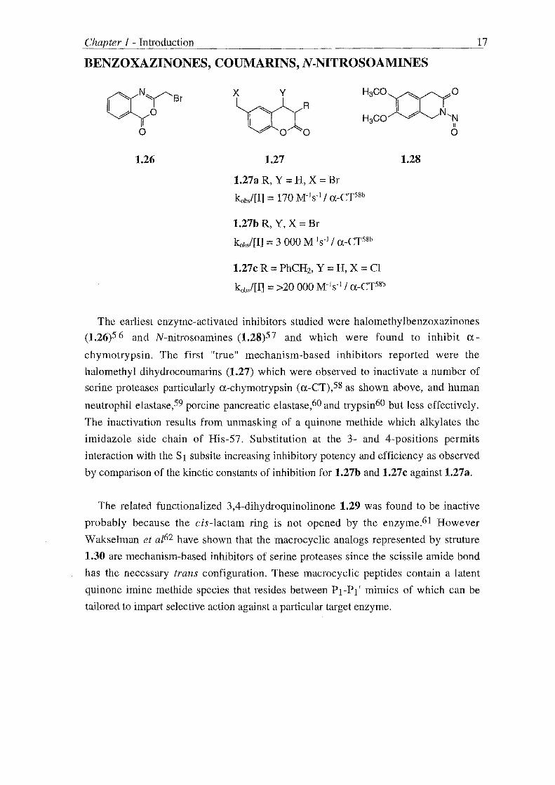
The earliest enzyme-activated inhibitors studied were halomethylbenzoxazinones
(1.26)56 and N-nitrosoamines (1.28)57 and which were found to inhibit a-
chymotrypsin. The first "true" mechanism-based inhibitors reported were the
halomethyl dihydrocoumarins (1.27) which were observed to inactivate a number of
serine proteases particularly a-chymotrypsin (a-CT),58 as shown above, and human
neutrophil elastase,59 porcine pancreatic elastase,60 and trypsin60 but less effectively.
The inactivation results from unmasking of a quinone methide which alkylates the
imidazole side chain of His-57. Substitution at the 3- and 4-positions permits
interaction with the SI subsite increasing inhibitory potency and efficiency as observed
by comparison of the kinetic constants of inhibition for 1.27b and 1.27c against 1.27a.
The related functionalized 3,4-dihydroquinolinone 1.29 was found to be inactive
probably because the cis-Iactam ring is not opened by the enzyme.61 However
Wakselman et al62 have shown that the macrocyclic analogs represented by struture
1.30 are mechanism-based inhibitors of serine proteases since the scissile amide bond
has the necessary trans configuration. These macrocyclic peptides contain a latent
quinone imine methide species that resides between PI-PI' mimics of which can be
tailored to impart selective action against a particular target enzyme.

1 - Introduction
CI
~Ph U~~O
1.29
inactive61
ISOCOUMARINS
y
rYYx l/yo
o
1.31
1.31aX = CI, Y = H
kob/[I] = 3 900 M-1s-l / HNE64
1.31b X, Y = CI
kob/[I] = 8 900 M-1s-1 / HNE65
1.30
PI = (L)-Arg
kubl[I] = 2300 M-1s-1 / urokinase62c
y
z~x l/yo
o
1.32
1.32a X = OCH3, Y = H, Z= N02
Alternate substrate68
1.32b X = OCH3, Y = CI, Z::::: NH2
kob/[I] = 10 000 M-1s-1 / HNE68
18
Isocoumarin derivatives 1.31 and 1.32 have been found to be mechanism-based
inhibitors of serine proteases particularly of human neutrophil elastase (HNE).63
Introduction of a second chlorine atom in the 4-position of the first generation inhibitor
1.31a64 gives 1.31b65 which is more potent due to increased electrophilicity of the
carbonyl group. Electrospray mass spectrometry studies of the inhibition of porcine
pancreatic elastase by 1.31b66 and proton inventories67 suggests the mechanism of
action involves an unmasked active site tethered acid chloride which, unless
hydrolysed, forms a salt linkage with His-57 to inactivate enzyme.
While 3-alkoxy-isocoumarins such as 1.32a are only alternate substrate inhibitors
lacking reactivity on ring-opening,68,69 introduction of a chlorine atom at the 4-position
gave inactivators, e. g. 1.32b, of human leukocyte elastase.67,68 The mechanism of
inactivation was found by X-ray analysis of a porcine pancreatic elastase-1.32b
complex69,70 to differ from that of the simple isocoumarins 1.31 in that an unmasked
quinone imine methide irreversibly alkylates His-57. The specificity of 1.32 can be
tailored by modifying the 3-alkoxy substituent such that 1.32b and 1.32c were found to

1 - Introduction 19
be more re,active towards human neutrophil elastase and a-chymotrypsin
respectively.67,68
HALOENOL LACTONES
1.33
1.33a R = Ph, X = Br
kinact/[I] = 23 M-1s-1 / a-CT72
1.34
1.34a R = Ph, X = Br
kinact/[I] = 73 000 M-1s-l / a-Crn
1.34b R = i-Pr, X = Br
(E)- kinact/[I] = 14500 M-1s-1 / HNE77
(Z)- kinac/[I] = 37 500 M-1s-1 / HNE77
1.34c R = Ph, X = I kinact![I] == 130 000 M-1s-l / a-CT72
1.34d R = naphthyl, X = I kinacJ[I] == 292 000 M-1s-1 / a-CT72
Both mono a-substituted 5- and 6-membered haloenollactones (1.33 and 1.34) have
been found to be mechanism-based inhibitors of serine proteases.7 1,72 These com
pounds were first proposed by Rand073 however it was a decade later that an efficient
synthetic route was developed.74 The mechanism of action involves ring-opening
followed by a tautomeric shift to reveal an a-halo ketone75 which, as indicated by
reactivation studies and molecular mechanics analysis, irreversibly alkylates the His-57
imidazole.75,76
The potency and efficiency of inactivation is related to the lactone ring size,
geometry, halogen and substitution. Six membered haloenollactones are more effective
than five membered rings (compare 1.33a and 1.34a) which may result from the fact
that the reactive a-halo ketone is on a longer tether and has better access to His-57. The
olefin geometry has a relatively moderate effect with (Z)-enols being more potent than
the corresponding (E)-enols [compare 1.34b (E) and (Z)] as does the halogen with iodo
enol lactones being more potent than bromo enol lactones (compare 1.34a and c).
Judicious modification of the a-substituent can impart selectivity of action. Haloenol
lactones with aryl a-substitutents (1.34a, c, and d) target specifically a-chymotrypsin 72
while those with an a-isopropyl substituent (1.34b) are selective for human neutrophil
elastase (HNE).77 The most potent inhibitor in the monosubstituted series, 1.34d, is

20
also very efficient with a partition ratio equal to 1.7. As a strategy for increasing
specificity haloenollactones have been incorporated into psuedo dipeptides.78,79 The
trans peptidyl haloenol lactone 1.35,79 which contains a proline-valine peptide
sequence typical of elastase substrates, has been found to be an effective in activator of
a-chymotrypsin (a-CT) and human neutrophil elastase (HNE) with specificity superior
to other mono substituted haloenollactones.
~/o Dyn-nu , oloXvBro
1.35
kob/[I] ::: 8 000 M-1s-] / a-CT79
kob/[I] ::: 1100 M-1s-I / HNE79
The "benzo" lactones represented by structures 1.36-8 were proposed to act by
unmasking a latent a-halo ketones as postulated for the halo enol lactones. The
isobenzofuranone derivative 1.36 and the isocoumarin 1.37 are not effective substrates
of serine proteases and hence the latent reactivity cannot be released. Although
isobenzofuranones of type 1.38 are potent inhibitors, the observation of biphasic
inactivation kinetics and the results of reactivation studies indicate that due to their poor
stability the benzopyran-l,4-dione is responsible for inactivation via a non mechanism
based acylation process.80
o ~~ ~ X R
1.36
Inactive80
1.37
Inactive80
o
0;0 Br CHBr-R
1.38
Acylating agent80

1 - Introduction
YNENOL LACTONES
R h //R'
A~~ o 0
1.39
1.39a R, R' = H
inactive82
1.39b R = PhCH2, R' = H
kinacJ[I] 7 600 M-1 S -1 / HNE82
1.39c R = PhCH2, R' = CH3
kinact/[I] 2.4 M-1s-1
/ HNE65
R
Y"l ~ #' OAO~
lAO
1.40a R = PhCH2
kinacJ[I] = 28 000 M-1s-1
/ HNE82
21
Five and six membered ynenol lactones represented by structures 1.39 and 1.40
respectively have been found to be mechanism based inhibitors similar to haloenol
lactones of serine proteases.81 ,82 The chemical competence of the mechanism of action
has been examined83 and involves unmasking of a reactive allene ketone which
irreversibly alkylates the enzyme although the site remains unknown. Five and six
membered and (E) and (Z) ynenol lactones are similarly equipotent. Substitution at the
a-position is required for inhibition (compare 1.39b and 1.39a) however substitution of
the acetylene reduces the rate of inactivation or eliminates it completely (compare 1.39c
and 1.39b). This is thought to be because of decreased rate of conversion to the reactive
allenone and/or active site geometries restricting alkylation.

1 Introduction
SUCCINIMIDES
R ~4
oANAo I
OL
1.41
1.41a R = i-Pr, L S02CH3
kobs/[I] = 3817 M-1s-1 / HLE87a
1.41b R = i-Pr, L = S02trans-styryl
kobs/[I] = >100 000 M-1s-1/ HLE87a
1.41c R ::; i-Bu, L = PO(OCH2Ph)
kobs/[I] ::; 6 180 M-1s-1 / HLE89
Acylating agent89
22
The series of succinimide inhibitors represented by structure 1.41 are unique in that
the reactive intermediate is generated through molecular rearrangement. 84,85 Proton
NMR studies support a mechanism which proceeds with reaction of Ser-195 to give a
ring-opened N-sulfonyloxycarboxamide which undergoes a Lossen rearrangement86 to
generate a reactive isocyanate if the N-substituent is a good leaving group. The reactive
isocyanate then acylates His-57 to form a doubly bound inactive enzyme-inhibitor
adduct. SAR study of 1.41 undertaken by Groutas et a187 has shown selectivity can be
obtained by modification of the C-3 substituent that is proposed to interact with the S 1
binding site of the enzyme. Introduction of a trans styry I group as the N-substituent
resulted in a dramatic increase in inhibitory activity due to proposed interaction with
the extended Sn' binding sites (compare 1.41a and 1.41b).
N-Phosphoryloxyimides are also known to undergo a Lossen rearrangement on
treatment with nucleophiles88 and hence compounds of type 1.41c were also examined
as mechanism-based inhibitors. However 31p NMR studies indicated these compounds
were only potent acylating agents.89 N-(Sulfonyloxyphthalimides) 1.42 have also been
found to inactivate serine proteases with a mechanism established by flourescence
spectroscopy to involve the Lossen rearrangement.90,91

1 - Introduction
o R~ ~N-OL
o
1.42
1.42a L = S02CH3' R = NH2
kob/[I] = 110000 M-1s-1 / HLE91
23
Other heterocyclic compounds, e.g. 1.43-46, which contain the masked isocyanate
have been investigated as potential mechanism-based inhibitors but have been found to
be less potent than the succinimide based derivatives 1.41.92-95 Of particular note is the
reversal of the trend in activity observed with the succinimide series in the
dihydrouracil inhibitors 1.44.93 Here incorporation of a trans-styryl group results in
less potent inhibition thought to reflect the increased steric demands of these six
membered inhibitors.
1.43
1.43aX, Y, Z = C
kob/[I] = 180 M-1s-l / HLE92
1.43b X =N, Y, Z = C
kob/[I] = 64 M-1s-1 / HLE92
1.43c Y = N, X, Z = C
kob/[I] = 58 M-1s-l / HLE92
1.43d X, Z =N, Y = C
kob/[I] 20 M-1s-l / HLE92
Ph 0
N~N-OS02R b~
o
1.45
R trans-styryl
Alternate substrate94
1.44
1.44aR= CH3
kob/[I] 3310 M-1s-1 / HLE93
1.44b R = trans-styryl
leab/[l] = 480 M-1s-1 / HLE93
o
~N-OS02CH3 Ph N~
Ph
1.46
Acylating agent95

1 - Introduction 24
SACCHARIN
Mechanism-based inhibition of serine proteases by saccharin derivatives was first
reported by Blasta et aZ96 based on the premise that incorporation of a leaving group
into known acylating agents,97 such as 1.47, would permit further inactivation.98
Groutas et aZ99 arrived at the design of inhbitors of type 1.48 on consideration of a
possible mechanism of inactivation based on the known chemistry of saccharin and its
derivatives towards nucleophiles.
o
01-v-N02
o 0 N02
1.47
1.48
L = halogen
AN =-N~
= -O~S"Ph o
(L)-Leu-N-Cbz
The potency of these compounds depends on the electron withdrawing properties of
the leaving group and modification of the saccharin nucleus to act as an enzyme
recognition element. Preliminary SAR studies undertaken by Hlasta et al99 identified 1-
phenyl mercapto tetrazole as a favourable leaving group while computer modelled
"docking" studies of substituted saccharins with human leukocyte elastase predicted
that substituents at the 4-position would interact with the Sl specificity pockets of
serine proteases resulting in the design of compound 1.49.103 Judicious choice of the 4-
substituent may therefore enhance potency as was observed on incorporation of a 4-iso
propyl substituent (compare 1.49a and 1.49b). The kinetic constant Ki* serves as a
measure of inhibitory activity for the following saccharin derivatives. A low value
corresponds to potent inhibition. All values quoted are for inhibition of human
leukocyte elastase.

1.49
1.49aR==H
1.49b R = i-Pr
K/ == O.3nM, t1/2 = 45min107
1.49c R = i-Pr, R' = OCH3
K/ = O.27nM, t1/2 260min107
25
Further SAR study included replacement of the mercapto tetrazole with aryl
ethers101 and phosphate esters, 102 a novel class of leaving group. Based on the results
of Smith et al103 with related compounds, aryl carboxylates were examined as leaving
groups resulting in the selection of 2,6-dichlorobenzoate (as in 1.50) based on its ability
to confer superior in vitro activity and stability. 104
1.50
1.50a R' = H
Ki* = O.03nM, t1/2 = 30min107
1.50b R' == OCH3
Ki* = O.023nM, t1/2 = 140min107
Clinical development of these saccharin derivatives has met the same hurdles
experienced with other mechanism-based inhibitors of serine proteases in that in vivo
activity by oral administration has been difficult to achieve because of poor hydrolytic
and metabolic stability.1 05 A probable cause of the low stability is the reactivity of
compounds 1.48-50 towards esterases. 106 It has been hypothesised that these
preferentially attack the carbonyl of the saccharin nucleus and hence increasing the
electron density and/or the sterk hindrance around this carbonyl would reduce esterase

1 - Introduction 26
cleavage and therefore improve stability.107 Introduction of an alkoxy group at the 6-
position (1.49c and 1.50b) lead to a dramatic increase in stability, due to combined
electronic and steric effects, and potency (Kt = O.OSnM) due to hydrogen bonding
between the C-6 alkoxy group and Val-216 of human leukocyte elastase. 108 However,
these compounds are very lipophilic with poor aqueous solubility and hence have poor
in vivo activity. To improve bioavailability, compounds such as 1.51 with aqueous
solubilizing substituents were prepared and though a decrease in stability was observed,
1.51 displays both potent in vitro and in vivo activity,l09
1.51 K/ = O.013nM, t1l2 = 36min109
Compounds with a modified saccharin skeleton have also been examined for
inhibitory activity against human leukocyte elastase. A class of inhibitors based on a
tetrahydrosaccharin heterocycle was shown to have different SAR than the
corresponding saccharin inhibitors with increased inhibitory activity being predicted by
modelling and observed with less sterically demanding C-4 substituents. Compound
1.52 was the most potent inhibitor in the series displaying similar activity to the bicyclo
derivative 1.53,110
1.52 1.53
Kt = O.03nM llO Kt = O.SnMll0
The biochemical rationale underlying the design of compound 1.54, a potential
mechanism-based inhibitor of human leukocyte cathepsin G (Cath G),111 is based on
the Gabriel-Colman rearrangement of either phthalimido- or saccharino-acetic esters or
ketones. I 12 Preliminary kinetic studies support the proposed mechanism which involves
Ser-195 induced ring-opening of the heterocycle and a prototropic shift of the resulting
imide anion to a carbanion. Subsequent elimination of the leaving group (L) gives a

1 - Introduction 27
reactive imine that undergoes a Michael reaction with His-57 to give doubly bound
inactive enzyme inhibitor. Compounds 1.55 and 1.56 have been found to be
mechanism-based inhibitors ofthe same ilk.113,1l4
1.54
L ::= leaving group
Z ::= activating group
1.54a L ::= F, Z = S02Ph
kob/[I] ::= 60 M-1s·1 / Cath Glll
1.56
1.55
L ::= leaving group
Z ::= activating group
1.55a L ::= F, Z = S02Ph
kobi[I] ::= 420 M-ls- l / HLEl13
kobsl[I] ::= 100 M-1s-1 / HLE1l4
Compounds of type 1.58, related to the saccharin example, have been found to be
mechanism-based inhibitors of serine proteases. 115,1l6 The mechanism of action of
these derivatives involves Ser-195 induced ring-opening of the heterocycle to give a
reactive electrophilic species that irreversibly alkylates the enzyme. In addition to
increasing the inhibitory activity by tailoring the R substituent and leaving group L to
interact with the Sl and Sn' subsites respectively of the enzyme the design of L has
considered the pathophysiology of disease states resulting from protease/anti-protease
imbalance.

1 - Introduction
1.57
1.57a R = PhCH2, L = ibuprofen
kob/[I] = 45 M-1 s-1 / HLE1l5
1.57b R = i-Pr
L = OPO(OCH2Ph)2
kinactiKI = 95000 M-1s-I / HLE116
28
Connective tissue diseases such as emphysema are associated with elevated levels of
oxidants and are inflammatory in nature. 117 Incorporation of an anti-inflammatory
agent such as ibuprofen for release into the surrounding milieu during the inhibition
process provides a second therapeutic effect. However obtention of this "dual funetion"
comes at a cost in potency as seen by comparison of 1.57a115 and 1.57b. 1l6
Based on the knowledge of the chemistry of the compounds represented by structure
1.58, these were proposed to be mechanism-based inhibitors of serine proteases. 118
1.58
1.58a RI = PhCH2, R2 = PhCH2
L == OPO(OCH2Ph)2
kinacl[l] == 6 000 000 M-1s-1 / HLE116
A noteworthy feature of the design process of these compounds was the use of
available X-ray crystal structures of human leukocyte elastase and inhibitor complexes
to model the binding of 1.58 in the active site. These studies suggested that the
heterocyclic ring may act as a highly effective peptidomimetic suitable for appending
peptidyl and nonpeptidyl recognition elements allowing the optimization of binding
interactions with the Sl sub site (through modification of Rt) and the S2-n subsites
(modification of R2). The choice of a good leaving group, (L = halogen, S02R, 02CR,
N-protected amino acids, phosphate esters),1l6,118,119 directly increases potency but L
may also be choosen such that interaction with the Sn' sub sites can be achieved
enhancing activity.

29
The optimisation of RI, R2 and L has lead to some of the most potent inhibitors of
serine proteases for example compound 1.58a116 which mimics the preferred PI residue
(valine) (Rl :::: CH(CH3h) and Pn and Pn' bulky residues of human leukocyte elastase
[R2 = PhCH2, L:::: OP(O)(OCH2Ph)]. Due to the dense functionalisation present in 1.58
this class of inhibitor could be considered to be general in nature as different serine
proteases can be targeted by judicious modification of Rl, R2 and L. The high stability
and efficiency (the partition ratio of compound 1.58a was found to be ca zero) of these
inhibitors enhances their suitability as potential in vivo modifiers.
CEPHALOSPORINS
(<;?)n R S
~5)3 L
a ~ R'
1.59
Inhibitors containing a ~-lactam nucleus are probably the most intensively studied
mechanism-based inhibitors of serine proteases and are unique in that they have been
used clinically for disease modification. The rationale underlying their design was
based on elucidation of their mode of action as antibiotics which inhibited bacterial
serine proteases 120 and ~-lactama.')es.l21 The first reported inhibitors were
cephalosporins represented by structure 1.59.122 These form a tetrahedral intermediate
on reaction with Ser-195 which collapses with loss of the C-3' leaving group to form a
reactive imine tethered in the active site which unless hydrolysed, slowly but
irreversibly alkylates His-57 to give one of three possible doubly bound inactive
enzyme-inhibitor complexes depending on the nature of the C-7 substituent.123-126
SAR study by the Merck groupI24,127-130 indicated activity was optimised by
modification of the substitution at the 2-, and 7-positions: the C-2 substituent is thought
to allow interaction with the Sn' sub sites while the C-7 substituent aetivates the lactam
ring towards nucleophilic attack and if it has the correct orientation (a-isomer) is
believed to bind in the enzyme's S1 specificity pocket. The oxidation state of the C-5
sulfur atom is also important with sulfones being more active than sulfoxides or
sulfides. Variations of the leaving group at the C-3' position did not significantly alter
activity. Whereas 1.60 was found to be the most potent in vitro inhibitor 1.61 was more
potent in vivo when administered intratracheally. The increased in vivo potency was
thought to be due to the C-2 amide imparting aqueous stability and hence optimisation
of both stability and potency lead to the synthesis of 1.62. This optimum compound still

30
lacked systemic activity due to poor stability in blood and a remedy was sought in the
investigation of simple moncyclic ~-lactams.131
CI O~ p
~P'~o~ o Ot-Bu
H CO O~ P 3', S
oH:&.OAC o NHt-Bu
1.60 1.61
kob/[I] = 161 000 M-1s- 1 / HLE128 kob/[I] = 2 200 M-1s-1 / HLE130
1.62
kob/[IJ = 3800 M-1s-1 / HLE 130
MONO CYCLIC P-LACTAMS
Two classes of azetidinones have been developed as mechanism based inhibitors of
serine proteases such as elastase132 and thrombin)33 Human cytomegalovirus
protease134 has also been targeted for potential treatment of herpes and inhibition
studies coupled with X -ray crystallography have lead to the discovery that this enzyme
ha'i a novel mechanism of hydrolysis which involves a catalytic triad of Ser-132, His-63
and His-157. 135 The first class (I) represented by structure 1.63136 has an electron
withdrawing group on the nitrogen and leaving group at the C-4 position: in the second
class (II) (1.64)136 the nature of the substituents is reversed. The binding and
mechanism of action were proposed131 ,137 to be the same as that described above for
the cephalosporin derived inhibitors excepting that class II azetidinones only form a
singly bound enzyme-inhibitor adduct which is functionally irreversible.138 The C-3
substituent occupies the Sl subsite of the target enzyme while that of the nitrogen atom
interacts with the Sn' subsites.136

·fuln"',,·y 1 - Introduction 31
1.63 1.64
kob/[I] = 5 615 M-1s-l I HNE136 kob/[I] = 5 615 M-1s-1 I HNE136
Structure-activity optimisation has lead to some of the most potent in activators of
human neutrophil elastase reported. Azetidinone 1.65139 is only 2.5 times less reactive
than the natural human neutrophil elastase inhibitor aI-proteinase inhibitor (kinact/KI = 2 200 000 M-Is-I)140 and possesses improved stability relating to the N-carbamoyl
group and the dialkyl substitution at C_3.139,141 Azetidinones related to 1.65 are among
the few compounds reported to be orally active human neutrophil elastase
inhibitors. 142,143
1.65
kob/[I] 867 000 M-1s- i I HNE139
This thesis is concerned with the study of substituted succinimide and glutarimides
which are potential mechanism-based inhibitors of the serine protease a-chymotrypsin.
Through the employment of general synthetic routes to these compounds it is a goal to
prepare a number of derivatives so that the relation between the extent and type of
functionalisation and substitution and inhibitory activity can be determined on assay
against a-chymotrypsin.

CHAPTER 2
SUBSTITUTED N-[(ACYL AND ALKYL ANF ARYL
SULFONYL)OXY]IMIDES

Chapter 2 - SubstitutedN-[(acyl and alkyl and aryl sulfonyl)oxy]imides 33
2.1 SYNTHETIC STRATEGY
A cornerstone of contemporary organic. synthesis is the principle of so-called
retrosynthetic analysis. The application of retrosynthetic analysis involves the
disconnection of larger target molecules into smaller molecules the chemist can
recognise as freely available starting materials either from a commercial or synthetic
source. There may exist several ways to disconnect a molecule on paper providing a
"tree" of alternatives for its synthesis. The route chosen is a product of logical
reasoning, based on the chemist's knowledge of reactivity and chemical reactions, and
intuition.
The potential mechanism-based inhibitors of serine proteases, 1.41, 2.1, and 2.2,
contain two common structural elements: an imide skeleton essential for enzyme
recognition and a reactive moiety essential for activity. Due to this similarity it could be
perceived that each inhibitor be prepared using slight modifications of one central
synthetic strategy using similar pools of precursors. In order to design such a strategy
retrosynthetic analysis of 1.41 was undertaken, being representative of the other target
inhibitors 2.1 and 2.2.
R 3h4
oANAO I
OL
1.41
L=S02R'
=C(O)R'
Rn'R R" 3 4
o ~ 0
X
2.2
x = leaving group
Rn'R R" 3 4
o N 0 I
OL
2.1
L=S02R"'
= C(O)R'"
The aim of the following discussion is to present briefly those disconnections that
exemplify the points and features that were considered in a desire to cultivate an

Chapter 2 - SubstitutedN-[(acyl and alkyl and aryl sulfonyl)oxy]imides 34
attractive and aesthetic general synthetic strategy. When considering the preliminary
disconnections of a target molecule a commonly employed strategy is to disect the
molecule in half. This often cultivates a shorter, more convergent synthesis than that
which is obtained from a retrosynthetic analysis that whittles away at the molecule's
skeleton. Following this principle, the two following retrosynthetic analyses were
examined to the exclusion of others.
FIRST RETROSYNTHETIC ANALYSIS OF 1.41
R
o~o I
Br
2.4
~o + OL
1.41
L::: S02R'
::: C(O)R'
Scheme 2.1 First retrosynthetic analysis of 1.41.
The arbitrary first disconnection that appeared favourable was to cleave the bond
between the nitrogen of the succinimide ring and the oxygen adjacent to the reactive
group L (as shown by the wavy line in Scheme 2.1). This would lead to the silver
alkylsulfonate and alkylcarboxylate salts and 2-substituted-N-bromosuccinimides 2.3 as
reagents. The retrosynthetic analysis continues with the functional group
interconversion (FGI) of 2.3 to the 2-substituted succinimides 2.4. These are readily

Chapter 2 - Substituted N-[(acyl and alkyl and aryl sulf.~o_ny,,-l-,,-)o_x~y,-,,-]_irm_· d_e_s ______ 35
prepared from either the respective succinic acid or if the substituent is an alkyl group,
from succinimide and the appropriate alkylhalide.
SECOND RETROSYNTHETIC ANALYSIS OF 1.41
The second strategy proposed for the synthesis of the target inhibitors is depicted by
the retrosynthetic analysis shown in Scheme 2.2. This proceeds by cleaving the bond
between the oxygen and the reactive moiety L which gives the 3-substituted-N
hydroxysuccinimides 2.5, and alkylsulfonic and alkylcarboxylic acid derivatives as
reagents. Disconnection of 2.5 proceeds by cleavage of the imide bonds, as indicated by
the wavy lines, to give hydroxylamine and 2-substituted succinic acid derivatives 2.6
and 2.7 as reagents.
2.6
2.6aX = Cl
2.6b X=OH
2.6c X = OR'
2.5
2.7
1.41
L= S02R'
=C(O)R'
R'S02X
R'C(O)X
Scheme 2.2 Second retrosynthetic analysis of target inhibitor 1.41

Chapter 2 - SubstitutedN-[(acyl and alkyl and aryl sulfonyl)oxy]imides 36
The above retrosynthetic analyses proposed two possible forward syntheses of the
target inhibitor. The chemical competency of each of these syntheses was then
investigated to identify the most favourable route to the target inhibitor 1.41. Of aid in
this investigation was the substantial literature describing the synthesis of succinic and
phthalic acid derivatives, however there existed little documentation, of which was
disjointed, a..'l to the synthesis of substituted succinate derivatives. An aim of this thesis,
therefore, was to make a systematic study of the preparation of these compounds.
FIRST FORWARD SYNTHESIS OF 1.41
2.8
B
A .. 0>00 H
2.9 2.4
D R
o~o I
OL
2.3 1.41
L = S02R'
L= C(O)R'
Scheme 2.3 A, 1) NaNH2, Ih, -78 DC 2) PhCH2Br, Ih, -78 DC
B, urea, 3h ca 200 DC
C, HOBr or Br2
D, AgOS02R' or Ag02CR', CH3CN, rt
C
The key 2-substituted succinimides 2.4 (Scheme 2.3) were prepared in two ways. In
the case where the R substituent of 2.4 is alkyl, it may be obtained by NaNH2 mediated
alkylation of succinimide.144 For example, 2-benzylsuccinimide 2.4 (R=PhCH2) was
prepared in 12% yield using this methodology which is discussed later in this chapter.

Chapter 2 - SubstitutedN-[(acyl and alkyl and aryl sulfonyl)oxy]imides 37
When this reaction was not applicable, as in the case of aromatic succinimides, e.g. 2.4
(R Ph), the 2-substituted succinimides were otherwise obtained by heating a mixture
of the corresponding succinic acid 2.8 with two equivalents of urea for 3h at ca 200 °C
(Step B, Scheme 2.3).1 45 Both 2-phenylsuccinimide 2.4 (R=Ph) and 2-
benzylsuccinimide 2.4 (R=PhCH2) were obtained in 65% yield in this manner.
The remainder of this synthetic route (Steps C - D, Scheme 2.3) was examined using
succinimide 2.9 as a model substrate. Due to the availability of N-bromosuccinimide
the bromination reaction of Step C (Scheme 2.3) was not immediately investigated,
however, it was anticipated that 2-substituted N-bromosuccinimides 2.3 could be
prepared following well established literature procedures. 146
The reaction of silver tosylate salts with alkylhalides to give alkyltosylates has been
documented147 but to our knowledge this methodology has not been expanded to
include a study of the reaction of halo-imides and silver sulfonate or acetate salts as a
means of preparing compounds of type 1.41. Initial studies using commercially
available N-bromosuccinimide (NBS) and silver (I) tosylate (prepared from reaction
between sodium tosylate and silver nitrate)148 failed and hence the preparation of the
target inhibitors 1.41 using the route displayed in Scheme 2.1 was abandoned.
SECOND FORWARD SYNTHESIS OF 1.41
The synthesis of the target inhibitor 1.41 was carried out according to the route
shown in Scheme 2.4.
2.6
2.6a X = CI
2.6bX OR
2.6cX= OR'
R
o~ 2.7
A
2.5
Scheme 2.4 A, H2NOH B, R'S02X or R'C(O)X
B
1.41
L= S02R'
L= C(O)R'

Chapter 2 - Substituted N-[(acyl and alkyl and aryl sulfonyl)oxyJimides 38
The intermediate 3-substituted N-hydroxysuccinimides 2.5 were prepared from 2-
substituted succinic acid derivatives 2.6 and 2.7 by reaction with hydroxylamine. It is
possible that on reaction of 2.6 and 2.7 with hydroxylamine (Step A, Scheme 2.4)
mono- and di-acylation could occur at either the oxygen or nitrogen atom to give a
mixture of products. 149 Jencks, however, has shown that acylation of the amino group
of hydroxylamine does not usually require protection of the hydroxyl group.1 50
However, as will be discussed, it proved more practical to utilise an O-protected form
of hydroxylamine - O-benzyloxyamine - in many cases. The choice of whether to use
hydroxylamine or O-benzyloxyamine was found to depend on the type of 2-substituted
succinic acid derivative used. Hence reaction of 2.6 or 2.7 with either hydroxylamine or
O-benzyloxyamine gave 3-substituted N-hydroxysuccinimides 2.5. Base mediated
coupling of these with alkylsulfonic and alkylcarboxylic acid derivatives (in the
presence of an acylation catalyst) (Step B of Scheme 2.4) then gave the desired
compounds 1.41.
A detailed discussion of the preparation of the succinimide inhibitors 1.41 according
to Scheme t.4 follows, beginning with the synthesis of 2-substituted succinic acids
which were prepared using three methods.

Chapter 2 - SubstitutedN-[(acyl and alkyl and aryl sulfonyl)oxy]imides 39
2.2 THE SYNTHESIS OF 2-SUBSTITUTED SUCCINIC ACIDS
METHOD A: MALONATE ESTER ROUTE
Monosubstituted succinic acids have been traditionally prepared using malonate ester
condensation reactions of the type shown in Scheme 2.5. 151 A range of 2-substituted
succinic acids can be obtained employing this methodology using different malonic
esters as starting materials.
A
2.10
C
2.12
..
Ph~02Et
Et02C C02Et
2.11
2.13
Scheme 2.5 A ethyl bromoacetate, NaOEt I EtOH, reflux 14.25h
B KOH I EtOH, reflux 4h
C 2.25h at 160-170 °C
B
Our initial goals were aimed at obtaining a good supply of 2-benzylsuccinic acid as
the corresponding inhibitors of type 1.41 (R = PhCH2, L = S02R') have been found to
be more specific for our target enzyme a-chymotrypsin (a-CT) than those with other
substitution (compare the inhibitory activities of 1.41a and 1.41d towards a
chymotrypsin which are shown below). 84
R
o~o J
OL
1.41
1.41a R = i- Pr, L = S02CH3
kob/[I] = 1 050 M-is·il a-CT84
1.41d R = PhCH2, L = S02CH3
kob/[I] = 9 000 M-ls·i; a-CT84
Chapter 2 - SubstitutedN-[(acyl and alkyl and aryl sulfonyl)oxy]imides 40
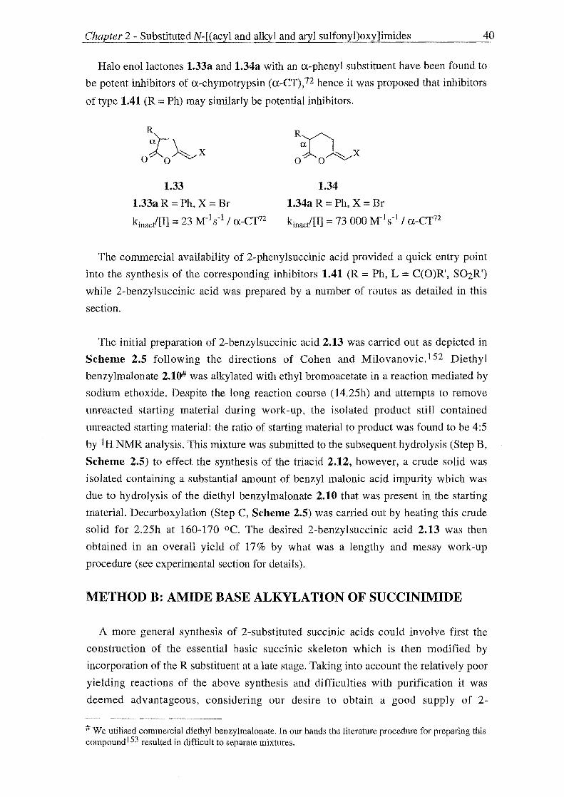
Halo enollactones 1.33a and 1.34a with an a-phenyl substituent have been found to
be potent inhibitors of a-chymotrypsin (a-CT),n hence it was proposed that inhibitors
of type 1.41 (R = Ph) may similarly be potential inhibitors.
R
~x o 0
1.33
1.33a R = Ph, X = Br
kinac/[I] = 23 M-1 s -1 / a-Cr72
1.34
1.34a R = Ph, X = Br
kinac/[I] = 73000 M-1s-1 / a-CT72
The commercial availability of 2-phenylsuccinic acid provided a quick entry point
into the synthesis of the corresponding inhibitors 1.41 (R = Ph, L = C(O)R', S02R')
while 2-benzylsuccinic acid was prepared by a number of routes as detailed in this
section.
The initial preparation of 2-benzylsuccinic acid 2.13 was carried out as depicted in
Scheme 2.5 following the directions of Cohen and Milovanovic.1 52 Diethyl
benzylmalonate 2.10# was alkylated with ethyl bromo acetate in a reaction mediated by
sodium ethoxide. Despite the long reaction course (14.25h) and attempts to remove
unreacted starting material during work-up, the isolated product still contained
unreacted starting material: the ratio of starting material to product was found to be 4:5
by IH NMR analysis. This mixture was submitted to the subsequent hydrolysis (Step B,
Scheme 2.5) to effect the synthesis of the triacid 2.12, however, a crude solid was
isolated containing a substantial amount of benzyl malonic acid impurity which was
due to hydrolysis of the diethyl benzylmalonate 2.10 that was present in the starting
material. Decarboxylation (Step C, Scheme 2.5) was carried out by heating this crude
solid for 2.25h at 160-170 oC. The desired 2-benzylsuccinic acid 2.13 was then
obtained in an overall yield of 17% by what was a lengthy and messy work-up
procedure (see experimental section for details).
METHOD B: AMIDE BASE ALKYLATION OF SUCCINIMIDE
A more general synthesis of 2-substituted succinic acids could involve first the
construction of the essential basic succinic skeleton which is then modified by
incorporation of the R substituent at a late stage. Taking into account the relatively poor
yielding reactions of the above synthesis and difficulties with purification it was
deemed advantageous, considering our desire to obtain a good supply of 2-
# We utilised commercial diethyl benzylmalonate. In our hands the literature procedure for preparing this compound153 resulted in difficult to separate mixtures.

Chapter 2 - SubstitutedN-[(acyl and alkyl and aryl sulfonyl)oxy]imides 41
benzyl succinic acid, that cleaner and more direct preparations of 2-alkylsuccinic acids
be examined.
Bryant and Hauser144 reported the potassium amide mediated benzylation of
succinimide 2.9 to give 2-benzylsuccinimide 2.14 from which 2-benzylsuccinic acid
2.13 can be obtained by acid hydrolysis (Scheme 2.6). The preparation of 2.14 was
carried out in the reported low yield of 29% due to competing reactions. A substantial
amount of higher C-alkylated product and a quantity of stilbene (generated from the
self-condensation of benzyl chloride - a reaction which is quite facile under these
conditions) were also obtained.
o~o A
H
2.9
Ph~
OAN~O H
2.14
B
Scheme 2.6 A, 1) KNH2 or NaNH2, NH3(l) 2) PhCH2X (X = Br, CI), NH3(l)
B, 6N HCI, reflux 1d
2.13
There is literature precedent that the yield of the key reaction (Step A) of Scheme 2.6
may be optimised by changing the reaction conditions.144,154 The yield of the mono
alkylated product appears to be dependent on the reaction temperature. Bryant and
Hauser isolated only higher C-alkylated products when the preparation of 2.14 was
carried out at higher temperatures in refluxing Et20 or toluene. Description of the
apparatus used for the above authors' preparation of 2.14 in 29% yield indicated the
reaction was carried outin refluxing ammonia (-33 OC) hence a higher yield (>29%) of
monoalkylated product may be realised if lower temperatures are used i.e. the reaction
is carried out at <-33 °C.
Wolfe and Rodgers154b report improved yields for the benzylation of disodio
glutarimide when potassium amide (65%) is replaced with sodium amide (80%).
Greater success was also observed when a higher molar equivalency of amide base and
longer alkylation periods were employed. The replacement of benzyl chloride with
benzyl bromide may also result in a higher yielding reaction. This advantage may be
offset in that the bromide may activate self condensation, producing greater amounts of
stilbene, relative to the chloride.
In consideration of the above the following procedure for the preparation of 2-
benzylsuccinimide was used. Succinimide 2.9 was added to a solution of sodium
amide155 in liquid ammonia to give disodio succinimide which was benzylated on

Chapter 2 - Substituted N-[(acyl and alkyl and aryl sulfonyl)oxy]imides 42
addition of a solution of benzylbromide in ether to give pure 2-benzylsuccinimide 2.14
in 12% yield. Acid hydrolysis of 2.14 with 6N Hel over a day period then gave 2-
benzylsuccinic acid 2.13 in quantatative yield. Although the yield of the monoalkylated
product was low, the reaction provides a direct and short two step synthesis of 2-
alkyl succinic acids. This route is also versatile in that it allows the preparation of a
number of different 2-alkylsuccinic acids by simply substituting the alkylating agent
used.
METHOD C: STOBBE CONDENSATION ROUTE
The Stobbe condensation 156 has been employed as a general method for the
preparation of 2-substituted succinic acid derivatives. 157 Groutas et a187a have reported
the preparation of 2-alkylsuccinic acids by the route shown (Scheme 2.7) using a
potassium t-butoxide (t-BuOK) mediated Stobbe condensation between the appropriate
aldehyde and diethyl succinate followed by reductive hydrogenation and hydrolysis.
Subsequently the preparation of 2-benzylsuccinic acid was undertaken using this route.
Sodium ethoxide was used to mediate the condensation between benzaldehyde (2.15)
and diethyl succinate (2.16) (Step A, Scheme 2.7) to give 2.17. Reductive
hydrogenation of 2.17 gave the corresponding saturated 2-benzy lsuccinic acid
monoethyl ester (2.18) as a yellow oil (60%). Hydrolysis of this ester was attempted a
number of times by treatment with ethanolic NaOH as prescribed by literature
procedures l52,157d however this was unsuccessful. Hence more vigorous reaction
conditions were employed and 2-benzylsuccinic acid 2.13 was successfully obtained in
82% yield (overall yield 37%) by refluxing 2.18 in ethanolic KOH for Sh.

Chapter 2 - SubstitutedN-[(acyl and alkyl and aryl sulfonyl)oxy]imides
° Ph~ + 1\ Et02C COzEt
H
2.15 2.16
C
2.18
A
..
Scheme 2.7 A NaOEt I EtOH, reflux 12h
B. H2/1O% Pd-C I EtOAc 19h
C. KOH I EtOH, reflux 5h
SUMMARY
2.17
2.13
43
B ..
The preparation of 2-substituted succinic acids by Method C (Scheme 2.7) as
represented by the synthesis of 2-benzylsuccinic acid 2.13 gave the best overall yield
(37% over three steps). The malonate ester route Method A (Scheme 2.5) gave 2.13 in
an overall yield of 16% over three steps while 2.13 was prepared in an overall yield of
12% in two steps using Method B (Scheme 2.6). On this basis, the method of choice
for the preparation of 2-substituted succinic acids is Method C. However the short two
step synthesis of Method B and its general nature make it more attractive excepting the
low yielding alkylation reaction (Step B, Scheme 2.6). The chemistry and practice of
this reaction was not as established as those in Method C, however there is literature
precedent to suggest that optimisation of this reaction is possible and once undertaken
Method B could provide the ideal route to the desired 2-alkylsuccinic acids.

Chapter 2 - SubstitutedN-[(acyl and alkyl and aryl su!fony"-I.L)0_x-"y-"-Jl_·1ll1_·_d_es ______ 44
2.3 THE SYNTHESIS OF 3-SUBSTITUTED-NHYDROXYSUCCINIMIDES
The next step towards the preparation of the desired inhibitors 1.41 is the synthesis
of the intermediate 3-substituted-N-hydroxysuccinimides 2.5 from the corresponding 2-
substituted succinic acid derivatives 2.6 and 2.7 (Scheme 2.8) by reaction with either
hydroxylamine (conditions A) or O-benzyloxyamine (conditions C). The following
discussion presents an investigation of the preparations of 3-alkyl-N
hydroxysuccinimides from 2-alkylsuccinic acid derivatives in order of increasing
utility.
2.6
2.6a X = CI
2.6bX=OH
2.6c X = OR'
2.7
Scheme 2.8 A H2NOH
A
B ...
B H2NOCH2Ph
C H2, Pd catalyst
D R'S02X or R'C(O)X
DIESTER
R
o~o , OH
2.5
jc R
o~o I
OCH2Ph
2.19
R D 0);).0 ..
I
OL
1.41
L::::S02R'
L:::: C(O)R'
The use of dicarboxylic acid diesters of the type 2.6c in reactions with 0-
benzyloxyamine favours formation of monoacylated N-benzyloxyhydroxamic acids
rather than the desired N-benzyloxyimides. N-Hydroxyphthalimide, however, has been
obtained in a one step synthesis by acidification of the sa1t obtained from reaction of
diethyl phthalate with hydroxylamine.I58 Reaction of diethyl 2-benzylsuccinate with an
ethanolic hydroxylamine solution failed, however, to give the proposed salt and
attempts to obtain 3-benzyl-N-hydroxysuccinimide 2.5 (R = PhCH2) using this
methodology was abandoned.

Chapter 2 - SubstitutedN-[(acyl and alkyl and aryl sulfonyl)oxy]imides 45
DIACID
It is possible to prepare imides from the diacid e.g. 2.6b and amine constituents
under relatively harsh dehydrating conditions. For example 2-benzylsuccinimide 2.14
was prepared by heating a mixture of 2-benzylsuccinic acid 2.13 and 2 equivalents of
urea for 3h at 170-200 °C (Scheme 2.9). However, it was thought unlikely that a
desirable preparation of 3-substituted N-hydroxysuccinnimides 2.5 could be realised by
the reaction between the appropriate 2-alkylsuccinic acid and hydroxylamine
hydrochloride despite the obvious attraction of the short one step synthesis. The
observation that hydroxylamines form explosive mixtures at elevated temperatures also
adds caution to such a synthesis. 149
A ..
2.13 2.14
Scheme 2.9 A, urea, 3h at ca 170-200 °C
A milder preparation of 3-alkyl-N-hydroxysuccinimides requires the use of activated
acid derivatives such as acid chlorides, e.g. 2.6c, anhydrides e.g. 2.7 or by activating the
diacid 2.6b in situ. A preparation of substituted N-benzyloxyglutarimides has been
reported by the EDAC/HOBt mediated condensation of O-benzyloxyamine and the
respective substituted glutaric acid.1 59 This methodology was successfully applied by
us to the preparation of a 4,4-dialkyl-N-benzyloxyglutarimide (see Section 2.7 of this
chapter) with the corresponding N-hydroxyglutarimide being obtained by the removal
of the O-benzyl protecting group by catalytic hydrogenation. The preparation of 3-
benzyl-N-benzyloxysuccinimide was attempted using this methodology but was
unsuccessful.
DIACID CHLORIDE
N-Hydroxyphthalimide has been prepared from reaction between the respective
diacid chloride and hydroxylamine. 160 A route to 3-alkyl-N-hydroxysuccinimides based
on this reaction first requires a reliable method for the preparation of substituted
succinic diacid chlorides. In general, acid chlorides are obtained by the treatment of the
parent acid with chlorinating agents for example succinyl dichloride has been obtained
by reaction of succinic acid and PC15.161 The preparation of 2-benzylsuccinyl chloride
was attempted by heating the acid with three molar equivalents of SOCh however the
isolated white solid was identified by NMR, melting point and mass spectrometry to be

Chapter 2 - Substituted N-[(acyl and alkyl and aryl sulfollyl)oxy]imides 46
the respective anhydride. A milder preparation was employed using oxalyl chloride and
DMF at room temperature but the anhydride was again obtained. Hence, in light of
these perhaps not suprising results, it was not possible to design a synthesis of 3-
substituted N-hydroxysuccinimides from diacid chlorides as their preparation favoured
activated ring closure to give the corresponding anhydride.
MONO ETHYL ESTER DERIVATIVES
The preparation of 3-alkyl-N-hydroxysuccinimides from 2-alkylsuccinic acid mono
ethyl ester derivatives was also investigated on the basis of study of the
functionalisation of the succinimide ring undertaken previously within the University of
Canterbury Department of Chemistry162 it was observed that methyl hydrogen
succinate 2.20 undergoes coupling to O-benzyloxyamine under standard DCCIHOBt
conditions to give methyl N-benzyloxysuccinamate 2.21. Treatment of 2.21 with either
Et3N or iso-propyl amine gave N-benzyloxysuccinmide 2.22 which on catalytic
hydrogenation gave 2.23.
A 1\ B • .. H3COl C COlH H3COl C CONHOCHlPh
2.20 2.21
oJ:;\o C O~O .. I I
OCHlPh OH
2.22 2.23
Scheme 2.10 A, HCl.H2NOCH2Ph, Et3N, DCC, HOBt, CH2Cb, overnight 0-5 °C-rt
B, Et3N, CH2Cl2, rt 2h
C, H2, 10% Pd-C, THF, 3h
Consequently possible preparations of substituted N-hydroxysuccinimides were
investigated by applying this methodology to substituted succinic acid mono ethyl
esters. Of the two esters available, ethyl 3-benzylidenesuccinate 2.17 and ethyl 3-
benzylsuccinate 2.18, obtained in the course of preparing 2-benzylsuccinic acid using
Stobbe condensation chemistry (see Scheme 2.7) a route to 3-benzyl-N
hydroxysuccinimide 2.26 was investigated from 2.18. The synthesis of 3-benzyl-N
hydroxysuccinimide 2.26 from 2.18 in five steps is shown below Scheme 2.11.

Chapter 2 - SubstitutedN-[(acyl and alkyl and aryl sulfonyl)oxy]imides ..
° 1\ Ph--1 +
H Et02C C02Et
A Ph~
Et02C C02H
..
2.15 2.16 2.17
Phh C .. Ph
h Et02C C02H H02C CONHOCH2Ph
2.18 2.24
Ph~ E .. o N 0
I
Ph~ o N 0
I
OCH2Ph OH
2.25 2.26
Scheme 2.11 A, NaOEt, EtOH, reflux 12h
B, H2, 10% Pd-C, EtOAc, 19h
B
D
C, HCl.H2NOCH2Ph, Et3N, DCC, HOBt, CH2Ch, overnight 0-5 °C-rt
D, p-TsOH, DCE, reflux 13h
E, H2, 10% Pd-C, THF, overnight
47
The mono ethyl ester 2.18 was prepared as discussed previously by reductive
hydrogenation of the Stobbe condensation product 2.17 in 46% yield (Steps A and B of
Scheme 2.11). Coupling of O-benzyloxyamine hydrochloride to 2.18 using standard
DCC / HOBt conditions gave a 2: 1 mixture, by 1 H NMR, of the ring opened N
benzyloxy hydroxamic acid derivative 2.24 and the ring closed N
benzy loxysuccinimide 2.25.
To complete conversion to the ring closed product 2.25 a solution of a sample of the
isolated mixture of 2.24 and 2.25 in CH2Ch was treated with triethylamine employing
the conditions used for the cyclisation of the model compound methyl N
benzyloxysuccinamate 2.21 discussed above (Scheme 2.10). Proton NMR analysis of
the isolated product indicated no further cyclisation had taken place implying that
harsher conditions were required.
To this end a neat synthetic sample of the mixturc obtained from the coupling of 2.18
and O-benzyloxyarnine hydrochloride was heated for 2h at 160 °C. The isolated product
was found by IH NMR to contain a 2:3 mixture of ring-opened 2.24 and ring-closed
2.25 implying that some further cyclisation had ocurred. The harsh conditions used

Chapter 2 - SubstitutedN-[(acyl and alkyl and aryl sulfonyl)oxy]imides 48
were deemed to be destructive considering that incomplete cyclisation took place and
therefore a milder alternative was sought.
Park et al163 have prepared a series of N-CBz-a-aminoglutarimides and
succinimides from the N-protected glutamic and aspartic acids respectively. The
ultimate step in the four step synthesis they employed involved cyclisation of methyl N
Cbz-a-aminoglutaramates and succinanamates by refluxing with 0.5 equivalent of p-
TsOH in
toluene for 8h in 65-82% yields. Consequently we tried these cyclisation conditions
only to find that the reaction mixture decomposed. However, on changing the solvent
from toluene to 1,2-dichloroethane (DCE) and refluxing for 13h we isolated the desired
ring-closed product 2.25 in 85% yield over the two steps (C and D of Scheme 2.11).
The desired 3-benzyl-N-hydroxysuccinimide 2.26 was then obtained by hydrogenolysis
of 2.25 in 72% yield (Step E , Scheme 2.11).
ANHYDRIDE
The synthesis of 3-alkylN-hydroxysuccinimides was carried out via an anhydride
using modifications of procedures adopted by Groutas et al.85 ,87
A
2.13
B ..
2.28
R' =H, CH2Ph
Scheme 2.12 A, AC20, reflux
.. Ph~
oJ.-..oAo
2.27
Ph~
OJ.-.N~O I
OR'
C [ 2.25 R' CH2Ph
2.26R' = H
B, H2NOR' (R' = H or CH2Ph), toluene or xylene, reflux
C, H2, 10% Pd-C, 1: 1 MeOHlEtOH, EtOAc, or THF
B
2-Benzylsuccinic acid 2.13, prepared as discussed earlier in this chapter, was
refluxed with excess acetic anhydride to give the anhydride 2.27 in 90% yields.

Chapter 2 - SubstitutedN-[(acyl and alkyl and aryl sulfonY"-I.L)0_x-Ly-=-]i_Inl_·d_e_s _____ --=49
However, it was found that the reaction of 2-benzylsuccinic anhydride 2.27 with 0-
benzyloxyamine was low yielding using the conditions described by Groutas et al.85
A review of the literature preparations149,164 of the model compounds N
benzyloxysuccinimide and N-benzyloxyphthalimide suggested that this reaction
proceeds by initial attack of O-benzyloxyamine with concomitant ring-opening to give
the intermediate which then undergoes ring closure with elimination of water to give the
N-benzyloxyimide. Depending on the reaction conditions (reaction time, temperature)
employed, either the ring opened intermediate or ring closed product can be obtained.
For example, N-benzyloxysuccinimide has been prepared from O-benzyloxyamine and
succinic anhydride in refluxing benzene with a reaction time of O.Sh but N
benzyloxysuccinamic acid was obtained employing a reaction time equal only to the
time required to mix the reagents.164
To ensure formation of the desired N-hydroxysuccinimides we sought to employ
longer reaction times than those used in literature preparations. Additional
improvements on the existing literature procedures were sought by employing
azeotropic removal of water from the reaction mixture to drive formation of the ring
closed N-benzyloxyimide and substituting the higher boiling xylenes (bp165 137-144
OC) for toluene (bp165 111 OC). On increasing the reaction time and employing the
azeotropic removal of water during the reaction we observed an increase in yield for the
preparation of 3-benzyl-N-benzyloxysuccinimide (78% cf 60%). On substituting
xylenes for toulene there was no observed improvement in yield, however, the work-up
of these reactions which involved evaporation of the reaction solvent proved more
difficult.
The original conditions employed by Groutas84 for the hydrogenolysis of 2.25 used a
1: 1 MeOHlEtOH solvent system, desirable because of the high solubility of H2 in both
MeOH and BtOH. Considering that both are nucleophilic it could be possible that
formation of ring-opened derivatives of type 2.28 could occur during the hydrogenation
reaction. Hence the work-up procedure includes evaporation of the solvent and then
reflux of a toluene solution of the resulting residue with azeotropic removal of water to
promote formation of the ring closed product. Substitution of 1: 1 MeOHlEtOH (61 %
yield) with the non-nucleophilic solvent THF (72%) resulted in higher yields and
simplified work-up procedures.
3-Phenyl-N-hydroxysuccinimide was also prepared using the reactions described
above (Scheme 2.13).

Chapter 2 - Substituted N-[(acyl and alkyl and aryl sulfonyl)oxy]imides
2.29
Ph
o~o I
OR'
C [ 2.31 R: = CH2Ph
2.32R =H
Scheme 2.13 A, AcCI, reflux
A
B, H2NOR' (R' = H, CH2Ph)
C, H2, 10% Pd-C, THF
2.30
50
B ..
Commercially available 2-phenylsuccinic acid (2.29) was refluxed with an excess of
acetyl chloride to give 2-phenylsuccinic anhydride (2.30) in 69% yield after
recrystallisation from toluene/petroleum ether.166 Addition of O-benzyloxyamine to a
refluxing solution of 2.30 in toluene followed by reflux with azeotropic removal of
water for 7 hours gave 3-phenyl-N -benzyloxysuccinimide (2.31) in 70% yield.
Hydrogenolysis of 2.31 in THF with 10% Pd-C for 4.5 hours gave 3-phenylN
hydroxysuccinimide (2.32) with 52% mass recovery after radial chromatography.
Despite this purification, inspection of the 1 H NMR spectrum still showed impurities.
Similar results have been observed on the hydrogenolysis of O-benzyl hydroxamates
similar to 2.31. 167 Despite using different types of palladium catalyst Pd-C (5%, 10% as
above, 20%), Pd black, 10% Pd-SrC03164 and varying the reaction pressure (l0-50psi)
the desired products were not obtained cleanly the corresponding amide being a major
impurity. The catalyst 5% PdlBaS04 proved to be more effective in the hydrogenation
of the described O-benzyl hydroxamates to the desired hydroxamic acids and hence
prompted its use to effect a clean synthesis of 2.32.
Hydrogenolysis at 50psi of 3-phenyl-N-benzyloxysuccinimide 2.31 in MeOH with
5% Pd/BaS04 for 8 hours gave 3-phenyl-N-hydroxysuccinimide 2.32 in 37% yield
(17% overall) and good purity after chromatography. Although a greatly reduced yield
was observed with this catalyst, the 1 H NMR spectrum of the crude product showed
less impurities than that of the reaction using 10% Pd-C as catalyst.

Chapter 2 - SubstitutedN-[(acyl and alkyl and aryl sulf.;.....0_nLyl....£..)_ox~yu]_im_._id_e--'-s ______ 51
The O-protected form of hydroxylamine, O-benzyloxyamine, has been preferred in
the above preparations of 2-substituted N-hydroxysuccinimides to reduce the risk of
side reactions. Jencks 150 has shown that acylation of the amino group of hydroxylamine
does not usually require protection of the hydroxyl group and hence shorter syntheses of
substituted N-hydroxysuccinimides may be perceived from reaction of the anhydrides
and hydroxylamine.
The model compounds N-hydroxysuccinimide and N-hydroxyphthalimide have been
obtained by reaction of the respective anhydride with hydroxylamine.168,169 N
Hydroxyphthalimide has been obtained by simply heating a mixture of hydroxylamine
hydrochloride, phthalic anhydride and Na2C03 in H20 for one hour.169 These reaction
conditions have also been used by Groutas in the preparation of 3-alkylthio-N
hydroxysuccinimides.87f
Both 3-benzyl- and 3-phenyl-N-hydroxysuccinimide (2.26 and 2.32) were prepared
in this manner. Hence 2.26 was prepared in 51 % yield by refluxing a solution of the 2-
benzylsuccinic anhydride 2.27, hydroxylamine hydrochloride (2 equiv.) and anhydrous
potassium carbonate (1 equiv.) for two hours and 2.32 by refluxing a solution of 2.30,
hydroxylamine hydrochloride and anhydrous sodium carbonate for five hours (82%
yield).

52 Chapter 2 - SubstitutedN-[(acyl and alkyl and aryl sulfonyl)oxy]imides ~~~----------------
2.4 THE DESIGN AND SYNTHESIS OF 3-SUBSTITUTED N-[(ACYL AND ALKYL AND ARYL SULFONYL) OXY]SUCCINIMIDES
The ultimate step in the synthesis of the target inhibitors 1.41 was the coupling of
alkylcarboxylic and alkylsulfonic acid derivatives to the 3-substituted N
hydroxysuccinimides 2.5 (Scheme 2.14).
~ ol~o
X X
R R 2.6 .. o):;Ao A 0);).0 ..
R ..
o):;Ao I I
OH OL
2.5 1.41
2.7 L== SOzR'
L == C(O)R'
Scheme 2.14 A, R'S02X or R'C(O)X
The object of this thesis was to examine the effect the L group of 1.41 had on
inhibitory activity. The design of these derivatives is discussed later but aspects
included the incorporation of favourable substitution capable of harnessing the binding
energy available through interaction with the Sn and Sn' subsites of a-chymotrypsin and
increasing the number of equivalents of reactive species released during inhibition.
The preparation of the N-[(sulfonyl)oxy]succinimide derivatives 1.41 (L == S02R') in
Table 2.1 was undertaken according to literature procedures for N-hydroxyphthalimide
or succinimide substrates,85,87,91,170 which prescribe the treatment of 2.5 with base
followed by the alkylcarboxylic and alkylsulfonic acid derivative. A variety of bases
have been used for this reaction including aqueous sodium bicarbonate and hydroxide,
and pyridine (two equivalents have been normally employed) or triethylamine if organic
solvents (benzene, dichloromethane, chloroform, toluene) have been used. Different
reaction times and temperatures have also been used with a continuum ranging from
20min at 0-5 °C followed by the same period at room temperature to room temperature
overnight to ultimately 40°C overnight.

Ctwpter 2 - Substituted N-[( acyl and alkyl and aryl sulfonyl)oxy]imides
R
o~o I
OH
2.26 R = PhCH2
2.32 R = Ph
A B C ..
R
o~o I
OL
1.41
L=S02R'
Conditions A Sulfonylation at 0-5 DC
B Sulfonylation at rt
C Sulfonylation at 50 DC
Compound R L
number
1.41d PhCH2 S02CH3
e Ph S02CH3
f PhCH2 S02 ~Ph
g PhCH2 S02-{ >
h PhCH2 S02-G-CH3
i Ph S02-G-CH3
j PhCH2 S02-{ ~ >
k PhCH2 N02
S02-{ ~ I PhCH2 CH3
~ S02 C02Et
rn PhCH2 o Ph
S020-N~ S02-{ ~ 0
n PhCH2 £(Ph o N 0
S02-{ > < )t-S026 Table 2.1 N-[(sulfonyl)oxy]succinimide inhibitors of type 1.41
Conditions
B
A
A
B
A
A
A
A
C
A
C
53

Chapter 2 - SubstitutedN-[(acyl and alkyl and aryl sulfonyl)oxy]imides 54
To effect the sulfonylation of 2.5 three distinct procedures were employed. The
mildest (Method A) involved treating an organic solution of 2.5 with either two
equivalents of pyridine or a slight excess of triethylamine followed by the appropriate
sulfonyl chloride for 0.5h at 0-5 °C and then allowing the reaction mixture to rise to
room temperature normally overnight. A second procedure (Method B) was a slight
modification of the first with the reaction being carried out at room temperature.
Dichloromethane was generally used as the reaction solvent except when solubility
problems were encountered requiring a third procedure (Method C) in which toluene
replaced dichloromethane and the reaction was carried out at an elevated temperature of
40-50°C.
When 2.26 was the substrate there was no noticeable difference in the nearly
quantatative yields observed between reactions mediated with pyridine or triethylamine
however when the latter was employed triethylamine hydrochloride deposited from the
reaction mixture requiring a filtration step in the work-up procedure. However treatment
of a solution of 3-phenyl-N-hydroxysuccinimide 2.32 in toluene with pyridine followed
by methanesulfonyl chloride using the conditions of Method B (24 hours at room
temperature) failed to give the desired compound 1.41e (R = Ph, L S02CH3) in
anything greater than ca 5-10% by 1 H NMR. Similar results were obtained using the
same reagents and employing the conditions of Method C with the temperature raised
to 50 °C for two hours. These results prompted a change in base. Employing Method
A, a solution of 2.32 in dichloromethane was treated with Hunigs base (N,N
diisopropylethylamine) followed by methanesulfonyl chloride which gave 1.41e in 98%
yield and high purity.
The Mitsunobu reaction 172 was also investigated as a method for the esterification of
compounds of this type, using N-hydroxysuccinimide as a model. A solution of N
hydroxysuccinimide, triphenylphosphine, diethylazodicarboxylate and methane sulfonic
acid was stirred at room temperature for 24 hours. Although similar compounds have
been prepared in this way, N-[(methanesulfonyl)oxy]succinimide was not obtained. 173
It has been found that the potency of the inhibitors 1.41 can be enhanced by
judicious choice of the R' substituent of the reactive moiety L. Substitution of the R'
group with improved electron withdrawing capabilites relative to the simple analogues
1.41d and 1.41e will result in increased inhibitory activity. In addition the R' substituent
may be modified to allow interaction with the secondary Sn' subsites of the serine
protease improving enzyme-inhibitor recognition and hence inhibitory activity. For
example Groutas et al87a observed a dramatic ten-fold increase enhancement of activity
against human leukocyte elastase on replacement of R' methyl substituent (1.41a) with a
trans-styryl group (1.41b).

Chapter 2 - Substituted N-[(acyl and alkyl and aryl sulfonyl)o){y]imides ---=---------
R
o~o I OL
1.41
1.41a R :;::: i- Pr, L :;::: S02CH3
kobi[I] :;::: 3 817 M-1s-l / HLE87a
1.41h R:;::: i-Pr, L :;::: S02transstyryl
k /[1] > 100 000 M-1s-1 / HLE87a obs
55
This result prompted the synthesis of derivatives of type 1.41 with R' aromatic
substituents that may utilise the potential increased binding energy available by
interaction with the Sn' subsites (for a definition see Section 1.1, Chapter 1). Hence
benzene sulfonyl chloride was coupled to 2.26 and p-toluenesulfony lchloride coupled to
both 2.26 and 2.32 to give inhibitors 1.41g-i respectively. The more sterically
demanding 2-napthalenesulfonyl chloride was coupled to 2.26 to give an inhibitor 1.41j
which extended further into the Su' subsites. To study the increased electron
withdrawing effects within this aromatic series 3-nitrobenzenesulfonyl chloride,
obtained from the available sodium sulfonate by reaction with PCIs/POC13,174 was
coupled to 2.26 to give 1.41k.

Chapter 2 - SubstitutedN-[(acyl and alkyl and aryl sulf()llyl)oxy]c!....i_m_id_e_s ______ 56
Ph
o~o I
OR
2.32
Ph~
O~N~O I
OR
2.26
1.41i
Ph~
O~N~O I ~Ph 0802
1.41f
Ph~
1.41g
O~N~O _
0802~CR3
Ph~
O~N~O OS02~ ~
1.41j
1.41h
1.41k
Figure 2.1 Aromatic series of succinimide inhibitors 1.41
The stereoselective synthesis of the 3-amino-3-alkyl-N-benzyloxysuccinimide 2.33
permitted the preparation via the N-hydroxysuccinimide intermediate 2.34 of imide
containing psuedo peptides 2.35,175 which were found to be inhibitors of serine
proteases with extension in the N direction.1 76 An aim was to expand this work
completed within the University of Canterbury Department of Chemistry to the
incorporation of an inhibitor of type 1.41 into a complete psuedo peptide sequence 2.36.

Chapter 2 - SubstitutedN-[(acyl and alkyl and aryl sulf()_n-,,--y--,,-I)_ox~Yc.=]_iIm_·_d_es ______ 57
H2n o N 0
I
OCH2 Ph
2.33
PI:::: PhCHz
2.34
PI:::: PhCHz
2.36
PI:::: PhCHz
2.35
PI ::::PhCH2
Figure 2.2 Synthetic Strategy for preparation of "extended" inhibitors
The strategy employed for the preparation of "extended" inhibitors of type 2.36
involved the synthesis of an alkylsulfonyl chloride with substitution similar to that of a
natural amino acid hence containing structural features able to improve enzyme
inhibitor recognition and the functionality to permit further extension by the proposed
coupling of other amino acids. Coupling of such a designed sulfonyl chloride to the
peptidic N-hydroxysuccinimide 2.34 using the reaction conditions described above
would give a peptidyl inhibitor of type 2.36. Compound 1.411 was targeted as an initial
step towards this goaL
The synthesis of the sulfonyl chloride 2.40176 designed as a Pn' alanine mimic and
the subsequent coupling reaction to give the corresponding inhibitor 1.411 extending in
the C-direction is shown in Scheme 2.15.
Commercially available 2-bromo-propionic acid (2.37), the starting point of the
above scheme, was esterified with SOCl2 and BtOH to give ethyl 2-bromopropionate
(2.38) in 30% yield. The potassium sulfonate salt 2.39 was obtained from the
Strecker178 reaction between an ethanolic solution of 2.38 and aqueous K2S03.
Activation with PCls and POCl3 gave the sulfonyl chloride 2.40 in 11 % yield which on
pyridine mediated coupling to 2.26 gave lAll in 28% yield after purification by flash
Si02 chromatography.
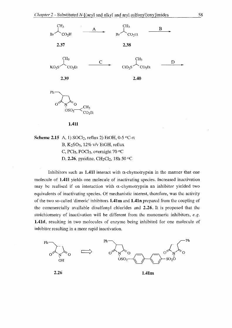
Chapter 2 - SubstitutedN-[(acyl and alkyl and aryl sulfonyl)oxy]imides
CH3 A
CH3
• A Bf C02H A
Bf C02Et
2.37 2.38
CH3 CH~ ~- C
CI02S A ~02Et • K03S C02Et
2.39
Ph~
OJ--N~O I CH3 OS 02--< C02Et
1.411
2.40
Scheme 2.15 A, 1) SOCh, reflux 2) BtOH, 0-5 DC-rt
B, K2S03, 12% v/v BtOH, reflux
C, PC15, POCh, overnight 70 DC
D, 2.26, pyridine, CH2Ch, I8h 50 DC
58
D •
Inhibitors such as 1.411 interact with a-chymotrypsin in the manner that one
molecule of 1.411 yields one molecule of inactivating species. Increased inactivation
may be realised if on interaction with a-chymotrypsin an inhibitor yielded two
equivalents of inactivating species. Of mechanistic interest, therefore, was the activity
of the two so-called 'dimeric' inhibitors 1.41m and 1.41n prepared from the coupling of
the commercially available disulfonyl chlorides and 2.26. It is proposed that the
stoichiometry of inactivation will be different from the monomeric inhibitors, e.g.
1.41d, resulting in two molecules of enzyme being inhibited for one molecule of
inhibitor resulting in a more rapid inactivation.
Ph~
OAN~O I
OH
2.26 1.41m

Chapt~r 2 - Substituted N-[(acyl and alkyl and aryl sulfonyl)oxy]imides ..
Ph~
OJ.-..N~O I
OR
2.26
Ph~ °VPh OJ.-..N~~S020--N
I - 0 OS02 ~ A
1.41n
59
The biochemical rationale underlying the design of N-sulfonyloxy succinimide
mechanism based inhibitors discussed above was based on the knowledge that these
derivatives are known to undergo spontaneous Lossen rearrangements on reaction with
nucleophiles to produce aliphatic isocyanates. These are known affinity label
inactivators of serine proteases reacting with His-57. The Lossen rearrangement was
first observed in amide systems88 and has also been observed in N
[(acyl)oxy]phthalimide derivatives179 and hence we proposed that N-acyloxy imides
1.41(R = Ph, PhCH2, L = C(O)R') may be potential mechanism based inhibitors
operating with the same mechanism of action as their sulfonylated counterparts 1.41 (R
= Ph, PhCH2, L = S02R').
Inhibitors of type 1.41 (R = Ph, PhCH2, L = C(O)R') , see Table 2.2, were prepared
by the base mediated coupling of either acetic anhdride with N,N-dimethyl-4-
aminopyridine (DMAP) as a reaction catalyst, or acetyl chloride to 2.26 and 2.32 to
give the acetyl derivatives 1.410 and 1.41p. Reaction of cinnamic acid with 2.26
mediated by triethylamine and catalysed by DMAP gave the proposed "dual" inhibitor
1.41q which in addition to inhibiting a-chymotrypsin will release cinnamic acid, a
known anti-oxidant, to combat the high level of free radicals associated with the disease
states resulting from unregulated a-chymotrypsin activity. To enhance inhibitory
activity through interaction with the enzyme's Sn' subsites the extended inhibitor 1.41r
was prepared by DCCIHOBt mediated coupling of 2.26 to N-benzyloxycarbonyl-(L)
phenylalanine (N-CBz-(L)-Phe).
The syntheses of 1.410-r by these methods gave crude products which required
further purification either by acid and base washing and crystallisation or
chromatography. Hence a cleaner acylation procedure was sought. Procopiou et al180
have published results describing the use of trimethylsilyl trifluoromethanesulfonate
(TMSOTf) as a catalyst in what are fast and efficient acylations of a variety of alcohols.
Applying this methodology initially to the synthesis of 1.410 a faster and cleaner
reaction in higher yield was observed requiring less equivalents of acylating agent and
catalyst. Specifically, reaction of 2.26 with AC20 (1.5 equiv.) and TMSOTf (0.02
equiv.) gave 1.410 as a white crystalline solid in 86% yield, whereas under the original
conditions, reaction of 2.28 and AC20 (2 equiv.), Et3N (1.1 equiv.) and DMAP (0.1

Chapter 2 - SubstitutedN-[(acyl and alkyl and aryl sulfonyl)oxy]imides
A R B R
0):).0 C 0):).0 D I I
OH E OL
2.26 R = PhCH2 1.41
2.32 R =Ph L=C(O)R'
Conditions A Et3N or i-PrNEt and DMAP catalysed acylation
B Et3N or i-PrNEt acylation
Compound
number
1.410
P
q
r
C TMSOTf acetylation
D TMSOTf acylation with in situ mixed anhydride
E DCC, HOBt mediated acid coupling
R L Conditions
PhCH2 Ac A,C
Ph Ac B
PhCH2 C~Ph A,D II 0
PhCH2 Ph, E _ 0
CANJlO ........... Ph II H 0
Table 2.2 Inhibitors of type 1.41 (R = Ph, PhCH2, L = C(O)R')
60
equiv.) gave 1.410 as a brown residue in 79% yield after purification by acid and base
washing.
Procopiou's method requires the use of anhydrides as acylating agents. The synthesis
of 1.41q and 1.41r would therefore require a supply of trans-cinnamic acid anhydride
and N-Cbz-(L)-phe acid anhydride both of which are not commercially available and
difficult to prepare. The use of mixed anhydrides generated in situ as acylating agents
was investigated as a solution to this problem hence the synthesis of 1.41p was
attempted using this methodology (Scheme 2.16).

Chapter 2 - SubstitutedN-[(acyl and alkyl and aryl sulfonyl)oxy]imides
Ph~C02H A
2.41
Scheme 2.16 AN-methyl morpholine, CIC(O)OEt, 0 °C
B 2.26, TMSOTf, overnight, 0-5 °C-rt
B
61
1.41q
Trans -cinnamic acid (2 equiv.) was treated with ethyl chloroformate (2 equiv.) and
N -methyl morpholine (2 equiv.) at 0 °C to generate the mixed ahydride 2.41. Reaction
of this with 2.26 and TMSOTf (0.02 equiv.) gave a brown residue that was found to
contain 2.41 and the desired product 1.41q in a 1: 1 ratio, by 1 H NMR. It was proposed
a cleaner reaction may be observed if the equivalency of the mixed anhydride and
therefore that of trans-cinnamic acid and N-methyl morpho line used be lowered.
Subsequently the reaction was repeated with only 1.05 equivalents of acid and base and
pure 1.41q as a white solid in 86% yield.

Chapter 2 - SubstitutedN-[(acyl and alkyl and aryl sulfonyl)oxy]imides 62
2.7 DESIGN AND SYNTHESIS OF SUBSTITUTED N-[(ACETYL AND METHANESULFONYL)OXY] GLUTARIMIDES
In order to study the effect ring size has on inhibitory activity a number of previously
unknown substituted N-[(acyl and alkylsulfonyl)oxy]glutarimides represented by
structure 2.1 were prepared which are represented in Table 2.3.
Compound R
number
2.1a Ph
2.1b Ph
2.1c PhCH2
2.1d H
2.1e H
2.lf H
R'
H
H
H
Ph
RlC).'R R" 3 4
o N 0 I OL
2.1
R"
H
H
H
H
R', R":::: cyc10hexyl
CH3 CH3
Table 4.1 Potential inhibitors of type 2.1
L
S02CH3
Ac
S02CH3
S02CH3
S02CH3
S02CH3
The a-C substituents of the related succinimide inhibitors 1.41 are proposed to
interact with the S1 subsite of a-chymotrypsin hence the potent and specific inhibition
of this enzyme by derivatives of type 1.41 with an aromatic a-C substituent as
illustrated by the inhibitory activities of 1.41d against human leukocyte elastase and a
chymotrypsin. It was proposed that by judicious choice of the extent and type of
substitution present on the glutarimide ring that similar increased activity could be
observed.
The retrosynthetic analysis of 2.1 (Scheme 2.17) proceeds in the same manner as the
second retrosynthetic analysis described for 1.41 (see Scheme 2.2, Section 2.1).
Disconnection of the bond between the oxygen and that of the reactive moiety (L) gives
substituted N-hydroxyglutarimides 2.42 and alkyl sulfonic and alky1carboxylic acid
derivatives. The preferred syntheses of 2-suhstituted N-hydroxysuccimides, the key
intermediates in the synthesis of derivatives of type 1.41, involved either the anhydride

Cl1apter 2 - SubstitutedN-[(acyl and alkyl and aryl sulfonyl)oxy]imides
R~"
O~N~O
'R R"
:00 I
OH
2.42
'R R" Rn 000
2.43
UFGI
R~" O~ ~O
OH OH
2.44
I O-~-L
2.1
:.:: C(O)R"'
Scheme 2.17 Retrosynthetic analysis of glutarimide inhibitors 2.1
63
or mono ethyl ester. The latter was prepared using the Stobbe condensation of diethyl
succinate and appropriate aldehyde. Diethyl glutarate cannot be used as a substrate in
this reaction 157c and hence an efficient route to N-hydroxyglutarimides can only be
realised through the anhydride. Hence disconnection of 2.42 gives the the glutaric acid
anhydride 2.43 and hydroxylamine. The anhydride 2.43 is derived from the
corresponding glutaric acid 2.44.
The substitution present on the glutarimide ring is directly obtained from that present
in the glutaric acid starting materials. The prepartion of potent inhibitors therefore
required a supply of aromatic glutaric acids. The aromatic 2- and 3-phenylglutaric acids

Chapter 2 - Substituted N-[(acyl and alkyl and aryl sulfonyl)oxy]imides 64
are commercially available and hence provided a quick entry point into the synthesis of
desired inhibitors 2.1 (R, R', R" is aromatic). Of particular interest was the inhibitory
activity of 2.1c (R == PhCH2, R', R" == H) which possesses the a-benzyl substitutent
found to impart higher inhibitory activity towards a-chymotrypsin in the succinimide
series (compare 1.41a and 1.41d above).
The required synthesis of 2-benzyl-glutaric acid 2.44b was carried out employing
Michael reaction chemistry181 following literature procedures for the preparation of 2-
alkyl glutaric acids.182
A ..
2.10 2.45
~ O~ ~O
OH OH
2.44b
Scheme 2.17 A, acrylonitrile, 30% KOH, MeOH, t-BuOH, 4.5h, rt
B, 68% RBr, Id, reflux
A Michael reaction between diethyl benzylmalonate 2.10 and acrylonitrile gave the
malonate eser 2.45 in 50% yield after distillation at reduced pressure. Acid hydrolysis of
2.47 then followed to give 2-benzylglutaric acid 2.44b in 72% yield.
As part of research being undertaken into anti-epilepsy agents within the University
of Canterbury Chemistry Department methodology for the preparation 3,3-disubstituted
glutaric acids 2.44 was being developed. To extend the scope of the SAR study of the
glutarimide inhibitor series 4,4-disubstituted inhibitors 2.1 (R == H, R', R" == alkyl) were
prepared from the corresponding 3,3-disubstituted glutaric acids.
Of the synthetic methods available for the synthesis of 3,3 disubstituted glutaric acid
derivatives the Guareschi route (Scheme 2.18) is the most favoured where applicable.
The formation of the key "Guareschi" imide intermediate 2.48 of Scheme 2.18 is
believed to occur in two steps at least. The starting ketone represented by structure 2.46
undergoes a Knoevenagel condensation with ethyl 2-cyanoacetate, which was used in
our synthesis, catalysed by the liquid ammonia solvent to give the a-~ unsaturated

Chapter 2 - Substituted N-[(acyl and alkyl and aryl sulfonyl)oxy]imides 65
compound 2.47. This undergoes a Michael reaction with 2-cyanoacetamide resulting
from solvolysis of ethyl cyanoacetate and concomitant cyclisation to give the Guareschi
imide 4,4-dialkyl-3,5-dicyanoglutarimide 2.48. Simple acid hydrolysis of 2.48 gives the
desired 3,3-disubstituted glutaric acids.
'R R"
'IT A
o
2.46
2.46a R', R" = CH3
2.48
2.48a R', R" = CH3
B
'R R"
Nc1
C02Et
2.47
'R R"
:1\0 OH OH
2.44
2.44e R = H, R', R" = CH3
Scheme 2.18 Guareschi route to 3,3-disubstituted glutaric acids
The Guareschi route has been applied to a number of substrates however the key
Guareschi imide intermediate has not been observed in a number of examples.185 This
occurs when the starting materials do not suit the chemistry of the Knoevenagel
condensation or if suitable give derivatives of type 2.47 which are unsuitable for the
following Michael reaction and ring closure. Hence the scope of the reaction is limited
to ketones which are not sterically hindered and less certainly those that have an
enolizable a-proton)86 Acetone fits this criteria and, therefore, it was proposed to use
this Guareschi imide methodology in the preparation of 3,3-dimethyl glutaric acid
2.44e. Cyclohexylglutaric acid 2.44d was also prepared in this manner and the
corresponding Guareschi imide was a generous gift from Dr. Rob McKeown.
Subsequently the synthesis of 3,3-dimethyl glutaric acid 2.44e was carried out using
the Guareschi route. The Guareschi imide 2.48a was prepared in 42% by acidification of
the ammonium salt that was deposited on storing a solution of ethylcyanoacetate and
acetone 2.46a in ethanol saturated with ammonia for three days at ca -5 °C. Subsequent
sulfuric acid hydrolysis of 2.48a gave the desired 3,3-dimethylglutaric acid 2.44e.
The preparation of the desired substituted N-[(alkylsulfonyl)oxy]glutarimide
inhibitors 2.2 from glutaric acid precursors was then carried out according to Scheme
2.19

Chapter 2 - Substituted N-[(ac::yl and alkyl and aryl sulfonyl)oxy]imides
'R R"
:1\0 OH OH
2.44
A
2.43a-e
'R R"
:00 I
OH
2.42a-e
2.44a R = Ph, R', R"= H
2.44b R = PhCH2, R', R" = H
2.44c R,R" = H, R' = Ph,
2.44d R=H, R', R" == cyc10hexyl
2.44e R= H, R', R" CH3
B ..
2.49a-d
D
2.1a-e
Scheme 2.19 A AC20, reflux
B H2NOCH2Ph, toluene, xylenes, reflux
C H2, 10% Pd-C, THF
Dbase, R'''S02CI, R'''C(O)CI
66
The variously substituted glutaric acids whether obtained from a commercial source
or synthesised as described above were treated with either acetyl chloride (in the case of
2.44a) or acetic anhydride (for 2.44b-e) under refluxing conditions to give the
respective anhydrides in high yields ranging from 84% for the preparation of 2-
phenyl glutaric anhydride 2.43a to 91 % for cy!cohexylglutaric anhydride 2.43d.
The anhydrides 2.43 were then refluxed with O-benzyloxyamine to give the N
protected glutarimides 2.49 in typically 80% yield. Hydrogenolysis of these gave the N
hydroxyglutarimides 2.42 in yields of 70% which were then treated with the appropriate
alkyl sulfony!Chloride or acid chloride to give the desired inhibitors. As observed with
the succinimide series of compounds this reaction proceeded in nearly quantatative
yield.

Chapter 2 SubstitutedN-[(acyl andalkyl and aryl sulfonyl)oxy]imides 67
Shorter routes to the desired inhibitors were realised by the reaction of
hydroxylamine directly with the anhydride 2.43. Both 4,4-dimethyl-N
hydroxyglutarimide 2.42e and 3-phenyl-N-hydroxyglutarimide were prepared in 50%
yield in this manner. A short route to 2.42e was also investigated by coupling 0-
benzyloxyamine directly to the acid 2.44e using EDAC however the low yield of this
reaction (12%) indicated this route was of poor preparative value considering the high
yields of the reactions of Scheme 2.19.

Chapter 2 - Substituted N-[( acyl and alkyl and aryl sulfonyl)oxy]itnides 68
2.6 SIJMMARY
A brief retrosynthetic analysis of the target inhibitor 1.41 suggested two general
syntheses. The chemical competency of these was examined and a route involving 3-
substituted N -hydroxysuccinimides as key intermediates was identified to be the most
desirable to the demise of a route proceeding via 2-substituted succinimides. This route
involved the reaction of succinimides with silver carboxylate and sulfonate salts as a
means of preparing inhibitors of type 1.41 but this reaction was unsuccessful.
The synthesis of 3-substituted N-hydroxysuccinimides 2.8 was carried out by
reaction of activated 2-substituted succinic acid derivatives and either hydroxylamine or
a protected form of this O-benzyloxyamine. The synthesis of 3-benzyl-N
hydroxysuccinimide was a key goal as inhibitors derived from this display increased
specificity for a-chymotrypsin. Consequently some effort was directed towards
investigating routes to this key intermediate from activated 2-benzylsuccinates requiring
a good synthetic supply of 2-benzylsuccinic acid. Three methods for the preparation of
the representative 2-benzylsuccinc acid were therefore compared. A route employing
the Stobbe condensation between benzaldehyde and diethylsuccinate gave the highest
overall yield and was preferred for the ease of its well established reactions. However a
short (two step) and general synthesis which involved the sodium amide mediated
benzylation of succinimide as a key reaction may provide the ideal route to 2-
alkylsuccinic acids with further optimisation.
The failed preparation of 3-benzyl-N-hydroxysuccinimide from the reaction of 2-
benzyl succinic acid activated in situ by treatment with the coupling reagent EDAC with
O-benzyloxyamine prompted the use of more activated derivatives. Of those derivatives
successful syntheses of 3-benzyl-N-hydroxysuccinimide were carried oput from the
anhydride and 2-benzylsuccinic acid mono ethyl ester in six and five steps respectively
with O-benzyloxyamine. The reaction of hydroxylamine and the anhydride proved to be
a successful preparation of 3-benzyl-N-hydroxysuccinimide in five steps whereas the
use of hydroxylamine in reaction with the mono ethyl ester failed.Of the two successful
five step syntheses a greater overall yield was observed with that route involving the
mono ethyl ester (30%) than that involving the anhydride (18%) and in conclusion this
synthesis was found to be the preferred preparation of 3-benzyl-N-hydroxysuccinimide.
To complete the synthesis of the target inhibitors 1.41 (R = Ph, PhCH2, L = S02R')
various sulfonyl chlorides were coupled to the 3-substituted N-hydroxysuccinimides
2.28 and 2.34 using modifications of literature procedures. The nature of the sulfonyl
chloride used affects the potency of the resulting inhibitors and hence is an important
design element. Literature SAR results indicated that bulky sulfonyl chlorides with

~C;hapter 2 - SubstitutedN-[(acyl and alkyl and aryl sulfonyl)oxyJimides 69
good electron withdrawing properties would furnish the most potent inhibitors in this
series and subsequently an aromatic series was studied. To add to a conceived strategy
of enhancing inhibitory activity by incorporating the inhibitors of type 1.41 into a
complete psuedo peptide sequence a tailored sulfonyl chloride was synthesised
permitting the preparation of an inhibitor extending in the C-direction. Two examples
of so-called "dimeric" inhibitors were prepared to study the effects of stoichiometry on
inhibition.
N-Acyloxyimides are also known to undergo a Lossen rearrangement when treated
with nucleophiles hence these were proposed to be potential mechanism-based
inhibitors of the same ilk as N-sulfonyloxyimides. 88 The acylation of 2.28 and 2.34
using Hunigs base or triethylamine with DMAP as a catalyst gave crude products. An
improved procedure employed the use of TMSOTf as a catalyst for acetylations which
was expanded to include acylations using mixed anhydride methodology. Acyl
chlorides and anhydrides were chosen so that a number of the the resulting inhibitors
possessed the same substitution as the related N-sulfonyloxyimides to provide a direct
comparison in activity. An extended inhibitor of type 1.41 (R = PhCH2, L = C(O)R')
was prepared by the coupling of N-protected phenylalanine to 2.28 .
To study the effect of ring size on inhibitory activity six membered glutarimide
inhbitors were prepared using the synthetic methodology developed above for the
succinimide series. The favoured route for the succinimides using the Stobbe
condensation was not applicable to the glutarimides series as this reaction cannot
tolerate glutarimide substrates and therefore the method employed proceeded through
the anhydride.
It was also desirable to examine what effect the type and extent of substitution had
on inhibitory activity which required a supply of both mono and disubstituted alkyl and
aromatic glutaric acids. The synthesis of a-benzylglutaric acid was effected through
Michael reaction chemistry and a number of dialkyl glutaric acids were prepared using
the Guareschi route. Glutarimide inhibitors were then prepared in good yield from the
respective acids through a four step synthetic sequence proceeding through the
anhydride, N-protected glutarimide and N-hydroxyglutarimide.

CHAPTER 3
a-CHYMOTRYPSIN INHIBITION ASSAYS

Inhibition 71
3.1 MECHANISM FOR INACTIVATION OF ctCHYMOTRYPSIN BY N-[(ACYL AND ALKYL AND ARYL SULFONYL) OXY]IMIDES
The mechanism of inactivation of a-chymotrypsin by the substituted N-[(acyl and
sulfonyl)oxy]imides (1.41 and 2.1) is proposed to be the same as that identified by
Groutas et a185 for the succinirnide inhibitor 1.41d. This mechanism is depicted in
Scheme 3.1.
R
3h4
oAN~O I OL
1.41
L=S02R'
= C(O)R'
1.41d
Rfl'R R" 3 4
o N 0 I
OL
2.1
= C(O)R'"
Accommodation of the inhibitor in the active-site of the enzyme through interactions
between the R substituent and the primary specificity site S 1 of the enzyme to give the
enzyme-inhibitor Michaelis complex (3.1), is followed by acylation of the active site
serine (Ser-195) to yield 3.2. Lossen rearrangement of 3.2 then generates an enzyme
bound isocyanate 3.3 which competes with deacylation which gives the free isocyanate
3.5. The enzyme bound isocyanate 3.3 then reacts rapidly with a nucleophile present in
the active site. In particular, acylation of the imidazole ring of the active site amino acid
His-57 (Pathway I) results in formation of 3.4, the likely final inactive enzyme species.
Hydrolysis of the Ser-195 ester linkage of 3.3 (Pathway II) results in the release of the
reactive free isocyanate 3.5 from the enzyme in a "turnover" event. Hydrolysis of the
isocyanate 3.3 (pathway III) gives the acyl enzyme 3.6. If stable, 3.6 may inactivate the
enzyme during its lifetime which depends on the rate of hydrolysis to the amine 3.7. It
is believed that the acyl enzyme 3.6 is short lived and hence pathway III will be acting
predominantly as a "turnover" pathway. The urea linkage of the inactive enzyme
inhibitor adduct 3.4 is also susceptible to hydrolysis, by analogy with related imidazole
N-carboxarnides,53 leading to fonnation of 3.5.

Inhibition 72
His-57 .---
Ser-195 y
~ 80r-195 N~
His-57
/ \""N~ o A H oy N 0 ""'~o ~ Asp-102
R H II 3.4 0
3.6 3.5
3.7 3.7 3.5
Scheme 3.1 Proposed mechanism of inhibition of a-chymotrypsin by 1.41 and 2.1

Chapter 3 - a-Chymotrypsin Inhibition Assay 73
The reactive enzyme bound isocyanate 3.3 reacts in three ways (Pathways I-In) all
of which ultimately lead to the release of free isocyanate 3.5 from the enzyme. This
undergoes rapid hydrolysis with concomitant decarboxylation to form the highlighted
amine 3.7. Aliphatic isocyanates, such as 3.5, have been shown to be act as inactivators
of serine proteases, including a-chymotrypsin,49 and hence it is thought that the
isocyanate 3.5 or the amine 3.7 may playa part in the inactivation process in concert
with the enzyme bound isocyanate 3.3.
Indirect evidence supporting the proposed mechanism (Scheme 3.1) has been
obtained.84,87e Reaction of equivalent amounts of the inhibitor lAId and sodium
methoxide in methanol resulted in formation of a mixture of two isomeric Lossen
rearrangement products 3.8 and 3.9 (Scheme 3.2).
PhL\ Ph 0 Ph
h A r~~CH3 + H3COyNH COZCH3 o N 0 1 o OCH
OSOzCH3 3 0
1.41d 3.8 3.9
Scheme 3.2 A, NaOCH3, CH30H, Ih, rt.
The isocyanate 3.10 has also been observed to react with imidazole to give the
imidazole N-carboxamide 3.11 in quantitative yield.
eCOzEt A
eCO~t ..
NJlN:it N"C H 'LJ "0
3.10 3.11
Scheme 3.3 A, imidazole, Ih, room temperature
The urea linkage of imidazole-N-carboximides such as 3.4 is labile to nucleophiles,
and hence treatment of inactivated enzyme with appropriate nucleophilic reagents
should result in reactivation of the enzyme. Reactivation of inactivated enzyme has
been observed in experiments undertaken with hydroxylamine and dithiothreitol, results
which support formation of 3.4.
Isocyanates 3.3 and 3.5, are both reactive towards nucleophiles. If an external
nucleophile is present during the inactivation process it will scavenge both 3.3 and 3.5,
and if these are involved in the inactivation process, the enzyme will be protected.
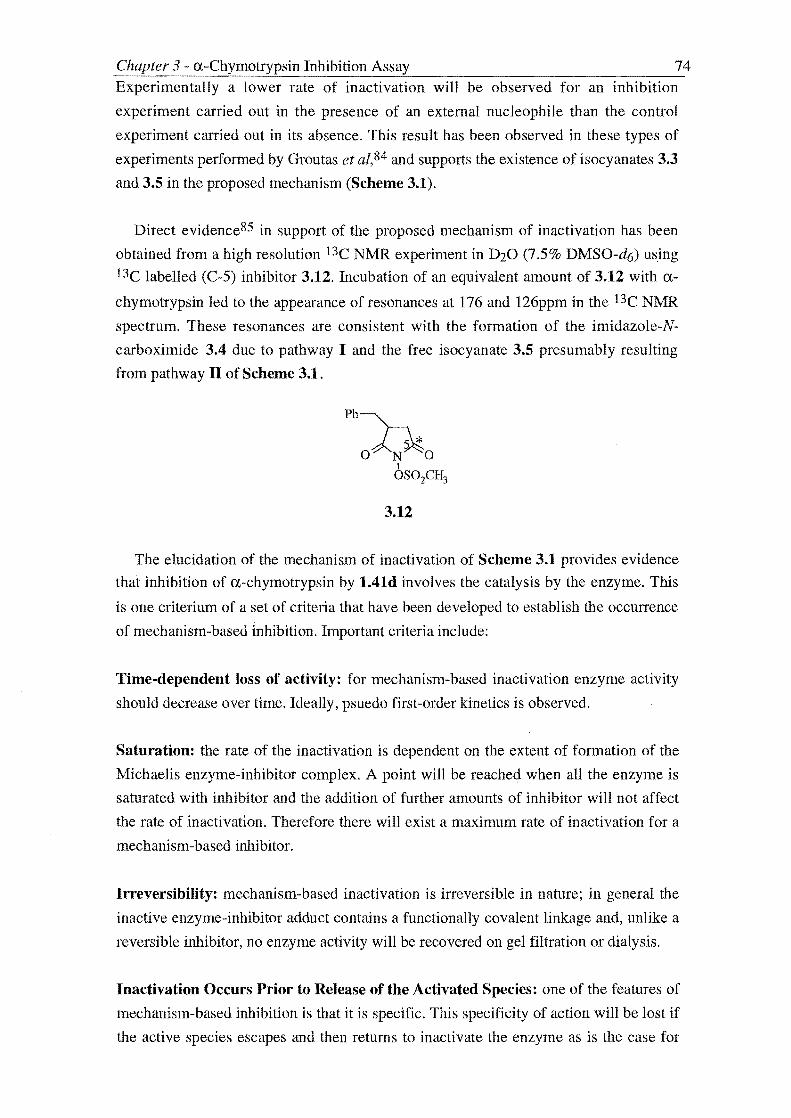
Chapter 3 a-Chymotrypsin Inhibition Assay 74 Experimentally a lower rate of inactivation will be observed for an inhibition
experiment carried out in the presence of an external nucleophile than the control
experiment carried out in its absence. This result has been observed in these types of
experiments performed by Groutas et al,84 and supports the existence of isocyanates 3.3
and 3.5 in the proposed mechanism (Scheme 3.1).
Direct evidence85 in support of the proposed mechanism of inactivation has been
obtained from a high resolution 13C NMR experiment in D20 (7.5% DMSO-d6) using
13C labelled (C-5) inhibitor 3.12. Incubation of an equivalent amount of 3.12 with a-
chymotrypsin led to the appearance of resonances at 176 and 126ppm in the 13C NMR
spectrum. These resonances are consistent with the formation of the imidazole-N
carboximide 3.4 due to pathway I and the free isocyanate 3.5 presumably resulting
from pathway II of Scheme 3.1.
3.12
The elucidation of the mechanism of inactivation of Scheme 3.1 provides evidence
that inhibition of a-chymotrypsin by 1.41d involves the catalysis by the enzyme. This
is one criterium of a set of criteria that have been developed to establish the occurrence
of mechanism-based Inhibition. Important criteria include:
Time-dependent loss of activity: for mechanism-based inactivation enzyme activity
should decrease over time. Ideally, psuedo first-order kinetics is observed.
Saturation: the rate of the inactivation is dependent on the extent of formation of the
Michaelis enzyme-inhibitor complex. A point will be reached when all the enzyme is
saturated with inhibitor and the addition of further amounts of inhibitor will not affect
the rate of inactivation. Therefore there will exist a maximum rate of inactivation for a
mechanism-based inhibitor.
Irreversibility: mechanism-based inactivation is irreversible in nature; in general the
inactive enzyme-inhibitor adduct contains a functionally covalent linkage and, unlike a
reversible inhibitor, no enzyme activity will be recovered on gel filtration or dialysis.
Inactivation Occurs Prior to Release of the Activated Species: one of the features of
mechanism-based inhibition is that it is specific. This specificity of action will be lost if
the active species escapes and then returns to inactivate the enzyme as is the case for

Inhibition 75 affinity labels or a so-called "paracatalytic" inhibitors. Mechanism-based inactivation
will show no "lag" phase and will not be affected by the presence of nuc1eophiles.
Partition Ratio: this concept is unique to mechanism-based inhbition. This term
introduced by Walsh, 54 describes the ratio of the number of latent inhibitor molecules
converted and released as product relative to each turnover leading to enzyme
inactivation. The partition ratio, therefore is a measure of the efficiency of the
inhibition.
Frequently in practice, as in this thesis, only a few of the criteria developed for
mechanism-based inactivation are investigated when a potential mechanism-based
inactivator is studied. However, to unambiguously charaterize an inactivator as being
mechanism-based the criteria above should be satisfied. The first two criteria define any
covalent or tightly bound, slow binding, non-covalent competitive inhibitor which
would be analysed in a different manner than a mechanism-based inhibitor. The last
three criteria, in combination, are definitive for a mechanism-based inhibitor. It is the
object of this chapter to discuss the assay of N-[(acyl and sulfonyl)oxy]imides 1.41 and
2.1 using kinetic models developed for mechanism-based inactivation. The question
then arises are we justified in using these models?
A preliminary answer to this question and a preview of the activity of compounds
1.41 and 2.1 can be provided by the results Groutas et aZ84,87a obtained in carrying out
experiments prescribed to establish mechanism-based inhibition for 1.41d. This
compound has been found to inhibit a-chymotrypsin in a time dependent manner with
the observation of pseudo first-order kinetics over 15 minutes. Experiments carried out
at increasing inhibitor concentration point to the a maximum rate of inactivation. The
urea linkage of the inactive enzyme (see Scheme 3.1, structure 3.4) is susceptible to
hydrolysis (t1l2 = Ih) and therefore inhibitors of type 1.41 and 2.1 will only be acting as
irreversible inhibitors over a short time.
Although the addition of the nuc1eophile dithiothreitol results in a lower rate of
inhibition it is believed that inactivation does occur before the reactive species escapes
the active site as there is no "lag" phase for inhibition. This result may be explained by
dithiothreitol entering the active site and reacting with the enzyme bound isocyanate
(Scheme 3.1, structure 3.3).
The partition ratio for 1.41d will depend on the reactivity of the enzyme bound
isocyanate, its rate of hydrolysis and hence diffusion from the active site, and the
proximity of the imidazole ring for carbamate bond formation. No partition ratio
analysis has been undertaken for 1.41d, however 13e NMR and reactivation studies

76
carried out by Groutas et aZ84,85 suggest that the rate of hydrolysis of the enzyme bound
isocyanate 3.3 and inactive enzyme 3.4 are significant. It is therefore proposed that the
partition ratios for inhibitors of type 1.41 and 2.1 will be far from the ideal value of
zero.
The predicted high partition ratios for 1.41 and 2.1 will have practical implications
for the inhibition assays as depletion of the inhibitor during assay will be significant
and problematic. It appears therefore that the N-[(acyl and sulfonyl)oxy]imide inhibitors
1.41 and 2.1 will only be acting as true mechanism-based inactivators at high
concentrations and over a short period of time after which deviation away from psuedo
first-order kinetics is expected. There is also evidence to suggest that a species other
than the initial compound l.4ld, most probably the amine 3.7, contribute to the
inactivation, although not initially.

Inhibition 77
5.2 INACTIVATION KINETICS
The evaluation of the inhibitory activity of a compound is routinely carried out using
two methods. The first procedure commonly referred to as the "incubation method" or
"pre-incubation method"71,72,79,187-9 as it name suggests involves incubating an assay
mixture containing the enzyme and an excess of inhibitor. Periodically, aliquots are
removed from this assay mixture and reacted with a "monitoring" substrate. The rate of
hydrolysis of the substrate is a measure of the enzyme activity of this sample. A
chromogenic substrate is used so that its rate of hydrolysis can be followed
spectrophotometrically by noting the proportional rate of increase in absorbance of its
hydrolysis product. The assay mixture contains only enzyme and inhibitor and hence
kinetic analysis involves consideration of only those two species.
The incubation method can be used only to assay relatively slow inhibitors because
sampling techniques are employed. Faster or more potent inhibitors cannot be
adequately examined using this method hence a second assay procedure capable of
measuring rapid inactivation is required. The procedure commonly employed is the
progress curve method.190-192 To an assay mixture containing "monitoring" substrate
and inhibitor an aliquot of enzyme is added, and the progress of the reaction followed
by the rate of hydrolysis of the substrate.
Practically, the incubation method of assay is more demanding. Several inhibition
and control experiments have to be undertaken to afford the rate measurements that on
simple analysis give the inhibitory activity of the compound being assayed. For the
incubation method the bimolecular constant kobs/[I] is used eXclusively as the index for
inhibitory activity. As discussed in the following section, for slow inhibitors kobJ[I]
serves as an approximation for the ratio kinact/Kr which is the ideal constant for
quantifying inhibition. For potent inhibitors this ratio is high, for weaker inhibitors it is
lower.
For those fast inhibitors able to be analysed using the incubation method kobsl[I]
approximates kinactlKr less well but kobs/Kr is still used as an index of potency, faster
inhibitors again displaying a higher value. A rigorous value for kinact/Kr can be
obtained using the incubation method but requires that experiments be carried out at
several different inhibitor concentrations (five are recommended)25 which adds to the
already high experimental demand for this method.
The progress curve method requires only one inhibition experiment for evaluation of
kobs/[I] but the data analysis is more complicated and time consuming. The expression
for kobs/[I] for the progress curve method is different from that for the incubation

Inhibition 78 method having a different dependence on the inhibitor concentration and including
substrate terms and hence the two are not compared. The ratio kinaet/Kr is exclusively
reported and can be determined rigorously using the progress curve method. Again
experiments are run at different inhibitor concentrations but the fact that the progress
curve method is less labour intensive facilitates these operations. Values for kinaet/Kr
can be compared between the two methods and a good agreement is normally
ob served. 62e
The assay mixture for the incubation method contains enzyme and inhibitor whereas
for the progress method the assay mixture contains enzyme, inhibitor, and substrate and
hence a more complicated kinetic analysis will be required. The kinetic models used in
the analysis of inhibitors using these two methods is discussed below.
INCUBATION METHOD KINETICS
The corresponding kinetic scheme for the interaction between enzyme and inhibitor
for the proposed mechanism of inactivation of a-chymotrypsin by 1.41 and 2.1 is
shown below (Scheme 3.4).
KI EJ
ka E-I
kz E-I' E-I" E+I .. •
I kd I k3 I k,
E+P E+P E+P
Scheme 3.4. Kinetic scheme for mechanism-based inhibitors of type 1.41 and 2.1
In this scheme E is the free enzyme; I is the N-acyl or sulfonyloxy imide inhibitor
1.41 or 2.4; E.I the Michaelis complex for the enzyme and inhibitor (structure 3.1 of
Scheme 3.1); E-I is the acyl enzyme (3.2); and E-I' is the reactive isocyanate (3.3)
tethered at the active site; E-I" is the inactivated enzyme with the inhibitor covalently
attached (3.4); and P represents the free isocyanate (3.5) and the amine (3.7) which are
the products resulting from hydrolysis of the inhibitor.
The inhibitor initially interacts with the enzyme in the same way as a substrate to
give an enzyme-inhibitor Michaelis complex E.L The binding of enzyme and inhibitor
is described by Kr the dissociation constant for the Michaelis complex. Acyl enzyme
formation (ka) proceeds as with a normal substrate, however, it is accompanied by the
release of the latent reactivity i.e. the Lossen rearrangement (k2) to the enzyme bound
isocyanate. Deacylation of the acyl enzyme (kd) may also occur via a so-called

Inhibition 79 "turnover" pathway which does not result in enzyme inactivation. Acylation of the
imidazole ring of His-57 by the enzyme bound isocyanate (k4) leads to inactivated
enzyme (E-1"). This competes with other "turnover" hydrolysis pathway (k3). The rate
constant for hydrolysis of the inactivated enzyme species (ks) determines the
"irreversibility" of the inactivator.
lung and Metcalfe187b have developed a kinetic model for mechanism-based
inhibitors extending the work undertaken by Kitz and Wilson187a for the kinetic
analysis of active-site directed irreversible inhibitors. Groutas et a184 ,8S,87 have
analysed inhibitors of type 1.41 (L = S02R') such as 1.41d, using the model of Jung
and Metcalfe and hence this model was adopted for the initial inhibition studies of this
thesis.
Since the progress of the overall inhibition reaction is monitored by the decrease in
total active enzyme (i. e. accumulation of E-I"), E.I, E~ I, and E~ l' are not observed
independently and hence Scheme 5.2 can be simplified to give the kinetic scheme
shown below (Scheme 3.5).
The assumption made in this approximation is that the inhibition reaction is carried
out with an excess of inhibitor so that the concentration of the Michaelis complex is
constant throughout the assay. If the inhibition experiment is designed so that these
ideal conditions are maintained then psuedo first-order kinetics will be observed.
E+1 E*1 .. E-1"
Scheme 3.5 Simplified incubation method kinetic scheme for mechanism-based
inhbitors 1.41 and 2.1
In this scheme E*I represents the kinetic composite of E.I, E~I, and E-I' and kinact is
the rate constant for the inactivation process. The rate constant kinact is a mixture of the
microscopic rate constants k2, k3, k4 and kS of Scheme 3.2. Waley 193 has applied a
steady-state hypothesis to the kinetic scheme for a general mechanism-based inactivator
which includes all the terms considered in Scheme 3.4 excepting ks as this is zero for
inactivation. The expression obtained for kinact is shown below in equation (3.1).
(3.1)
From equation (3.1) it is observed that the magnitude of kinact is proportional to k2,
the rate constant for the Lossen rearrangement to the inactivating species; and k4, the

80 Chapter 3 - a-Chymotrypsin Inhibition Assay --~-------------------------------
rate constant for the reaction between the inactivating species and His-57 to form
inactive enzyme. The magnitude of kin act is inversely proportional to k3, the rate
constant for the turnover pathways which result in no net inactivation.
Typically k2 is found to be the rate determining step in the inhibition process, in this
case equation (3.1) reduces to:
(3.2)
From equation (3.2) the magnitude of kin act will be proportional directly to k3 and k4
(but in a more complicated manner) but inversely proportional to k3.
The ratio kinacJKr provides a measure of inhibitory activity. A potent inhibitor will
have a high kinactlKr value whereas a weaker inhibitor will have a lower value. Hence a
potent inhibitor will have a low Kr, demonstrating that it has high binding affinity for a-
chymotrypsin and a high kin act demonstrating that the Lossen rearrangement and
reaction of the resulting isocyanate with His-57 is rapid, but deacylation and hydrolysis
of the isocyanate and inactive enzyme are slow.
The kinetic scheme above (Scheme 3.5) can be analysed by the method of Kitz and
Wilson.l87 Provided the enzyme-inhibitor solution is extensively diluted before assay,
and that the inhibitor concentration is at such an excess over enzyme that it remains
constant dUling the course of the assay it can be shown that:
[E] In[Eo]
where
kobs _ kinact - Kr
1 + [I]
(3.3)
(3.4)
Experimentally, the quantity [E] is measured by the rate of hydrolysis of the
monitoring substrate in the presence of inhibitor, Vt. The quantity [Eo] is measured by
the rate of hydrolysis of monitoring substrate in the absence of inhibitor, vo, such that
equation (3.3) can also be expressed as:
1 Vt nVo
(3.5)

Inhibition
Therefore, a plot of In Vt, which serves as a measure of enzyme activity, versus time Vo
should give a straight line with a slope of -kobs.
Looking at equation (3.4):
kobs (3.6)
If [I] « Kr, for a poor inhibitor, then:
kobs ~ kinact. [I]
Kr kobs
"'" kinact .. [I] KI~~ (3.7)
Thus, k[I]s can be an approximation for the second order rate constant (k~~ct)
81
The inhibitory constant kinactIKr can be obtained in a more rigorous manner using
the method of Khz and Wilson.1 87a The experimental quantity kobs is obtained for a
number of inhibition experiments where the excess of inhibitor over enzyme is varied.
The half-lives for inhibition, (t1l2), for these experiments are then simply calculated
using equation (3.8) which is derived from equation (3.5):
ln2 t1l2=
kobs (3.8)
If the t1l2 values for inhibition are plotted against the reciprocal of the inhibitor
concentrations, according to equation (3.9), obtained from substitution of equation (3.8)
into equation (3.6) , two possibilities can exist (Figure 3.1).
t1/2 = ~2 + 1~2KI kmact kmact
(3.9)
Of the plots displayed in Figure 3.1 Graph A depicts the case when enzyme
saturation is observed while Graph B depicts the situation when saturating conditions
do not exist. The inhibitory constant kinactlKr can be only obtained from the gradient
and y-intercept of Graph A as shown, demonstrating the importance of maintaining
saturating inhibitor concentrations during the course of the enzyme assay.

hUfYlAt-rur",'n Inhibition 82
A B
]n2lkinact
1/[1]
Figure 3.1 Kitz and Wilson plot for an inhibitor exhibiting saturation (A) or (B) not
exhibiting saturation
To determine kinactlKr using Graph A requires first the calculation of t1/2 from kobs
as described above and then of the gradient and y-intercepts. A modified Kitz and
Wilson plot of [I]/kobs versus [I] (Figure 3.2) according to equation (3.10) , derived
from equation (3.6), also gives a straight line with positive slope and postive y
intercept. A value for kinactlKr can be obtained directly from the y-intercept of this
graph and hence more simply than a determination using Graph A of Figure 3.1 and
without the required t1/2 calculations.
l!L = ~ + -.L!L (3.10) kobs kinact kinact
[I]lkobs _1_
kinact
Krlkinact
[1]
Figure 3.2 Plot of [I]lkobs versus [1] to determine kinactlKr
PROGRESS CURVE KINETICS
The kinetic scheme for the interaction of enzyme, inhibitor and substrate for the
proposed mechanism of inactivation of a-chymotrypsin by 1.41 and 2.1 is shown
below.

Chapter 3 - a-Chymotrypsin Inhibition Assay 83
I +
E+S E.S .. E+P
KI
ka E~r
k4 E-I" E.I .. E~I ..
jku j k3 I ks
E+P E+P E+P
Scheme 3.6 Progress curve method kinetic scheme for mechanism-based inhibitors of
type 1.41 and 2.1
This kinetic scheme can be analysed according to the method of Tsou. 190 Provided
that the rate of the enzyme-substrate reaction is faster than the enzyme-inhibitor
reaction and the inhibitor and substrate concentrations remain constant during the
course of the assay it can be shown that there will be an exponential release of product
with time according to equation (3.11).
At t = 00, from equation (3.11):
V
kobs
(3.11)
(3.12)
where kobs is the rate constant for the observed inhibition and V the rate of the enzyme
substrate reaction in the absence of inhibitor which are given by:
k kinact + [10]
obs = S [10] + KI[1 + k~]
(3.13)
v

At ..... "n"·.n Inhibition 84
(3.14)
To determine the kinetic constants for irreversible inhibition, kobs/[I] and kinact!KI,
using the progress curve kinetic method, a suitable irreversible curve of type A (Figure
3.3) which is described by equation (3.11) must be obtained. Therefore an important
experimental task when using the progress curve method is the design of inhibition
conditions so that this irrevesible curve is obtained. This involves changing inhibitor
and substrate concentrations, however, the change must be such that the inhibitor and
substrate are present at saturating levels. Once a suitable curve is obtained analysis can
be carried out as follows.
v/ekobs
Absorbance
Figure 3.3 Irreversible Curve A
, ,
" v -ji·-- ...... --
" , . , . • I
time
A
From equations (3.11) and (3.12) it can be simply shown that:
In([poo] - [PrJ) = In[Poo] - kobs.t (3.15)
The quantities [Pt] and [Poo] can be interchanged with the corresponding
absorbances, At and Aend, which can be directly read off the absorbance versus time
curves obtained experimentally (see Figure 3.3). Therefore equation (3.15) can also be
expressed as:
In(Aend - At) = In(Aend Ie) - kobs.t (3.16)
Hence kobs can be obtained from the slope of the initial portion of the logarithmic
plot In(Aend - At) versus time using linear regression analysis. Alternatively kobs can be
determined by fitting experimental absorbance versus time data to equation (5.9) using
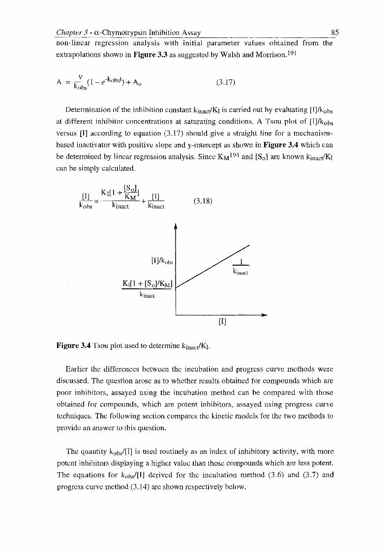
Chapter 3 - a-Chymotrypsin Inhibition Assay ---"----
85 non-linear regression analysis with initial parameter values obtained from the
extrapolations shown in Figure 3.3 as suggested by Walsh and Morrison.191
(3.17)
Determination of the inhibition constant kinact/KI is carried out by evaluating [I]/kobs
at different inhibitor concentrations at saturating conditions. A Tsou plot of [J]/kobs
versus [I] according to equation (3.17) should give a straight line for a mechanism
based inactivator with positive slope and y-intercept as shown in :Figure 3.4 which can
be determined by linear regression analysis. Since KM 194 and [So] are known kinact/KI
can be simply calculated.
[So] [I] KI[1 + KM] [I] --= +--kobs kinact kinact
[I]/kobs
KI[1 + [So]/KMJ
kinact
(3.18)
Figure 3.4 Tsou plot used to determine kinacVKI.
_1_ kinact
[I]
Earlier the differences between the incubation and progress curve methods were
discussed. The question arose as to whether results obtained for compounds which are
poor inhibitors, assayed using the incubation method can be compared with those
obtained for compounds, which are potent inhibitors, assayed using progress curve
techniques. The following section compares the kinetic models for the two methods to
provide an answer to this question.
The quantity kobs/[I] is used routinely as an index of inhibitory activity, with more
potent inhibitors displaying a higher value than those compounds which are less potent.
The equations for kobs/[I] derived for the incubation method (3.6) and (3.7) and
progress curve method (3.14) are shown respectively below.

Inhibition 86
(3.6)
(3.7)
(3.14)
It can be observed that both the equations are not independent of the inhibitor
concentration and cannot therefore serve as an index of inhibitory activity unless
inhibitors are compared at the same inhibitor concentration in which case kobsl[I]
values can only be compared if obtained using the same assay technique; no valid
comparison can be made between the two methods. As discussed above, if a poor
inhibitor is being assayed using the incubation method kobs/[1] serves as an
approximation for kinact/Kr, however this is not the case for compounds assayed using
progress curve kinetics.
The equations for the experimental quantity [IJlkobs for the incubation (3.10) and
progress curve method (3.18) are shown respectively below.
[I] KI [I] --=--+--kobs kin act kinact
(3.10)
K [·1 [So]] [1J I + KM [1] --= +--kobs kinact kinact
(3.18)
Observation of these equations shows that they are of the same form and
demonstrate the same dependence with the inhibitor concentration [I]. Hence it is by
considering the term [I]/kobs that the two methods converge; plots according to these
equations will show the same trends (Figure 3.5) and as discussed can be manipulated
to determine a value for kinactiKI which is independent of which method is used.

Chapter 3 - a-Chymotrypsin Inhibition Assay
A
[I]/kobs
[1]
[I]/kobs
K I [1 + [So]/KM ]
kinact
87
B
[I]
Figure 3.5 Analytical graphs for incubation method (A) and progress curve method (B)
3.3 EXPERIMENTAL PROCEDURES
GENERAL
Kinetic assays were performed using a Hewlett-Packard 8451A single beam diode
array spectrophotometer and 1cm path length quartz cuvettes incubated in a customised
heating block fitted to the spectrophotometer. a-Chymotrypsin (bovine pancreatic;
three times crystallised and free of autolysis products and low molecular contaminants)
and N-Succ-(Alah-Pro-Phe-p-nitroanilide were obtained from Sigma Chemical
company. N-Benzoyl-L-tyrosine ethyl ester (BTEE)195 was purchased from Aldrich.
All kinetic studies were performed at 25°C.
The following solutions were prepared fresh for assay:
O.lM KH2P041K2HP04 potassium phosphate buffer, pH 7.2. Prepared from a O.lM
solution of KH2P04 in distilled H20 and adjusting the pH of this solution with 5M
NaOH as monitored by a meter previously calibrated with sodium phthalate buffer, pH
4.01.
a-Chymotrypsin: Stock solution of 20J.lM (4.3mg in lOmL O.lM phosphate buffer).
Bovine pancreatic a-chymotrypsin has Mr = 21 600 Da.196 The stock solution was
diluted 5-fold in phosphate buffer before use, to give a concentration of 4J.lM.
N-Succ-(Alah-Pro-Phe-p-nitroanilide: Stock solution 120J.lM in phosphate buffer. This
required slight warming for dissolution.
BTEE: Stock solution 120~ in 1:1:2 MeOHlH20/phosphate buffer.

Chapter 3 - a-Chymotrypsin Inhibition Assay 88 Inhibitor solutions: Stock solutions of 5mM or lOmM in CH3CN were prepared and
were diluted when required by the assay.
INCUBATION ASSAY PROCEDURE
Typically, a solution of a-chymotrypsin in O.lM (potassium) phosphate buffer
(lmL, 41lM, final concentration 3.922IlM) was incubated at 250C with various
concentrations of inhibitor (three-, five-, ten- or 25-fold excess over enzyme) added as a
volume [xllL of a 5mM or more dilute stock MeCN inhibitor solution plus a volume
(20-x)IlL of acetonitrile (MeCN)]. Aliquots (25!lL) were withdrawn at typically 1,3,5,
7, 9, 11, 15, 20 and 30min intervals (though assays were carried out at longer time
intervals) and added to quartz cuvettes containing a solution of either BTEE in 1:1:2
MeOH/H20/buffer (lmL, 120/lM) for compounds l.4ld-q and 2.lb and 2.d-f; or N
Succ-(Alah-Pro-Phe-p-nitroanilide in buffer (ImL, 1201lM) for compounds l.4le and
hand 2.1f and 2.le held in a heating block thermostated at 25°C.
Reaction progress was then followed immediately by recording the increase of
absorbance at 256nm (BTEE) or 410nm (N-Succ-(Alah-Pro-Phe-p-nitroanilide) for 80s
relative to a "blank" cuvette containing BTEE (lmL, 120llM in 1:1:2
MeOH/H20/buffer, final concentration 117/lM) or N-Succ-(Alah-Pro-Phe-p
nitroanilide (lmL, 120llM in buffer) and buffer (25IlL).
The absorbance versus time data obtained were linear plots with slope equal to the
rate of reaction, (in the presence of inhibitor), Vt. This was calculated by linear
regressional analysis of data over 20-70s. The rate of reaction (in the absence of
inhibitor) Vo was determined in the same manner but enzyme was incubated with 20llL
MeCN. Vo was observed to remain constant over the 30min period of incubation and
hence an average value was used in subsequent calulations. The ratio vo/vt is a measure
of residual enzyme activity.I 87a
PROGRESS CURVE PROCEDURE
Typically into a ImL quartz cuvette 9581lL of the stock solution of BTEE (final
concentration 117J.1M) and a solution of inhibitor in acetonitrile were combined. The
inhibitor was added as a volume x mL of a 15mM or more dilute stock inhibitor
solution in acetonitrile plus a volume (25-x)IlL of acetonitrile. The final concentrations
of inhibitor ranged from 0.189J.1M (three-fold excess over enzyme) to 18.25J.1M (250-
fold excess over enzyme). This solution was incubated for five minutes at 25 °C after
which 17/lL of the buffered enzyme stock solution (final concentration 0.073J.1M) was
added and the reaction progress was then followed immediately by recording the change

Chapter 3 - a-Chymotrypsin Inhibition Ass~ ____________ ~ ____ 89
in absorbance at A, ::: 256nm over a time interval not greater than 30min. The change in
absorbance was measured relative to a "blank" cuvette containing 958~L of BTEE
stock solution, 25~ acetonitrile and 17~L phosphate buffer.
A control experiment in the absence of inhibitor was also routinely carried out in the
same manner excepting 958~L of the stock BTEE and 25~L of acetonitrile only were
incubated before addition of the enzyme. The reaction was monitored as above.
The time delay involved from addition (at t::: 0) of the 17~L aliquot of the buffered
enzyme stock solution to the incubated inhibitor-substrate solution in an open cuvette,
removal of the cuvette from the water bath, placement of the cuvette's plastic cap,
agitation of the cuvette to mix the assay solution and placement of the cuvette into the
incubated cell of the spectrophotometer and finally employment of the latter took
approximately 15s in our experience. This mixing time was noted for each inhibition
experiment and subsequently was entered into the analysis of absorbance data.
Three types of absorbance versus time curve were generally obtained and are shown
in Figure 3.6. Graph A depicts a progress curve typical of irreversible inhibition and
can be analysed using the Tsou logarithmic plot of In(Aend-At) versus time, or by non
linear regression analysis to obtain a value for the observed inhibiton rate constant kobs.
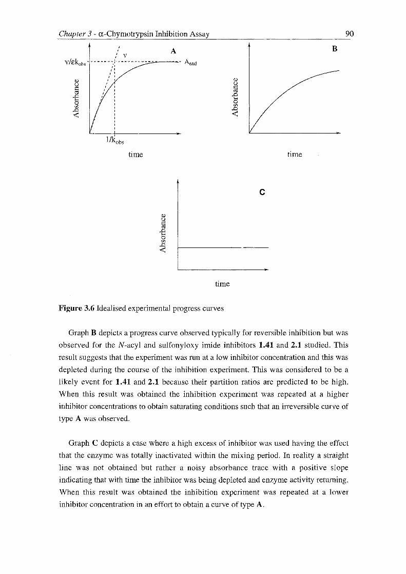
, I
,
r.1"r,,, .... <",n Inhibition
J J
" V - i' - _ ...... ,',
" I' I • I •
J I
•
1lkobs
time
A
time
Figure 3.6 Idealised experimental progress curves
90
B
time
c
Graph B depicts a progress curve observed typically for reversible inhibition but was
observed for the N-acyl and sulfonyloxy imide inhibitors 1.41 and 2.1 studied. This
result suggests that the experiment was run at a low inhibitor concentration and this was
depleted during the course of the inhibition experiment. This was considered to be a
likely event for 1.41 and 2.1 because their partition ratios are predicted to be high.
When this result was obtained the inhibition experiment was repeated at a higher
inhibitor concentrations to obtain saturating conditions such that an irreversible curve of
type A was observed.
Graph C depicts a case where a high excess of inhibitor was used having the effect
that the enzyme was totally inactivated within the mixing period. In reality a straight
line was not obtained but rather a noisy absorbance trace with a positive slope
indicating that with time the inhibitor was being depleted and enzyme activity returning.
When this result was obtained the inhibition experiment was repeated at a lower
inhibitor concentration in an effort to obtain a curve of type A.

Chapter 3 - a-Chymotrypsin Inhibition Assay 91 The major task therefore when using the progress curve assay method is finding
suitable inhibitor concentrations that give curves of type A suitable for analysis of kobs.
Several inhibition experiments were carried out to determine kobs at different inhibitor
concentrations and the resulting data analysed using a Tsou plot of [I]/kobs versus m from which kinactiKI can be obtained.

Chapter 3 - a-Chymotrypsin Inhibition Assay 92
5.4 ASSAY RESULTS
Each inhibitor of type 1.41 and 2.1 shown in Tables 3.1 and 3.2 respectively was
assayed using the incubation method detailed above to quickly determine the activity
time profile. Subsequent experiments depended on the potency and type of inhibition
observed in these preliminary experiments. In general five results were obtained from
the incubation assay.
a) The compounds were inactive.
b) The inhibitors inactivated a-chymotrypsin in a time-dependent manner at
such a rate that a number of data points could be obtained for analysis.
The inhibition constants kobs and kobs/[I] were then determined as
described above.
c) A rapid loss in enzymic activity was observed followed by a rapid regain
of this activity. It was proposed that these compounds were acting as
poor alternate substrate inhibitors. No further analysis was undertaken.
d) The rate of inactivation was too fast to be determined using the
incubation assay. These potent inhibitors were assayed using
progress curve kinetic methods.
e) The compounds were of such potency that they could notbe measured
accurately using the progress curve method at an inhibitor concentration
of ten-fold excess over enzyme.

Chapter 3 a-Chymotrypsin Inhibition Assay R
o):;Ao
Compound R number 1.4ld PhCH2
e Ph
f PhCH2
g PhCH2
h PhCH2
i Ph
j PhCH2
k PhCH2
1 PhCH2
m PhCH2
n PhCH2
I OL
1.41
L
S02CH3
S02CH3
S02 ~Ph
S02-{ )
S02-Q-CH3
S02-Q-CH3
S02-\ ~ > N02
S02-\ ~ CH3
A S02 C02Et
o Ph
S020-\=l S02-\ ~ 0
J:(Ph --0--0- ¥ 0
S02 ~ /; ~ /; S020
Table 3.1 N-[(sulfonyl)oxy]imide inhibitors of type 1.41
93

Chapter 3 a-Chymotrypsin Inhibition Assay
R
):~o
Compound R number
0 PhCH2
P Ph
q PhCH2
r PhCH2
L
Ac
Ac
c~Ph II 0
0
I OL
1.41
Ph':,
CANAO ........... Ph II H 0
Table 3.2 N-[(acyl)oxy]imide inhibitors of type 1.41
Compound R
number
2.1a Ph
2.1b Ph
2.1c PhCH2
2.1d H
2.1e H
2.lf H
R'
H
H
H
Ph
RY" ~~y,l o N 0
I OL
2.1
R"
H
H
H
H
R', R" = cyc10hexyl
CH3 CH3
L
S02CH3
Ac
S02CH3
S02CH3
S02CH3
S02CH3
Table 3.3 N-[(acyl and alkylsulfonyl)oxy]glutarimide inhibitors of type 2.1
94

95 S;hapter 3 - a-Chymotrypsin Inhibition Assay --------~--~~~~~------~----~----~----
INACTIVE COMPOUNDS
When no appreciable net loss of enzyme activity was monitored over the time course
of the experiment the compounds being assayed were considered to be inactive. In these
cases the compound concentration was increased up to 50-fold excess over enzyme in
an attempt to increase the rate of inactivation so that it could be measured, however,
stm no inhibitory activity was observed. Figure 3.7 depicts the results obtained on
assay of 1.410 which is representative of the N-[(acyl)oxy]succinimides 1.410-r which
were found to be inactive.
90
80~-r~~~r-~~~~~r-~~~~~
o 10 20 30 40 50 60 70
time (min)
Figure 3.7 Assay of inhibitory activity of compound 1.41 against a-chymotrypsin. a
Chymotrypsin (4!lM) was incubated with (A, 4JlM; B, lOOJlM) and enzyme activity
assayed using either N-Succ-(Alah-Pro-Phe-p-nitroanilide (A, 117JlM) or BTEE (B,
117JlM).
The oscillating nature of the graph suggests that there is a period of inhibition
followed by a temporary regain in activity. This trend could be explained in the terms
that several inactivating species are present in the assay mixture each of which only
inhibit the enzyme for a transient period before being metabolised by the enzyme.
These results are more likely to be explained, however, by the error associated with the
sampling measurements used in the incubation method. Although the individual rate
measurements are precise (± 1-2% error observed) they are difficult to reproduce
accurately; hence erroneous results can give the impression of enzyme inhibition or
activation. Therefore experiments need to be run in duplicate or triplicate for true
activity versus time profiles to become evident.

Chapter 3 - a-Chymotrypsin Inhibit_io_n_A_ss_a-=...y ________________ 9_6
TIME DEPENDENT INHIBITORS
Once it was shown by the appropriate logarithmic plot that inhibition was psuedo
first order, work was undertaken to optimise the inhibitor concentration so that an
accurate value of kobs was obtained. In practice this required finding the highest
inhibitor concentration that gave linearity over as many data points over the time course
of the assay. The analysis of the inhibition of a-chymotrypsin by compound 2.lf whose
activity versus time profile (Figure 3.8) serves as a representative example.
120~----------------------------------------~
100 .)
2.lf 60 -
40 - 13
20 -
0~--~--r-~--~--~--~1--~---r--~13~-~~·--~--~ o 20 40 60
time (min)
80 100 120
Figure 3.8 Time-dependent inactivation of a-chymotrypsin by 2.lf. a-Chymotrypsin
(4,-"M) was incubated with 2.lf (100J-LM) and enzyme activity assayed using N-Succ
(Alah-Pro-Phe-p-nitroanilide (l17J-LM).
Figures 3.9 and 3.10 display logarithmic plots obtained from the activity versus time
data of Figure 3.8 firstly over the full time course of the assay (100 minutes) and then
over the initial nine minutes.
Observation of the logarithmic plot displayed in Figure 3.9 indicates there is
deviation away from the psuedo first-order kinetics with time. The logarithmic plot
displays three phases. Initially psuedo-first order kinetics is observed. There is no "lag
phase" indicating immediate inhibition of a -chymotrypsin is due to the N-

Chapter 3 - a-Chymotrypsin Inhibition Assay 97 mesylglutarimide rather than a metabolite. This initial inhibition phase is followed by
an upward deviation away from psuedo-first-order kinetics i.e. the rate of inactivation
decreases with time. This may be accounted for by depletion of inhibitor due to
turnover. The final phase depicts a downward deviation indicating a second period of
inactivation of the enzyme at a slower rate than that observed initially. These
observations are consistent with possibly a hydrolysis product of the N
mesylglutarimide being a potential inhibitor of a-chymotrypsin. Over the time course
of the assay this species has increased beyond a threshold concentration such that has
returned to inactivate the cnzyme.
Given that there is deviation from psuedo-first order kinetics as the time course of
the assay increases linear regression analysis of data to determine kobs was carried out
only over the initial portion of the logarithmic plot of Figure 3.9 as shown in Figure
3.10. The initial data points are more dependable as indicated by comparison of the
correlation coefficients of the regressions carried out for the plots of :Figures 3.9 (r2 = 0.827) and 3.10 (r2 = 0.965). Analysis of the data in Figure 3.10 gives a value of 39 M
Is-1 for kobs/[I] for derivative 2.lfwhen N-Succ-(Alah-Pro-Phe-p-nitroanilide was used
as the incubation assay substrate.
0 .e-.... Ii>-.... t: <
<:Ii e .... -2 IS = ~ -3 ~
= - -4
-5 0 2000 4000 6000 8000
time (s)
Figure 3.9 Logarithmic plot of enzyme activity versus time over 100 minutes for the
data displayed in Figure 3.8.

Chapter 3 a-Chymotrypsin Inhibition Assay 98
t- o . :0' 't: .... o N 0 .... I CJ
< OS02CHg <1.1
~ -1 2.1f lSI
~ ~
= -2 -o 100 200 300 400 500 600
time (s)
Figure 3.10 Logarithmic plot of enzyme activity versus time over nine minutes for
inhibition data displayed in Figure 3.8.
The inhibition of a-chymotrypsin by 2.1f was then studied at different inhibitor
concentrations at 15-, 20-, 25-, 30-fold excess over enzyme with the ultimate aim of
determining a rigorous value for kinact/Kr for this inhibitor. The trend of the kobs values
with inhibitor concentration is shown below (see Figure 3.11).
3~------------------------------~
2.1f
s15 fold
s 20-fold
I I I
50 60 70 80
I!l
30-fold
I!l
40-fold
• I I
90 100 110 120
[I] (f.LM)
Figure 3.11 Plot of kobs versus [10] for compound 2.1f

Chapter 3 - a-Chymotrypsin Inhibition Assay 99 As observed from Figure 3.11 the value for kobs is increasing with inhibitor
concentration but the graph is characterized by an asymptote indicating there is a
maximum value for kobs. This results confirms that saturating kinetics are being
observed for the inhibition of a-chymotrypsin by compound 2.U which is one of the
features that identifies true mechanism-based inactivation (see Section 3.1 for a
discussion).
The analysis of this data using the Kitz and Wilson plot described previously
(Section 3.2) for determination of kinacJKI is shown below (Figure 3.12).
t1l2 (s) 400 - 2.1f EI
300 - EI
EI
200 J I J I
0.008 0.010 0.012 0.014 0.016 0.018
1/[1] (IlM-1)
Figure 3.12 Kitz and Wilson plot for inhibition of a-chymotrypsin by 2.U
The above plot is of the type obtained when non-saturating conditions are prevalent.
Linear regression analysis of this plot gives the y-intercept as negative and hence a
value for kinact/Kr cannot be obtained. This result may be explained in the tenus that the
kinact for the compound is high, however, analysis by the incubation method has shown
this compound to be a poor inhibitor which would normally possess a relatively low
kinact value. The error for each individual calculation of t1/2 at each of the four inhibitor
concentrations is high 15%) and will contribute to the non-saturating shape of the
graph. However the above graph is distinguished by a sharp rise in t1/2 at a lower
inhibitor concentration and therefore it could be considered that analysis at this
inhibitor concentration ([10 ] = 59mM, I5-fold excess over enzyme) has been
undertaken in non-saturating conditions.
THE LIBRARY UNIVERSITY OF CANTERBURY
CHRISTCHURCH, N.Z.

Atr,:rnC'-'n Inhibition 100
A Kitz and Wilson plot of the first three data points of Figure 3.12 which were
deemed to be obtained from inhibition experments carried out at saturating inhibitor
concentrations is shown below (Figure 3.13).
380
360 Hn' o N 0
340 I OS02CH3
tl/2 (s) 320 2.lf
300
280
260~~--~~~--~~~--~-r~--~~
0.008 0.009 0.010 0.011 0.012 0.013 0.014
Figure 3.13 Khz and Wilson plot for inhibition of a-chymotrypsin by 2.lf at saturating
conditions
The above plot is of the type obtained at saturating conditions permitting analysis of
kinactIKr which was determined to be 37 M- 1s-1 (kinact = 0.006 s-1, Kr = 156 JiM). A
plot of [I]/kobs versus [I] also gave a value of 37 M-1s-1.
This highlights the caution required when using kobs/[I] values as approximations for
kinactlKr. For a poor inhibitor, the kobs/[I] values calculated from inhibition experiments
where the inhibitor concentration is less than the Kr serve as an approximation for
kinact!I(r. The data points of Figure 3.13 were obtained from inhibition experiments
carried out at inhibitor concentrations 20-fold (78JiM), 25-fold (98~), and 30-fold
(l17JiM) excess over enzyme. The kobsl[I] values calculated from these experiments
were 24 M-1s-l, 24 M-1s-1 and 21 M-1s-l respectively. These inhibitor concentrations
are lower than the Kr calculated for 2.lf (156~LM) but the kobs/[I] values poorly
approximate kinacVKr.
In a series of other experiments a determination of kinactlKr was undertaken using
the progress curve method. A plot of [I]/kobs versus [I] gave a plot typical of non
saturating conditions hence kinact/Kr was unable to be calculated. However a value for
kinact (0.003s- 1) was obtained from the slope of this graph and is of the same magnitUde

Inhibition 101
of that value obtained from the incubation method above (kinact = 0.006s-1). On this
basis there appears to be a good agreement between the two methods for compound
2.lf.
ALTERNATE SUBSTRATE INHIDITORS
Alternate substrate inhibitors 71,80,97(a),197 function as substrates of the target
enzyme; however instead of chemically reacting with the enzyme, the alternate
substrate becomes bound so tightly to the active site that further access by natural
substrate molecules is prevented.
Compounds 1.41 and 2.1 may act as alternate substrate inhibitors if on formation of
the acyl enzyme the Lossen rearrangement is unable to occur. In this case the enzyme
would remain inhibited for the lifetime of the acyl enzyme species. The goal therefore
in the design of alternate substrate inhibitors is to prolong the life span of the acyl
enzyme by hindering the deacylation process using strategies based on either electronic
or steric reasoning. 189 If the deacylation process is rapid then the inhibitors will merely
be acting as substrates of the enzyme.
Compounds 1.41e and 1.41h displayed activity time profiles consistent with
alternate substrate inhibitors that only transiently inhibit the enzyme. As the results
obtained on assay of compound 1.41e (Figure 3.14) show there is initial rapid
inhibition of the enzyme the rate of inhibition increasing for increased inhibitor
concentration (compare graphs A and B). The enzyme then remains inhibited for a
period of time dependent on the initial concentration of inhibitor ([laD used in the
experiment. When [10 ] was ten-fold excess over enzyme this period was ca 7minutes
but increased to ten minutes when [Io] was increased to 25-fold excess over enzyme.
During this period the inactive enzyme is actively being hydrolysed but there still
remains enough inhibitor to elicit inactivation until a point where this is depleted and
enzyme activity is regained.

n"ln.Tr,,',","'1n Inhibition 102
120
100 Ph
o~o ,-." I
~ 80 OS02CH3 '-' :;.... ,...,
1.41e ..... " ..... ,...,
----a--~ 60 -< S :;.... N
40 = ~
20
o 10 20 30
time (min)
40 50 60
Figure 3.14 Assay of inhibitory activity of compound 1.41e against a-chymotrypsin.
a-Chymotrypsin (4JlM) was incubated with 1.41e CA, 40JlM; B, 100JlM) in O.lM
phosphate buffer, pH = 7.2, and 2.5% CH3CN. Aliquots (25JlL) were removed
periodically and assayed for activity for enzymic activity using N-Succ-(Alah-Pro-Phe
p-nitroanilide (A, B 117JlM) according to the incubation assay procedure discussed
previously.
POTENT INHIBITORS
The rate of inhibition of a-chymotrypsin by compounds 1.41d, 1.4lf, 1.41g, 1.41h,
1.41j, l.4lli, 1.41m, 1.41n, 2.1a, 2.1c, 2.1d, 2.1e was too fast to be determined using
the incubation assay indicating that these were potent inhibitors. To obtain a
measurable rate the inhibitor concentration was reduced but even at the lower inhibitor
concentrations employed some inhibitors still inactivated a-chymotrypsin at a rate too
rapid to be measured by the incubation assay. With other inhibitors an increased
number of assay points was able to be obtained however substantial deviation from
psuedo first order kinetics was observed.
In another attempt to determine a value for the inhibitory activity of these potent
inhibitors using the incubation method the sampling periods were changed for analysis.
• A B

nT"Tl""Tl Inhibition 103
In a typical incubation experiment aliquots of the enzyme-inhibitor assay mixture were
withdrawn at 1,3, 5,7,9 etc. minute intervals. To obtain a greater number of data
points for the initial period of inhibition aliquots were withdrawn at smaller time
intervals requiring that the duration of the data set collection for the calculation of Vt, be
decreased from 80s to 50s. The inhibitory activity of 1.41d was assayed in this manner
(Figure 3.15) and the subsequent logarithmic plot is shown below (Figure 3.16).
120
100 P».o ,-....
~ '-' I ~ ..... 80 OS02CH3 . ~ ...... .....
1.41d I;j 60 < ~
~ 40 ~ I!I
~ I!I
20 I!I
0 r'
0 100 200 300 400
time (s)
Figure 3.15 Assay of inhibitory activity of compound 1.41d against a-chymotrypsin.
Incubation of a-chymotrypsin (4rnM) with 1.41d (21mM, 5-fold excess over enzyme)
in O.IM phosphate buffer, pH = 7.2, with 2.5% CH3CN. Aliquots were withdrawn at t =
0.5, 1.75, 3min intervals before the enzyme was totally inactivated.
c o . P».o .....
t;.-..... j ..... OS02CH3 I;j
< ~ e -1 1:.1 1.41d ~ N
= ~
~
= -2 --3~------~------.-------~------~
o 100 200
time (s)

nu'mn.1" .. ,,'n,,',n Inhibition 104
Figure 3.16 Logarithmic plot of enzyme activity versus time over 200 seconds for the
data displayed in Figure 3.15.
A logarithmic plot with negative slope is obtained but as alluded above there is
substantial deviation away from psuedo first-order kinetics (r2 = 0.886). Therefore
kobs/[I] (502 M-1s-l) will serve as a poor approximation ofkinacJKr, but none the less is
a useful figure as it serves as a lower limit for kinactIKI. Comparison with the value
obtained for the glutarimide inhibitor 2.lf (kinacJKI = 37 M-1s-I, see previous section)
indicates that 1.41d is at least 13-fold more potent than 2.1f. Both 1.41d and 2.lf
contain a mesylate leaving group hence the difference in inhibitory activity is related to
the different ring size and substitution possessed by these compounds.
The activity versus time profile of the inhibition of a-chymotrypsin by compound
2.1d (Figure 3.17) serves to illustrate the activity displayed by compounds that were
found to be potent inhibitors over an extended period of time.
120
100 .. ,-...
~ '-' ..... 80 -...... .... .... ~ c.i 60 --< ~
& 40 -
= ~ 20
~
0 0
• / "
/ '. I \
./.A,.\ .~II ~ ...
20 40 .,.
60
time (min)
2.1d
80 100 120
---a- A _.- .• -.- B --iIII-- C
Figure 3.17 Assay of inhibitory activity of compound 2.1d against a-chymotrypsin.
Incubation of a-chymotrypsin (4~) with 2.1d CA, 1O~, 3-fold excess over enzyme;
B, C, 20flM, 5-fold excess over enzyme) in 0.1M phosphate buffer, pH = 7.2 with 2.5%
CH3CN. Aliquots (25flL) were withdrawn periodically and assayed for enzymic
activity using BTEE employing the incubation method.
Incubation assay of compound 2.1d carried out at an inhibitor concentration five
fold excess over enzyme (data sets B and C of Figure 3.17) resulted in a rapid loss of

Chapter 3 a-Chymotrypsin Inhibition Assay 105 enzyme activity followed by a partial regain in enzyme activity (ca t = 20-30min) then
a second rapid and ireversible loss of activity. A subsequent experiment was carried out
at a lower inhibitor concentration (three-fold excess over enzyme) (Figure 3.17, Data
Set A) in an attempt to lower the rate of inactivation and hence obtain data suitable for
analysis. Less rapid inactivation was observed «1 % activity remaining after seven
minutes) followed by a smaller recovery of enzymic activity compared to experiments
B and C. After this partial regain in enzymic activity a second irreversible loss was also
observed.
These results are similar to those obtained for the assay of compound 2.lf (see
Figures 3.8-3.10) and support an inhibition scenario where initial inactivation of a-
chymotrypsin is due to the unreactive imides 1.41 and 2.1 acting as "tme" mechanism
based inactivators. Hydrolysis of the inactive enzyme and inhibitor results in a
temporary regain in enzyme activity followed by a second but less rapid phase of
inactivation thought to be due to a hydrolysis product of the imides. These rates of
hydrolysis appear to be greater for more potent inhibitors. For example, there is a
smaller recovery of enzyme activity for the inhibition of a-chymotrypsin by 2.1d at
three-fold excess over enzyme than at five-fold excess (compare experiment A with B
and C of Figure 3.17). To whit there was only a slight recovery of enzyme activity
observed on assay of compound 2.lf (see Figures 3.8 and 3.9).
Assay of 1.41d using the incubation method gave results indicating that the rate of
inactivation of a-chymoptrypsin by this compound was too rapid to measured by this
technique. The inhibitory activity was then evaluated using progress curve kinetics.
Groutas et a184 have previously assayed 1.41d using this technique obtaining a value for
kobs/[IJ of 9000 M-1s-l. This experiment was repeated and the results are shown below.

Chapter 3 - a-Chymotrypsin Inhibition Assay 106
0.10
0.08 -III
III III III III III III III III III III III Aend liE
III
CI.i III t.I
= 0.06 -~ III ,.Q Phb '"" 0 rrJ
,.Q 0.04- III < '<1 o N 0
I I!I OS02CH3
0.02 -
1.41d
0.00 . I
0 10 20 30 40
time (min)
Figure 3.18 Progress curve assay of inhibitory activity of a-chymotrypsin of compound
1.41d against a-chymotrypsin. a-Chymotrypsin (0.073f1M) was added to an incubated
solution of 1.41d (0. 189j.LM; 3-fold excess over enzyme) and BTEE (117j.LM). Reaction
progress was monitored by change in absorbance at 'A = 256nm.
Analysis of this curve according to the graphical method of Tsou is shown below
(Figure 3.19).
-2
-3
~
< -4 I
'" = <l)
< '--' -5 .s
-6
-7 0 2
Ph~
o..J..NAo
3
I
OS02CH3
1.41d
4
time (min)
5
Figure 3.19 Initial portion of Tsou plot obtained for absorbance curve (see Figure 3.18)

Chapter 3 a-Chymotrypsin Inhibition Assay 107
There is a good correlation between the data and the kinetic model (r2 = 0.962) and a
value for kobsl[I] of 64 497 M-1s-l ± 10% was obtained. The ± 10% error in this
measurement, which is typical of progress curve measurements, was determined by
addition of the error evaluated by the regression process in the calculation of kobs (±
5%) and that in the experimental measurement of the inhibitor concentration, which
incorporated weighing of the inhibitor to prepare the stock solution and subsequent
dilution of this as required by each inhibition experiment.
It appears therefore that the progress curve measurements are more accurate than
those obtained with the incubation method where errors of up to ± 15% were calculated
for determination of kobs which reflects the greater number of experimental
measurements that need to made to determine kobs when using the incubation method.
However results were obtained for identical progress curve inhibition experiments that
were far beyond within 10% of each other even though the same buffer, enzyme,
inhibitor and substrate solutions had been used. It is considered that the extent of
mixing and the time taken to achieve this are of critical importance in obtaining
accurate measurements. Even though a mixing time of not more than 20 seconds was
strictly adhered to the extent of mixing is a hard variable to control. To minimise the
error in measurements experiments were repeated so that two values for kobs within
10% of each other were obtained.
The fact that the logarithmic plots conceal alot of error in progress curve
measurements contributes to the difference in the value we obtained for the inhibition of
a-chymotrypsin by 1.41d (kobsf[I] :::: 64 497M-1s-l) to that obtained by Grouta..'l et al84
(kobs/[I] :::: 9 000 M-1s-l).
These values differ by an order in magnitude and illustrate the care that needs to be
taken when comparing inhibition results between laboratories.
Of the experimental details offered by Groutas et al84 in publishing their kobsf[I]
value the only difference between the experimental conditions we employed is the use
of DMSO as opposed to CH3CN however the volume of these solvents used in the
assay is small and is considered to have a negligible effect compared to other assay
variables. These include inhibitor purity and the freshness of the solutions used in the
assay; the loss of enzyme activity of a stock solution of enzyme in phosphate buffer was
noted and stock solutions of BTEE should be prepared daily.195b The above
experiments have also been run at low inhibitor concentration of 3-fold excess over
enzyme and hence there will be deviation away from the kinetic model used; this will
also contribute to the discrepancy between the two results.

Inhibition 108
As stressed earlier it is important that inhibition experiments are carried out at
saturating inhibitor and substrate concentrations for the steady-state assumption of the
kinetic model to remain valid. It is quoted that less than ten percent of the substrate
should undergo hydrolysis during the assay so that the steady-state for enzyme-substrate
reaction of an inhibition experiment does not differ from that of the control
experiment.198 The extent of hydrolysis can be calculated directly from the absorbance
versus time curve using the Beer-Lambert Law. Calculation for the above curve (Figure
3.18) indicates that 70% of BTEE has undergone hydrolysis.
An experiment run at this low inhibitor concentration could be considered to be
overstretching the steady-state assumption of the kinetic model even though other
researchers have reported assay conditions with similar low inhibitor excesses. It has
been shown that an inhibitor with a low partition ratio can be analysed at low inhibitor
excess with little deviation away from psuedo first-order kinetics.25 There is evidence
to suggest, however, that significant "turnover" pathways for N-acyl and sulfonyloxy
imide inhibitors 1.41 and 2.1 exist and hence their partition ratios will be high.
For accurate measurement of kobs/[I] using the progress curve method high
concentrations of inhbitor will have to be used to achieve the all important saturating
conditions. This was observed in the inhibition of a-chymotrypsin by compound 2.1f
where a Kitz and Wilson plot to test for saturating conditions gave a result indicative of
non-saturating conditions when low inhibitor concentrations were employed (see
Figure 3.12).
The results of the inhibition of a-chymotrypsin by 1.41d at higher concentrations is
shown below (Figure 3.20).

Chapter 3 a-Chymotrypsin Inhibit_io_n_A_s_s_ay"-----______ ~ ________ ]_09
0.10..,.-----------------,
0.08
0.06 I!I • •• I!I A Phb • g • B
0.04 a. g iii! C o N 0
~.aP I
OS02CH3
0.02 .1'.11
fiafll
1.41d
0.00 1:1 I I I
0 1 0 20 30 40
time (min)
Figure 3.20 Progress curves for inhibition of a-chymotrypsin by 1.41d. a
Chymotrypsin (0.073).lM) was added to an incubated solution of 1.41d (A, 0.189!lM, 3-
fold excess over enzyme; B, 0.365mM, 5-fold excess; C, 0.949).lM, 13-fold excess) and
BTEE (l17).lM). Reaction progress was monitored by change in absorbance at A = 256nm.
From :Figure 3.20 it can be observed that curves Band C fall over more sharply than
does curve A. Hence the rate of inhibition of a-chymotrypsin is more rapid at higher
concentrations. Analysis of curve B gives kobsl[l] = 17 790 M-1s-l (r2 = 0.989) and
kobs/[l] = 3 562 M-1s-l for curve C (r2 = 0.993) and as alluded the correlation between
data and model is better for higher inhibitor concentrations. Suprisingly the Aend values
are higher for these curves than that of curve A suggesting the extent of substrate
hydrolysis is greater. Curve C has the appearance of a curve associated normally with
reversible inhibition. These results suggest that the inhibitor is being depleted
significantly during the course of the experiment making the maintainence of saturating
conditions difficult for this inhibitor. This is confirmed by the Tsou plot of [IJlkobs
versus [IJ for 1.41d (Figure 3.21).

Chapter 3 a-Chymotrypsin Inhibition Assay
1000
800
600
[I]/kobs (/lMs)
400
200
0 0 1
Ph~
O~N~O I
OS02CH3
1.41d
[I] (/lM)
Figure 3.21 Tsou plot of [I]/kobs versus [1] for 1.41d
110
2
The above plot is of the type obtained when non-saturating conditions are prevalent
characterized by a poor correlation (r2 = 0.886). The y-intercept is negative and hence a
value for kinactlKr can not be obtained however a value for kinact (0.00 13s-1) was
obtained from the slope which provides valuable information regarding the inhibition
process.
The graph above however is distinguished by a sharp rise in [I]/kobs at higher
inhibitor concentrations and therefore it could be considered that these have been
obtained at the necessary saturating conditions required for analysis. A plot of these two
data points cut the positive y-axis and subsequently an approximate value of kinactlKr
was found to be 4 653 M-1s-l. Comparison with the kinactlKr value obtained for the
glutarimide inhibitor 2.1f (kinact/Kr = 37 M-1s-l) indicates that 1.41d is IOO-fold more
potent. Comparison of the kinact and Kr values obtained for these inhibitors provides the
valuable insight that 1.41d owes its increa'ied potency to greater binding affinity for a
chymotrypsin.
To allow an accurate kinactlKr analysis a number of experiments have to be carried
out at high inhibitor concentration however this was found to be excruciatingly
frustrating to achieve as illustrated by the progress curve analysis of compound 1.41g.

nUTY\AT''''''''C'Tl Inhibition 111
0.10
III 1!I1lI1lI1lI1lI1!I1!I1lI1lI1lI!i1!l
• • •• • 0.08 III
••• A end III • II1II II1II II1II II1II II1II II1II II1II II1II III
(jj I!I • II1II I;j a II1II
0.06 I%J • of Ph III A III
o~o 0 III • • B til
,.Q 0.04 - III II1II C -<
-<:l I!I J
f OS02Ph
0.02 ~111 ., 1.41g
0.00 0 10 20 30 40
time (min)
Figure 3.22 Progress curves for inhibition of a-chymotrypsin by 1.41g. a
Chymotrypsin (O.073J.LM) was added to an incubated solution of 1.41g (A, O.189!lM,
[1o]/[Eo] = 3; B, 0.365mM, [Io]/[Eo] = 5; C, O.55mM, [1o]/[EoJ = 8) and BTEE
(l17J.LM). Reaction progress was monitored by change in absorbance at A = 256nm.
Figure 3.22 displays the progress curves obtained for inhibition of a-chymotrypsin
by 1.41g obtained at three different inhibitor concentrations. As the inhibitor
concentration increases the curves fall over more steeply as seen in the trend from curve
A to curve C. The initial rates of reaction are also observed to decrease reflecting
increased rate of inhibition and the Aend value is lower hence less substrate has been
hydrolysed. Therefore saturating conditions are more likely to be prevalent in the
inhibition experiment C than A or B.
This is confirmed by the Tsou plot of [1]/kobs versus [I] for 1.41g which has a
negative y-intercept. As observed in the Tsou plot for 1.41d (Figure 3.21) there is a
sharp rise from a data point considered to be obtained in non-saturating conditions to
data points obtained at saturating conditions. A plot of [1]lkobs versus [I] for these two
points (not shown) also cuts the negative y-axis.
To determine kinactlKI for compound 1.41g it was therefore necessary that
experiments be carried out at higher inhibitor concentrations. However, in an
experiment carried out at an inhibitor concentration ten-fold excess over enzyme the
enzyme was found to be totally inactivated within the mixing period as characterized by
a flat absorbance versus time graph (see graph C of Figure 3.6). It was not possible,

112
therefore, to obtain a value of kinact/Kr for this compound as the rate of inhibition at
saturating inhibitor concentrations was too rapid to be measured.
These results indicate that the progress curve method is inadequate for measuring the
inhibition of a-chymotrypsin by 1.41£. The potent activity displayed by 1.41£ is such
that only the activity at low inhibitor concentrations can be measured however these
measurements will be inaccurate and deviate away from the pseudo first-order kinetics
of the modeL An accurate measurement of the rate of inhibition of a-chymotrypsin by
compound 1.41g in order to determine kinactlKr would require the use of stopped-flow
methods.
The results described above still, however, provide a qualitative description of
structure and activity relation when compared to those obtained for other inhibitors at
the same concentration. For example, the inhibition experiment carried out for 1.41d at
an inhibitor concentration ten-fold excess over enzyme resulted in a normal progress
curve (see progress curve C of Figure 3.20) whereas, as above, 1.41g totally inactivated
a-chymotrypsin at this concentration. Therefore the inhibition of a-chymotrypsin by
1.41g is more rapid than that of 1.41d.
300
Ph~
200 o N 0 I
[I]/l{obs OS02Ph
!lMs 1.41g
100
0.0 0.1 0.2 0.3 0.4 0.5 0.6
[I] JlM
Figure 3.23 Tsou plot of [l]/kobs versus [I] for 1.41g
For an accurate measurement of kinact/Kr we require kobs values at saturating
inhibitor concentrations. Accuracy will be improved if a greater number of inhibition
concentrations are used. A larger data set has been obtained for the glutarimide inhibitor
2.1g, with [I]/kobs values being obtained from inhibitor concentrations ranging from

Chapter 3 - a-Chymotrypsin Inhibition Assay 113
0.189JlM (three-fold excess over enzyme) to 18.2SJlM (2S0-fold excess over enzyme).
The Kitz and Wilson plot for 2.1e is shown below (Figure3.17).
[I]lkobs
JlMs
2.-------------------------T-----,
2.1e
o..;:.t-----.------r------.,.......----\ 0.00 0.01 0.02
Figure 3.24 Tsou plot of [I]/kobs versus [1] for 2.1e
Again a straight line with negative slope is obtained from which a value for kinact
(0.009s- 1) can be obtained however the y-intercept is not positive, which does not
permit analysis of kinactlKJ.
The results discussed for the succinimide inhibitors 1.41d and 1.41f and the
glutarirnide inhibitor 2.1g were representative for the other potent inhibitors of type
1.41 and 2.1. Due to the inability to calculate kinactIKI which was to be used as the
measure of inhibitory activity providing the basis for structure activity comparison other
means have to be used.
As discussed earlier kobsl[I] is conventionally used when comparing the potencies of
inhibitors. A high kobsl[I] value for a compound indicates that it is a more potent
inhibitor than a compound with a lower kobi[I] value. The constant kobsl[I] is used
routinely to compare compounds assayed using the incubation method however this
term is not independent of inhibitor concentration for potent inhibitors a'lsayed using
the progress curve method; hence for a valid comparison to be made experiments for
each inhibitor will have to be run at the same inhibitor concentration (an inhibitor
concentration of ten-fold exces over enzyme was chosen for this study).

Chapter 3 - a-Chymotrypsin Inhibition Assay 114 In noting the trends in kobs/[I] values for compounds assayed using the progress
curve method we found that, suprisingly, those compounds which were determined to
be more potent inhibitors by the incubation method possessed lower kobs/[I] values than
those determined to be weaker. For example, compound 1.41d could not be analysed
accurately using the incubation method because it inactivated a-chymotrypsin rapidly
within four minutes at an inhibitor concentration five-fold excess over enzyme. By
decreasing the time intervals used in the sampling a lower limit for kobs/[I] was able to
be calculated (kobs/[I] z kinact/Kr = 502 M-1s-l) indicating that 1.41d was at least 13-
fold more potent than the glutarimide 2.1f (kinacJKr = 37 M-1s-l). Compound 2.1f only
inactivated a-chymotrypsin within 100minutes at an inhibitor concentration 25-fold
excess over enzyme. On progress curve analysis the converse trend in the kobs/[I] values
was observed; 1.41d (3 562 M-1s-l) versus 2.1f (7888 M-1s-l).
This trend is also observed in comparing the inhibitory activities of compounds
1.41d and 2.1e. Compound 2.1e, like 1.41d, could not be assayed accurately using the
incubation method because it rapidly inactivated a-chymotrypsin. Assay at a low
inhibitor concentration 3-fold excess over enzyme served to provide a lower limit for
kinacJKr for the inhibition of a-chymotrypsin by 2.1e (kob/[I] z kinactlKr 403 M -1 s-l)
hence 1.41d (kobs/[I] z kinactlKr =502 M-1s-l) is more potent.
Progress curve assay of these compounds confirmed this result. At a concentration of
twenty-fold excess over enzyme compound 1.41d totally inactivated a-chymotrypsin
within the mixing time. In comparison, at a concentration of 2.1e 250-fold excess over
enzyme a-chymotrypsin remained active. Based on these results 1.41d is more potent
than 2.1e but this compound has a lower kobs/[I] value 1.41d (kobs/[I] == 3 562 M-1 s-1)
versus 2.1e (kobs/[I] == 14321 M-1s-l).
A preliminary explanation for the reversal in trends for kobJ[I] may lie in
consideration of the inhibitor concentration dependence for kobs/[I]. For the
mechanism-based inhibitors of type 1.41 and 2.1 studied [I]/kobs is observed to increase
with inhibitor concentration. For kobs/[I] the converse will be observed i.e. kobs/[I]
decreases at increased inhibitor concentration. A tentative hypothesis predicts a lower
kobs/[I] value for a more potent inhibitor if for inhibition experiments run at the same
inhibitor concentration the more potent inhibitor is acting as the weak inhibitor but at a
higher concentration.
In the absence of a theoretical basis for an explanation as to why more potent
inhibitors possess lower kobs/[I] values when assayed using progress curve kinetics this
method for comparison has its basis in the empirical observations described for the
representative inhibitors above.

Chapter 3 - a-Chymotrypsin Inhibition Assay 115 In the following structure-activity discussion the activities of inhibitors have
beencompared on the basis of kobs/[I] values obtained from inhibition experiments run
at a ten-fold excess of inhibitor over enzyme where there exists no more rigorous
information regarding the inhibition process. Hence compounds possessing a low
kobs/[I] when assayed employing the progress curve are considered to be more potent
inhibitors than compounds with high values.

Chapter 3 - a-Chymotrypsin Inhibition Assay
3.5 STRUCTURE-ACTIVITY DISCUSSION
Compound R number 1.41d PhCH2
: e Ph
f PhCH2
g PhCH2
h PhCH2
i Ph
j PhCH2
k PhCH2
L
S02CH3
S02CH3
R
o):;Ao I OL
1.41
S02 ~Ph
S02-{ > S02-Q-CH3
S02-Q-CH3
SOz-{ < ) N02
SOz-{ )
kinact (s-l) 0.0013
A
A
A
A
A
0.0004D
O.003D
TABLE 3.3 Inhibition of a-chymotrypsin by inhibitors of type 1.41
kobs/[I] (M-Is-I)
3562B
Alternate Substrate 6471B
C
C
Alternate Substrate
826B
6427B
A Unable to be calculated because alternate substrate inhibitor or inactivated a
chymotrypsin too rapidly at [lorEo] > 10 for kobs/[I] values to be determined.
B Value obtained from progress curve analysis [lorEo] = 10; [Eo] = 0.073f..lM; [S]
= 117f..lM. The lower the value the more potent the inhibitor.
C Inhibition too rapid to be measured by progress curve method at [lorEo] = 10;
[Eo] 0.073IlM; [S] = 1171lM.
116
D Value obtained from slope of Tsou plot of [I]/kobs versus [I] by linear regression
analysis

Chapter 3 a-Chymotrypsin Inhibition Assay ~------------~~----------------
117 In comparison to the "reference" inhibitor 1.41d it can be seen that the incorporation
of an aromatic substitutent can result in an increase in inhibitory activity. On the basis
that both 1.4ld and 1.41f totally inactivated a-chymotrypsin at an inhibitor excess 20-
fold over enzyme these compounds are essentially equipotent however a dramatic
increase in activity was observed with compounds 1.4lg and 1.4Ih. Both inhibited a
chymotrypsin at a rate too fast to be measured at an inhibitor concentration ten-fold
excess over enzyme suggesting that these have successfully harnessed the binding
energy available in interacting with the Sn I subsites of a-chymotrypsin. Compound
1.4lj was very potent with a kobs/[I] value of 826 M-1s-I (a low value indicates potent
inhibition) but less so than 1.4lg and h. This perhaps suggests that its larger bulk
dissuades favourable binding which corroborates with the result obtained with 1.41f,
also a bulkier inhibitor, which showed no enhancement in inhibitory activity relative to
1.41d.
Compound 1.41k was designed with the aim of enhancing the activity of 1.4lg as the
increased electron withdrawing properties of the nitro group would be thought to
increase inhibitory activity in two ways. The rate of the Lossen rearrangement has been
determined to be related directly to the strength of the acid that is released during
formation of the isocyanate. 86 It is considered that m-nitrobenzene sulfonic acid which
is released during the inhibition of a-chymotrypsin by 1.4lk is a stronger acid than
benzene sulfonic acid released during the inhibition cycle of 1.4lg due to the inductive
effect of the nitro group. A more rapid Lossen rearrangement leads to an increase in
kinact resulting in more potent inhibition. The increased electron withdrawing ability of
the nitro group is also thought to have a secondary influence on the potency of
inhibitors by increasing the electrophilicity of the scissile carbonyl of the succinimide
ring. This would activate formation of the acyl-enzyme again increasing kinact.
Despite the prediction of increased inhibitory activity the converse was observed,
with 1.41k found to be less potent than 1.41g but comparable inactivity to 1.41f. It must
be noted that the increased electron withdrawing effect of the nitro group in 1.4lk may
also increase the rate of deacylation so that it competes with the Lossen rearrangement
such that kinact may remain constant for 1.4lk and 1.41g. A definitive comparison of
the kinact values for 1.4lg and 1.41k was not possible however as the inhibition of a-
chymotrypsin by 1.41g was too rapid to measure. However a comparison of the kinact
values of compounds 1.4lj (kinact = 0.0004 s-I), which is thought to have similar
electronic properties to 1.4lg, and 1.4lk (kinact = 0.003 s-1) does indicate there is a
favourable ten-fold increase in kinact. Despite this ten-fold increase compound 1.41g is
more potent, hence it is quite likely that compound 1.4lk lacks the topology to interact
with a-chymotrypsin to the same extent as does 1.4lg hence the lower inhibitory
activity.

Chapter 3 - a-Chymotrypsin Inhibition Assay 118 Compound 1.4li in comparison to compound 1.41k has an electron donating methyl
group on the phenyl ring. This compound was found to have comparable activity to
1.41g hence the methyl group has no effect on inhibitory activity i.e. the electron
donating and steric effects of this group are minimal.
Compounds 1.41e and l.4li, both of which have a phenyl substituent (R) rather than
the benzyl substituent of the compounds discussed above, were found only to be
transient alternate substrate inhibitors of a-chymotrypsin. This result may be explained
in the terms that the Lossen rearrangement to form the inactivating isocyanate has not
occurred or if it has, the binding of these compounds to a-chymotrypsin has been such
that the resulting isocyanate does not have the correct geometry or proximity to react
with His-57. The Lossen rearrangement has been found to not only depend upon the
strength of the departing acid as discussed above but also on the electron releasing
power of the substitution present in the resultant isocyanate.86 On this basis it may be
considered that the benzyl substituent is more favourable in promoting the Lossen
rearrangement and binds more effectively with the S 1 pocket of a-chymotrypsin. The
results obtained with compounds 1.41e and l.4li indicate that a benzyl substituent is
required if a compound is to exhibit mechanism-based irreversible inhibition.
The increased activity of the aromatic compounds 1.41g and 1.41h relative to the
simple derivative 1.41d is almost certainly due to a favourable hydrophobic interaction
between the phenyl ring contained within the fomer with either Phe-81 or Phe-192
which are located in the Sn' sub sites of a-chymotrypsin.199 To harness this available
binding energy and in an attempt to further increase enzyme-inhibitor recognition
compound 1.411 (Table 3.4) was prepared which incorporated the design of the L group
as an amino acid mimic. Initial design plans focused on the synthesis of the sulfonyl
chloride 3.15, a phenylalanine mimic, that on coupling to 2.25 would give compound
3.13 that possesses an aromatic substituent incorporated into an amino acid like
substituent. An attempted synthesis of 3.13 using the Strecker reaction with the
phenylalanine derivative 3.14 however was unsuccessful presumably due to the steric
crowding of the a-centre of 3.14 a conclusion which is supported by documented low
yields of the Strecker reaction when using secondary alkyl halides. 173
As shown below in Table 3.4 1.41k, which lacks the desired aromatic substitution,
was found only to be a moderate inhibitor of a-chymotrypsin.

Inhibition 119 Ph
B,fC02Et
3.14
~A P~
Phn Ph
+ CI02/C02Et
Ph o N 0
o 6s02 ='-CC0
2Et
I
OR
2.25 3.15 3.13
Scheme 3.7 A, 1) K2S03, EtOHIH20, reflux 2) PCls, POCh, 70 °C
Compound R L kin act kobs/[l] number (s-I) (M-Is-I) 1.411 PhCH2 CR3 A B
SOZA~02Et m PhCH2 o Ph O.OOlC 1609D
S020-N~ S02~ ~ 0
n PhCH2 ;:(Pn 0.0l2c 12589D
-o--ci- Ii 0 SOz ~;/ ~;/ SOzO
Table 3.4 Inhibition of a-chymotrypsin by derivatives of type 1.41
A Unable to be calculated
B Value (kobs/[I] = 278 M-Is·l) obtained from incubation analysis using N-Succ
(Alah-Pro-Phe-p-nitroanilide [IolEo] = 10; [Eo] = 0.073JlM; [S] = 117JlM. Note
that this value cannot be compared to those obtained for 1.41m and n.
C Determined from slope of a plot of [I]/kobs versus [1]
D Value obtained from progress curve analysis [lolEo] = 10; [Eo] = 0.073JlM; [S]
= 117JlM. The lower the value the more potent the inhibitor.

(";hapter 3 - a-Chymotrypsin Inhibition Assay 120 The "dimeric" compounds 1.4lm and 1.4ln were designed to release a species
capable of inhibiting a-chymotrypsin providing a second cycle of inhibition. It was
thought that compound 1.4lm might be more potent than the related monomeric
compound 1.41f. Although these compounds showed reasonable activity, the opposite
trend was observed suggesting that the second inactivating species is ineffectual,
probably because it escapes from the enzyme active site.
If a second species derived from the initial compounds 1.41m and n was involved in
the inhibition process these compounds would display complicated multiphasic kinetic
behaviour relative to the monomeric compounds, however, in the preliminary studies
undertaken this was not observed. Further development of these types of inhibitors may
include incorporation of structural features, i.e. macrocycles, to prevent the release of
the second inactivating species from the active site.
Of interest is the difference in the kinact values for the "dimeric" inhibitors 1.4lm
and l.4ln. There is a ten-fold increase in kinact between the meta substituted compound
1.4lm and the para substituted compound l.4ln however the former is the more potent
inhibitor (as indicated by the lower kobs/[I] value; a lower value indicates more potent
inhibition. This result suggests that l.4ln binds less favourably to a-chymotrypsin than
1.4lm, perhaps due to its increased steric demands, resulting in poorer inhibition. This
is consistent with the trend observed with the monomeric compounds where compounds
1.41f and 1.4lj were found to be less potent than 1.41g and h. In the same manner the
"extended" dimeric compound l.4ln was found to be two-fold less potent than 1,411 on
comparison of kobsf[I] values.
Compound R L Result number 1.410 PhCH2 Ac Inactive
p Ph Ac Inactive
q PhCH2 c~Ph Inactive II 0
r PhCH2 Ph, ~ 0
Inactive
C'lN.JlO ........... Ph I 11 H
0
Table 3.5 Inhibition results for N-[(acyl)oxy]succinimides
All the N-[(acyl)oxy]succinimides 1.410-r displayed in Table 3.5 were found to be
inactive suggesting that an acyl leaving group is unfavourable for inhibition. To
improve the activity of 1.410 compound 1.4lq was prepared which has a better leaving

Inhibition 121 group as evident by comparison of the pKa values of the respective acids (pKa acetic
acid 4.74, pKa trans-cinnamic acid 4.44), however this compound too was inactive. An
attempt was also made to impart activity to this series by increasing enzyme-inhibitor
recognition and to this end compound 1.41r which contains N-Cbz (L)-phenylalanine as
a leaving group was prepared however this too was found to be inactive.
The inactivity of the N-[( acyl)oxy ]succinimides of type 1.410-r appears to be due to
the lack of a suitable leaving group but may also be rationalised in terms of their ability
to mimic a natural peptide substrate. The crystal structures of compounds 1.410-q were
determined during the course of this thesis and comparison to a crystal struture of an
active inhibitor may prove valuable in determining if N-[(acyl)oxy]succinimides
possess the favourable binding properties that are required for inhibition. This proposed
analysis is only approximate as the crystal structures which are obtained in the solid
state give no indication of the conformations acquired by compounds in the active site.
The crystal structure of 1.41f, whose inhibitory activity against both a-chymotrypsin
and human leukocyte elastase is discussed above, has been solved,87g and is pictured in
Figure 3.18 in contrast to a topological picture obtained for 1.41q from data (see
Appendix) provided by the refinement for this compound. A comparison of 1.41f with
1.41q is thought to be representative of the other N-[(acyl)oxy]succinimides 1.410, p,
and r as 1.41q and 1.4f both contain a trans-styryl group hence the differences in
conformations will be illustrated more clearly.
The first point to note on comparison of the inactive N-[(acyl)oxy]succinimide 1.41q
and the active N-[(sulfonyl)oxy]succinimide 1.41f (Figure3.18) is that 1.41q lacks the
typical horseshoe or H shape of 1.41f which is commonly observed in the crystal
structures of dipeptides containing (L)-phenylalanine. This characteristic conformation
results from the two amino acid side chains extending from the backbone of the
dipeptide and running approximately paralleJ.200-203 This geometry displayed by the
natural substrates of serine proteases is reflected in the complementary shape of the
active sites of these enzymes. 199,204
It therefore appears that the sulfonyl(oxy) segment of potent inhbitors not only
imparts favourable electronic properties for inhibition but has a role in setting the
geometry of the inhibitor such that it mimics a natural substrate. The carbonyl group
lacks this geometry and imposes a linear and flat conformation to the N
[(acyl)oxy]imides which is unfavourable for binding to serine proteases.

rnT',In<",n Inhibition
o
Ph-...r'YO-~~ o K Ph o
1.41q
122
Figure 3.18 Comparison of crystal structure for 1.4lf (above) against that for 1.41q
(below)
The results obtained on assay of the six-membered glutarimide compounds 2.1a-f
against a-chymotrypsin are presented in Table 3.6. These compounds provided a
valuable insight into the importance ring size has with regards to structure-activity
relation.

Compound R
number
2.1a Ph
2.1b Ph
2.1c PhCH2
2.1d H
2.1e H
2.lf H
Inhibition
R' R"
H H
H H
H H
Ph H
cyc10hexyl
CH3 CH3
Rn'R R" 3 4
o N 0 I OL
2.1
L==S02R'"
==C(O)R'"
L kinact (s-l)
S02CH3 0.011
Ac C
S02CH3 C
S02CH3 C
S02CH3 0.009
S02CH3 0.006E
kinactfKI (M-is-I)
100000A
C
C
C
C
37E
Table 3.6 Inhibition of a-chymotrypsin by derivatives of type 2.1
A Values determined from Tsou plot of [I]/kobs versus [I]
kobs/[I] (M-l s-i)
7784B
15845
D
452B
14321B
F
B Value obtained from progress curve analysis [IolEo] == 10; [Eo] == 0.073!lM; [S]
== 117!lM. The lower the value the more potent the inhibitor.
123
C Unable to be calculated due to inactivation of a-chymotrypsin being too rapid at
[IolEo] = 10 or Tsou plot of [I]/kobs versus [I] having a negative y-intercept.
D Inhibition too rapid to be measured by progress curve method at [IoIEo] == 10;
[Eo] == 0.073mM; [S] == 117!lM.
E Values obtained from Kitz and Wilson plot oft1l2 versus 1/[1]
F Value (kobs/[I] = 20 M-1s-l) obtained from incubation method analysis [IolEo] =
25; [Eo] = 3.922!lM; [S] = 117!lM. Note that this value eannot be compared with
those obtained using the progress curve method.

Chapter 3 - a-Chymotrypsin Inhibition Assay 124
The N-mesyl glutarimide 2.1fwas found to be a poor inhibitor (kinactlKI = 37 M-1s-
1) when assayed using the incubation method. However, the glutarimide 2.le which
possesses more suitable substitution to interact with the hydrophobic S 1 pocket was
found to be a more potent inhibitor. This compound was unable to be assayed
accurately using the incubation method although a lower limit for kinact/KI was
calculated to be 408 M-1s-l hence 2.le is at least ll-fold more potent than 2.1f. An
analysis using the progress curve method gave a kobs/[I] value of 14321 M- ts-l hence
2.1£ is less active than the five-membered succinimide compounds (see Table 3.3). The
difference in the observed potencies of these compounds lies in consideration of their
binding affinity for a-chymotrypsin since their kinact values were found to be of the
same magnitude; KI for the more potent 2.le must therefore be lower than the value for
2.1£ (KI = 156~M) calculated from a Kitz and Wilson plot of tl/2 versus 11[1].
The introduction of an aromatic substituent onto the ring resulted in a dramatic 2 000
fold increase in inhibitory activity as shown by comparison of kinactlKI values for 2.1£
(kinactlKI = 37 M-1s-l) and 2.la (kinactlKJ = 100 000 M-Is·l) which has a favourable a
phenyl substituent. The kinact value for 2.la was only slightly higher than that for 2.1£
hence the dramatic increase in activity is due to increased binding affinity as confirmed
by the comparison of the KJ values for 2.la (0.1~M) and 2.1£ (156~lM).
Although no kinact/KI analysis was made for 2.Id comparison of the kobs/[IJ values
obtained for 2.la (kobJ[I] = 7 784 M-ts-I) and 2.ld (kobs/[I] 452 M-1s-l) indicate that
the placement of the phenyl substituent at the ~-position leads to an increase in
inhibitory activity (note that studies have shown that a lower value corresponds to more
potent inhibition). This may suggest that 2.ld may be able to bind in a different manner
in the active-site projecting both the phenyl substituent and a portion of the glutarimide
ring into the SI specificity pocket such that this compound can mimick the p]
phenylalanine residue of the natural substrate while retaining the correct conformation
to react with Ser-195.
As observed in the succinimide series incorporation of an a-benzyl substituent onto
the glutarimide ring afforded the most potent inhibition observed in the glutarimide
series. Compound 2.lc totally inactivated a-chymotrypsin at a concentration lO-fold
excess over enzyme within the mixing period and is therefore of comparable activity to
the succinimides l.4Ig and h.
As mentioned previously these potent compounds would require the use of stopped
flow methods to determine kinact/l<]:. However an indication of their relative potencies
can be obtained by comparison of the kinact values obtained for the succinimide and
glutarimide series. With the exception of the "dimeric" inhibitor l.4In whose kinact is

125 Chapter 3 - a-Chymotrypsin Inhibition Assay ----------~--------------------------------
of the same magnitude the kinact values of the glutarimide inhibitors are an order of
magnitude higher.
Assuming that compounds 1.41g and h, and 2.1c have comparable binding affinity
for a-chymotrypsin it appears that 2.1c may be the more potent inhibitor due to the
higher kinact value observed in the glutarimide series. This trend has been reported in
the comparison of five and six membered haloenol lactones discussed previously
(Section 1.3, Chapter 1) where the higher inhibitory activity of the six membered
derivatives was rationalised in the terms that the a-halo ketone reactive species
unmasked was on a longer tether and therefore closer to His-57 promoting formation of
the inactive enzyme.
Suprisingly, the N-acetylglutarimide 2.1h showed inhibitory activity against a
chymotrypsin. If the mechanism of inactivation of a-chymotrypsin by 2.1h involves thc
Lossen rearrangement the question arises as to why the related
N[(acyl)oxy]succinimides did not exhibit similar activity. The differences in activity
between the N-acyloxy glutarimide and succinimide derivatives could be explained in
the terms that the glutarimide binds more favourably to a-chymotrypsin and in such a
way that allows a mode of inhibition to proceed which is unavailable to the
succinimides.
In summary, we assayed the inhibitory activity of the series of five- and six
membered imides represented by structures 1.41 and 2.1 against a-chymotrypsin using
both the incubation method, for slower inhibitors, and the progress curve method for
faster more potent inhibitors.
R 3h4
oAN~O I
OL
1.41
L=S02R'
= C(O)R'
Rn'R R" 3 4
o N 0 I
OL
2.1
=C(O)R'"
These results suggest that for potent inhibition to be observed the ring substitution
must be aromatic or hydrophobic in nature; alkyl type substitution resulted in poor
inhibition. A benzyl substituent was found to be optimum for activity being equivalent
to the side chain of phenyalanine the preferred PI residue of natural substrates of a
chymotrypsin.

Inhibition 126
No activity was observed in the succinimide compounds 1.41o~r, probably due to the
fact that an acyl group is not a sufficiently good leaving group. Suprisingly, activity was
observed in the glutarimide example 2.1b suggesting that the six-membered ring has
favourable binding properties.
An increase in inhibitory activity in the succinmide series was observed when the R
substituent of the leaving group (OS02R) was aromatic. It is thought that this R group
may be interacting favourably with the Sn' subsites of a-chymotrypsin and their
increased activity owing to increased binding energy. However not all aromatic type
inhibitors of type 1.41 displayed a favourable enhancement in activity; those derivatives
with bulkier subtituents e.g. 1.4lf and 1.4li displayed slightly decreased potency
indicating there are steric limitations to the extent of binding with the Sn' subsites.
The glutarimides 2.1a, e and f were analysed to have higher kinact values than the
succinimides 1.41. This is thought to result from an increase in the rate of reaction of
the reactive isocyanate formed in the Lossen rearrangement with His-57. This is
explained in the terms that the isocyanate is on a longer tether and hence closer to His-
57 for glutarimides. Due to the glutarirnide compounds possessing higher kinact values it
is thought that appropriately substituted derivatives, having high binding affinity for a-
chymotrypsin, will display the optimum activity of the inhibitors whose assay has been
discussed in this chapter. The greater number of substitution sites on the glutarimide
ling will also make these compounds more amenable to the development of imides as
potent and selective mechanism-based inactivators of serine proteases.

CHAPTER 4
ENANTIOPURE N-[(ALKYLSULFONYL)OXY]
SUCCINIMIDES AND
INHIBITION

Chapter 4 - Enantiopure N-[(sulfqnyl)oxy]succinimides and inhibition 127
4.1 CHIRALITY AND BIODISCRIMINATION
Chirality is a property of molecules which defines important aspects of our
biochemistry and interaction with the environment. It also determines the biological
activity of molecules finding wider everyday use as pharmaceuticals, agrochemicals,
and foodstuffs.
Biological activity arises from the interaction of a compound with a biomolecule
such as an enzyme or a receptor. These biomolecules are constructed from amino acids
and carbohydrates - nature's own chiral building blocks - and hence are chiral
themselves. If the compound is chiral then the type and magnitude of the interaction,
and therefore the biological response, is related to its stereochemistry. Thus the
stereoisomers of an organic compound are likely to interact differently with its
biomolecule.205,206
This biodiscrimination is manifest throughout nature,207,208 and has long been
recognised in taste and odour perception209-211 and other physiological responses.
Perhaps the most striking example is the stereoselective action of the enzymes and
receptors responsible for the chemical reactions that instill life. The enzymes of our
body, with few exceptions, metabolize only the (L)-enantiomers of amino acids and the
(D)-enantiomers of carbohydrates and receptors act on the spatial elements these
stereochemistries dictate.
TYPES OF BIODISCRIMINATION
In general, there are four different ways in which stereoisomers act on their target
chiral biomolecule.212 Of these four, only three are observed when studying a single
enzyme system in vitro as in this thesis. In one situation the biological activity resides
totally with one stereoisomer while another is devoid of activity: for example the (S,S)
peptidyl haloenol lactone 1.35 is active against a-chymotrypsin (a-CT) and human
neutrophil elastase (HNE) while the (S,R)-peptidyl haloenollactone 4.1 is inactive at
the same concentration.79
)"1" o. Dy"-BU o o:lsro
(S,S)
1.35
kobs/[I] = 8 000 M-1s-l/a-CT79
kob/[I] = 1100 M-1s-1/HNE79
(S,R)
4.1
inactive

Chapter 4 - Enantiopure N-[(sulfonyl)oxy]succinimides and inhibition 128
Alternatively, the stereisomers may both be active but differ in potency. This case is
illustrated by the reversible inhibition of human neutrophil elastase (HNE) by peptidyl
trifluoromethylketones (see Section 1.3, Chapter 1 for a discussion). Compound 1.1536
has the correct (S)-configuration at the a-position to mimick the natural peptide and
hence is 100-fold more active than the epimer 4.236 which has an unfavourable non
natural (R)-configuration, and two-fold more active than the racemate 4.3.
'-..../
Cbz-Val-Prer-~~CF3 H 0
: CF Cbz-Val-Pro-N~ 3
H 0
1.15 4.2
K j = 0.0008 11M / HNE36 K j = 0.015 11M / HNE36
Cbz-Val-Pro-~~CF3 H 0
4.3
Ki = 0.0016 11M / HNE192
Discussion of the biodiscrimination displayed in the above two situations has lent
itself to the nomenclature proposed by Lehmann et al.213 The more biologically active
stereoisomer has been named the eutomer; the corresponding less potent or inactive
isomer is then called the distomer. The ratio of the activities (eutomer:distomer), the so
called eudismic ratio, is a measure of the degree of stereoselectivity of the biological
activity. The eudismic ratio for the above example (Kil.1S:Ki4.3) is ca 200 which is a
value typical of chiral medicinal agents.214
The remaining biodiscrimination type observed is relatively rare. In this situation
both stereoisomers may have similar or equal activities as illustrated by the inhibition
of Type 1 human steroid Sa-reductase by 4.4 and 4.5.215

J:..hapter 4 - Enantiopure N-[(sulfonyl)()xy]succinimides and inhibition
CI
o
4.4
Ki,app == 9nM
Type 1 Steroid Sa-reductase
o
4.5
Ki,app == 10nM
CI
Type 1 Steroid Sa-reductase
129
The stereoselective inhibition of serine proteases by mechanism-based inhibitors of
type 1.41 has been undertaken by Groutas et al. 87e The results for the inhibition of
human leukocyte elastase by (-)-(R)- and (+)-(8 )-1.41d are shown below.
Ph-"
o~o I
OS02CH3
(-)-(R)
1.41d
k2iKr == 330 M-1s-11 HLE87e
(+)-(8)
1.41d
k2/KI = 1500 M-1s-11 HLE87e
The eudismic ratio for the above example is equal to five suggesting there is low
stereo selectivity for the inhibition which has been rationalised in the terms that both
enantiomers of 1.41d can display the correct spatial orientation to be accomodated in
the active site.
Human leukocyte elastase has a long and shallow active site and its substrate
specificity at Sl has been found to be regulated by substrate length.216 The preferred PI
residue is valine however the hydrophobic Sl pocket of human leukocyte elastase will
also accomodate residues such as alanine and phenylalanine with short substrates. With
extended substrates the specficity demands increase and only those substrates with
valine as the PI residue are accomodated, in the same manner the stereo selectivity of
inhibition will increase a commonly observed observation that is known as Pfeiffer's
rule. 217 The low stereoselectivity of inhibition observed does not mean that human
leukocyte elastase acts with low stereospecificity; it is thought that with longer
inhibitors the difference in rates of inhibition for stereoisomers will be larger and a
higher eudismic ratio observed.
Groutas et al87e obtained similar results to those above for human leukocyte elastase
when investigating a-chymotrypsin i.e. (-)-(R)- and (+)-(8)-1.41d were equipotent.

Chapter 4 - Enantiopure N-[(sulfonyl)oxy]succinimides and inhibition 130
This enzyme does not display the broad substrate specificity of human leukocyte
elastase and invoking Pfeiffer's rule it may have been expected that the two
enantiomers would display different levels of activity. The stereoselective action of a-
chymotrypsin has been explained in terms of binding requirements to specific structural
motifs in a model of the active site proposed by Cohen and Milovanovic in a study of
the absolute steric course of hydrolysis of a number of esters of substituted succinic and
propionic acids.152 Figure 4.1 depicts the binding of diethyl 2-benzylsuccinate (4.6) in
the active site proposed by these authors.
4.6
JJ. a-chymotrypsin
a-chymotrypsin active site model
(R)-4.6 accomodated (8)-4.6 no fit
Figure 4.1 The binding of diethyl 2-benzylsuccinate 4.6 in Cohen and Milovanovic's
active site model for a-chymotrypsin
From Figure 4.1 only (R)-4.6 is hydrolysed by a-chymotrypsin as it is the
enantiomer that has the correct spatial orientation to bind properly within the active
site. For this enantiomer, the phenyl ring is juxtaposed so that it can interact favourably
with the Sl hydrohobic binding pocket while the scissile bond is aligned correctly in
the hydrolytic site, N. The a-carbethoxymethylene group is in the correct position to
permit favourable hydrogen bonding with the acylamino binding pocket Am. The a-H
of (R)-4.6 points toward the interior of the active site at a domain H of restricted
volume, contiguous with Ar.
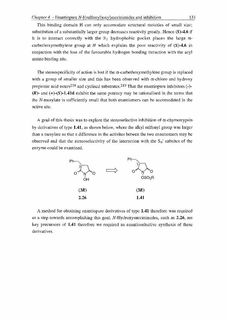
Chapter 4 - Enantiopure N-[(sulfonyl)oxy]succinimides and inhibition 131
This binding domain H can only accomodate structural moieties of small size;
substitution of a substantially larger group decreases reactivity greatly. Hence (8)-4.6 if
it is to interact correctly with the S 1 hydrophobic pocket places the large a
carbethoxymethylene group at H which explains the poor reactivity of (8)-4.6 in
conjuction with the loss of the favourable hydrogen bonding iteraction with the acyl
amino binding site.
The stereospecificity of action is lost if the a-carbethoxymethylene group is replaced
with a group of smaller size and this has been observed with a-chloro and hydroxy
propionic acid esters218 and cyclised substrates.219 That the enantiopure inhibitors (-)
(R)- and (+ )-(8)-1.41d exhibit the same potency may be rationalised in the terms that
the N-mesylate is sufficiently small that both enantiomers can be accomodated in the
active site.
A goal of this thesis was to explore the stereoselective inhibition of a-chymotrypsin
by derivatives of type 1.41, as shown below, where the alkyl sulfonyl group was larger
than a mesylate so that a difference in the activites betwen the two enantiomers may be
observed and that the stereoselectivity of the interaction with the Sn' subsites of the
enzyme could be examined.
Ph-" "
O~O I
OH
(3R)
2.26
(3R)
1.41
A method for obtaining enantiopure derivatives of type 1.41 therefore was required
as a step towards accomplishing this goal. N-Hydroxysuccinimides, such as 2.26, are
key precursors of 1.41 therefore we required an enantioselective synthesis of these
derivatives.

Chapter 4 - Enantiopure N-[(sulfonyl)oxy]succinirnides and inhibition
4.2 METHODS SUCCINATES
FOR OBTAINING 132
CHIRAL
Biodiscrimination has had implications for the agrochemical and foodstuffs
industries: chiral herbicides and plant growth factors display biodiscrirnination, and
large industries have developed for the stereoselective synthesis of (L)-amino acids as
food supplements and (D)-glucoses as non nutritive sweeteners.220,221 Chiral medicinal
agents have been known to exhibit biodiscrimination as long as 80 years ag0222 and
this phenomenon abounds in the medical field223 and hence has held the greatest
consequence for the pharmaceutical industry.
Industry's requirement for chiral pharmaceuticals, agrochemicals and foodstuffs has
fueled research into the synthesis of enantiopure compounds and subsequently the
analysis of the enantiomer compositions of molecules. This has had a huge "wash-over"
effect for academic pursuit as there exist several ways of obtaining chiral, non-racemic
compounds essential for the study of nature's stereoselective processes, whether that be
the investigation of the stereospecificity of an enzyme's catalytic action, or mimicking
her biosynthetic prowess by threading the stereochemical labyrinth of her structurally
diverse and complicated molecules through asymmetric synthesis. 224
There exist a number of methods for the preparation of chiral succinates and these
are discussed briefly in turn.
CLASSICAL RESOLUTION
Traditionally, optically pure compounds have been obtained by classical resolution
techniques at a favourable stage during the synthesis of their racemates. The use of
enantiomerically pure starting materials and enzymes in synthesis and of the latter in
kinetic resolutions and the advent of stereoselective synthesis has reduced, though not
totally, the need for classical resolution. A synthesis purporting all of these design
principles can still be subject to rabid racemization requiring resolution of
intermediates and products.
Earlier in this thesis (Section 2.1, Chapter 2) 2-substituted succinic acids were
identified as intermediates in the synthesis of 3-substituted N-hydroxysuccinimides key
precursors in the preparation of the target inhibitors 1.41. These acids also represent a
favourable target for classical resolution. The resolution of 2-substituted succinic acids
in particular 2-benzylsuccinic acid 2.13 by the optically active amine C+)-(R)
methybenzylamine (MBA) followed by the alkaloid strychnine (Scheme 4.1) has been
carried out by Fredga.225 Succinic acid mono esters featured in one synthesis of 2-

Chapter 4. - EnaIltiopure N-[(sulfonyl)0.xy]succinimides and inhibition 133
substituted succinic acids employing Stobbe condensation methodology (Method C,
Scheme 2.5, Chapter 2) and these also present themselves as targets for classical
resolution. The resolution of methyl 2-isopropylsuccinate by both enantiomers of
methylbenzylamine has been achieved.226
A resolution procedure may prove wasteful if the undesirable enantiomer cannot be
used in a different application or recycled. The above resolution procedures are
economical in this sense as both enantiomers of the target inhibitors 1.41 provide
valuable information regarding the stereospecifics of inhibition of a-chymotrypsin.
Classical resolution therefore is a competitive route to desired enantiopure inhibitors
1.41 considering the resolving agents employed, strychnine and methylbenzylamine,
are both commercially available.
Classical resolution, however, involves numerous recrystallisations each with
inherent loss of material, and is lengthy (in terms of time). For example, the resolution
of racemic 2-benzylsuccinic acid 2.13 (Figure 4.1) as described in the literature,225
was undertaken using pure 2.13, requiring repeated recrystallisation of the material
obtained from synthesis (in our experience four recrystallisations from water were
required).
The racemic acid 2.13 is treated with two equivalents of (+ )-(R)-methylbenzylamine
to give the diastereoisomeric salts 4.7 and 4.8. The required separation of these is trivial
as 4.8 crystallises from the reaction solution, and 4.7 remains in the mother liquor.
Repeated recrystallisation of salt 4.8 from H20 completes the resolution of (-)-(S)-2-
benzyl succinic acid 2.13 ([a]o = -27.2°) and this is obtained by decomposition of the
purified salt 4.8 with dilute H2S04 and extractive work-up with Et20. Salt 4.7 is
decomposed to give impure (+)-(R)-2-benzylsuccinic acid 2.13 which is then further
resolved by treatment with strychnine, the resulting salt 4.7 requiring no purification to
give pure (R)~2.13 ([a]D = +27.00 ) which is obtained as described for (S)~2.13.

Chapter 4 - Enantiopure N-[(sulfonyl)oxy]succinimides and inhibition
4.7
(+,+ )-salt stays in solution
Ph-" '}-,
H02C C02H
(R)
2.13
impure [U]D = + 19°
(-)-strychnine
4.9
(+,-)-salt crystallises
(RIS)
racemic
2.13
QH3
H2N~Ph (+)-(R) -MBA
4.8
(-,+ )-salt crystallises
(S)
2.13
[ulD = -27.2°
Ph-" "'1\
H02C C02H
(R)
2.13
[ulD = +27.0°
134
Scheme 4.1 Classical resolution of racemic 2-benzy lsuccinic acid 2.13 by (+)
methylbenzylamine (MBA) and (-)-strychnine.

Chapter 4 - Enantiopure N-[(sulfonyl)oxy]succinimides and in_h_i_b_it_io_n _______ l_35
In addition to developing resolution procedures for 2-substituted succinic acids
Fredga has undertaken the groundwork in determining the absolute configurations of
the (+)- and (-)-enantiomers of the acids obtained. The method he has exclusively used
has been that of quasi-racemates.227 In brief, it has been shown that (+)-2-
benzylsuccinic acid has the same configuration as (+ )-malic acid whose absolute (R)
configuration has been unequivocably established.228
The specific rotations obtained by Fredga for the above resolution are considered to
be the maximum specific rotations for (+ )-(R)- and (-)-(8)-2.13 and have therefore been
used routinely in the determination of the optical purity of samples prepared using other
methods as seen below.
KINETIC ENZYMIC RESOLUTION
The length of a classical resolution however does not present itself in the resolution
of racemates by enzymes. On presentation of a racemic substrate the enzyme due to its
chirality behaves in an stereoselective manner reacting exclusively with one
stereoisomer leaving the other untouched. As discussed earlier the (+ )-(R) enantiomer
of racemic diethyl 2-benzylsuccinate 4.6 can only be accommodated in the active site
of a-chymotrypsin and is hydrolysed to (+)-(R)-ethy12-benzylsuccinate (3.11). The (-)-
(S) enantiomer of 4.6 is left untouched as observed in Scheme 4.2 which depicts the
enzymic kinetic resolution of 4.6 by a-chymotrypsin.
Ph"h a-chymotrypsin Ph-" Ph"h '" > ~ + Et02C C02Et H02C C02Et Et02C C02Et
4.6 1Sh
(R) (8)
4.10 4.6
JA JA
Ph-" Ph"h ", ~
H02C C02H H02C C02H
(R) (8)
2.13 2.13
Scheme 4.2 A, NaOEt, EtOH, rt

Chapter 4 Enant~opure N-[(sulfonyl)oxy]succinimides and inhibition 136
Both (R)-4.10 and (S)-4.6 can be hydrolysed with ethanolic sodium hydroxide to
give the corresponding (R)- and (S)- acids 2.13 whose rotations ([a,]25D = +27° and
[a.]25D -270 respectively) were found to be comparable to the definitive rotations
determined by Fredga above; (R)-2.13 ([a,]25D = 27.00) and (S)-2.13 ([a,]25D ::= -27.2°)
indicating that the products obtained by the enzyme kinetic resolution were of high
purity.
This resolution procedure has the potential to return amounts of both enantiomers of
the racemic substrate in a day period. However high concentrations of a,-chymotrypsin
have to be used making this method prohibitive considering the supply of this enzyme
in our laboratory and the hydrolysis of 4.6 is slow and with no suitable pH-Stat
apparatus available, the 18h duration of the hydrolysis course to be followed manually
makes this approach attractive to only an insomnic chemist.
Enantiopure inhibitors can then be prepared from the enantiopure 2-benzylsuccinic
acid obtained from the above classical and enzymic resolutions employing the
methodology of Scheme 4.3 used in the synthesis of racemic inhibitors of type 1.41
discussed in Chapter 2. The reactions of Scheme 4.3 which depicts the representative
synthesis of (S)-1.41d have been shown to be devoid of racemization by Groutas et
al87e who found the product inhibitors prepared by this route to be enantiopure by
chiral shift reagent determination.229
(S)
2.13
Phb o N 0
I
OR'
(S)
C [ 2.25 R' ::= CH2Ph y... 2.26 R'::= H
D
Scheme 4.3 A AC20, reflux
B H2NOCH2Ph, toluene, reflux
C H2/10% Pd-CITHF
D CH3S02Cl, pyridine, CH2Ch
Ph~
oJ-..o~o
(S)
2.27
B

Chapter 4 - Enantiopure N-[(sulfonyl)oxy]succinimides and inhibition 137
ASYMMETRIC SYNTHESIS
Asymmetric synthesis provides the chemist with the power to employ synthetic
methods that not only result in the desired transformations of starting materials to
products but control the absolute stereochemistry of the stereogenic centres which are
created as a result of the synthetic operation.
An asymmetric synthesis of chiral succi nates is therefore more flexible than the
previous methods discussed. Methods that have been employed include the synthesis of
chiral succinates from naturally occurring substrates,230 a strategy widely known as the
chiron approach.231 This method has been used in the synthesis of imide containing
psuedo peptide sequences 2.37 from (L)-phenylalanine (4.11). The stereochemistry
residing in 4.11 is incorporated into the key intermediate 2.36 which mediates the
stereochemistry in the final product 2.37. However, to its disadvantage the chi ron
approach often involves multi-step syntheses.
H2NX
C02H
( H Ph
4.11 2.36
o
-~:Q-OS02R' Ph 0
2.37
Scheme 4.4 Synthesis of 3-benzylsuccinimide containing pseudo peptides
The asymmetric hydrogenation of unsaturated succinate derivatives has been
undertaken232 however methods based on the principle of relayed asymmetric
induction have proved popular. In this process asymmetric information is introduced to
the racemic substrate before the reaction where the new asymmetric centre is created
and then the chiral originator, a so-called chiral auxiliary is removed. Two auxiliaries
that have furnished short syntheses of chiral alkyl succinates are iron acyl complexes233
and the chiral oxazolidinones developed by Evans. 234
Evan's chiral oxazolidinones prepared from naturally ocurring amino acids such as
valine and phenylalanine are readily available. Of the chiral auxiliaries derived from
these two amino acids those prepared from phenylalanine are crystalline not requiring
the extra purification steps associated with the valinol derived auxiliaries and contain a
good chromophore facilitating TLC analysis. The phenylalanine oxazolidinones are
easily attached to the racemic substrate and recycled and hence were the chiral auxiliary
of choice presenting an attractive route to the desired chiral alkyl succinate
intermediates.

Chapter 4 - Enantiopure N-[(sulfonyl)oxy]succinimides and inhibitio.:....n ____ _ 138
The mode of asymmetric induction by Evans chiral auxiliaries lies in the
consideration of enolate chemistry. For a reaction at the a-position to be stereos elective
several conditons have to be fulfilled among the most important being the geometry of
the enolate formed. Factors which can control the stereoselectivity of enolate formation
include choice of base, solvent, temperature and metal counter ion but most importantly
from the viewpoint of this thesis the chiral auxiliary. In this respect the Evans
oxazolidinones are attractive in that they give exclusively (Z)-enolates.
A second factor in determining the stereochemical outcome in the reaction is that the
chiral auxiliary imposes a strong facial bias for the approach of the eletrophilic
alkylating agent towards the enolate. It is here the substitution present the chiral
auxiliary comes into playas the approach of the electrophile occurs on the less hindered
face of the enolate.
Alkylation and aldol additions of N-acyl compounds derived from chiral
oxazolidinones proceed with high diastereoselectivity as illustrated by relevant
literature examples of the synthesis of chiral succinates. In 1981 Evans235 prepared
(2R)-benzylsuccinic acid employing the aldol addition of a N-acyloxazolidinone boron
enolate derived from(L)-valinol. The key aldol adduct was prepared in 99%
diastereoisomeric excess as determined by gas capillary chromatography which was
degraded to give (2R)-benzylsuccinic acid whose optical purity was judged to be 98%
by comparison to the maximum specific rotation obtained by Fredga.225
In 1988, Fadel and Salaun236 reported the preparation of chiral succinic acid diesters
employing the distereoselective alkylation (>95% as determined by IH NMR) of (L)
valinol derived N-acyloxazolidinones. The enantiomeric excess of the esters was
determined by chiral shift reagent to be >95%.
In 1993, Crimmins et al237 described a stereoselective synthesis (>95%
diastereoisomeric excess) of succinic acid mono esters employing sodium base
mediated alkylation of a chiral N-acyloxazolidinone derived from (L)-phenylalanine.
This methodology was repeated in 1998 by Levy et al226 and McClure et a1238 who
reported the synthesis of chiral succinate derivatives with high diastereoselectivity.
The trend in these examples illustrates how Evans oxazolidinones are widely utilised
for the synthesis of chiral compounds and that the stereochemistry of these reactions
has been so rigorously established that present day synthetic communications rarely
involve a critique on the stereoselectivity of these reactions-an example being patents
filed for the synthesis of renin inhibitors using oxazolidinone methodology.239

Chapter1~nantiopure N-[(sulfonyl)oxy]succinimides and inhibition 139
4.3 SYNTHESIS AND INHIBITORY ACTIVITY OF AN ENANTIOPURE N-[(ALKYLSULFONYL)OXY] SUCCINIMIDE
To incorporate the Evans oxazolidinone methodology into a synthesis of an
enantiopure inhibitor of type 1.41 a retrosynthetic analysis was carried out. The
disconnection of (R)-l.4ld serves as a representative example and is shown below
(Scheme 4.5).
Ph-"
o~o
Ph-"
I
OS02CH3
(R)
l.4ld
o~o
Ph-"
h o X X 0
4.12
X=OH, OR
I
OH
(R)
2.26
Scheme 4.5 Retrosynthetic analysis of (R)-l.4ld
The retrosynthetic analysis proceeds with disconnections discussed earlier in the
synthesis of the racemic counterpart. Disconnection of the bond between the oxygen
and the reactive moiety L of (R)-1.41d gives (R)-3-substituted N-hydroxysuccinimide
(R)-2.26 and alkylsulfonic and alky1carboxylic acid derivatives as reagents.

Chapter 4 - Enantiopure N-[(sulfonyl)oxy]succinimides and inhibition 140
Disconnection of (R)-2.26 proceeds by cleavage of the imide bonds, as indicated, to
give hydroxylamine and the chiral 3-alkylsuccinate reagent 4.12.
Retrosynthetic analysis of the key chiral succinate reagent 4.12 proceeded as shown
in Scheme 4.6. Dissection of 4.12 as indicated gives the two synthons 4.13 and 4.14
which correspond to the chira12-benzylpropionate reagent 4.15 and bromo acetate esters
4.16. Disconnection of 4.15 gives the chiral auxiliary 4.17 and 3-phenylpropionyl
chloride 4.18.
(NH o-{
o
4.17
III
4.15
Ph-, \ ~
o;:~o X X
4.12
X=OH,OR
4.18
+~ X 0
4.14
III
Scheme 4.6 Disconnection of 4.12 using a chiral propionate synthon

Chapter 4 - Enantiopure N-[(sulfonyl)oxy]succinimides and inhibition 141
The starting point of the synthesis of an enantiopure inhibitor was the preparation of
the chiral oxazolidinone 4.17 in two steps (Steps A-B of Scheme 4.7) from (L)
phenylalanine 4.11.
A B
4.11 4.19
o A o NH
~Ph 4.17 4.15
Scheme 4.7 A 1) BF3.0Et, 0.75h, rt, 2h reflux, 2) BH3.S(CH3h, 12.25h rt,
11.25h reflux, 3) 5N NaOH, overnight
B diethyl carbonate, K2C03, 4h, 136°C
C 1) n-BuLi, THF, 0.5h, -78 °C 2) 3-phenylpropionyl chloride, overnight
-78°C rt
An initial preparation of (L)-phenyalaninol 4.19 was carried out using lithium
aluminium hydride reduction of phenylalanine. Work up of this reaction was hindered
by a persistent emulsion, which was removed by filtration through ceHte but probably
contributed to the low observed yield for the reaction (10% ).
The low yield prompted the use of another method and hence the synthesis of (L)
phenylalaninol was carried out by borane reduction of (L)-phenylalanine according to
the literature procedure (Step A, Scheme 4.7).240 Thus (L)-phenylalanine was treated
with boron trifluoride etherate followed by borane-dimethyl sulfide (1.15 equiv.).
Aqueous alkali was added and the resulting reaction slurry refluxed overnight.
Extractive work-up gave (S)-phenylalaninol 4.19 in 91 % yield. Little emulsion was
observed with this method and although this intricate procedure was lengthy (three days
in total) and involved hazardous reagents and conditions a higher yield was observed.
An added advantage is that borane reductions typically proceed with less
racemisation.240
A mixture of 4.19 and diethyl carbonate (2 equiv.) and K2C03 (0.5 equiv.) was
heated for 4h at 136 °C to give the oxazolidinone chiral auxiliary 4.17 in 98 % yield.

Chapter 4 - Enantiopure N-[(sulfonyl)oxy]succinimides and inhibition 142
However when this reaction was repeated the results above proved difficult to
reproduce; charring of the reaction mixture occurred on heating which on work-up gave
a crude brown mass which proved difficult to purify by recrystallisation due to oiling
problems. However we found the sucess of the subsequent acylation step such that
these unfavourable samples of 4.17 could be used without hindering the yield of the
reaction.
The chiral oxazolidinone 4.17 was acylated following literature directions239,241 by
treatment with n-BuLi (1.05 equiv.) followed by 3-phenylpropionyl chloride (1.1
equiv.), itself obtained from treatment of 3-phenylpropionic acid with SOCl2 (1.2
equiv.) under refluxing conditions, to give crude 4.15. This was purified by flash
chromatography to give pure 4.15 as a white solid in 83% yield. The sucess of this
reaction depended much on the quality of the n-BuLi and thus the reagent was
frequently titrated to determine an accurate active concentration. The reaction was
repeated in similar high yields illustrating the ease in which these compounds can be
prepared highlighting the advantageous use of chiral auxiliaries derived from
phenylalanine.
The key stereoselective reaction of the synthetic scheme was then carried out by
treating 4.15 with LDA at -78°C to give the corresponding chelated lithium enol ate
4.17 which was alkylated by excess ethyl bromo acetate as the reaction solution was
allowed to warm to room temperature overnight (Scheme 4.8).
4.15
(S,R)
4.21
+
(S,S)
4.22
4.20
B
Scheme 4.8 A 1) LDA -78°C Ih 2) ethylbromoacetate, overnight, -78°C - rt
..

Chapter 4 - Enantiopure N-[(sulf(myl)oxy]succinimides and inhibition 143
Work-up gave a brown residue which was purified by flash chromatagraphy. Proton
NMR analysis of this product gave a spectrum consistent with the predicted (S,R)
diastereoisomer 4.21 but also indicated the presence of a second component in ca 5%
by 1 H NMR. This was determined to be either the minor diastereoisomer 4.22 or
starting material 4.15. Homonuclear decoupling experiments were carried out to make a
definitive identification especially concentrating on locating a second triplet and quartet
which would only be attrituable to the minor diastereoisomer 4.22. These experiments
proved difficult to analyse due to overlapping signals especially the methylene protons
of the ethyl ester and those of the oxazolidinone ring of 4.21 since the irradiations in
this region of the spectrum (3 4.00ppm) were to be of important diagnostic value.
Confirmation of the presence of staring material in the alkylation product was
obtained by comparison to an authentic spectrum of 4.15. Of note, the l3e NMR
spectrum revcaled no presence of the minor diastereoisomer 4.22. Further evidence for
the presence of 4.15 in the alkylation product was obtained from LiOH/30% H202
hydrolysis of a sample of the alkylation product. Proton NMR analysis of the product
obtained indicated the presence of 3-phenyl propionic acid presumably arising from
hydrolysis of 4.15.
Attempts were made to further purify the alkylation product by repeating the
chromatography lowering the polarity of the eluting solvent, using more silica, and
collecting smaller fractions. However no effective separation of 4.15 and 4.21 could be
effected.
The presence of starting material in the alkylation product suggested that formation
of the lithium enolate 4.20 had not been quantative. Possible causes included not
maintaining the reaction at -78 oe during enolisation such that the enol ate formed could
be undergoing decomposition via a ketene pathway. A product of this decomposition
would be the chiral oxazolidinone 4.17 however inspection of the 1 H NMR spectrum of
the crude product for the NH resonance diagnostic for 4.17 indicated its absence. This
placed the quality of the LDA into question and consequently a new solution was
employed which was then titrated according to literature procedure242 and found to be
1.8M a significant deviation from the quoted 2.0M.
The alkylation procedure was repeated and in addition to purification by
chromatography the product was recrystallised. The white solid obtained possessed a
sharp melting point (60-62 OC) however 1 H NMR analysis indicated the presence of the
disastereoisomers 4.21 and 4.22 in the ratio of 10: 1, by examining the integrals of the
resonances assigned as the methyl protons of the ethyl esters of this diastereoisomeric

Chapter 4 - Enantiopure N-[(sulfonyl)oxy]succinimides an-'---.-d_i_n_hl_·b_it_io'-n _____ -'---.-1_44
pair, equivalent to a diastereoisomeric excess of 91 % (see Figure 4.2 for the
appropriate IH NMR spectrum).
The assignment of the spectrum displayed in Figure 4.2 was undertaken by carrying
out several homo nuclear decoupling experiments aided by comparison with spectra
obtained for the chiral oxazolidinone 4.17 and N-acyloxazolidinone 4.15. The spectrum
can be divided into three parts which at several points overlap. The triplets (0-1.25
ppm) according to the methyl protons of the ethyl esters of the diastereoisomers 4.21
and 4.22 are easily distinguished and due to their chemical equivalence, unlike the
resonances of the diastereotopic protons featured in the rest of the spectrum, appear as a
triplet. Further downfield (02.4-3.3 ppm) are signals resulting from the merging of two
spin systems made up of a series of doublets of doublets. The first doublet of doublets
downfield is due to the one of the benzylic protons of the 4-benzyl substituent of the
oxazolidinone ring. The other proton of this type then merges with the resonances for
the benzylic protons of the 2'-benzyl substituent which in turn merge with the
resonances for the methylene protons in the a-positon relative to the carbonyl of the
ethyl ester. The protons of the oxazolidinone ring overlap with the quartet of the ethyl
ester at 0-4.0ppm. Of the two protons at the 3-postion of the oxazolidinone ring one is
a triplet due to coupling constants around this ring being of the same magnitude. The 4-
H of the oxazolidinone ring overlaps with the 2'-H at 0-4.5 ppm which is shifted
considerably downfield from the spin system it is a part of.

Chapter 4 - Synthesis of enaI1tiopure N[(sulfonyl)oxyJimides and inhibition 145
+
~
[;
o~ t
o=<z:( 0
1.1') -~ r
U")
0?
"" <:r
Vl
<::r
Figure 4.2 Selected region of IH NMR spectrum for product obtained on alkylation of
4.15 by ethyl bromoacetate

Chapter 4 - El1antiopure N-[(sulfonyl)o:xy]succinimides and inhibition 146
To further probe the nature of the alkylation reaction the t-butyl ester 4.23 was
prepared. The purpose of its preparation lay in generating a simpler spectrum which
could give a clearer picture as to the presence of the minor diastereoisomer. It was also
considered that the increased bulk of the t-butyl ester relative to the ethyl ester may
increase the diastereoisomeric excess observed for this reaction.
A ..
4.15
o 0
O)lN¥Ph
~Ph~C02t-Bu
(S,R)
4.23
(S,S)
4.24
Scheme 4.9 A, 1) LDA -78°C 1h 2) tert-butyl bromoacetate, overnight, -78°C rt
Hence the N-acyloxazolidinone was treated with LDA (1.2 equiv.) and the resulting
anion was alkylated with t-butyl bromoacetate to give a crude product which was
purified by flash chromatography to give a white solid in 44% yield. This white solid
was further purified by recrystallisation to a give a product that as observed with the
ethyl ester had a sharp melting point (105-7 DC). Proton NMR analysis of this product
provided a spectrum consistent with formation of 4.23. Inspection of the region of the
spectrum for 4.23 displayed in Figure 4.3 indicated no presence of the minor isomer
4.24.
The region of the spectrum for 4.23 displayed in Figure 4.3 contains the same
featues as that discussed for the spectrum of 4.22. A series of doublets of doublets (0
2.35-3.3 ppm) precedes the two protons at the 3-position of the oxazolidinone ring
further downfield. The coupling existing between the protons of the oxazolidinone ring
is made clearer in the case of the t-butyl ester confirming that two of the constants are
of the same magnitude. As observed in the spectrum of 4.23 the NCH proton of the ring
and that at the 2' position overlap.
Further confirmation of the alkylation product containing only the major
diastereoisomer 4.23 was provided by inspection of the alkyl region of the l3C NMR
spectrum (see Figure 4.4), which showed no signs of the minor diastereoisomer 4.24.

Chapter 4 ~Synthesis of enantiopure N[(sulfonyl)oxy]imides and inhibition 147
:::l >:Q
,.r::: I ... ... N
j""} -,.r::: "' ... :=(:i o '"
.... 0::;
0) ~ o=("i
0
,-., ~
~ ('l CIj" ~ '-'
It) 1"""i
~
E 0. 0.
N
to · N
<0
N
C)
· (»
N · (»
~~ ~ (»
'-
t [~
b '"
N
'"
Figure 4.3 Selected region of IH NMR spectrum of product obtained on alkylation of
4.15 by tert-butyl bromoacetate

Chapter 4 - Synthesis of enantiopure N[(sulfonyl)oxy]imides and inhibition 148
In
;:l (')
o:::l , ..c ..... A... '" ) ,,,},,, .- ~
~ N
:~J r,,5' ~ 0
'-" """
0
In
"""
= In
..c ~ 0...
0) ~ ~
k I.() :t \I) 1'""'1 \I)
o=<zJ ~ ~
l 0
t 0
<0
}-~ ....::;:: ~ \I)
~-<0
~-
Figure 4.4 Selected region of l3e NMR spectrum of product obtained on alkylation of
4.15 by tert-butyl bromoacetate

C~apter 4 - Enantiopure N-[(sulfonyl)0x.y]succinimides and inhibition 149
The absolute stereochemistry of the ethyl ester 4.21 was determined by subsequent
LiOH/30% H202 hydrolysis to give 4.25 (Scheme 4.10) whose measured positive
specific rotation ([a]D24 = 20.30 (c = 1.6, EtOAc» correlates to the (R) enantiomer on
the basis of the work of Fredga discussed earlier.
(S,R)
4.21
A
Scheme 4.10 A, LiOH, 30% H202, lh, 0-5 °C
..
(R)
4.25
The synthesis of (R)-2.26 then proceeded using methodology developed in the
synthesis of racemic 2.26 (Scheme 4.11). DCC/HOBt mediated coupling of 0-
benzyloxyamine to (R)-4.25 followed by reflux in DCE gave (R)-2.25 ([aJ21D = -67.80
(c =4.5, EtOAc) in 58% yield over two steps. Hydrogenolysis in the presence of 10%
Pd-C then gave (-)-(R)-3-benzyl-N-hydroxysuccinimide, (R)-2.26, ([a]24n = -78.00 (c =
3.0, EtOAc) in 91 % yield.
o
HO~Ph
(R)
4.25
A .-rPh
O,)..N~O I
OR'
(R)
B [ 2.25 R' CH2Ph
'-+- 2.26 R' = H
Scheme 4.11 A 1) HCl.H2NOCH2Ph, Et3N, DCC, HOBt, CH2CI2, overnight, 0-5 °C
rt 2) reflux, DCE, 20h
B H2, lO% Pd-C, THF
To stringently determine the optical purity of (R)-2.26 the optical purity or
enantiomeric excesses of the intermediates in the synthesis of (R)-2.26 would have to
be determined. Although Step B of Scheme 4.11 has been reported to proceed free of
racemisation87e no such study has been made for the reactions of Step A. A definitive
analysis of the optical purity of (R)-2.26 could be obtained from coupling to a chiral
amino acid and inspection of the diastereoisomeric excess obtained or by chiral shift
determination. However the high diastereoisomeric excess (>90 %) observed in the

Chapter 4 -~nantiopure N-[ (sulfonyl )oxy]succinimides:.:...a:.:...n:.:...d_i_n __ h-'ib:.:...i-'ti_on _____ ---..:l=--:5_0
alkylation of 4.15 by ethyl bromoacetate, and the high optical rotations observed
indicated that (R)-2.26 had been obtained in good optical purity.
To examine the stereospecificity of action of an inhibitor of type 1.41 we needed to
chose an inhibitor which had good binding affinity to a-chymotrypsin so that the
stereoselectivity of action was high (invoking Pfeiffers rule)217 and of a magnitude that
there would exist a measurable difference in the activities between the racemate and the
enantiopure derivative. The R substituent also had to be of greater size than methyl, as
both (+)-(R)- and (-)-(S)-1.41d exhibited the same potency towards a-chymotrypsin
which has been rationalised in the terms that the methyl substituent is sman enough to
allow both enantiomers to be accomodated in the active site.
Of the race mates whose inhibitory activity was discussed in Chapter 3 compound
1.41f suited our needs. This compound was shown to have high binding affinity
towards a-chymotrypsin and inhibited this enzyme at a measurable rate compared to
other aromatics which were of such potency that they could not be measured using the
progress curve method. The trans-styryl substituent is larger than the methyl substituent
and it was therefore predicted that there would be an observed difference in actvity
between (R)-1.41f and its racemate.
Ph-" "
O~O I
OR
(R)
2.26
A
(R)
1.41f R = trans-styryl
1.41d R = CH3
Scheme 4.12 A, trans-styrylsulfonyl chloride, pyridine, overnight 50 °C
The inhibitory activity of (R)-1.41f against a-chymotrypsin was then assayed using
the progress curve method. When experiments were carried out at an inhibitor
concentration at 20-fold excess over enzyme a difference in the activity of (R)-1.41f
and its racemate was observed. At this concentration the racemate was found to
inactivate a-chymotrypsin totally within the mixing period whereas the (R)-enantiomer
gave a typical progress curve indicating the enzyme was still active. These preliminary
inhibition studies indicate that the the racemate is more potent than the (R)-enantiomer.
These preliminary inhibition studies indicate that the the racemate is more potent
than the (R)-enantiomer. This result suggests that the (S)-enatiomer is also active, and

Chapter 4 - Enantiopure N-[(sulfonyl)oxy]succinimides and inhibition 151
of greater potency than the (R)-enantiomer enriching the activity of the racemate. It
appears also that both enantiomers are active towards a-chymotrypsin. On the basis of
kobs/[I] values the less active (R)-enantiomer (kobs/[I] = 6 944 M-1s-l) still compares
favourably with the succinimide and glutarimide inhibitors discussed in Chapter 3
being equipotent to the aromatic succinimide 1.41k (kobsl[I] = 6 427 M-1s-l). The
observed activity of (R)-1.4lf reflects the high binding affinity to a-chymotrypsin that
compounds exhibiting aromatic substitution possess.
For a definitive analysis of the stereoselectivity of inhibition of a-chymotrypsin by
compound 1.4lf and other inhibitors of this type a route to the (S)-enantiomer of the
important N-hydroxysuccinimide precursor 2.26 is required.
As depicted in Figure 4.13 this may be achieved by using (R)-phenylalanine
(Strategy 1) as the starting material or by using a different metal counter ion (Strategy
2) so that the enolate is not chelated. Both have the effect of inducing the opposite
sense of alkylation to the route described above.

Chapter 4 - Enantiopure N-[(sulfonyl)oxy]succinimides and inhibition 152
0 0 Li 0 0
O)lNj~Ph 0" '0
~,Rr\:Ph c:=:=> O)lN~Ph LJ
~"'-Ph LJ ""'-Ph
"'-Ph C02Et
(R) 4.27 (R,S)
4.26 4.28
Strategy 1
0 0 0 OM 0 0
O)lNj~Ph c:=:=> OJlN~Ph c:=:=> O,RN~Ph ~Ph ~Ph ~Ph C02Et
(S) 4.29 (S,R)
4.15 4.30
Strategy 2
0 ,-Ph
4.28 HO~Ph 040 4.30 I C02Et OR
(S) (S)
4.25 2.26
Figure 4.13 Strategies for the synthesis of (S)-2.26
A route towards (8)-2.26 employing Strategy 1 would require the synthesis of the
(R)-N-acyloxazolidinone 4.26 using the lengthy borane reduction of (R)-phenylalanine
and subsequent treatment with diethyl carbonate and acylation steps described for the
preparation of the (S)-N-acyloxazolidinone 4.15. An improved route to chiral
oxazolidinones came to our attention during the above work which involved a one-pot
two-step synthesis.243 Employment of such methodology would lead to a short
synthesis of (S)-2.26.
A route to (S)-2.26 using Strategy 2 was more practical as it made use of reagents
already prepared above.

Chapter 4 - Enan!iopure N-[(sulfonyl)oxy]succinimides and inhibition 153
An attractive alternative synthesis of chiral succinates would be one where a number
of derivatives could be prepared by substituting the alkylating agent used. To achieve
this goal a different type of chiral enolate (4.31) to 4.27 and 4.29 was employed.
A
4.17
4.32
..
3 steps
'--------,>
4.31
(S)
2.26
B X "
Scheme 4.14 A, 1) n-BuLi, THF, O.5h, -78 DC 2) 3-carbomethoxy-propionyl chloride,
overnight -78 DC - rt
B 1) TiCI4, Hunigs Base, 2) PhCH2Br
Acylation of the chiral oxazolidinone with 3-carbomethoxy-propionyl chloride,
prepared according to literature procedure,244 gave the N-acyloxazolidinone 4.32.
Attempted enolisation of this by TiC14 and Hunigs base and subsequent alkylation with
benzyl bromide gave a product whose IH NMR indicated the presence of a mixture of
alkylated products and starting material. The mixture of the alkylation products may be
explained in terms that the ester also activates the P-position towards alkylation as well
as the desired a-position. Hence future development of this route may involve methods
of directing the alkylation to one position.
In summary a synthetic route to enantiopure N-[(sulfonyl)oxy]imide inhibitors of
type 1.41was undertaken employing well established Evans oxazolidinone chemistry.
An appropriately substituted racemic propionate substrate was attached to a chiral
oxazolidinone to give a key N-acyloxazolidinone. This was treated with LDA to give
the corresponding lithium enolate which was alkylated by both ethyl bromo acetate and
tert-butyl acetate, reactions which proceeded with high stereoselectivity as evidenced
by both 1 Hand l3C NMR. Hydrolysis of the alkylation product obtained with ethyl
bromo acetate with LiOH/30% H202 gave enantiopure (R)-succinic acid mono ethyl
ester. Employing methodology developed in the synthesis of racemic inhibitors the key
enantiopure N-hydroxysuccinimide precursor (R)-2.26 was prepared and then coupled

Chapter 4 - Enantiopure N-[(sulfonyl)oxy]succinimides and inhibition 154
to trans-styryl sulfonyl chloride to give an enantiopure inhibitor (R)-1.4lf with
sufficient sterically demanding substitution that a difference in inhibitory activities
between itself and its racemate may be observed. Preliminary inhibition studies
undertaken employing the progress curve method indicated that the racemic inhbitor
1.4lf was more potent than (R)-1.4lf suggesting that the (S)-enantiomer is the more
active of the two enantiomers.
CHAPTERS
SUBSTITUTED N-[(4-HYDROXYMETHYL)
PHENYL]IMIDES: NOVEL INHIBITORS

Chapter 5 - Substituted N~[( 4-hydroxymethyl)phenyl]imides: Novel inhibitors 156
5.1 DESIGN AND PROPOSED MECHANISM OF INHIBITION
The glutarimide and succinimide based inhibitors 1.41 and 2.1 which were discussed
in previous chapters, all operate with the same mode of inactivation. The key step in
their proposed mechanism of inactivation is a Lossen rearrangement of an N-acyl or
sulfonyloxy carboxamide to give a reactive electrophilic isocyanate that reacts further
with the enzyme to give a doubly bound inactive enzyme-inhibitor complex (see
Section 1.3, Chapter 1).
R ~4
oANAO I
OL
R~"
O~N~O I
OL
2.1
L=SOzR'
= C(O)R'
Other classes of mechanism-based inhibitors operate by presenting a reactive species
other than isocyanate in the active site of the enzyme, for example halo enol and ynenol
lactones contain a-halo ketones and allenones respectively (see Section 1.3, Chapter 1).
With the goal of designing new classes of mechanism-based imide inhibitors we aimed
at incorporating electrophilic species previously observed to be involved in mechanism
based inhibition into the succinimide and glutarimide skeletons.
Halomethyl coumarins of type 1.27, are known mechanism-based inhibitors of serine
proteases. These operate by unmasking a 4-hydroxybenzylhalide which decomposes to
give a reactive quinone methide which alkylates His-57 inactivating the enzyme. The
functionalised 3,4-dihydroquinoline 1.29 was proposed to inactivate serine proteases by
unmasking a quinone imine methide but it was found to be inactive probably because
the cis-Iactam ring was not hydrolysed by the enzyme. However incorporation of the
latent methide of 1.29 into a macrocyclic peptide possessing the correct trans
configuration resulted in a series of active inhibitors represented by 1.30.

Chapter 5 - SubstitutedN-[(4-hydroxymethyl)phenyl]imides: Novel inhib~_to_rs_' ___ 1_57
~R Uo~o
1.27
1.27c R = PhCH2, Y = H, X = Cl
kobi[I] >20000 M-1s-11 a_CT58b
1.30
PI = (L)-Arg
CI
~Ph VN~O
H
1.29
inactive61
kobi[I] 2 300 M-is-l I urokinase62c
Based on the knowledge that imides readily undergo enzyme induced ring opening,
the imides 2.2 and 5.1 containing the latent quinone imine methide were proposed to be
able to liberate activity on hydrolysis. The compounds would then be potentia]
mechanism-based inhibitors of serine protea'ies.
R~"
o~NAo
~ X
2.2 5.1
The proposed mechanism of the release of latent reactivity (Scheme 5.1), shown for
2.2, would proceed with accommodation of the imide inhibitor in the active site of the
enzyme through favourable interaction of the imide ring substitution with the S 1 pocket
of the enzyme to give the Michaelis enzyme -inhibitor complex 6.2. Ser-195 induced
opening of imide ring to gives the acyl enzyme species 6.3. Charge transfer through the
aromatic ring as shown with release of the leaving group results in a reactive quinone
imine methide tethered in the active site of the enzyme, see structure 5.4. This will then

Chcpter 5 - Substituted N-[(4-hydr()xymethyl)pheny1]imides: Novel inhibitors 158
His·57 ----Ser·195 ~
",N~ O-H'\"~·'H
" " " '" -0 If Asp-102
o
5.2
o ~
,~~o )N~J!'H R~~~ .~ 5.4
\ "R OH
':q~ o ~OH
5.6
5.5
Scheme 5.1 Proposed mechanism of inactivation of a-chymotrypsin by representative
inhibitor 2.2

Chapter 5 - Substituted N-[( 4-hy~ro~ymethyl)phenyl]imides: Novel inhibitors 159
undergo reaction with an enzyme nucleophile such as His-57 to give the likely inactive
enzyme species 5.5. The quinone imine methide may also undergo hydrolysis to give
5.6 and liberating free enzyme in a turnover pathway.

Chapter 5 - Substituted N-[( 4-hydroxymethyl)phenyljimides: Novel inhibitors 160
5.2 SYNTHESIS
A retrosynthetic analysis of 5.1 (Scheme 5.2) identified a synthetic strategy similar
to that used for the preparation of substituted racemic N-hydroxyglutarimides and
succinimides discussed in Chapter 2. The first disconnection of 5.1 involves a
functional group interconversion to the N-(4-hydroxymethyl)phenyl imide 5.7.
Disconnection of the imide ring then follows, as shown, to give the diacid derivatives
2.7 and 2.8 and p-hydroxymethylbenzylamine 5.8 as reagents. It was considered
therefore that the synthesis of the target inhibitors of types 2.2 and 5.1 could be
wrought using the methodology developed for preparation of the related N
benzyloxyimides (see Chapter 2) replacing 0 -benzyloxyamine with p
hydroxymethylaniline, or a potential analogue.
R
h o N 0
~ X
X = leaving group
5.1
UFOI R
h o N 0
~ OH
5.7
U Rn R
o~o NH2~ H02C C02H _ OH
2.8 2.7 5.8
Scheme 5.2 Retrosynthetic analysis of representative imide 5.1
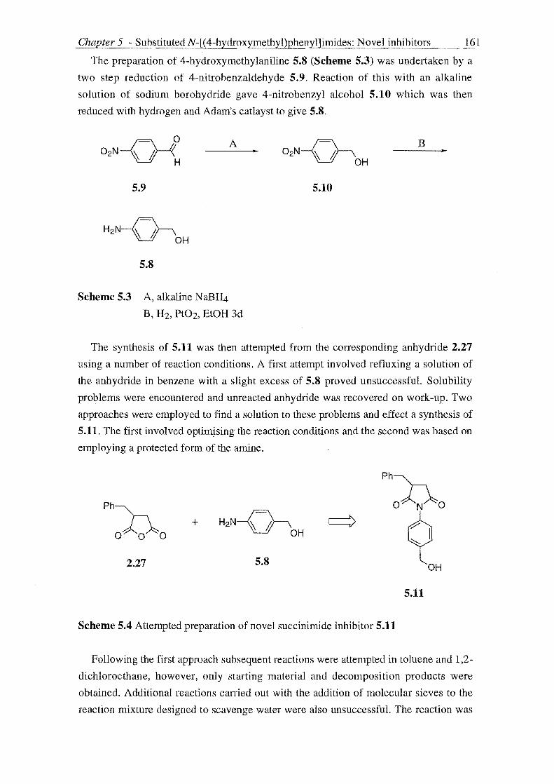
Chapter 5 - Substituted N-[(4-hydroxymethyl)phenyl]imides: Novel inhibitors 161
The preparation of 4-hydroxymethylaniline 5.S (Scheme 5.3) was undertaken by a
two step reduction of 4-nitrobenzaldehyde 5.9. Reaction of this with an alkaline
solution of sodium borohydride gave 4-nitrobenzyl alcohol 5.10 which was then
reduced with hydrogen and Adam's catlayst to give 5.S.
A B ..
5.9 5.10
5.S
Scheme 5.3 A, alkaline N aBH4
B, H2, Pt02, EtOH 3d
The synthesis of 5.11 was then attempted from the corresponding anhydride 2.27
using a number of reaction conditions. A first attempt involved refluxing a solution of
the anhydride in benzene with a slight excess of 5.S proved unsuccessfuL Solubility
problems were encountered and unreacted anhydride was recovered on work-up. Two
approaches were employed to find a solution to these problems and effect a synthesis of
5.11. The first involved optimising the reaction conditions and the second was based on
employing a protected form of the amine.
Ph~
oAo~o
2.27
+
5.S
Scheme 5.4 Attempted preparation of novel succinimide inhibitor 5.11
5.11
Following the first approach subsequent reactions were attempted in toluene and 1,2-
dichloroethane, however, only starting material and decomposition products were
obtained. Additional reactions carried out with the addition of molecular sieves to the
reaction mixture designed to scavenge water were also unsuccessful. The reaction was

Chapter 5 - SubstitutedN-[(4-hydro.xymethyl)phenyl]imides: Novel inhibitors 162
also attempted with two equivalents of p-hydroxymethylaniline and in pyridine
however this was method was also unsuccessful.
Considering the prior success of these reactions when using O-benzyloxyamine
focus turned to consideration of using a protected form of the aniline 5.8 such that the
modified properties may facilitate the reaction. The choice of an acetate protecting
group was influenced by the desire to also incorporate a leaving group at this position
so that a potential inhibitor may be obtained. The attempted synthesis of 5.14 is shown
below (Scheme 5.5).
02N~ A 02N~ .. OH OAc
5.10 5.12
fB Ph=n + H2N~
o 0 0 OAc
2.27 5.13
Scheme 5.5 A AC20, pyridine, overnight, rt
B Pt02, BtOH, overnight
Ph=n o N 0
c:::>
~ OAc
5.14
The starting material for the above synthesis 4-nitrobenzylalcohol 5.10 was
acetylated by reaction with acetic anhydride in pyridine to give p-nitrobenzylacetate
5.12. Subsequent hydrogenation to give 4-acetoxymethylaniline 5.13 the key precursor
to 5.14 however was unsuccessful with the molecule fragmenting during the reaction as
indicated by the presence of 4-nitrotoluene in the isolated product as analysed by IH
NMR.
With the proposed routes to the desired succinimide inhibitors 5.1 unsuccessful,
focus turned to the preparation of a glutarimide derivative of type 2.2. A mild method
for the preparation of N-substituted glutarimides involves the reaction of amines with
the appropriate acids mediated by coupling reagents such as dicyclohexylcarbodiimide
(DCC)246 and 1-(3-dimethylamino)-propyl)-3-ethyl-carbodiimide hydrochloride

Chapter 5 - SubstitutedN-[(4-hydroxymethyl)phenyl]imides: Novel inhibitors 163
(EDAC) 159 which was used in a successful preparation of a N -benzyloxyglutarimide
(see Section 2.4, Chapter 2 for a discussion) following the directions of Zhu et al.159
The method of Zhu et al159 was then applied to the synthesis of 5.15 (Scheme 5.6).
This was undertaken by the EDAC mediated coupling of the amine 5.8 to 3,3-
dimethylglutaric acid 2.44e in the presence of triethylamine. Work-up of the reaction
mixture gave a crude solid which was purified by flash Si02 chromatography to give
pure 5.15 in 12% yield.
H3XH3
O~ ~O A
OH OH
2.44e
5.15
Scheme 5.6 A, EDAC, HOBt, Et3N, 6.8, overnight 0-5 DC - rt
Compound 5.15 was then assayed against a-chymotrypsin (Figure 5.1) using the
incubation method as described in Chapter 3 to make a preliminary determination of the
inhibitory activity.
The activity versus time profile of compound 5.15 (Figure 5.1) indicates that over a
period of thirty minutes there is no net loss of activity. However as the inhibitor
concentration was increased to 50-fold excess over enzyme it appears there is an initial
loss of enzyme activity followed quickly, however by a regain in enzyme activity. This
suggests that compound 5.15 is acting as a transient alternate substrate inhibitor of a
chymotrypsin in which the acyl-enzyme species (structure 5.3 of Scheme 5.1) inhibits
the enzyme for a period equal to its lifetime. This description of the activity displayed
by 5.15 is tentative for as discussed previously the error involved in incubation method
measurements is significant 20%).

Chapter 5 - SubstitutedNJ(4-hydroxymethyl)phtmyl]imides: Novel inhibitors 164
120
110 ~
~ '-" ..... 100 ..... .... ~ -a-- A .... ..... ~ 90 • B ~ a.l II C e ..... 80 N
= ~ 70
60 0 10 20 30 40
time (min)
Figure 5.1 Time dependent inactivation of a-chymotrypsin by 5.15. a-Chymotrypsin
(4J.lM) was incubated with 6.15 (A, 1OJ.1M; B, lOOJ.lM; C, 200J.lM) and enzymic activity
assayed using either N-Succ-(Alah-Pro-Phe-p-nitroanilide (A, 117J.lM) or BTEE (B, C,
117J.lM).
Study of the mechanism of inactivation proposed for these compounds (Scheme 5.1)
indicates design efforts to enhance the activity of these compounds will be based on
increasing the rate of formation of the initial Michaelis inhibitor enzyme complex (KI)
and the rates of formation of the reactive quinone methide and alkylation of the active
site imidazole ring of His-57. Modification of the substituents on the imide ring to
mimic the substitution present in a natural substrate of a-chymotrypsin would be
expected to enhance activity. The a or ~ substituents of the ring are supposed to interact
with the S 1 subsite of a-chymotrypsin and hence it is forecast that the introduction of
aromatic substituents on the ring would lead to increased activity. This SAR trend was
observed in the N-[(methanesulfonyl)oxy]glutarimide series assayed in Chapter 3. The
glutarimide inhibitor 2.1f, which possesses dimethyl substitution was found to be a poor
inhibitor of a-chymotrypsin (kobs/[I] = 25 M-1s·l) when assayed by the incubation
method, whereas compound 2.1d, which possesses aromatic substitution was too potent
to be measured accurately by the incubation method. On this basis, it is perhaps not
suprising that 5.15 did not show significant activity.

Chapter 5 - Substituted N-[( 4-hydroxymethyl)pheny l]imides: Novel inhibitors
oJ)o I
OS02CH3
2.1d
Too potent to be measured by incubation method
2.U kob/[I] = 25 M-1s-1; a-CT
165
The inhibitory activity may also be enhanced by increasing the rate of formation of
the quinone imine methide. This may be accomplished by substituting the OH group of
5.15 with a better leaving group. Determination of a good leaving group can be made
by comparison of the relative strengths of the parent acids hence it was supposed that
incorporation of X = halogen into structure 5.15 in the place of the hydroxy goup would
result in more potent inhibitors (compare the strengths of HCl and H20). This trend has
been observed in the series of saccharin inhibitors of type 1.48 developed by Groutas et
al99 as seen by comparing the inhibitory activities of 1.48b and 1.48a.
1.48 1.48aL=OH
inactive99 .
1.48b L = Cl
kobsf[I] > 49 000 M"ls-l; HLE99
1.48c L = F kobsf[I] > 4 750 M-1s-1; HLE99
These SAR considerations suggested that compound 5.17 may be a more potent
inhibitor of a-chymotrypsin than 5.15, because it has both more suitable substitution to
interact favourably with a-chymotrypsin and a better leaving group. Hence 5.17
became a synthetic target. EDAC mediated coupling of 4-hydroxymethylaniline 5.8 to
the commercially available 3-phenylglutaric acid (Scheme 5.7) gave a solid that on
analysis by thin layer chromatography (TLC) was found to be unsuitable for
purification by flash Si02 chromatography. Purification was then carried out following
the directions of Seth et al.246 The solid was dissolved in 5% aqueous sodium
carbonate and this aqueous solution was washed with dichloromethane to remove
impurities such as 5.8. Acidification of the aqueous layer and organic extraction gave
5.16 which was further purified by recrystallisation.

Chapter 5 - SubstitutedN-[(4-hydroxymethyl)phenyl]imides: Novel inhibitors 166
A 000 •
~ OH
2.44a 5.16
Scheme 5.7 A, EDAC, HOBt, 5.8, Et3N, 0-5 °C rt overnight
Chlorination of 5.16 to afford 5.17 was then attempted using the method adopted by
Groutas et al99 in the preparation of 1.48b from N-hydroxymethylsaccharin 1.48a. A
solution of 5.16 in ether was treated with thionyl chloride (5 equiv.) for two days at
room temperature however 1 H NMR analysis of the product indicated the reaction was
unsuccessful and an altenative procedure was sought.
000 000 ~
>
~ OH CI
5.16 5.17
The chlorination of primary alcohols using CC14-PPh3 has been well documentecJ247
and was attractive as the reaction is both mild and rapid. A trial chlorination of 6.15
was undertaken employing this methodology however this too was unsuccessful.
Although only preliminary results, the lack of success observed in these reactions may
be due to the inherent reactivity of 5.17. This may undergo ring-opening to give the
quinone imine methide which reacts with any nucleophiles present in the reaction
mixture. Proton NMR analysis of the product isolated from the attempted chlorination
using thionyl chloride indicated the presence of a charateristic triplet and quartet of an
ethyl ester perhaps formed from reaction of the methide with ethanol.
In summary we have developed a preliminary synthetic approach to a glutarimide
designed to liberate reactivity on hydrolysis. Initial studies have failed to identify an
inhibitor of a-chymotrypsin. The lack of inhibitory activity may be reasoned in that this

Chapter 5 - SubstitutedN-[(4-hydroxyme!hyl)phenyl]imides: Novel inhibitors 167
glutarimide does not act as a substrate of a-chymotrypsin and therefore cannot undergo
the required ring-opening necessary to unmask the quinone imine methide. Considering
the structure of the acyl enzyme intermediate in the formation of the methide the lone
pair on the nitrogen may not be available to move through the aromatic ring to displace
the leaving group as it is delocalised by the neighbouring carbonyl group.
Attempts to introduce activity into this series involved incorporation of ring
substitution able to interact with a-chymotrypsin and improving the leaving group.
Synthesis of a compound purporting both these features was unsuccessful maybe due to
the irony that the synthesis is undone by the activity it is trying to create.
CHAPTER 6
EXPERIMENTAL

169
6.1 GENERAL
Melting points (mp) were measured using an Electrothermal apparatus and are
uncorrected. Optical rotations were measured on a JASCO J-20C recording
spectropolarimeter at the Na line (J... = 589nm). The specific optical rotation at the stated
temperature [a]TD is given in units of lO-I degcm2 g-l with the sample concentration,
c, in the indicated solvent given in units of g lOOcm-3.
IR spectra were recorded on a Schimadzu FTIR-8201PC spectrophotometer.
Samples were run as KBr discs (KBr), Nujol mulls between KBr plates (NujollKBr),
CHCb and CDCl3 solutions, or as a CHCb film between NaCI plates (CHCI3/NaCI).
Data is presented as the wavenumbers (cm-I) at which the maxima absorptions of
diagnostic value occur.
Proton (300MHz) and 13C (75MHz) nuclear magnetic resonance (NMR)
spectroscopy was performed with either a Varian CFT300 or XL300 spectrometer.
CDCI3, with Me4Si as an internal standard, a mixture of CDCb and a small amount of
DMSO-d6 (CDC13IDMSO-d6), DMSO-d6, and D20 (lH NMR only) were used as
solvents.
Proton CDCl3 and CDCI3IDMSO-d6 spectra were referenced to the (CH3)4Si signal
at a chemical shift (0) of 0.00 parts per million (ppm) and 13C NMR spectra to the
central peak of the CDCl3 triplet at 0 77.0ppm. Proton DMSO-d6 spectra were
referenced to the residual (CH3hSO signal at 0 2.50ppm and 13C NMR spectra to the
central peak of (CH3hSO quintuplet at 0 39.6. Proton D20 spectra were referenced to
the residual H20 signal at 0 4.70ppm.
Proton NMR data are recorded as follows: chemical shift in parts per million (0)
accurate to two decimal places (multiplicity (s: singlet, d: doublet, t: triplet, q: quartet,
m: multiplet, dd: doublet of doublets, ABq: AB quartet, etc., b indicates some
broadening of the signal), integrated intensities, coupling constant(s) in Hz accurate to
one decimal place, and assignment as the underlined proton(s) within the given
structural fragment). Data for 13C NMR are recorded as the chemical shifts observed
accurate to one decimal place. If required, 2D NMR experiments were carried out using
a Varian CFT300 spectrometer to aid assignment.
Mass spectra were obtained using a Kratos MS80RF Aspectrometer operating at
70eV. The molecular ion (M+) only, if present, is reported accurate to four decimal
places when using High Resolution Mass Spectrometry (HRMS) in the indicated
operated mode +CI (Chemical Ionisation), +EI (Electron Ionisation) or FAB (Fast Atom

170
Bombardment). If the molecular ion was sufficiently weak so as not to be recorded
significantly high mass ions and the more intense low mass ions accurate to one
decimal place are reported. The molecular ion (M+), if present, significantly high mass
ions and the more intense low mass ions accurate to the nearest mass unit are reported
when using Low Resolution Mass Spectrometry (LRMS) in the indicated operation
mode as above. Data are presented in the folowing order: mlz value, the attributed
fragmentation; relative intensity as a percentage of the base peak.
Elemental analyses were performed by the Campbell Microanalytical Laboratory,
University of Otago, Dunedin.
Solvents were freshly dried and distilled according to the following well established
procedures248,249 and then stored in N2 immediately prior to use. Tetrahydrofuran
(THF) , and diethyl ether (ether, Et20) were purified by distillation from sodium
benzophenone ketyl. Ethanol (EtOH) and Methanol (MeOH) were purified by
distillation from magnesium ethoxide or methoxide respectively. Benzene,
dichloromethane (CH2Clz) and toluene were purified by distillation from CaH2. N,N
Dimethylformamide (DMF) was dried by the method of Burfield and Smithers.250
Petroleum ether refers to the fraction of boiling range 50-70 oC. Reagents were purified
prior to use according to literature procedures.249 The commonly used bases N,N
diisopropylethylamine (Hunigs base), pyridine, and triethylamine (Et3N) were purified
by distillation from CaH2 or P205.
Organic reactions requiring anhydrous conditions were performed with solvents and
reagents as prepared above under a dry N2 atmosphere with glassware baked in an oven
set at 110 °C overnight then assembled hot and 'flame' dried with a bunsen burner or
heat gun under N2 or vacuum immediately prior to use. Reaction temperatures refer to
the external bath temperature, A reaction carried out at room temperature (rt) refers to a
temperature of ca 20 °C. A workup procedure carried out in the usual manner involved
separation of the organic extract or phase, drying with Na2S04 or MgS04, filtration to
remove the dessicant and then evaporation of the bulk of the solvent using a rotary
evaporator, the last traces being removed under high vacuum, Flash chromatography
was conducted according to the method of Still et al 251 using Merck 60 PF254 type
silica geL
Diethyl benzyl malonate (2.10), 2-phenylsuccinic acid (2.30), 3-phenylglutaric acid
(2.44c), 2-phenylglutaric anhydride (2.43a), ~-tral1s-styryl sulfonyl chloride, 2-
naphthalene sulfonyl chloride, 4,4'-biphenyldisulfonyl chloride were obtained from
Aldrich Chemical Company, Milwaukee, USA. 2-Phenylglutaric anhydride 2.43a was
purified by treatment with AC20 before use. 3-Chlorosulfonyl benzoic acid and 1,3-

bis(chlorosulfonyl)benzene were obtained from Maybridge Chemical Co. Ltd, Tintagel,
Cornwall, UK.
4,4-Cyc1ohexyl-3,5-dicyanoglutarimide (2.48b) was generously gifted by Dr. R. H.
McKeown.

6.2 CHAPTER 2 EXPERIMENTAL
SYNTHESIS OF 2-SUBSTITUTED SUCCINIC ACIDS
Method A: Malonate Ester Route
DiethyI2-carboethoxy-2-benzylsuccinate (2.11)152
A solution of sodium ethylate was prepared by slowly dissolving sodium (2.332g,
O.lOlmol) in EtOH (3SmL). Diethyl benzyl malonate (2.10) (24.890g, O.lOOmol) and
ethyl bromoacetate (11mL, 16.566g, 0.099mol) were added slowly in succession. The
resulting solution was refluxed for 14.25h, concentrated, made slightly acidic (pH=6,
Universal Indicator Paper) with 3N H2S04 and extracted with Et20. The Et20 extract
was worked up in the usual manner to give a yellow oil (28.49g) containing a 4:5
mixture, by IH NMR, of unreacted 2.10 and 2.11. This crude product was used without
further purification. IH NMR 2.11 (CDCl3) 01.17-1.29 (m, 9H, 3xOCH2CH3), 2.85 (s,
2H, CH2C02Et), 3.39 (s, 2H, CH2Ph), 4.12-4.24 (m, 6H, 3xOCH2CH3), 7.08-7.26 (m,
5H, ArH).
2-Carboxyl-2-benzylsuccinic acid (2.12)152
The above crude product (28.49g) was boiled under reflux with KOH (21.338g,
0.380mol) in EtOH (120ml) for 4h. The reaction mixture was diluted with H20 (55ml),
and extracted with Et20. The aqueous layer was acidifided (pH=I, Universal Indicator
Paper) with 6M HCI, and extracted with Et20. The extracts were worked up in the usual
manner to give a brown residue (l3.127g) containing a 12:15 mixture, by IH NMR, of
benzyl malonic acid (from the hydrolysis of 2.10 present in the starting material) and
2.12: IH NMR 2.12 (CDCb) 02.85 (s, 2H, CH2Ph), 3.33 (s, 2H, CfuC02H), 7.14-7.58
(m, SH, ArH), 9.66 (bs, 3H, 3xCOllI).
2-Benzylsuccinic acid (2.13)152
The above mixture (l3.127g) was heated in an oil bath set at 160-170 DC for 2.25h.
The reaction mixture was allowed to cool to rt and then refridgerated. The resultant
brown mass was treated with boiling H20 to give an immiscible mixture consisting of
an upper aqueous phase and lower brown oil. The aqueous phase was decanted and
allowed to cool to rt during which 2.13 crystallised as a white solid. More acid was
obtained by repeated treatments and concentration of the mother liquors. Total yield
(3.307g, overall yield 17%); mp 160-1 °C (H20, glittering white rectangular plates)
lit.252 mp 160-1 °C; IR (KEr) 'Umax 3022.1 (OH), 1697.8 (C=O) em-I; IH NMR

173
(CDCbIDMSO-d6) 6 2.31 (dd, IH, .J = 4.9, 17.1Hz, HCHPh), 2.58 (dd, IH, .J = 8.3,
16.6Hz, HCHPh), 2.78 (dd, IH, .J = 4.9, 1O.2Hz, HCHC(O)), 3.01-3.08 (m, 2H,
HCHC(O) and CH), 7.18-7.30 (m, 5H); HRMS (+EI, M+) ca1cd. 208.0736, found
208.0736.
Method B: Amide base alkylation of succinimide
2.Benzylsuccinimide (2.14)144
Liquid NH3 (60mL) was transferred to a three necked reaction flask cooled to -78 °C
(dry-ice/acetone bath) equipped with a dry-ice/acetone condenser and a droppping
funnel. A small amount of Na was added followed by FeN03.9H20 (90mg). After the
blue colour was discharged to give a charcoal like reaction solution, Na (total amount
added 1.634g, 0.071mol) was then added portionwise to give a grey suspension of
NaNH2. This was stirred for a Ih at -78 °C after which succinimide (3.178g, 0.032mol)
was added portionwise and the resulting grey suspension stirred for a further hour. The
dropping funnel was charged with a solution of benzyl bromide (3.826mL, 5.490g,
0.032mol) in anhydrous Et20 (lOmL) and this was added rapidly to the reaction
suspension. The dropping funnel was rinsed with an additional quantity of Et20 (ImL)
and the resulting pale yellow suspension stirred for Ih and the reaction quenched by
pouring the reaction mixture into a beaker containing NH4CI (5.649g, 0.106mol, 3.3
equiv.). The NH3 was allowed to evaporate while being replaced by the slow addition
of El20 (50mL). A mixture of conc HCI (27mL) and ice (250mL) was added and the
resulting mixture was stirred until the ice melted. The Et20 phase was separated and the
aqueous phase extracted with Et20. The organics were combined and washed with H20
and worked up in the usual manner to give a creamy yellow solid. This was dissolved in
Et20 and extracted thoroughly with 5% aqueous NaOH. The alkaline extracts were
washed with Et20 and then acidified (pH=I, Universal Indicator Paper) with 6N HCI
and then extracted with Et20. Work-up in the usual manner gave a white solid (755mg,
12%): mp 81-4 °C (lit. 144 mp 92.5-95 OC); IR (KBr) umax 3170.8 (NH) , 1776.3 and
1705.0 (C=O) cm-1; IH NMR (CDCI3) 62,48 (dd, 1H, .J = 4.9, 1805Hz, HCH2Ph) 2.71
(dd, IH,.J = 8.8, 1805Hz, HCHPh) 2.84 (dd, IH,.J = 9.5, 14,4Hz, HCHC(O)) 3.13-3.25
(m, 2H, HCHC(O) and CHC(O)), 7.17-7.34 (m, 5H, ArH); 13C NMR (CDCb) 634.4,
36.1,42.7, 127.0, 128.8, 128.9, 137.1, 180.1, 189.6; LRMS m/z (+EI) 189 (67, M+),
161 (3), 146 (17),117 (2), 103 (5), 91 (100),78 (8), 65 (13),51 (8).

2-Benzylsuccinic acid (2.13)
2.14 (684mg, 4mmol) was refluxed with 6N HCI (15mL) for 26h. The reaction
solution was allowed to cool to rt during which a white solid cystallised. This reaction
mixture was evaporated to dryness and sufficient Et20 and saturated aqueous NH4Cl
solution were added to dissolve the white solid. The Et20 phase was worked up in the
usual manner to give 2.13 as a white solid (760mg, quantitative yield): mp 152-155 oC
(lit. 252 mp 160-1 oc). Spectral data as given above.
Method C: Stobbe Condensation Route
Ethyl2-benzylidenesuccinate (2.17)157d
A solution of sodium ethoxide was prepared by slowly dissolving sodium (1.476g,
0.064mol, 1.23 equiv.) in EtOH (25ml) under a N2 atmosphere. Diethyl succinate
(8.6ml, 9.004g, 0.052mol) was added dropwise and the resulting milky mixture heated
until reflux at which point benzaldehyde (5.3ml, 5.533g, 0.052mol) was added
dropwise. The resulting yellow reaction mixture was refluxed for 12h. The now brown
reaction mixture was concentrated and dissolved in H20 (lOOmL) and then extracted
with Et20 (2xlOOmL). The aqueous layer was acidified (pH:l, Universal Indicator
paper) with 6N HCI (20mL) and this was extracted with Et20 (2x100mL). The organic
extracts were worked up in the usual manner to give crude ethyl 2-benzylidenesuccinate
2.17 as a viscous brown syrup (9.262g, 76%): IH NMR 0 1.35 (t, 3H, J 7.1Hz,
CH2CH3), 3.58 (s, 2H, CH2C02H), 431 (q, 2H, J: 7.1, 7.3Hz, CH2CH3), 7.34-7.44
(m, 5H, ArH), 7.93 (bs, 1H, NH); 13C NMR 0 14.1, 33.6, 61.4, 125.5, 128.7, 129.0,
134.7,1423,167.4,177.2; HRMS (+EI, M+) calcd. 234.0893, found 234.0892.
Ethyl 2-benzylsuccinate (2.18)
A solution of ethyl 2-benzylidenesuccinate 2.17 (9.144g, 0.039mol) in EtOAc
(20mL) was hydrogenated in the presence of 10% palladium on carbon (1. 141g) for
19h. The reaction mixture was filtered through a column packed with both celite and
Na2S04. The fUter cake was washed with a small quantity of EtOAc and the filtrate and
washings were combined and evaporated to give 2.18 as a yellow oil (5.541g, 60%): IH
NMR (CDCI3) 0 1.18 (t, 3H, J = 7.1Hz, OCH2CH3), 2.43 (dd, 1H, J: 4.4, 17.1Hz,
HCHPh), 2.66-2.81 (m, 2H, HCHPh and HCHC(O», 3.01-3.13 (m, 2H, HCHCeO) and
CHC(O», 4.12 (q, 2H, J = 6.8, 7.4Hz, OCfuCH3), 7.14-7.31 (m, 5H, ArH); 13C NMR
o 14.0,34.7,37.6,42.8,60.8, 126.7, 128.5, 129.0, 138.0, 174.0, 177.8.

175
2-Benzylsnccinic acid (2.13)152
A solution of ethyl 2-benzylsuccinate 2.18 (2.416g, O.OlOmol) in 1: 1 EtOHIH20
(20mL) with KOH (2.23g, 0.040mol, 4 equiv.) was refluxed for 5h. The reaction
solution was concentrated and extracted with EtOAc. The aqueous layer was acidified
(pH=l, Universal Indicator paper) with 6N HCI and then extracted with EtOAc. The
organic extracts were worked up in the usual manner to give 2.13 as a white solid
(1.745g, 82%). Spectral data data as given above.
THE SYNTHESIS OF 2-SUBSTITUTED SUCCINIC ACID ANHYDRIDES
2·Phenylsnccinic anhydride (2.30)
A mixture of 2-phenylsuccinic acid 2.29 (5.000g, 26mmol) and acetyl chloride
(l0.5mL, 11.592g, 129mmol) was refluxed for lh. The excess acetyl chloride was
evaporated and the resultant liquid distilled at reduced pressure to give a pale yellow
solid. Recrystallisation from toluene/petroleum ether gave 2.30 as a white solid (3.123g,
69%): mp 51-3 °c (lit.166 mp 53-5 OC); IR (KBR) 'Umax 1867.0 and 1791.7 (C=O) cm-I;
IH NMR (CDCI3) 83.13 (dd, IH, J = 6.9, 19.1Hz, HCH), 3.47 (dd, 1H, J = 10.3,
19.1Hz, HCH), 4.35 (dd, IH, J = 6.8, lO.2Hz, CH), 7.26-7.29 (m, 2H, ArH), 7.40-7.45
(m, 3H, ArH); 13C NMR (CDCb) 8 36.5, 46.5, 127.4, 128.6, 129.4, 134.7, 169.9,
172.0.
2.Benzylsnccinic anhydride (2.27)84
A mixture of 2-benzylsuccinic acid (2.13) (2.055g, O.OlOmol) and AC20 (9.6ml,
10.387 g, 0.102mol, 10.2 equiv.) was refluxed for 1h. Evaporation of solvent gave a
crude white solid which was triturated with ice-cold petroleum ether to give 2.27 as a
white solid (1.766g, 94%): mp 97-9 oC (lit. 84 mp 95-7 OC); IR (CDCb) 'Umax 1868.9
and 1785.0 (C=O) cm- I; IH NMR (CDCb) 82.72 (dd, 1H, J= 6.3, l8.5Hz, HCHPh),
2.95 (dd, 1H, J = 5.3, 14.1Hz, HCHPh), 3.02 (dd, 1H, J = 8.3, 14.2Hz, HCHC(O»,
3.24 (dd, 1H, J = 4.9, 14.2Hz, HCHC(O», 3.40-3.50 (m, 1H, CH), 7.17 (d, 2H, J = 6.3Hz, ArH), 7.27-7.38 (m, 3H, ArH); 13C NMR (CDCI3) 832.9, 36.0,42.1, 127.6,
135.8,169.6,173.1,128.9,129.1; HRMS (+EI, M+) calcd. 190.0630, found 190.0630.
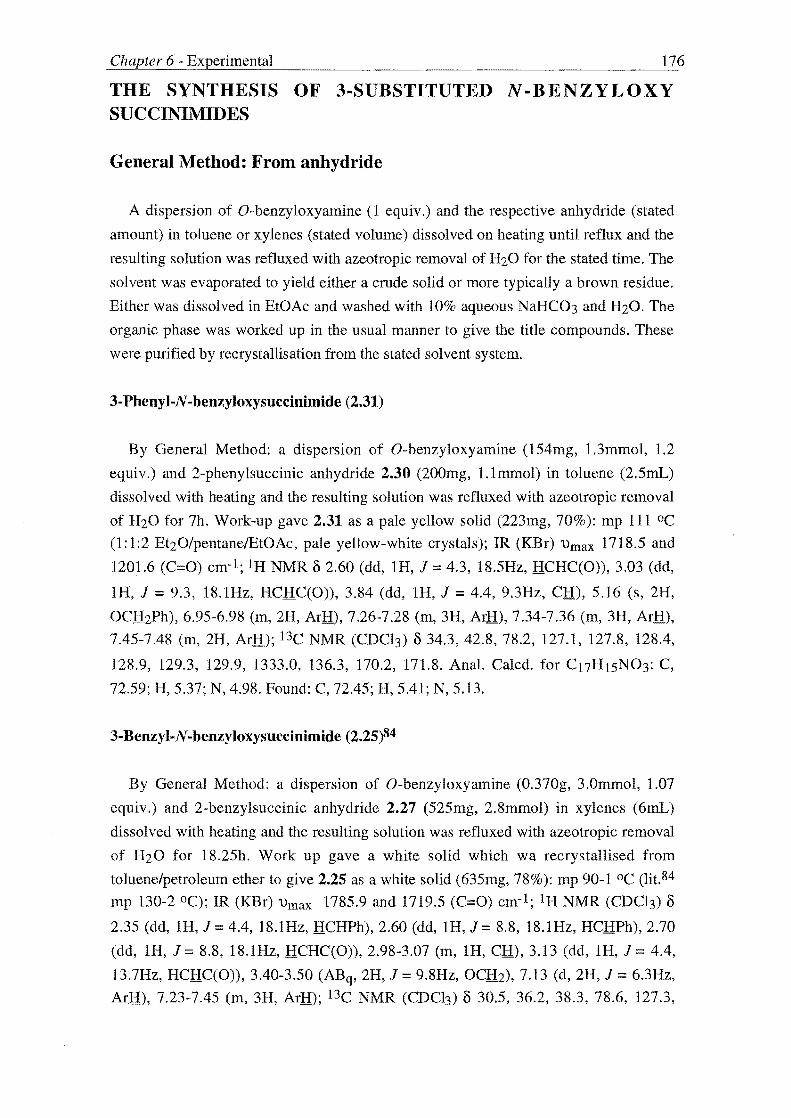
176
THE SYNTHESIS OF 3-SUBSTITUTED N·BENZYLOXY SUCCINIMIDES
General Method: From anhydride
A dispersion of O-benzyloxyamine (l equiv.) and the respective anhydride (stated
amount) in toluene or xylenes (stated volume) dissolved on heating until reflux and the
resulting solution was refluxed with azeotropic removal of H20 for the stated time. The
solvent was evaporated to yield either a crude solid or more typically a brown residue.
Either was dissolved in EtOAc and washed with 10% aqueous NaHC03 and H20. The
organic phase was worked up in the usual manner to give the title compounds. These
were purified by recrystallisation from the stated solvent system.
3-Phenyl-N -benzyloxysuccinimide (2.31)
By General Method: a dispersion of O-benzyloxyamine (154mg, 1.3mmol, 1.2
equiv.) and 2-phenylsuccinic anhydride 2.30 (200mg, 1.1mmol) in toluene (2.5mL)
dissolved with heating and the resulting solution was refluxed with azeotropic removal
of H20 for 7h. Work-up gave 2.31 as a pale yellow solid (223mg, 70%): mp 111°C
(1:1:2 Et20/pentanelEtOAc, pale yellow-white crystals); IR (KBr) 'Urn ax 1718.5 and
1201.6 (C=O) cm-I; IH NMR 3 2.60 (dd, IH, J = 4.3, 18.5Hz, HCHC(O)), 3.03 (dd,
IR, J = 9.3, 18.1Hz, HCHC(O», 3.84 (dd, IH, J = 4.4, 9.3Hz, CH), 5.16 (s, 2H,
OCH2Ph), 6.95-6.98 (m, 2H, ArH), 7.26-7.28 (m, 3H, ArH), 7.34-7.36 (m, 3H, ArH),
7.45-7.48 (m, 2H, ArH); 13C NMR (CDCI3) 3 34.3, 42.8, 78.2, 127.1, 127.8, 128.4,
128.9, 129.3, 129.9, 1333.0, 136.3, 170.2, 171.8. Anal. Calcd. for C17R 15N03: C,
72.59; H, 5.37; N, 4.98. Found: C, 72.45; H, 5.41; N, 5.13.
3-Benzyl-N -benzyloxysuccinimide (2.25)84
By General Method: a dispersion of O-benzyloxyamine (0.370g, 3.0mmol, 1.07
equiv.) and 2-benzylsuccinic anhydride 2.27 (525mg, 2.8mmol) in xylenes (6mL)
dissolved with heating and the resulting solution was refluxed with azeotropic removal
of H20 for 18.25h. Work up gave a white solid which wa recrystallised from
toluene/petroleum ether to give 2.25 as a white solid (635mg, 78%): mp 90-1 °C (lit.84
mp 130-2 °C); IR (KBr) 'Urnax 1785.9 and 1719.5 (C=O) cm-I; IH NMR (CDCI3) 3
2.35 (dd, IH, J= 4.4, 18.1Hz, HCHPh), 2.60 (dd, IH, J= 8.8, 18.1Hz, HCHPh), 2.70
(dd, IH, J = 8.8, 18.1Hz, HCHC(O», 2.98-3.07 (m, IH, CH), 3.13 (dd, IH, J 4.4,
13.7Hz, HCHC(O)), 3.40-3.50 (ABq, 2H, J = 9.8Hz, OCfu), 7.13 (d, 2H, J = 6.3Hz,
ArH), 7.23-7.45 (m, 3H, ArH); 13C NMR (CDCh) 3 30.5, 36.2, 38.3, 78.6, 127.3,

177
128.5,128.9,129.1, 129.4, 130.0, 133.2, 136.3, 170.4, 173.3; HRMS (+CI, MH+) calcd.
296.1287, found 296.1287.
Alternatively 2.25 was prepared in the following manner from ethyl 3-benzylsuccinate
2.18.
A solution of ethyl 2~benzylsuccinate 2.18 (193mg, 0.81mmol) in CH2Cl2 (2mL)
was added to a solution of O-benzyloxyamine hydrochloride (130mg, 0.81mmol) and
Et3N (141.2/lL, 103mg, 1.02mmol, 1.26 equiv.) in CH2Ch (2mL) that had been stirring
for 10 min under N2 at 0-5 °C (ice-salt bath). HOBt (11Omg, 0.81mmol) and then DCC
(l65mg, 0.80mmol) were added successively and the resulting yellow solution was
stirred for 21h while the temperature was allowed to come to rt. The reSUlting white
slurry was filtered to remove the white DCU solid formed during the reaction and this
was washed with a small quantity of CH2Ch. The filtrate and the washings were
combined and evaporated to give a brown residue. This was taken up into the minimum
amount of EtOAc and the suspended DCU was filtered and washed with a small
quantity of EOAc and the filtrate and washings were combined and washed with H20
(lOmL) and worked up in the usual manner to give a brown residue (287mg). A sample
(233mg) of this was purified by flash Si02 chromatography eluting with 1:3
EtOAc/petroleum ether to give 3-benzyl-N-benzyloxysuccinimide 2.25 as a white solid
(33mg) with physical data identical to that given above, and then eluting with EtOAc to
give 3-(carboethoxy)-3-benzylpropionyl-N-benzyloxyamide 2.24 as a white solid
(109mg): mp 90-91 oC; IH NMR (CDCb) 8 1.18 (t, 3H, J = 7.1Hz, OCH2C.tb), 2.81
(dd, tH, J = 7.8, 13.7Hz, HCHPh), 2.97-3.03 (m, 2H, HCHPh and HCHC(O», 3.17-
3.65 (m, 2H, HCHC(O) and CHC(O», 4.09 (q, 2H, J = 6.8, 7.3Hz, OCH2CH3), 4.86
(bs, 2H, OCH2Ph), 7.15-7.37 (m, 1OH, ArH), 8.16 (bs, tH, NH).
A solution of 2.24 (92mg, 0.27mmol) andp-TsOH (26mg, 0.14mmol, 0.5 equiv.) in
1,2-dichloroethane (DCE) (5mL) was refluxed with azeotropic removal of EtOH for
13h. The solvent was evaporated to give 2.25 as a pale brown solid (l06mg, quantative
yield). Total yield 2.25 t39mg, (85%). Spectral data as above.
THE SYNTHESIS OF 3-SUBSTITUTED N -HYDROXY SUCCINIMIDES
Method A: From 3-substitutedN -benzyloxysuccinimide
A solution of the respective substituted N-(benzyloxy)succinimide (stated amount) in
the stated solvent (stated volume) was hydrogenated in the presence of the indicated
palladium catalyst (stated amount) for the stated time. The reaction mixture was filtered

through celite and Na2S04 and this cake washed with a small quantity of solvent. The
filtrate and washings were combined and evaporated to give the title N
hydroxysucccinimides. These were purified, if required, by either chromatagraphy or
recrystallisation using the stated solvent system.
Method B: From 2-substituted succinic acid anhydride
The respective anhydride (stated amount) was added to a solution of hydroxylamine
hydrochloride (1.2 equiv.) and K2C03 (0.6-1equiv.) in distilled H20 (stated volume).
This mixture was refluxed for the stated time. The hot solution was acidified (pH= 1,
Universal Indicator Paper) with 10% or conc. HCI, allowed to cool to rt, and then
extracted with EtOAc. The combined extracts were worked up in the usual manner to
give the titleN-hydroxysuccinimide.
3-Phenyl-N-hydroxysuccinimide (2.32)
By Method A: a solution of 3-phenyl-N-benzyloxysuccinimide 2.31 (40mg,
0.14mmol) in THF (lmL) was hydrogenated for 4.25h in the presence of 10%
palladium on carbon (5mg). Work-up gave a yellow oil which was purified by radial
chromatography eluting with 1:4 EtOAc/petroleum ether to give 2.32 as a pale yellow
oil (14mg, 53%): mp 109-111 oC (EtOAc/petroleum ether, white crystals); IR
(CHCI3/NaCI) umax 3118.7 (OH), 1787.9 and 1693.4 (C=O) cm- l ; lH NMR (DMSO
d6) 02.73 (dd, 1H, J= 4.4, 17.5Hz, HCH), 3.17 (dd, 1H, J= 9.2, 18.0Hz, HCH), 4.14
(dd, 1H, J = 4.4, 8.8Hz, CH), 7.31-43 (m, 5H, ArH), 10.78 (bs, lH, NOH); l3C NMR
(CDC13) 034.2,42.8,127.7,128.0,129.0,137.9,171.8,173.5; LRMS (+EI, mlz) 191
(M+, 18), 175 (25), 121 (35), 104 (100), 78 (45), 51 (55). Anal. Calcd. for ClOH9N03:
C, 62.83; H, 4.74; N, 7.33. Found: C, 62.67; H, 4.68; N, 7.36.
By Method A: a solution of 3-phenyl-N-benzyloxysuccinimide 2.31 (40mg,
0.14mmol) in MeOH (7.5mL) was hydrogenated at 50psi for 8.5h in the presence of 5%
palladium on BaS04. Work up gave a pale yellow oil which was purified by radial
chromatography eluting with 1:4 EtOAc/petroleum ether to give a colourless oil. This
was recrystallised from EtOAc/petroleum ether to give 2.32 as a white solid (lOmg,
37%): mp 108-9 oC. Physical data as given above.
By Method B: 2-phenylsuccinic anhydride 2.30 (500mg, 2.9mmol) was added to a
solution of hydroxylamine hydrochloride (237mg, 3.4mmol, 1.2 equiv.) and K2C03
(236mg, 1.7mmol, 0.6 equiv.) in distilled H20 (7.SmL). This mixture was refluxed for
5h. Subsequent work up as described gave 2.32 as a pale yellow-white solid (440mg,
82%). Physical data as above.

3-Benzyl-N -hydroxysuccinimide (2.26)84
By Method A: a solution of 3-Benzyl-N-benzyloxysuccinimide 2.25 (202mg,
0.7mmol) in 1:1 MeOHlEtOH (7mL) hydrogenated in the presence of 10% palladium
on carbon (71mg) for 19.5h. Work up as described gave 2.26 as a white solid (86mg,
61%): mp 105-110 °c (lit. 84 mp 120-1°C); IR (KBr) 'Umax 3224.8 (OH) , 1770.5 and
1697.2 (C=O) em-I; IH NMR (CDCI3/DMSO·<h) 3 2.13 (dd, 1H, J = 4.4, 18.1Hz,
HCHPh) 2.41 (dd, 1H, J = 8.8, IS. 1Hz, HCHPh) 2.62 (dd, IH, J = S.6, 13.5Hz,
HCHC(O» 2.83-2.91 (m, 1H, CHC(O», 2.97 (dd, 1H, J = 4.4, 13.7Hz, HCHC(O»,
6.92-7.25 (m, 5H, ArH): 13C NMR (CDCI3IMSO-d6) 330.3,35.9, 38.0, 126.7, 128.5,
128.7,136.6,171.3,174.1; HRMS (+EI, M+) calcd. 205.0739, found 205.0739. Anal.
Calcd for CUHllN03.l/2H20: C, 61.66; H, 5.65. Found C, 62.08; H, 5.21.
By Method A: a solution of 3-benzyl-N-benzyloxysuccinimide 2.25 (5.565g,
19mmol) in THF (SOmL) was hydrogenated in the presence of 10% palladium on
carbon (1.S96g). Work-up as described gave 2.26 as a white solid (2.780g, 72%): mp =
119-120 °C (lit. 84 mp = 120-1 °C). Physical data as above.
By Method B: 2-benzylsuccinic anhydride 2.27 (266mg, 1.4mmol) was added to a
solution of hydroxylamine hydrochloride (201mg, 2.8mmol) and anhydrous K2C03
(20Omg, 1.4mmol, 1 equiv.) in H20 (7mL) and the resulting mixture refluxed for 1.75h.
Acidification with three drops of conc. HCI and subsequent work up gave 2.26 as a
white solid (299mg, 51 %): mp 11O-17°C (lit. 84 mp = 120-1 °C ). Physical data as
above.

180
THE SYNTHESIS OF 3-SUBSTITUTED N-[(ALKYL AND ARYLSULFONY]OXY] SUCCINIMIDES
Method A: Sulfonylation at 0-5 oC then rt
The appropriate sulfonyl chloride (1.5 equiv.) was added slowly to a solution of the
N-hydroxysuccinimide (stated amount) and either Et3N or i-PrNEt or pyridine (1.1
equiv.) in CH2Ch or toluene (stated volume) stirring under N2 at 0-5 oC (ice-salt bath).
Stirring was continued for the stated time after which the ice-salt bath was removed and
the reaction mixture allowed to come to rt while stirring over the stated period. In
preparations where CH2Ch had been used as the solvent, the reaction solution was
diluted and then washed with H20, 10% HCI or 10% aqueous citric acid, and 10% or
saturated aqueous NaHC03. The organic phase was worked up in the usual manner to
give the title compound. In preparations where toluene had been used the reaction
solution was evaporated and the resulting residue dissolved in EtOAc and worked up in
the same manner as described for a reaction carried out in CH2Ch. Further purification
if required was carried out as described per compound.
Method B: Sulfonylation at rt
The appropriate sulfonyl chloride was (1-1.5 equiv.) was added slowly to solution of
N-hydroxy imide (stated amount) and either Et3NH or iprNEt (1.1 equiv.) or pyridine
(2 equiv.) in CH2Ch or toluene (stated volume) stirring at rt in N2. Stirring was
continued for the stated time. Work up was carried out as described for Method A.
Method C: Sulfonylation at 50 °c
The appropriate sulfonyl chloride (l equiv.) was added to a solution of N
hydroxyimide (stated amount) and pyridine (2 equiv.) in toluene (stated volume)
stirring at 40 °C. The temperature was elevated to 50 °C and stirring was continued for
the stated time. The reaction solution was then allowed to cool to rt and work up carried
out as per Methods A and B.
3-Benzyl-N-[(methanesulfonyl)oxy]succinimide (1.41d)84
By Method B: MeS02Cl (l6.3J.1L, 24mg, 0.21mmol) was added to a solution of 3-
benzyl-N-hydroxysuccinimide 2.26 (44mg, 0.21mmol) and pyridine (33.9~L, 33mg,
0.42mmol, 2 equiv.) in toluene (lmL). Stirring was continued for 12h. Work up in the
manner described for a reaction carried out in toluene gave a white solid (59mg,
quantatative yield); mp 70-1°C (lit. 84 mp 70-1°C); 1 H NMR (CDCb) 8 2.55 (dd, 1H, J
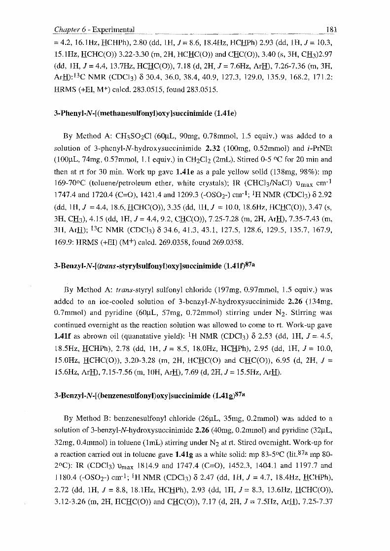
181
= 4.2, 16.1Hz, HCHPh), 2.80 (dd, 1H, J::: 8.6, 18.4Hz, HCHPh) 2.93 (dd, lH, J::: 10.3,
15.1Hz, HCHC(O)) 3.22-3.30 (m, 2H, HCHC(O)) and CHC(O)), 3.40 (s, 3H, CH3)2.97
(dd, IH, J::: 4.4, 13.7Hz, HCHC(O»), 7.18 (d, 2H, J::: 7.6Hz, ArH), 7.26-7.36 (m, 3H,
ArH):13C NMR (CDCb) cS 30.4, 36.0, 38.4, 40.9, 127.3, 129.0, 135.9, 168.2, 171.2:
HRMS (+EI, M+) calcd. 283.0515, found 283.0515.
3-Phenyl-N -[ (methanesulfonyl)oxy ]succinimide (1.41e)
By Method A: CH3S02CI (60IlL, 90mg, 0.78mmol, 1.5 equiv.) was added to a
solution of 3-phenyl-N-hydroxysuccinimide 2.32 (100mg, 0.52mmol) and i-PrNEt
(lOOIlL, 74mg, 0.57mmol, 1.1 equiv.) in CH2Cl2 (2mL). Stirred 0-5 °C for 20 min and
then at rt for 30 min. Work up gave 1.41e as a pale yellow solid (138mg, 98%): mp
169-70oC (toluene/petroleum ether, white crystals); IR (CHCb/NaCl) 'Umax cm- I
1747.4 and 1720.4 (C:::O), 1421.4 and 1209.3 (-OS02-) cm-I; IH NMR (CDC13) cS 2.92
(dd, IH, J ::: 4.4, 18.6, HCHC(O»), 3.35 (dd, IH, J ::: 10.0, 18.6Hz, HCHC(O», 3.47 (s,
3H, CH3), 4.15 (dd, IH, J::: 4.4, 9.2, CHC(O», 7.25-7.28 (m, 2H, ArH), 7.35-7.43 (m,
3H, ArH); 13C NMR (CDCI3) cS 34.6, 41.3, 43.1, 127.5, 128.6, 129.5, 135.7, 167.9,
169.9: HRMS (+EI) (M+) calcd. 269.0358, found 269.0358.
3-Benzyl-N-[(trans-styrylsulfonyl)oxy]succinimide (1.41f)87a
By Method A: trans-styryl sulfonyl chloride (197mg, 0.97mmol, 1.5 equiv.) was
added to an ice-cooled solution of 3-benzyl-N-hydroxysuccinimide 2.26 (134mg,
0.7mmol) and pyridine (60!lL, 57mg, 0.72mmol) stirring under N2. Stirring was
continued overnight as the reaction solution was allowed to come to rt. Work-up gave
1.4lf as abrown oil (quanatative yield): IH NMR (CDCb) cS 2.53 (dd, 1H, J::: 4.5,
18.5Hz, HCHPh), 2.78 (dd, 1H, J::: 8.5, 18.0Hz, HCHPh) , 2.95 (dd, 1H, J = 10.0,
15.0Hz, HCHC(O»), 3.20-3.28 (m, 2H, HCHC(O) and CHC(O», 6.95 (d, 2H, J = 15.6Hz, ArID, 7.15-7.56 (m, lOH, ArH), 7.69 (d, 2H, J::: 15.5Hz, ArH).
3· Benzyl-N-[ (benzenesulfonyl)oxy ]succinimide (l.41g)87a
By Method B: benzene sulfonyl chloride (26!lL, 35mg, 0.2mmol) was added to a
solution of 3-benzyl-N-hydroxysuccinimide 2.26 (40mg, 0.2mmol) and pyridine (32!lL,
32mg, O.4mmol) in toluene (ImL) stirring under N2 at rt. Stired overnight. Work-up for
a reaction carried out in toluene gave 1.41g as a white solid: mp 83-5°C (lit,87a mp 80-
2°C): IR (CDCI3) 'Umax 1814.9 and 1747.4 (C:::O), 1452.3, 1404.1 and 1197.7 and
1180.4 (-OS02-) em-I; IH NMR (CDCI3) cS 2.47 (dd, IH, J::: 4.7, 18.4Hz, HCHPh) ,
2.72 (dd, 1H, J = 8.8, 18.1Hz, HCHPh), 2.93 (dd, IH, J = 8.3, 13.6Hz, HCHC(O»,
3.12-3.26 (m, 2H, HCHC(O» and CHC(O», 7.17 (d, 2H, J = 7.5Hz, ArH), 7.25-7.37
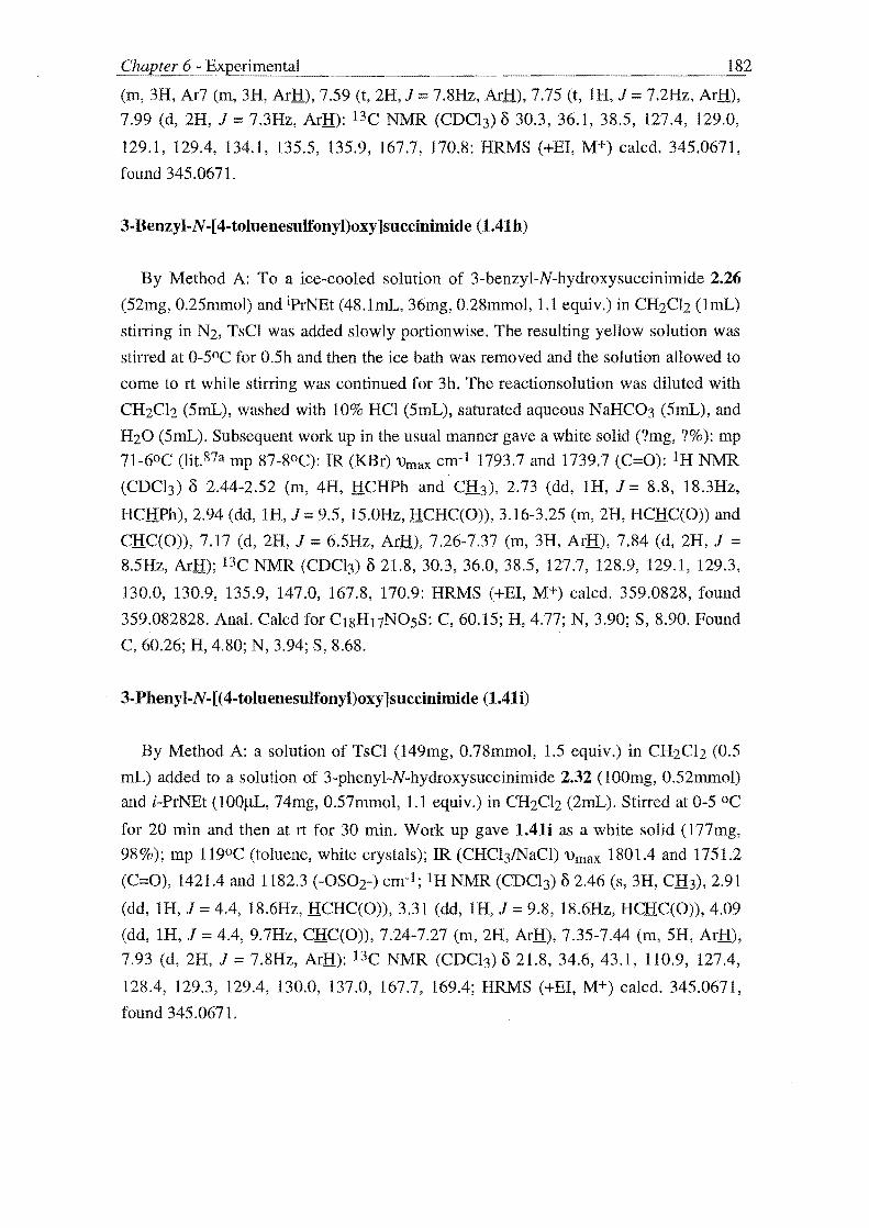
(m, 3H, Ar7 (m, 3H, ArH), 7.59 (t, 2H, J = 7.8Hz, ArH), 7.75 (t, IH, J = 7.2Hz, ArH),
7.99 (d, 2H, J = 7.3Hz, ArH): l3C NMR (CDCI3) 0 30.3, 36.1, 38.5, 127.4, 129.0,
129.1, 129.4, 134.1, 135.5, 135.9, 167.7, 170.8: HRMS (+EI, M+) calcd. 345.0671,
found 345.0671.
3-Benzyl-N-[ 4-toluenesulfonyl)oxy ]succinimide (1.4Ih)
By Method A: To a ice-cooled solution of 3-benzyl-N-hydroxysuccinimide 2.26
(52mg, 0.25mmol) and iprNEt (48.1mL, 36mg, 0.28mmol, 1.1 equiv.) in CH2Ch (lmL)
stirring in N2, TsCI was added slowly portion wise. The resulting yellow solution was
stirred at 0-5°C for 0.5h and then the ice bath was removed and the solution allowed to
come to rt while stirring was continued for 3h. The reactionsolution was diluted with
CH2Ch (5mL), washed with 10% HCI (5mL), saturated aqueous NaHC03 (5mL) , and
H20 (5mL). Subsequent work up in the usual manner gave a white solid (?mg, ?%): mp
71-6°C (lit.87a mp 87-8°C): IR (KBr) 'Urnax cm- I 1793.7 and 1739.7 (C=O): IH NMR
(CDC13) 0 2.44-2.52 (m, 4H, HCHPh and Cfu), 2.73 (dd, IH, J = 8.8, 18.3Hz,
HCHPh), 2.94 (dd, IH, J= 9.5, 15.0Hz, HCHC(O», 3.16-3.25 (m, 2H, HCHC(O» and
CHC(O», 7.17 (d, 2H, J = 6.5Hz, ArH), 7.26-7.37 (m, 3H, ArH), 7.84 (d, 2H, J = 8.5Hz, ArH); l3C NMR (CDCI3) 021.8,30.3,36.0,38.5, 127.7, 128.9, 129.1, 129.3,
130.0, 130.9, 135.9, 147.0, 167.8, 170.9: HRMS (+EI, M+) calcd. 359.0828, found
359.082828. Anal. Calcd for CI8H17NOSS: C, 60.15; H, 4.77; N, 3.90; S, 8.90. Found
C, 60.26; H, 4.80; N, 3.94; S, 8.68.
3-Phenyl-N-[(4-toluenesulfonyl)oxy]succinimide (lAli)
By Method A: a solution of TsCI (149mg, 0.78mmol, 1.5 equiv.) in CH2Ch (0.5
mL) added to a solution of 3-phenyl-N-hydroxysuccinimide 2.32 (lOOmg, 0.52mmol)
and i-PrNEt (lOOJlL, 74mg, 0.57mmol, 1.1 equiv.) in CH2Ch (2mL). Stirred at 0-5 °C
for 20 min and then at rt for 30 min. Work up gave l.4li as a white solid (177mg,
98%); mp 119°C (toluene, white crystals); IR (CHC13/NaCl) 'Urn ax 1801.4 and 1751.2
(C=O), 1421.4 and 1182.3 (-OS02-) em-I; IH NMR (CDC13) 0 2.46 (s, 3H, CH3), 2.91
(dd, IH, J = 4.4, 18.6Hz, HCHC(O», 3.31 (dd, IH, J = 9.8, 18.6Hz, HCHC(O», 4.09
(dd, IH, J 4.4, 9.7Hz, CHC(O», 7.24-7.27 (m, 2H, ArH), 7.35-7.44 (m, 5H, ArH),
7.93 (d, 2H, J = 7.8Hz, ArH): l3C NMR (CDC13) 0 21.8, 34.6, 43.1, 110.9, 127.4,
128.4, 129.3, 129.4, 130.0, 137.0, 167.7, 169.4; HRMS (+EI, M+) calcd. 345.0671,
found 345.0671.

183
3-Benzyl-N-[(2-napthylsulfonyl)oxy]succinimide (1.41j)
By Method A: 2-napthylsulfonyl chloride (33mg, 0.15mmol, 1.1 equiv.) was added
to a solution of 3-benzyl-N-hydroxysuccinimide 2.25 (30mg, 0.15mmol) and i-PrNEt
(26~L, 21mg, 0.16mmol, 1.1 equiv.) in CH2Cl2 (5mL). Stirred at 0-5 °C for 0.5h and
then at rt for 1h. Work up by diluting with CH2Ch and washing with H20, 10% HCI
and 10% aqueous NaHC03 gave 1.41j as a white solid (57mg, 99%); IH NMR (CDC!))
62.50 (dd, IH, J = 4.4, 18.5Hz, HCHPh), 2.74 (dd, 1H, J = 9.1, 18.4Hz, HCHPh), 2.95
(dd, IH, J = 8.8, 14.7Hz, HCHC(O) 3.16-3.27 (m, 2H, HCHC(O» and CHC(O), 7.15-
7.18 (m, 2H, ArH), 7.26-7.37 (m, 3H, ArH), 7.63-7.7.75 (m, 2H, ArH), 7.93-8.05 (m,
2H, ArH), 8.56 (s, IH, ArH):13C NMR (CDCl)) 6 30.3,36.0,38.5, 121.3, 123.2, 127.4,
128.0,128.1,128.9,129.0,12.1,129.6,129.7,130.2, 130.8,131.8,131.9,135.9,136.0,
1648, 167.8, 170.9: HRMS (+EI, M+) calcd. 395.0828, found 395.0828.
3-Benzyl-N -[ (3-nitrobenzenesulfonyl)oxy ]succinimide (1.41k)
By Method A: a solution of 3-nitrobenzene sulfonyl chloride (120mg, 0.55mmol, 1.5
equiv) was added to a solution of 3-benzyl-N-hydroxysuccinimide 2.25 (75mg,
0.36mmol) and Et3N (55.7mL, 4Omg, 0.40mmol, 1.1equiv.) in CH2Cb (4mL). Stirred
0-5°C Ih and then rt 4.5h. Work up as described gave a brown residue which was
purified by flash Si02 chromatography eluting with 3:7 EtOAc/petroleum ether to give
a brown oil (quantative yield): IR (CDC!) 'Umax 1813.0 and 1747.4 (C=O) cm- 1; IH
NMR (CDCI) 6 2.51 (dd, IH, J = 4.4, 18.6Hz, HCHPh), 2.77 (dd, 1H, J = 8.8, 18.6Hz,
HCHPh), 2.94 (dd, IH, J= 8.8, 13.7Hz, HCHC(O», 3.16 (dd, IH, J = 4.9, 14.2Hz,
HCHC(O», 3.24-3.33 (m, lH, CHC(O», 7.17 (d, 2H, J = 7.3Hz, ArH), 7.23-7.34 (m,
3H, ArH), 7.83 (t, lH, J = 7.8Hz, ArH), 8.24 (d, 1H, J = 8.3Hz, ArH), 8.55 (d, 1H, J =
803Hz, ArH), 8.75 (s, IH, ArH); 13C NMR (CDCI) 6 30.3, 36.0, 38.5, 124.5, 127.6,
129.1, 129.1, 129.7, 130.8, 134.5, 135.6, 136.6, 148.2, 167.5, 170.6: HRMS (+EI, M+)
calcd. 390.0522, found 390.0522.
Synthesis of 3-benzyl-N -[ (2-carbethoxy -ethanesulfonyl)oxy ]succinimide (1.411)
1. Freshly distilled 2-bromopropionic acid 2.37 (l8mL, 30.6g, 0.2mol) was added
dropwise to SOCh (20mL, 32.740g, 0.275mol, 1.4 equiv.) stirring at 30-40°C. After the
addition was completed the resulting solution was refluxed for 2h. Excess SOCh was
removed by distilllation at aspirator pressure to give a dark brown liquid which was
then distilled at reduced pressure to give the intermediate 2-bromopropionoyl chloride
as a colourless liquid (10.192g, 30%). This acid chloride was transferred with toluene
washings (2mL) to a dropping funnel and was added dropwise to dry EtOH (4mL,
3.140g, 0.068mol, 1.15 equiv.) stirring at 0-5°C (ice-salt bath). After the addition was

184
completed, stirring was continued at that same temperature for 2h at which point the
ice-salt bath was removed and the reaction solution allowed to come to rt while being
stirred for 2.75d. The reaction solution was poured into H20 (lOmL) and the organic
layer that separated washed with the same volume of 10% aqueous NaHC03 and H20
and worked up in the usual manner to give the ester 2.38 as a clear oil (6.629g, 18%
overall): bp 22°C 10.02rnrnHg; IH NMR (CDCI3) 8 1.27 (t, 3H,J = 7.1Hz, CH2CH3),
1.79 (d, 3H, J = 6.8Hz, CHCHJ.), 4.20 (q, 2H, J = 6.8, 7.4Hz, CHZCH3), 4.33 (q, lH, J
= 6.8, 6.9Hz, CHCH3).
2. A solution of ethyl 2-bromopropionate 2.38 (1.31g, 7mmol) and K2S03 (1.312g,
8m mol) in 12%:88% EtOHlH20 (l7mL) was refluxed for 1.5h. The solvent was
evaporated in vacuo to give a white solid. This crude salt 2.39 (1.271g, 80%) was dried
in an oven set at 110°C for 2d and then used without further purification. 1 H NMR
(D20) 8 1.25 (t, 3H,J = 7.3Hz, CH2CH1), 1.48 (d, 3H, J = 6.9Hz, CHCHJ.), 3.93 (q, IH,
J = 6.8, 7JHz, CHCH3), 4.22 (q, 2H, J = 7.1, 7.3Hz, ClliCH3).
3. A suspension of potassium ethyl 2-sulfopropionate 2.39 (1.271g, 6mmol) and
PCls (1.250g, 6mmol) in POCh (l2mL) was stirred vigorously at 80°C for 2.5h under a
N2 atmosphere. The reaction mixture was allowed to cool to rt and then filtered under a
N2 atmosphere directly into a modified Claisen flask and short Vigreux column set up
for distillation. The salt cake was washed with a small quantity of POCh and this
washing and the yellow coloured filtrate were distilled at aspirator pressure to give a
mixture of a small quantity of white solid, presumably unremoved NaC!, and brown
residue. This mixture was distilled at reduced pressure to give 2.40 as a yellow coloured
liquid with a characteristic piquant odour (122mg, 11 %): bp 52°C 10.02mmHg; IH
NMR (CDCh) 8 1.35 (t, 3H,J = 7.1Hz, CH2CH3), 1.86 (d, 3H, J = 6.8Hz, CHCfu),
4.30-4.38 (m, 2H, CH2CH3), 4.50 (q, IH, J = 6.8, 7.4Hz, CHCH3).
4. By Method C: a solution of ethyl (2-chlorosulfonyl)propionate 2.40 (122mg,
0.61mmol) in toluene (1.5mL) was added dropwise to a solution of 3-benzyl-N
hydroxysuccinimide 2.26 (125mg, 0.61rnrnol) and pyridine (98.7JlL, 97mg, 1.2rnmol,2
equiv.) in toluene (1.5mL) stirring in N2 at rt. The resulting white slurry was stirred at rt
for 15min after which the tenperature was raised to 50°C during which the slurry
cleared to give a brown coloured solution. Stirring was then continued for 18h. The
reaction solution was evaporated to give a brown residue which was dissolved in EtOAc
and wahed with 2N HCI. The aqueous layer was re-extracted with EtOAc and the
organics combined and worked up in the usual manner to give a brown coloured residue
(151mg, 67%). Purification of 101mg of this residue by flash Si02 chromatography
eluting with 1: 1 EtOAc/petroleum ether gave 1.411 as a brown coloured residue (64mg,
28%); IR (CDC13) 'Umax 1801.4 and 1747.4 (C=O), 1409.9 and 1190.0 (-S02-) cm- I ;

185
IH NMR (CDC13) {) 1.32 (t, 3H, J :=: 7.3Hz, CH2CH3), 1.85 (dd, 3H, J :=: 2.3, 7.3Hz,
CHCH3), 2.55 (dd, IH, J:=: 3, 17.5Hz, HCHPh), 2.80 (dd, IH, J:=: 8.8, 18.3Hz, HCHPh),
2.87-2.97 (m, IH, H2CCHCH2), 3.24-3.30 (m, 2H, CHCH2), 4.31 (q, 2H, J = 7.0,
7.3Hz) 4.67 (q, IH, J = 6.0, 7.0Hz, CHCH3), 7.17-7.35 (m, 5H, ArH); l3C NMR
(CDCI3) {) 13.4, 13.9, 30.5, 36.1, 36.3, 38.5, 38.6, 63.2, 65.3, 127.4, 128.5, 129.0,
129.0, 129.2, 135.9, 164.3, 168.1, 171.0, 171.0; HRMS (+E1, M+) calcd. 369.0883,
found 369.0882.
3-Benzyl-N -[ (3-{ 3-benzyl-N-succinimidylsulfonate }benzenesulfonyl)oxy]
succinimide (l.4ln)
By Method A: 1,3-bis(chlorosulfonyl)benzene (32mg, 0.12mmol) was added slowy
to a solution of 3-benzyl-N-hydroxysuccinimide 2.26 (45mg, 0.22mmol, 2 equiv.) and i
PrNEt (42J..iL, 31mg, 0.24mmol, 2.2 equiv.) stirring under N2 at 0-5 DC. Stirring
continued at 0-5 °C for 0.5h and then for 11h at rt. Work-up gave l.4ln as a white solid
(22mg, 17%) after crystallisation from toluene/petroleum ether: mp 95-100 °C; 1R
(CDC13) 'Umax 1813.0 and 1747.4 (C=O) cm-I; IH NMR (CDC13) {) 2.50 (dd, IH, J = 4.4, 18.5Hz, HCHPh), 2.75 (dd, IH, J = 9.0, 18.3Hz, HCHPh), 2.94 (dd, IH, J = 8.6,
l3.9Hz, HCHC(O», 3.17-3.27 (m, 2H, HCHC(O) and CHC(O», 7.l3-7.36 (m, 5H,
ArH) , 7.86 (t, IH, J 8.lHz, ArH), 8.35 (d, 2H, J = 83Hz, ArH), 8.59 (s, IH, ArH);
J3C NMR (CDC13) {) 30.4,36.0, 38.5, 127.4, 129.0, 130.1, 130.8, 135.5, l35.8, 136.1,
167.6, 170.7; HRMS (FAB, MH+) calcd. 613.0951, found 613.0951.
3-Benzyl-N-[(4'-{3-benzyl-N
succinimidyl}sulfonatebiphenylsulfonyl)oxy ]succinimide (l.4lm)
By Method C: 4,4-biphenyl disulfonyl chloride (173mg, 0.5mmol) was added slowly
portionwise to a solution 3-benzyl-N-hydroxysuccinimide (202mg, Immol, 2 equiv.)
and pyridine (160.1mL, 157mg, 2mmol, 4 equiv.) in toluene (8mL) stirring at 400 C in
N2. The temperature was raised to 500 C and stirring continued for 2h. Solvent was
evaporated and the resulting mixture consisting of a brown residue and a white solid
was dissolved in the minimum amount of EtOAc and washed with H20. The aqueous
layer was reextracted with EtOAc and the extracts combined and worked up in the usual
manner to give a yellow solid. Purification by flash Si02 chromatography eluting with
CH2Cb gave l.4lm as a brown oil (117mg, 34%): 1R (CDCb) 'Umax 18l3.0 and
1747.4 (C=O) em-I; IH NMR (CDCI3) {) 2.50 (dd, IH, J = 4.4, 18.5Hz, HCHPh), 2.75
(dd, IH, J = 9.0, 18.3Hz, HCHPh), 2.94 (dd, IH, J:=: 8.6, 13.9Hz, HCHC(O», 3.17-3.27
(m, 2H, HCHC(O) and CHC(O», 7.13-7.36 (m, 5H, ArH), 7.86 (t, IH, J:=: 8.1Hz, ArH),
8.35 (d, 2H, J = 8.3Hz, ArH), 8.59 (s, IH, ArH); 13C NMR (CDC13) {) 30.4,36.0, 38.5,
127.4, 129.0, 130.1, l30.8, 135.5, 135.8, 136.1, 167.6, 170.7; HRMS (FAB, MH+)

186
calcd. 613.0951, found 613.0951. Anal. Calcd. for C34H2SN201OS2.1.5H20: C, 57.17;
H, 3.95; N, 3.92. Found C, 56.96; H, 4.14; N, 3.93.
THE SYNTHESIS OF 3·SUBSTITUTED N-[(ACYL)OXY] SUCCINIMIDES
Method A: Et3N/iPrNEt and DMAP catalysed acylation.
Acetic anhydride or the appropriate acid chloride (l equiv.) was added to a solution
of 3-substituted N-hydroxysuccinimide (stated amount), DMAP (0.1 equiv.), and Et3N
or i-PrNEt (l equiv.) in CH2C12 (stated volume) which had been stirring for 5min under
N2 at r1. Stirring was then continued at rt overnight. The reaction solution was diluted
with EtOAc and washed with 10% aqueous citric acid and H20 and then O.5N KHC03
and H20 again. The organic phase was then worked up in the usual manner to give the
title compounds. These were purified by crystallisation from the stated solvent system.
MethodB: Et3N li-PrNEt catalysed acylation.
The appropriate acid chloride (1.5 equiv.) was added to a solution of N-hydroxy
imide (stated amount) and Et3NH or iPrNEt (1.1 equiv.) in CH2Cl2 stirring at 0-50 C
(ice-salt bath). Stirring was continued at that temperature for the stated time, (typically
at least 20min), then the ice-salt bath removed and the reaction mixture allowed to come
to rt while stirring for the stated period (typically at least 0.5h). The reaction solution
was diluted with CH2Cb and washed with H20, 10% HCI and saturated NaHC03. The
organic phase was worked up in the usual manner to give the title compounds in high
purity by 1 HNMR.
Method C: TMSOTf catalysed acetylation.
TMSOTf (0.02 equiv.) was added to a solution of N-hydroxy imide (stated amount)
and AC20 (1.5 equiv.) in CH2C12 which had been stirring in N2 for lOmin at 0-50 C
(ice-salt bath). The resulting solution was allowed to come to rt while stirring overnight.
This was then treatedwith saturated aqueous NaHC03 and extracted with CH2C12. The
organic extract was separated, washed with 10% aqueous NaHC03 and worked up in
the usual manner to give the title compounds as crystalline solids.
Method D: TMSOTf catalysed acylation with in situ mixed anhydride
A solution of the appropriate acid (stated amount) in CH2Cb was treated with N
methylmorpholine and ethyl chloroformate to give an in situ mixed anhydride. After

187
this solution was stirred for the stated period the N-hydroxysuccinimide (stated amount)
was added followed by TMSOTf (0.02 equiv.) and the reaction stirred while being
allowed to come to rt over the stated period. Saturated aqueous NaHC03 was added to
the reaction mixture and the organic phase then separated and washed with saturated
aqueous NH4CI and H2O. Work-up in the normal manner gave the title compound.
Method E: DCC, HOBt mediated acid coupling
HOBt (stated amount) and acid (stated amount) were added to an ice cooled solution
of the N-hydroxysuccinimide (stated amount) in CH2Cl2 stirring under N2. The
resulting solution was stirred for the stated period under N2 at O-S °c after which DCC
(stated amount) was added and the resulting mixture stirred for the indicated period
while the reaction mixture was allowed to come to rt. The reaction mixture was
evaporated and the resulting product suspended in EtOAc (stated amount). This
suspension was then filtered to remove the by-product DCU and the filter cake washed
with a small quantity of EtOAc. The filtrate and washings were combined and washed
with O.5N aqueous KHC03 and H20 and the organic phase worked up in the usual
manner to give the title compound.
3-Benzyl-N -[ (acetyl)oxy ]succinimide (1.410)
By Method A: AC20 (SS.7JlL, 6Omg, 0.6mmol) was added to a solution of 3-benzyl
N-hydroxysuccinimide 2.26 (lllmg, 0 6mmol), DMAP (8mg, 0.06mmoL 0.1 equiv.),
and Et3N (82.2mL, 60mg, 0.6mmol) in CH2Cl2 (6mL) which had been stirring for Smin
under N2 at rt. Work-up gave 1.410 as a brown residue (108mg, 79%): mp 107-90 C
(EtOAc/petroleum ether, amber coloured crystals); IH NMR (CDCI3) 8 2.22 (s, 3H,
C(O)Cfu), 2.43 (dd, IH, J = 3.4, 18.1Hz, HCHPh), 2.62-2.81 (m, 2H, HCHPh and
HCHC(O», 3.16 (bs, 2H, HCHC(O) and CHC(O», 7.08-7.26 (m, SH, ArH).;. l3C NMR
(CDC13) 8 17.S, 30.7, 36.3, 38.7, 127.2, 128.6, 128.9, 136.3, 16S.S, 168.S, 171.S;
HRMS (+E1, M+) calcd. 247.084S, found 247.084S.
By method C: TMSOTf (4JlL, Smg, 0.02mmol, 0.02 equiv.) was added to a solution
of 3-benzyl-N-hydroxysuccinimide 2.26 (202mg, 0.99mmol) and AC20 (l40IlL,
lSlmg, l.Smmol, I.S equiv.) in CH2C12 (6mL) that had been stirring at O-S °C under
N2 for 10 min. Stirrind was continued as the reaction solutionwas allowed to come to rt
overnight. Work-up as described above gave 1.410 as a brown coloured residue which
on standing at rt over a period of one week solidified to afford 1.410 as a white
crystalline solid (209mg, 86%). Physical data as given above.

188
3~Phenyl~N~[ (acetyl)oxy ]succinimide (1.41p)
By Method B: AcCI (28IlL, 3lmg, 0.39mmol) was added to a solution of 3-phenyl
N-hydroxysuccinimide 2.32 (SOmg, 0.26mmol) and i-PrNEt (49.9JlL, 37mg, 0.29mmol,
1.1 equiv.) in CH2Clz (1mL). Work-up gave 1.41p as an off-white solid (4Smg, 74%):
mp 74 oC (Et20/petroleum ether, colourless crystals); IR (CDCb) 'Umax 1789.8 and
1743.5 (C=O) cm-I; IH NMR (CDCh) (32.37 (s, 3H, CH3), 2.89 (dd, IH, J ::: 404, 19.0,
HCHC(O)), 3.33 (dd, JH, J ::: 9.8, 18.SHz, HCHC(O)), 4.12 (bs, IH, CHC(O)), 7.31-
7043 (m, SH, ArH); l3C NMR (CDCI3) (3 17.6, 3S.3, 43.S, 126.6, 127.S, 128.3, 129.3,
129.6, 130.2, 16S.6, 168.3; HRMS (+EI, M+) calcd. 233.0688, found 233.0688. Anal.
Ca1cd. for C12HUN04: C, 61.80; H, 4.7S; N, 6.01. Found: C, 62.10; H, 4.81; N, 6.2S.
3~Benzyl-N-[(trans-cinnamoyl)oxy]succinimide (1.41q)
By Method A: trans-cinnamoyl chloride (70mg, 0.42mmol, 1.0S equiv.) was added
slowly portionwise to a solution of 3-benzyl-N-hydroxysuccinimide 2.26 (83mg,
OAmmol), DMAP (4mg, 0.03mmol), and i-PrNEt (70AJlL, S2mg, OAmmol) in CH2Cl2
(7mL) that had been stirring for Smin under N2 at rt. The resulting reaction solution was
stirred for 14.Sh. Work-up gave 1.41q as a white soid (lOOmg, 7S%): mp 1OS-70 C
(EtOAc/petroleum ether, white crystals); IH NMR (CDCI3) (3 2.S7 (bd, JH, J = 18.0Hz,
HCHPh), 2.76-2.93 (m, 2H, HCHPh and HCHC(O)), 3.29 (bs, 2H, HCHC(O) and CH)
6.S9 (d, IH, J = 16.1Hz, C(H):CHPh), 7.93 (d, 1H, J::: 16.1Hz, C(O)CH:C(H)Ph1. l3C
NMR (CDCI3) (330.8,36.2,38.8, IlLS, 127.2, 127.8, 128.6, 128.9, 128.9 129.0, 131.S,
13304,149.9,162.0,168.7,171.7; HRMS (+EI) (M+) ca1cd 33S.11S8, found 33S.11S8.
Anal. Calcd for C2oH170~: C, 71.64; H, S.07; N, 4.18. Found: C, 71.12; H, S.12; N,
4.10.
By Method D: N-methylmorpholine (11SmL, 106mg, 1.0Smmol) and ethyl
chloroformate (104mL, 118mg, 1.09mmol, 1.1 equiv.) were added to an ice cooled
solution of cinnamic acid (1S6mg, 1. OSmmol , 1.0S equiv.) in CH2Cl2 (SmL). The
resulting solution was stirred at O-S °C under N2 for 10 min. 3-Benzyl-N
hydroxysuccinimide 2.26 (206mg, Immol) and TMSOTf (4mL, Smg, 0.02mmol) were
then added and the resulting reaction solution allowed to warm to rt while stirring
overnight. Work-up gave 1.41q as a white solid (289mg, 86%). Spectral data as above.
3-Benzyl-N -[ (N -benzyloxycarbonyl-( L) -phenylalanyl)oxy]succinimide (1.41r)
By Method E: HOBt (66mg, OA9mmol) and N-Cbz-(L)-phenylalanine (147mg,
OA9mmol) were added to an ice-cooled solution of 3-benzyl-N-hydroxysucccnimide
2.26 (1OImg, O.SOmmol) in CH2C12 (9mL) stirring under N2. This solution was stirred

189
at 0-5 DC under N2 for 15min after which DCC (114mg, 0.55mmol) was added and the
solution allowed to warm to rt while being stirred for 43h. The reaction solution was
evaporated and the resulting mixture of a brown residue and white solid was suspended
in the minimum amount of EtOAc. This was then filtered to remove the by-product
DCU and the filer cake washed with a small quantity of EtOAc. The filtrate and
washings were combined and washed with 0.5N KHC03 and H20 and the organics
worked-up in the normal manner to give a brown residue (l98g, 83%): 1 H NMR
(CDCb) 82.56 (1H, dd, J = 3.9, 18.1 Hz, HCHPh), 2.78-2.93 (2H, m, HCHPh and
HCHC(O», 3.29-3.43 (m, 2H, HCHC(O) and CH), 4.62-4.66 (m, 1H, C(O)CHNH),
5.10 (s, 2H, OCH2Ph), 5.24 (d, lH, J = 6.3Hz, NH), 7.09-7.38 (m, lOH, ArH); l3C
NMR 8 24.6,25.3,29.6,30.6,30.7,33.3,36.1,37.6,37.8, 38.1, 38.4, 38.7, 52.9, 54.6,
54.8,67.0,67.3,77.2,127.0, 127.1, 127.3, 127.4, 128.0, 128.1, 128.2, 128.4, 128.5,
128.7, 128.8, 128.9, 129.3, 129.5, 134.3, 135.6, 135.8, 136.0, 136.2, 136.6, 155.5,
155.9,167.4,168.2,171.1,171.8,174.5,174.88; HRMS (+EI) calcd. 486.1792, found
486.1791.
THE SYNTHESIS OF 2NALKYLGLUTARIC ACIDS
Method A: Michael Reaction Route
DiethyI2-benzyl-2-cyanoethyl malonate (2.45)182a
Acrylonitrile (2.4mL, 1.934g, 36mmol) was added slowly to a solution of diethyl
benzyl malonate (8.7mL, 9.257g, 37mmol) and 30% methanolic KOH (201mg) in
tBuOH (13mL) stirring inN2 at such a rate that the temperature of the reaction mixture
did not exceed 40°C. Stirring was continued at rt for a further 4.5h. The reaction
mixture was acidified (pH=l, Universal Indicator Paper) with glacial CH3C02H and 6N
HCI then concentrated and extracted with Et20. The Et20 extract was washed with an
equal volume of H20 and worked up in the usual manner to give an oily brown residue.
This was then distilled under reduced pressure (lmmHg) to give three liquid fractions.
The first (1.620g) and second (1.045g) fractions were collected in the ranges 124-50 oC
and 150-70 °C and contained, by IH NMR, 21:4 and 14:11 unreacted diethyl benzyl
malonate/diethyl benzyl-2-cyanoethyl malonate respectively. The third fraction was
found to contain pure product and after standing at rt for 19d slowly crystallised to give
2.45 as a white solid (5.616g, 50%): bp 170-96 DC/1mmHg (lit. 182a bp 175-80
oCllmmHg); IH NMR (CDCI3) 8 1.27 (t, 6H, J = 7.3Hz, 2CH2CH3), 2.11 (t, 2H, J =
7.8Hz, CH2CN), 2.44 (t, 2H, J = 7.8Hz, CfuCH2), 3.26 (s, 2H, PhCH2), 4.23 (q, 4H, J
= 6.9, 7.3Hz, 2CH2CH3), 7.06-7.09 (m, 2H, ArID, 7.24-7.31 (m, 3H, ArH): l3C NMR
(CDC]3) 8 13.0, 13.8, 28.6, 39.4, 57.5, 61.7, 119.0, 127.3, 128.4, 129.7, 134.8, 169.9:

190
HRMS (+E1, M+) calcd. 289.1315, found 289.1314. Anal. Calcd for C17H21N04: C,
66.41; H, 6.62; N, 4.84. Found: C, 66.17; H, 6.50; N, 4.71.
2-BenzyIglutaric acid (2.44b)
A mixture of 2.45 (2.249g, 7mmol) and 68% HBr (l8mL) was refluxed for ld. The
solution was then evaporated to give a mixture of a brown liquid and a white solid
(presumably NH4Br). Sufficient H20 was added to dissolve the NH4Br and the
resulting mixture was extracted with Et20. Work up in the usual manner gave 2.44b as
a brown oil (l.174g, 72%): IH NMR (CDCI3) 01.78 (q, 2H, J = 7.3, 7.8Hz), 2.26-2.39
(m, 2H), 2.67 (dd, IH, J = 7.3, 14.lHz), 2.80 (dd, 1H, J = 6.4, 13.2Hz), 2.94 (dd, 1H, J
= 8.3, 13.6Hz), 7.26-7.39 (m, SH, ArH); l3C NMR (CDCI3) 0 25.9, 31.5, 37.8,46.2,
126.5, 128.4, 128.8, 138.2.
THE SYNTHESIS OF 3,3-DIALKYLGLUTARIC ACIDS
4,4-DimethyI-3,5-dicyanoglutarimmide (2.48a) 185
A solution of EtOH saturated with NH3 was prepared by bubbling NH3(g) through
EtOH (200mL) at ca. -S DC for 6h. This solution was added to a mixture of ethyl
cyanoacetate (l06mL 113g, 1mol) and acetone (37mL, 29.267g, 0.5mol) and the
resulting mixture was stored in the freezer for 3d. Cold EtOH (200mL) was added and
the mixture stirred thoroughly and returned to the freezer. After this period the reaction
mixture was allowed to warm to rt and the yellow salt that had deposited over this
period was collected by suction filtration and washed with a small quantity of Et20 to
remove ethyl cyanoacetate and acetone and then air-dried. The resulting pale yellow
solid was dissolved in boiling H20 and cone. HCI added until this solution was acid to
Congo Red Paper after which an additional volume of cone. HCI (SOmL) was added.
On cooling 2.48a crystallised as a white solid (40.87g, 42%); IR (KBr) 'Umax 3215.1
(NH), 1712.7 and 1749.3 (C=O); IH NMR (DMSO-d6) 0 1.27 (s, 3H, CH3), 1.44 (s,
3H, CH3), 4.88 (s, 2H, 2xCll), 12.23 (bs, lH, NH); l3C NMR (DMSO-d6) 19.9,26.0,
39.1,46.9,114.6,164.7; HRMS (FAB, M+) calcd. 191.069S, found 191.0695.
3,3-CycIohexyIglutaric acid (2.44d)253
4,4-Cyc1ohexyl-3,S-dicyano-glutarimide 2.49b (2.020g, 9mmol) was dissolved in
cone. H2S04 (5mL) with gentle warming. Water (SmL) was added slowy and the
resultant solution heated to reflux becoming a black mixture containing a suspension of
fine white solid. At this stage some foaming problems were experienced leaving
material adhered to the insides of the reaction flask and reflux condenser. This was

191
washed back down into the reaction mixture with a small quantity of H20. Reflux was
continued overnight during which period the described suspension dissolved to give a
clear black solution. This was diluted carefully with H20 (lOmL) and extracted
thoroughly with Et20. The Et20 extracts were combined and washed with H20 and
then extracted with saturated aqueous NaHC03. The NaHC03 extracts were acidfied
(pH:::: 1 , Universal Indicator Paper) with conc. HCI and then extracted with Et20. Work
up in the usual manner gave 2.44d as a white solid (1.259g, 72%): mp 179-820C (lit.253
mp 182-3 °C).
3,3-Dimethylglutaric acid (2.44e)185
Compound 2.48a (23.22g, 0.12mol) was dissolved in conc. H2S04 (62mL) with
slight warming. Water (62mL) was then added slowly and the resulting mixture
refluxed for 1.5d. The reaction mixture was diluted with H20 (50mL) and extracted
with Et20. The organic extracts were combined and washed with H20 and then worked
up in the normal manner to give a pale yellow solid (l5.19g, 78%); mp 99-102 °C
(lit.1 85 mp 102-3 °C); IR (KBr) 'Urn ax 2881.5 (OH), 1689.5 (C=O) cm- I ; IH NMR
(DMSO-d6) b 1.14 (s, 6H, 2xCH3), 2.38 (s, 4H, 2xCH2C(O)).
THE SYNTHESIS OF SUBSTITUTED GLUTARIC ANHYDRIDES
General Method
A mixture of the glutaric acid (stated amount) and excess AcCI or AC20 (stated
amount) was refluxed for the stated time. The solvent - a mixture of the excess AcCI or
AC20 and AcOH which was formed during reaction - was removed by downward
distillaton at aspirator pressure to give either liquids or more typically brown to black
residues. These were distilled at reduced pressure, or recrystallised from the stated
solvent system, as indicated, to give the title anhydride.
2.Phenylglutaric anhydride (2.43a)
A mixture of 2-phenylglutaric acid 2.44a (2.500g, 13mmol) and AcCI (5.200mL,
61mmol, 4.7equiv.) was refluxed for 1h. The excess AcC} was evaporated and the
resultant liquid was distilled at reduced pressure to give a pale yellow solid.
Recrystallisation from toluene/petroleum ether gave 2.43a as a white solid (2.081g,
84%); IR (CHCl)lNaCI) 'Urnax 1813.0 and 1762.8 (C=O) cm-I ; IHNMR (CDCh) b 2.28
(m, 2H, CH2C(O)), 2.85 (m, 2H, CHCH2), 3.92 (dd, 1H, J = 6.3, 8.7Hz, Cm, 7.33 (m,
5H, ArH); l3C NMR (CDC13) b 24.4, 29.4, 46.8, 127.8, 128.0, 128.9, 135.9, 166.5.

2-Benzylglutaric anhydride (2.43b)
A mixture of 2-benzylglutaric acid 2.44b (2.131g, 9.6rnrnol) and AC20 (5.978mL,
6.468g, 0.063mol, 6.6 equiv.) was refluxed for 2d. Distillation of the reaction mixture at
reduced pressure gave 2.43b as a pale yellow solid (1.354g, 69%): mp 80-1 DC
(toluene/petroleum ether); IR (KEr) 'Urn ax 1801.4 and 1755.1 (C=O) cm-1; IH NMR
(CDCI3) 8 1.64-1.78 (m, IH, HCHPh), 1.89-1.98 (m, IH, HCHPh), 2.55-2.67 (m, IH,
CHHCHCH2), 2.76-2.91 (m, 3H, CHHCHCH2 and C(O)CH2), 3.44 (q, 1H, J = 8.8,
18.1Hz, CHC(O)) 7.19-7.36 (m, 5H, ArH); 13C NMR (CDCI3) 821.2,29.9,36.1,42.3,
127.1,128.8, 129.1, 137.2, 166.5, 168.7.
3-Phenylglutaric anhydride (2.43c)
A mixture of 3-phenylglutaric acid (764mg, 4rnrnol) and AC20 (1.900rnL, 2.056g,
20mmol, 5 equiv.) was refluxed for 20.75h. The solvent was removed to give a brown
mass. This was dissolved in toluene, activated carbon added and the mixture filtered
with celite. The celite cake was washed with a small quantity of toluene and the filtrate
and washings combined and evaporated to yield a pale yellow solid (629mg, 89%): mp
101-3 DC: IH NMR (CDCI3) 82.87 (dd, 2H, J = 11.2, 17.6Hz, CH2C(O)), 3.11 (dd, 2H,
J = 4.4, 17.6Hz, CH2C(O)), 3.38-3.48 (m, IH, CH), 7.18-7.43 (m, 5H, ArH); 13C NMR
(CDCI3) 8 37.1, 41.0, 126.2, 128.1, 129.3,139.1,165.9.
3,3-Cyclohexylglutaric anhydride (2.43d)253
A mixture of 3,3-cyclohexylglutaric acid 2.44d (848mg, 4mmol) and AC20
(0.793mL 858mg, 8mmol, 2equiv.) was refluxed for 2h 20min. The solvent was
removed as per the general method to give a brown mass that was distilled at reduced
pressure to give 2.43d as a white solid (691mg, (91 %): mp 68-9 DC (lit. 253 mp 73 DC
(benzene/petroleum ether): bp 150 DC/5mrnHg (lit. 253 bp 213 DC/36mrnHg); IR (KEr)
'Urnax 1807.2 and 1755.1 (C=O) cm-I; IH NMR (CDCI3) 81.43-1.53 (m, lOH,5xCH2),
2.66 (s, 4H, 2xCH2C(O)); 13C NMR (CDCI3) 8 21.3,25.2,32.2,35.9,41.5, 166.3.
3,3-Dimethylglutaric anhydride (2.43e)
3,3-Dimethyl glutaric acid 2.44e (14.945g, 0.093mol) and AC20 (55rnL, 59.51Og,
O.583mol, 6.3 equiv.) were placed in a 2-necked round bottom flask set up for
downward distillation with a thermometer dipping into the reaction mixture. This was
heated, with distillation of AcOH and AC20, until the temperature reached 180 DC. The
resultant dark brown mass was allowed to cool to rt while stirring and then dissolved in
the minimum amount of boiling toluene. Activated carbon was added, celite added and

193
the mixture filtered. On cooling to rt a white solid crystallised from the filtrate. This
was reheated then recooled and petroleum ether was added upon which 2.44e
precipitated as a white solid which was isolated by suction filtration. Yield 11.703g,
(89%): mp 122-5 °C (lit.254 mp 122-3°C); IH NMR (CDC13) 81.15 (s, 6H, 2CH3), 2.61
(s, 4H, 2xCH2C(0»: 13C NMR (CDCI3) 8129.1, 129.1, 130.0, 133.7, 139.8, 167.3.
THE SYNTHESIS OF SUBSTITUTED N-BENZYLOXYGLUTARIMIDES
Method A: From anhydride
A dispersion of O-benzyloxyamine (1 equiv.) and the respective anhydride (stated
amount) in toluene or xylenes (stated volume) was refluxed with azeotropic removal of
H20 for the stated time. The solvent was evaporated to yield either a crude solid or
more typically a brown residue. Subsequent work up depended on this result. A solid
was purified by recrystalllisation from the stated solvent system to give the title
compounds as white solids. A residue was dissolved in the minimum of EtOAc (stated
volume) and washed with an equal volume of 10% aqueous NaHC03 and H20. The
organic phase was worked up in the usual manner to give the title compounds as solids.
These were purified, if required, by recrystallisation from the stated solvent system.
:Method B: From acid
O-Benzyloxyamine (1.2 equiv.), Et3NH (5 equiv.) and HOBt (2.2 equiv.) were added
in successsion to a solution of acid (stated amount) in CH2Ch (stated volume) stirring
in N2 at 0-5 °C (ice-salt bath). Stirring W&'I) continued for ten min and then EDAC (2.08
equiv.) was added. The resulting solution was allowed to come to rt while stirring
overnight. The reaction solution was washed with H20, 10% HCI, and saturated
aqueous NaHC03. Work up in the usual manner gave the desired product as a crude
oily solid. This was purified by flash chromatography eluting with the stated solvent
system.
3-Benzyl-N-benzyloxyglutarimide (2.49b)
By Method A: a dispersion of 2-benzylglutaric anhydride 2.43b (418mg, 2mmol)
and O-benzyloxyamine (263mg, 2.lmmol, 1.05 equiv.) in xylenes (5roL) was heated
until dissolution and refluxed with azeotropic removal of H20 overnight. Work-up gave
a white solid (436mg, 69%): mp 91-4 °C (toluene/petroleum ether); IH NMR (CDCI3)
8 1.51-1.59 (m, IH, HCHPh), 1.65-1.85 (m, IH, HCHPh), 2,45-2.57 (m, IH,
CHHCHCH2), 2.64-2.81 (m, CHHCHCH2 and CH2C(O», 3,40 (dd, IH, J = 3.5,

13.0Hz, CHC(O», 5.01 (s, 2H, OCfuPh), 7.16-7.52 (m, lOH, ArH); 13C NMR (CDCh)
() 21.5,32.1,36.1,44.6, 77.95, 126.8, 128.3, 128.6, 129.1, 129.1, 130.0, 133.8, 137.9,
168.1,170.3; HRMS (+CI, MH+) calcd. 310.14440, found 310.1443.
4-Phenyl-N -benzyloxyglutarimide (2.49c)
By Method A: a dispersion of 3-phenylglutaric anhydride 2.43c (286mg, l.5mrnol)
and O-benzyloxyarnine (196mg, 1.6mmol, 1.07 equiv.) in toluene (l1rnl) refluxed with
azeotropic removal of H20 for 19h. Work up gave 2.49c as a white solid (425mg,
96%): mp 164-7 °C; IR 'Umax (KEr) 1743.5 and 1691.5 (C=O) em-I; IH NMR (CDC13)
() 2.83-3.05 (m, 4H, 2xCH2C(O», 3.27-3.37 (m, IH, CH), 5.03 (s, 2H, OCH2Ph), 7.18
(d, 2H, J = 8.3Hz, ArH), 7.26-7.40 (m, 6H, ArH), 7.51-7.55 (m, 2H, ArH); 13C NMR
(CDC13) () 34.5, 40.2, 78.2, 126.2, 127.7, 128.4, 129.1, 129.1, 130.0, 133.7, 139.8,
167.3; HRMS (+EI, mlz) 190.1 (M - OCH2Ph, 11), calcd. 289.1315, found 289.1314.
4,4-Cyclohexyl-N-benzyloxyglutarimide (2.49d)
A dispersion of 3,3-cyclohexylglutaric anhydride 2.43d (478g, 2.6mmol) and 0-
benzyloxyamine (338mg, 2.7mmol) in xylene (6ruL) was refluxed with azeotropic
removal of H20 overnight. The solvent was evaporated in vacuo to give a white solid
which was dissolved in the minimum amount of EtOAc and washed with 10% aqueous
NaHC03. Work-up in the usual manner gave 2.49d as a purified white solid (618mg,
83%): IR (KEr) 1741.6 and 1705.0 (C=O) em-I; IH NMR (CDCL3) () 1.33-1.58 (m,
lOH, 5xCH2), 2.61 (s, 2H, CH2C(O», 5.03 (s, 2H, OCH2), 7.35-7.37 (m, 3H, ArH),
7.51-7.54 (m, 2H, ArH); 13C NMR (CDCL3) () 21.4,25.5,32.2,35.8,44.4,78.0,128.4,
129.1, 129.9,170.1; HRMS (+EI, M+) calcd. 287.1522, found 287.1522.
4,4-Dimethyl-N -benzyloxyglutarimide (2.4ge)
By Method A: a dispersion of 3,3-dimethyl glutaric anhydride 2.43e (3 oOrng ,
2.l1mmol) and O-benzyloxyamine (265mg, 2.15mmol, 1.02 equiv.) in xylene (4.3mL)
was heated until dissolution and then refluxed with removal of H20 for 15h. The
resultant solution was allowed to cool to rt and then refridgerated overnight during
which 2.4ge crystallised as a white solid. This was isolated by withdrawing the solvent
with a pasteur pipette. Yield (375mg, 72%): mp 112-4 oC; IR (KEr) 'Umax 1733.9 and
1693.4 (C=O) em-I; 1 H NMR (CDCh) () 1.07 (s, 6H, 2xCfu), 2.56 (t, 2H,
2xCH2C(O», 5.02 (s, 2H, OCH2Ph), 7.36-7.38 (m, 3H, ArH), 7.52-7.53 (m, 2H, ArH);
13C NMR (CDCI3) () 27.5,29.4,46.7, 78.0, 128.4, 129.1, 129.9, 133.8, 167.6; HRMS
(FAB, MH+) calcd. 248.1287, found 248.1287. Anal. Calcd. for CI4H17N03: C, 68.02;
H, 6.88; N, 5.67. Found: C, 67.72; H, 6.96; N, 5.63.

195
By Method B: with O-Benzyloxyarnine (43mg, 0.35mmol, 1.2 equiv.) in CH2Cl2
(2mL), Et3N (202~L, 147mg, 1.45mmol, 5 equiv.) and HOBt (88mg, 0.65mol, 2.2
equiv.) were added in successsion to a solution of 3, 3-dimethylglutaric acid (46mg,
0.29mmol) in CH2Ch (4mL). EDAC (115mg, 0.60mmol, 2.08 equiv.) was added. Work
up gave an oily white solid which was purified by flash Si02 chromatography eluting
with 1:4 EtOAc/petroleum ether to give 2.4ge as a white solid (10 mg, 13%). Physical
data as above.
THE SYNTHESIS OF SUBSTITUTED N -HYDROXY GLUTARIMIDES
Method A: From 3-substitutedN -benzyloxysuccinimide
A solution of the respective substituted N-(benzyloxy)succinimide (stated amount) in
the stated solvent (stated volume) was hydrogenated in the presence of the indicated
palladium catalyst (stated amount) for the stated time. The reaction mixture was filtered
through celite and Na2S04 and this cake washed with a small quantity of solvent. The
filtrate and washings were combined and evaporated to give the title N
hydroxysucccinimides. These were purified, if required, by either chromatagraphy or
recrystallisation using the stated solvent system.
Method B: From 2-substituted succinic acid anhydride
The respective anhydride (stated amount) was added to a solution of hydroxylamine
hydrochloride (1.2 equiv.) and K2C03 (0.6-1equiv.) in distilled H20 (stated volume).
This mixture was refluxed for the stated time. The hot solution was acidified (pH= 1,
Universal Indicator Paper) with 10% or conc. HCI, allowed to cool to rt, and then
extracted with EtOAc. The combined extracts were worked up in the usual manner to
give the titleN-hydroxysuccinimide.
3-Phenyl-N-hydroxyglutarimide (2.42a)
By Method B: 2-phenylglutaric anhydride 2.43a (500mg, 2.6mmol) was added to a
solution of hydroxylamine hydrochloride (220mg, 3.2mmol, 1.2 equiv.) and K2C03
(218mg, 1.6mmol, 0.6 equiv.) in distilled H20 (7.5mL). This mixture was refluxed for
5h. Acidification with 10% HCI and subsequent work up gave a pale yellow oil
(420mg, 77%). This oil was heated to 160°C for lh at ca. 2mmHg. Decolourisation of
the resultant oil with charcoal gave 2.42a as a pale yellow oil (218mg, 56%): IR
(CHCI3/NaCl) 'Umax 1768.6 and 1643.2 (C=O) cm- I ; IH NMR (DMSO-d6) (52.27 (m,

196
2H, CH2C(O», 2.86 (m, 2H, CHCfu), 4.07 (dd, 1H, J = 4.9, 11.2Hz, CH), 7.29 (m, 5H,
ArH), 10.35 (bs, 1H, NOH); 13C NMR (CDCl3) 8 24.8,31.5,48.3, 126.9, 127.6, 128.3,
138.9,168.6,170.0; HRMS (+EI, M+) caIed. 205.0739, found 205.0739.
3.Benzyl·N·hydroxyglutarimide (2.42b)
By Method A: a solution of 3-benzyl-N-benzyloxyglutarirnide 2.49b (l98mg,
0.64mmol) in THF (5mL) was hydrogenated in the presence of 10% palladium on
carbon (66mg) for 14h. Work-up gave 2.42b as a white solid (l16mg, 83%); mp 62-5
oC; IR (CDC13) 'Umax 3350.1 (OH) , 1739.7 and 1681.8 (C=O) cm- I ; IH NMR (CDC13)
8 1.61-1.75 (m, 1H, HCHPh), 1.83-1.93 (m, 1H, HCHPh) , 2.55-2.67 (m, 1H,
CHHCHCH2), 2.72-2.94 (m, 3H, CHHCHCH2 and C(O)CH2), 3.45 (dd, IH, J = 2.9,
14.2Hz, CHC(O»), 7.18-7.34 (m, 5H, ArID; 13C NMR (CDCl3) 8 21.5,31.0,36.0,43.6,
126.9, 128.7, 129.1, 137.6, 166.9, 169.2; HRMS (+EI) caIed. 219.0896, found
219.0895.
4· Phenyl-N .hydroxyglutarimide (2.42c)
By Method A: a solution of 4-phenyl-N-benzyloxyglutarimide 2.49c (347mg,
1.2mmol) in THF (6mL) was hydrogenated in the presence of 10% palladium on carbon
for 17.25h. Work-up gave 2.42c as a brown residue (221mg, 90%); 1 H NMR
(CDC13IDMSO-d6) 82.90-3.06 (m, 4H, 2xCH2), 3.37-3.49 (m, 1H, CH), 7.23-7.39 (m
5H, ArH) , 10.03 (bs, 1H, NOH):13C NMR (CDC13IDMSO-d6) 8 33.9, 39.2, 125.8,
126.9,128.4, 139.8, 167.2.
4,4·Cyclohexyl-N·hydroxyglutarimide (2.42d)
By Method A: a solution of 4, 4-Cyclohexyl-N-(benzyloxy)glutarimide 2.49d
(618mg, 2.2mmol) in THF (5mL) was hydrogenated for 72h in the presence of 10%
palladium on carbon (206mg). Work-up gave 2.42d as a white solid (371mg, 86%)
which was purified by crystallisation from toluene/petroleum ether to give a white solid
(305mg, 82%): mp 99-102 oC; IR (CDCI3) 'Umax 3352.1 (OH) , 1735.8 and 1678 (C=O)
cm- I ; l3C NMR (CDCI3) 8 21.3,25.5, 35.9,43.4, 167.2: HRMS (+EI, M+) calcd.
197.1053, found 197.1052.
4,4·Dimethyl-N .hydroxyglutarimide (2.42e)
By Method A: a solution of 3,3-dimethyl-N-(benzyloxy)glutarimide 2.4ge (95mg,
0.38mmol) in THF (5mL) was hydrogenated in the presence of 10% Pd/C (43mg) for
7h. Work up as described gave a brown oil which on standing at rt crystallised.

Recrystallisation from toluene/petroleum ether gave a purified white solid (35mg,
59%): mp 56-9 °C; IR (KBr) umax 3496.7 (OH), 1734.9 and 1676.0 (C=O) cm- I; IH
NMR (CDCb) 0 1.17 (s, 6H, 2xCH3), 2.67 (s, 4H, 2xCH2C(0»; l3C NMR (CDCI3) 0
27.7,29,9,45.6, 166.5; HRMS (+EI, M+) calcd. 157.0739, found 157.0739.
THE SYNTHESIS OF SUBSTITUTED N-[(ACYL AND ALKYL) OXY] GLUTARIMIDES
3-Phenyl-N-[(methanesulfonyl)oxy]glutarimide (2.Ia)
Methane sulfonyl chloride (34mL, 50mg, 0.44mmol, 1.5 equiv.) was added to an ice
cooled solution of 3-phenyl-N-hydroxyglutarimide 2.42a (60mg, 0.29mmol) and i
PrNEt (55IlL, 41mg, 0.32mmol) in CH2Cl2 (2mL) stirring under N2. The resulting
solution was stirrd at 0-5 °C for 20min followed by 30min at rt. Work-up gave 2.Ia as a
pale yellow oil (25mg, 30%): IR (CHCblNaCI) umax 1793.7 and 1724.2 (C=O), 1375.2
and 1215.1 (-S02- ) cm- I ; IH NMR (CDCI3) 02.28-2.34 (m, 2H, CHCH2), 2.87-2.93
(m, 2H, CH2CH2), 3.43 (s, 3H, OS02CH3), 4.05 (dd, IH, J = 5.3, 9.2Hz, CH), 7.23-
7.40 (m, 5H, ArH); l3C NMR (CDCb) 25.0, 31.5, 40.7, 49.5, 128.0, 128.1, 129.0,
129.1,166.6,168.3; HRMS (+EI) calcd. 283.0515, found 283.0514.
3-Phenyl-N-[ (acetyl)oxy ]glutarimide (2.Ib)
Acetyl chloride (52mL, 57mg, 0.73mmol, 1.5 equiv.) was added to an ice-cooled
solution of 3-phenyl-N-hydroxyglutarimide 2.42a (lOOmg, 0.49mmol) and i-PrNEt
(94IlL, 70mg, 0.54mmol, 1. 1 equiv.). The resulting solution was stined for 20min under
N2 at 0-5 oC and allowed to warm to rt while stirring for a further 0.5h. Work-up gave
2.Ib as a pale yellow oil (34mg, 28% yield): IH NMR (CDCb) 02.30 (m, 5H, CHCH2
and CH3), 2.85 (m, 2H, CH2C(0», 4.06 (m, IH, CH), 7.32 (m, 5H, ArH); l3C NMR
(CDCb) 0 25.0, 31.5, 40.7, 49.5, 128.0, 128.1, 129.1, 136.4, 166.6, 168.3; IR
(CHCblNaCl) umax 1813.3 and 1751.2 (C=O) cm- 1; HRMS (+EI, M+) calcd.
247.0845, found 247.0845.
3-Benzyl-N -[ (methanesulfonyl)oxy ]glutarimide (2.Ic)
Methane sulfonyl chloride (39.3IlL, 58mg, 0.51mmol, 1.5 equiv.) was added to an
ice-cooled solution of 3-benzyl-N-hydroxyglutarimide 2.42b (74mg, 0.34mmol) and
Et3N (51.8!lL, 38mg, 0.37mmol) in CH2Cb (5mL) stitTing under N2. Stirred at 0-5 °C
for 0.5h and then at rt overnight. Work-up gave a brown coloured residue which was
purified by flash Si02 chromatography to give 2.Ic as a white solid (79mg, 79%): mp
86-7 °C; IR (CDCI3) 'Utnax 1762.8 and 1720.0 (C=O), 1388.7 and 1247.9 (-S02-) cm-I;

198
IH NMR (CDCI3) 0 1.67-1.81 (m, IH, HCHPh), 1.89-1.98 (m, IH, HCHPh), 2.63-3.01
(m, 4H, CHCH2 and CH2C(0», 3.42 (s, 3H, ArCH3), 3.47 (dd, IH, J = 4.2, 14.0 Hz,
CH), 7.19-7.36 (m, 5H, ArH); l3C NMR (CDCb) 021.2, 32.0, 35.9,40.7,44.8, 127.0,
128.8,129.1,137.4,166.7,169.1; HRMS (+EI, M+) calcd. 297.0671, found 297.0671.
AnaL Calcd. for C13HlSNOSS: C, 52.51; H, 5.09; N, 4.71; S, 10.76. Found: C, 52.51;
H, 5.30; N, 4.68; S, 10.71.
4-Phenyl-N -[ (methanesulfonyl)oxy ]glutarimide (2.ld)
Methane sulfonylchloride (l3.2mL, 20mg, O.l7mmol) was added to a solution of 4-
phenyl-N-hydroxyglutarimide 2.42c (35mg, 0.17mmol) and pyridine (27.7mL, 27mg,
0.34mmol, 2 equiv.) in CH2C12 (3mL) and the resulting solution stirred overnight under
N2 at rt. The reaction solution was washed with H20, 10% aqueous HCl and saturated
aqueous NaHC03. The organic phase was worked-up in the normal manner to give 2.ld
as a brown solid which was purified by trituration with ice-cold petroleum ether. Yield
46mg (94%): mp 101-3 DC; IR (CDCI3) 1759.0 and 1720.0 (C=O), 1379.0 and 1226.6 (
S02-) em-I; IH NMR (CDCb) 0 2.99-3.18 (m, 4H, 2xCH2C(0», 3.43 (s, 3H,
OS02CH3), 3.46 -3.56 (lH, m, CH), 7.22-7.42 (m, 5H, ArH); l3C NMR 0 34.2, 40.1,
40.7, 126.3, 128.0, 129.2,139.2, 166.0; HRMS (+EI) calcd. 283.0515, found 283.0515.
4,4-Cyclohexyl-N-[(methanesulfonyl)oxy]glutarimide (2.le)
Methane sulfonyl chloride (41.8IlL, 62mg, 0.54mmol, 1.5 equiv.) was added to an
ice-cooled solution of 4,4-cyc1ohexyl-N-hydroxyglutarimide 2.42d (70mg, 0.36mmol)
and Et3NH (55.2J.1L, 40mg, 0.40mmol, 1.1 equiv.) in CH2Cl2 (3mL). Stirred at 0-5 DC
for 20min then at rt overnight. Brown residue obtained on work up which was purified
by flash chromatography eluting with 2:3 EtOAc/petroleum ether to give 2.le as a
white solid (5Img, 52%): mp 80-81 DC; IR (CDCl]) 1)max 1759.0 and 1724.2 (C=O)
cm-1; IH NMR (CDCl]) 0 1.50 (bs, 10H, 5CH2), 2.76 (s, 4H, 2CfuC(0», 3.42 (s, 3H,
CH3); 13C NMR (CDCI3) 021.4,25.4,32.5,35.7,40.7,44.2, 166.3; HRMS (+EI, M+)
ca1cd. 275.0828, found 275.0828. Anal. Calcd for Cl1H17NOSS: C, 47.99; H, 6.23; N,
5.09; S, 11.62. Found: C, 48.12; H, 6.31; N, 5.10; S, 11.71.
4,4-Dimethyl-N -[ (methanesulfonyl)oxy ]glutarimide (2.1t)
Methane sulfonyl chloride (49.5mL, 73mg, 0.64mmol) was added to a solution of
4,4-dimethyl-N -hydroxyglutarimide 2.42e (lOOmg, 0.64mmol) and pyridine (104~,
101mg, 1.28mmol, 2 equiv.) in toluene (4mL) which had been stirring at 40 DC under
N2. The temperature was elevated to 50°C and the reaction solution stirred for 3d at
this temperature. The reactio solution was evaporated to give a brownresidue that was

dissolved in the minimum anount of EtOAc and washed with H20, 10% aqueous HC1,
and saturated aqueous N aHC03 and the organic phase worked up in the normal manner
to give 2.1f as a white solid (quantative yield): mp 86-88 DC; IR (CDCh) umax 1764.7
and 1724.2 (C=O), 1386.7 and 1232.4 (-S02-) em-I; IH NMR (CDC13) d 1.17 (s, 6H,
2xCH3), 2.70 (s, 4H, 2xCfuC(O)), 3.42 (s, 3H, OS02CH3); 13C NMR (CDCl3) d 27.4,
29.7,40.6,46.4,166.3; HRMS (+EI) calcd. 235.0515, found 235.0515.

6.3 CHAPTER 4 EXPERIMENTAL
(S)-phenylalaninol (4.19)
Method A: LiALH4 reduction of (S)·Phenylalanine
To a suspension of LiAIH4 (1.723g, 0.045mol, 1.5 equiv.) in THF (100mL) (L)
phenylalanine (5.lg, 0.03mol) was added portionwise and the resulting mixture refluxed
gently for 6h. The reaction mixture was allowed to cool to rt after the which IN
aqueous NaOH (5mL) was added and the reaction mixture refluxed for a further 0.5h.
The reaction mixture was again allowed to cool to rt and diluted with 0.5N NaOH (ca
100mL). The aqueous and organic layers were then left to separate but due to an
emulsion this mixture was filtered through celite. The aqueous and organic phases of
the filtrate were then separated, the aqueous phase extracted with EtOAc and the
organic extracts combined and worked up in the usual manner to give an oily white
solid (454mg, 10%): IH NMR (CDCI3) 8 1.89 (bs, IH, NH), 2.53 (dd, IH, J = 8.8,
13.5Hz, HCHPh), 2.79 (dd, IH, J = 5.0, 13.5Hz, HCHPh), 3.09-3.17 (m, IH, CH), 3.38
(dd, IH, J = 7.0, 1O.5Hz, HCHOH), 3.64 (dd, IH, J = 3.8, lO.5Hz, HCHOH), 7.18-7.31
(m, 5H, ArH).
Method B: Borane reduction of (S)-Phenylalanine
Freshly distilled BF3.0Et (0.76OmL, 877mg, 6.2mmol) was added over a period of
40 min to a white slurry of (S)-Phenylalanine (l.OOlg, 6.1mmol) in THF (3.2mL)
stirring in N2 at rt. The mixture was heated uptil reflux. Reflux was continued for 2h,
resulting in a colourless, homogeneous solution. BH3.SMe2 (0.670mL, 529mg, 7mmol,
1.15 equiv.) was then added via a dropping funnel to the refluxing solution followed by
a THF (I mL) washing of the funnel over a period of 2.7 5h and reflux was continued for
4.5h. The solution was allowed to cool to rt while stirring for 12.25h. The reaction
solution was again heated to reflux and this was maintained for 0.5h after which
additional BH3.SMe2 (0.2mL, 158mg, 2.1mmol) was added dropwise and the resulting
solution refluxed for a further 4.25h. THF (lmL) and 5N aqueous NaoH (4mL) were
added in succession and the resulting mixture consisiting of an upper white phase and a
clear lower phase was refluxed overnight. The reaction mixture was allowed to coo] to
iT and then filtered. The residual solids were washed with a 1:1 TIIFIH20 (2mL) rinsing
of the reaction flask and THF (2mL). The filtrate and washings were combined and
evaporated to give a white slurry which was extracted with CH2Ch. Work up in the
usual manner gave 4.19 as a white solid (603mg, 65%). More product (232mg, 25%)
was obtained on a second extraction procedure. Total yield 835mg (91 %): mp 90-2 °C
(EtOAc, fine white needles) (lit. 240 mp 88.5-91 OC); IH NMR (CDCI3) 82.16 (bs, IH,

201
NH), 2.51 (dd, 1H, J = 8.8, 13.6Hz, HCHPh), 2.79 (dd, 1H, J = 5.2, 13.5Hz, HCHPh),
3.07-3.15 (m, 1H, CPh), 3.07-3.15 (m, IH, CH), 3.39 (dd, IH, J = 7.1, 10.Hz,
HCHOH), 3.63 (dd, IH, J 3.9, 1O.7Hz, HCHOH), 7.18-7.34 (m, 5H, ArH); l3C NMR
(CDC13) () 40.8, 54.2, 66.3, 126.4, 128.6, 129.2, 138.6; HRMS (+EI, M+) 149.0 (57),
120.1 (47),103.1 (10),91.1 (20),77.0 (8), 60.1 (M - PhCH2, 100).
(S)-4-Benzyl-2-oxazolidinone (4.17)240
A mixture of (S)-phenylalaninol 4.19 (407mg, 3mmol), K2C03 (38mg, 0.3rnmol, 0.1
equiv.) and diethyl carbonate (0.679mL, 662mg, 6mmol, 2 equiv.) was heated in an oil
bath set at 136°C for 4h. The resuling mixture consisting of a white solid suspended in
a yellow solution was allowed to cool to rt and then diluted with CH2Ch and washed
with H20. The organic phase was worked up in the usual manner to give 4.17 as a
white solid (468mg, 98%): mp 85-7 °C (EtOAc/petroleum ether, white plates) (lit.240
mp 84.5-86.5 DC); IH NMR (CDCI3) () 2.81-2.94 (m, 2H, CH2Ph), 4.05-4.16 (m, 2H,
HCHO and CH), 4.39-4.46 (m, IH, HCHO), 6.04 (bs, 1H, NH), 7.15-7.19 (m, 2H,
ArH), 7.24-7.36 (m, 3H, ArH); l3C NMR (CDCh) () 41.3, 53.7, 69.5, 127.1, 128.9,
129.0, 135.9,159.6: HRMS (+EI, M+) ca1cd. 177.0790, found 177.0790.
N-3-Phenylpropionyl-(S)-4-benzyl-2-oxazolidinone (4.15)239a
n-BuLi (l.3M in hexane) (14.9mL, 19mmol, 1.05 equiv.) was added dropwise to a
solution of (S)-4-benzyl-2-oxazolidinone 4.17 (3.245g, 18mmol) in THF (l3mL)
stirring in N2 at -78°C. The resulting dark red solution was stirred for 1h after which a
solution of 3-phenylpropionyl chloride (3.360g, 20mmol, 1.11 equiv.) in THF (6mL)
was added slowly dropwise. The resulting amber reaction solution was allowed to corne
to rt while stirring was continued overnight. Brine (20mL) was added and the solvent
evaporated to give an immiscible mixture consisting of a brown oily residue and
aqueous layer. This was extracted with CH2C12 to give a brown residue that was
purified by flash chromatography eluting with 1:4 EtOAc/petroleum ether to give 4.15
as a white solid (4.623g, 83%): mp 105-6 °C (lit.239a mp 92-5 DC); IR (CDC13) 'Umax 1782.1 and 1701.1 (C=O) em-I; IH NMR () 2.75 (dd, 1H, J = 9.5, 13.4Hz, CHHCHPh),
2.95-3.10 (m, 2H, CHHCHPh and CH2HCHPh), 3.19-3.38 (m, 3H, CH2HCHPh and
CH2C(O»), 4.13-4.21 (m, 2H, CH20), 4.62-4.70 (m, 1H, NCH), 7.16-7.35 (m, lOH,
ArH); l3C NMR (CDC13) () 30.2, 37.1,37.8, 55.1, 66.2, 126.2, 127.3, 128.4, 128.6,
128.9, 129.4, 135.2, 140.4, 153.4, 172.4; HRMS (+EI, M+) ca1cd. 309.1366, found
309.1365.

Chapter 6 - ~xperimental 202
N -[3-Ethoxycarbonyl-(2R)-benzyl-1-oxopropylJ-(S)-4-benzyl-2-oxazolidinone
(4.21)
A 1.8M solution of LDA in THFlhexanes (0. 64mL, 1. 15mmol, 1.26 equiv.) was
added slowly dropwise to a solution of (S)-4-benzyl-2-oxazolidinone (280mg,
0.91mmol) in THF (5mL) stirring under a N2 atmosphere at -78°C (dry ice-acetone
bath). The resulting solution was stirred for Ih at -78°C after which a solution of ethyl
bromo acetate (2l2~L, 319mg, 1.91mmol) in THF (ImL) was added slowly dropwise.
The bath was packed with dry ice and the temperature allowed to come to rt while
stirring was continued overnight. Brine (6mL) was added and the bulk of the THF
evaporated to give a white slurry which was extracted with EtOAc. Work up of the
extract in the usual manner gave a brown residue which was purified by flash Si02
chromatography eluting with 1:4 EtOAc/petroleum ether to give 4.21 as a brown
residue (177mg, 49%): mp 60-2 °C (EtOAc/petroleum ether); IH NMR (CDCh) 02.45
(dd, IH, J = 4.2, 17.2Hz, NCHHCHPh), 2.62-2.77 (m, 2H, NCHHCHPh and
C(O)CHHCHPh), 2.89-3.06 (m, 2H, C(O)CHHCHPh and HCHC(O)), 3.32 (dd, lH, J =
3.2, 13.4Hz, HCHC(O)), 3.94 (t, IH, J = 8.3Hz, HCHO), 4.05-4.12 (m, 3H, HCHO and
OCH2CH3), 4.49-4.60 (m, 2H, NCH and C(O)CH), 713-7.37 (m, 10H, ArH); l3C NMR
(CDCI3) 0 14.1, 35.5, 37.5, 38.3, 41.0, 55.6, 60.7, 65.9, 126.8, 127.2, 128.5, 128.9,
129.2, 129.5, 135.6, 137.8, 152.9, 171.8, 175.1; HRMS (+EI) calcd. 395.1734, found.
395.1733.
N -[3-tert-Butoxycarbonyl-(2R)-benzyl-1-oxopropylJ-(S) -( 4S) -benzyl-2-
oxazolidinone (4.23)
LDA 1.8 M solution in THFlhexanes (2.4mL, 4.3mmol, 1.2 equiv.) was slowly
added dropwise to a solution of 4-(S)-benzyl-2-oxazolidinone 4.15 (1.116g, 3.6mmol)
in THF (lOmL) stirring in N2 at -78°C (dry ice-acetone bath). The resulting solution
was stirred for Ih after which a solution of tert-butyl bromo acetate (l.458mL, 1.761g,
9mmol, 2.5 equiv.) in THF (2mL) was added slowly dropwise. The bath was packed
with dry ice and the temperature allowed to come to rt while stirring was continued
overnight. Brine (l2mL) was added and the bulk of the THF evaporated to give an
immiscible solution consisting of a brown residue and an aqueous phase. This was
extracted with EtOAc (30mL) then washed with brine (30mL) and worked up in the
usual manner to give a brown residue (94mg). Purification by flash Si02
chromatography eluting with 1:4 EtOAclpetroleum ether gave 4.23 as a pale yellow
solid (675mg, 44%): mp 105-7 °C (EtOAc/petroleum ether, crystals); IH NMR
(CDCb) 0 1.40 (s, 9H, O(CH3h), 2.37 (dd, 1H, J = 4.2, 16.8Hz, NHCHHCHPh), 2.64
(dd, 1H, J = 9.0, 12.9Hz, NHCHHCHPh), 2.73 (dd, 1H, J = 10.0, 13.4Hz,
C(O)CHHCHPh), 2.84 (dd, lH, J = 10.7, 17.lHz, C(O)CHHCHPh), 3.01 (dd, J = 6.3,

203
12.7Hz, CHHCHC(O», 3.30 (dd, IH, J 3.4, 13.7Hz, CHHCHC(O», 3.92 (t, IH, J = 8.3Hz, HCHO), 4.07 (dd, IH, J = 2.4, 8.8Hz, HCHO), 4.44-4.57 (m, 2H, NCH and
CHC(O», 7.13-7.37 (m, lOH, ArH); 13c NMR 15 (CDCI3) 28.0,36.8,37.6,38.3,41.2,
55.6, 65.9, 80.8, 126.7, 127.2, 128.4, 128.9, 129.3, 129.5, 135.7, 138.0, 152.9, 171.2,
175.4; HRMS (+EI) calcd. 423.2047 found. 423.2046.
(+ )-(R)-Ethyl-3-Benzylsuccinate (4.25)
30% H202 (36f.LL, OAmmol, 4 equiv.) was added dropwise to a solution of (4S)-(-)-4-
benzyl-N-[(2R)-benzy 1-3-( ethoxycarbony l)propiony 1]-2-oxazolidinone 4.21 (35mg,
0.09mmol) in 4:1 THFIH20 (4mL). LiOH.H20 (6mg, 0.14mmol, 1.6 equiv.) was added
and the reslting mixture was stirred for Ih after the LiOH.H20 had dissolved. Aqueous
1.345M Na2S03 (265f.LL, O.4mmol, 4 equiv.) was added and the bulk of the THF
evaporated to give a mixture consisting of a brown residue and an aqueous phase. This
mixture was extracted with CH2Cl2 to remove the chiral oxazolidinone auxiliary 4.17
and the aqueous layer was acidified with 2N HCI and then extracted with EtOAc. The
EtOAc extracts were worked up in the usual manner to give (R)-ethyl-3-benzylsuccinate
4.25 as a brown residue (17mg, 81%): [a]24D = 20.30 (c = 1.6, EtOAc); IR 'Umax
(CDCl3) 1728.1 (C=O) cm- I; IH NMR (CDCI3) 15 1.22 (t, 3, J = 7.z, OCH2CH3), 2040
(dd, IH, J 4.4, 17. 1Hz, HCHPh), 2.59-2.82 (m, 2H, HCHPh and HCHC(O», 3.10-
3.19 (m, 2H, HCHC(O) and CHC(O», 4.10 (q, 2H, J = 6.8, 7.3Hz, OCH2CH3), 7.17-
7.33 (m, 5H, ArH); l3c NMR (CDC13) 15 14.0, 34.7, 37.3,42.8, 60.8, 126.8, 128.9,
129.0, 137.9, 171.7, 180.2; HRMS (+EI) ca1cd. 236.1049, found 236.1049.
(-)-(R)-3-Benzyl-N-benzyloxysuccinimide «R)-2.25)
A solution of (R)-ethyl 2-benzylsuccinate 4.25 (l40mg, 0.59mmol) in CH2Cl2 (7mL)
was added to a solution of 0 -benzyloxyamine hydrochloride (94mg, 0.59mmol) and
Et3N (104f.LL, 76mg, 0.75mmol, 1.27 equiv.) in CH2Cl2 (4mL) which had been stirring
for 0.25h under N2 at 0-5 °c (ice-salt bath). HOBt (80mg, 0.59mmol) and then DCC
(l23mg, 0.59mmol) were added successively and the resulting solution stirred
overnight while the temperature was allowed to come to rt. The reaction slurry was
evaporated to give a white solid that was suspended in EtOAc (8mL) and this mixture
was filtered to remove the DCU byproduct. The reaction flask was rinsed with EtOAC
(2mL) and this was used to wash the DCU filter cake. The filtrate and the washings
were combined and evaporated to give a brown residue (294mg) containing a 2: 1
mixture, by IH NMR, of 3-ethoxycarbonyl-(3R)-benzylpropionyl N-benzyloxy amide
4.26 and (3R)-benzyl-N-benzyloxysuccinimide (R)-2.25.

Chapter 6 - Experimental 204
A solution of this residue (294mg) and p-TsOH.1H20 (57mg, 0.30mmol, 0.5 equiv.)
in 1,2-DCE (lOmL) was refluxed with azeotropic removal of EtOH for 20h. The solvent
was evaporated to give a brown residue (0.335g) which was submitted to flash Si02
chromatography eluting with 1:4 EtOAc/petroleum ether to give (R)-3-benzyl-N
benzyloxysuccinimide (R)-2.25 as a white solid (lOlmg, overall yield 58%): mp 86-88
°C; [a]21D = -67.80 (c = 4.5, EtOAc); IH NMR (CDC13) 0 2.28 (dd, 1H, ] = 4.4,
18.1Hz, HCHPh), 2.53 (dd, 1H, ] = 8.8, 18.1Hz, HCHPh), 2.70 (dd, 1H, ]= 8.3,
13.7Hz, HCHC(O», 2.92-3.00 (m, 1H, CHC(O», 3.06 (dd, 1H,] =:: 4.2, 13.4Hz,
HCHC(O», 7.06 (d, 2H,] = 6.8Hz, ArID, 7.17-7.38 (m, 8H, ArH); 13C NMR 0 30.5,
36.2,38.3,78.5,127.2,128.5, 128.9,129.1,129.4, 130.0,133.2, 136.3, 170.4, 173.3.
(-)-(R)-3-Benzyl-N-hydroxysuccinimide «R)-2.26)
A solution of (-)-(R)-3-benzyl-N -hydroxysuccinimide (R)-2.25 (lOlmg, 0.34mmol)
in EtOAc (4mL) was hydrogenated in the presence of 10% palladium on carbon (33mg)
for 7h. The reaction mixture was filtered through celite and Na2S04. This filter cake
was washed with a small quantity of EtOAc and the filtrate and the washings were
combined and evaporated to give (R)-2.26 as a white solid (64mg, 91%): mp 142-3 °C;
[a]24D = -78.00 (c = 3.0, EtOAc); IH NMR (CDC13IDMSO-d6) 0 2.35-2.45 (m, 1H,
HCHPh), 2.60-2.71 (m, IH, HCHPh), 2.82-2.92 (m, 1H, HCHC(O», 3.08-3.15 (m, 1H,
HCHC(O», 3.21-3.27 (m, IH, CH), 7.17-7.39 (m, 5H, ArH); 13C NMR
(CDCI3IDMSO-d6) 030.3,36.0,38.1,126.8,128.6,128.8,136.6,171.1,174.0.
(R)-3-Benzyl-N-[(trans-styrylsulfonyl)oxy]succinimide «R)-1.41t)
Trans-Styrylsulfonyl chloride (90mg, 0.44mmol, 1.5 equiv.) was added to a solution
of (-)-(R)-3-benzyl-N-hydroxysuccinirnide (R)-2.26 (59mg, 0.29mmol) and pyridine
(47!lL, 46mg, 0.58mmol, 2 equiv.) in CH2Ch (5mL) which had been stirring under N2
at rt. The resulting solution was then stirred overnight. The reaction solution was then
diluted with CH2Cb (5mL) and then washed with H20, 10% HCI and saturated
aqueous NaHC03. The organic phase was worked up in the normal manner to give (R)-
1.4lf as a brown oil (quantative yield): IH NMR (CDCI3) 0 2.53 (dd, 1H, ] = 4.4,
18.6Hz, HCHPh), 2.78 (dd, 1H, ] =:: 8.5, 183Hz, HCHPh), 2.95 (dd, 1H,] =:: 10.0, 15.4,
HCHC(O», 3.20-3.30 (m, 2H, HCHC(O) and CHCeO», 6.94 (d, 1H, ] = 15.6Hz,
C:CHPh), 7.15-7.35 (m, 5H, ArH), 7.68 (d, lH, ] =:: 15.6Hz, HC:C ); HRMS (+EI)
calcd. 371.0828, found 371.0828.

205
N -3-Carbomethoxy-propionyl-(S)-4-benzyl-2-oxazolidinone (4.32)
n-BuLi (1.6M in hexane) (540JlL, 0.86mmol, 1.02 equiv.) was added dropwise over
15min to a solution of (S)-4-benzyl-2-oxazolidinone 4.17 (l50mg, 0.85mmol) in THF
(3mL) stirring under N2 at -78°C. A solution of 3-carbomethoxy-propionyl chloride
(140mg, 0.93mmol, 1.1 equiv.) in THF (lmL) was added in one portion and the
resulting reaction solution was stirred overnight while being allowed to come to rt. The
reaction solution was quenched with saturated aqueous NI4CI and the resulting mixture
evaporated and then extracted with CH2Ch. The organic extracts were washed with IN
aqueous NaOH and then worked up in the normal manner to give a brown residue
(198mg, 80%); IR (KBr) umax 1782.1, 1735.8 and 1703.0 (C=O) cm- I ; IH NMR
(CDC13) 0 2.69-2.83 (m, 3H, HCHPh and CH2CH2C02CH3), 3.24-3.31 (m, 3H,
HCHPh and C(O)CH2,CH2), 3.72 (s, OCH3), 4.16-4.26 (m, 2H, CHCH20), 4.64-4.72
(m, IH, NCH), 7.20-7.37 (m, 5H, ArH); l3C NMR (CDCb) 0 28.0, 30.8, 37.7, 51.8,
55.1,66.3,127.3,128.9,129.4,135.1,153.4,171.8,172.8.

206
6.4 CHAPTER 5 EXPERIMENTAL
4-Nitro-benzyl alcohol (5.10)
An alkaline solution of NaBH4 was prepared by dissolving NaBH4 (950mg,
25mmol, 038 equiv.) in 2N NaOH (1.33mL) and H20 (12mL). This alkaline solution
was added to a yellow solution of 4-nitrobenzaldehyde 5.9 (lOg, 66mmol) in CH30H
(65mL) at such a rate that the internal reaction temperature was maintained below 25
DC. The reaction solution turned orange almost immediately and when a sample
withdTawn from the reaction mixture was treated with 3N H2S04 H2(g) evolved
indicating the reaction was complete. The reaction mixture was then evaporated to give
a yellow oil which was diluted with H20 (60mL) and extracted with Et20 (2 x 60mL).
The Et20 extracts were worked-up in the normal manner to give a pale yellow solid
(6.628g, 66%): mp 92-4 DC; IH NMR (DMSO-d6) 04.74 (d, 2H, J = 5.4Hz, CfuOH),
5.65 (t, lH, J = 5.9Hz, CH20H), 7.66 (d, 2H, J = 8.8Hz, ArH), 8.24 (d, 2H, J = 73Hz,
ArID; l3C NMR (DMSO-d6) 061.9, 123.0, 126.7, 146.1, 150.5.
4-Hydroxymethyl aniline (5.8)
A solution of 4-nitro-benzyl alcohol 6.10 (200mg, 1.3mmol) in EtOH (3mL) was
hydrogenated inthe presence of Pt02 (l5mg) for 28h. The reaction mixture was filtered
through celite and the filtrate was evaporated to give a brown residue that solidified
overnight to afford 6.8 as a brown solid (l29mg, 78%): mp 43°C (lit.245a mp 63-4 DC);
IH NMR (CDCI3) 02.96 (bs, 2H, NH2), 4.54 (s, 2H, CH20H), 6.67 (d, 2H, J = 83Hz),
7.15 (d, 2H, J = 83Hz); l3C NMR (CDC13) 0 65.1,115.1,128.7,131.1,145.9; HRMS
(+EI) calcd. 123.0685, found 123.0684.
4,4-Dimethyl-N -[ (4-hydroxymethyl)phenyl]glutarimide (5.15)
EDAC (260mg, 1. 36mmol, 2.09 equiv.) was added to a solution of 3,3-
dimethyl glutaric acid 2.44e (104mg, 0.65mmol), 4-hydroxymethylaniline 5.8 (97mg,
0.78mmol, 1.2 equiv.) and HOBt (193mg, 1.43mmol, 2.2 equiv.) and Et3N (453mL,
329mg, 3.25mmol, 5 equiv.) in CH2Cb (lOmL) stirring under N2 at 0-5 DC. Stirring
was continued overnight as the reaction mixture was allowed to warm to rt. The
reaction mixture was then washed with H20 (lOmL), and the aqueous phase extracted
with CH2Cb. The organics were combined and washed with 10% aqueous HCI and
then worked up in the normal manner to give a brown residue which was purified by
flash Si02 chromatography, eluting with 7: 13 EtOAc/petroleum ether to give 5.15 as a
brown residue (20mg, 12%): IR (CDCI3) umax 1732.0 and 1683.7 (C=O) cm- I ; IH
NMR (CDC13) 01.21 (s, 6H, 2xCH3), 2.67 (s, 2xCH2C(O), 4.70 (s, 2H, OCH2Ph), 7.07

207
(d, 2H, J= 7.8Hz, ArH), 7.45 (d, 2H, J= 8.3Hz, ArH); 13CNMR (CDCb) 827.7,29.3,
46.5,64.7, 127.7, 128.4, 134.1, 141.4, 172.0; HRMS (+EI) ca1cd. 247.1209, found
247.1208.
4-Phenyl-N -[ (4-hydroxyrnethyI}phenyl]glutarimide (5.16)
Aniline 5.8 (108mg, 0.89mmol), HOBt (214mg, 1.6mmol, 2.2 equiv.) and Et3N
(502mL, 364mg, 3.6mmol, 5 equiv.) were added in succession to a solution of 3-
phenyl glutaric acid (150mg, 0.72mmol) in CH2C12 (lOmL) stirring under N2 at 0-5 DC.
EDAC (288mg, 1.5 mmol, 2.08 equiv.) was added to the above solution and stirring
was continued overnight whilethe reaction solution was allowed to come to rt. The
reaction solution was washed with H20 and 2N HCI and the organic phase worked up
in the normal manner to give crude 5.16 as an orange solid (l38mg, 64% mass
recovery): mp 68 DC (EtOAc/petroleum ether); IR (CDCl}) 'Umax 1735.8 and 1687.6
(C=O); IH NMR (CDCl}) 83.00 (dd, 2H, J = 11.0, 16.6Hz, CH2C(O)), 3.16 (dd, 2H, J
4.4, 17.1Hz, CH2C(O)), 3.53-3.61 (m, lH, CH), 4.74 (s, 2H, CH20H), 7.10 (d, 2H, J
= 8.3Hz, ArH), 7.23-7.49 (m, 7H, ArH); HRMS (+EI) calcd. 295.1209 found 295.1208.

REFERENCES
1. a) Lynch, D. R; Snyder, S. H. Ann. Rev. Biochem. 1986, 55, 773.
b) McKelvy, J. F. Ann. Rev. Neurosci. 1986,9,415.
c) Neurath, H. Science 1984, 224, 350.
d) Thorsett, E. D.; Wyvratt, M. J. in Neuropeptidase Inhibitors Ed. Turner,
AJ., Ellis Horwood, Chichester, 1987, 229.
2. Polgar, L. Mechanisms of Protease Action CRC Press Inc, Boca Raton, 1989.
208
3. Rich, D. H. in Comprehensive Medicinal Chemistry, The Rational Design,
Mechanistic Study and Therapeutic Application of Chemical Compounds, Vol.2:
Enzymes and Other Molecular Targets, Ed. Hansch, c.; Sammes, P. G.; Taylor,
J. B. Pergamon Press, Oxford, 1990, 391 and references therein.
4. Fischer, G. Nat. Prod. Rep., 1988,5,465.
5. Advances in Experimental Medicine and Biology, Vol. 240, Proteases II
Potential Role in Health and Disease, Ed. HorJ, W. H.; Heidland, A., Plenum
Press, New York London, 1988.
6. Stroud, R M. Sci. Am. 1974, 74.
7. Walsh, C. Enzymatic Reaction Mechanisms W.H. Freeman and Company, San
Francisco, 1979, 53.
8. Kraut, J. in Ann. Rev. Biochem. Ed. Snell, E. E; 1977,46,331.
9. a) Blow, D. M. in The Enzymes, Vol. III Hydrolysis: Peptide Bonds Ed. Boyer,
P. 3rd edn, Academic Press, New York and London, 1971,185.
b) Hess, G. P. in The Enzymes, Vol. III Hydrolysis: Peptide Bonds Ed. Boyer, P.
3rd edn, Academic Press, New York and London, 1971, 213.
c) Blackburn, S. Enzyme Structure and Function Marcel Dekker, New York,
1976, 11.
d) Fink, A L. in Enzyme Mechanisms Ed. Page, M. 1.; Williams, AThe Royal
Society of Chemistry, London, 1987, 159.
10. Edwards, P. D.; Bernstein, P. R Med. Res. Rev. 1994, 14, 127.
11. a) Schechter, 1.; Berger, A Biochem. Biophys. Res. Commun. 1967,27, 157.
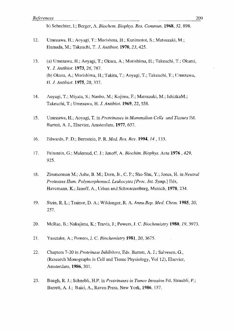
b) Schechter, 1; Berger, A. Biochem. Biophys. Res. Commun. 1968,32,898.
12. Umezawa, H.; Aoyagi, T.; Morishma, H.; Kunimotot, S.; Matsuzaki, M.;
Hamada, M.; Takeuchi, T. J. Antibiot. 1970,23,425.
209
13. (a) Umezawa, H.; Aoyagi, T.; Okura, A.; Morishima, H.; Takeuchi, T.; Okami,
Y. J. Antibiot. 1973, 26, 787.
(b) Okura, A.; Morishima, H.; Takita, T.; Aoyagi, T.; Takeuchi, T.; Umezawa,
H. J. Antibiot. 1975, 28, 337.
14. Aoyagi, T.; Miyata, S.; Nanbo, M.; Kojima, F.; Matsuzaki, M.; IshizkaM.;
Takeuchi, T.; Umezawa, H. J. Antibiot.1969, 22, 558.
15. Umezawa, H.; Aoyagi, T. in Proteinases in Mammalian Cells and Tissues Ed.
Barrett, A. J., Elsevier, Amsterdam, 1977, 637.
16. Edwards, P. D.; Bernstein, P. R. Med. Res. Rev. 1994, 14 , 133.
17. Feinstein, G.; Malemud, C. 1.; Janoff, A. Biochim. Biophys. Acta 1976,429,
925.
18. Zimmerman M.; Ashe, B. M.; Dorn, Jr., C. P.; Shu-Shu, Y.; Jones, H. in Neutral
Proteases Hum. Polymorphonucl. Leukocytes [Proc. Int. Symp.] Eds,
Havemann, K.; Janoff, A., Urban and Schwarzenburg, Munich, 1978, 234.
19. Stein, R. L.; Trainor, D. A.; Wildonger, R. A. Annu.Rep. Med. Chem. 1985,20,
237.
20. McRae, B.; Nakajima, K; Travis, 1.; Powers, J. C. Biochemistry 1980, 19, 3973.
21. Yasutake, A.; Powers, J. C. Biochemistry 1981, 20,3675.
22. Chapters 7-20 in Proteinase Inhibitors, Eds. Barrett, A. J.; Salvesen, G.,
(Research Monographs in Cell and Tissue Physiology, Vol 12), Elsevier,
Amsterdam, 1986, 301.
Baugh, R. 1.; Schnebli, H.P. in Proteinases in Tumor Invasion Ed. StraubH, P.;
Ban-ett, A. J.; Baici, A., Raven Press, New York, 1986, 157.

24. a) Pulmonary Emphysema and Proteolysis Ed. Mittman, c., Academic Press,
New York, 1972; 1-537.
b) Sprung, C. L.; Schultz, D. R; Clerch, A R New Engl. J. Med. 1984,304,
1301.
c) Velvart, M. Rheumatol. Int. 1981,1, 121.
25. Silverman, R B. Mechanism-Based Enzyme Inactivation: Chemistry and
Enzymology Vol. I, CRC Press, Boca Raton, FL, 1988.
26. Lippert, B.; Jung, M. J.; Metcalf, B. W. Brain. Res. Bull. 1980,5 (2), 375.
27. Lentini, A; Farchione, F.; Ternai, B.; KreuA-Ongarjnukool, N.; Tovivich, P.
Biol. Chem. Hoppe-Seyler 1987, 368, 369.
28. Digenis, G. A; Agha, B. 1.; Tsuji, K.; Kato, M.; Shingoi, M. J. Med. Chem.
1986,29, 1468.
29. Dutta, A S.; Giles, M. B.; Gormley, J. J.; Williams, 1. S.; Kusner, E. 1. J. Chem.
Soc. Perkin Trans. 1987,111, 1.
30. Westerik,1. 0.; Wolfenden, R J. Bioi. Chem. 1972,247,8195.
31. Edwards, P. D.; Meyer, Jr., F.; Vjayalakshmi, J.; Tuthill, P. A.; Andisik, D.
A; Gomes, B.; Strimpler, A J. Am. Chem. Soc. 1992,114, 1854.
32. Wiley, R. A.; Rich, D. H. J. Med. Chem. 1993,13,327.
33. Imperiali, B.; Abeles, R H. Tetrahedron Lett. 1986,27, 135.
34. Imperiali, B.; Abeles, R. H. Biochemistry 1986,25,3760.
35. Bergeson, S. H.; Edwards, P. D.; Krell, R D.; Shaw, A; Stein, R L.; Stein, M.
M.; Strimpler, A M.; Trainor, D. A; Wildonger, R A; Wolanin, D. 1. 193
National Meeting of the American Chemical Society (Abstracts of Papers),
Denver, Colorado, April5-10, 1987.
36. Edwards, P. D. Tetrahedron Lett. 1992,33,4279.
37. Stein, M. M.; Wildonger, R A; Trainor, D. A; Edwards, P. D.; Yee, Y. K.;
Lewis,1. J.; Zottola, M. A; Williams, J. c.; Strimpler, A. M. Eleventh

American Peptide Symposium, (Abstracts of Papers), University of California,
San Diego, California, July 9-14,1989.
38. Edwards, P. D.; Bernstein, P. R Med. Res. Rev. 1994,14, 145.
39. Peet, N. P.; Burkhart, J. P.; Angelastro, M. R; Giroux, E. L.; Mehdi, S.; Bey,
P.;Kolb, M.; Neises, B.; Schirlin, D. J. Med. Chem. 1990,33,394.
211
40. Hori, H.; Yasutake, A; Minematsu, Y.; Powers, J. C. in Peptides, Structure and
Function (Proceedings of the Ninth American Peptide Symposium), Eds. Deber,
C. M.; Hruby, V. J.; Kopple, K. D. Pierce Chern. Co., Rockford, Illinois, 1985,
819.
41. Mehdi, S.; Angelastro, M. R; Burkhart, J. P.; Koehl, J. P.; Peet, N. P.; Bey, P.
Biochem. Biophys. Res. Commun. 1990,166,595.
42. Stein, M. M.; Wildonger, R A; Trainor, D. A; Edwards, P. D.; Yee, Y. K.;
Lewis, 1. J.; Zottola, M. A.; Williams, 1. c.; Strimpler, A. M. in Pep tides,
Structure and Function (Proceedings of the Eleventh American Peptide
Symposium), Eds., River, 1. E.; Marshall, G. R ESCOM, Leiden, 1990,369.
43. (a) Kettner, C. A; Shenvi, A B. J. Bioi. Chem. 1984,259, 15106.
(b) Kettner, C. A.; Bone, R; Agard, D. A; Bachovchin, W. W. Biochemistry
1988,27, 7682.
44. (a) Knabb, R M.; Kettner, C. A; Timmermans, P. B. M. W. M.; Reilly, T.M.
Thromb. Haemostas. 1992,67,56.
(b) Kettner, C. A; Mersinger, L. J.; Knabb, R M. J. Bioi. Chem. 1990,265,
18289.
(c) Quan, M. L.; Wit yak, J.; Dominguez, c.; Duncia, J. V.; Kettner, C. A.; Ellis,
C. D.; Liauw, A Y; Park, J. M.; Santella, J. B.; Knabb, R M.; Thoolen, M. 1.;
Weber, P. C.; Wexler, R R Bioorg. Med. Chem. Lett. 1997,7,1595.
45. (a) Tian, Z. -Q.; Brown, B. B.; Mack, D. P.; Hutton, C. A; Bartlett, P. A J.
Org. Chem. 1997,62,514 and references therein.
(b) Edwards, P. D.; Berstein 148-152 and references therein.
46. Shaw, E. in Enzyme Inhibitors as Drugs Ed., Sandler, M. MacMillan Press,
1980,25.

47. (a) Lamden, L. A.; Bartlett, P. A. Biochem. Biophys. Res. Commun. 1983, 112,
1085.
(b) Bartlett, P. A; Lamden, L. A Bioorg. Chem. 1986,14, 356.
(c) Oleksyszyn, J.; Powers, J. C. Biochem. Biophys. Res. Commun. 1989, 161,
143.
(d) Oleksyszyn, J.; Powers, J. C. Biochemistry 1991, 30, 485.
48. (a) Lively, M. 0.; Powers, J. C. Biochim. Biophys. Acta 1978, 525, 171.
(b) Yoshimura, T.; Barker, L. N.; Powers, J. C. J. Biol. Chem. 1982,257,5077.
49. (a) Brown, W. E.; Wold, F. Science 1971, 174, 608.
(b) Brown, W. . Wold, F. Biochemistry 1973, 12, 828.
(c) Brown, W. . Wold, F. Biochemistry 1973, 12, 835.
(d) Brown, W. E. Biochemistry 1975, 14, 5079.
50. Miyano, M.; Deason, J. R.; Nakao, A; Stealey,M. A; Villamil, C. I.; Sohn, D.
D.; Meuller, RAJ. Med. Chem. 1988,31, 1052.
51. Kawabata, K.; Suzuki, M.; Sugitani, M.; Imaki, K.; Toda, M.; Miyamoto, T.
Biochem. Biophys. Res. Commun. 1991, 177,814.
52. Silverman, R B. The Organic Chemistry of Drug Design and Drug Action,
Academic Press, San Diego and London, 1992, 185-7.
53. (a) Groutas, W. c.; Badger, R c.; Ocain, T. D.; Felker, D.; Frankson, J.;
Theodorakis, M. C. Biochem. Biophys. Res. Commun. 1080,95,1890.
(b) Walker, B.; Elmore, D. T. Biochem. J. 1984,221,277.
(c) Groutas, W. c.; Abrams, W. R; Theodorakis, M. c.; Kasper, A M.; Rude,
S. A; Badger, R c.; Ocain, T. D.; Miller, K. . Moi, M. K.; Brubaker, M. J.;
Davis, . S.; Zandler, M. E. J. Med. Chem. 1985,28, 204.
(d) Groutas, W. C.; Brubaker, M. J.; Zandler, M. . Mazo-Gray, V.; Rude, S.
A.; Crowley, J. P.; Castrisos, J. c.; Dunshee, D. A; Giri, P. K. J. Med. Chem.
1986, 29, 1302.
54. Walsh, C. Tetrahedron 1982,38,871.
55. (a) Walsh, C.; Cromartie, T.; Marcotte, P.; Spencer, R Methods Enzymol. 1978,
53D,437.
(b) Wang, E.; Walsh, C. Biochemistry 1978, 17,1313.

56. Alazard, R; Bechet, 1. 1.; Dupaix, A; Yon, 1. Biochim. Biophys. Acta 1973,
309,379.
57. (a) White, E. H.; Roswell, D. F.; Politzer, 1. R.; Branchini, B. R. J. Am. Chem.
Soc. 1975,97,2290.
213
(b) White, E. H.; Roswell, D. F.; Politzer, I. R; Branchini, B. R. Meth. Enzymol.
1977,46,216.
(c) White, E. H.; Jelinski, Politzer, 1. R; Branchini, B. R; Roswell, D. F. J. Am.
Chem. Soc. 1981,103,4231.
58. (a) Bechet, 1. J.; Dupaix, A; Yon, J.; Wakselman, M.; Robert, J. -L.; Vilkas, M.
Eur. J. Biochem. 1973,35,527.
(b) Bechet, 1. -J.; Dupaix, A; Blagoeva, 1. Biochimie 1977, 59, 231.
59. Vilain, A -C.; Okochi, V.; Vergely, I.; Reboud-Ravaux, M.; Mazaleyrat, 1. -P.;
Wakselman, M. Biochim. Biophys. Acta 1991, 401, 1076.
60. Bechet, J. -J.; Dupaix, A; Roucous, C.; Bonamy, A -M. Biochimie 1977, 59,
241.
61. Decodts, G.; Wakselman, M. Eur. J. Med. Chem. 1983, 18, 107.
62. (a) Wakselman, M. Nouv. J. Chim.1983, 7,439.
(b) Wakselman, M.; Mazalyerat, J. P.; Xie, 1.; Montagne, J. J.; Vilain, A -c.; Reboud-Ravaux, M. Eur. J. Med. Chem.1991, 26,699.
(c) Wakselman, M.; Xie, 1.; Mazalyerat, 1. P.; Boggetto, N.; Vilain, A -C.;
Montagne, J. J.; Reboud-Ravaux, M. J. Med. Chem. 1993,36,1539.
63. Powers, J. C.; Kam, C. -M.; Narasimhan, L.; Oleksyszym, J.; Hernandez, M. A;
Ueda, T. J. Cell. Biochem. 1989, 39, 33.
64. Harper J. W.; Hemmi, K.; Powers, J. C. J. Am. Chem. Soc. 1983, 105,6518.
65. Harper J. W.; Hemmi, K.; Powers, J. C. Biochemistry 1985,24, 1831.
66. Alpin, R T.; Robinson, C. V.; Schofield, C. J.; Westwood, N. J. J. Chem. Soc.
Chem. Commun. 1992, 1650.
67. Harper, J. W.; Powers, J. C. J. Am:. Chem. Soc. 1984,106, 7618.

68. Harper, 1. W.; Powers, 1. C. Biochemistry 1985, 24, 7200.
69. Meyer, Jr., E. F.; Presta, L. G.; Radhkrishnan, R J. Am. Chern. Soc. 1985, 107,
4091.
70. Vijayalakshmi, 1.; Meyer, Jr., F.; Kam, C. -M.; Powers, 1. C. Biochemistry
1991,30,2175.
71. Chakravarty, .K.; Krafft, G. A; Katzenellenbogen J. A 1. Biol. Chern. 1982,
257,610.
214
72. Daniels, S. B.; Cooney, E.; Sofia, M. 1.; Chakravarty, P. K.; Katzenellenbogen
1. A J. Bioi. Chern. 1983, 258 , 15046.
73. Rando, R R Science 1974, 185, 321.
74. Krafft, G. A; Katzenellenbogen, 1. A J. Am. Chern. Soc. 1981, 103, 5459.
75. Daniels, S. B.; Katzenellenbogen, J. A Biochemistry 1986, 25, 1436.
76. Naruto, S.; Motoc, I.; Marshall, G. R; Daniels, S. B.; Sofia, M. J.;
Katzenellenbogen,1. A J. Am. Chern. Soc. 1985, 107, 5262.
77. Katzenellenbogen, J. A.; Rai, R; Dai, W. Bioorg. Med. Chern. Lett. 1992,2,
1399.
78. Sofia, M. J.; Chakravarty, P. K.; Katzenellenbogen, J. A J. Org. Chern.
1983,48, 3318.
79. Reed, P. E.; Katzenellenbogen, 1. A J. BioI. Chern. 1991,266, 13.
80. Hemmi, K.; Harper, J. W.; Powers, 1. C. Biochemistry 1985, 24, 1841.
81. Tam, T. F.; Spencer, R W.; Thomas, M.; Copp, L. 1.; Krantz, A. 1. Am.
Chern. Soc. 1984, 106, 6849.
82. Copp, L. J.; Krantz, A; Spencer, R W. Biochemistry 1987,26, 169.
83. Spencer, R W.; Tam, T. F.; Thomas, E.; Robinson, V. J.; Krantz, A. J. Am.
Chem. Soc. 1986, 108,5589.

215
84. Groutas, W. C.; P. K Giri, P. K; I. P. Crowley, I. P.; Castrisos, I. C; Brubaker,
M. I. Biochem. Biophys. Res. Commun. 1986,141, 741.
85. Groutas, W. Co; Stanga, M. A; Brubaker, M. I. J. Am. Chem. Soc. 1989,
111,1931.
86. (a) Lossen, H. Liebigs Ann. Chem. 1869,150,314.
Cb) Lossen, W. Liebigs Ann. Chem. 1872,161,347.
(c) Lossen, W. Liebigs Ann. Chem. 1877,186, 1.
Cd) Lossen, W. Liebigs Ann. Chem. 1889,252, 170.
(e) Lossen, W. Liebigs Ann. Chem. 1894,281,169.
87. (a) Groutas, W. Co; Brubaker, M. I.; Stanga, M. A; Castrisos, 1. C.; Crowley, I.
P.; Schats, E. 1. J. Med. Chem. 1989,32, 1607.
(b) Groutas, W. Co; Hoidal, M. I.; Brubaker, M. 1.; Stanga, M. A.;
Venkataraman, R.; Gray, B. H. Rao, N. V.I. Med. Chem. 1990,33,1085.
(c) Groutas, W. C.; Stanga, M. A; Castrisos, I. C.; Schatz, E. I.; Brubaker, M. I.
J. Pharm. Sci. 1990, 79, 886.
Cd) Groutas, W. C.; Brubaker, M. I.; Venkataraman, R.; Epp, I. B.; Stanga, M.
A; McClenahan, 1. I. Arch. Biochem. Biophys. 1992,294, 144.
(e) Groutas, W. Co; Brubaker, M. I.; Venkataraman, R.; Stanga, M. A. Arch.
Biochem. Biophys. 1992,297, 144.
(f) Groutas, W. Co; Venkataraman, R.; Brubaker, M. I.; Epp, I. B.; Chong, L. S.;
Stanga, M. S.; McClenahan I.I.; Tagusagawa, F. Biochim. Biophys. Acta. 1993,
1164,283.
(g) Groutas, W. Co; Brubaker, M.J.; Chong, L. S.; Epp, 1. 8.; Huang, H.; Keller,
Co E.; McClenahan I.I.; Givens, R. S.; Singh, R.; Zandler, M. ; Karr, P. A;
Tagusagawa, F. Drug Design and Discovery 1994, 11, 149.
88. Bauer, L.; Exner, O. Angew. Chem. Int. Ed. 1974, 13, 376.
89. Groutas, W. C.; Venkataraman, R.; Brubaker, M. J.; Stanga, M. A
Biochemistry 1991,30,4132.
90. Neumann, u.; Gutschow, M. J. Biol. Chem. 1994,269,21561.
91. KelTigan, I. E.; Shirley, J. I. Bioorg. Med. Chem. Lett. 1996, 6, 451.

92. Groutas, W. c.; Castrisos J. c.; Stanga M. A; Kuang, R; Venkataraman, R;
Epp, 1. B.; Brubaker, M. J.; Chong, L. S. Bioorg. Med. Chern. Lett. 1993,3,
1163.
93. Groutas, W. c.; Huang, H.; Epp, J. B.; Bruaker, M. J.; Keller, C. E.;
McClenahan, J. J. Bioorg. Med. Chern. Lett. 1992,2, 1565.
94. Groutas, W. c.; Chong, L. S.; Epp, J. B.; Venkataraman, R; Brubaker, M. J.;
Stanga, M. A; Eun-Hong, K.; Keller, C. E. Bioorg. Med. Chern. Lett. 1992,2,
1571.
95. Pinto, I. P.; West, A; Debouck, C. M.; DiLella, A G.; Gorniak, J. G.;
ODonnell, K. c.; O'Shannessy, Patel, A; Jarvest, R L. Bioorg. Med. Chern.
Lett. 1996, 6, 2467.
216
96. Hlasta, D. J.; Bell, M. R; Boaz, N. W.; Court, J. J.; Desai, R c.; Franke, C. A.;
Mura, A J.; Subramanyam, C.; Dunlap, R P. Bioorg. Med. Chern. Lett. 1994,
4, 1801.
97. (a) Zimmerman, M.; Morman, H.; Mulvey, D.; Jones, H.; Frankshun, R; Ashe,
B. M. 1. Biol. Chern. 1980, 255, 9848.
(b) Ashe, B. M.; Clark, R L.; Jones, H.; Zimmerman, M. 1. Biol. Chern. 1980,
256,11603.
98. Reczek, 1. A; Welter, T. R Eur. Pat. Appl. 198256444.
99. (a) Groutas, W. C.; Brubaker, M. 1.; Venkataraman, R; Epp, J. B.; Houser
Archield, N.; Chong, L. S.; McClenahan, J. 1. Bioorg. Med. Chern. Letts. 1992,
2,175.
(b) Groutas, W. C.; Houser-Archield, N.; Chong, L. S.; Venkataraman, R; Epp,
J. B.; Huang, H.; McClenahan, 1. 1. Med. Chern. 1993,36,3178.
100. Hlasta, D. 1.; Court, 1. J.; Desai, R c.; Talomie, T. G.; Shen, J. Bioorg. Med.
Chern. Letts. 1996,6,2941.
101. Subramanyam, c.; Bell, M. R; Ferguson, E.; Gordon, R 1.; Dunlap, R P.;
Franke, C. A; Mura, A J. Bioorg. Med. Chern. Letts. 1995, 5,319.
102. Desai, R c.; Court, 1. 1.; Ferguson, E.; Gordon, R J.; Hlasta, D. 1. 1. Med.
Chern. 1995,38, 1571.

103. Smith, R A; Copp, L. 1.; Coles, P. 1.; Pauls, H. W.; Robinson, V. 1.; Spencer,
R W.; Heard, S. B.; Krantz, A 1. Arn. Chern. Soc. 1988, 110,4429.
217
104. Subramanyam, C.; Bell, M. R; Carabateas, P. M.; Court, 1. J.; Dority, Jr., J. A;
Ferguson, E. W.; Gordon, R J.; Hlasta, D. 1.; Kumar, V.; Saindane, M. 1. Med.
Chern. 1994, 37, 2623.
105. Edwards, P. D.; Bernstein, P. R Med. Res. Rev. 1994, 14,127 and references
cited on pp. 180-5.
106. Williams, F. M. Pharmac. Ther. 1987,34, 99.
107. Hlasta, D. 1.; Bell, M. R.; Court, J. 1.; Cundy, K c,; Desai, R C.; Ferguson, E.
W.; Gordon, R J.; Kumar, v.; Maycock, A L.; Subramanyam, C, Bioorg. Med.
Chern. Letts. 1995,5,331.
108. Desai, R C.; Dunlap, R P.; Farrell, R P.; Ferguson, E.; Franke, C. A; Gordon,
R; Hlasta, D. J.; Talomie, T. G. Bioorg. Med. Chern. Lett. 1995, 5, 105.
109. Hlasta, D. 1.; Subramanyam, C.; Bell, M. R; Carabateas, P. M.; Court, 1. J.;
Desai, R c,; Drozd, M. L.; Eickhoff, W. M.; Ferguson, E. W.; Gordon, R J.;
Johnson, J. J.; Kumar, v.; Maycock, A. L.; Mueller, K. R.; Pagani, E. D.;
Robinson, D. T.; Saindane, M. T.; Silver, P. J.; Subramanian, S.; Dunlap R P.;
Franke, C. A; Mura, A J.; Rowlands, A 1. Med. Chern. 1995,38, 739.
110. Subramanyam, C.; Bell, M. R; Ghose, A K; Kumar, V.; Dunlap, R P.; Franke,
C. A; Mura, A J. Bioorg. Med. Chern. Lett. 1995,5, 325.
111. Groutas,W. C.; Chong, L. S.; Venkataraman, R; Epp, 1. B.; Kuang, R;
Brubaker, M. 1.; Houser-Archfield, N.; Huang, H.; McClenahan, J. J. Biochern.
Biophys. Res. Cornrnun. 1993, 194, 1491.
112. (a) Gabriel, S.; Colman, J. Ber. 1900, 33, 980.
(b) Hill, 1. H. M. 1. Org. Chern. 1965, 30, 620.
(c) Abe, K; Yamamoto, S.; Matsui, K.; Yakugaku Zasshi 1956, 76, 1058.
113. Groutas, W. C.; Chong, L. S.; Venkataraman, R Biochern. Biophys. Res.
Cornmun. 1993, 197, 730.

218
114. Groutas, W. C.; Chong, L. S.; Venkataraman, R; Huang, H.; Epp, J. B.; Kuang,
R Bioorg. Med. Chern. Lett. 1993, 3, 2745.
115. Groutas, W. C.; Brubaker. M. J.; Venkataraman, R; Epp, J. B.; Houser
Archield, N.; Chong, L. S.; McClenahan, J. J. Bioorg. Med. Chern. Lett. 1992,2,
175.
116. Kuang, R; Venkataraman, R; Ruan, S.; Groutas, W. C. Bioorg. Med. Chern.
Lett. 1998, 8, 539.
117. Konstau, M. W.; Vargo, K. M.; Davis, P. B. Am. Rev. Resp. Dis. 1990,141,
186.
118. Groutas, W. c.; Kuang, R; Venkataraman, R Biochem. Biophys. Res.
Commun. 1994, 198, 341.
119. Groutas, W. C.; Kuang, R; Venkataraman, R; Epp, J. B.; Ruan, S.; Prakash, O.
Biochemistry 1997, 36, 4739.
120. Ghuysen, J. -M. in The Bacterial D-D Carboxypeptidesltranspeptidase Enzyme
System, University of Tokyo Press, 1976,115-143.
121. (a) Brenner, D. G.; Knowles, J. R Biochemistry 1981,20,3680.
(b) Brenner, D. G.; Knowles, J. R Biochemistry 1984,23,5833.
122. Doherty, J. B.; Ashe, B. M.; Argenbright, L. W.; Barker, P. L.; Bonney, R J.;
Chandler, G. 0.; Dahlgren, M. E.; Dorn, Jr., C. P.; Finke, P. ; Firestone, R A;
Fletcher, D.; Hagmann, W. K.; Mumford, R; O'grady, L.; Maycock, A. L.;
Pisano, J. M.; Shah, S. K.; Thompson, K. R.; Zimmerman, M. Nature 1986,
322, 192.
123. Green, B. G.; Weston, H.; Ashe, B. M.; Doherty, 1. B.; Finke, P. E.; Hagmann,
W. K.; Lark, M.; Mao, 1.; Maycock, A L.; Moore, V.; Mumford, R; Shah, S.
K.; Walaovits, L.; Knight, W. B. Arch. Biochem. Biophys. 1991,286,284.
124. Shah, S. K.; Brause, K. A.; Chandler, G. 0.; Finke, P. E.; Ashe, B. M.; Weston,
H.; Knight, W. B.; Maycock, A L.; Doherty, J. B. 1. Med. Chern. 1990, 33,
2529.

125. Navia, M. A; Springer J. P.; Lin, T. -Y.; Williams, H. R; Firestone, R A;
Pisano, J. M.; Doherty, J. B.; Finke, P. E.; Hoogsteen, K Nature 1987327, 79.
219
126, Knight, W. B.; Maycock, A L.; Green, B. G.; Ashe, B. M.; Gale P.; Weston, E.;
Finke, P. E.; Hagmann, W. K; Shah, S. K; Doherty, J. B. Biochemistry 1992,
31,4980.
127. Hagmann, W. K; O'Grady, L. A; Ashe, B. M.; Dahlgren, M. E.; Weston, H.;
Maycock, A L.; Knight, W. B.; Doherty, J. B.; Pisano, J. M.; Shah, S. K
Eur. J. Med. Chem. 1989,24,599.
128. Doherty, J. B.; Ashe, B. M.; Barker, P. L.; Blacklock, T. J.; Butcher, J. W.;
Chandler, G. 0.; Dahlgren, M. E.; Davies, P. ; Dorn, Jr., C. P.; Finke, P. E.;
Firestone, R A; Hagmann, W. K; Halgren, T.; Knight, W. B.; Maycock, A L.;
Navia, M. A.; O'Grady, L. A; Pisano, J. M.; Shah, S. K; Thompson, K R;
Weston, H.; Zimmerman, M. J. Med. Chem. 1990,33,2513.
129. Finke, P. E.; Ashe, B. M.; Knight, W. B.; Maycock, A L.; Navia, M. A; Shah,
S. K; Thompson, K R; Underwood, D. J.; Weston, H.; Zimmerman, M.;
Doherty J. B. J. Med. Chem. 1990, 33, 2522.
130. Finke, P. E.; Shah, S. K; Ashe, B. M.; Ball, R G.; Blacklock, T. J.; Bonney, R
J.; Brause, KA; Chandler, G. 0.; Cotton, M.; Davies, P.; Dellea, P. S.; Dorn,
Jr., C. P.; Fletcher, D. S.; O'Grady, L A; Hagmann, W. K; Hand, K. M.;
Knight, W. B.; Maycock, A L.; Mumford, R A.; Osinga, D. G.; Sohar, P.;
Thompson, K R; Weston, H.;Doherty, J. B. Firestone, R A; Halgren, T.;
Knight, W. B.; Maycock, A L.; Navia, M. A; O'Grady, L.; Pisano, J. M.; Shah,
S. K; Thompson, K R; Weston, H.; Zimmerman, M. J. Med. Chem. 1992,35,
3731.
131. Firestone, R A; Barker, P. L.; Pisano, J. M.; Ashe, B. M.; Dahlgren, M. E.
Tetrahedron 1990, 46, 2255.
132. Shah, K S.; Dorn, Jr., C. P.; Finke, P. E.; Hale, J. J.; Hagmann, W. K.; Brause,
K A; Chandler, G. 0.; Kissenger, A L.; Ashe, B. M.; Weston, H.; Knight, W.
B.; Maycock, A L.; Dellea, P. S.; Flechter, D. S.; Hand, K M.; Mumford, R
A; Underwood, D. J.; Doherty J. B. J. Med. Chem. 1992, 35, 3745.
133. Han, T. W.; Trehan, A. K; Wright, J. J. K; Federici, M. E.; Seiler, S. M.;
Meanwell, N. A Bioorg. Med. Chern. 1995,3,1123.

References 220
134. Borthwick, A. D.; Weingarten, G.; Haley, T. M.; Tomaszewski, M.; Wang, W.;
Hu, Z.; Bedared, J.; Jin, H.; Yuen, L.; Mansour, T. S. Bioorg. Med. Chem. Lett.
1998, 8, 365.
135. (a) Tong, L.; Qian, C.; Massariol, M. -J.; Ronneau, P. R; Cordingley, M. G.;
Lagace, L. Nature 1996, 383, 272.
(b) Qiu, X.; Culp, J. S.; DiLella, A. G.; Helmig, B.; Hoog, S. S.; Jason, . A;
Smith, W. W.; Abdel-Meguid, S. S. Nature 1996, 383, 275.
(c) Shieh, H. -J.; Kurumbail, R G.; Stevens AM.; Stegean, R A.; Sturman, E.
J.; Pak, J. Y.; Wittwer, A J.; Palmier, M.O.; Wiegand, R C.; Holwerda, B. C.;
Stallings, W. C. Nature 1996, 383,279.
(d) Chen. P.; Tsue, H.; Almassy, R J.; Gribskov, C. L.; Katoh, S; Vanderpool,
D. L.; Margosiak, S. A.; Pinko, C.; Matthews, D. A; Kan, C. -C. Cell 1996, 86,
835.
136. Hagmann, W. K; Shah, S. K; Dorn, Jr. C. P.; O'Grady, L. A; Hale, J. J.; Finke,
P. E.; Thompson, K R; Brause, K A; Ashe, B. M.; Weston, H.; Dahlgren, M.
E.; Maycock, A L.; Dellea, P. S.; Hand, K M.; Osinga, D. G.; Bonney, R. J.;
Davies, P.; Fletcher, D. S.; Doherty, J. B. Bioorg. Med. Chem. Lett. 1991, 1,
545.
137. Knight, W. B.; Chabin, R; Green, B. Arch. Biochem. Biophys. 1992,296,704.
138. Knight, W. B.; Swiderek, K. M.; Sakuma, T.; Calaycay, J.; Shively, J. E.; Lee,
T. D.; Covey, T. R; Shushan, B.; Green, B. C.; Chabin, R; Shah, S. K;
Mumford, R; Dickinson, T. A; Griffin, P. R Biochemistry 1993, 32, 2031.
139. Knight, W. B.; Green, B. G.; Chabin, R M.; Gale, P.; Maycock, A L. Weston,
H.; Kuo, D. W.; Westler, W. M.; Dorn, Jr., C. P.; Finke, P. E.; Hagmann, W. K;
Hale, J. J.; Liesch, J.; MacCross, M.; Navia, M. A.; Shah,S. K; Underwood, D.;
Doherty, J. B. Biochemistry 1992,31,8160.
140. Edwards, P. D.; Bernstein, P. R Med. Res. Rev. 1994, 14, 168.
141. Hagmann, W. K; Thompson, K R; Shah, S. K; Finke, P. E.; Ashe, B. M.;
Weston, H.; Maycock, A. L.; Doherty, J. B. Bioorg. Med. Chem. Lett. 1992,2,
681.

221
142. Shah, S. K.; Dorn, Jr., C. P.; Finke, P. . Hale, J. I; Hagmann, W. K; Brause,
K A.; Chandler, G. 0.; Kissinger, A. L.; Ashe, B. M.; Weston, H.; Knight, W.
B.; Maycock, A. L.; Dellea P. S.; Fletcher, D. S.; Hand, K. M.; Mumford, R. A.;
Underwood, D. J.; Doherty, J. B. J. Med. Chem. 1992,35,3745.
143. Hagmann, W. K; Kissinger, A. L.; Shah, S. K; Finke, P. E.; Dorn, Jr., C. P.;
Brause, K A.; Ashe, B. M.; Weston, H.; Maycock, A. L.; Knight, W. B.;
Dellea, P. S.; Fletcher, D. S.; Hand, K. M.; Osinga, D.; Davies, P.; Doherty, J. B.
J. Med. Chem. 1993,36,771.
144. Byrant, D. R.; Hauser, C. R. J. Am. Chem. Soc. 1961, 83, 3468.
145. Hargreaves, M. K.; Pritchard, J. G.; Dave, H. R. Chem. Rev. 1970, 70,439.
146. Fujisaki, S.; Hamura, S.; Eguchi, H.; Nishida, A. Bull. Chem. Soc. Jpn. 1993,
66,2426.
147. Kornblum, N.; Jones W. J.; Anderson, G. J. J. Am. Chem. Soc. 1959,81,4113.
148. Fieser L. F.; Fieser, M. Reagents for Organic Synthesis, Wiley, 19692,371.
149. Sandler, S. R.; Karo, W. in Organic Functional Group Preparations Academic
Press, New York, 2nd ed, 1983, 482.
150. Jencks, W. P. J. Am. Chem. Soc. 1958, 80, 4581.
151. (a) Fittig. R.; Roders. P. Ann. 1890,256,95.
(b) Lucus, R.; Papadakis, M. Z. Ann. Chim. 1032,18,32.
(c) Baeyer, A.; Perkin, W. H. Ber. 1884, 17,449.
152. Cohen, S. G.; Milovanovic, A. J. Am. Chem. Soc. 1968, 90, 3495.
153. Marvel C. S. Org. Syn. Call. Vol. III, Ed., Horning, C. 1955, 705.
154. (a) Brown, R. F. C. Aust. J. Chem. 1964,17, 154.
(b) Wolfe, J. F.; Rogers T. G. J. Org. Chem. 1970,35,3600.
(c) Goehring, R. R.; Greenwood, T. D.; Nwokogu, G. C.; Pisipati, J. S.; Rogers,
T. G.; Wolfe, J. F. J. Med. Chem 1990, 33, 926.

155. Fieser, L. F.; Fieser, M. Reagentsfor Organic Synthesis, Wiley, New York,
1966, 1, 1034-5 and references therein.
156. Stobbe, H. Ann. 1890, 281, 282.
157. (a) Overberger, C. G.; Roberts, C. W. 1. Am. Chem. Soc. 1949, 71, 3618.
(b) Billet, Mlle. D. Bull. Soc. Chim. Fr. 1949, 16, 297.
222
(c) Johnson, W. S.; Daub, G. H. Organic Reactions Eds., Adams, R.; Adkins, H.
(decu); Blatt, A H.; Cope, A C.; McGrew, F. c.; Niemann, C.; Snyder, H. R.,
Wiley, New York, 1951,6, 1.
(d) Doulut, S.; Dubuc, 1.; Rodriguez, M.;Vecchini, F.; Fulcrand, H.; Barelli, H.;
Chec1er, F.; Bourdel, ; Aumelas, A; Lallement, J. C.; Kitabgi, P.; Costentin
J.; Martinez, J. 1. Med. Chem. 1993,36, 1369.
158. Bauer, L.; Miarka, S. V.I. Am. Chem. Soc. 1957, 79, 1983.
159. Zhu, Z.; Chuong, P.M.; Lemoine, P.; Tomas, T.; Galons, H. Heterocycles 1996,
43,1923.
160. Cohn, L.Ann. 1880,205,295.
161. Morrell, G. J. 1. Am. Chem. Soc. 1914, 105, 1733.
162. Oldham, M. D. Ph. D. 12 Month Progress Report, Department of Chemistry,
University of Canterbury, 1993.
163. Park, M.; Lee, J.; Choi, J. Bioorg. Med. Chem. Lett. 1996, 6, 1297.
164. Ames, D. E.; Grey, T.F.; 1. Chem. Soc. 1955, 631.
165. Aldrich Catalogue Handbok of Fine Chemicals, 1996-7.
166. Price, C. C.; Tomisek, A J. 1. Org. Chem. 1943,65,439.
167. Nikam, S. S.; Kornberg, B. E.; Johnson, D. R.; Doherty, A M. Tetrahedron
Lett. 1995,36,197.
168. Wang, K.-T.; Braltesani, D. N.; Weinstein, B. 1. Het. Chem.1966, 3, 98.
169. Orndoff, W. R.; Pratt, D. S.; Am. Chem.l. 1912,47,89.

170. (a) Buess, C. M.; Bauer, L. J. Org. Chem. 1955,20,33.
(b) Kuhle, Wegler. Ann. 1985, 616, 183.
171. Mitsunobu, O. Synthesis 1981, 1.
172. Growchowski, E.; Jurczak, 1. Synthesis 1977,277.
173. Sammes, M. P.; Wylie, C. M.; Hoggett, 1. G. J. Chem. Soc. 1971, 2151.
174. Abell A. D.; Oldham, M. D. J. Org. Chem.1997, 62,1509.
175. Oldham, M. D. Ph. D. Thesis Department of Chemistry, University of
Canterbury, 1998.
223
176. Le Berre, A.; Etienne, A; Desmazieres, B. Bull. Soc. Chim. Fr. 1975,3-4,807.
177. Strecker, Ann. 1868, 148, 90.
178. Rougny, A; Daudon, M. Bull. Soc. Chim. Fr. 1976, 833.
179. Procopiou, P. A; Baugh S. P. D.; Flack S. S.; Inglis G. G. A J. Chem. Soc.
Chem. Commun. 1996, 2625.
180. Puterbaugh, W. H. J. Am. Chem. Soc. 1962,27,4010.
181. Michael, J. J. Prakt. Chem. 1887,35,349.
182. (a) Bruson, H. A; Reiner, T. W. J. Am. Chem. Soc. 1943, 65,23.
(b) Ansell, M. F.; Hey, H. J. Chem. Soc. 1950, 1683 and references therein.
183. 1. Guareschi, IChem. ZhZ. 1901, 1,579.
184. (a) Knoevenagel, Ber. 1894, 27, 2346.
(b) Jones, G. Organic Reactions 1967,15,204.
185. Handley, G. J.; Nelson, E. R.; Somers, T. C. J. Aust. Chem. 1960,13, 129.
186. (a) Bergmann, D.; Ginsburg, D.; Pappo, R. Organic Reactions 1959, 10, 179-
555.

224
(b) Kon, G. A R; Thorpe, J. F. 1. Chem. Soc. 1919, 115, 686.
187. (a) Khz, R; Wilson, 1. B. 1. BioI. Chem. 1962,237,3245.
(b) Jung, M. J.; Metcalf, B. W. Biochem. Biophys. Res. Commun. 1975,67,301
188. Oleksyszyn, J.; Powers, J. C. Biochemistry 1991, 30, 485.
189. Moorman, A R; Abeles, R H. 1. Am. Chem. Soc. 1982, 104,6785.
190. Tian, W.-X.; Tsou, C. L. Biochemistry 1982, 21, 1028.
191. Morrison, J. F.; Walsh, C. T. Adv. Enzymol1988, 61,201.
192. Cha, S. Biochem. Pharmacol. 1975,24,217.
193. Waley, S. G. Biochem. 1. 1980, 185, 771.
194. Berezin,1. V.; Kazanshaya, N. F.; Klyosev, A A. FEBS Lett. 1971,15,121.
195. (a) Hummel, B. C. W. Can. 1. Biochem. 1959,37,1393.
(b) Walsh, K. A; Wilcox, P. E. Meth. Enzymol. 1970, 19, 33.
196. Schwert, G. W.; Kaufman, S. 1. Bioi. Chem.1951, 190,807.
197. (a) Baek, D. -J.; Reed, P. E.; Daniels, S. B.; Katzenellenbogen, J. A
Biochemistry 1990, 29, 4305.
(b) Krantz, A; Spencer, R W.; Tam. T. F.; Liak, T. J.; Copp, L. J.; Thomas, E.
M.; Rafferty, S. P. 1. Med.Chem. 1990,33,464.
(c) Reed, P. E.; Katzenellenbogen, J. A 1. Org. Chem. 1991,56,2624.
(d) Baek, D. -J.; Daniels, S. B.; Reed, P. E.; Katzenellenbogen, J. A 1. Org.
Chem. 1989,54, 3963.
(e) Stein, R L.; Strimpler, AM.; Viscarello, B. R; Wildonger, R A.; Mauger,
R c.; Trainor, D. A Biochemistry 1987,26,4126.
(f) Hedstrom, E.; Moorman, A R; Dobbs, J.; Abeles, R H. Biochemistry 1984,
23, 1753.
198. Bieth, J. G. Meth. Enzymol. 1995,248,59.

199. Navia, M. A; McKeever, B. M.; Springer, J. P.; Lin, T. -Y.; Williams, H. R.;
Fluder, E. M.; Dopm, Jr., C. P.; Hoogsteen, K. Proc. Natl. Acad. Sci. 1989,86,
7.
200. Zydowsky, T.M.; Dellaria, Jr., J. F; Nellans, H. N. 1. Org. Chem. 1988,53,
5607.
201. Karle, L L.; Flippen-Anderson, J. L. Acta Crystallogr. 1989, C45, 791.
202. Krause, J. A.; Baures, P. W.; Eggleston, D. S. Acta Crystallogr. 1993, B49,
123.
225
203. Delettre, P. J.; Berthou, J.; Lifchitz, A; Jones, P. Acta Crystallogr. 1988 , C44,
902.
204. (a) Bode, W.; Meyer, E.; Powers, J. C. Biochemistry 1989,28, 1951.
(b) Brubaker, M. J.; Groutas, W. C.; Hoidal, J. R Rao, N. V. Biochem. Biophys.
Res. Commun.1992, 188,1318.
205. Procter, G. Asymmetric Synthesis, Oxford University Press, Oxford, 1996, 1.
206. Eliel, E. L.; Wilen, S. H. Stereochemistry of Organic Compounds, Wiley, New
York, 1994, 201-208.
207. Craig, D. P.; Mellor, D. P. Top. Curro Chem. 1976, 63, 1.
208. Holmstedt, B. The Use of Eantiomers in Biological Studies: A Historical
Review in Chirality and Biological Activity, Eds. Holmstedt, B.; Frank, H.;
Testa, B. Liss, New York, 1990, Chapter 1.
209. Piutti, A Heabd. Seances. Acad. Sci. 1886, 103, 134.
210. Bentley, R Molecular Asymmetry in Biology, Academic, New York, 1969, 1,
286.
211. Ohloff, G. Experientia 1986, 42,271.
212. Powell, J. R; Ambre, J. J.; Ruo, T. L The Efficacy and Toxicity of Drug
Stereoisomers in Drug Stereochemistry. Analytical Methods and Pharmacology,
Eds. Wainer, 1. W.; Drayer, D. Marcel Dekker, New York, 1988, 245.

213. Lehmann, F. P. A.; Rodrigues de Miranda, J. F.; Ariens, E. 1. Prog. Drug. Res.
1976,20, 101.
214. Ariens, E. J. Trends Pharmacol. Sci . 1986, 7,200.
215. Abell, A. D.; Erhard, K. . Hwa-Kwo, Y.; Yamashita, D. S.; Brandt, M.;
Mohammed, H.; Levy, M. A.; Holt, D. A. Bioorg. Med. Chem. Lett. 1994, 4,
1365.
216. Stein, R L.; Srimpler, A. M.; Edwards, P. D.; Lewis, J. J.; Mauger, R c.; Schwartz, J. A.; Stein, M. M.; Trainor, D. A.; Wildonger, R A.; Zottola, M. A.
Biochemistry 1987, 26, 2682.
217. Pfeiffer, C. C. Science 1956, 124, 29.
218. Snoke, J. E.; Neurath, H. Chem. Rev. 1950,46,69.
219. Cohen, S. G.; Schultz, R M. Proc. Natl. Acad. Sci. 1967,57,243.
220. Schallenberger, R S.; Acree, T. E.; Lee, C. Y. Nature 1969,221,555.
221. Levin, G. V. US Patent 4262032, Apr. 14, 1981; Chem. Abstr. 1981,95,
78771h.
222. Cushny, A. R The Biological Relations of Optically Isomeric Substances,
Williams and Wilkins, Baltimore, 1926.
223. Eliel, E. L.; Wilen, S. H. Stereochemistry of Organic Compounds, Wiley, New
York, 1994, 205 and references therein.
224. (a) Mander, L. N. Stereoselective Synthesis in W Eliel, E. L.; Wilen, S. H.
Stereochemistry of Organic Compounds, Wiley, New York, 1994, Chapter 12,
835 and references therein.
226
(b) Koskinen, A. Asymmetric Synthesis of Natural Products, Wiley, Chichester,
1993,29.
225. Fredga, A. Arkiv Kemi Mineral. Geol. 1948, 26B, (11) in Newman, P. Optical
Resolution Procedures for Chemical Compounds, 2, Acids, 1981,2, (1), 542.
226. Levy, D.E.; Lapierre, F.; Liang, W.; Ye, W.; Lange, C. W.; Li, X.; Grobelny,
D.; Casabonne, M.; Tyrrell, D.; Holme, K.; Nadzan, A; Galardy, R. E. J. Med.
Chem. 1998,41, 199.
227. (a) Fredga, A; Palm, O. Arkiv Kemi Mineral. Geol. 1949, 26A, (26),9; Chem.
Abstr. 1949,43,6611.
(b) Fredga, A Arkiv Kemi 1953, 6, 277.
(c) Matell, M. Arkiv Kemi 1953,5, 17.
(d) Fredga, A Arkiv Kemi Mineral. Geol. 1941, 14B, (27), 8.
(e) Fredga, A Arkiv Kemi Mineral. Geol. 1942, 15B, (23), 1.
(f) Fredga, A Tetrahedron 1960, 8, 126.
228. Elie1, E. L.; Wilen, S. H. Stereochemistry (if Organic Compounds, Wiley, New
York, 1994, 126.
229. (a) Whitesides, G. M.; Lewis, D. W. J. Am. Chem. Soc., 1970,92,6979.
(b) Parker, D. Chem. Rev. 1991,91, 1441.
230. (a) Bajwa, 1. S.; Miller, M. J. J. Org. Chem. 1983, 48, 1114.
(b) Mori, K.; Iwasawa, H. Tetrahedron 1980,36,87.
231. Hanessian, S. Total Synthesis of Natural Products: The 'Chiron' Approach
Pergamon Press, Oxford, 1986.
227
232. (a) Kawano, H.; Ishii, Y; Ikariya, T.; Saburi, M.; Yoshikawa, S.; Uchida, Y;
Kumobayashi, H. Tetrahedron Lett. 1987,28, 1905 and references cited therein.
(b) Hung, C. W.; Wong, H. N. C. Tetrahedron Lett. 1987,28,2393.
233. (a) Bashiardes, G.; Collingwood, S. P.; Davies, S. G.; Preston, S. C. J.
Organomet. Chem. 1989,364, C29.
(b) Bashiardes, G.; Collingwood, S. P.; Davies, S. G.; Preston, S. C. J. Chem.
Soc. Perkin Trans. 1989, 1162.
234. (a) Evans, D. A Aldrichimica Acta. 1982, 15, 23.
(b) Evans, D. A in Asymmetric Synthesis, Ed. Morrison, J. D., Academic Press,
New York, 1984,3,87-90.
(c) Evans, D. A; Takacs, J. M.; McGee, L. R.; Mathre, D. J.; Bartroli, J. Pure
and Appl. Chem. 1981,53,1109.
(d) Evans, D. A; Ennis, M. D.; Mathre, D. 1. J. Am. Chem. Soc. 1982, 104,
1737.

References
235. Evans, D. A; Bartroli, 1.; Shih, T. L. 1. Am. Chem. Soc. 1981, 103,2127.
236. Fadel, A; Salaun, 1. Tetrahedron Lett. 1988,29,6257.
237. Beckett, R P.; Crimin, M. 1.; Davis, M. H.; Spavold, Z. Synlett 1993, 137.
238. McClure, K. F.; Axt, M. Z. Bioorg. Med. Chem. Lett. 1998, 8,143.
239. (a) Sanko Company Limited, Eur. Pat. 0 383 635 A2, 1986.
(b) Sanko Company Limited, Eur. Pat. 0228 192 A2, 1986.
240. Gage 1. R; Evans, D. A Org. Synth. 1989, 68, 77 and references therein.
241. Gage 1. R; Evans, D. A Org. Synth. 1989, 68, 83 and references therein.
242. Ireland, R E.; Meissner, R S. 1. Org. Chem. 199156,4566.
243. Sudharshan, M.; Hultin, P. G. SynIe tt. 1997, 171.
244. Cason, J. Org. Synth. Coll. Vol. III, Wiley, New York, 1955,169.
245. (a) Nystrom R F.; Brown, W. G. 1. Am. Chem. Soc. 1947, 69, 2548.
(b) Grice, R; Owen, L. H. 1. Chem. Soc. 1963,1947.
246. Witiak, D. T.; Seth, S. K.; Baizman, E. R; Weibel, S. L.; Wolf, H. H. 1. Med.
Chem. 1972, 15,1117.
247. Kocienski, P. 1.; Cernigliaro, G.; Feldsyein, G. 1. Org. Chem. 1977,42,353.
248. Vogel's Textbook of Practical Organic Chemistry, 5th edn, Furniss, B. S.;
Hannaford, A J.; Smith, P. W. G.; Tatchell, A R 1989.
249. Perrin, D. D.; Armarego, W. L. F. Pwjication of Laboratory Chemicals, 3rd
edn, Pergamon Press, London, 1988.
250. Burfield, D. R; Smithers, R H. 1. Org. Chem. 1978,43, 3966.
251. Still, W. c.; Kahn, M.; Mitra, A 1. Org. Chem., 1978,43,2923.
228

229
252. Weizman, A. J. Org. Chern., 1943,8,285.
253. Thole, F. B.; Thorpe, J. F. J. Chem. Soc. 1911,99,466.
254. Benica, W. S.; Wilson, C. O. J. An'ler. Ph arm. Assn. [Sci. Edn.], 1950,39,451.

APPENDIX

Appendix
DATA OBTAINED DETERMINATION SUCCINIMIDE (1.410)
231
FROM CRYSTAL STRUCTURE OF 3-BENZYL-N-[(ACETYL)OXY]
05
Figure A.l Labelled drawing of 1.410
Atom X Y Z U(eq)
01 7989(4) 692(4) 3430(2) 38
02 6068(4) -837(4) 4275(2) 42
05 9951(4) 2018(4) 4888(2) 46
011 6554(5) 2638(4) 3261(3) 68
N1 7958(8) 720(5) 4408(3) 30
C1 7256(5) 1869(6) 2895(3) 36
C2 6958(5) -100(5) 4766(3) 30
C3 7260(5) 169(5) 5844(3) 29
C4 8676(5) 960(5) 6054(3) 30
C5 8985(5) 1356(5) 5089(3) 30
C6 7182(6) -1234(5) 6415(3) 38
C7 7376(5) -946(5) 7495(3) 31
C8 6366(6) -227(5) 7880(3) 38
C9 6556(7) 40(5) 8864(4) 47
ClO 7720(11) -408(6) 9477(4) 45
C11 8793(7) -1122(6) 9108(4) 49
C12 8605(6) -1374(6) 8112(3) 40
C13 7280(7) 1618(7) 1868(3) 53
Table A.l Atomic coordinates [x 104] (standard deviation) and equivalent
displacement parameters [A2 x 103] for 1.410. U(eq) is defined as one third of the trace
of the orthogonalized Uij tensor.

Appendix 232
01-N1 1.383(5) 01-C1 1.419(5)
02-C2 1.202(5) 05-C5 1.201(5)
011-C1 1.171(5) N1-C2 1.403(7)
NI-C5 1.368(7) C1-C13 1.470(6)
C2-C3 1.507(6) C3-C4 1.534(6)
C3-C6 1.527(6) C4-C5 1.495(6)
C6-C7 1.517(6) C7-C8 1.387(6)
C7-C12 1.387(7) C8-C9 1.382(6)
C9-ClO 1.344(10) ClO-C11 1.418(9)
C1l-Cl2 1.396(7)
Table A.2 Bond lengths for 1.410
N1-01-C1 113.8(4) 01-NI-C2 120.2(5)
01-N1-C5 123.0(6) C2-N1-C5 116.1(4)
01-C1-011 120.0(4) 01-C1-CI3 107.2(4)
011-C1-C13 130.8(5) 02-C2-Nl 124.6(5)
02-C2-C3 130.0(5) N1-C2-C3 105.4( 4)
C2-C3-C4 104.9(3) C2-C3-C6 111.8(4)
C4-C3-C6 115.8(4) C3-C4-C5 106.1(3)
05-C5-N1 123.2(5) 05-C5-C4 130.4(4)
N1-C5-C4 106.3(4) C3-C6-C7 111.8(4)
C6-C7-C8 121.4(5) C6-C7-CI2 119.7(5)
C8-C7-CI2 118.9(4) C7-C8-C9 121.0(5)
C8-C9-ClO 121.0(6) C9-ClO-C11 119.6(5)
C lO-C 11-C 12 119.2(6) C7-C12-C11 120.3(5)
Table A.3 Bond Angles [0] (standard deviation) for 1.410

Appendix 233
Atom Ull U22 U33 U23 U13 U12
01 45(2) 49(2) 23(2) 4(1) 11(2) 10(2)
02 36(2) 51(2) 39(2) -9(2) 5(2) -9(2)
05 45(2) 51(2) 44(2) -2(2) 18(20 -10(2)
011 92(4) 63(2) 49(2) 10(2) 13(2) 22(3)
Nl 28(3) 40(3) 23(2) 1(2) 9(2) -6(3)
C1 41(3) 50(3) 17(2) 4(2) 7(2) 23(3)
C2 26(3) 29(3) 40(3) 2(2) 14(2) 1(2)
C3 33(3) 31(2) 28(2) -4(2) 14(2) 2(2)
C4 33(3) 30(2) 27(2) -4(2) 5(2) 5(2)
C5 32(3) 28(2) 31(2) -2(2) 8(2) 3(2)
C6 50(4) 28(3) 38(3) -1(2) 17(3) -6(2)
C7 41(3) 27(2) 27(2) 5(2) 9(2) -3(2)
C8 46(3) 30(3) 40(3) 4(2) 12(2) 1(2)
C9 72(4) 32(3) 41(30 -3(2) 23(3) -1(3)
ClO 75(8) 34(3) 28(3) -7(2) 14(4) -8(4)
Cll 52(4) 43(3) 44(3) 7(3) -9(3) -13(3)
C12 42(3) 38(3) 40(3) -2(2) 7(2) 1(3)
C13 57(4) 68(4) 36(3) 14(3) 15(3) 3(4)
Table A.4 Anisotropic displacement parameters [A.2 x 103] for 1.410. The anisotropic
displacement factor exponent takes the form: -27t2 [(ha*)2U 11 + ... + 2hka*b*U 12]
Atom X Y Z U(eq)
H3 6548 842 5989 35
H4A 8625 1829 6438 36
H4B 9401 327 6406 36
H6A 7902 -1903 6299 45
H6B 6279 -1694 6187 45
H8 5546 80 7470 46
H9 5868 538 9105 56
HlO 7821 -251 10140 54
Hll 9611 -1419 9524 59
H12 9307 -1830 7861 48
H13A 8015 2187 1689 79
H13B 6396 1900 1476 79
H13C 7444 602 1767 79
Table A.S Hydrogen coordinates [x 104] and isotropic displacement parameters [A2 x
103] for 1.410

Appendix 234
CRYSTAL STRUCTURE DETERMINATION OF 3-PHENYL-N[(ACETYL)OXY]SUCCINIMIDE (1.41p)
Figure A.2 Labelled drawing of 1.41p showing 50% thermal ellipsoid probability for
non-hydrogen atoms, with hydrogens as small spheres of arbitrary size
Single-crystal data collection was performed at 293K on a Siemens P4 diffractometer
using graphite-monochromatised Mo-Ka radiation (A = 0.71073A). The crystal used
was a rectangular prism with dimensions 0.23 x 0.30 x 0.63 mm. Compound 3
C12HIIN04 (Mr = 233.22) crystallised from diethyl ether-petroleum ether in the
monoclinic system, space group C2/c, a = 19.542(3), b = 5.7830(10), c = 20.626(4) A, R 0 3 . -3 a = y = 90°, f-' = 107.570(10)°, V = 2222.2(7) A, Z = 8, Deale = 1.394 Mg m .
Absorption coefficient 0.106mm-J, 8 range for data collection 2.07 - 24.99, index
ranges -22 ~ h ~ 8, 0 ~ k ~ 6, -24 ~ I ~ 23, data/restraints/parameters 194610/155,
goodness of fit on F was 0.817, final R indices [1>20'(1)] RJ = 0.0404, WR2 = 0.0752,
R indices (all data) RJ = 0.0853, wR2 = 0.0845, largest difference peak and hole 0.187
and -0. 16gek3.
The unit cell parameters were obtained by least-squares refinement of the setting
angles of 42 reflections with 6.894° ~ 28 ~ 24.910° from a Siemens P4 diffractometer.
A unique data set was measured at 293K within 28max = 57° limit (Omega scans). Of
the 2908 reflections obtained, 1946 were unique (Rint = 0.0473) and were used in the
full-matrix least-squares refinement.A.l The intensities of 3 standard reflections,
measured every 97 relections throughout the data collection, showed only 1.55% decay.
The structure was solved by direct methods.A.2 Hydrogen atoms were fixed in idealised
positions. All non-hydrogen atoms were refined with anisotropic atomic displacement
parameters. Neutral scattering factors and anomalous dispersion corrections for non
hydrogen atoms were taken from Thers and Hamilton.A.3

Appendix 235
Atom X Y Z U(ea)
01 613(1) 7595(3) 4080(1) 43(1)
02 248(1) 7382(3) 5279(1) 40(1)
05 1992(1) 5656(3) 4316(1) 37(1)
011 1230(1) 10772(3) 4552(1) 43(1)
Nl 1011(1) 6460(4) 4665(1) 30(1)
Cl 774(1) 9944(4) 4099(1) 30(1)
C2. 772(1) 6399(4) 5228(1) 31(1)
C3 1290(1) 4806(4) 5728(1) 30(1)
C4 1951(1) 4712(4) 5463(1) 29(1)
C5 1689(1) 5608(4) 4745(1) 29(1)
C6 1426(1) 5593(4) 6459(1) 25(1)
C7 1750(1) 7667(4) 6683(1) 32(1)
C8 1867(1 ) 8363(4) 7343(1) 34(1)
C9 1665(1) 6950(5) 7793(1) 36(1)
CI0 1331(1) 4880(5) 7581(1) 39(1)
Cll 1208(1) 4198(5) 6905(1) 34(1)
C12 289(1) 11080(5) 3487(1) 40(1)
Table A.6 Atomic coordinates [x 104] and equivalent isotropic displacement
parameters [A2 x 103] for 1.41 p. U(eq) is defined as one third of the trace of the
orthogonalized Uij tensor.
01l-C1 1.180(3) 01-N1 1.388(2)
01-Cl 1.393(3) 05-C5 1.205(3)
02-C2 1.203(3) NI-C2 1.376(3)
N1-C2 1.377(3) CI2-Cl 1.483(3)
C5-C4 1.505(3) C4-C3 1.548(3)
C3-C6 1.520(3) C3-C2 1.520(3)
C6-C7 1.370(3) C6-C11 1.384(3)
Cll-CI0 1.398(3) ClO-C9 1.371(4)
C9-C8 1.379(3) C8-C7 1.372(3)
Table A.7 Selected bond lengths [A] for 1.41p

Appendix 236
N1-01-C1 112.0(2) C2-NI-C5 116.7(2)
C2-N1-01 120.1(2) C5-N1-01 122.6(2)
01l-Cl-01 121.9(2) 011-CI-C12 129.0(2)
01C1-C12 109.1(2) 05-C5-N1 124.9(2)
05-C5-C4 129.4(2) Nl-C5-C4 105.6(2)
C5-C4-C3 105.3(2) C6-C3-C2 111.9(2)
C6-C3-C4 116.5(2) C2-C3-C4 103.8(2)
02-C2-N1 125.2(2) 02-C2-C3 129.6(2)
N1-C2-C3 105.2(2) C7-C6-C11 119.3(2)
C7-C6-C3 121.4(2) C11-C6-C3 119.2(2)
C6-Cll-ClO 120.3(3) C9-C10-C11 119.1(3)
C10-C9-C8 120.6(2) C7-C8-C9 119.8(2)
C6-C7-C8 120.9(2)
Table A.S Selected bond angles [0] for 1.41p
Atom Uu U22 U33 U23 UB U12
01 38(1) 33(1) 25(1) 1(1) 0(1) -3(1)
02 29(1) 54(1) 38(1) 4(1) 11(1) 8(1)
05 30(1) 23(2) 34(1) -6(1) 19(1) -5(1)
011 35(1) 35(1) 54(1) -5(1) 3(1) -5(1)
N1 29(1) 33(1) 25(1) 5(1) 6(1) 3(1)
C1 29(1) 33(2) 31(1) 0(1) 13(1) -2(1)
C2 23(1) 34(2) 33(1) -3(1) 7(1) -3(1)
C3 31(1) 27(2) 31(1) 2(1) 9(1) -1(1)
C4 28(1) 32(2) 30(1) -4(1) 8(1) 1(1)
C5 30(1) 23(2) 34(1) -8(1) 7(1) -5(1)
C6 22(1) 28(1) 24(1) 4(1) 5(1) 6(1)
C7 30(1) 33(2) 34(1) 3(1) 10(1) -2(12)
C8 30(2) 32(2) 39(2) -3(1) 7(1) 1(1)
C9 31(2) 49(2) 2791) -2(1) 10(1) 7(1)
ClO 36(2) 48(2) 3692) 10(1) 16(1) 1(2)
Cl1 30(2) 32(2) 39(2) -3(1) 7(1) 1(1)
C12 30(1) 33(2) 34(1) 3(1) 10(1) -2(1)
Table A.9 Anisotropic displacement parameters [A2 x 103] for 1.41p. The anisotropic
displacement factor exponent takes the form: -2n2 [ (ha*)2U 11 + ... + 2hka*b*U 12]

Appendix 237
Atom X Y Z U(eq)
H3 1076(1) 3258(4) 5682(1) 36
H4A 2336(1) 5676(4) 5739(1) 36
H4B 2126(1) 3139(4) 5472(1) 36
H7 1893(1) 8615(4) 6384(1) 39
H8 2081(1) 9785(4) 7488(1) 41
H9 1757(1) 7406(5) 8243(1) 43
HlO 1187(1) 3942(5) 7882(1) 47
Hll 979(1) 2800(5) 6755(1) 41
H12A -191(2) 11087(22) 3517(4) 48
H12B 301(6) 10247(14) 3088(1) 48
H12C 445(5) 12642(9) 3460(4) 48
Table A.10 Hydrogen coordinates [x 104] and isotropic displacement parameters
(Axl03) for 1.41p

Appendix 238
DATA OBTAINED FROM CRYSTAL STRUCTURE DETERMINATION OF 3-BENZYL-N-[(TRANS-CINNAMOYL) OXY]SUCCINIMIDE (1.41£1)
Figure A.3 Labelled drawing of 1.41q
Atom X Y Z U(eq)
01 1608(1) 5045(6) 1(1) 50
02 1982(1) 5138(7) 1255(1) 59
05 1876(1) 908(6) -377(1) 57
011 818(1) 3915(6) 129(1) 57
N1 1857(1) 3276(7) 370(1) 47
C1 1032(2) 5266(9) -92(2) 41
C2 2051(2) 3518(10) 991(2) 51
C3 2362(2) 1383(8) 1241(2) 51
C4 2339(2) -29(9) 687(2) 48
C5 2003(2) 1344(9) 149(2) 46
C6 2939(2) 1941(9) 1672(2) 61
C7 3293(2) -24(9) 1941(2) 47
C8 3202(2 1411(10) 2359(2) 61
C9 3533(2) -3173(10) 2604(2) 48
C10 3991(2) -3693(8) 2437(2) 54
C11 4084(2) 2286(9) 2022(2) 60
C12 3746(2) -5639(10) 1774(2) 57
C13 822(2) 7236(8) 445(2) 42
C14 302(2) 7865(9) 556(2) 49
C15 36(2) 9834(8) -892(2) 41
Table A.ll Atomic coordinates [x 104] (standard deviation) and equivalent isotropic
displacement parameters [A2 x 103] for 1.41q. U(eq) is defined as one third of the trace
of the orthogonalized Uij tensor.

Appendix 239
Atom X Y Z U(eq)
C16 271(2) 11210(8) -1211(2) 48
C17 -9(2) 13023(9) 1523(2) 58
C18 539(2) 13536(9) -1522(2) 62
C19 -766(2) 12219(10) -1211(2) 60
C20 -504(2) 10388(9) -904(2) 51
Table A.ll (cont.) Atomic coordinates [x 1()4] (standard deviation) and equivalent
isotropic displacement parameters [A2 x 103] for 1.41q. U(eq) is defined as one third of
the trace of the orthogonalized Uij tensor.
Ol-N1 1.380(4) Ol-C1 1.419(5)
02-C2 1.201(6) 05-C5 1.204(5)
011-C1 1.198(5) N1-C2 1.388(6)
N1-C5 1.376(6) C1-C13 1.436(6)
C2-C3 1.513(7) C3-C4 1.549(6)
C3-C6 1.508(6) C4-C5 1.508(6)
C6-C7 1.485(7) C7-C8 1.376(6)
C7-C12 1.392(6) C8-C9 1.349(7)
C9-C10 1.399(6) ClO-Cll 1.381(6)
Cll-C12 1.342(7) C13-C14 1.320(5)
C14-C15 1.451(6) C15-C16 1.391(6)
C15-C20 1.413(6) C16-C17 1.365(6)
C17-C18 1.392(6) C18-C19 ] .347(7)
C19-C20 1.357(7)
Table A.12 Bond lengths [A] (standard deviation) for 1.41q

Appendix 240
N1-01-C1 113.0(3) 01-N1-C2 121.9(3)
01-N1-C5 116.8(4) C2-NI-C5 120.5(4)
01-C1-011 118.7(4) Ol-CI-C13 108.7(4)
01l-CI-CI3 132.5(4) 02-C2-Nl 125.2(5)
02-C2-C3 129.4(4) NI-C2-C3 105.7(4)
C2-C3-C4 105.7(4) C2-C3-C6 109.4(4)
C4-C3-C6 115.5(4) C3-C4-C5 105.2(4)
05-C5-Nl 124.7(5) 05-C5-C4 128.8(5)
NI-C5-C4 106.5(4) C3-C6-C7 114.8(5)
C6-C7-C8 123.7 C6-C7-CI2 120.3(4)
C8-C7-CI2 115.9(4) C7-C8-C9 122.8(4)
C8-C9-ClO 120.9(4) C9-CIO-Cl1 116.2(4)
ClO-C11-C12 122.3(4) C7-C12-C11 121.8(5)
C1-CI3-C14 119.6(4) C13-C14-C16 125.8(5)
C14-C15-C16 125.0(4) C14-CI5-C20 117.7(4)
CI6-CI5-C20 117.3(4) CI5-CI6-CI7 121.5(4)
C16-C17-CI9 119.9(5) CI7-C18-C19 118.9(5)
C 18-C 19-C20 122.7(5) C 15-C20-C 19 119.7(5)
Table A.13 Bond Angles [0] (standard deviation) for 1.41q
Atom Ul1 U22 U33 U23 U13 U12
01 3292) 71(3) 49(2) 22(2) 16(1) 6(2)
02 59(2) 69(3) 52(2) 3(2) 24(2) 15(2)
05 51(2) 82(3) 39(2) -1(2) 16(1) -13(2)
011 43(2) 75(3) 56(2) 11(2) 21(2) -3(2)
N1 37(3) 68(3) 36(2) 13(2) 1492) 10(2)
C1 37(3) 53(3) 34(2) 0(2) 13(2) -8(2)
C2 35(3 78(4) 44(3) 2(3) 18(2) 2(3)
C3 44(3) 58(4) 49(3) 9(2) 15(2) 4(3)
C4 45(3) 55(3) 43(2) 1(2) 14(2) -0(2)
C5 29(3) 65(4) 47(3) -2(3) 19(2) -11(2)
C6 28(3) 63(4) 37(2) 3(2) 10(2) -4(2)
C7 39(3) 63(4) 32(2) 1(2) 5(2) 7(3)
C8 33(3) 105(5) 45(3) 12(3) 14(2) 13(3)
C9 49(3) 89(5) 51(3) 22(3) 15(2) -6(3)
Table A.14 Anisotropic displacement parameters [A2 x 103] for 1.41q. The anisotropic
displacement factor exponent takes the form: -2n2 [(ha*)2U 11 + ... + 2hka*b*U 12]

Appendix 241
Atom U11 U22 U33 U23 U13 U12
ClO 53(3) 50(3) 49(3) 5(3) 7(2) 9(3)
C11 50(3) 79(4) 56(3) 8(3) 25(3) 17(3)
C12 49(3) 81(4) 44(3) 98(3) 20(2) 10(3)
C13 28(3) 63(4) 37(2) 3(2) 10(2) -4(2)
C14 41(3) 72(4) 35(2) -5(2) 15(2) -7(3)
C15 49(3) 36(3) 30(2) -3(2) 4(2) 1(2)
C16 40(3) 56(4) 45(3) 2(3) 10(2) -4(3)
C17 50(3) 55(4) 60(3) 12(3) 7(2) -6(3)
C18 58(4) 48(4) 59(3) 0(3) -4(3) 5(3)
C19 52(3) 67(4) 54(3) -15(3) 11(3) 13(3)
C20 45(3) 69(4) 42(2) -1(3) 20(2) 3(3)
Table A.14 (cont.) Anisotropic displacement parameters [A2 x 103] for 1.4Iq. The
anisotropic displacement factor exponent takes the form: -2n2 [ (ha*)2U 11 + ... + 2hka *b *U 12]
Atom X Y Z U(eq)
H3 2161 583 1462 61
H4A 2712 -296 686 58
H4B 2160 -1455 688 58
H6A 3127 2832 1461 73
H6B 2904 2851 1994 73
H8 2900 -1121 2478 73
H9 3456 -4054 2887 77
HlO 4219 -4916 2596 65
H11 4393 -2543 1911 72
H12 3817 295 1483 68
H13 1052 8063 -593 50
H14 85 6967 -41 59
H16 626 10888 -1212 58
H17 155 13914 -1736 70
H18 -732 14767 -1733 74
H19 -1118 12581 -1207 72
H2O 626 10888 -1212 58
Table A.IS Hydrogen coordinates [x 104] and isotropic displacement parameters [A2 x
103] for 1.4Iq

Appendix 242
REFERENCES
A.l Sheldrick, G. M. SHELXL 93, J. Appl. Crystallogr., in press.
A.2 Sheldrick, G. M. SHELXL 86, Acta Crystallogr., Sect. A. 1990,46,467.
A.3 International Tables for Crystallography, Vol C, Eds. Ibers, J. A.; Hamilton, W.
C., Kynoch Press, Birmingham, 1992.