Variability in expression of Bothrops insularis snake venom proteases: An ontogenetic approach
Upload
independentCategory
view
1download
0
13
J. Exp. Med.
The Rockefeller University Press • 0022-1007/97/01/13/17 $2.00Volume 185, Number 1, January 6, 1997 13–29
Secretory Granule Proteases in Rat Mast Cells. Cloning of 10Different Serine Proteases and a Carboxypeptidase A fromVarious Rat Mast Cell Populations
By Claudia Lützelschwab,
*
Gunnar Pejler,
‡
Maria Aveskogh,
*
and Lars Hellman
*
From the
*
Department of Medical Immunology and Microbiology, University of Uppsala, Biomedical Center, S-751 23 Uppsala, Sweden; and
‡
Swedish University of Agricultural Sciences, Department of Veterinary Medical Chemistry, Biomedical Center, S-751 23 Uppsala, Sweden
Summary
Two of the major rat mast cell proteases, rat mast cell protease 1 (RMCP-1) and RMCP-2,have for many years served as important phenotypic markers for studies of various aspects of mastcell (MC) biology. However, except for these proteases only fragmentary information has beenavailable on the structure and complexity of proteases expressed by different subpopulations ofrat MCs. To address these questions, cDNA libraries were constructed from freshly isolated ratperitoneal MCs and from the rat mucosal MC line RBL-1. cDNA clones for 10 differentserine proteases (RMCP-1-10), and the MC carboxypeptidase A were isolated and character-ized. Six of these proteases have not been isolated previously. Based on their protease content,three separate subpopulations of MCs were identified. Connective tissue MCs (CTMCs) fromthe ear and peritoneum express the chymases RMCP-1 and -5, the tryptases RMCP-6, and -7and the carboxypeptidase A. However, based on a large difference in the level of expression ofRMCP-7, CTMCs of these two organs may be regarded as two separate subpopulations.RMCP-2 and the three closely related proteases of the RMCP-8 subfamily were identified asthe major mucosal MC proteases in rat. In contrast to what has been reported for human MCs,no expression of cathepsin G or cathepsin G–like proteases was detected in any of the rat MCpopulations. To determine mRNA frequencies for the various proteases expressed by normaltissue MCs, an unamplified peritoneal MC cDNA library was screened with a panel of mono-specific cDNA probes. These results showed that peritoneal MCs are highly specialized effectorcells with mRNA frequencies for the major proteases in the range of several percent of the totalmRNA pool.
M
ast cells (MCs)
1
are highly specialized immune effec-tor cells of importance in both host defense and in
allergic reactions. They are distributed throughout the bodyand are often found in close association with blood vesselsand nerves. In rodents, at least two populations of MCs havebeen identified based on differences in tissue distribution,histochemical-staining properties and biochemical pheno-type (1–5). Connective tissue MCs (CTMCs), which is thedominant subpopulation in the skin and peritoneal cavity,stain safranin positive, contain high amounts of histamine,
and store predominantly heparin proteoglycans in theirsecretory granules. Mucosal MCs (MMCs), which are foundmainly in the lung and the intestinal mucosae, contain pre-dominantly chondroitin sulfate proteoglycan (6, 7), do notstain with safranin and contain comparably low amounts ofhistamine (8). In addition, MCs synthesize and store intheir secretory granules large amounts of serine proteases aswell as a carboxypeptidase A. The MC serine proteases areclassified as either chymases (chymotrypsin-like substratespecificity) or tryptases (trypsin-like substrate specificity).
In both rodents and man, MC heterogeneity is associatedwith differential expression of the various granule proteases.Mouse MC populations contain different combinations ofat least seven different serine proteases, denoted mouse MCproteases 1 through 7 (MMCP-1-7), and a MC-specific car-boxypeptidase A (CPA) (9–17). In humans, based on theirprotease content, two distinct subpopulations of MCs havebeen identified (18). MCs containing both tryptase and
1
Abbreviations used in this paper:
3
H-DFP, (
3
H)diisopropyl fluorophosphate;aa, amino acid; BMMC, bone marrow-derived mast cell; CPA, carboxy-peptidase A; CTMC, connective tissue mast cell; DFP, diisopropyl fluo-rophosphate; IgERI, the IgE high-affinity receptor; MC, mast cell;MMC, mucosal mast cell; MMCP, mouse mast cell protease; MTC, mas-tocytoma tumor cell; nt, nucleotide(s); pos., position; RMCP, rat mastcell protease.
on Decem
ber 26, 2013jem
.rupress.orgD
ownloaded from
Published January 1, 1997

14
Rat Mast Cell Proteases
chymase (MC
TC
) and MCs containing only tryptase (MC
T
).These two populations can be regarded as the counterpartsof rodent CTMCs and MMCs, respectively. In rats, it haspreviously been shown that MC granules contain bothchymase and tryptase activity (19, 20). However, until re-cently only two chymases had been identified, rat mast cellprotease 1 (RMCP-1) and RMCP-2 (20-23). RMCP-1 isthe most abundant serine protease expressed by rat CTMCswhereas RMCP-2 is the major mucosal MC protease (5).Recently, two additional serine proteases were isolated byPCR amplification from rat serosal MCs: the rat tryptase(the counterpart of MMCP-6) and an additional chymasenamed RMCP-3 (24, 25). The latter protease is the ratcounterpart of mouse MMCP-5 (10).
The aim of this investigation has been to isolate andcharacterize the different proteases expressed by various ratMC populations and to study their expression in compari-son to other characteristic differentiation markers for ratMCs. We report the cloning of 10 different serine proteasesand the MC carboxypeptidase A from two different ratMC populations. In addition, monospecific DNA probeswere constructed and used to estimate the mRNA levelsfor the various proteases in relation to other hematopoieticdifferentiation markers.
Materials and Methods
Purification of MCs.
Peritoneal cells from Sprague-Dawley rats(females, 10–15 wk old) were collected by peritoneal washingwith 0.025 M Tris-HCl, pH 7.6, containing 0.12 M NaCl and0.01 M EDTA. MC (connective tissue type) of
z
95% purity (themajority of the contaminating cells were red blood cells), asjudged by staining with toluidine blue, were prepared by densitygradient centrifugation on metrizamide as described by Sterk andIshizaka (26).
Cell Lines.
The RBL-1 cell line was originally establishedfrom cells of a rat with basophilic leukemia (27). This cell line,which shows many phenotypic characteristics of rat MMCs, wasmaintained in F-DMEM supplemented with 5% fetal calf serum,2 mM
L
-glutamine/ml, and 50
m
g/ml gentamycin.
Labeling of MC Proteases with (
3
H)Diisopropyl Fluorophosphate
MCs (
z
2
3
10
6
cells) were solubilized by adding 250
m
l of PBS/1M NaCl/0.5% Triton X-100. 70
m
l of the solubilisate was mixedwith 400
m
l of H
2
O/0.1% Triton X-100, followed by addition of50
m
l of a 0.17 mM solution of [
3
H]diisopropyl fluorophosphate(
3
H-DFP) (6 Ci/mmol; New England Nuclear, Boston, MA).After 1 h incubation, the reaction was terminated by the additionof 250
m
l of SDS-PAGE sample buffer. A sample (200
m
l) of theincubation mixture was subjected to SDS-PAGE analysis on a 20-cm-long 10–18% gradient gel, according to the method of Laem-mli (28). Electrophoresis was performed at 12 mA for
z
18 h. Af-ter electrophoresis, the gel was stained with Coomassie brilliantblue and subsequently subjected to fluorography. NH
2
-terminalsequences were determined as previously described (29).
PCR.
Degenerate PCR primers directed against conservedregions surrounding the histidine and serine residues characteristicfor the catalytic triad of all trypsin-related serine proteases weresynthesized. These primers were used in several PCR reactionsunder different conditions to isolate the central regions of he-matopoietic serine proteases from the rat mucosal MC line RBL-1.
The primer design and the conditions used for the PCR reactionswill be described in a separate communication. In brief, a PCRreaction was performed using RBL-1 cDNA as template. ThePCR product was loaded on a 1% low melting point agarose gel,and the DNA from a broad band of
z
500 bp in size was isolatedand cloned into a plasmid vector. After transformation the result-ing colonies were screened on two identical filters with differentprobes. The filters were either hybridized with a RMCP-2-spe-cific probe or with one of the PCR primers as probe. Clonesshowing positive hybridization signals with the PCR primer butwere negative with the RMCP-2-specific probe were selected forfurther analysis. The sizes of the inserts were determined by re-striction enzyme digestion followed by agarose gel separation, andclones with inserts of appropriate sizes (420–550 bp) were sub-jected to nucleotide sequence analysis. Inserts containing openreading frames and showing homologies in critical amino acid (aa)residues with other serine proteases were selected for further analysis.
Construction and Screening of
l
Phage cDNA Libraries.
CulturedRBL-1 cells and freshly isolated, purified rat peritoneal MCs wereused as starting material for mRNA purification followed by theconstruction of two separate cDNA libraries (Pharmacia TimeSaver cDNA synthesis kit; Pharmacia, Uppsala, Sweden). ThecDNA was ligated into the single EcoRI site of the
l
−
gt 10 vec-tor and the ligated DNA was packaged into phage particles withan in vitro packaging system (Stratagene, La Jolla, CA). A libraryof
z
50,000 recombinants was obtained for the RBL-1 cell lineand a library of
z
2.5
3
10
6
recombinants was obtained for the ratserosal MCs.
The libraries were spread as a monolayer of the
Escherichia coli
C-600 Hfl strain with a titer of
z
25,000 clones/138-mm plateand the plaques were subsequently transferred to Hybond N
1
fil-ters (Amersham Int., Amersham, Buckinghamshire, England).The filters were screened with labeled fragments from the clonedPCR products for RMCP-3, -4, -8, -9 and 10 or with fragmentsfrom cDNA clones encoding MMCP-4, -5, -6, -7, and mouseMC CPA as probes (10, 13, 31). Filters were washed at low ormedium stringency (6
3
SSC, 0.5% SDS or 2
3
SSC, 0.5% SDS).Autoradiography was performed for 24–48 h on Kodak ExomatAR film (Eastman-Kodak Company, Rochester, NY). Positiveplaques were purified from each screening, phage DNA was pre-pared and the inserts were subcloned into the pGEM-2 vector.After detailed restriction endonuclease mapping of the cDNAclones, fragments of appropriate sizes were subcloned into thepGEM-blue vector and were sequenced by the dideoxy chain-termination method (32). All nucleotide sequences presented inthis study were established by sequencing both strands of theDNA insert.
Amino Acid Sequence Alignments.
Nucleotide or aa sequencesfor a large panel of mature (NH
2
-terminally processed) hema-topoietic serine proteases and some selected non-hematopoieticserine proteases were compared using the pairwise algorithm in theDNASTAR program (for Macintosh; DNASTAR, Ltd. London,UK). The original references for amino acid sequences are as fol-lows: MMCP-1 (10), MMCP 2 (11), MMCP-4A (12), MMCP-4B(10), MMCP-L (12), MMCP-5 (10, 14), MMCP-6 (13, 31),MMCP-7 (15), RMCP-1 (21), RMCP-2 (22), a rat granzymelike serine protease isolated from rat duodenum which is identicalto the RMCP-10 isolated by us (33), mouse granzyme A (34),human granzyme A (35), mouse granzyme B (36), mouse gran-zyme C (37), mouse granzyme D (38), mouse granzyme E (39),mouse granzyme F (40), human granzyme B (41), human gran-zyme H (42), rat NK protease 1 (43), the human chymase (44),the human tryptase 1 HMCP 1 (
a
-tryptase) (45), the human
on Decem
ber 26, 2013jem
.rupress.orgD
ownloaded from
Published January 1, 1997
15
Lützelschwab et al.
tryptase 2 HMCP 2 (
b
-tryptase) (45a), human cathepsin G (46),human azurocidin (47), human protease 3 (myeloblastin) (48),human neutrophil elastase (49), human adipsin (complement fac-tor D) (49a), mouse adipsin (50), human pancreatic trypsin I (51),human pancreatic trypsin II (51), and human pancreatic chymo-trypsin (52).
Construction of Monospecific DNA Probes for the Different MC Pro-teases.
Fragments of sizes from 165 to 220 bp from the 3
9
termi-nal end of the cDNA clones encoding RMCP-1 (R-I), RMCP-2(R-II), RMCP-3 (R-III), RMCP-4 (R-IV), RMCP-6 (R-VI),and RMCP-7 (R-VII) were excised and inserted into the plasmidvectors pGEM-blue or pGEM-2. The sequences of the insertswere confirmed. The inserts were excised from the plasmid vec-tors of the different clones by restriction enzyme cleavage. Thepurified fragments were labeled by random priming and wereused as probes for the various phage library screenings and for theNorthern blot analysis.
mRNA Frequency Analysis.
The specific 3
9
fragments forRMCP-1,-2,-3,-4,-6,-7, and the full-length cDNAs coding forRMCP-5, -8, R-CPA and the mouse heparin core protein (53,54)
were used as probes to determine the mRNA frequencies forthese proteins by screening of the unamplified rat peritoneal MCcDNA library. In addition, nearly full-length cDNA probes forthe rat
a
chain of the high-affinity receptor for IgE (IgERI-
a
),the mouse
g
chain of the
IgERI, the rat Mac-2 cell surfacemarker (the IgE binding protein), mouse lysozyme, a partialcDNA clone for the 3
9
region of the rat c-kit receptor, a cDNAcovering the entire coding region of mouse
a
2
-microglobulin,and cDNA clones covering the entire coding regions of MouseGATA-1 and mouse Pu.1
were used as probes in the screening ofthis cDNA library. These latter cDNAs have been cloned byPCR amplification or direct isolation from cDNA libraries fromvarious mouse and rat hematopoietic cell lines. In addition tothese cDNA probes we also used a few oligonucleotide probes,one directed against the 3
9
terminal region of the rat IgE receptor
b
-chain, and one degenerate oligonucleotide directed against aconserved motif in the zinc finger region of members of theKrüppel-related zinc finger proteins. Positive signals from 4–8 fil-ters were carefully counted and presented in Fig. 8 as number ofpositive signals in 100,000 phage plaques. The size of the cDNAlibrary is
z
2.5
3
10
6
recombinants and the number of plaquesscreened with each probe was in the range of 100,000–200,000.Since the cDNA library was constructed with oligo-dT as primersthe majority of all clones contain the 3
9
ends of the mRNAs. Anumber of clones from each of the above described screeningsand cloning of the various proteases have been analyzed and allclones were found to contain the 3
9
end of the mRNAs. Severalplatings of the unamplified library were made with
z
25,000 re-combinants/138-mm plate. One additional 10-fold dilution wasmade and a few plates were plated at this density to determine theactual number of plaques in the screening. Only filters with thehigh density were used for the screenings, and the filters wereused only up to a maximum of four consecutive rounds of hy-bridizations. The results were reconfirmed by at least two inde-pendent experiments.
Northern Blot Analysis.
Total cellular RNA was prepared fromcultured RBL-1 cells, several organs from Sprague Dawley rats(lung, intestine, spleen, liver, ears, and peritoneal MCs) and fromears of an additional panel of rat strains (Brown Norway, HoodedLister, Dark Agouti, and Lewis). The RNA was isolated by theguanidinium thiocyanate method (55). Poly(A)
1
RNA was puri-fied from total RNA using oligo dT-coupled magnetic beads fol-lowing the procedure described by the manufacturer (PolyATract,
mRNA Isolation system II; Promega, Madison, WI).
RNA wasseparated on a 1.0% agarose gel containing 0.2 M formaldehyde,and was blotted onto Hybond N
1
nylon membranes (AmershamInt.). Membranes were hybridized with the different probes in6
3
SSC, 3
3
Denhardt’s solution, 0.5% SDS, 2 mM EDTA, and100
m
g yeast RNA/ml at 65
8
C and were washed four times at65
8
C during 60 min under high stringency conditions (0.1
3
SSC, 0.1% SDS).
Genomic Southern Blot Analysis.
Genomic DNA was pre-pared from Sprague Dawley rat liver as previously described (56).20
m
g
of genomic DNA was digested to completion with the ap-propriate restriction enzymes and was fractionated through 0.6%agarose gels. The DNA was transferred to N
1
Nylon membranes(Amersham) by blotting in 0.4 M NaOH. Blotting with a Vacu-Blot unit (Pharmacia, Uppsala, Sweden) was completed in 3 hand the DNA was cross-linked to the membrane by UV-irradia-tion (GS Gene Linker; BioRad Laboratories, Hercules, CA). Hy-bridizations were carried out overnight in a solution containing6
3
SSC, 3
3
Denhardts solution, 0.5% SDS, 2 mM EDTA, and100
m
g yeast RNA/ml at 65
8
C. Subsequently, the filters werewashed four times at 65
8
C during 60 min under medium strin-gency conditions (2
3
SSC, 0.5% SDS). The filters were hybrid-ized with a
32
P-labeled probe consisting of a purified cDNA frag-ment from the RMCP-8 clone.
Results
Cellular extracts from peritoneal MCs were analyzed bySDS-PAGE (Fig. 1, lane
A
). A dominant band correspond-ing to an apparent molecular mass of
z
29 kD was detectedalong with other major bands of
z
32 and 17 kD. In addi-tion, a
z
38-kD band and several faint bands of
z
28,
z
31and
z
9-13 kD could be distinguished. Our previous stud-ies have shown that the 29-kD protein gives an NH
2
-ter-minal sequence identical to that of mature RMCP-1 (57).Attempts were made to identify additional bands by NH
2
-terminal aa sequence analysis. Most of the bands yieldedmixtures of several sequences, or the NH
2
-terminals ap-peared to be blocked. However, the NH
2
-terminal se-quence: Asn-Phe-Tyr-Ser-Asn-Leu-His-Asp-Ile-Met-Leuwas obtained for a 13-kD band. This sequence correspondsto an internal portion of RMCP-1 starting at Asn82 (num-bering according to Le Trong et al., 1987[21]), indicating acleavage at the Tyr81-Asn82 bond. Since cleavage after ar-omatic aa residues is a characteristic feature of MC chy-mases (29, 58); it appears likely that the cleavage is catalysedby either RMCP-1 (auto catalysis) or by RMCP-5, theother major chymase of rat CTMCs. A
z
12-kD band gavethe NH
2
-terminal sequence Ile-Ile-Gly-Gly-Val-Glu, indi-cating that it is an RMCP-1 fragment containing the intactNH
2
-terminal. When the MC extracts were radiolabeledwith
3
H-DFP, major incorporation of radioactivity was ob-served for the
z
32- and
z29-kD bands, indicating thatthese bands correspond to serine proteases (Fig. 1, lane B).Minor incorporation of label was also observed for a 17-kDpolypeptide as well as into polypeptides of z15 and z10kD. Since the latter DFP-binding proteins appear to be toosmall to represent intact serine proteases it is likely that theycorrespond to various proteolytic fragments.
on Decem
ber 26, 2013jem
.rupress.orgD
ownloaded from
Published January 1, 1997

16 Rat Mast Cell Proteases
Cloning and Structural Analysis of Five Proteases fromRat Serosal MCs
mRNA was isolated from purified peritoneal MCs andused as starting material for the construction of a l-gt10cDNA library of a size of z2.5 3 106 individual recombi-nants. Fragments originating from cDNA clones encoding themajor CTMC proteases in the mouse, MMCP-4, -5, -6 , -7,and the mouse MC carboxypeptidase A were used asprobes to isolate cDNA clones for the corresponding ratproteases. The isolated rat proteases were numbered ac-cording to their mouse counterparts: RMCP-5 (MMCP-5),RMCP-6 (MMCP-6), RMCP-7 (MMCP-7), and rat car-boxypeptidase A. However, the nomenclature of the ratcounterpart to MMCP-4, RMCP-1 cannot be changed dueto historical reasons. The characteristics of the differentcDNA clones encoding the five major CTMC proteasesand some of the biochemical properties of the correspond-ing proteases are listed below. A summary of their bio-chemical characteristics is presented in Table 1.
RMCP-1. A cDNA clone encoding the entire codingregion of the major rat MC chymase RMCP-1 was isolated(Fig. 2 A, see also Fig. 5 A). A comparison was made withthe previously published partial sequence determined byPCR amplification (23) and no differences between thenucleotide sequences were found (23). RMCP-1 is trans-lated as a 260-aa primary polypeptide with an 18-aa-longsignal sequence, a 2-aa-long NH2-terminal propeptide(Glu-Glu), and a 13-aa-long COOH-terminal propeptide.
The processed mature protease of 227 aa has a molecularmass of 25,193 (excluding potential carbohydrate additions), anet charge of 119.1 (Arg 1 Lys 5 34; Asp 1 Glu 5 16)and contains no potential N-glycosylation sites. (The nu-cleotide sequence is deposited in GenBank under the ac-cession number U67915.)
RMCP-5. A nearly full-length cDNA clone for the ratchymase RMCP-5 was isolated (Figs. 2 A and 5 A). Theclone contains an open reading frame of 740 bp encodingan 19-aa signal sequence, a 2-aa activation peptide (Gly-Glu) and the entire coding region of the mature protein,but lacks the first A of the initiation codon. The coding re-gion is followed by a 39 untranslated region of 203 bp withthe canonical AATAAA polyadenylation signal located at
Figure 1. SDS-PAGE analysisof MC extracts. Purified perito-neal MCs (z2 3 106 cells) weresolubilized and mixed with 3H-DFP (see Materials and Meth-ods). Lane A shows a Coomassieblue stained gel separation of theMC extract. Lane B shows thesame lane after fluorographywhich visualises proteins that co-valently bind 3H-DFP.
Table 1. Physico-Chemical Characteristics of VariousMC Proteases
Mr(Da) No. aaNet
chargeNo. of
cysteinesIsoelectric
point*
RMCP-1‡ 25193 227 119.1 6 9.7MMCP-4 25269 227 119.1 6 9.8RMCP-2 25046 227 15.2 6 8.4MMCP-1 24958 226 14.1 6 8.2MMCP-2 24693 224 17.7 6 8.8RMCP-3 25443 226 19.2 6 9.0RMCP-4 24987 226 112.1 6 9.5RMCP-5 25392 226 114.1 7 9.5MMCP-5 25345 226 113.2 7 9.4H-Chymase 25032 226 113.1 7 9.5RMCP-6 27464 245 24.6 8 6.1MMCP-6 27440 245 23.1 8 6.5RMCP-7 27434 245 24.6 8 6.1MMCP-7 27413 245 27.9 8 5.9H-a Tryptase 27425 244 22.6 8 6.4H-b Tryptase 27446 245 21.3 8 6.7RMCP-8 25248 228 110.6 9 9.0RMCP-9 25249 228 112.3 8 9.4R-CPA 35701 308 118.1 5 9.5M-CPA 35822 308 117.9 5 9.5H-CPA 36057 308 116.9 5 9.5
*pH 7.0‡terminal processed protein.Molecular masses (Mr), no. of amino acids (aa), net charges, no. of cys-teines and isoelectric points of the NH2-terminal processed rat, mouse,and human MC-specific neutral proteases.
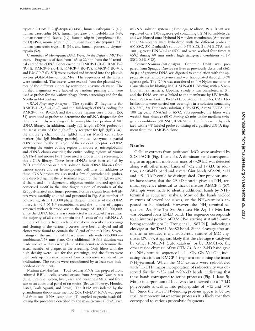
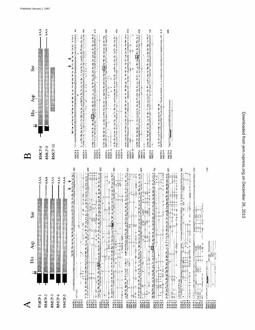
Figure 2. Nucleotide sequences for rat MC proteases. The cleavage sites for the signal sequence and the activation peptide are marked by arrows. Thepositions of the three aa of the catalytic triad are shown within boxes. The polyadenylation signal is underlined. Nucleotide numbers per line are depictedat the right of the figure. Schematic drawings of the different cDNA clones are shown at the top of the figure where the coding regions for the matureproteins are indicated by grey boxes, and the signal sequences and the activation peptides are indicated by black and white boxes, respectively. (A) Com-parative analysis of the complete nucleotide sequences coding for RMCP-1, -2, -3, -4, and -5 and the deduced amino acid sequence for RMCP-1. (B)Comparative analysis of RMCP-8, -9, and -10 and the deduced aa sequence for RMCP-8.
on Decem
ber 26, 2013jem
.rupress.orgD
ownloaded from
Published January 1, 1997
17 Lützelschwab et al.
on Decem
ber 26, 2013jem
.rupress.orgD
ownloaded from
Published January 1, 1997

18 Rat Mast Cell Proteases
position (pos.) 921. The mature protein of 226 aa has amolecular mass of 25,392 and has one putative N-glycosy-lation site (Asn-X-Ser/Thr) at position 58 of the matureprotein. The processed proteolytically active RMCP-5 is abasic protein with a net charge of 114.1 (Arg 1 Lys 5 29;Asp 1 Glu 5 16). (The nucleotide sequence is deposited inGenBank under the accession number U67908.) The se-quence differs in two positions compared with the previ-ously published sequence of RMCP-5 (two additional nu-cleotides, T in pos. 810 and a G in pos. 811) (25).
RMCP-6. A cDNA clone containing the entire codingregion for the rat tryptase RMCP-6 was isolated. Thisclone contains an open reading frame of 824 bp encoding asignal sequence of 19 aa, a 10-aa activation peptide (Ala-Pro-Cys-Pro-Val-Lys-Gln-Arg-Val-Gly) and the entire cod-ing region of the mature protein (Figs. 3 and 5 C). Thecoding region is followed by a 39 untranslated region of
246 bp with the non-canonical ATTAAA polyadenylationsignal located at position 1061. The mature protein of 245 aahas a molecular mass of 27,464 and contains two putativeN-glycosylation sites at positions 75 and 102 of the matureprotein. The processed proteolytically active RMCP-6 isan acidic protein with a net charge of 24.6 (Arg 1 Lys 519; Asp 1 Glu 5 25). (The nucleotide sequence is depos-ited in GenBank under the accession number U67909.)The sequence differs in five positions from the previouslypublished sequence of the rat tryptase (A pos. 399, C pos.432, C pos. 748, T pos. 946, and one base pair deleted atposition 1024) (24).
RMCP-7. A cDNA clone containing the entire codingregion for a second rat MC tryptase, denoted RMCP-7,was isolated (Fig. 3 and 5 C). The open reading frame of822 bp encodes a signal sequence of 18 aa, a 10-aa activa-tion peptide (Ala-Phe-Ser-Leu-Ala-Met-Phe-Arg-Glu-Gly),
Figure 3. Nucleotide se-quences for MC tryptases. Thefigure shows a comparative anal-ysis of the nucleotide sequencesfor the cDNAs encoding themouse and rat MC tryptases:RMCP-6, MMCP-6, RMCP-7,and MMCP-7, and the deducedaa sequence for RMCP-6. Thecleavage sites for the signal se-quence and the activation pep-tide are marked by arrows. Thepositions of the three aa of thecatalytic triad are shown withinboxes. The polyadenylation sig-nal is underlined. Nucleotidenumbers per line are depicted atthe right of the figure. A sche-matic drawing of the RMCP-6and -7 cDNA clones is shown atthe top of the figure where thecoding region for the matureprotein is indicated by a greybox, and the signal sequence andthe activation peptide are indi-cated by black and white boxes,respectively.
on Decem
ber 26, 2013jem
.rupress.orgD
ownloaded from
Published January 1, 1997

19 Lützelschwab et al.
and the entire coding region for the mature protein. Thecoding region is followed by a 39 untranslated region of184 bp with the canonical AATAAA polyadenylationsignal located at position 1204 (Fig. 3). The mature proteinof 245 aa has a molecular mass of 27,434 and two putativeN-glycosylation sites located at positions 21 and 102. Theprocessed proteolytically active RMCP-7 is an acidic pro-tein with a net charge of 24.6 (Arg 1 Lys 5 17; Asp 1Glu 5 23). (The nucleotide sequence is deposited in Gen-Bank under the accession number U67910.)
Rat Mast Cell Carboxypeptidase A. A nearly full-lengthcDNA clone for rat MC carboxypeptidase A (R-CPA) wasisolated (Figs. 4 and 5 D). This clone contains an open
reading frame of 1,254 bp encoding approximately two-thirds of the signal sequence, a 94-aa activation peptide andthe entire coding region of the mature protein, but lacksthe initiation codon and the NH2-terminal end of the sig-nal sequence. The coding region is followed by a 39 un-translated region of 178 bp with the canonical AATAAApolyadenylation signal located at position 1399. The ma-ture protein of 308 aa has an molecular mass of 35701 andtwo putative N-glycosylation sites at positions 127 and 133.The processed proteolytically active R-CPA is a basic pro-tein with a net charge of 118.1 (Arg 1 Lys 5 46; Asp 1Glu 5 29). (The nucleotide sequence is deposited in Gen-Bank under the accession number U67914.)
Figure 4. Nucleotide andamino acid sequences for MCcarboxypeptidases. The figureshows a comparative analysis ofthe nucleotide sequences encod-ing rat (R-CPA), mouse (M-CPA), and human (H-CPA) MCcarboxypeptidase A, and the de-duced aa sequence for R-CPA.The cleavage sites for the signalsequence and the activation pep-tide are marked by arrows. Thepolyadenylation signal is under-lined. Nucleotide numbers perline are depicted at the right ofthe figure. A schematic drawingof the different cDNA clones isshown at the top of the figurewhere the coding region for themature protein is indicated by agrey box, and the signal se-quence and the activation pep-tide are indicated by black andwhite boxes, respectively.
on Decem
ber 26, 2013jem
.rupress.orgD
ownloaded from
Published January 1, 1997

20 Rat Mast Cell Proteases
Cloning and Structural Analysis of Five Different Serine Proteases Expressed in the Rat Mucosal MC line RBL-1
Mucosal MCs are difficult to isolate as a pure population.Therefore, we decided to use the mucosal MC line RBL-1as a source of mRNA for the isolation of rat mucosal MCproteases. The RBL-1 cell line has been widely used as animportant in vitro model for studies of rodent MC biologyand is considered to be a good representative of rat MMC(59). A RBL-1 cDNA library of z50,000 independent re-combinants was constructed. Only one rat MMC protease,RMCP-2 (the rat homologue to MMCP-1) has previouslybeen identified. Except for MMCP-1, the only additionalMMC protease isolated in mouse, MMCP-2, could not beused as probe because of strong cross-hybridization be-
tween the different MC chymases. We therefore decidedto use a novel technique based on the use of degeneratePCR primers directed against conserved regions surround-ing the histidine and serine residues of the catalytic triadcharacteristic for all proteolytically active members of thelarge multigene family of trypsin-related serine proteases.Using this technique, five novel proteases, designatedRMCP-3, -4, -8, -9, and -10 were isolated from the RBL-1cell line. Based on a high degree of sequence identity withpreviously isolated rat MC chymases, RMCP-3 and -4were classified as chymases. RMCP-8, -9, and 10 are allhighly homologous and may be regarded as a new subfam-
Figure 5. Amino acid se-quence comparison of MC pro-teases. Sequence comparisons of(A) the rat chymases: RMCP-1,-2, -3, -4, and -5; (B) theRMCP-8 subfamily of serineproteases including RMCP-8, -9,and -10; (C) the mouse and rattryptases: RMCP-6, MMCP-6,RMCP-7, and MMCP-7; and(D) three different mammalianMC carboxypeptidases: rat (R-CPA), mouse (M-CPA), and hu-man (H-CPA) carboxypeptidaseA. Arrows indicate the cleavagesites for the activation peptides.The grey arrow indicates the au-tocatalytic initial cleavage in-volved in the removal of theactivation peptide (70). The re-maining two aa of the activationpeptide are removed by thedipeptidyl peptidase I, leading tothe same result as does the singleactivation step of the MC chy-mases (70). The positions of thethree aa of the catalytic triad aremarked with asterisks. The num-ber of aa per line is depicted atthe right side of the figure.
on Decem
ber 26, 2013jem
.rupress.orgD
ownloaded from
Published January 1, 1997

21 Lützelschwab et al.
ily of MC proteases, more closely related to the T cellgranzymes than to the previously characterised MC pro-teases (see Fig. 6). The cleavage specificities of RMCP-8,-9,and -10 have not yet been determined. To obtain the com-plete sequences of these proteases, the z500-bp insert frag-ments were used as probes in screenings of the RBL-1cDNA library. Together with a full-length cDNA copy ofthe previously published RMCP-2, full-length or nearlyfull-length cDNA clones were isolated for RMCP-3, -4, -8,and -9. No full-length clone was obtained for RMCP-10(see below). The nucleotide sequences of the RMCP-3and -4 clones are depicted in Fig. 2 A, and the sequences ofthe clones encoding RMCP-8,-9, and -10 are shown inFig. 2 B. The deduced aa sequences of these clones are de-picted in Figs. 5, A and B. Fig. 6 shows a comparative anal-ysis of the rat MC serine proteases with other rat, mouse,and human serine proteases. The characteristics of theclones, encoding the five different serine proteases isolated
from the RBL-1 cDNA library, and some of the biochem-ical properties of these proteases are listed below. A sum-mary of their biochemical characteristics is presented in Ta-ble 1.
RMCP-2. A cDNA clone containing the entire codingregion for RMCP-2 was isolated (Figs. 2 A and 5 A). Onlythe 59- and 39-terminal 250 bp were subjected to nucle-otide sequence analysis and were found to be identical tothe previously published sequence for RMCP-2 (22).RMCP-2 is translated as a 247-aa primary polypeptidewith an 18-aa-long signal sequence and a 2-aa long nega-tively charged NH2-terminal propeptide (Glu-Glu). Theenzymatically active processed protease has a net charge of15.2 (Arg 1 Lys 5 25; Asp 1 Glu 5 21) and a molecularmass of 25,046. RMCP-2 is also processed in its COOH-terminal end, where 3 aa are removed after translation (22).One putative N-glycosylation site (Asn-X-Ser/Thr) is lo-cated in the COOH-terminal end of the protein.
Figure 6. A comparative analysis basedon the amino acid sequence identities be-tween a panel of mammalian hematopoie-tic serine proteases. MC serine proteasesare shown in black. The rat MC serineproteases are indicated by asterisks. Thepercent of divergence among the differentproteases, as indicated by the branchpoints, is depicted at the bottom of thefigure.
on Decem
ber 26, 2013jem
.rupress.orgD
ownloaded from
Published January 1, 1997

22 Rat Mast Cell Proteases
RMCP-3. A clone containing a partial sequence forRMCP-3 was isolated. This clone contains an open read-ing frame of 717-bp encoding half of the signal sequence, a2-aa activation peptide (Glu-Glu) and the entire coding re-gion of the mature protein (Figs. 2 A and 5 A). The codingregion is followed by a 39 untranslated region of 172 bpwith the canonical AATAAA polyadenylation signal lo-cated at position 868. The mature protein of 226 aa has amolecular mass of 25,443, and contains no putative N-gly-cosylation sites. The processed proteolytically active RMCP-3is a basic protein with a net charge of 19.2 (Arg 1 Lys 528; Asp 1 Glu 5 20). (The nucleotide sequence is depos-ited in GenBank under the accession number U67888.)
RMCP-4. A cDNA clone containing the entire codingregion for RMCP-4 was isolated. The clone has an insert sizeof 959 bp with an open reading frame of 741 bp flanked by15-bp 59 and 175-bp 39 untranslated regions (Figs. 2 A and5 A). A canonical polyadenylation signal (AATAAA) is lo-cated at position 938. The primary translated product hasan 18-aa signal peptide and a 2-aa negatively charged acti-vation peptide (Glu-Glu). The enzymatically active 226-aaprotease has a molecular mass of 24,987 and no N-linkedglycosylation sites. The mature basic protein has a net chargeof 112.1 (Arg 1 Lys 5 26; Asp 1 Glu 5 15). (The nucle-
otide sequence is deposited in GenBank under the acces-sion number U67907.)
RMCP-8. A cDNA clone containing the entire codingregion for RMCP-8 was isolated (Figs. 2 B and 5 A). Theclone starts 10 bp upstream from the initiation codon andcontains an open reading frame of 747 bp followed by a 39untranslated region of 82 bp. A canonical polyadenylationsignal is located at position 814. The coding region startswith a signal sequence of 18 aa followed by a 2-aa activa-tion peptide (Gly-Glu). The mature protein of 228 aminoacids has a molecular mass of 25,248 and contains four pu-tative N-glycosylation sites at positions 47, 132, 160, and169. The active protease is a basic protein with a net chargeof 110.6 (Arg 1 Lys 5 27; Asp 1 Glu 5 17). (The nucle-otide sequence is deposited in GenBank under the acces-sion number U67911.)
RMCP-9. A nearly full-length cDNA clone for RMCP-9was isolated (Figs. 2 B and 5 B). The 804-bp cDNA clonehas an open reading frame of 717 bp, a 39 untranslated re-gion of 82 bp and contains a canonical polyadenylation sig-nal at position 783. The unprocessed protein has an 18-aasignal peptide and a 2-aa activation peptide (Gly-Glu). Theactive basic enzyme has 228 aa with a molecular mass of25,249, a net charge of 112.3 (Arg 1 Lys 5 27; Asp 1Glu 5 15), and two putative glycosylation sites at positions132 and 160. (The nucleotide sequence is deposited inGenBank under the accession number U67912.)
RMCP-10. A clone containing the partial sequence ofRMCP-10 was isolated by PCR amplification with theHis-Ser primers (see above). The obtained sequence coversthe central region between the His and Ser residues ofthe catalytic triad (Figs. 2 B and 5 B). (The nucleotidesequence is deposited in GenBank under the accessionnumber U67913.) Except for a single base pair difference,this sequence was found to be identical to a cDNA previ-ously isolated from rat duodenum by Amerik et al. (33)(Granzyme-like protein II). This single base pair differencemay have been caused by the low accuracy of the Taqpolymerase used during the isolation of RMCP-10.
Construction of Monospecific ProbesSeveral of the MC serine proteases show a high degree
of sequence identity, making it difficult to use the entireinserts of the clones as probes in Northern blot, Southernblot, or plaque hybridizations. To solve this problem, thesequences for the different proteases were compared andregions with maximal divergence were selected as regionsof interest for the construction of specific probes. Such re-gions were found in the 39 non-coding regions of all thedifferent mRNAs. Fragments located within these regionswere excised from the cDNA clones and subcloned intoplasmid vectors. The probes for RMCP-1, -2, -3, -4, -6,and -7 were all in the range of 165–220 nucleotides inlength. For RMCP-5, -8, and CPA the entire inserts of theoriginal cDNA clones were used as probes. To study thespecificity of these probes, the insert fragments were puri-fied, labeled and used in a dot blot analysis. All probes, ex-
Figure 7. Dot blot analysis of probes for rat MC proteases. The probesfor RMCP-1 (R-I), RMCP-2 (R-II), RMCP-3 (R-III), RMCP-4 (R-IV),RMCP-6 (R-VI), and RMCP-7 (R-VII) were 160–220-bp fragmentsoriginating from the 39 non-coding regions of the respective cDNAclones (see Materials and Methods). The probes for RMCP-5 (R-V),RMCP-8 (R-VIII), and carboxypeptidase A (CPA) are the entire insertsof the corresponding full-length cDNA clones. Cross hybridization be-tween RMCP-8, -9, and -10 was seen when using the R-VIII probe.
on Decem
ber 26, 2013jem
.rupress.orgD
ownloaded from
Published January 1, 1997

23 Lützelschwab et al.
cept for the RMCP-8 probe, were found to be monospe-cific (Fig. 7). Because of the high degree of homologyshared by RMCP-8, -9, and -10 it has not been possible toexcise any fragment of a size large enough to label by ran-dom priming, and still maintain mono-specificity (Figs. 2 Band 7). Consequently, in all hybridizations performed withthe RMCP-8 probe, we detect all three members of thissubfamily of serine proteases.
mRNA Frequency DeterminationBoth CTMCs and MMCs store very large amounts of
the various proteases in their secretory granules. Based onstudies of the protein content in different MC subpopula-tions, it has been estimated that granule proteases constituteup to 50% of the total cellular protein (60). One importantquestion in MC biology is whether the large amounts ofstored proteases is also correlated with a high synthesis rate.One alternative possibility is that the turnover is minimaland that the synthesis rate thereby may be relatively low. Infavour of this hypothesis, previous studies of mRNA levelsfor the various MC proteases in transformed MCs haveshown that these levels actually are relatively low (Hellman,L., unpublished observations). However, these results maybe questioned as being a phenomenon related to the trans-formed phenotype of the cell lines and not representativeof mature tissue MCs. To address this question, we deter-mined the mRNA frequencies for the various MC pro-teases, some housekeeping genes and a few cell surface re-ceptor genes expressed by normal tissue MCs. For thispurpose, an unamplified cDNA library was screened forpositive hybridization signals with a panel of probes. Allexcept one of these probes (the RMCP-8 probe) weremonospecific and nearly all contained sequences originat-ing from the 39 ends of the mRNAs (see Materials andMethods for details of the various probes).
The results showed that peritoneal MCs are highly spe-cialized cells, which most likely constantly produce rela-tively high amounts of the various proteases. The mRNAfrequencies of the most abundant proteases were in therange of 0.4–2.5 % of the total number of plaques in thescreenings (Fig. 8). Actin and GAPDH showed substan-tially lower frequencies, in the range of 0.03–0.4%. A panelof cell surface markers: the three subunits (a, b, and g) ofthe IgE high-affinity receptor (IgERI), the Mac-2, and thec-kit receptor were all present at a level of 0.1% or below.The number of IgE receptors on CTMCs has previouslybeen estimated at z300,000/cell (26), a relatively lownumber compared to the number of stored proteases mole-cules in the granules, which also is reflected by the lownumber of clones corresponding to the three IgE receptorsubunits (0.1, 0.028, and 0.034% of the plaques for the a,b, and g chains, respectively). The very low level of c-kitwas unexpected. However, growth factor receptors areknown to be present at comparably low levels on the sur-face of most cell types (61). Further, the cDNA library wasanalysed for the presence of several additional hematopoie-tic granule proteins. A quite substantial number of clonesfor lysozyme were detected (235 in 100,000 plaques). 223clones for the heparin proteoglycan core protein (53, 54),and 34 clones for a deacetylase (62) were detected in 100,000plaques. Both of these proteins are involved in the synthesisof heparin proteoglycans. No positive signals were ob-served with cDNA probes against either a2-microglobulin(63), TNF-a, IL-4, or IL-5. The mRNA frequencies for afew transcription factors were also determined. We de-tected 17 and 10 clones in 100,000 plaques for Pu-1 and
Figure 8. mRNA frequencies for various MC proteins. The expressionlevels of different proteins synthesized by rat serosal MCs were deter-mined from the number of individual clones for these mRNAs in an un-amplified rat peritoneal MC cDNA library. mRNA frequencies are ex-pressed at the right side of each bar as positive hybridization signals per100,000 plaques. (A) mRNA frequencies for the secretory granule pro-teins: RMCP-1, -2, -3, -4, -5, -6, -7, and -8, cathepsin G (Cath.G), car-boxypeptidase A (CPA), and the heparin proteoglycan core protein (Hep-arin c.p.). (B) mRNA frequencies for a number of cell surface markers: thea, b, and g chains of the IgE high-affinity receptor, the IgE binding pro-tein (Mac-2), and the stem cell factor receptor (c-kit). Glyceraldehyde-3-phosphate dehydrogenase (GAPDH) and actin were used as controls.
on Decem
ber 26, 2013jem
.rupress.orgD
ownloaded from
Published January 1, 1997

24 Rat Mast Cell Proteases
GATA-1, respectively. These values may be slight underes-timates, since these probes do not contain sequences fromthe 39 non-coding regions of the mRNAs and clonesshorter than 300–500 bp may thereby escape detection. Wealso screened the library with a 62-bp degenerate oligonu-cleotide against conserved regions of the finger and spacerregions of Krüppel-related zinc finger proteins (64). Only31 positive signals were found in 100,000 plaques. We canfrom these data conclude that the transcription factors andcell surface markers tested are expressed at much lower lev-els compared to the most abundant granule components.
To obtain an estimate of what the number of plaquesrepresents in actual mRNA frequencies, the sizes of the in-serts in eleven independent clones were determined. 8 outof 11 of the clones had inserts of a size of 600 bp or largerwhereas the remaining clones had inserts that were toosmall to be visualized on a 2% agarose gel (probably z50bp). Therefore, these latter clones probably contain onlyvery short adapter fragments. Taking into considerationthat z70% of the clones contain mRNA sequences, thisresults in estimated mRNA frequencies z30% higher thanthe actual number of positive signals in 100,000 plaques,
Figure 9. Northern and Southern blot analysis. (A) Northern blot analysis of poly(A)1RNA isolated from the ears of Lewis, Brown Norway, HoodedLister, Dark Agouti, and Sprague Dawley rats, poly(A)1RNA from purified peritoneal MCs (PMC; from Sprague Dawley rats) and poly(A)1RNA iso-lated from rat liver (Sprague Dawley). These mRNAs were analyzed for the expression of RMCP-1 (R-I), RMCP-5 (R-V), RMCP-6 (R-VI), RMCP-7(R-VII), RMCP-8 (R-VIII), carboxypeptidase A (CPA), and the a chain of the IgΕRI using the specific probes described in Materials and Methods. (B)Northern blot analysis of poly(A)1RNA from: Sprague Dawley lung, intestine, liver, spleen, Lewis ears and from the RBL-1 cell line. The various mRNAswere analyzed with probes for RMCP-1 (R-I), RMCP-2 (R-II), RMCP-3 (R-III), RMCP-4 (R-IV), RMCP-5 (R-V), RMCP-8 (R-VIII), carbox-ypeptidase A (CPA), or the a chain of the IgΕRI using the specific probes described in Materials and Methods. In both A and B, a b-actin probe wasused to normalise the amount of RNA loaded in each lane to facilitate a comparative analysis between different organs and cell lines. (C) Genomic South-ern blot analysis of DNA from Sprague Dawley rats. The DNA was cleaved with three different restriction enzymes, BamHI, BglII, and EcoRI. Sizemarkers are indicated at the left side of C. The blot was hybridized with the entire insert from the full-length cDNA clone for RMCP-8 and the blot waswashed under medium stringency conditions (2 3 SSC and 0.5% SDS at 658C).
on Decem
ber 26, 2013jem
.rupress.orgD
ownloaded from
Published January 1, 1997

25 Lützelschwab et al.
and consequently, mRNA frequencies for the most abun-dant proteases in the range of 0.5–3.5% of the total mRNApool.
Northern Blot AnalysisTo study the distribution of the different MC proteases
in various rat tissues, mRNA was prepared from differentorgans of Sprague Dawley rats, from ears of several addi-tional rat strains and from purified peritoneal MCs. Thepurified peritoneal MCs represent an essentially pure popu-lation of CTMCs. In the ear, the majority of the MCspresent are of the connective tissue subtype, although itshould be emphasized that the percentage of MCs in thistissue is relatively low. In mRNA isolated from both earsand from peritoneal MCs, Northern blot analysis showedhigh expression levels for the chymases RMCP-1 and -5,the tryptase RMCP-6 and the MC carboxypeptidase A(Fig. 9 A). In mRNA isolated from the ears of several dif-ferent strains of rats we detected relatively high levels ofRMCP-7 (Fig. 9 A). In contrast, peritoneal MCs expressedonly low levels of this protease compared with the expres-sion levels for RMCP-1, -5, -6, and CPA. These results arethus in agreement with the results from the mRNA fre-quency analysis described above (Fig. 8). With respect totheir RMCP-7 content, CTMCs of ears and peritoneummay therefore be regarded as two distinct subpopulations ofMCs. When comparing different strains of rats, most MCcomponents showed similar patterns of expression. How-ever, the amounts of mRNA for the various MC proteinswere different among the various strains, indicating that thenumber of MCs may differ.
The mucosal MC protease RMCP-2 was undetectablein a Northern blot analysis of RNA from both ears andperitoneum (data not shown). Further, only two clones forRMCP-2 were detected in 100,000 clones of the serosalMC library (Fig. 8), indicating that MC populations inboth the peritoneal cavity and the ears are essentially de-void of mucosal MCs. As detected by Northern blot analy-sis, ear and peritoneal MCs showed only low expression ofRMCP-8 (Fig. 9). In addition, only 14 positive clones forRMCP-8 were detected in 100,000 clones of the serosalMC library (Fig. 8). This shows that the RMCP-8 subfam-ily of proteases is expressed only at a very low level in thesetwo CTMC populations. No hybridization signals forRMCP-3 and -4 were detected in MCs of ear (Fig. 9 B) orperitoneum (data not shown), and no positive plaques werefound during the screening of the serosal MC library (Fig.8). From these data we can conclude that the MC popula-tions of these organs most likely completely lack expressionof both of these proteases.
Cathepsin G or a cathepsin G–like protease has been de-tected previously both in human MCs and in a mouse MCtumour (65 and data not shown). To determine whethercathepsin G is expressed also by rat MCs, a cDNA fragmentcontaining the entire coding region of mouse neutrophilcathepsin G was used as probe in a Northern blot analysisof mRNA from ear and peritoneal MCs. No hybridization
signals were detected by Northern blot analysis (data notshown) and no positive plaques were found in the serosalMC library after screening under low stringency conditions(Fig. 8). This indicates that mature rat CTMCs completelylack the expression of cathepsin G or cathepsin G–like pro-teases. In addition, the RBL-1 cell line has previously beenanalyzed by Northern blots and found to be negative formRNA encoding cathepsin G or a cathepsin G-like pro-tease (data not shown).
mRNA isolated from the RBL-1 cell line was analyzed,together with mRNA from rat lung, small intestine, ears, andliver for expression of the various MC proteases (Fig. 9 B).The RBL-1 cells expressed RMCP-2, -8, and low levels ofRMCP-1, -3, and -4, but no RMCP-5 (Fig. 9 B). Theliver RNA was negative for all of these proteases. In earswe detected the previously identified CTMC proteasesRMCP-1 and -5, but no hybridization signals for RMCP-2,-3, and -4. In mRNA isolated from small intestine wefound relatively strong hybridization signals for RMCP-2and the proteases of the RMCP-8 family. A low level ofRMCP-1 and quite significant levels of RMCP-5 were alsodetected (Fig. 9 B). In contrast, no visible bands were de-tected with the RMCP-3- and -4-specific probes (Fig. 9 B).In addition to MMCs, the small intestine probably containsa small fraction of CTMCs originating from the sub-mucosaand the muscular layer of this organ. This may explain thedetection of RMCP-1 and -5 expression in this tissue.However, the amount of mRNA for RMCP-5 seems toolarge to originate only from the CTMC population. Onepossibility, which we presently can not exclude is that ratMMCs also express RMCP-5. Lung is considered to have amixture of both mucosal and connective tissue MCs. Ac-cordingly, this organ showed expression of both MMC andCTMC proteases (Fig. 9 B).
Genomic Southern Blot Analysis of RMCP-8 and of Closely Related Genes in the Rat Genome
As previously described, cDNA clones for three closelyrelated proteases of the RMCP-8 subfamily (RMCP-8,-9,and -10) were isolated from the RBL-1 cDNA library. Toestimate the total number of closely related members of thissubfamily in the rat genome, a Southern blot analysis wasperformed with genomic DNA from Sprague Dawley andWistar rats. The blots were hybridized using the full-lengthcDNA for RMCP-8 as a probe. The pattern of bands onthe blots were found to be identical in both strains of rats(Fig. 9 C, and data not shown). The results show multiplebands in the size range of 3–20 kb after cleavage with BamHI, BglII, or EcoRI. The exact number is not possible todetermine until information is available from genomicclones for one or several of these proteases. However, arough estimate indicates three to five potential genes.
Discussion
We present in this communication the cloning of elevendifferent proteases from various rat MC populations. Five
on Decem
ber 26, 2013jem
.rupress.orgD
ownloaded from
Published January 1, 1997

26 Rat Mast Cell Proteases
of the proteases have been isolated previously as full-lengthor partial clones either by conventional cDNA cloning(RMCP-2, and RMCP-10 [Granzyme-like protease II]) orby PCR amplification (RMCP-1, -5, and -6) (22–25, 33).In two of these three proteases, RMCP-5 and -6, severaldifferences in sequence were found between ours and thesequences determined by PCR amplification by Ide et al.(Figs. 2 A and 3) (24, 25). We favor the explanation thatmost of these differences are allelic differences between dif-ferent strains of rat, as the sequences originate from two dif-ferent strains, Sprague-Dawley and Lewis (24, 25). However,we can not exclude that some of the differences are causedby the low accuracy of the Taq polymerase used during thePCR amplification. To minimize potential misunderstand-ings upon comparisons of mouse and rat MC proteases wehave suggested a nomenclature which as close as possibleharmonises between the corresponding proteases in thesetwo species. This nomenclature has also been used by otherinvestigators in the field (66).
The cloning of the various rat MC proteases has made itpossible to construct monospecific cDNA probes for mostof these enzymes. These probes have been used as specifictools to estimate mRNA frequencies for the different pro-teases in normal freshly isolated peritoneal MCs. Thescreening of the large unamplified cDNA library showedthat serosal MCs are highly specialized cells which mostlikely continuously produce large amounts of the varioussecretory granule proteases, and with mRNA frequenciesfor these proteases in the range of several percent of the to-tal mRNA pool. Such high expression levels have previ-ously only been detected in terminally differentiated cells,and can be compared to, e.g., the expression of immuno-globulins in B lymphocyte plasma cells, where the immu-noglobulin mRNA may account for 5–10% of the totalmRNA pool.
The very high levels of mRNA for the different pro-teases actualises an important question concerning the roleof MCs during normal tissue maintenance and tissue re-modelling. If MCs store and only infrequently release theproteases (upon activation by allergen challenge or underinflammatory conditions) there would not be a need for acontinuous protease production at such a high level, even ifthe amount stored in the cell is very high. Possible explana-tions for this phenomenon are either that MCs continu-ously release quite substantial amounts of their granule con-tents, or that a fraction of the proteases are never stored butare secreted without previous passage through the granulecompartment. Alternatively, there may be a high rate of in-tracellular turnover of the proteases. Bone macrophages,osteoclasts, are known to actively participate in the degra-dation of non-cellular components and thereby help tomaintain the elasticity of the bone (67, 68). Possibly, CT-MCs have a similar function in the degradation of collagen,proteoglycans, fibronectin and other components of theconnective tissue and thereby in the maintenance of theelasticity and function of this tissue.
We have also shown that there is a very tight regulation
of the different proteases in the various MC populations.The typical CTMC proteases RMCP-1, -5, -6, and CPAwere found at very high frequencies in the serosal MC li-brary, whereas a low number of, or a total lack of clones forthe MMC proteases RMCP-2, -3, -4, and the members ofthe RMCP-8 family were detected in the same library. Incontrast, the MMC line RBL-1 and rat small intestineshowed strong expression of the typical MMC proteaseRMCP-2 and the RMCP-8 subfamily, but only weak ex-pression of the CTMC proteases. The separate subpopula-tions of MCs must thereby be considered as highly special-ized cells where the protease genes are tightly regulated andexpressed only in one out of several distinct subpopula-tions.
Mouse and rat MCs show a high degree of similarity intheir protease expression. CTMCs of both species expresstwo dominant chymases, RMCP-1/MMCP-4 and RMCP-5/MMCP-5, which are each others homologues. CTMCs ofboth species also express two tryptases, RMCP-6/MMCP-6and RMCP-7/MMCP-7, and the MC carboxypeptidaseA. In addition, in both species the expression of RMCP-7/MMCP-7 is considerably higher in skin CTMCs than inperitoneal CTMCs. When comparing MMCs of the twospecies, both similarities and also certain important differencesare found. In both species the major MMC proteases areRMCP-2/MMCP-1 and, probably, the RMCP-8 subfamily.We have recently isolated the cDNA for a mouse MC pro-tease, denoted MMCP-8, homologous to the RMCP-8 sub-family (data not shown). In the mouse genome, MMCP-8is probably the only gene encoding serine proteases of thissubfamily. In contrast, studies by genomic Southern blotanalysis indicated three or possibly four or five members ofthe RMCP-8 subfamily in rat (Fig. 9 C). Additional differ-ences between MMCs derived from the two species includethe fact that no direct homologue of MMCP-2 has yetbeen isolated in rat, and that no homologues of RMCP-3and -4 have been isolated from mouse MCs (Fig. 7). Oneadditional difference may also lie in the expression patternfor RMCP-5. We have detected substantial amounts ofRMCP-5 mRNA in uninfected intestinal tissue, whereasmouse MMCs lack the expression of MMCP-5 (14). More-over, Ide et al. observed a variable expression of RMCP-5during cultivation of the RBL-2H3 cell line and they alsodetected a slight increase in the amounts of RMCP-5 dur-ing parasite infection of rat jejunum (25). In addition, theMC subclass specificity of RMCP-3 and -4 is not clear.We find low but detectable levels in several different sub-lines of RBL-1, but no expression in normal uninfected in-testine. Possibly, RMCP-3 and -4 may be upregulated dur-ing parasite infection, in a similar fashion as has been reportedfor another MC protease, MMCP-L (12).
Despite the present characterization of cDNAs for threenew MC proteases, the RMCP-8 subfamily, none of theseenzymes have been identified at the protein level. A possi-ble explanation for this may lie in both extensive and vari-able glycosylation of these proteases. The members of theRMCP-8 subfamily of proteases have several potential gly-
on Decem
ber 26, 2013jem
.rupress.orgD
ownloaded from
Published January 1, 1997

27 Lützelschwab et al.
cosylation sites (see Results). In a two-dimensional gelanalysis of rat MMC proteases, Abe et al. (60) showed thatfreshly isolated MMCs from parasite infected rats gave anumber of quite abundant but relatively fuzzy bands in thesize range of 24–32 kD. Since none of these bands reactedwith an RMCP-2-specific antiserum (60), it is possible thatsome of them represent differently glycosylated forms of pro-teases belonging to the RMCP-8 subfamily. This is not un-likely, in view of the fact that the majority of the MC-pro-teases are present in several differently glycosylated forms,and that these carbohydrates are exclusively N-linked (60,66, 69).
The expression of cathepsin G or cathepsin G–like pro-teases in MCs from different mammals still remains an openquestion. In the present study, no expression of cathepsinG–like proteases was detected in any of the various rat MC
populations tested. In contrast, we have previously detectedsignificant levels of cathepsin G in mouse CTMC-like MCtumor cells (data not shown), and further, Schechter et al.have detected a cathepsin G–like protease in human MCs(65). We do not know whether these discrepancies reflect aspecies difference or if cathepsin G is expressed only in cer-tain populations of immature MCs.
In summary, this investigation has led to a detailed pic-ture of the various proteases and other differentiationmarkers expressed by different populations of rat MCs. Thespecific reagents originating from these studies can now beused as important tools in the analysis of the various biolog-ical functions associated with these proteases and as specificmarkers for studies of both early and late events during MCdifferentiation.
We are grateful to Dr. Anders Karlström for performing NH2-terminal sequence analysis. We would alsolike to thank Dr. David Eaker for linguistic revision of the manuscript. This investigation was supported bygrants from the Swedish Natural Sciences Research Council.
Address correspondence to Dr. Lars Hellman, Department of Medical Immunology and Microbiology, Uni-versity of Uppsala, Biomedical Center, Box 582, S-751 23 Uppsala, Sweden.
Received for publication 4 September 1996 and in revised form 21 October 1996.
References1. Enerbäck, L. 1966. Mast cells in rat gastrointestinal mucosa 1.
Effects of fixation. Acta Pathol. Microbiol. Scand. 66:289–302.2. Enerbäck, L. 1966. Mast cells in rat gastrointestinal mucosa 2.
Dye-binding and metachromatic properties. Acta Pathol. Mi-crobiol. Scand. 66:303–312.
3. Wingren, U., and L. Enerbäck. 1983. Mucosal mast cells ofthe rat intestine: a re-evaluation of fixation and staining prop-erties, with special reference to protein blocking and solubilityof the granular glycosaminoglycan. Histochem. J. 15:571–582.
4. Stevens, R.L., H.R. Katz, D.C. Seldin, and K.F. Austen.1986. In Biochemical Characteristics Distinguish Subclasses ofMammalian Mast Cells. Raven Press, New York. 183–203.
5. Gibson, S., and H.R. Miller. 1986. Mast cell subsets in the ratdistinguished immunohistochemically by their content ofserine proteinases. Immunology. 58:101–104.
6. Enerbäck, L., S.O. Kolset, M. Kusche, A. Hjerpe, and U.Lindahl. 1985. Glycosaminoglycans in rat mucosal mast cells.Biochem. J. 227:661–668.
7. Stevens, R.L., T.D. Lee, D.C. Seldin, K.F. Austen, A.D. Be-fus, and J. Bienenstock. 1986. Intestinal mucosal mast cellsfrom rats infected with Nippostrongylus brasiliensis containprotease-resistant chondroitin sulfate di-B proteoglycans. J.Immunol. 137:291–295.
8. Aldenborg, F., and L. Enerbäck. 1986. Histamine contentand mast cell numbers in tissues of normal and athymic rats.Agents Actions. 17:454–459.
9. Newlands, G.F., S. Gibson, D.P. Knox, R. Grencis, D.Wakelin, and H.R. Miller. 1987. Characterization and mastcell origin of a chymotrypsin-like proteinase isolated from in-testines of mice infected with Trichinella spiralis. Immunology.
62:629–634.10. Huang, R., T. Blom, and L. Hellman. 1991. Cloning and
structural analysis of MMCP-1, MMCP-4 and MMCP-5,three mouse mast cell-specific serine proteases. Eur. J. Immu-nol. 21:1611–1621.
11. Serafin, W.E., D.S. Reynolds, S. Rogelj, W.S. Lane, G.A.Conder, S.S. Johnson, K.F. Austen, and R.L. Stevens. 1990.Identification and molecular cloning of a novel mouse mu-cosal mast cell serine protease. J. Biol. Chem. 265:423-429.
12. Serafin, W.E., T.P. Sullivan, G.A. Conder, A. Ebhrahimi, P.Marcham, S. Jonhson, K.F. Austen, and D.S. Reynolds.1991. Cloning of the cDNA and Gene for Mouse Cell Pro-tease 4. J. Biol. Chem. 266:1934–1941.
13. Reynolds, D.S., D.S. Gurley, A.K.F. Austen, and W.E. Sera-fin. 1991. Cloning of the cDNA and gene of the mouse mastcell protease-6. J. Biol. Chem. 266:3847–3853.
14. McNeil, H.P., K.F. Austen, L.L. Somerville, M.F. Gurish,and R.L. Stevens. 1991. Molecular cloning of the mousemast cell protease-5 gene. A novel secretory granule proteaseexpressed early in the differentiation of serosal mast cells. J.Biol. Chem. 266:20316–20322.
15. McNeil, H.P., D.S. Reynolds, V. Schiller, N. Ghildyal, D.S.Gurley, K.F. Austen, and R.L. Stevens. 1992. Isolation, char-acterization, and transcription of the gene encoding mousemast cell protease 7. Proc. Natl. Acad. Sci. USA. 89:11174–11178.
16. Reynolds, D.S., R.L. Stevens, D.S. Gurley, W.S. Lane, K.F.Austen, and W.E. Serafin. 1989. Isolation and molecularcloning of mast cell carboxypeptidase A. J. Biol. Chem. 264:20094–20099.
on Decem
ber 26, 2013jem
.rupress.orgD
ownloaded from
Published January 1, 1997

28 Rat Mast Cell Proteases
17. Serafin, W.E., E.T. Dayton, P.M. Gravallese, K.F. Austen,and R.L. Stevens. 1987. Carboxypeptidase A in mouse mastcells. Identification, characterization, and use as a differentia-tion marker. J. Immunol. 139:3771–3776.
18. Irani, A.A., N.M. Schechter, S.S. Craig, G. DeBlois, andL.B. Schwartz. 1986. Two types of human mast cells thathave distinct neutral protease compositions. Proc. Natl. Acad.Sci. USA. 83:4464–4468.
19. Kido, H., N. Fukusen, and N. Katunuma. 1985. Chymo-trypsin- and trypsin-type serine proteases in rat mast cells:properties and functions. Arch. Biochem. Biophys. 239:436–443.
20. Kido, H., K. Izumi, H. Otsuka, N. Fukusen, Y. Kato, and N.Katunima. 1986. A Chymotrypsin-type serine protease inRat Basophilic Leukemia cells: evidence for its immunologicidentity with atypical mast cell protease. J. Immunol. 136:1061–1065.
21. Le Trong, H., D.C. Parmelee, K.A. Walsh, H. Neurath, andR.G. Woodbury. 1987. Amino acid sequence of rat mast cellprotease I (chymase). Biochemistry. 26:6988–6994.
22. Benfey, P.N., F.H. Yin, and P. Leder. 1987. Cloning of themast cell protease, RMCP II. Evidence for cell-specific ex-pression and a multi-gene family. J. Biol. Chem. 262:5377–5384.
23. Rouleau, A., M. Garbarg, J.C. Schwartz, and M. Ruat. 1994.Molecular cloning of rat mast cell protease 1 and develop-ment of specific probes for its gene transcript. Biochem Bio-phys. Res. Commun. 199:593–602.
24. Ide, H., H. Itoh, M. Tomita, Y. Murakumo, T. Kobayashi,H. Maruyama, Y. Osada, and Y. Nawa. 1995. cDNA se-quencing and expression of rat mast cell tryptase. J. Biochem.118:210–215.
25. Ide, H., H. Itoh, M. Tomita, Y. Murakumo, T. Kobayashi,H. Maruyama, Y. Osada, and Y. Nawa. 1995. Cloning of thecDNA encoding a novel rat mast-cell proteinase, rMCP-3,and its expression in comparison with other rat mast-cell pro-teinases. Biochem. J. 311:675–680.
26. Sterk, A.R., and T. Ishizaka. 1982. Binding properties of IgEreceptors on normal mouse mast cells. J. Immunol. 128:838–843.
27. Eccleston, E., B.J. Leonard, J.S. Lowe, and H.J. Welford.1973. Basophilic leukaemia in the albino rat and a demon-stration of the basopoietin. Nature, New Biol. 244:73–76.
28. Laemmli, U.K. 1970. Cleavage of structural proteins duringthe assembly of the head of bacteriophage T4. Nature (Lond.).227:680–685.
29. Pejler, G., A.R. Karlstrom, and L. Berg. 1995. Identificationof the proteolytic thrombin fragments formed after cleavagewith rat mast cell protease 1. Eur. J. Biochem. 227:102–107.
30. Deleted in proof.31. Huang, R., and L.Hellman. 1991. Cloning and structural
analysis of a novel mouse mast cell specific serine protease theMMCP-6: a connective tissue mast cell specific tryptase. XIVInternational Congress of Allergology and Clinical Immunol-ogy, Kyoto, Japan. Abstract no 125.
32. Sanger, F., S. Niclen, and A.R. Coulson. 1977. DNA se-quencing with chain-terminating inhibitors. Proc. Natl. Acad.Sci. USA. 74:5463–5467.
33. Amerik, A.Y., S.V. Yarovoi, V.G. Grigorenko, and V.K. An-tonov. 1993. Identification, sequence analysis, and character-ization of cDNA clones encoding two granzyme-like serineproteinases from rat duodenum. FEBS Lett. 324:226–230.
34. Gershenfeld, H.K., and I.L. Weissman. 1986. Cloning of acDNA for a T cell-specific serine protease from a cytotoxic Tlymphocyte. Science (Wash. DC). 232:854–858.
35. Gershenfeld, H.K., R.J. Hershberger, T.B. Shows, and I.L.Weissman. 1988. Cloning and chromosomal assignment of ahuman cDNA encoding a T cell- and natural killer cell-spe-cific trypsin-like serine protease. Proc. Natl. Acad. Sci. USA.85:1184–1188.
36. Lobe, C.G., C. Upton, B. Duggan, N. Ehrman, M. Letellier,J. Bell, G. McFadden, and R.C. Bleackley. 1988. Organiza-tion of two genes encoding cytotoxic T lymphocyte-specificserine proteases CCPI and CCPII. Biochemistry. 27:6941–6946.
37. Jenne, D., C. Rey, D. Masson, K.K. Stanley, J. Herz, G.Plaetinck, and J. Tschopp. 1988. cDNA cloning of granzymeC, a granule-associated serine protease of cytolytic T lym-phocytes. J. Immunol. 140:318–323.
38. Jenne, D., C. Rey, J.A. Haefliger, B.Y. Qiao, P. Groscurth,and J. Tschopp. 1988. Identification and sequencing ofcDNA clones encoding the granule-associated serine pro-teases granzymes D, E, and F of cytolytic T lymphocytes.Proc. Natl. Acad. Sci. USA. 85:4814–4818.
39. Bleackley, R.C., B. Duggan, N. Ehrman, and C.G. Lobe.1988. Isolation of two cDNA sequences which encode cyto-toxic cell proteases. FEBS Lett. 234:153–159.
40. Kwon, B.S., D. Kestler, E. Lee, M. Wakulchik, and J.D.Young. 1988. Isolation and sequence analysis of serine pro-tease cDNAs from mouse cytolytic T lymphocytes. J. Exp.Med. 168:1839–1854.
41. Klein, J.L., T.B. Shows, B. Dupont, and J.A. Trapani. 1989.Genomic organization and chromosomal assignment for aserine protease gene (CSPB) expressed by human cytotoxiclymphocytes. Genomics. 5:110–117.
42. Haddad, P., D. Jenne, J. Tschopp, M.V. Clement, M.D.Mathieu, and M. Sasportes. 1991. Structure and evolutionaryorigin of the human granzyme H gene. Int. Immunol. 3:57–66.
43. Zunino, S.J., R.C. Bleackley, J. Martinez, and D. Hudig.1990. RNKP-1, a novel natural killer-associated serine pro-tease gene cloned from RNK-16 cytotoxic lymphocytes. J.Immunol. 144:2001–2009.
44. Caughey, G.H., E.H. Zerweck, and P. Vanderslice. 1991.Structure, chromosomal assignment, and deduced amino acidsequence of a human gene for mast cell chymase. J. Biol.Chem. 266:12956–12963.
45. Miller, J.S., E.H. Westin, and L.B. Schwartz. 1989. Cloningand characterization of complementary DNA for humantryptase. J. Clin. Invest. 84:1188–1195.
45a.Vanderslice, P., S.M. Ballinger, E.K. Tam, S.M. Goldstein,C.S. Craik, and G.H. Caughey. 1990. Human mast celltryptase: multiple cDNAs and genes reveal a multigene serineprotease family. Proc. Natl. Acad. Sci. USA. 87:3811–3815.
46. Salvesen, G., D. Farley, J. Shuman, A. Przybyla, C. Reilly,and J. Travis. 1987. Molecular cloning of human cathepsinG: structural similarity to mast cell and cytotoxic T lympho-cyte proteinases. Biochemistry. 26:2289–2293.
47. Morgan, J.G., T. Sukiennicki, H.A. Pereira, J.K. Spitznagel,M.H. Guerra, and J.W. Larrick. 1991. Cloning of the cDNAfor the serine protease homolog CAP37/azurocidin, a micro-bicidal and chemotactic protein from human granulocytes. J.Immunol. 147:3210–3214.
48. Bories, D., M.C. Raynal, D.H. Solomon, Z. Darzynkie-witcz, and Y.E. Cayre. 1989. Down-regulation of a serineprotease, myeloblastin, causes growth arrest and differentia-tion of promyelocytic leukemia cells. Cell. 59:959–968.
49. Takahashi, H., T. Nukiwa, K. Yoshimura, C.D. Quick, D.J.States, M.D. Holmes, J. Whang-Peng, T. Knutsen, and R.
on Decem
ber 26, 2013jem
.rupress.orgD
ownloaded from
Published January 1, 1997

29 Lützelschwab et al.
G. Crystal. 1988. Structure of the human neutrophil elastasegene. J. Biol. Chem. 263:14739–14747.
49a.White, R.T., D.L. Damm, N. Hancock, B.S. Rosen, B.L.Lowell, P. Usher, J.S. Flier, and B.M. Spiegelman. 1992. Hu-man adipsin is identical to complement factor D and is ex-pressed at high levels in adipose tissue. J. Biol. Chem. 267:9210–9213.
50. Min, H.Y., and B.M. Spiegelman. 1986. Adipsin, the adipo-cyte serine protease: gene structure and control of expressionby tumor necrosis factor. Nucl. Acids Res. 14:8879–8892.
51. Emi, M., Y. Nakamura, M. Ogawa, T. Yamamoto, T.Nishide, T. Mori, and K. Matsubara. 1986. Cloning, charac-terization and nucleotide sequences of two cDNAs encodinghuman pancreatic trypsinogens. Gene (Amst.).41:305–310.
52. Tomita, N., Y. Izumoto, A. Horii, S. Doi, H. Yokouchi, M.Ogawa, T. Mori, and K. Matsubara. 1989. Molecular cloningand nucleotide sequence of human pancreatic prechymotryp-sinogen cDNA. Biochem. Biophys. Res. Commun. 158:569–575.
53. Kjellén, L., I. Pettersson, P. Lillhager, M.-L. Steen, U. Pet-tersson, P. Lehtonen, T. Karlsson, E. Rouslahti, and L. Hell-man. 1989. Primary structure of a mouse mastocytoma pro-teoglycan core protein. Biochem. J. 263:105–113.
54. Angerth, T., R. Huang, M. Aveskogh, I. Pettersson, L.Kjellén, and L. Hellman. 1990. Cloning and structural analy-sis of a gene encoding a mouse mastocytoma proteoglycancore protein; analysis of its evolutionary relation to threecross hybridizing regions in the mouse genome. Gene (Amst.).93:235–240.
55. Chomczynki, P., and N. Sacchi. 1987. Single-step method ofRNA isolation by acid guanidium thiocyanate-phenol-chlo-roform extraction. Analytical Biochem. 162:156–159.
56. Sambrock, J., E.F. Frisch, and T. Maniatis. 1989. MolecularCloning, a laboratory manual. Second edition. Cold SpringHarbor Laboratory Press, Cold Spring Harbor, NY. 9.14.
57. Pejler, G., K. Söderström, and A. Karlström. 1994. Inactiva-tion of thrombin by a complex between rat mast-cell protease1 and heparin proteoglycan. Biochem. J. 299:507–513.
58. Powers, J.C., T. Tanaka, J.W. Harper, Y. Minematsu, L. Barker,D. Lincoln, K.V. Crumley, J.E. Fraki, N.M. Schechter, G.G.Lazarus et al. 1985. Mammalian chymotrypsin-like enzymes.Comparative reactivities of rat mast cell proteases, human anddog skin chymases, and human cathepsin G with peptide4-nitroanilide substrates and with peptide chloromethyl ke-tone and sulfonyl fluoride inhibitors. Biochemistry. 24:2048–2058.
59. Seldin, D.C., S. Adelman, K.F. Austen, R.L. Stevens, A.Hein, J.P. Caulfield, and R.G. Woodbury. 1985. Homologyof the rat basophilic leukemia cell and the rat mucosal mast
cell. Proc. Natl. Acad. Sci. USA. 82:3871–3875.60. Abe, T., M. Swieter, T. Imai, N. Den Hollander, and A.D.
Befus. 1990. Mast cell heterogeneity: two-dimensional gelelectrophoretic analyses of rat peritoneal and intestinal mu-cosal mast cells. Eur. J. Immunol. 20:1941-1947.
61. Nicola, N.A. 1989. Hemopoietic cell growth factors andtheir receptors. Annu. Rev. Biochem. 58:45–77.
62. Eriksson, I., D. Sandback, B. Ek, U. Lindahl, and L. Kjellen.1994. cDNA cloning and sequencing of mouse mastocytomaglucosaminyl N-deacetylase/N-sulfotransferase, an enzymeinvolved in the biosynthesis of heparin. J. Biol. Chem. 269:10438-443.
63. Xu, S.Y., M. Carlson, A. Engstrom, R. Garcia, C.G. Peter-son, and P. Venge. 1994. Purification and characterization ofa human neutrophil lipocalin (HNL) from the secondarygranules of human neutrophils. Scand. J. Clin. Lab. Invest. 54:365–376.
64. Åbrink, M., M. Aveskogh, and L. Hellman. 1995. Isolationof cDNA clones for 42 different Kruppel-related zinc fingerproteins expressed in the human monoblast cell line U-937.DNA Cell Biol. 14:125–136.
65. Schechter, N.M., A.-M.A. Irani, J.L. Sprows, J. Abernethy,B. Wintroub. and L.B. Schwartz. 1990. Identification of acathepsin G-like proteinase in the MCTC type of human mastcells. J.Immunol. 145:2652–2661.
66. Befus, A.D., B. Chin, J. Pick, S. Evans, S. Osborn, and J.Forstrom. 1995. Proteinases of rat mast cells. Peritoneal butnot intestinal mucosal mast cells express mast cell proteinase 5and carboxypeptidase A. J. Immunol. 155:4406–4411.
67. Yoshida, H., S. Hayashi, T. Kunisada, M. Ogawa, S. Nish-ikawa, H. Okamura, T. Sudo, L.D. Shultz, and S. Nishikawa.1990. The murine mutation osteopetrosis is in the coding re-gion of the macrophage colony stimulating factor gene. Na-ture (Lond.). 345:442–444.
68. Felix, R., M.G. Cecchini, and H. Fleisch. 1990. Macrophagecolony stimulating factor restores in vivo bone resorption inthe op/op osteopetrotic mouse. Endocrinology. 127:2592–2594.
69. Newlands, G.F., D.P. Knox, S.R. Pirie-Shepherd, and H.R.Miller. 1993. Biochemical and immunological characteriza-tion of multiple glycoforms of mouse mast cell protease 1:comparison with an isolated murine serosal mast cell protease(MMCP-4). Biochem. J. 294:127–135.
70. Sakai, K., S. Ren, and L. B. Schwartz. 1996. A novel hep-arin-dependent processing pathway for human tryptase. Au-tocatalysis followed by activation with dipeptidyl peptidase I.J. Clin. Invest. 97:988–995.
on Decem
ber 26, 2013jem
.rupress.orgD
ownloaded from
Published January 1, 1997
Copyright © 2022 FDOKUMEN