Novel diagnostic features of dysferlinopathies
18
Novel Diagnostic Features of Dysferlinopathies Xiomara Q. Rosales, M.D. 1,2,4 , Julie M. Gastier-Foster, Ph.D. 2,4 , Sarah Lewis 1,2 , Malik Vinod, Ph.D. 1,2 , Devon L. Thrush, M.S. 2,4 , Caroline Astbury, Ph.D. 2,4 , Robert Pyatt, Ph.D. 2,4 , Shalini Reshmi, Ph.D. 2,4 , Zarife Sahenk, M.D. Ph.D. 1,2,4 , and Jerry R. Mendell, M.D. 1,2,4 1 Neuromuscular Center, Columbus, OH 2 Department of Pediatrics, Neurology, and Center for Gene Therapy, Columbus, OH 3 Department of Pathology and Laboratory Medicine, Columbus, OH 4 The Research Institute at Nationwide Children's Hospital, and The Ohio State University, Columbus, OH Abstract Introduction—Prior reports of dysferlinopathy suggest a clinically heterogeneous group of patients. We identified specific novel molecular and phenotypic features that help distinguish dysferlinopathies from other forms of limb-girdle muscular dystrophy (LGMD). Methods—A detailed history, physical exam, protein and mutation analysis of genomic DNA was done in all subjects. Results—Five of 21 confirmed DYSF gene mutations were not previously reported. A distinct “bulge” of the deltoid muscle in combination with other findings was a striking feature in all patients. Six subjects had atypical calf enlargement, and three of these exhibited a paradoxical pattern of dysferlin expression: severely reduced by direct immunfluorescence with overexpression by western blots. Six patients showed amyloid deposits in muscle that extended these findings to new domains of the dysferlin gene including the C2G domain. Correlative studies showed co-localization of amyloid with deposition of dysferlin. Discussion—This data further serves to guide clinicians facing the expensive task of molecular characterization of patients with an LGMD phenotype. Keywords muscular dystrophy; LGMD2B; dysferlin; amyloid; calf myopathy Introduction Limb girdle muscular dystrophies (LGMD) include 7 autosomal dominant and 14 autosomal recessive disorders. 1-3 Based on our own and studies of others, 2 a specific diagnosis can be a laborious exercise and yields a confirmed molecular diagnosis in fewer than 60% of cases. Mutations in the gene that encodes dysferlin (DYSF) cause allelic autosomal recessive disorders that arise from mutations in the same genetic locus on chromosome 2p13. 4,5 The classification of the dysferlinopathies suggests four distinct phenotypes that include LGMD2B with predominantly proximal weakness, Miyoshi myopathy (MM) with calf Corresponding Author: Jerry R. Mendell, M.D., Center for Gene Therapy, Research Institute of Nationwide Children's Hospital, 700 Children's Dr., Columbus, OH 43205 [email protected]. Material included in this manuscript was presented in the annual meeting at the American Academy of Neurology in Seattle, WA., on April 29, 2009. NIH Public Access Author Manuscript Muscle Nerve. Author manuscript; available in PMC 2011 July 1. Published in final edited form as: Muscle Nerve. 2010 July ; 42(1): 14–21. doi:10.1002/mus.21650. NIH-PA Author Manuscript NIH-PA Author Manuscript NIH-PA Author Manuscript
Transcript of Novel diagnostic features of dysferlinopathies
Novel Diagnostic Features of Dysferlinopathies
Xiomara Q. Rosales, M.D.1,2,4, Julie M. Gastier-Foster, Ph.D.2,4, Sarah Lewis1,2, MalikVinod, Ph.D.1,2, Devon L. Thrush, M.S.2,4, Caroline Astbury, Ph.D.2,4, Robert Pyatt, Ph.D.2,4,Shalini Reshmi, Ph.D.2,4, Zarife Sahenk, M.D. Ph.D.1,2,4, and Jerry R. Mendell, M.D.1,2,4
1Neuromuscular Center, Columbus, OH2Department of Pediatrics, Neurology, and Center for Gene Therapy, Columbus, OH3Department of Pathology and Laboratory Medicine, Columbus, OH4The Research Institute at Nationwide Children's Hospital, and The Ohio State University,Columbus, OH
AbstractIntroduction—Prior reports of dysferlinopathy suggest a clinically heterogeneous group ofpatients. We identified specific novel molecular and phenotypic features that help distinguishdysferlinopathies from other forms of limb-girdle muscular dystrophy (LGMD).
Methods—A detailed history, physical exam, protein and mutation analysis of genomic DNAwas done in all subjects.
Results—Five of 21 confirmed DYSF gene mutations were not previously reported. A distinct“bulge” of the deltoid muscle in combination with other findings was a striking feature in allpatients. Six subjects had atypical calf enlargement, and three of these exhibited a paradoxicalpattern of dysferlin expression: severely reduced by direct immunfluorescence withoverexpression by western blots. Six patients showed amyloid deposits in muscle that extendedthese findings to new domains of the dysferlin gene including the C2G domain. Correlative studiesshowed co-localization of amyloid with deposition of dysferlin.
Discussion—This data further serves to guide clinicians facing the expensive task of molecularcharacterization of patients with an LGMD phenotype.
Keywordsmuscular dystrophy; LGMD2B; dysferlin; amyloid; calf myopathy
IntroductionLimb girdle muscular dystrophies (LGMD) include 7 autosomal dominant and 14 autosomalrecessive disorders.1-3 Based on our own and studies of others,2 a specific diagnosis can bea laborious exercise and yields a confirmed molecular diagnosis in fewer than 60% of cases.Mutations in the gene that encodes dysferlin (DYSF) cause allelic autosomal recessivedisorders that arise from mutations in the same genetic locus on chromosome 2p13.4,5 Theclassification of the dysferlinopathies suggests four distinct phenotypes that includeLGMD2B with predominantly proximal weakness, Miyoshi myopathy (MM) with calf
Corresponding Author: Jerry R. Mendell, M.D., Center for Gene Therapy, Research Institute of Nationwide Children's Hospital, 700Children's Dr., Columbus, OH 43205 [email protected] included in this manuscript was presented in the annual meeting at the American Academy of Neurology in Seattle, WA., onApril 29, 2009.
NIH Public AccessAuthor ManuscriptMuscle Nerve. Author manuscript; available in PMC 2011 July 1.
Published in final edited form as:Muscle Nerve. 2010 July ; 42(1): 14–21. doi:10.1002/mus.21650.
NIH
-PA Author Manuscript
NIH
-PA Author Manuscript
NIH
-PA Author Manuscript

muscle weakness and atrophy, a distal anterior compartment myopathy (DACM) withtibialis muscle atrophy, and a less common subtype with rigid spine syndrome.6-10 Clinicalexperience, however, suggests that there is significant overlap in presentation of thesepresumably distinct entities.7,10,11 In addition, in some families, the same mutation can beassociated with LGMD2B, MM, or DACM.12-15 As new treatment strategies evolve formuscular dystrophies, and enrollment in clinical trials is dependent on a specific diagnosis,the goal for most neuromuscular centers is to arrive at a specific diagnosis, proven bymolecular analysis. In cases of suspected dysferlin deficiency, the large size of the dysferlingene discourages molecular testing. In our dysferlin patient cohort of 21 subjects withconfirmed DYSF gene mutations we have identified novel mutations and phenotypic featuresthat provide insight for further testing.
MethodsSubjects
Twenty one patients were evaluated through an NIH supported LGMD characterizationstudy (NIAMS U54 AR050733-05). Consistent information was obtained for each patient toestablish ethnic and geographical origin, family history, consanguinity, age at onset, initialdistribution of symptoms, pattern of muscle involvement, ambulatory status, diseaseprogression, and serum creatine kinase (CK) levels.
Muscle Biopsy and Peripheral Blood Monocyte Analysis for DysferlinSkeletal muscle sections fixed in acetone (10-12 μm) were stained with NCL-Hamletmonoclonal antibody (Novocastra Laboratories Ltd.) to dysferlin diluted 1:10. Secondaryantibody consisted of fluorecein-conjugated goat anti-mouse IgG diluted 1:200. Dysferlinwas also analyzed by western blots using muscle homogenates prepared in SDS lysis buffer(125mM Tris-HCl pH 6.8, 4% SDS, 4M urea & protease inhibitor cocktail). For eachsubject, 15 μg of protein lysate were electrophoresed on 3-8% Tris-acetate NuPage gels(Invitrogen) and transferred to PVDF membrane (Amersham Biosciences). After blockingfor 1hr in 5% nonfat dry milk in TBST (100 mM Tris-HCl, pH 8.0, 167 mM NaCl, 0.1%Tween), the western blot (WB) was incubated overnight with NCL-Hamlet (1:5000)dysferlin antibody followed by horseradish-peroxidase–labeled goat antimouse IgG (GEHealthcare). Immunoreactive bands were visualized with the use of the ECL Plus Westernblotting detection system (GE Healthcare) and Hyperfilm ECL (Amersham Biosciences).Signal intensities were measured with ImageQuantTL software (GE Healthcare). Forperipheral blood monocyte analysis, cells were isolated from whole blood using a Ficoll-Hypaque density gradient followed by protein extraction. Western blots for dysferlin wereperformed as described above (except 25 μg of protein lysate were electrophoresed). Similarmethods have been described by Ho et al.16
Sequence Analysis of the Dysferlin (DYSF) GeneGenomic DNA was isolated from each patient (PureGene, Qiagen) as previously described.17 Each of the 55 DYSF exons and at least 50 intronic bases were amplified using primersdesigned by Primer 3.0 (Whitehead Institute: http://www.wi.mit.edu/). The primers weretagged at the 5′ end with either the M13F (sense primers) or M13R (antisense primers)sequence. All exons were amplified with 35 cycles of 94° C for 30 seconds, 60° C for 30sequences, and 72° C for 45 seconds. Bidirectional sequence analysis was performed usingthe M13F and M13R primers, as well as internal primers for larger exons, using the AppliedBiosystems Big Dye Terminator 3.1 sequencing mix. All sequences were aligned to thereference sequence (NM_003494) using Sequencer (GeneCodes Corp) alignment software.For any nucleotide change suspected to be a pathogenic mutation, the exon was reamplifiedand resequenced in order to confirm that the mutation was not an amplification artifact.
Rosales et al. Page 2
Muscle Nerve. Author manuscript; available in PMC 2011 July 1.
NIH
-PA Author Manuscript
NIH
-PA Author Manuscript
NIH
-PA Author Manuscript
ResultsGenetic Analysis
Twenty one patients were found to have DYSF gene mutations. One subject, the son of aconsanguineous marriage, had an affected mother. Four subjects were Hispanic, one wasAsian-Indian, one African-American, and the others were Caucasian non-Hispanic. Weidentified 27 different DYSF mutations (Fig. 1), five of which are novel (Table 1). Noapparent mutational hot spots were observed. The mutations can be characterized as follows:eight missense, seven splice-site, five nonsense resulting in premature termination codons,and seven frameshift mutations. Twenty-three mutations affected C2 domains, and threeaffected the DysF domain (SMART nomenclature 18). In 15 of these cases, other proteinswere affected (Table 2): calpain-3 (9 subjects) and caveolin-3 (1 subject) were reduced, andutrophin was upregulated (5 subjects). The secondary reductions of calpain-3 and caveolin-3have been reported in patients with primary dysferlinopathy, but further information isneeded to understand the functional implications and interactions between these proteins.19-25 The cause for upregulation of utrophin is more elusive in dysferlin deficiency. Therewas no alteration of dystrophin to account for this finding in our cases.26-28
Clinical FeaturesIn this cohort of 21 subjects (Table 3), the mean age of onset was 19 years (range from 3 -34 yrs). The experience of patients in this cohort was similar to that recently described byKlinge et al.29 Twenty-four percent of patients presented symptoms before age 15, and themajority described participating in physical activities without limitations prior to the onsetof symptoms. One patient was a very successful wrestler up to the age of 21, when hismuscle condition became apparent after a sports related injury. Another patient was also arenowned boxer until his late twenties when he became aware of his muscle disease after adirect knee injury in a car accident. Anecdotal reports of traumatic events coincident withthe onset illness in individuals with pre-symptomatic fitness have been described in otherpatients with dysferlinopathies.29-31 Eight subjects became wheelchair-dependent at a meanage of 38 years old (range 24-54). Serum CK levels were strikingly divergent (6248 U/L ±5091, normal 37-430 U/L), ranging from normal (280 U/L) to markedly elevated (17972 U/L). All 21 patients, ages 24 to 60 years (mean age 38 ± 9.7), displayed a pattern of bothproximal and distal muscle upper and lower extremity weakness at the time of examinationby our group (Table 3 shows distribution of weakness at onset). A consistent findingexhibited in all patients was preservation of the deltoid muscle. This resulted in a distinct“bulge” in contrast to the surrounding muscle groups of the shoulder girdle, scapularstabilizers, and upper arm muscles (trapezius, levator scapulae, teres major and minor,pectoralis major, biceps) (Fig. 2). The biceps atrophy and preserved deltoids present astriking contrast not previously commented on in previous reports of LGMD2B patients.The sarcoglycanopathies also have relative preservation of deltoids and markedly atrophiedbiceps but are distinguished from the dysferlin deficient patients when there is associatedcalf muscle atrophy. Rare FSHD patients with facial sparing may also have overlappingfeatures, but they can usually be distinguished by asymmetric weakness, axillary folds,scapular winging with riding up of the scapula from the frontal view, and pectoralis majormuscle atrophy.32,33 Phenotypic uncertainty arises, however, when DYSF gene mutationsare associated with calf enlargement, as we have seen in six of the twenty one subjectsincluding patients #3, #4, #8, #10, #18, and #20 (Table 3; Fig. 3). Patients who had calfmuscle hypertrophy were previously misdiagnosed with Becker muscular dystrophy. Aslowly progressive course of muscle weakness was common to all subjects.
Rosales et al. Page 3
Muscle Nerve. Author manuscript; available in PMC 2011 July 1.
NIH
-PA Author Manuscript
NIH
-PA Author Manuscript
NIH
-PA Author Manuscript

Histology and Immunohistochemical analysisThe muscle biopsies showed non-specific dystrophic features to varying degrees with fibersize variability, muscle fiber necrosis, and endomysial connective tissue proliferation.Inflammatory infiltrates, mainly perivascular, were observed in several cases as previouslydescribed.34-36 The most distinctive histological finding was the presence of amyloiddeposits identified by Congo red (Fig. 4) in blood vessel walls and in the perimysialconnective tissue in the muscle biopsies of six dysferlin deficient subjects (Table 2, Figure1). This unique finding has previously been described in LGMD2B37,38 and is distinct tothis form of dystrophy. It should not be confused with the intracellular deposits observedwithin the muscle fibers of sporadic inclusion body myositis subjects.39 To ensure amyloidspecificity for dysferlinopathies we performed Congo red stains on muscle biopsies from thefollowing cases with proven LGMD mutations (part of our LGMD characterization study):LGMD2A (calpain-3 deficiency, n = 5), LGMD2C (gamma-SG deficiency, n = 3),LGMD2D (alpha-SG deficiency, n = 6), LGMD2E (beta-SG deficiency, n = 5), LGMD2I(FKRP deficiency, n = 5) and LGMD1B (lamin A/B deficiency, n = 5). None were found tohave amyloid deposits in the muscle. In the cohort we report here, amyloidogenic mutationsincluded exons not previously described that extend over a gene distribution from exons5-53 (patient 2 = exons 17 and 52; patient 3 = exon 36; patient 5 = exons 49 and 53; patient6 = exons 49; patient 8 = exon 36; patient 15 = exons 5 and 52), (Figure 1). In prior reportsthe amyloidogenic dysferlin mutations preferentially clustered toward the N- terminal regionbetween exons 7-16, corresponding to the C2B and C2C domains of DysF.37,38 In addition,we were able to unequivocally demonstrate dysferlin co-localized to the amyloid deposits inarterioles in the muscle biopsy specimen of patient 3 (exon 36 mutation), (Fig. 4). Thisremoves any doubt that amyloid is derived from the mutant dysferlin. It most likelyrepresents a fragment of the full length protein that has been degraded and then folded toform a β-pleated sheet with typical green birefringence under polarized light. Similarfindings were demonstrated by Spuler et al.,37,38 in their cases.
Another previously unreported, unique finding in our cohort was the paradoxical reductionor absence of dysferlin expression by immunofluorescence (IF) on muscle sections (Fig. 5),compared with overexpression (increased by 2-5 fold over normal) of this same protein byWB in three of the 21 cases (Table 2 patients 3, 8, 10), (Fig. 6). These three patients haveDYSF gene mutations that have been reported in previous publications, 7, 15, 40,41 and listedin the Leiden database. Patients #3 and #8 have heterozygous mutations at one allele at exon36 (c.3892A>G). A recently published algorithm that derives a pathogenicity score formissense mutations suggests that this amino acid substitution, isoleucine to valine, is in anon-pathologic range.42 Despite the fact that the mutation has been called into question,these patients fulfill criteria for dysferlinopathy based upon: 1) severely reduced (#3) andabsent (#8) dysferlin membrane staining; 2) the presence of amyloid in their muscle that ishighly specific for LGMD. The third (patient 10) has two dysferlin mutations, one ofunequivocal pathogenicity [exon 37, c.3992G>T, sequence change p.(R133L)] clearlydefining this patient with a dysferlinopathy. The second mutation is controversial (exon 29,c.3065G>A) with disagreement as to whether this nucleotide change is a polymorphism,overrepresented in normal population43,44 or should carry a designation of “probablypathogenic”.41 Of particular interest, patients 3 and 10 returned for analysis of peripheralblood monocyte dysferlin performed by WB. In both subjects, dysferlin was present in themonocyte WB (patient 3 done by a commercial laboratory; patients 3 and 10 done by ourlaboratory at Nationwide Children's Hospital). Control cases, patients 1, 5, 15, and 21showed absent monocyte dysferlin and these same cases also demonstrated reduced orabsent dysferlin by IF and WBs of skeletal muscle. It is important to emphasize thatdysferlin may be abnormal in the cytoplasm as a non-specific finding, but the results ofmembrane staining were the most predictive of gene mutation in our series.
Rosales et al. Page 4
Muscle Nerve. Author manuscript; available in PMC 2011 July 1.
NIH
-PA Author Manuscript
NIH
-PA Author Manuscript
NIH
-PA Author Manuscript

DiscussionDefining a specific LGMD diagnosis for patients is a challenging objective forneuromuscular clinicians. The data presented in this study further arm clinicians withadditional information to help attain this goal. Potential experimental treatment strategiesrely on a specific molecular diagnosis45-48 prior to entry into a clinical trial. Presently, onlyabout 60% 2 of LGMD patients can be further classified into one of the 14 recessive or 7dominant forms of the overall group of LGMDs. The dysferlinopathies represent a particulardiagnostic challenge because of the size of the gene and the expense incurred in genesequencing, since hot spots for mutations have not been identified. The observationsprovided in this cohort analysis will strengthen the clinical suspicion and provide a rationalefor dysferlin gene sequencing. For example, the bulging deltoid with loss of muscle bulk insurrounding muscles combined with either typical lower leg posterior compartment (calfmuscle) atrophy 49-51 or calf muscle hypertrophy should be considered to be a signature ofthe disease. Under such conditions the muscle biopsy evaluation must include dysferlinstaining. Further novel features detected in this cohort can be summarized as follows: 1)First, the presence of amyloid deposits in the muscle is unique to LGMD2B, different fromall other LGMDs. Although, this has previously been reported, 37,38 the findings in thepatient group in this analysis confirms and extends the observation by recognizing thatamyloidogenic domains extend over a wide distribution from N- to C-terminal. In thisreport, we added important confirmatory co-localization observations linking amyloid todysferlin. Presumably these are misfolded dysferlin proteins that emphasize the importanceof including Congo red staining as part of the muscle biopsy analysis in the LGMD work up.2) Secondly, the discrepancy on muscle biopsy between the absence of dysferlin by direct IFstains on muscle sections and the finding of overexpressed amounts by WB potentially leadsto misdiagnosis. The exact mechanism for this profile has not been established but mayrepresent a mutant protein that fails to insert into the muscle membrane. This finding wasobserved in a patient 10 (Table 1) with an unequivocal DYSF mutation with a pathologicamino acid substitution [p.(R133L) reported in the Leiden database. The other two cases(patients 3 and 8) have the c.3892A>G mutation that is less certain to be disease causing. Itshould be considered, however, that in the Italian family where this mutation was firstconsidered to be disease-causing, 7, 15 three siblings with the same nucleotide change eachdemonstrated the classic phenotypes of dysferlinopathy: Myoshi myopathy, LGMD, andDACM. This still does not preclude the obvious possibility that this is a familial non-pathogenic mutation. 3) In addition to the discrepancy between dysferlin IF (severelyreduced) and WB (overexpressed) on muscle tissue, our study demonstrates a furtherdifference with peripheral blood monocytes where dysferlin appeared normal (patient 10with established pathologic mutation). Thus, if the index of suspicion is high, a musclebiopsy or DNA test for dysferlin is still warranted despite positive dysferlin in monocytes. 4)The final contribution of this report is the addition of five novel dysferlin mutations that willease the potential burden of molecular diagnosis. An integrated summary of the clinical,biopsy and gene mutation findings can be found in the tables 1-3.
AcknowledgmentsThis work was supported by NIH NIAMS U54 AR050733-05, Jesse's Journey, and the Muscular DystrophyAssociation
References1. Guglieri M, Straub V, Bushby K, Lochmuller H. Limb-girdle muscular dystrophies. Curr Opin
Neurol. 2008; 21:576–584. [PubMed: 18769252]
Rosales et al. Page 5
Muscle Nerve. Author manuscript; available in PMC 2011 July 1.
NIH
-PA Author Manuscript
NIH
-PA Author Manuscript
NIH
-PA Author Manuscript

2. Guglieri M, Magri F, D'Angelo MG, Prelle A, Morandi L, Rodolico C, et al. Clinical, molecular,and protein correlations in a large sample of genetically diagnosed Italian limb girdle musculardystrophy patients. Hum Mut. 2008; 29:258–266. [PubMed: 17994539]
3. Jarry J, Rioux MF, Bolduc V, Robitaille Y, Khoury V, Thiffault I, et al. A novel autosomalrecessive limb-girdle muscular dystrophy with quadriceps atrophy maps to 11p13-p12. Brain. 2007;130:368–380. [PubMed: 17008331]
4. Bashir R, Keers S, Strachan T, Passos-Bueno R, Zatz M, Weissenbach J, et al. Genetic and physicalmapping at the limb-girdle muscular dystrophy locus (LGMD2B) on chromosome 2p. Genomics.1996; 33:46–52. [PubMed: 8617508]
5. Bejaoui K, Hirabayashi K, Hentati F, Haines JL, Ben Hamida C, Belal S, et al. Linkage of Miyoshimyopathy (distal autosomal recessive muscular dystrophy) locus to chromosome 2p12-14.Neurology. 1995; 45:768–772. [PubMed: 7723968]
6. Bashir R, Britton S, Strachan T, Keers S, Vafiadaki E, Lako M, et al. A gene related toCaenorhabditis elegans spermatogenesis factor fer-1 is mutated in limb-girdle muscular dystrophytype 2B. Nat Genet. 1998; 20:37–42. [PubMed: 9731527]
7. Liu J, Aoki M, Illa I, Wu C, Fardeau M, Angelini C, et al. Dysferlin, a novel skeletal muscle gene, ismutated in Miyoshi myopathy and limb girdle muscular dystrophy. Nat Genet. 1998; 20:31–36.[PubMed: 9731526]
8. Illa I, Serrano-Munuera C, Gallardo E, Lasa A, Rojas-Garcia R, Palmer J, et al. Distal anteriorcompartment myopathy: a dysferlin mutation causing a new muscular dystrophy phenotype. AnnNeurol. 2001; 49:130–134. [PubMed: 11198284]
9. Seror P, Krahn M, Laforet P, Leturcq F, Maisonobe T. Complete fatty degeneration of lumbarerector spinae muscles caused by a primary dysferlinopathy. Muscle Nerve. 2008; 37:410–414.[PubMed: 17932988]
10. Nagashima T, Chuma T, Mano Y, Goto Y, Hayashi YK, Minami N, et al. Dysferlinopathyassociated with rigid spine syndrome. Neuropathology. 2004; 24:341–346. [PubMed: 15641596]
11. Ueyama H, Kumamoto T, Horinouchi H, Fujimoto S, Aono H, Tsuda T. Clinical heterogeneity indysferlinopathy. Int Med. 2002; 41:532–536.
12. Weiler T, Bashir R, Anderson LV, Davison K, Moss JA, Britton S, et al. Identical mutation inpatients with limb girdle muscular dystrophy type 2B or Miyoshi myopathy suggests a role formodifier gene(s). Hum Mol Genet. 1999; 8:871–877. [PubMed: 10196377]
13. Weiler T, Greenberg CR, Nylen E, Halliday W, Morgan K, Eggertson D, et al. Limb-girdlemuscular dystrophy and Miyoshi myopathy in an aboriginal Canadian kindred map to LGMD2Band segregate with the same haplotype. Am J Hum Genet. 1996; 59:872–878. [PubMed: 8808603]
14. Illarioshkin SN, Ivanova-Smolenskaya IA, Tanaka H, Poleshchuk VV, Markova ED, Tsuji S.Refined genetic location of the chromosome 2p-linked progressive muscular dystrophy gene.Genomics. 1997; 42:345–348. [PubMed: 9192858]
15. Aoki M, Liu J, Richard I, Bashir R, Britton S, Keers SM, et al. Genomic organization of thedysferlin gene and novel mutations in Miyoshi myopathy. Neurology. 2001; 57:271–278.[PubMed: 11468312]
16. Ho M, Gallardo E, McKenna-Yasek D, De Luna N, Illa I, Brown RH Jr. A novel, blood-baseddiagnostic assay for limb girdle muscular dystrophy 2B and Miyoshi myopathy. Ann Neurol.2002; 51:129–133. [PubMed: 11782994]
17. D'Souza G, Sugar E, Ruby W, Gravitt P, Gillison M. Analysis of the effect of DNA purification ondetection of human papillomavirus in oral rinse samples by PCR. J Clin Microbiol. 2005;43:5526–5535. [PubMed: 16272481]
18. Patel P, Harris R, Geddes SM, et al. Solution structure of the inner DysF domain of myoferlin andimplications for limb girdle muscular dystrophy type 2B. J Mol Biol. 2008; 379:981–990.[PubMed: 18495154]
19. Glover L, Brown RH Jr. Dysferlin in membrane trafficking and patch repair. Traffic. 2007; 8:785–794. [PubMed: 17547707]
20. Huang Y, Verheesen P, Roussis A, Frankhuizen W, Ginjaar I, Haldane F, et al. Protein studies indysferlinopathy patients using llama-derived antibody fragments selected by phage display. Eur JHum Genet. 2005; 13:721–730. [PubMed: 15827562]
Rosales et al. Page 6
Muscle Nerve. Author manuscript; available in PMC 2011 July 1.
NIH
-PA Author Manuscript
NIH
-PA Author Manuscript
NIH
-PA Author Manuscript

21. Anderson LV, Harrison RM, Pogue R, Vafiadaki E, Pollitt C, Davison K, et al. Secondaryreduction in calpain 3 expression in patients with limb girdle muscular dystrophy type 2B andMiyoshi myopathy (primary dysferlinopathies). Neuromuscul Disord. 2000; 10:553–559.[PubMed: 11053681]
22. Walter MC, Braun C, Vorgerd M, Poppe M, Thirion C, Schmidt C, et al. Variable reduction ofcaveolin-3 in patients with LGMD2B/MM. J Neurol. 2003; 250:1431–1438. [PubMed: 14673575]
23. Matsuda C, Hayashi YK, Ogawa M, Aoki M, Murayama K, Nishino I, et al. The sarcolemmalproteins dysferlin and caveolin-3 interact in skeletal muscle. Hum Mol Genet. 2001; 10:1761–1766. [PubMed: 11532985]
24. Chrobakova T, Hermanova M, Kroupova I, Vondracek P, Marikova T, Mazanec R, et al.Mutations in Czech LGMD2A patients revealed by analysis of calpain3 mRNA and theirphenotypic outcome. Neuromuscul Disord. 2004; 14:659–665. [PubMed: 15351423]
25. Hernandez-Deviez DJ, Martin S, Laval SH, Lo HP, Cooper ST, North KN, et al. Aberrant dysferlintrafficking in cells lacking caveolin or expressing dystrophy mutants of caveolin-3. Hum MolGenet. 2006; 15:129–142. [PubMed: 16319126]
26. Mizuno Y, Nonaka I, Hirai S, Ozawa E. Reciprocal expression of dystrophin and utrophin inmuscles of Duchenne muscular dystrophy patients, female DMD-carriers and control subjects. JNeurol Sci. 1993; 119:43–52. [PubMed: 8246010]
27. Kawajiri M, Mitsui T, Kawai H, Kobunai T, Tsuchihashi T, Saito S. Dystrophin, utrophin andbeta-dystroglycan expression in skeletal muscle from patients with Becker muscular dystrophy. JNeuropathy Exp Neurol. 1996; 55:896–903.
28. Kleopa KA, Drousiotou A, Mavrikiou E, Ormiston A, Kyriakides T. Naturally occurring utrophincorrelates with disease severity in Duchenne muscular dystrophy. Hum Mol Genet. 2006;15:1623–1628. [PubMed: 16595608]
29. Klinge L, Aboumousa A, Eagle M, Hudson J, Sarkozy A, Vita G, et al. New aspects on patientsaffected by dysferlin deficient muscular dystrophy. J Neurol Neurosurg Psychiatry. 2009 June 14.Epub.
30. Nagaraju K, Rawat R, Veszelovszky E, Thapliyal R, Kesari A, Sparks S, et al. Dysferlin deficiencyenhances monocyte phagocytosis: a model for the inflammatory onset of limb-girdle musculardystrophy 2B. Am J Pathol. 2008; 172:774–785. [PubMed: 18276788]
31. Mahjneh I, Marconi G, Bushby K, Anderson LV, Tolvanen-Mahjneh H, Somer H. Dysferlinopathy(LGMD2B): a 23-year follow-up study of 10 patients homozygous for the same frameshiftingdysferlin mutations. Neuromuscul Disord. 2001; 11:20–26. [PubMed: 11166162]
32. Felice KJ, North WA, Moore SA, Mathews KD. FSH dystrophy 4q35 deletion in patientspresenting with facial-sparing scapular myopathy. Neurology. 2000; 54:1927–1931. [PubMed:10822431]
33. Felice KJ, Moore SA. Unusual clinical presentations in patients harboring the facioscapulohumeraldystrophy 4q35 deletion. Muscle Nerve. 2001; 24:352–356. [PubMed: 11353419]
34. Gallardo E, Rojas-Garcia R, de Luna N, Pou A, Brown RH Jr, Illa I. Inflammation in dysferlinmyopathy: immunohistochemical characterization of 13 patients. Neurology. 2001; 57:2136–2138.[PubMed: 11739845]
35. Anderson LV, Davison K, Moss JA, Young C, Cullen MJ, Walsh J, et al. Dysferlin is a plasmamembrane protein and is expressed early in human development. Hum Mol Genet. 1999; 8:855–861. [PubMed: 10196375]
36. McNally EM, Ly CT, Rosenmann H, Mitrani Rosenbaum S, Jiang W, Anderson LV, et al. Splicingmutation in dysferlin produces limb-girdle muscular dystrophy with inflammation. Am J MedGenet. 2000; 91:305–312. [PubMed: 10766988]
37. Carl M, Rocken C, Spuler S. Amyloidosis in muscular dystrophy. Der Pathologe. 2009; 30:235–239. [PubMed: 19326120]
38. Spuler S, Carl M, Zabojszcza J, Straub V, Bushby K, Moore SA, et al. Dysferlin-deficientmuscular dystrophy features amyloidosis. Ann Neurol. 2008; 63:323–328. [PubMed: 18306167]
39. Mendell JR, Sahenk Z, Gales T, Paul L. Amyloid filaments in inclusion body myosits. Novelfindings provide insight into nature of filaments. Arch Neurol. 1991; 48:1229–1234. [PubMed:1668977]
Rosales et al. Page 7
Muscle Nerve. Author manuscript; available in PMC 2011 July 1.
NIH
-PA Author Manuscript
NIH
-PA Author Manuscript
NIH
-PA Author Manuscript

40. Fanin M, Nascimbeni AC, Angelini C. Muscle protein analysis in the detection of heterozygotesfor recessive limb girdle muscular dystrophy type 2B and 2E. Neuromuscul Disord. 2006; 16:792–799. [PubMed: 16934466]
41. Krahn M, Beroud C, Labelle V, Nguyen K, Bernard R, Bassez G, et al. Analysis of the DYSFmutational spectrum in a large cohort of patients. Hum Mutat. 2009; 30:E345–375. [PubMed:18853459]
42. Frédéric MY, Lalande M, Boileau C, et al. UMD-predictor, a new prediction tool for nucleotidesubstitution pathogenicity -- application to four genes: FBN1, FBN2, TGFBR1, and TGFBR2.Hum Mutat. 2009; 30:952–959. [PubMed: 19370756]
43. Cagliani R, Fortunato F, Giorda R, et al. Molecular analysis of LGMD-2B and MM patients:identification of novel DYSF mutations and possible founder effect in the Italian population.Neuromuscul Disord. 2003; 13:788–795. [PubMed: 14678801]
44. Kawabe K, Goto K, Nishino I, Angelini C, Hayashi YK. Dysferlin mutation analysis in a group ofItalian patients with limb-girdle muscular dystrophy and Miyoshi myopathy. Eur J Neurol. 2004;11:657–661. [PubMed: 15469449]
45. Hattori H, Nagata E, Oya Y, Takahashi T, Aoki M, Ito D, et al. A novel compound heterozygousdysferlin mutation in Miyoshi myopathy siblings responding to dantrolene. Eur J Neurol. 2007;14:1288–1291. [PubMed: 17868276]
46. Leriche-Guerin K, Anderson LV, Wrogemann K, Roy B, Goulet M, Tremblay JP. Dysferlinexpression after normal myoblast transplantation in SCID and in SJL mice. Neuromuscul Disord.2002; 12:167–173. [PubMed: 11738359]
47. Kong KY, Ren J, Kraus M, Finklestein SP, Brown RH Jr. Human umbilical cord blood cellsdifferentiate into muscle in sjl muscular dystrophy mice. Stem cells. 2004; 22:981–993. [PubMed:15536189]
48. Vieira NM, Bueno CR Jr, Brandalise V, Moraes LV, Zucconi E, Secco M, et al. SJL dystrophicmice express a significant amount of human muscle proteins following systemic delivery ofhuman adipose-derived stromal cells without immunosuppression. Stem cells. 2008; 26:2391–2398. [PubMed: 18583542]
49. Ueyama H, Kumamoto T, Nagao S, Masuda T, Horinouchi H, Fujimoto S, et al. A new dysferlingene mutation in two Japanese families with limb-girdle muscular dystrophy 2B and Miyoshimyopathy. Neuromuscul Disord. 2001; 11:139–145. [PubMed: 11257469]
50. Suzuki N, Aoki M, Takahashi T, Takano D, Asano M, Shiga Y, et al. Novel dysferlin mutationsand characteristic muscle atrophy in late-onset Miyoshi myopathy. Muscle Nerve. 2004; 29:721–723. [PubMed: 15116377]
51. Katz JS, Rando TA, Barohn RJ, Saperstein DS, Jackson CE, Wicklund M, et al. Late-onset distalmuscular dystrophy affecting the posterior calves. Muscle Nerve. 2003; 28:443–448. [PubMed:14506716]
Abbreviations
BMD Becker muscular dystrophy
CK creatine kinase
C2 domain calcium sensitive domain of the dysferlin gene
C2B the second calcium sensitive domain of the dysferlin gene
C2C the third calcium sensitive domain of the dysferlin gene
C2G the seventh calcium sensitive domain of the dysferlin gene
DACM distal anterior compartment myopathy
DMD Duchene muscular dystrophy
DNA Deoxyribonucleic acid
Rosales et al. Page 8
Muscle Nerve. Author manuscript; available in PMC 2011 July 1.
NIH
-PA Author Manuscript
NIH
-PA Author Manuscript
NIH
-PA Author Manuscript

DYSF dysferlin gene
ECL enhanced chemiluminescence
FKRP fukutin related protein
FSHD Facioscapulohumeral Muscular Dystrophy
IF Immunofluorescence
IHC immunohistochemistry
LGMD limb girdle muscular dystrophy
LGMD1B limb girdle muscular dystrophy type 1B
LGMD2A limb girdle muscular dystrophy type 2A
LGMD2B limb girdle muscular dystrophy type 2B
LGMD2C limb girdle muscular dystrophy type 2C
LGMD2D limb girdle muscular dystrophy type 2D
LGMD2E limb girdle muscular dystrophy type 2E
LGMD2I limb girdle muscular dystrophy type 2I
MM miyoshi myopathy
M13F sense primers
M13R antisense primers
NIAMS National Institute of Arthritis and Musculoskeletal and Skin Diseases
NIH National Institute of Health
PBMC Peripheral blood mononuclear cell
PBS phosphate-buffered saline
PVDF Polyvinylidene fluoride
SDS sodium dodecyl sulfate
SG sarcoglycan
TBST Tris buffered saline Tween
WB western blot
Rosales et al. Page 9
Muscle Nerve. Author manuscript; available in PMC 2011 July 1.
NIH
-PA Author Manuscript
NIH
-PA Author Manuscript
NIH
-PA Author Manuscript

Figure 1.Localization of the mutations in the dysferlin gene and the corresponding exons and proteindomains. A green background color was used to distinguish the patients with amyloiddeposits in their muscle biopsy. The number inside the box corresponds to the number of thepatient in Table 2.
Rosales et al. Page 10
Muscle Nerve. Author manuscript; available in PMC 2011 July 1.
NIH
-PA Author Manuscript
NIH
-PA Author Manuscript
NIH
-PA Author Manuscript
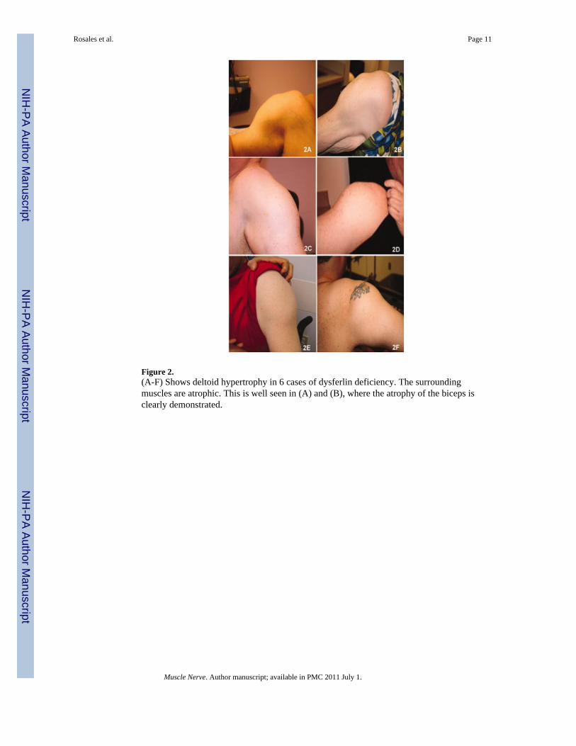
Figure 2.(A-F) Shows deltoid hypertrophy in 6 cases of dysferlin deficiency. The surroundingmuscles are atrophic. This is well seen in (A) and (B), where the atrophy of the biceps isclearly demonstrated.
Rosales et al. Page 11
Muscle Nerve. Author manuscript; available in PMC 2011 July 1.
NIH
-PA Author Manuscript
NIH
-PA Author Manuscript
NIH
-PA Author Manuscript

Figure 3.(A,B) Calf muscle hypertrophy in patients with dysferlin gene mutations. These patients hadbeen previously mislabeled with a diagnosis of Becker muscular dystrophy.
Rosales et al. Page 12
Muscle Nerve. Author manuscript; available in PMC 2011 July 1.
NIH
-PA Author Manuscript
NIH
-PA Author Manuscript
NIH
-PA Author Manuscript

Figure 4.(A) Congo red stain shows amyloid deposited in the smooth muscle layer of a skeletalmuscle arteriole in patient 3. (B) Using polarized light, the green birefringence of amyloid isseen. (C) In a consecutive serial section, dysferlin deposition is seen in this same bloodvessel (NCL-Hamlet Novocastra Laboratories Ltd.). (D) In the same section a blood vesselwall lacks dysferlin deposition; only the autofluorescence of the internal elastic membrane(tunica intima) and adventitia (tunica externa) can be seen, but the smooth muscle of thetunica media is devoid of dysferlin.
Rosales et al. Page 13
Muscle Nerve. Author manuscript; available in PMC 2011 July 1.
NIH
-PA Author Manuscript
NIH
-PA Author Manuscript
NIH
-PA Author Manuscript

Figure 5.(A) Dysferlin staining in the sarcolemma of normal muscle; (B) negative control (normalmuscle without dysferlin antibody stained only with goat anti-mouse IgG); (C) reduceddysferlin membrane staining is seen in patient 2 (Table 2); (D) absent dysferlin membranestaining in patient 8 (Table 2). NCL-Hamlet antibody Novocastra Laboratories Ltd in A,C,D.
Rosales et al. Page 14
Muscle Nerve. Author manuscript; available in PMC 2011 July 1.
NIH
-PA Author Manuscript
NIH
-PA Author Manuscript
NIH
-PA Author Manuscript

Figure 6.Western blot in LGMD2B patients. Total protein extracted from the patient muscle sampleswas probed with dysferlin antibody (upper panel). Lane 1: a normal control; lanes 2-4:LGMD2B patients showing overexpression of dysferlin in patients 3, 8, 10, Table 2); bandsignal intensities were quantified by ImageQuant T2 software (GE Healthcare) and were to 2to 5 fold increased over normal controls; lanes 5-8: LGMD2B patients showing no dysferlinexpression. Protein loading is assessed by probing with muscle specific actin antibody(lower panel).
Rosales et al. Page 15
Muscle Nerve. Author manuscript; available in PMC 2011 July 1.
NIH
-PA Author Manuscript
NIH
-PA Author Manuscript
NIH
-PA Author Manuscript

NIH
-PA Author Manuscript
NIH
-PA Author Manuscript
NIH
-PA Author Manuscript
Rosales et al. Page 16
Table 1
Dysferlin mutations in the 21 patients.
Patient Mutation
1 [c.2643+1G>A]+[c.4577A>C]
2 [c.1481-1G>A]+[c.5836_5839del]
3 [c.3892A>G]
4 [c.3843+2T>A]+[c.5698_5699delAG]
5 [c.5497G>T]+[c.5946+1G>A]
6 [c.5509G>A]+[c.5509G>A]
7 [c.610C>T]+[c.1120G>C]
8 [c.3892A>G]
9 [c.353delT]+[c.5444G>T]
10 [c.3065G>A; c.3992G>T]
11 [c.4685dupT]+[c.4685dupT]
12* [c.1392dupA]+[c.3516_3517delTT]
13* [c.1392dupA]+[c.3516_3517delTT]
14 [c.3041A>G]+[c.5526-1G>A]
15* [c.610C>T]+ [c.5884C>T]
16* [c.610C>T]+ [c.5884C>T]
17 [c.5668-7G>A]
18 [c.1120G>C]+[c.5713C>T]
19 [c.855+1delG]+[c.3505insC]
20 [c.3041A>G]+[c.3041A>G]
21 [c.3478C>T]+[c.3478C>T]
*12, 13 siblings; 15, 16 brothers.
In bold: novel mutations.
Muscle Nerve. Author manuscript; available in PMC 2011 July 1.

NIH
-PA Author Manuscript
NIH
-PA Author Manuscript
NIH
-PA Author Manuscript
Rosales et al. Page 17
Tabl
e 2
His
topa
thol
ogy
feat
ures
.
Imm
unoh
isto
chem
istr
yW
este
rn B
lot
Patie
ntD
ysfe
rlin
Mem
bran
eD
ysfe
rlin
Cyt
opla
smC
aveo
lin-3
Utr
ophi
nD
ysfe
rlin
Cal
pain
-3
1A
bsen
tA
bsen
tN
orm
alN
orm
alN
DN
D
2Am
yloi
dSe
vere
ly re
duce
dA
bsen
tN
orm
alN
orm
alA
bsen
tR
educ
ed
3Am
yloi
dSe
vere
ly re
duce
dPr
esen
tN
orm
alN
orm
alN
orm
al (2
-5 fo
ld in
crea
sed)
Posi
tive
4Se
vere
ly re
duce
dA
bsen
tN
orm
alN
orm
alA
bsen
tR
educ
ed
5Am
yloi
dSe
vere
ly re
duce
dA
bsen
tN
orm
alU
preg
ulat
edA
bsen
tPo
sitiv
e
6Am
yloi
dA
bsen
tA
bsen
tR
educ
edN
orm
alA
bsen
tPo
sitiv
e
7Se
vere
ly re
duce
dA
bsen
tN
orm
alN
orm
alN
DN
D
8Am
yloi
dA
bsen
tA
bsen
tN
orm
alN
orm
alN
orm
al (2
-5 fo
ld in
crea
sed
Posi
tive
9A
bsen
tA
bsen
tN
orm
alN
orm
alR
educ
edPo
sitiv
e
10Se
vere
ly re
duce
dA
bsen
tN
orm
alN
orm
alN
orm
al (2
-5 fo
ld in
crea
sed
Red
uced
11A
bsen
tA
bsen
tN
orm
alN
orm
alA
bsen
tR
educ
ed
12A
bsen
tA
bsen
tN
orm
alN
orm
alA
bsen
tPo
sitiv
e
13A
bsen
tA
bsen
tN
orm
alN
orm
alA
bsen
tPo
sitiv
e
14A
bsen
tA
bsen
tN
orm
alN
orm
alA
bsen
tR
educ
ed
15A
myl
oid
Seve
rely
redu
ced
Abs
ent
Nor
mal
Upr
egul
ated
ND
ND
16N
DN
DN
DN
DN
DN
D
17Se
vere
ly re
duce
dA
bsen
tN
orm
alU
preg
ulat
edA
bsen
tR
educ
ed
18Se
vere
ly re
duce
dA
bsen
tN
orm
alU
preg
ulat
edA
bsen
tR
educ
ed
19N
DN
DN
DN
DN
DN
D
20A
bsen
tA
bsen
tN
orm
alN
orm
alA
bsen
tR
educ
ed
21Se
vere
ly re
duce
dA
bsen
tN
orm
alU
preg
ulat
edA
bsen
tR
educ
ed
Am
yloi
d in
mus
cle
biop
sy; N
D=
not d
one.
Muscle Nerve. Author manuscript; available in PMC 2011 July 1.

NIH
-PA Author Manuscript
NIH
-PA Author Manuscript
NIH
-PA Author Manuscript
Rosales et al. Page 18
Tabl
e 3
Phen
otyp
ic fe
atur
es.
Clin
ical
Dat
a
Patie
ntSe
x/A
geA
ge o
f Ons
etO
nset
mod
eA
ge =
loss
of a
mbu
latio
nC
K
1M
/48
30D
UE+
PLE
Am
bula
tory
3481
2M
/40
17D
LE28
7938
3†M
/46
27PL
E46
9468
4†F/
4734
PLE
Am
bula
tory
4393
5M
/35
18D
LEA
mbu
lato
ryN
D
6M
/38
17D
LEA
mbu
lato
ry17
972
7M
/34
22PL
EA
mbu
lato
ry31
56
8†M
/32
3PL
EA
mbu
lato
ry28
0
9M
/31
19PU
EA
mbu
lato
ry14
650
10†
M/4
66
PLE
4342
8
11F/
2522
DLE
Am
bula
tory
2661
12*
M/4
121
PLE
3569
5
13*
F/42
16D
LE31
320
14M
/26
21D
LEA
mbu
lato
ry11
040
15*
M/6
015
DLE
5417
58
16*
M/5
214
DLE
45N
D
17M
/24
20D
LEA
mbu
lato
ry88
77
18†
F/31
19PL
EA
mbu
lato
ry30
46
19M
/33
20D
LEA
mbu
lato
ry84
77
20†
M/4
224
PLE
Am
bula
tory
4000
21M
/25
12PL
E24
1014
7
* 12, 1
3 si
blin
gs; 1
5, 1
6 br
othe
rs.
† Patie
nts w
ith c
alf h
yper
troph
y; C
K, c
reat
ine
kina
se; D
LE, d
ista
l low
er e
xtre
mity
; DU
E, d
ista
l upp
er e
xtre
mity
; PLE
, pro
xim
al lo
wer
ext
rem
ity; P
UE,
pro
xim
al u
pper
ext
rem
ity; N
D, n
ot d
one.
Muscle Nerve. Author manuscript; available in PMC 2011 July 1.