Identification of Novel Biomarkers With Diagnostic Value and ...
13
Identification of Novel Biomarkers With Diagnostic Value and Immune Infiltration in Burn Injury Sitong Zhou 1 , Kangchun Wang 2 , Jingru Wang 3 , Jia He 3 , Wenlian Zheng 4 , Chengmin Long 4 , Xiaodong Chen 3 * and Ronghua Yang 5 * 1 Department of Dermatology, the First People’s Hospital of Foshan, Foshan, China, 2 Department of Organ Transplantation and Hepatobiliary, the First Affiliated Hospital of China Medical University, Shenyang, China, 3 Department of Burn Surgery and Skin Regeneration, the First People’s Hospital of Foshan, Foshan, China, 4 Graduate School, Guangdong Medical University, Zhanjiang, China, 5 Department of Burn and Plastic Surgery, Guangzhou First People’s Hospital, School of Medicine, South China University of Technology, Guangzhou, China Burn injury is an intractable problem in the field of surgery where screening relevant target genes and exploring pathological mechanisms through bioinformatic methods has become a necessity. Herein, we integrated three burn injury mRNA microarray datasets from the Gene Expression Omnibus database to analyze the hub differentially expressed genes (DEGs) between burn injury patient samples and healthy human samples; we conducted multiple functional enrichment analyses and constructed the protein–protein interaction (PPI) network. Finally, we evaluated the immune infiltration in the burn injury microenvironment. A total of 84 intersection DEGs (32 upregulated and 52 downregulated) were screened in burn injury patients via integrated analyses. Upregulated genes were primarily enriched in regulation of T cell activation, regulation of response to DNA damage stimulus, positive regulation of innate immune response, positive regulation of defense response. We also identi fied 10 hub genes from the PPI network (CCNB2, MYO10, TTK, POLQ, VASP, TIMP1, CDK16, MMP1, ZYX, and PKMYT1). Next, we found that 22 immune cells were substantially changed during the burn injury by CIBERSORT. In addition, we verified that VASP and POLQ are two novel diagnostic markers in burn processes with high diagnostic efficacy via immunohistochemistry. In summary, we identified several key genes involved in burn injury and provided a favorable basis for elucidating the molecular mechanisms of burn injury through comprehensive bioinformatic analysis. Keywords: burn injury, inflammation, immune infiltration, functional enrichment analysis, CIBERSORT INTRODUCTION Burn injury primarily refers to damage to tissues, including skin and mucous membranes, caused by heat. In some severe cases, it can also involve subcutaneous and submucosal tissues and even internal organs. According to statistics from the World Health Organization, approximately 300,000 people die from burn injuries worldwide every year (Roshangar et al., 2019). In particular, the healing of a deep burn injury is a complex process involving multiple factors and an intractable problem in the surgical field (Gerber et al., 2019). Burn injury not only damages the cells, tissues, and blood vessels but also affects the release of various growth factors and cytokines. Severe burns may be accompanied by insufficient blood supply, severe infections, and even sepsis, which will seriously prolong the wound healing time. In addition, a burn injury usually requires long-term treatment and multiple Edited by: Yang Gao, Nankai University, China Reviewed by: Lei Yang, Yale University, United States Guozhi Wu, Lanzhou University Medical College, China *Correspondence: Xiaodong Chen [email protected] Ronghua Yang [email protected] Specialty section: This article was submitted to Computational Genomics, a section of the journal Frontiers in Genetics Received: 06 December 2021 Accepted: 31 January 2022 Published: 22 March 2022 Citation: Zhou S, Wang K, Wang J, He J, Zheng W, Long C, Chen X and Yang R (2022) Identification of Novel Biomarkers With Diagnostic Value and Immune Infiltration in Burn Injury. Front. Genet. 13:829841. doi: 10.3389/fgene.2022.829841 Frontiers in Genetics | www.frontiersin.org March 2022 | Volume 13 | Article 829841 1 ORIGINAL RESEARCH published: 22 March 2022 doi: 10.3389/fgene.2022.829841
-
Upload
khangminh22 -
Category
Documents
-
view
2 -
download
0
Transcript of Identification of Novel Biomarkers With Diagnostic Value and ...
Identification of Novel BiomarkersWith Diagnostic Value and ImmuneInfiltration in Burn InjurySitong Zhou1, Kangchun Wang2, Jingru Wang3, Jia He3, Wenlian Zheng4, Chengmin Long4,Xiaodong Chen3* and Ronghua Yang5*
1Department of Dermatology, the First People’s Hospital of Foshan, Foshan, China, 2Department of Organ Transplantation andHepatobiliary, the First Affiliated Hospital of China Medical University, Shenyang, China, 3Department of Burn Surgery and SkinRegeneration, the First People’s Hospital of Foshan, Foshan, China, 4Graduate School, Guangdong Medical University,Zhanjiang, China, 5Department of Burn and Plastic Surgery, Guangzhou First People’s Hospital, School of Medicine, South ChinaUniversity of Technology, Guangzhou, China
Burn injury is an intractable problem in the field of surgery where screening relevant targetgenes and exploring pathological mechanisms through bioinformatic methods has become anecessity. Herein, we integrated three burn injury mRNA microarray datasets from the GeneExpression Omnibus database to analyze the hub differentially expressed genes (DEGs)between burn injury patient samples and healthy human samples; we conducted multiplefunctional enrichment analyses and constructed the protein–protein interaction (PPI) network.Finally, we evaluated the immune infiltration in the burn injury microenvironment. A total of 84intersection DEGs (32 upregulated and 52 downregulated) were screened in burn injurypatients via integrated analyses. Upregulated genes were primarily enriched in regulation ofT cell activation, regulation of response to DNA damage stimulus, positive regulation of innateimmune response, positive regulation of defense response. We also identified 10 hub genesfrom the PPI network (CCNB2, MYO10, TTK, POLQ, VASP, TIMP1, CDK16, MMP1, ZYX,and PKMYT1). Next, we found that 22 immune cells were substantially changed during theburn injury by CIBERSORT. In addition, we verified that VASP and POLQ are two noveldiagnostic markers in burn processes with high diagnostic efficacy viaimmunohistochemistry. In summary, we identified several key genes involved in burninjury and provided a favorable basis for elucidating the molecular mechanisms of burninjury through comprehensive bioinformatic analysis.
Keywords: burn injury, inflammation, immune infiltration, functional enrichment analysis, CIBERSORT
INTRODUCTION
Burn injury primarily refers to damage to tissues, including skin and mucous membranes, caused byheat. In some severe cases, it can also involve subcutaneous and submucosal tissues and even internalorgans. According to statistics from the World Health Organization, approximately 300,000 peopledie from burn injuries worldwide every year (Roshangar et al., 2019). In particular, the healing of adeep burn injury is a complex process involving multiple factors and an intractable problem in thesurgical field (Gerber et al., 2019). Burn injury not only damages the cells, tissues, and blood vesselsbut also affects the release of various growth factors and cytokines. Severe burns may be accompaniedby insufficient blood supply, severe infections, and even sepsis, which will seriously prolong thewound healing time. In addition, a burn injury usually requires long-term treatment and multiple
Edited by:Yang Gao,
Nankai University, China
Reviewed by:Lei Yang,
Yale University, United StatesGuozhi Wu,
Lanzhou University Medical College,China
*Correspondence:Xiaodong Chen
[email protected] Yang
Specialty section:This article was submitted to
Computational Genomics,a section of the journalFrontiers in Genetics
Received: 06 December 2021Accepted: 31 January 2022Published: 22 March 2022
Citation:Zhou S, Wang K, Wang J, He J,
Zheng W, Long C, Chen X and Yang R(2022) Identification of Novel
Biomarkers With Diagnostic Value andImmune Infiltration in Burn Injury.
Front. Genet. 13:829841.doi: 10.3389/fgene.2022.829841
Frontiers in Genetics | www.frontiersin.org March 2022 | Volume 13 | Article 8298411
ORIGINAL RESEARCHpublished: 22 March 2022
doi: 10.3389/fgene.2022.829841
reconstructive surgical operations, which may cause irreversibledamage to the patient’s mental health. Studies have shown thatburn patients often show a tendency of depression and requirelong-term psychological treatment, which seriously affects theirlife quality (Smolle et al., 2017).
Currently, bioinformatic analysis is an important tool foranalyzing the expression data and screening for target genes inmany diseases. The full use of gene detection technology andbioinformatics will effectively explore the mechanism of variousdiseases, including burn injury. Numerous studies have revealed thatthere are many gene expression changes at different stages of burninjury (Fang et al., 2020). A strong immune and stress responsedisorder is one of the most important features in the early stage ofburn injury, and many studies indicate that the high-mobility groupbox protein 1, a nuclear protein, is increased in burn injury patients,which is due to it passive release from damaged cells (Lantos et al.,2010). In the repair stage, various molecules are involved in scarformation. Transforming growth factor β1 (TGF-β1) can mediatefibrosis of wounds and form hypertrophic scars after burning;Smad7 can negatively regulate the TGF-β1/Smad pathway,thereby preventing fibrosis mediated by TGF-β1 (Zhang et al.,2020). Molecular changes can also be observed in some seriousburn complications. Cystic fibrosis transmembrane conductanceregulator and downstream signaling were critical in modulatingthe gut ischemia and hypoxia post severe burn, which resulted insepsis and multiple organ failure (Liu et al., 2020).
Most burn injury–related studies use single microarrays ormouse microarrays (Gao et al., 2016; Zou et al., 2017). However,key differentially expressed genes (DEGs) and biologicalpathways identified in these burn injury studies present a highfalse-positive rate in a single microarray and a low level ofevidence for mouse microarrays, limiting the accuracy of theresults. Further comprehensive analysis is needed to identify thekey molecular markers and diagnostic targets. This studyintegrates three burn injury mRNA microarray datasets fromthe Gene Expression Omnibus (GEO) database to analyze the hubDEGs between burn injury patients and healthy human samples.We conducted multiple functional enrichment analyses andconstructed a protein–protein interaction (PPI) network;finally, we explored the immune infiltration in the burn injurymicroenvironment and we verified that VASP and POLQ are twonovel diagnostic markers in burn processes with high diagnosticefficacy via immunohistochemistry. Our study aimed to identifyseveral key genes involved in burn injury and provide a favorablebasis for elucidating its underlying molecular mechanisms.
MATERIALS AND METHODS
Data Source and Differential ExpressionAnalysisWe downloaded three microarray datasets of burn injury tissueand normal tissue from the GEO dataset1 for further analysis(GSE8056, GSE19743, GSE37069) (Barrett et al., 2013). Next, we
performed principal component analysis (PCA) and differentialexpression analysis (Ringnér, 2008). We divided the data into twogroups: the burn injury group and the normal group and used theR package “limma” to analyze datasets and screen out DEGs(Ritchie et al., 2015). Thresholds of |log2FC| > 1.0 and an adjustedp-value < 0.05 were selected.
Functional Enrichment AnalysisMetascape2 is a powerful online tool for analyzing gene functionannotation (Zhou et al., 2019) and provides gene enrichmentanalysis and PPI network analyses. This tool was used to analyzethe gene ontology (GO) and the Kyoto Encyclopedia of Genesand Genomes (KEGG) pathway enrichment analysis. In addition,a series of analyses were performed in the DisGeNET database(Piñero et al., 2017), Trrust database (Han et al., 2018), andPaGenbase database (Pan et al., 2013).
Construction of PPI NetworkThe STRING database3 is a practical online tool that can be usedto construct PPI networks (Szklarczyk et al., 2015). Since theupregulated DEGs were highly enriched in functions andpathways that are closely related to burn injury and immuneresponse, all the upregulated DEGs were included in the databasefor analysis, and the interaction threshold was set to 0.4. TheCytoscape software was used for visualization (Doncheva et al.,2019), and CytoHubba was used to analyze hub genes in thenetwork (Chin et al., 2014). In the PPI network, we screened 10hub genes, and correlation was shown by different shades ofcolors.
Gene Set Enrichment Analysis (GSEA)To identify the signal transduction pathways that weredifferentially activated between the burn injury group and thenormal group, we selected an ordered list of genes through the“limma” R package and then performed a gene set enrichmentanalysis.
Expression Level and Receiver OperatingCharacteristic (ROC) Curves of Top 10 HubGenes.We analyzed the expression levels of 10 hub genes in burn injuryand normal tissues in the GSE37069 dataset and constructed acolumnar scatter plot. In addition, using the package “pROC” toperform the ROC analysis (Robin et al., 2011), the specificity andsensitivity of each gene were obtained, and the area under thecurve (AUC) of each hub gene was calculated.
CIBERSORTCIBERSORT4 can enumerate cell type composition in geneexpression data through deconvolution algorithm, and it hasbeen used to evaluate immune cell infiltration in many
1http://www.ncbi.nlm.nih.gov/geo/.
2http://metascape.org.3http://string-db.org.4http://cibersort.stanford.edu/.
Frontiers in Genetics | www.frontiersin.org March 2022 | Volume 13 | Article 8298412
Zhou et al. Novel Biomarkers in Burn Injury
diseases, estimated abundances of immunocytes had beenassessed by 22 given kinds of immunocytes accompanyingwith 1,000 permutations. Then, the immune cell matrix wasvisualized by the R package of “ggplot2”. Finally, weconstructed the correlation heatmap for visualizing thecorrelation of infiltrating immune cells by “corrplot” package.
Patient Tissue SpecimensFifteen clinical samples were obtained from the Chinese Hanpopulation from 2020 to 2021. This study was approved by theEthics Committee of Foshan First People’s Hospital (AF-SOP-20-1.4-0). All subjects provided written informed consent, inaccordance with the Declaration of Helsinki.
Immunohistochemistry (IHC) Staining andHistologic ScoringParaffin-embedded tissues were sectioned at 4 μm for IHCanalysis. Antigen retrieval was performed by incubating thesamples in citrate buffer (pH 6.0) for 15 min. After blocking
with a mixture of methanol and 0.75% hydrogen peroxide,sections were incubated overnight with primary antibody(VASP, Signalway Antibody, 1:100; POLQ, SignalwayAntibody, 1:50), followed by incubation with a secondaryantibody conjugated with horseradish peroxidase (goat anti-rabbit, 1:500, Cell Signaling Technology). Sections werewashed three times with phosphate-buffered saline andincubated with diaminobenzidine. The process of histologicscoring and analysis was as described in our previous study(Zhou et al., 2021).
RESULTS
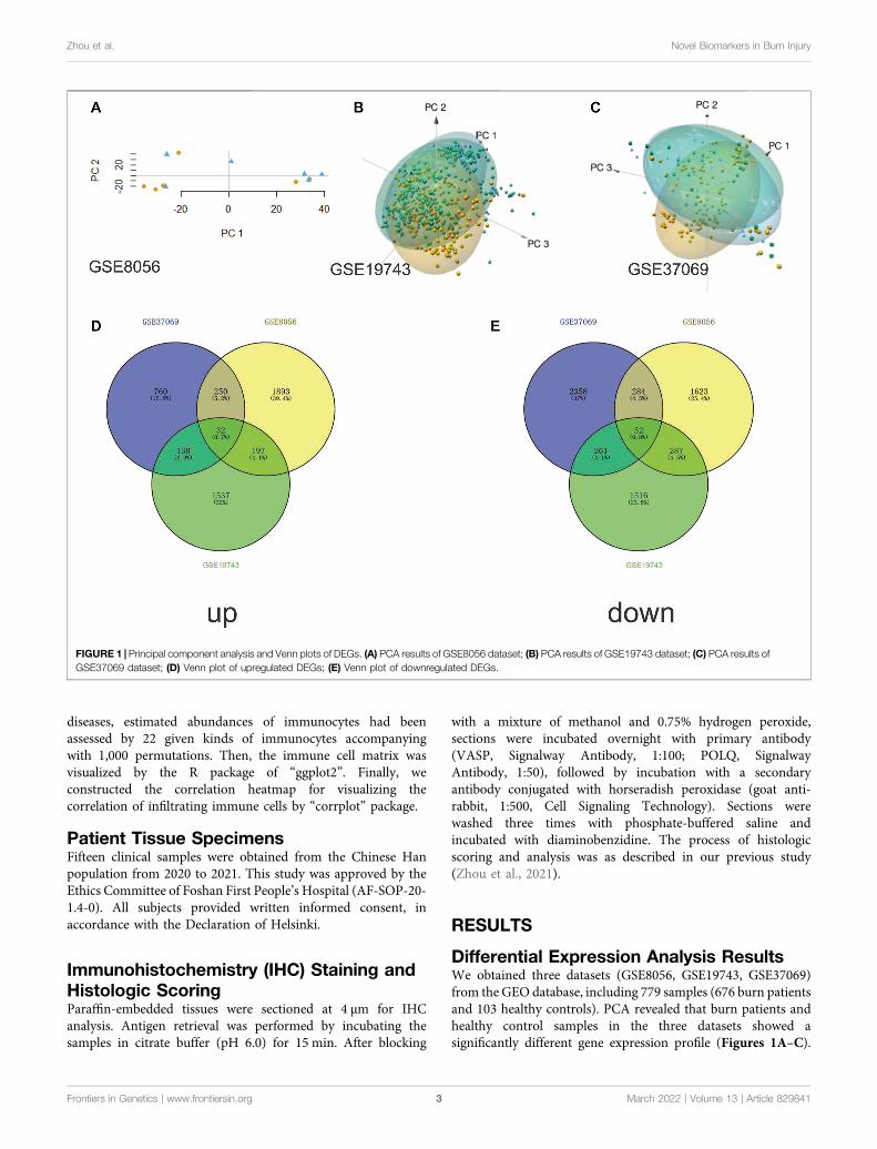
Differential Expression Analysis ResultsWe obtained three datasets (GSE8056, GSE19743, GSE37069)from the GEO database, including 779 samples (676 burn patientsand 103 healthy controls). PCA revealed that burn patients andhealthy control samples in the three datasets showed asignificantly different gene expression profile (Figures 1A–C).
FIGURE 1 | Principal component analysis and Venn plots of DEGs. (A) PCA results of GSE8056 dataset; (B) PCA results of GSE19743 dataset; (C) PCA results ofGSE37069 dataset; (D) Venn plot of upregulated DEGs; (E) Venn plot of downregulated DEGs.
Frontiers in Genetics | www.frontiersin.org March 2022 | Volume 13 | Article 8298413
Zhou et al. Novel Biomarkers in Burn Injury

FIGURE 2 | Differential expression analysis in three datasets. (A) The gene expression heatmap of important DEGs in the GSE8056 dataset; (B) the volcano plot ofthe differential expression analysis in the GSE8056 dataset; (C) the gene expression heatmap of important DEGs in the GSE19743 dataset; (D) volcano plot of differentialexpression analysis in GSE19743 dataset; (E) the gene expression heatmap of important DEGs in the GSE37069 dataset; (F) volcano plot of differential expressionanalysis in GSE37069 dataset.
Frontiers in Genetics | www.frontiersin.org March 2022 | Volume 13 | Article 8298414
Zhou et al. Novel Biomarkers in Burn Injury
Next, we separately conducted a differential expression analysis ofthe three datasets and analyzed their intersection. Finally, 32upregulated and 52 downregulated DEGs were obtained (Figures1D,E). In addition, volcano maps of DEGs and heatmaps ofimportant DEGs are displayed in Figure 2.
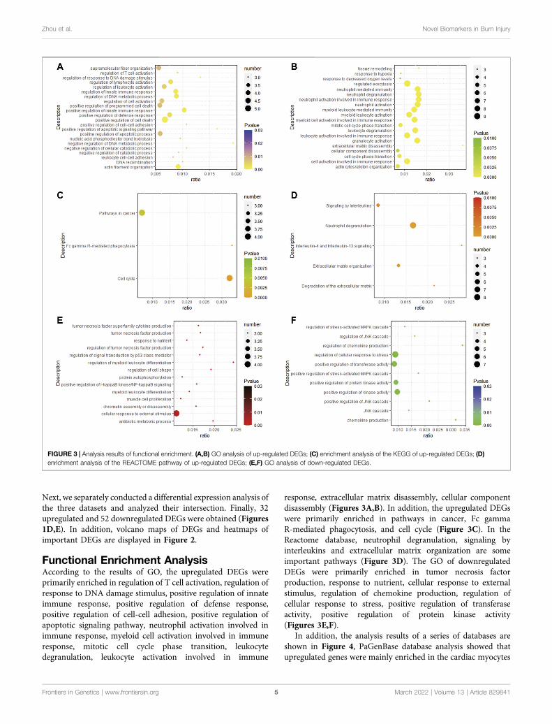
Functional Enrichment AnalysisAccording to the results of GO, the upregulated DEGs wereprimarily enriched in regulation of T cell activation, regulation ofresponse to DNA damage stimulus, positive regulation of innateimmune response, positive regulation of defense response,positive regulation of cell-cell adhesion, positive regulation ofapoptotic signaling pathway, neutrophil activation involved inimmune response, myeloid cell activation involved in immuneresponse, mitotic cell cycle phase transition, leukocytedegranulation, leukocyte activation involved in immune
response, extracellular matrix disassembly, cellular componentdisassembly (Figures 3A,B). In addition, the upregulated DEGswere primarily enriched in pathways in cancer, Fc gammaR-mediated phagocytosis, and cell cycle (Figure 3C). In theReactome database, neutrophil degranulation, signaling byinterleukins and extracellular matrix organization are someimportant pathways (Figure 3D). The GO of downregulatedDEGs were primarily enriched in tumor necrosis factorproduction, response to nutrient, cellular response to externalstimulus, regulation of chemokine production, regulation ofcellular response to stress, positive regulation of transferaseactivity, positive regulation of protein kinase activity(Figures 3E,F).
In addition, the analysis results of a series of databases areshown in Figure 4, PaGenBase database analysis showed thatupregulated genes were mainly enriched in the cardiac myocytes
FIGURE 3 | Analysis results of functional enrichment. (A,B) GO analysis of up-regulated DEGs; (C) enrichment analysis of the KEGG of up-regulated DEGs; (D)enrichment analysis of the REACTOME pathway of up-regulated DEGs; (E,F) GO analysis of down-regulated DEGs.
Frontiers in Genetics | www.frontiersin.org March 2022 | Volume 13 | Article 8298415
Zhou et al. Novel Biomarkers in Burn Injury
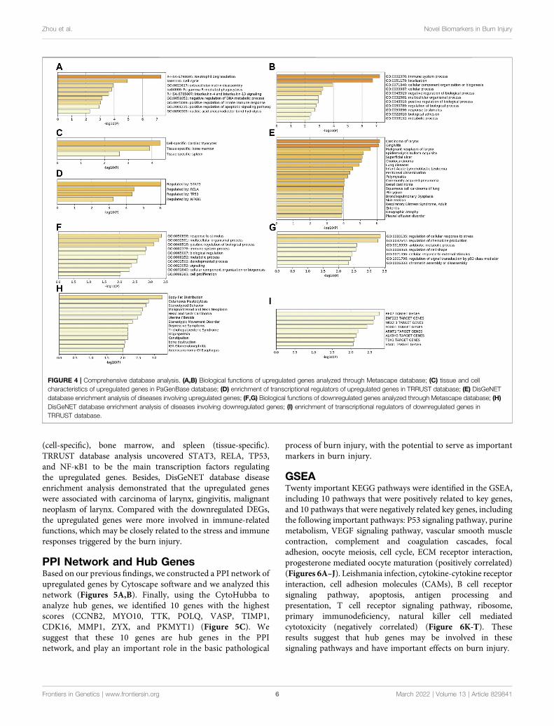
(cell-specific), bone marrow, and spleen (tissue-specific).TRRUST database analysis uncovered STAT3, RELA, TP53,and NF-κB1 to be the main transcription factors regulatingthe upregulated genes. Besides, DisGeNET database diseaseenrichment analysis demonstrated that the upregulated geneswere associated with carcinoma of larynx, gingivitis, malignantneoplasm of larynx. Compared with the downregulated DEGs,the upregulated genes were more involved in immune-relatedfunctions, which may be closely related to the stress and immuneresponses triggered by the burn injury.
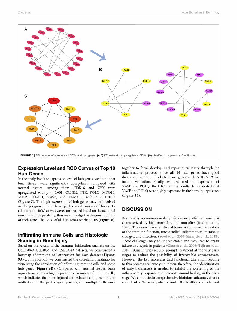
PPI Network and Hub GenesBased on our previous findings, we constructed a PPI network ofupregulated genes by Cytoscape software and we analyzed thisnetwork (Figures 5A,B). Finally, using the CytoHubba toanalyze hub genes, we identified 10 genes with the highestscores (CCNB2, MYO10, TTK, POLQ, VASP, TIMP1,CDK16, MMP1, ZYX, and PKMYT1) (Figure 5C). Wesuggest that these 10 genes are hub genes in the PPInetwork, and play an important role in the basic pathological
process of burn injury, with the potential to serve as importantmarkers in burn injury.
GSEATwenty important KEGG pathways were identified in the GSEA,including 10 pathways that were positively related to key genes,and 10 pathways that were negatively related key genes, includingthe following important pathways: P53 signaling pathway, purinemetabolism, VEGF signaling pathway, vascular smooth musclecontraction, complement and coagulation cascades, focaladhesion, oocyte meiosis, cell cycle, ECM receptor interaction,progesterone mediated oocyte maturation (positively correlated)(Figures 6A–J). Leishmania infection, cytokine-cytokine receptorinteraction, cell adhesion molecules (CAMs), B cell receptorsignaling pathway, apoptosis, antigen processing andpresentation, T cell receptor signaling pathway, ribosome,primary immunodeficiency, natural killer cell mediatedcytotoxicity (negatively correlated) (Figure 6K-T). Theseresults suggest that hub genes may be involved in thesesignaling pathways and have important effects on burn injury.
FIGURE 4 | Comprehensive database analysis. (A,B) Biological functions of upregulated genes analyzed through Metascape database; (C) tissue and cellcharacteristics of upregulated genes in PaGenBase database; (D) enrichment of transcriptional regulators of upregulated genes in TRRUST database; (E) DisGeNETdatabase enrichment analysis of diseases involving upregulated genes; (F,G) Biological functions of downregulated genes analyzed through Metascape database; (H)DisGeNET database enrichment analysis of diseases involving downregulated genes; (I) enrichment of transcriptional regulators of downregulated genes inTRRUST database.
Frontiers in Genetics | www.frontiersin.org March 2022 | Volume 13 | Article 8298416
Zhou et al. Novel Biomarkers in Burn Injury
Expression Level and ROC Curves of Top 10Hub GenesIn the analysis of the expression level of hub genes, we found thatburn tissues were significantly upregulated compared withnormal tissues. Among them, CDK16 and ZYX wereupregulated with p < 0.001, CCNB2, TTK, POLQ, MYO10,MMP1, TIMP1, VASP, and PKMYT1 with p < 0.0001(Figure 7). The high expression of hub genes may be involvedin the progression and basic pathological process of burns. Inaddition, the ROC curves were constructed based on the acquiredsensitivity and specificity, thus we can judge the diagnostic abilityof each gene. The AUC of all hub genes reached 0.68 (Figure 8).
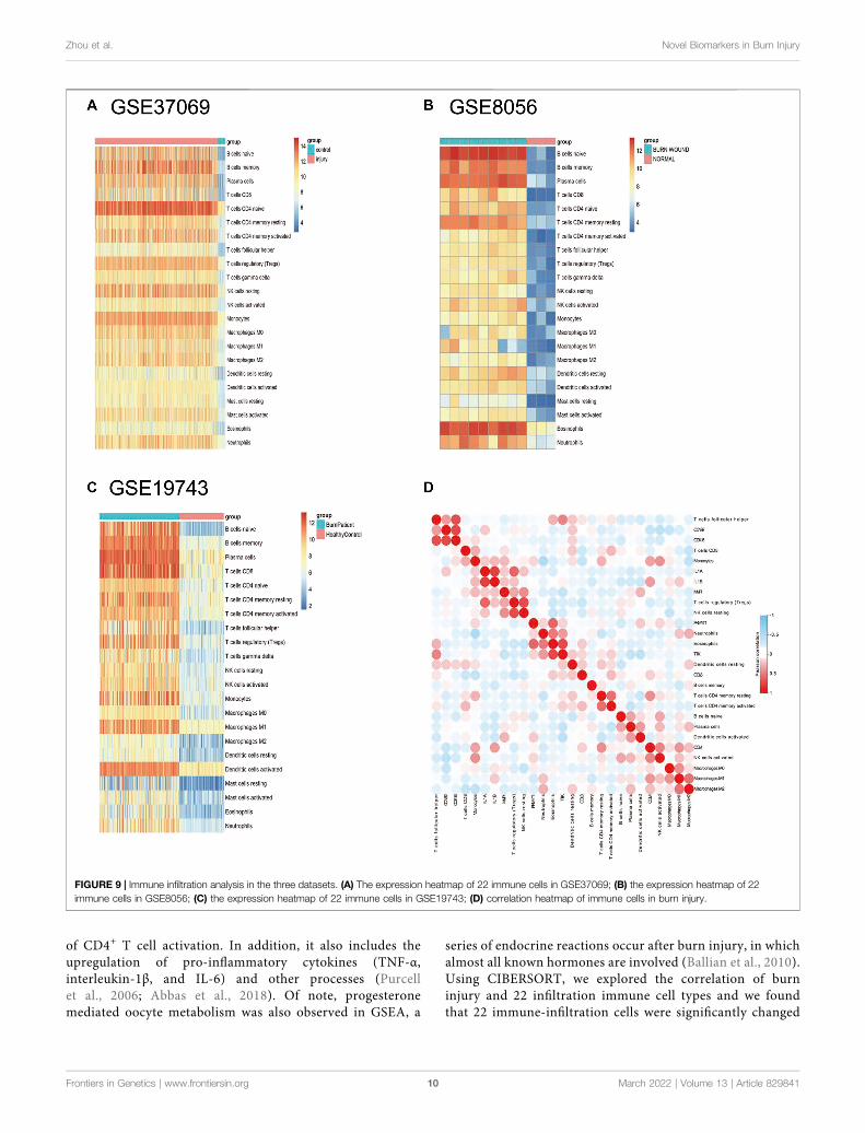
Infiltrating Immune Cells and HistologicScoring in Burn InjuryBased on the results of the immune infiltration analysis on theGSE37069, GSE8056, and GSE19743 datasets, we constructed aheatmap of immune cell expression for each dataset (Figures9A–C). In addition, we constructed the correlation heatmap forvisualizing the correlation of infiltrating immune cells and somehub genes (Figure 9D). Compared with normal tissues, burninjury tissues have a high expression of a variety of immune cells,which indicates that burn-injured tissues have a complex immuneinfiltration in the pathological process, and multiple cells work
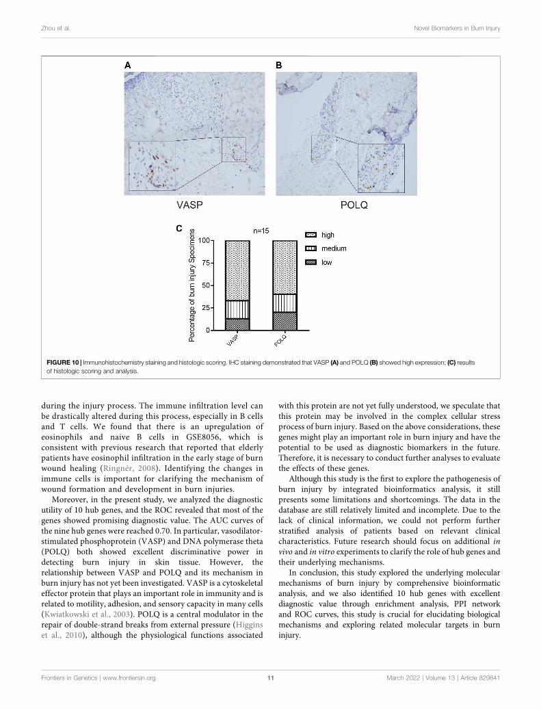
together to form, develop, and repair burn injury through theinflammatory process. Since all 10 hub genes have gooddiagnostic values, we selected two genes with AUC >0.9 forfurther validation. Finally, we evaluated the expression ofVASP and POLQ, the IHC staining results demonstrated thatVASP and POLQ were highly expressed in the burn injury tissues(Figure 10).
DISCUSSION
Burn injury is common in daily life and may affect anyone, it ischaracterized by high morbidity and mortality (Jeschke et al.,2020). The main characteristics of burns are abnormal activationof the immune function, uncontrolled inflammation, metabolicchanges, and infections (Sood et al., 2016; Stanojcic et al., 2018).These challenges may be unpredictable and may lead to organfailure and sepsis in patients (Church et al., 2006; Tejiram et al.,2019). Burn injuries require prompt treatment at the very earlystages to reduce the possibility of irreversible consequences.However, the key molecules and functional alterations leadingto this process are largely unknown; therefore, the identificationof early biomarkers is needed to inhibit the worsening of theinflammatory response and promote wound healing in the earlystage. We conducted a comprehensive bioinformatic analysis on acohort of 676 burn patients and 103 healthy controls and
FIGURE 5 | PPI network of upregulated DEGs and hub genes. (A,B) PPI network of up-regulation DEGs; (C) identified hub genes by CytoHubba.
Frontiers in Genetics | www.frontiersin.org March 2022 | Volume 13 | Article 8298417
Zhou et al. Novel Biomarkers in Burn Injury
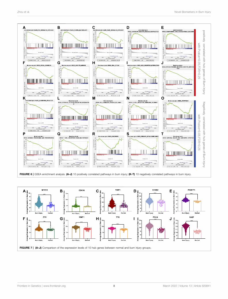
FIGURE 6 | GSEA enrichment analysis. (A-J) 10 positively correlated pathways in burn injury; (K-T) 10 negatively correlated pathways in burn injury.
FIGURE 7 | (A–J) Comparison of the expression levels of 10 hub genes between normal and burn injury groups.
Frontiers in Genetics | www.frontiersin.org March 2022 | Volume 13 | Article 8298418
Zhou et al. Novel Biomarkers in Burn Injury
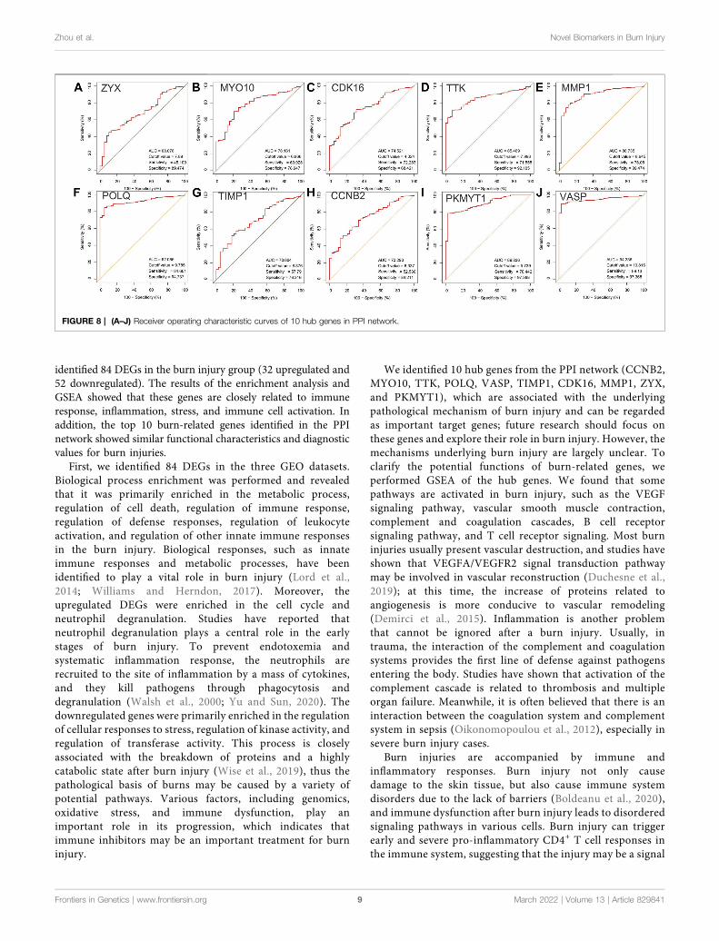
identified 84 DEGs in the burn injury group (32 upregulated and52 downregulated). The results of the enrichment analysis andGSEA showed that these genes are closely related to immuneresponse, inflammation, stress, and immune cell activation. Inaddition, the top 10 burn-related genes identified in the PPInetwork showed similar functional characteristics and diagnosticvalues for burn injuries.
First, we identified 84 DEGs in the three GEO datasets.Biological process enrichment was performed and revealedthat it was primarily enriched in the metabolic process,regulation of cell death, regulation of immune response,regulation of defense responses, regulation of leukocyteactivation, and regulation of other innate immune responsesin the burn injury. Biological responses, such as innateimmune responses and metabolic processes, have beenidentified to play a vital role in burn injury (Lord et al.,2014; Williams and Herndon, 2017). Moreover, theupregulated DEGs were enriched in the cell cycle andneutrophil degranulation. Studies have reported thatneutrophil degranulation plays a central role in the earlystages of burn injury. To prevent endotoxemia andsystematic inflammation response, the neutrophils arerecruited to the site of inflammation by a mass of cytokines,and they kill pathogens through phagocytosis anddegranulation (Walsh et al., 2000; Yu and Sun, 2020). Thedownregulated genes were primarily enriched in the regulationof cellular responses to stress, regulation of kinase activity, andregulation of transferase activity. This process is closelyassociated with the breakdown of proteins and a highlycatabolic state after burn injury (Wise et al., 2019), thus thepathological basis of burns may be caused by a variety ofpotential pathways. Various factors, including genomics,oxidative stress, and immune dysfunction, play animportant role in its progression, which indicates thatimmune inhibitors may be an important treatment for burninjury.
We identified 10 hub genes from the PPI network (CCNB2,MYO10, TTK, POLQ, VASP, TIMP1, CDK16, MMP1, ZYX,and PKMYT1), which are associated with the underlyingpathological mechanism of burn injury and can be regardedas important target genes; future research should focus onthese genes and explore their role in burn injury. However, themechanisms underlying burn injury are largely unclear. Toclarify the potential functions of burn-related genes, weperformed GSEA of the hub genes. We found that somepathways are activated in burn injury, such as the VEGFsignaling pathway, vascular smooth muscle contraction,complement and coagulation cascades, B cell receptorsignaling pathway, and T cell receptor signaling. Most burninjuries usually present vascular destruction, and studies haveshown that VEGFA/VEGFR2 signal transduction pathwaymay be involved in vascular reconstruction (Duchesne et al.,2019); at this time, the increase of proteins related toangiogenesis is more conducive to vascular remodeling(Demirci et al., 2015). Inflammation is another problemthat cannot be ignored after a burn injury. Usually, intrauma, the interaction of the complement and coagulationsystems provides the first line of defense against pathogensentering the body. Studies have shown that activation of thecomplement cascade is related to thrombosis and multipleorgan failure. Meanwhile, it is often believed that there is aninteraction between the coagulation system and complementsystem in sepsis (Oikonomopoulou et al., 2012), especially insevere burn injury cases.
Burn injuries are accompanied by immune andinflammatory responses. Burn injury not only causedamage to the skin tissue, but also cause immune systemdisorders due to the lack of barriers (Boldeanu et al., 2020),and immune dysfunction after burn injury leads to disorderedsignaling pathways in various cells. Burn injury can triggerearly and severe pro-inflammatory CD4+ T cell responses inthe immune system, suggesting that the injury may be a signal
FIGURE 8 | (A–J) Receiver operating characteristic curves of 10 hub genes in PPI network.
Frontiers in Genetics | www.frontiersin.org March 2022 | Volume 13 | Article 8298419
Zhou et al. Novel Biomarkers in Burn Injury
of CD4+ T cell activation. In addition, it also includes theupregulation of pro-inflammatory cytokines (TNF-α,interleukin-1β, and IL-6) and other processes (Purcellet al., 2006; Abbas et al., 2018). Of note, progesteronemediated oocyte metabolism was also observed in GSEA, a
series of endocrine reactions occur after burn injury, in whichalmost all known hormones are involved (Ballian et al., 2010).Using CIBERSORT, we explored the correlation of burninjury and 22 infiltration immune cell types and we foundthat 22 immune-infiltration cells were significantly changed
FIGURE 9 | Immune infiltration analysis in the three datasets. (A) The expression heatmap of 22 immune cells in GSE37069; (B) the expression heatmap of 22immune cells in GSE8056; (C) the expression heatmap of 22 immune cells in GSE19743; (D) correlation heatmap of immune cells in burn injury.
Frontiers in Genetics | www.frontiersin.org March 2022 | Volume 13 | Article 82984110
Zhou et al. Novel Biomarkers in Burn Injury
during the injury process. The immune infiltration level canbe drastically altered during this process, especially in B cellsand T cells. We found that there is an upregulation ofeosinophils and naive B cells in GSE8056, which isconsistent with previous research that reported that elderlypatients have eosinophil infiltration in the early stage of burnwound healing (Ringnér, 2008). Identifying the changes inimmune cells is important for clarifying the mechanism ofwound formation and development in burn injuries.
Moreover, in the present study, we analyzed the diagnosticutility of 10 hub genes, and the ROC revealed that most of thegenes showed promising diagnostic value. The AUC curves ofthe nine hub genes were reached 0.70. In particular, vasodilator-stimulated phosphoprotein (VASP) and DNA polymerase theta(POLQ) both showed excellent discriminative power indetecting burn injury in skin tissue. However, therelationship between VASP and POLQ and its mechanism inburn injury has not yet been investigated. VASP is a cytoskeletaleffector protein that plays an important role in immunity and isrelated to motility, adhesion, and sensory capacity in many cells(Kwiatkowski et al., 2003). POLQ is a central modulator in therepair of double-strand breaks from external pressure (Higginset al., 2010), although the physiological functions associated
with this protein are not yet fully understood, we speculate thatthis protein may be involved in the complex cellular stressprocess of burn injury. Based on the above considerations, thesegenes might play an important role in burn injury and have thepotential to be used as diagnostic biomarkers in the future.Therefore, it is necessary to conduct further analyses to evaluatethe effects of these genes.
Although this study is the first to explore the pathogenesis ofburn injury by integrated bioinformatics analysis, it stillpresents some limitations and shortcomings. The data in thedatabase are still relatively limited and incomplete. Due to thelack of clinical information, we could not perform furtherstratified analysis of patients based on relevant clinicalcharacteristics. Future research should focus on additional invivo and in vitro experiments to clarify the role of hub genes andtheir underlying mechanisms.
In conclusion, this study explored the underlying molecularmechanisms of burn injury by comprehensive bioinformaticanalysis, and we also identified 10 hub genes with excellentdiagnostic value through enrichment analysis, PPI networkand ROC curves, this study is crucial for elucidating biologicalmechanisms and exploring related molecular targets in burninjury.
FIGURE 10 | Immunohistochemistry staining and histologic scoring. IHC staining demonstrated that VASP (A) and POLQ (B) showed high expression; (C) resultsof histologic scoring and analysis.
Frontiers in Genetics | www.frontiersin.org March 2022 | Volume 13 | Article 82984111
Zhou et al. Novel Biomarkers in Burn Injury
DATA AVAILABILITY STATEMENT
This study analyzed three publicly available datasets, whichcan be found in the Gene Expression Omnibus (GEO)database.This data can be found here: GSE8056, GSE19743,and GSE37069.
ETHICS STATEMENT
The studies involving human participants were reviewed andapproved by Ethics Committee of Foshan First People’s Hospital(AF-SOP-20-1.4-0). The patients/participants provided theirwritten informed consent to participate in this study.
AUTHOR CONTRIBUTIONS
SZ and XC conceived and designed the study. RY and KWdesigned and carried out bioinformatics and statistical analyses,visualization, and drafted the manuscript. JW, JH, WZ, and CLparticipated in editing the manuscript. All authors contributed tothe article and approved the submitted version.
FUNDING
This study was supported by the National Natural ScienceFoundation of China (No.82002913) and Guangdong Basicand Applied Basic Research Foundation (2021B1515120036).
REFERENCES
Abbas, O. L., Özatik, O., Gönen, Z. B., Öğüt, S., Entok, E., Özatik, F. Y., et al. (2018).Prevention of Burn Wound Progression by Mesenchymal Stem CellTransplantation. Ann. Plast. Surg. 81 (6), 715–724. doi:10.1097/sap.0000000000001620
Ballian, N., Rabiee, A., Andersen, D. K., Elahi, D., and Gibson, B. R. (2010). GlucoseMetabolism in Burn Patients: the Role of Insulin and Other EndocrineHormones. Burns 36 (5), 599–605. doi:10.1016/j.burns.2009.11.008
Barrett, T., Wilhite, S. E., Ledoux, P., Evangelista, C., Kim, I. F., Tomashevsky, M.,et al. (2013). NCBI GEO: Archive for Functional Genomics Data Sets-Update.Nucleic Acids Res. 41, D991–D995. (Database issue). doi:10.1093/nar/gks1193
Boldeanu, L., Boldeanu, M., Bogdan, M., Meca, A., Coman, C., Buca, B., et al.(2020). Immunological Approaches and Therapy in burns (Review). Exp. Ther.Med. 20 (3), 2361–2367. doi:10.3892/etm.2020.8932
Chin, C.-H., Chen, S.-H., Wu, H.-H., Ho, C.-W., Ko, M.-T., and Lin, C.-Y. (2014).cytoHubba: Identifying Hub Objects and Sub-networks from ComplexInteractome. BMC Syst. Biol. 8 (Suppl. 4), S11. doi:10.1186/1752-0509-8-s4-s11
Church, D., Elsayed, S., Reid, O., Winston, B., and Lindsay, R. (2006). BurnWoundInfections. Clin. Microbiol. Rev. 19 (2), 403–434. doi:10.1128/cmr.19.2.403-434.2006
Demirci, S., Doğan, A., Karakuş, E., Halıcı, Z., Topçu, A., Demirci, E., et al. (2015).Boron and Poloxamer (F68 and F127) Containing Hydrogel Formulation forBurn Wound Healing. Biol. Trace Elem. Res. 168 (1), 169–180. doi:10.1007/s12011-015-0338-z
Doncheva, N. T., Morris, J. H., Gorodkin, J., and Jensen, L. J. (2019). CytoscapeStringApp: Network Analysis and Visualization of Proteomics Data.J. Proteome Res. 18 (2), 623–632. doi:10.1021/acs.jproteome.8b00702
Duchesne, C., Banzet, S., Lataillade, J. J., Rousseau, A., and Frescaline, N. (2019).Cold Atmospheric Plasma Modulates Endothelial Nitric Oxide SynthaseSignalling and Enhances Burn Wound Neovascularisation. J. Pathol. 249 (3),368–380. doi:10.1002/path.5323
Fang, X., Duan, S.-F., Gong, Y.-Z., Wang, F., and Chen, X.-L. (2020). Identificationof Key Genes Associated with Changes in the Host Response to Severe BurnShock: A Bioinformatics Analysis with Data from the Gene ExpressionOmnibus (GEO) Database. J. Inflamm. Res. 13, 1029–1041. doi:10.2147/jir.S282722
Gao, Y., Nai, W., Yang, L., Lu, Z., Shi, P., Jin, H., et al. (2016). Construction of anImmunorelated Protein-Protein Interaction Network for Clarifying theMechanism of Burn. Burns 42 (2), 405–413. doi:10.1016/j.burns.2015.06.015
Gerber, L. H., Bush, H., Holavanahalli, R., Esselman, P., Schneider, J., Heinemann,A., et al. (2019). A Scoping Review of Burn Rehabilitation PublicationsIncorporating Functional Outcomes. Burns 45 (5), 1005–1013. doi:10.1016/j.burns.2018.09.029
Han, H., Cho, J.-W., Lee, S., Yun, A., Kim, H., Bae, D., et al. (2018). TRRUST V2: anExpanded Reference Database of Human and Mouse TranscriptionalRegulatory Interactions. Nucleic Acids Res. 46 (D1), D380–d386. doi:10.1093/nar/gkx1013
Higgins, G. S., Prevo, R., Lee, Y.-F., Helleday, T., Muschel, R. J., Taylor, S., et al.(2010). A Small Interfering RNA Screen of Genes Involved in DNA RepairIdentifies Tumor-specific Radiosensitization by POLQ Knockdown. CancerRes. 70 (7), 2984–2993. doi:10.1158/0008-5472.Can-09-4040
Jeschke, M. G., van Baar, M. E., Choudhry, M. A., Chung, K. K., Gibran, N. S., andLogsetty, S. (2020). Burn Injury. Nat. Rev. Dis. Primers 6 (1), 11. doi:10.1038/s41572-020-0145-5
Kwiatkowski, A. V., Gertler, F. B., and Loureiro, J. J. (2003). Function andRegulation of Ena/VASP Proteins. Trends Cel Biol. 13 (7), 386–392. doi:10.1016/s0962-8924(03)00130-2
Lantos, J., Földi, V., Rőth, E., Wéber, G., Bogár, L., and Csontos, C. (2010). BurnTrauma Induces Early Hmgb1 Release in Patients. Shock 33 (6), 562–567.doi:10.1097/SHK.0b013e3181cd8c88
Liu, X., Chen, Y., You, B., Peng, Y., Chen, Y., Yang, Z., et al. (2021). MolecularMechanism Mediating Enteric Bacterial Translocation after Severe Burn: theRole of Cystic Fibrosis Transmembrane Conductance Regulator. Burns Trauma9, tkaa042. doi:10.1093/burnst/tkaa042
Lord, J. M., Midwinter, M. J., Chen, Y.-F., Belli, A., Brohi, K., Kovacs, E. J., et al.(2014). The Systemic Immune Response to Trauma: an Overview ofPathophysiology and Treatment. The Lancet 384 (9952), 1455–1465. doi:10.1016/s0140-6736(14)60687-5
Oikonomopoulou, K., Ricklin, D., Ward, P. A., and Lambris, J. D. (2012).Interactions between Coagulation and Complement-Their Role inInflammation. Semin. Immunopathol 34 (1), 151–165. doi:10.1007/s00281-011-0280-x
Pan, J.-B., Hu, S.-C., Shi, D., Cai, M.-C., Li, Y.-B., Zou, Q., et al. (2013). PaGenBase:a Pattern Gene Database for the Global and Dynamic Understanding of GeneFunction. PLoS One 8 (12), e80747. doi:10.1371/journal.pone.0080747
Piñero, J., Bravo, À., Queralt-Rosinach, N., Gutiérrez-Sacristán, A., Deu-Pons, J.,Centeno, E., et al. (2017). DisGeNET: a Comprehensive Platform IntegratingInformation on Human Disease-Associated Genes and Variants. Nucleic AcidsRes. 45 (D1), D833–d839. doi:10.1093/nar/gkw943
Purcell, E. M., Dolan, S. M., Kriynovich, S., Mannick, J. A., and Lederer, J. A.(2006). Burn Injury Induces an Early Activation Response by Lymph NodeCD4+ T Cells. Shock 25 (2), 135–140. doi:10.1097/01.shk.0000190824.51653.32
Ringnér, M. (2008). What Is Principal Component Analysis?. Nat. Biotechnol. 26(3), 303–304. doi:10.1038/nbt0308-303
Ritchie, M. E., Phipson, B.,Wu, D., Hu, Y., Law, C.W., Shi, W., et al. (2015). Limmapowers Differential Expression Analyses for RNA-Sequencing and MicroarrayStudies. Nucleic Acids Res. 43 (7), e47. doi:10.1093/nar/gkv007
Robin, X., Turck, N., Hainard, A., Tiberti, N., Lisacek, F., Sanchez, J.-C., et al.(2011). pROC: an Open-Source Package for R and S+ to Analyze and CompareROC Curves. BMC Bioinformatics 12, 77. doi:10.1186/1471-2105-12-77
Roshangar, L., Soleimani Rad, J., Kheirjou, R., Reza Ranjkesh, M., and FerdowsiKhosroshahi, A. (2019). Skin Burns: Review of Molecular Mechanisms andTherapeutic Approaches. Wounds 31 (12), 308–315.
Smolle, C., Cambiaso-Daniel, J., Forbes, A. A., Wurzer, P., Hundeshagen, G.,Branski, L. K., et al. (2017). Recent Trends in Burn EpidemiologyWorldwide: ASystematic Review. Burns 43 (2), 249–257. doi:10.1016/j.burns.2016.08.013
Frontiers in Genetics | www.frontiersin.org March 2022 | Volume 13 | Article 82984112
Zhou et al. Novel Biomarkers in Burn Injury
Sood, R. F., Gibran, N. S., Arnoldo, B. D., Gamelli, R. L., Herndon, D. N., andTompkins, R. G. (2016). Early Leukocyte Gene Expression Associated with Age,Burn Size, and Inhalation Injury in Severely Burned Adults. J. Trauma AcuteCare Surg. 80 (2), 250–257. doi:10.1097/ta.0000000000000905
Stanojcic, M., Abdullahi, A., Rehou, S., Parousis, A., and Jeschke, M. G. (2018).Pathophysiological Response to Burn Injury in Adults. Ann. Surg. 267 (3),576–584. doi:10.1097/sla.0000000000002097
Szklarczyk, D., Franceschini, A., Wyder, S., Forslund, K., Heller, D., Huerta-Cepas,J., et al. (2015). STRING V10: Protein-Protein Interaction Networks, Integratedover the Tree of Life.Nucleic Acids Res. 43 (Database issue), D447–D452. doi:10.1093/nar/gku1003
Tejiram, S., Romanowski, K. S., and Palmieri, T. L. (2019). Initial Management ofSevere Burn Injury. Curr. Opin. Crit. Care 25 (6), 647–652. doi:10.1097/mcc.0000000000000662
Walsh, N. P., Blannin, A. K., Bishop, N. C., Robson, P. J., and Gleeson, M. (2000).Effect of Oral Glutamine Supplementation on Human NeutrophilLipopolysaccharide-Stimulated Degranulation Following Prolonged Exercise.Int. J. Sport Nutr. Exerc. Metab. 10 (1), 39–50. doi:10.1123/ijsnem.10.1.39
Williams, F. N., and Herndon, D. N. (2017). Metabolic and EndocrineConsiderations after Burn Injury. Clin. Plast. Surg. 44 (3), 541–553. doi:10.1016/j.cps.2017.02.013
Wise, A. K., Hromatka, K. A., and Miller, K. R. (2019). Energy Expenditure andProtein Requirements Following Burn Injury.Nutr. Clin. Pract. 34 (5), 673–680.doi:10.1002/ncp.10390
Yu, Y., and Sun, B. (2020). Autophagy-mediated Regulation of Neutrophils andClinical Applications. Burns Trauma 8, tkz001. doi:10.1093/burnst/tkz001
Zhang, Z., Liu, C., Chen, B., Tang, W., Liu, Z., Cao, W., et al. (2021). Smad7 Down-Regulation via Ubiquitin Degradation Mediated by Smurf2 in Fibroblasts ofHypertrophic Scars in Burned Patients. Burns 47, 1333–1341. doi:10.1016/j.burns.2020.12.017
Zhou, Y., Chang, M., Zhou, B., Pache, L., Khodabakhshi, A. H., Tanaseichuk, O.,et al. (2019). Metascape Provides a Biologist-Oriented Resource for the Analysisof Systems-Level Datasets. Nat. Commun. 10 (1), 1523. doi:10.1038/s41467-019-09234-6
Zhou, S., Ouyang, W., Zhang, X., Liao, L., Pi, X., Yang, R., et al. (2021).UTRN Inhibits Melanoma Growth by Suppressing P38 and JNK/c-JunSignaling Pathways. Cancer Cel Int 21 (1), 88. doi:10.1186/s12935-021-01768-4
Zou, Q., Gao, Y. B., Jin, H., Lu, Z. Y., Shi, P. W., and Yang, L. (2017). Screening ofBiomarkers Related with Leukocyte Responses Early after Burn Injury in Miceby Differential Gene Expression Profiling. Nan Fang Yi Ke Da Xue Xue Bao 37(6), 767–773. doi:10.3969/j.issn.1673-4254.2017.06.09
Conflict of Interest: The authors declare that the research was conducted in theabsence of any commercial or financial relationships that could be construed as apotential conflict of interest.
Publisher’s Note: All claims expressed in this article are solely those of the authorsand do not necessarily represent those of their affiliated organizations, or those ofthe publisher, the editors, and the reviewers. Any product that may be evaluated inthis article, or claim that may be made by its manufacturer, is not guaranteed orendorsed by the publisher.
Copyright © 2022 Zhou, Wang, Wang, He, Zheng, Long, Chen and Yang. This is anopen-access article distributed under the terms of the Creative Commons AttributionLicense (CC BY). The use, distribution or reproduction in other forums is permitted,provided the original author(s) and the copyright owner(s) are credited and that theoriginal publication in this journal is cited, in accordance with accepted academicpractice. No use, distribution or reproduction is permitted which does not complywith these terms.
Frontiers in Genetics | www.frontiersin.org March 2022 | Volume 13 | Article 82984113
Zhou et al. Novel Biomarkers in Burn Injury