Ph.D Thesis Diversity and Antagonistic Activity of Endophytic ...
157
1 Ph.D Thesis Diversity and Antagonistic Activity of Endophytic Fungi from Sweet Cherry and Pepper Neda Haddad Derafshi Supervisors: Dr. Krisztián Halász Prof. Dr. Noémi Lukács Department of Plant Physiology and Plant Biochemistry Budapest Corvinus University 2015 DOI: 10.14267/phd.2015062
-
Upload
khangminh22 -
Category
Documents
-
view
0 -
download
0
Transcript of Ph.D Thesis Diversity and Antagonistic Activity of Endophytic ...
1
Ph.D Thesis
Diversity and Antagonistic Activity of Endophytic Fungi from
Sweet Cherry and Pepper
Neda Haddad Derafshi
Supervisors:
Dr. Krisztián Halász
Prof. Dr. Noémi Lukács
Department of Plant Physiology and Plant Biochemistry
Budapest Corvinus University
2015
DOI: 10.14267/phd.2015062
2
Ph.D School
Name: Doctoral School of Horticultural Sciences Field: Crop Sciences and Horticulture Head of Ph.D School: Dr. Magdolna Tóth
Academic Professor, DSc Department of Fruit Sciences, Faculty of Horticultural Science, Corvinus University of Budapest
Supervisors: Dr. Noémi Lukács
Academic Professor, DSc Department of Plant Physiology and Plant Biochemistry, Faculty of Horticultural Science, Corvinus University of Budapest Dr. Krisztián Halász Assistant Professor, Ph.D Department of Plant Physiology and Plant Biochemistry, Faculty of Horticultural Science, Corvinus University of Budapest
The applicant met the requirement of the Ph.D regulations of Corvinus University of Budapest
and the thesis is accepted for the defence process.
…………………………….... …..………..……………… …..………..………………
Prof. Dr. Tóth Magdolna Prof. Dr. Noémi Lukács Dr. Krisztián Halász Head of the Ph.D School Supervisor Supervisor
DOI: 10.14267/phd.2015062
3
According to the Doctoral Council of Life Sciences of Corvinus University of Budapest on 9th June, 2015, the following committee was designated for the public discussion:
COMMITTEE:
Chair: Dr. Mária Hőhn, CSc,
Corvinus University of Budapest
Members: Dr. Zoltán Illyés, Ph.D
Zalaegerszeg-Botfa
Dr. Zoltán Bratek, Ph.D
Eötvös Loránd University
Dr. Marietta Horváthné Petróczy, Ph.D
Corvinus University of Budapest
Opponents: Dr. István Parádi, Ph.D
Eötvös Loránd University
Dr. Erzsébet Koósné Szathmáry, Ph.D
Corvinus University of Budapest
Secretary: Dr. Erzsébet Kissné Bába, Ph.D
Corvinus University of Budapes
DOI: 10.14267/phd.2015062

4
Table of Contents 1. Introduction ................................................................................................................................. 6
2. Literature Review...................................................................................................................... 10
2.1. Definition of endophytes .................................................................................................... 10
2.2. Ecology............................................................................................................................... 11
2.3. Biology ............................................................................................................................... 12
2.3.1. Reproduction and transmission mode of endophytic fungi ......................................... 12
2.3.2. Partner fidelity and evolution of virulence .................................................................. 14
2.4. Impacts of plant-endophyte interactions on endophyte diversity....................................... 15
2.5. Pathophysiological aspects of plant-endophyte symbiosis ................................................ 21
2.5.1. Host protection against herbivores .............................................................................. 22
2.5.2. Effects to stress tolerance of the host plant ................................................................. 22
2.5.3. Induction of plant resistance to pathogens .................................................................. 23
2.6. Agricultural use of endophytic fungi ................................................................................. 24
2.7. Occurrence and biodiversity of endophytic fungi .............................................................. 25
2.8. Current status of the issue in the area of the present study ................................................ 28
2.9. Endophytic fungi in Capsicum ........................................................................................... 30
3. Materials and Methods .............................................................................................................. 31
3.1. Locality and sampling strategy of the study....................................................................... 31
3.2. Tissue preparation .............................................................................................................. 32
3.3. Primary isolation of endophytic fungi ................................................................................ 33
3.4. Single spore isolation of endophytic fungi ......................................................................... 33
3.5. Morphological study .......................................................................................................... 34
3.6. PCR amplification of ribosomal internal transcribed spacer regions ................................. 34
3.7. Analysis of endophyte – pathogen antagonism by dual culture method ............................ 37
3.8. Statistical analysis .............................................................................................................. 38
3.9. Sampling, cultivation and identification of endophytic fungi in Capsicum annuum L. .... 39
4. Results ....................................................................................................................................... 40
4.1. Identified endophytic fungi associated with Prunus sp., rootstocks .................................. 41
4.2. Diversity of identified endophytic fungi in different sampling periods ............................. 41
DOI: 10.14267/phd.2015062

5
4.3. Pattern of colonization and distribution of endophytic fungi on examined cherry rootstocks .................................................................................................................................. 52
4.4: Relative frequency of identified endophytic fungi on cherry rootstocks ........................... 58
4.5. Endophytic fungi associated with Capsicum annuum L. ................................................... 67
4.6. The evolutionary relationship of identified fungal endophyte taxa ................................... 70
4.7. Dual-culture of identified endophytic fungi with selected pathogens and potential antagonistic activities ................................................................................................................ 74
4.8. Novel scientific achievements by the present study .......................................................... 92
5. Discussion ................................................................................................................................. 93
5.1. Characterization of endophytic fungi isolated from cherry rootstocks .............................. 94
5.2. Impact of host species, colonized tissue, and temporal changes on biodiversity of endophytic fungi ........................................................................................................................ 97
5.2.1. Sweet cherry ................................................................................................................ 97
5.2.2. Capsicum annuum L. ................................................................................................. 101
5.3. Pathogen growth inhibition by endophytic fungi ............................................................. 102
Summary ..................................................................................................................................... 105
Acknowledgement ...................................................................................................................... 108
Appendix ..................................................................................................................................... 109
A1. Biodiversity of endophytic fungi associated with Prunus sp., rootstocks ........................ 109
A.1.1. Biodiversity of endophytic fungi in Prunus mahaleb L., rootstocks ........................ 109
A.1.2. Biodiversity of endophytic fungi associated with other Prunus sp., rootstocks ....... 125
A.2. Radial growth measurements for antagonistic activity of selected fungal endophytes ... 132
References ................................................................................................................................... 143
DOI: 10.14267/phd.2015062

6
1. Introduction
Synthetic agents, widely used for medicinal and industrial purposes, are also essential to reduce
economical losses caused by pathogens in agriculture. Due to their effect on nature and to the
health risks of their application reduction of their use would be highly advisible. Recently, there
has been an emerging attempt to explore nature-friendly compounds which could substitute for
chemically synthesized products. A growing body of research have perceived plants as „bio-
factories‟ of potentially valuable bioactive compounds, but slow growing rate and harvesting of
rare, endangered species pose a risk in themselves, therefore this approach is not generally
applicable. This is why alternative sources are outmost essential, since organic synthesis of
natural products is not yet economically feasible and cost-effective (Pereira & Castro, 2014). By
searching for new potential sources of novel bioactive molecules special attention has been given
to symbiotic microorganisms that are associated with a wide-range of plant species and are
termed as „endophytes‟(Guo et al., 2008).
Endophytic symbionts including bacteria and fungi live within plant tissues without causing
any obvious negative effects and have been found in every plant species examined to date. It
became evident that endophytes are rich sources of bioactive natural products, and many
different agents have been isolated from these microorganisms with promising applications in
development of natural drugs and other industrial products. According to Berdy (Berdy, 2005)
more than 20,000 bioactive metabolites are of microbial origin. Fungi are among the most
important groups of eukaryotic organisms that are well known for producing many novel
metabolites which are directly used as drugs or function as lead structures for various bioactive
products. The success of several drugs such as the antibiotic penicillin from Penicillium sp., the
immunosuppressant cyclosporine from Tolypocladium inflatum and Cylindrocarpon lucidum, the
antifungal agent griseofulvin from Penicillium griseofulvum fungus, the cholesterol biosynthesis
inhibitor lovastatin from Aspergillus terreus, and β-lactam antibiotics from various fungal taxa,
has shifted the focus of drug discovery from plants to these microorganisms (Gunatilaka, 2006);
(Kock et al., 2001); (Stadler & Keller, 2008). In addition to the importance of fungal endophytes
and their metabolites in development of new pharmaceutical compounds, studies in recent
decades have suggested a positive impact on the host plant as well.
DOI: 10.14267/phd.2015062
7
Endophytic fungi were first studied in plants in temperate regions, but recently these studies
were extended to tropical plants as well. All plants maintain associations with fungal endophytes
and epibionts. These associations between fungi and plants are generally a cryptic phenomenon
in Nature. Fungal endophytes may inhabit tissues of roots, stems, branches, twigs, bark, leaves,
petioles, flowers, fruit, and seeds, including xylem of all available plant organs. These fungi are
alleged to affect the ecology of plants, by frequently enhancing the capacity of host plants to
survive and resist environmental and biological stresses through mechanisms that are only
partially understood. It is also believed that endophytes have important roles in plant protection,
acting against herbivores, insects and pathogens of the host and may also increase plant
resistance to pathogens and biotic and abiotic stresses (Ahlholm et al., 2002); (Kogel et al.,
2006). However, association between plants and micro-symbionts has been controversial.
Symbiosis between a fungus and a plant is a widespread phenomenon in nature. The outcome of
such an interaction can vary in a seamless manner from mutualism to parasitism. In most cases,
the host plant does not suffer; in fact it often gains an advantage from colonization by a fungus.
This benefit is based on a fine-tuned balance between the demands of the invader and the plant
response. If the interaction becomes unbalanced, disease symptoms may appear or the fungus is
excluded by induced host defense reactions. Symbioses of plants with beneficial or neutral
endophytes share many common attributes with plant interactions with pathogens. Recent
findings derived from studies on host- endophytic fungi interactions have improved our
understanding of causative factors that determine symbiotic or parasitic behavior of endophytes.
Besides available data could help to infer how plants avoid exploitation by detrimental parasites
but benefit from mutualistic endophytes (Faeth & Fagan, 2002); (Kogel et al., 2006).
Contribution of endophytes to biological functions of the host plant was primarily studied in
cool season grasses, i.e. grasses that most actively grow in the spring and fall when the soil
temprature is about 18°C or lower. Endophyte-infected grasses tend to be comparatively more
vigorous, especially under conditions of minimal fertilization and irrigation. Infected plants were
found to produce greater numbers of tillers and roots making them more drought-tolerant, more
competitive with weed species, to recover more rapidly from injury, and to be generally more
persistent in the field. The higher performance was particularly notable under stressful
conditions, such as high temperature or nutrient and water deficiency (Clay, 1992). The result of
the endophytes presence is a grass that is highly suitable for medium to low input situations.
DOI: 10.14267/phd.2015062

8
Endophyte-infested grasses have also shown high resistance to foliar-feeding insects.
Biologically active alkaloids were found only in infested grasses. The insecticidal effects
produced by these compounds may deter insects from feeding or cause “antibiosis” effects which
alter the life cycle of the insect (Clay, 1992); (Leuchtmann, 1992). Among fungi-derived
alkaloids, the effect of loline alkaloids (saturated 1-aminopyrrolizidines with an oxygen bridge)
that occur almost exclusively in many grasses associated with fungal endophytes of the genera
Epichloë or Neotyphodium, on the host resistance against aphids has been well investigated
(Westendorf et al., 1993).
While systemic endophytes in grasses of agronomic importance have been systematically
studied, the interactions between host plants and endophytes in natural populations and
communities are poorly understood. The emerging picture from the limited studies of
horizontally (spore) transmitted endophytes in plants suggests that (i) they are very abundant and
common as localized infections in all types of plants, ranging from algae to angiosperms; (ii)
they are extremely diverse, particularly in longer-lived woody plants, and (iii) have the same
attributes as other macro-communities, including seasonality, sequential changes, dominant and
rare, and/or generalist and specialist species (Faeth & Fagan, 2002); (Saikkonen et al., 2004).
It has been documented that higher non-grass plants furnish complex, multilayered, spatially
and temporally diverse habitats that support species-rich assemblages of microorganisms.
Microfungi are dominant components of those communities, colonizing foliar and twig surfaces
(epiphytes), internal tissues of foliage (foliar endophytes), young and old bark (bark endophyte),
roots, fruits, flowers, seed and wood (xylem endophytes and wood decomposers). Increasing
interest in cryptic occupation of internal tissues of healthy plants by endophytic micro-fungi has
led to a growing awareness that higher plants likely harbor a reservoir of undiscovered fungi
(Arnold et al., 2007); (Rodriguez et al., 2009 b). It is assumed that the structure of endophytic
assemblages within the same species may vary not only due to the geographical differences, but
also to changes in climate conditions in the region (Arnold et al., 2003). Considering the
presence of endophytes in every known plant species, such characteristics make fungal
endophytes as one the most diverse components of the biomass that are dynamically being
modified to adjust to the environmental changes and to host physiology (Aly et al., 2011). It is
estimated that there are approximately 1 million fungal endophyte species worldwide (Ganley et
al., 2004); however, only a fraction has been described and explored to date.
DOI: 10.14267/phd.2015062

9
Hungary has an eminent potential for horticultural production due to its geographical situation
and agro-ecological conditions. Along with other Central and Eastern European countries,
Hungary has started to improve the state‟s capacities for attending the world market of
horticultural products. In the last two decades sweet cherry (Prunus avium) received an
increasing attention. As most other fruit plants, sweet cherry is grafted. To produce high quality
products for the fresh market vigorous rootstocks well adapted to the regional climate and soil
condition are needed, this is why different strains of P. mahaleb have been introduced as
rootstocks in Hungary (Gyeviki et al., 2008). To develop the cultivation of sweet cherry in large
scales an integrated research program, from basic to applied science, is fundamentally needed to
improve productivity of domestic species and their resistance to stresses and natural pathogens.
Fungal endophyte assemblages associated with their host plant may have an influence on
pathophysiology of the host. As there has been no data regarding the biodiversity of endophytic
fungi and their association with sweet cherry, the present study was carried out to obtain the
following objectives:
1- Determining the biodiversity of endophytic fungi in sweet cheryy grafted on different P.
mahaleb rootstocks
2- Identification of potentialy host-specific or or tissue-specific (leaf, twig, and root) strains
and their dynamic changes during the growing season.
3- Evaluation of anti-microbial activities of isolated fungal endophytes.
In additon to sweet cherry we also investigated the endophytic fungi of pepper under different
growing conditions and in different cultivars. My task in the latter studies was to help to start the
cultures and to monohyphenate / monosporulate the individual strains and establish the strain
collection.
DOI: 10.14267/phd.2015062

10
2. Literature Review
2.1. Definition of endophytes
During the past 30 years the terms endophyte and endophytic fungi have appeared frequently in
the mycological literature to describe the internal mycota of living plants. Although the origin of
the term “endophyte” can be traced back to the nineteenth century, its contemporary meaning is
different from the original one. Today most commonly used definition is that of Petrini et al.
(Petrini et al., 1992). In the broadest sense, endophytic fungi are fungi that colonize living plant
tissue without causing any immediate, overt negative effects (Hirsch & Kapulnik, 1998). This
definition includes virtually the entire spectrum of symbiotic interactions in which fungi and
plants participate: parasitism, commensalism, and mutualism. For grass hosts (primarily
Poaceae), the word endophyte has been used to denote a particular type of systemic,
nonpathogenic symbiosis. Grass endophytes provide their hosts with a number of benefits, such
as protection against herbivory and pathogens, and thereby increase their fitness (Saikkonen et
al., 2004). Taxonomically these fungi are primarily Neotyphodium anamorphs of Balansiae
(Clavicipitaceae); they colonize leaf, culms, and root tissues of species of cool-season grasses
extensively and are transmitted in their hosts‟ seeds. Sporulation on the host is suppressed
completely, and host and fungus function together essentially as a single organism. These
symptomless endophytes of Lolium, Festuca, and other genera of pooid grasses are interspecific
hybrid strains derived from Epichloë species that cause partial or complete host sterility (Moon
et al., 1999).
One of the early publications describing an endophytic fungus was by Freeman in 1904, where
he has made reference to four other papers on endophytes that were published in 1898. This
paper described a fungus in Persian darnel - an annual grass that today is considered a
troublesome weed by many wheat farmers (Schardl et al., 2004); (Schulz & Boyle, 2005).
Between 1930-1990, several discoveries prompted a series of studies in which similar
asymptomatic endophytes were recorded in a wide range of grasses (Rodriguez et al., 2009 a).
More recent reports describe European endophytes, endophytes of palms, grasses and woody
plants (Alvarez-Loayza et al., 2011); (Petrini et al., 1992); (Schulz & Boyle, 2005).
DOI: 10.14267/phd.2015062

11
2.2. Ecology
Many of the fungi commonly reported as endophytes are regarded as minor or secondary
pathogens by forest pathologists. Their common occurrences in both healthy and diseased tissues
underscore the uncertainty of boundaries separating endophytes, facultative pathogens, and latent
pathogens. Pathogenic fungi capable of symptomless occupation of their hosts in part of the
infection cycle ,“quiescent infections” (Selosse et al., 2004), and strains with impaired virulence
can be considered endophytes (Schardl & An, 1993) as well as a variety of commensal saprobic
and mutualistic fungi that have cryptic, non-apparent patterns of host colonization. Fungi
described as “endophytic” characteristically exhibit a prolonged, inconspicuous period in which
growth and colonization cease temporarily, resuming after a physical or maturational change in
the host (Stepniewska & Kuzniar, 2013); (Zuccaro et al., 2014). This episodic growth is a
defined feature of endophytes, whether they ultimately are considered commensal saprobes,
latent pathogens or protective mutualists. Although such a definition may seem too broad, most
fungal biologists agree that the species composition of the internal mycobiota is distinct for
various hosts, organs, and tissues although some species of endophytic infections also may be
found in the epiphytic or rhizosphere mycobiota (Saikkonen et al., 2004).
Endophytic fungi are polyphyletic; mostly belonging to ascomycetes and to anamorphic fungi
(Aly et al., 2011); (Arnold et al., 2007). There are nearly 300,000 plant species on earth and each
individual plant is host to one or more endophytes, and many of them may colonize only certain
hosts. It has been estimated that there may be as many as one million different endophytic fungal
taxa, thus endophytes may be hyper diverse (Strobel & Daisy, 2003); (Petrini et al., 1992).
Endophytes occur in almost all known plants such as algae (Hawas et al., 2012), mosses and
lichens (U'Ren et al., 2010), liverworts (Pressel et al., 2008), ferns and fern allies (Higginbotham
et al., 2013); (Del Olmo-Ruiz & Arnold, 2014), numerous angiosperms and gymnosperms,
including tropical palms, broad-leaved trees (Moricca & Ragazzi, 2008); (Arnold & Herre,
2003); (Clay, 1992); (Petrini et al., 1992), diverse herbaceous annuals, and many deciduous and
evergreen perennials, in all known plant-growing regions from xeric to mesic temperate and
tropical environments and from extreme arctic to alpine, temperate, tropical and boreal forests
(Petrini et al., 1992).
DOI: 10.14267/phd.2015062

12
2.3. Biology
Endophytes may be transmitted either vertically (from parent to offspring) or horizontally
(from individual to unrelated individual). Vertically transmitted fungal endophytes are asexual
and transmit via fungal hyphae penetrating the host's seeds (e.g., Neotyphodium). Since their
reproductive fitness is intimately tied to that of their host plant, these fungi are often mutualistic.
Conversely, horizontally transmitted fungal endophytes are sexual and transmit via spores that
can be spread by wind and/or insect vectors. Since they spread similarly to pathogens,
horizontally transmitted endophytes are often closely related to pathogenic fungi, although they
are not pathogenic themselves (Selosse et al., 2004). It is generally believed that variations in
sexual reproduction and modes of transmission can cause variations in symbiotic traits of plant-
fungus interaction. These differences among endophytes, in concert with biotic and abiotic
environmental factors, are likely to have implications for genotypic diversity, generation time,
spatial and temporal distribution of endophytes and the nature of host-fungus interplay.
Life history traits, such as the mode of transmission, largely determine the spatial and temporal
distribution of endophytes (Saikkonen et al., 2004). Vertically transmitted grass-endophytes
usually produce considerable mycelial biomass within the host, sometimes throughout the whole
plant and always along the stem to developing flower heads and seeds. The generation time of
vertically transmitted grass-endophytes is relatively long, often covering several grass
generations. In contrast, abundance and diversity of horizontally transmitted endophytes in plants
accumulate throughout the growing season, mostly in foliage (Faeth & Sullivan, 2003);
(Saikkonen et al., 2004). Individual endophyte infections are localized and the mycelial biomass
remains very low relative to plant biomass. Spores are usually dispersed from senescent and
abscised leaves, and thus the lifespan of foliage limits the lifespan of most endophytes inhabiting
woody plants. The spatial and temporal patterns of endophytes differ not only between grasses
and trees, but also between evergreen and deciduous trees (Arnold et al., 2007).
2.3.1. Reproduction and transmission mode of endophytic fungi Reproductive and transmission modes of endophytic fungi are often used synonymously to
refer to their spread within the host and among the population of host plants. They are, however,
clearly different processes, whereby reproduction mode specifies the sexual or asexual
characteristics of the process, while the mode of transmission describes mechanisms by which
DOI: 10.14267/phd.2015062

13
fungal infections are distributed. So far, there are two known transmission modes for fungal
endophytes:
Fungal hyphae may grow clonally into host seeds and are thereby transmitted to offspring of
infested plants which is commonly termed as vertical transmission. Alternatively, the fungus
may produce spores and promote horizontal transmission. To fully understand the ecological and
evolutionary consequences of these life history strategies, however, it is essential to recognize
that fungi may produce either mitotic asexual or meiotic sexual spores. Thus, asexual
reproduction of fungi is possible through vertical transmission via host seeds and horizontal
transmission by spores, or possibly hyphae, whereas sexual reproduction requires production of
sexual spores and is therefore always horizontal (Tadych et al., 2014). The reproductive and
transmission mode of the fungus appears to be adapted to the life history of the host, particularly
the growth pattern, expected lifetime, and age of sexual maturity of the plant. The vast majority
of ecological literature on fungal endophytes associated with grasses has focused on two related
fungal genera, Neotyphodium and Epichloë. Both of them occur as systemic infection (i.e,.
growing throughout the host plant to developing inflorescence and seeds), and are transmitted
vertically from maternal plants to offspring. Neotyphodium endophytes are assumed to be strictly
vertically transmitted, and thus, considered „trapped‟ in the host plant (Clay, 1992); (Eaton et al.,
2011). In contrast, Epichloë endophytes can also be transmitted sexually by spores (Clay, 1992);
(Schardl et al., 2004). However, contagious spread should not be ruled out even in
Neotyphodium endophytes because they produce asexual conidia on growth media and on living
plants (di Menna et al., 2012); (White & Torres, 2010). Recent evidence indicates horizontal
transmission in natural grass populations (Tadych et al., 2014). Foliar endophytes of woody
plants are non-systemic and transmitted horizontally by spores from plant to plant, usually
causing highly restricted local infections. Endophytes of woody plants have also been detected in
seeds and acorns (Petrini et al., 1992), but vertical transmission of woody plant endophytes is
probably rare (Saikkonen et al., 2004). Many tree-endophytes also produce asexual spores, and
horizontal transmission and sexual reproduction of some fungal species is likely to result in
relatively higher genotypic diversity in populations of fungal endophytes in trees than in grasses.
Reproduction and transmission modes are well recognized as important factors related to the
epidemiology and evolution of virulence in parasite and pathogen interactions (Herre et al.,
1999); (Herre et al., 2007). Mode of transmission, pattern of endophyte infections, architecture
DOI: 10.14267/phd.2015062

14
and lifespan of the host and the fungus likely affect the probability of endophyte-plant
interactions occurring along the continuum from antagonistic to mutualistic interactions (Clay,
1992); (Tsai et al., 1994). Saikkonen et al. (Saikkonen et al., 2002) suggested that exclusively
vertically transmitted asexual grass endophytes are more likely to fall nearer the mutualistic end
of the interaction continuum compared with mixed strategy (both vertically and horizontally) or
only horizontally transmitted endophytes. However, strict vertical transmission does not
guarantee mutualistic interactions with the host (Clay, 1992); (Faeth, 2009); (Saikkonen et al.,
2004).
2.3.2. Partner fidelity and evolution of virulence Evolutionary theory predicts that vertical transmission should align the interests of partners
toward mutualistic associations, whereas horizontal transmission, with increased opportunities
for contagious spread, should promote the evolution of increased virulence (Clay, 1992); (Herre
et al. 1999). Most empirical literature on endophytes generally supports this theory. Interactions
between Neotyphodium endophytes and grasses represent an extreme form of partner fidelity,
because the fungus spreads only with seeds of infected plants, and thus it is fully dependent on
the host plant for survival and reproduction. Neotyphodium interactions are often found as
mutualistic, lending support to the theory. In contrast, other grass endophytes, such as some
Epichloë species, with mixed modes of transmission, may incur severe costs to the host by
producing fungal sexual structures (stromata) in the plant inflorescences thereby decreasing seed
production of the host plant. In general, endophytes that are transmitted horizontally by spores
are often either neutral or parasitic (Ahlholm et al., 2002); (Saikkonen et al., 2004) , even though
these endophytes as well were originally proposed as defensive mutualists against rapidly
evolving herbivores (Faeth & Shochat, 2010). Although vertically transmitted endophytes appear
to be selected for lowered virulence, their interactions with grasses do not necessarily remain
mutualistic and evolutionary stable for several reasons. First, costs and benefits of the partners
are not symmetric, even in mutualistic plant-endophyte symbioses. The symbiosis is critical for
long-term survival and reproduction of the fungus, which has presumably lost the independent
phase of its life cycle. Alternatively, the fungus may only minimally increase plant survival and
reproduction. Recent empirical evidence suggests in some environments and for some
endophyte-host combinations, that endophytes may reduce host growth and reproduction, further
skewing the relative cost and benefits of association between partners (Ahlholm et al., 2002);
DOI: 10.14267/phd.2015062

15
(Sullivan & Faeth, 2004). Another important destabilizing factor is the mismatch between
genetic diversity of the host grass and asexual endophytes. Asexual, vertically transmitted
endophytes, such as Neotyphodium, have greatly reduced genetic diversity and exhibit very low
gene flow in natural populations (Faeth & Sullivan, 2003). Increased benefits of endophyte are
typically manifested through increased production or diversity of endophytic alkaloids (Faeth &
Sullivan, 2003). The consequence of this strategy is that the majority of vertically transmitted
endophytes of native grasses may only be weakly mutualistic, such that genetically limited
haplotypes can persist over time in an ever-changing (genetically) host background. Endophyte-
host associations that are strongly mutualistic (i.e., great benefits) may also be highly harmful in
terms of high or diverse alkaloid production. Faeth and Fagan (Faeth & Fagan, 2002) reviewed
the literature and found far fewer native grass-endophyte associations that were highly toxic to
herbivores than expected based upon estimated species of grasses infected with Neotyphodium,
contrary to prevailing ideas of endophytic mutualisms. The strategy of many seed-borne
endophytes may be: do little harm but provide few benefits. In fact, when genetic diversity of the
host grass is low, more mutualistic associations are expected because more constant plant
genotypic backgrounds appear generation after generation. This appears exactly the case in
agronomic grasses such as tall fescue and perennial ryegrass, well known for high and diverse
alkaloid production that inhibits herbivores. Cultivars of these agronomic plants are highly
inbred and exhibit much lower genetic diversity than their native counter-parts (Saikkonen et al.,
2004); (Saari et al., 2010).
2.4. Impacts of plant-endophyte interactions on endophyte diversity
Natural selection operates on heritable properties of individuals, and sexual reproduction
promotes genetic variability through outcrossing, permitting rapid response to changing selection
pressures (Ahlholm et al., 2002); (Muller & Krauss, 2005). Sexual reproduction also removes
accumulating deleterious mutations (Muller & Krauss, 2005). Thus in theory, although loss of
sexual reproduction may provide short-term benefits, it should increase probability of extinction
of plant mutualistic fungi. Interestingly, however, in about 20% of all known fungi, including
Neotyphodium endophytes, sex has never been observed in nature, although some of them may
be very old (Schardl, 2001). There are two hypotheses that may explain how asexual endophytes
may be able to cope with changing selection pressures. First, fitness of fungus is intertwined with
DOI: 10.14267/phd.2015062

16
the fitness of the host plant. As for C-endophytes, although only one fungal genotype is
transmitted vertically to seed progeny, novel genetic combinations of vertically transmitted
endophytes and their hosts are formed regularly through sexual reproduction of hosts. Thus, the
fungus may be buffered by its outcrossing host that evolves rapidly enough in the face of
environmental changes. Recent evidence also indicates the importance of interactive effects of
fungal and plant genotypes, which affect the mutual fitness of the fungus and the host plant.
Faeth and fagan (Faeth & Fagan, 2002) found that plant genotype rather than endophyte
haplotype or environmental conditions determined mainly the mycotoxin levels within the
examined population of Arizona fescue (Festuca arizonica).
Species interactions, even obligate mutualisms, are generally accepted as being based on
mutual exploitation rather than reciprocal altruism, with sanctions imposed against
overexploitation by either partner (Rowan & Knowlton, 1995). Theory predicts that sporulating
endophytes should range from negative to positive in their interactions with host plants, and that
contagious spreading should favor less-mutualistic interactions ((Faeth & Fagan, 2002);
(Saikkonen et al., 2004). However, the costs of systemic and vertically transmitted endophytes
have been underestimated in earlier literature, the costs of harboring endophytes were assumed to
be negligible (Faeth & Sullivan, 2003). Clearly, systemic Epichloë endophytes that form
stromata which surround and destroy developing inflorescences (choke disease) during the
sexual phase of the fungus, are obviously costly and act parasitically (Meijer & Leuchtmann,
2001); (Moon et al., 1999); (Ahlholm et al., 2002). The cost of systemic endophytic infections in
native grasses have been overlooked because the vast majority of studies have been conducted
under enriched resource environments, either in agronomic environments or green-houses using
fertilized standard potting soil, and agronomic grass cultivars (Faeth & Fagan, 2002). According
to life history theory, competition for limited resources is assumed to result in negative
correlations (i.e. trade-offs) between competing functions, such as growth, reproduction,
maintenance, and defense (Hamilton et al., 2010).
Nonclavicipitaceous (NC)- endophytes are highly diverse, representing a polyphyletic
assemblage of primarily ascomycetous fungi with diverse and often poorly defined or unknown
ecological roles. NC- endophytes have been recovered from every major lineage of land plants
and from all terrestrial ecosystems, including both agro-ecosystems and biomes ranging from the
tropics to the tundra (Arnold et al., 2007). NC- endophytes can be differentiated into three
DOI: 10.14267/phd.2015062

17
functional classes based on host colonization patterns, mechanism of transmission between host
generations, in planta biodiversity levels and ecological function (Table 2.1). Although all three
classes have broad host ranges, Class 2 endophytes may grow in both above- and below-ground
tissues. By contrast, Class 3 and 4 endophytes are restricted to above-ground tissues and roots,
respectively. Colonization of host tissues also differs: Class 3 endophytes form highly localized
infections, while Class 2 and 4 endophytes are capable of extensive tissue colonization. In
general, the diversity of Class 2 endophytes in individual host plants is quite limited, whereas the
diversity of Class 3 endophytes within a host plant or tissue can be extremely high, e.g. >20
species recorded from a single tropical leaf (Arnold et al., 2003). The diversity of Class 4
endophytes within individual plants has not been sufficiently evaluated. Differences in in planta
biodiversity of Class 2 and 3 endophytes may reflect differences in host colonization and
transmission patterns: although members of both classes are transmitted horizontally, Class 2
endophytes also are transmitted vertically via seed coats, seeds or rhizomes (Cannon &
Simmons, 2002); (Arnold et al., 2007).
Class 2 endophytes comprise a diversity of species, all of which are members of the Dikarya
(Ascomycota or Basidiomycota). Most belong to the Ascomycota, with a minority of
Basidiomycota. Members of the former are restricted to the Pezizomycotina, wherein they
represent several classes. Class 2 endophytes within the Basidiomycota include a few members
of the Agaricomycotina and Pucciniomycotina. Class 2 endophytes are distinct from the other
NC- endophytes because in general they colonize roots, stems and leaves; are capable of forming
extensive infections within plants; are transmitted via seed coats and/or rhizomes; have low
abundance in the rhizosphere; confer habitat-adapted fitness benefits in addition to nonhabitat-
adapted benefits; and typically have high infection frequencies (90–100%) in plants growing in
high-stress habitats (Rodriguez et al., 2009 a) (Table 2.1).
DOI: 10.14267/phd.2015062
18
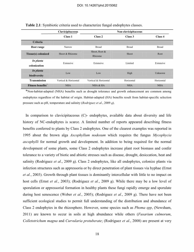
Table 2.1: Symbiotic criteria used to characterize fungal endophytes classes.
Clavicipitaceous
Class 1
Class 2
Non-clavicipitaceous
Class 3
Class 4
Criteria
Host range Narrow Broad Broad Broad
Tissue(s) colonized Shoot & Rhizome Shoot, Root &
Rhizome Shoot Root
In planta
colonization Extensive Extensive Limited Extensive
In planta
biodiversity Low Low High Unknown
Transmission Vertical & Horizontal Vertical & Horizontal Horizontal Horizontal
Fitness benefits* NHA NHA & HA NHA NHA
*Non-habitat-adapted (NHA) benefits such as drought tolerance and growth enhancement are common among
endophytes regardless of the habitat of origin. Habitat-adapted (HA) benefits result from habitat-specific selection
pressure such as pH, temperature and salinity (Rodriguez et al., 2009 a).
In comparison to clavicipitaceous (C)- endophytes, available data about diversity and life
history of NC-endophytes is scarce. A limited number of reports appeared describing fitness
benefits conferred to plants by Class 2 endophytes. One of the clearest examples was reported in
1995 about the brown alga Ascophyllum nodosum which requires the fungus Mycophycia
ascophylli for normal growth and development. In addition to being required for the normal
development of some plants, some Class 2 endophytes increase plant root biomass and confer
tolerance to a variety of biotic and abiotic stresses such as disease, drought, desiccation, heat and
salinity (Rodriguez et al., 2009 a). Class 2 endophytes, like all endophytes, colonize plants via
infection structures such as appressoria or by direct penetration of plant tissues via hyphae (Ernst
et al., 2003). Growth through plant tissues is dominantly intercellular with little to no impact on
host cells (Ernst et al., 2003); (Rodriguez et al., 2009 a). While there may be a low level of
sporulation or appressorial formation in healthy plants these fungi rapidly emerge and sporulate
during host senescence (Weber et al., 2005); (Rodriguez et al., 2009 a). There have not been
sufficient ecological studies to permit full understanding of the distribution and abundance of
Class 2 endophytes in the rhizosphere. However, some species such as Phoma spp, (Newsham,
2011) are known to occur in soils at high abundance while others (Fusarium culmorum,
Colletotrichum magna and Curvularia protuberate; (Rodriguez et al., 2008) are present at very
DOI: 10.14267/phd.2015062

19
low abundance. Analysis of soil fungi typically involves making soil suspensions, dilution
plating and enumerating colony-forming units; therefore, it is difficult to discern the ability of
endophytes to propagate in soil (Selosse et al., 2004); (Rodriguez et al., 2009 a). Although these
characteristics highlight horizontal transmission, Class 2 endophytes often are transmitted
vertically: they can be passed from maternal plants via seed coats (Rodriguez, et al., 2009 a).
Interestingly, culturable Class 2 endophytes can grow on a variety of simple media. The
prevalence of these fungi in plant hosts and their abundance in soils suggest that some of them
are unable to compete outside hosts, while others may have multiple lifestyles (symbiotic or
saprophytic) (Rodriguez et al., 2009 a).
Like Class 2 endophytes, the majority of Class 3 endophytes are members of the
Dikaryomycota (Ascomycota or Basidiomycota), with a special concentration in the Ascomycota.
Class 3 endophytes are distinguished on the basis of their occurrence primarily or exclusively in
above-ground tissues, horizontal transmission, the formation of highly localized infections, the
potential to confer benefits or costs on hosts that are not necessarily habitat-specific and
extremely high in planta biodiversity (Rodriguez et al., 2009 a); (Table 2.1). Class 3 endophytes
include the hyper-diverse endophytic fungi associated with leaves of tropical trees (Arnold &
Lutzoni, 2007); (Gamboa et al., 2002), as well as the highly diverse associates of above-ground
tissues of nonvascular plants, seedless vascular plants, conifers, and woody and herbaceous
angiosperms in biomes ranging from tropical forests to boreal and Arctic/Antarctic communities
(Petrini et al., 1992); (Rodriguez et al., 2009 b). In addition to occurring within photosynthetic
tissues, Class 3 endophytes are found in flowers and fruits, as well as in asymptomatic wood and
inner bark (Verma et al., 2007); (Rodriguez et al., 2009 a). Class 3 endophytes are especially
notable for their high diversity within individual host tissues, plants and populations. For
example, apparently healthy leaves in tropical forests contain numerous, independent infections,
rather than systemic or otherwise extensive growth of hyphae (Arnold & Herre, 2003); (Arnold
et al., 2003). The biomass resulting from any given infection is very low, such that each leaf
represents a densely packed mosaic of diverse endophyte species (Rodriguez et al., 2009 b). In
tropical forests in central Panama, where 100% of mature leaves of diverse trees and shrubs
typically contain endophytes, individual leaves may harbor up to one isolate per 2 mm2 of leaf
tissue and frequently contain dozens of species. Different leaves on the same tree may have quite
distinctive assemblages of endophytic fungi (Arnold et al., 2003); (Gamboa et al., 2002).
DOI: 10.14267/phd.2015062

20
Individual plants may harbor hundreds of species, and plant species across their native ranges
may be inhabited by thousands of species. This tremendous diversity, showcased in some
tropical plants and localities, is not exclusively a tropical phenomenon: plants in temperate and
boreal communities also harbor an astonishing richness of Class 3 endophytes. For example,
Higgins et al. (2007) identified >50 species among the examined 280 boreal and arctic
endophyte isolates. Although horizontally transmitted, Class 3 endophytes are typically distinct
from pathogens associated with the same host species (Ganley et al., 2004) and from epiphyllous
fungi even on the same leaves (Santamaria & Bayman, 2005), but their distinctiveness from
saprotrophic communities is debated (Selosse et al., 2009). Class 3 endophytes are rarely
isolated from seeds (Arnold et al., 2003); (Ganley et al., 2004).
The diversity of Class 3 endophytes raises several questions regarding their ecological roles.
Indeed, most recent studies of Class 3 endophytes have focused on characterizing bioactive
strains or enumeration of species, leaving aside the potential ecological roles of these fungi in
planta (Arnold et al., 2003) or their evolutionary implications for plants, although it is hardly
possible to set up general rules given the occurrence of tens to hundreds of phylogenetically
diverse endophytic fungi within the foliage of a single host (Rodriguez et al., 2009 b); (Arnold &
Herre, 2003). Class 3 endophytes reproduce by hyphal fragmentation and/or by the production of
sexual or asexual spores on dead or senescent tissue (Herre et al., 2007). Spores and hyphal
fragments may be released passively, by herbivores or by physical disturbances such as wind or
rain (Rodriguez et al., 2009 b). Some, such as Phyllosticta sp., produce slimy spores that rely at
least in part on rain for dispersal, while the Ingoldian fungi produce spores that depend on water
for dispersal and infection (Selosse et al., 2009). In general, seedlings raised under sterile
conditions do not contain culturable Class 3 endophytes, highlighting a key difference relative to
Class 2 endophytes (which may be transmitted vertically). Colonization by Class 3 endophytes
proceeds rapidly given the presence of airborne inoculum and high relative humidity or wetting
of leaf surfaces by dew, rain or fog (Arnold & Herre, 2003).
Class 4 endophytes were primarily described by Merlin in 1922 as brown to black pigmented
fungi associated with terrestrial plant roots. Presently, these fungi are referred to as „dark septate
endophytes‟ (DSE) and are grouped together as Class 4 endophytes. In general, Class 4
endophytes are primarily ascomycetous fungi that are conidial or sterile and that form melanized
structures such as inter- and intracellular hyphae and microsclerotia in the roots. DSE have little
DOI: 10.14267/phd.2015062

21
host or habitat specificity; they have been reported in association with ≈600 plants including
plants that are non-mycorrhizal, from Antarctic, Arctic, alpine, sub-alpine, and temperate zones,
as well as from African coastal plains and lowlands, and from some tropical ecosystems
(Jumpponen et al. 1998). DSE are often found in boreal and temperate forests associated with the
fine roots of trees and shrubs, especially of conifers (Reininger & Sieber, 2012). These fungi are
not thought to be pathogenic, as they are observed on healthy fine roots, and in this context, may
be referred to as endophytes. DSE are found worldwide, are prevalent in high-stress
environments, and appear to be ubiquitous and abundant across various ecosystems. Collectively,
these observations suggest that DSE may play an important role in the ecophysiology of plants.
However, little is still known about the role of these elusive fungal symbionts (Rodriguez et al.,
2009 a). DSE appear to represent a large and interesting class of endophytes that have still not
been well defined taxonomically and/or ecologically. Presently, the presence of asexual, darkly
pigmented, septate endophytes in plant roots is the primary criterion for DSE designation. Class
4 endophytes were found associated with 587 plants species representing 320 genera and 114
families. Colonization studies were conducted using five described anamorphic taxa of DSE
(Chloridium paucisporum, Leptodontidium orchidicola, Phialocephala dimorphosphora,
Phialocephala fortinii and Phialophora finlandia) under natural and experimental conditions
(inoculation of root systems in pots). Collectively, these DSE species had a large host range
and/or lacked host specificity (Jumpponen et al., 1998); (Mandyam et al., 2010); (Mandyam et
al., 2013). Because of the presence of DSE in soils and plant roots, transmission is most likely
horizontal and proceeds by mycelial fragmentation and dispersal of conidia (Jumpponen et al.,
1998). Although anamorph–teleomorph connections have not yet been identified for most DSE,
the possibility of sexual reproduction should not be discounted (Rodriguez et al., 2009 a).
2.5. Pathophysiological aspects of plant-endophyte symbiosis
A variety of relationships exist between fungal endophytes and their host plants, ranging from
mutualistic or symbiotic to antagonistic or slightly pathogenic effects. Recent studies of
endophytic fungi and their relationships with host plants have elucidated that plant-endophyte
mutualism has not only a crucial role in biological functions of both parties, but directs the
ecophysiology of host plant and their symbionts to enhanced ability to adapt environmental
stresses throughout the evolutionary time (Chaudhari et al., 2014). Although there has been
DOI: 10.14267/phd.2015062

22
extensive research in plant stress responses, it is not known why so few species are able to
colonize high stress habitats. However, plant stress research rarely takes into consideration a
ubiquitous aspect of plant-fungi symbiosis. It has been indicated that fitness benefits conferred
by mutualistic fungi contribute to or are responsible for plant adaptation to stress (Schulz &
Boyle, 2005); (Selosse et al., 2004). Collectively, mutualistic fungi may confer tolerance to
drought, metals, disease, heat, and herbivory, and/or promote growth and nutrient acquisition. It
has become clear that at least some plants are unable to endure habitat-imposed abiotic and biotic
stresses in the absence of fungal endophytes (Schardl & An, 1993).
2.5.1. Host protection against herbivores Much has been published on the highly specific nature of grass-endophyte symbiosis and the
effects of fungal alkaloids on vertebrate and invertebrate herbivores. Pervasive systemic
colonization of host tissue with endophyte hyphae ensures that herbivores, whether large
mammals or small arthropods, encounter fungal metabolites while consuming the plant tissues.
Most clavicipitaceous endophytes enhance resistance of hosts to insect feeding (Stepniewska &
Kuzniar, 2013). Tintjer & Rudgers (2006) found that deterrence of insect herbivory depends on
the fungal strain and growth stage of the plant. Other studies have provided evidence for anti-
nematode activity of Class 1 endophytes as well (Strobel & Daisy, 2003). However, research has
also shown that some Class 1 endophytes do not provide insect or nematode resistance to host
plants, and have highlighted the importance of examining native plants under natural conditions
in determining endophyte-conferred benefits (Saikkonen et al., 2004). Because of several
examples where endophytes do not appear to provide defensive benefits to host plants, some
investigators have questioned the tendency to classify C-endophytes as defensive mutualists
(Hawas et al., 2012). There is evidence that Class 4 endophytes may also involved in host
defense against herbivores by production of secondary metabolites (Rodriguez et al., 2009 a).
2.5.2. Effects to stress tolerance of the host plant All plants are known to initiate complex biosynthetic responses to elevated temperatures.
These involve the synthesis of heat shock proteins and antioxidant systems as well as
adjustments in osmotic potential and membrane lipid composition (Pressel et al., 2008).
Interaction between host plant and endophytic fungi may cause a drastic enhancement of plant
resistance to heat stress as shown in the Dichanthelium-Curvularia system. Laboratory and field
studies revealed that the plant Dichanthelium lanuginosum is only able to grow in geothermal
DOI: 10.14267/phd.2015062

23
soil at temperatures as high as 57º C when it harbors the endophytic fungus Curvularia sp.
infected with a dsRNA virus (U'Ren et al., 2010).
Although all plants respond to water deficit, only a few species are drought-tolerant not
showing detrimental impacts of water stress (Higginbotham et al., 2013). However, there are
numerous reports describing drought tolerance conferred to plants by fungal symbionts (Del
Olmo-Ruiz & Arnold, 2014). The mechanism of symbiont conferred drought tolerance is not
known, although it is thought to involve osmotic adjustments and/or altered stomatal activity.
Some mycorrhizal fungi can also confer salt tolerance. The physiological basis of fungal-
conferred salt tolerance has not been investigated but this appears to be a generalized
phenomenon occurring in several plant species including banana, tomato and lettuce. It is a
methodological problem, that research is usually focused on one plant-fungus interaction and not
on the complex symbiotic partnerships that are more common in nature (Moricca & Ragazzi,
2008).
It is assumed that at least some Class 2 endophytes are mutualistic, conferring positive fitness
benefits to hosts while also obtaining nutrition for growth and reproduction from host tissues,
and avoiding abiotic stress via symbiosis. Class 2 endophytes commonly increase plant biomass
under stressful conditions, while plants infected by multiple Class 3 endophytes typically show
no observable change in growth rate, biomass accumulation, root/shoot ratio, or other easily
quantifiable characteristics following inoculation under in vivo conditions (Arnold et al., 2003).
2.5.3. Induction of plant resistance to pathogens It has been recently demonstrated that endophyte fungal have the ability to protect the host
from diseases and to limit the damage caused by pathogen microorganisms. Many fungal
endophytes produce secondary metabolites and some of these compounds are antifungal and
antibacterial strongly inhibiting the growth of other microorganisms including plant pathogens
(Arnold et al., 2003). It has been implied that some fungal species may switch between
pathogenic and mutualistic life styles under certain circunstances (Clay, 1992); (Arnold et al.,
2003). In addition, a single fungal isolate can express pathogenicity in certain plant species, and
commensalism or mutualism in others. When non-symbiotic plants respond to pathogen
challenge activation of their defense response is slower and weaker than that of their symbiotic
counterparts, suggesting that communication between host and symbiont increases the ability of
plants to perceive a pathogen and rapidly activate its defense systems. Some studies indicated
DOI: 10.14267/phd.2015062
24
that Class 3 endophytes can be mutualistic, despite the fact that several aspects of their ecology
(i.e. high diversity within hosts and horizontal transmission) are more frequently associated with
parasitic or pathogenic lifestyles (Rodriguez et al., 2009 a). The outcome of plant-pathogen-
endophyte interaction probably depends on the endophytic niche. Endophytic recognition and
colonization may lead to rapid occupation of the ecological niche and leave no space for
pathogens, that could be the common and main reason for the protective action of endophytes
(Petrini et al., 1992).
2.6. Agricultural use of endophytic fungi
In addition to providing ideal research systems for testing ecological and evolutionary theory,
endophytes also have broad economic applications. Since endophytes can affect virtually every
type of plant-plant, plant-pathogen, and plant-herbivore interaction (Selosse et al., 2004) (Petrini
et al., 1992), any human activities (agriculture, deforestation, pollution, etc.), which alter
diversity of endophyte-plant interactions, may have unpredictable effects on population
dynamics and community structure of plants, pathogens and herbivores in terrestrial ecosystems.
Direct anti-herbivore properties of endophytes (particularly in grasses) have already been
exploited, for example in:
1. Biocontrol through developing natural pesticides or improvement of herbivore-resistant
cultivars by introducing biologically active (e.g. high mycotoxin producing) fungal strains into
cultivars (Tadych et al., 2014). On the other hand, colonization of plants with nonpathogenic
fungi and bacteria can lead to induced systemic resistance (ISR) in the host plant. Induced
resistance is a plant-mediated biocontrol mechanism whereby the biocontrol agent and the
phytopathogen do not make physical contact with one another. Plants react to the presence of a
pathogen with a rapid expression of defense-related genes. Thus in addition to economic value,
endophytes may lower investments in chemical pest control by providing environmentally
friendly and energy-efficient biocontrol, and help consumers to avoid remnants of chemical
pesticides in the crop (Tsai et al., 1994).
2. Economic value may also arise from understanding harmful effects in agricultural
production. Mycotoxins cause decreased weight gain of livestock and animal disorders. In this
context, endophytic fungi have been largely ignored in European grass-ecosystems although
DOI: 10.14267/phd.2015062

25
most pasture grasses used in the northern hemisphere are of Eurasian origin and infected with
endophytes (Saikkonen et al., 2004).
3. Alternative fungal strains which do not produce mycotoxins harmful to vertebrates but
increase plant growth, seed production, seed germination rate and stress tolerance can be used to
increase productivity when introduced to the cultivars used as forage. This has already been
accomplished for some tall fescue and perennial ryegrass cultivars (Rodriguez et al., 2009 b).
4. As a direct result of the role that secondary metabolites of endophytic fungi may play in
Nature, they may ultimately be shown to have applicability in medicine. Endophytic fungi very
frequently occur in herbs and in medicinal plants. Many studies investigate their effect on
secondary metabolites and the possibility of their use to produce rare and expensive products
such as taxol (Saikkonen et al., 2002).
2.7. Occurrence and biodiversity of endophytic fungi
Although many studies illustrate endophyte diversity in different ecologies, there is no reliable
estimation of the number of endophytic species, of their host- and tissue-selectivity, since
environmental factors have a complex effect on these features. Most endophytes isolated to date
have been Ascomycetes and their anamorphs; however, several endophytes belonging to
Basidiomycetes have also been observed, but their colonization rate varied greatly. Some of the
common endophytes not only exist in a broad range of plant species, but also have different
relative frequency in every host. In contrast, some other endophytic fungi have high specificity
for the host plant (Eaton et al., 2011); (Ahlholm et al., 2002).
Available data regarding biodiversity of fungal endophytes mainly refer to species which have
been isolated from plants in temperate and tropical regions. As a general overview, endophytes
isolated from tropical plant species are highly diverse indicating relatively lower host-specificity
in comparison to that in temperate zones.
Methods are currently used for identification of endophytic fungi include morphological
studies and determination of phylogenic relationships between isolated morphotaxa. The nuclear
ribosomal RNA (rRNA) cistron has been used for fungal diagnostics and phylogenetics for more
than 20 years. The eukaryotic rRNA cistron consists of the 18S, 5.8S, and 28S rRNA genes
transcribed as a unit by RNA polymerase I. Posttranscriptional processes split the cistron,
removing two internal transcribed spacers (ITS). These two spacers, including the 5.8S gene, are
DOI: 10.14267/phd.2015062

26
usually referred to as the ITS region (Schardl, 2001). The 18S nuclear ribosomal small subunit
rRNA gene (SSU) is commonly used in phylogenetics, and although its homolog (16S) is often
used as a species diagnostic for bacteria, it has fewer hypervariable domains in fungi. The 28S
nuclear ribosomal large subunit rRNA gene (LSU) sometimes discriminates species on its own
or combined with the ITS. Eukaryotic ribosomal RNA genes (known as ribosomal DNA or
rDNA) are found as parts of repeat units that are arranged in tandem arrays, located at the
chromosomal sites known as nucleolar organizing regions (NORs) (Faeth & Fagan, 2002). Each
repeat unit consists of a transcribed region (having genes for 18S, 5.8S and 26S rRNAs and of
the external transcribed spacers ETS1 and ETS2 and a non-transcribed spacer (NTS) region. In
the transcribed region, internal transcribed spacers (ITS) are found on either side of 5.8S rRNA
gene and are described as ITS1 and ITS2. The length and sequences of ITS regions of rDNA
repeats are believed to be fast evolving and therefore may vary. Universal PCR primers designed
from highly conserved regions flanking the ITS and its relatively small size (600-700 bp) enable
easy amplification of ITS region due to high copy number (up to 30000 per cell) of rDNA
repeats. This makes the ITS region an interesting subject for evolutionary/phylogenetic as well as
bio- geographic investigations (Faeth & Fagan, 2002); (Faeth, 2009).
Higgins et al. (Higgins et al., 2007) examined endophytic fungi associated with 11 Poaceae
species in a lowland tropical forest at Barro Colorado Island, Panama, and suggested prevalent
host generalism in tropical forest grasses. In another study at the same region 418 endophyte
morphospecies, most of which (59%) originated from a single isolate, were isolated from leaves
of Heisteria concinna and Ouratea lucens, and researchers suggested spatial heterogeneity of
endophytes in tropical forests (di Menna et al., 2012). Molecular sequence data from 1403
endophyte strains showed that endophytes increase in incidence, diversity and host range from
arctic to tropical sites. Endophyte communities from higher latitudes constituted of relatively few
species whereas tropical endophyte assemblages were dominated by a small number of classes
with a very large number of endophytic species (di Menna et al., 2012). From leaves of coffee
trees (Coffea arabica) in Puerto Rico a total of 821 endophyte colonies were isolated and
grouped into 131 morphospecies. The four most common non-sporulating strains were identified
by sequencing the nuclear ribosomal internal transcribed spacer (ITS) region as Xylaria (two
isolates), Botryosphaeria and Guignardia. Of the most common genera, Pestalotia and
Botryosphaeria were significantly higher represented among epiphytes, while Colletotrichum,
DOI: 10.14267/phd.2015062

27
Xylaria, and Guignardia were significantly more common as endophytes. Surprisingly, more
morphospecies occurred as endophytes than as epiphytes in leaves of coffee tree (Faeth &
Shochat, 2010).
Bernardi-Wenzel et al. (2010) discussed that one or two endophyte species are frequently
predominant in a specific host, while other isolates are rare. The close association between
endophytes and plant species, with a high degree of specialization of interactions, is a possible
indication that the species have evolved together. According to their report, genera Alternaria,
Cochliobolus, Diaporthe, Epicoccum, Guignardia, Phoma, and Phomopsis, were identified;
rDNA sequence analysis showed intra-species variability among endophyte isolates of the genus
Phomopsis sp., in Brazilian Luehea divaricata Mart. (Tiliaceae) (White & Torres, 2010).
Association of Phomopsis, Diaporthe, Dothideomycete, and Cordyceps genera with Trichilia
elegans (Meliaceae) and Cochliobolus, Alternaria, Curvularia, Phomopsis, Diaporthe and
Phoma with Sapindus saponaria (Herre et al., 1999) has been also evaluated showing the
predominance of Phomopsis sp. in these host plants (Herre et al., 2007) .
According to Davis and Shaw (2008), differences between endophytic communities may
correlate with geographic distances rather than with host phylogeny. Therefore identification of
fungal endophyte populations in various geographical and ecological regions is necessary to infer
more comprehensive view of endophyte diversity worldwide. They collected liverworts in North
Carolina, Washington, Idaho, British Columbia, Germany and New Zealand and identified
endophytes using culture-based and molecular methods. They reported that 53 – 88% of the
major lineages of filamentous Ascomycetes recovered belonged to the Xylariales and there was
no significant difference in species richness between regional endophyte communities, however,
North Carolina and New Zealand had richer communities than Germany and the Pacific
Northwest. The authors assumed that this pattern reflects lower per-host endophyte density and
prevalence of a common, shared sequence group in Germany and the Pacific Northwest. They
also tested regional and host specificity of the isolates and reported that endophyte floras of hosts
within a geographic area are more similar to one another than to those of closely related hosts in
different locations (Faeth, 2009).
A similar situation has been observed in temperate and arctic regions. A study on 4 species of
the carnivorous pitcher plant genus Sarracenia: S. minor, S. oreophila, S. purpurea, and S.
psittacina collected from savanna and temperate zones in North America resulted in isolation of
DOI: 10.14267/phd.2015062

28
twelve taxa of fungi, 8 Ascomycota and 4 Basidiomycota. Authors stated that their study was the
first prooving that Coniothyrium/Paraconiothyrium, Penicillium, Cryptosporiopsis, Phomopsis
and Colletotrichum spp. are endophytes of Sarraceniaceae and the first report describing fungal
endophytes in leaves of a carnivorous plant family. The isolation of Colletotrichum spp. from
multiple Sarracenia individuals of all 4 species at locations 300+ miles apart and in different
years, led to the conclusion that at least this fungal genus is a true pitcher plant endophyte (Herre
et al., 1999). Another report from temperate zone (South Korea) described endophytes in leaf
and root samples of Taraxacum coreanum (white dandelion). Of the 72 isolates recovered, 39
were from leaves and 33 from roots with an isolation frequency of 54% and 46%, respectively.
Based on ITS sequence analysis, 72 isolates were classified into 19 genera of which 17 were
Ascomycota and 2 Basidiomycota. Diverse genera were found, dominated by Alternaria,
Cladosporium, Fusarium and Phoma. Out of the 19 genera, Apodus, Ceriporia, Dothideales,
Leptodontidium, Nemania, Neoplaconema, Phaeosphaeria, Plectosphaerella and Terfezia were
new to Korea. Seventy two isolates were screened for antifungal activity, of which 10 isolates
(14%) were found active at least against one of the tested fungi (Sullivan & Faeth, 2004). Osono
and Masuya (2012) examined the diversity and species composition of endophytic fungi in
leaves of 11 species of Betulaceae, with reference to climatic, tree species, and seasonal
variations. A total of 186 fungal isolates were obtained from 190 leaves collected in a subalpine
forest, a cool temperate forest, and a subtropical forest in Japan, and the most frequent taxonomic
units were found as Muscodor sp. and Nemania sp. in Xylariaceae, followed by Gnomonia sp.,
Glomerella acutata, Apiosporopsis sp., Asteroma sp., and Cladosporium cladosporioides. They
assumed that the seasonal changes in composition of fungal endophytes assemblages in leaves of
Betula was higher in subalpine forests than in cool temperate forests (Faeth & Sullivan, 2003).
2.8. Current status of the issue in the area of the present study
Yet, there is a lack of information regarding the features of endophytic fungal communities in
different host plants in Europe. Available data are principally originating from studies on
characterization of bioactive products of endophytes with industrial or medicinal applications.
Nonetheless, understanding the composition and dynamics of endophytic assemblages and
impacts of host-specificity and tissue-colonization of these symbionts on physiology, is
fundamental to improve the existing knowledge about the bioecology of plant-endophyte
DOI: 10.14267/phd.2015062

29
mutualism and is required to pave the lane toward finding novel bio-agents with pesticidal,
medicinal and industrial applications.
Located in Central Europe, Hungary possesses a promising natural phyto-geographical
condition. The state‟s floristic feature encompasses Central European Circumboreal Region
within Boreal (Holarctic) Kingdom and according to The World Wide Fund for Nature (WWF),
the territory of Hungary belongs to the eco-region of Pannonian mixed forests. The land of
Hungary includes:
a) the Little Hungarian Plain (tectonic basin) (Kisalföld) and Transdanubia which encloses
Lake Balaton and Lake Hévíz, the largest lake in Central Europe and the largest thermal lake in
the world, respectively.
b) the Great Hungarian Plain (Alföld) that includes the largest natural grass land in Europe
(Hortobágy National Park) and foothills of the Carpathians.
Almost a fifth of the country is forested, however, only 10 percent is natural forest. Hungary is
home to some 2200 flowering plant species although many of them are thought to be immigrant
species because of the state‟s topography and the transitional climate.
Despite the fact that research on endophytic fungi associated with plants has gained much
interest the available data is rather insufficient about endophytes in Hungary. Among recent
papers, one report described the identification of Coelomycetous fungi classified in Ascochyta,
Phoma, and Phyllosticta which are known as pathogens of soybeans in Hungary (Faeth & Fagan,
2002). Gonda et al. (2013) demonstrated the contribution of endophytic fungi to metabolite
stability/instability in leaves of a medicinal plant (Plantago lanceolata L.) and showed the
association of genera Epicoccum, Bipolaris, Cladosporium, Leptosphaerulina, Aspergillus,
Eurotium and Penicillium with this host plant. Data are also available on orchid-associated
mycorrhizal fungi where the authors claimed that they had isolated Ceratobasidiaceae,
Epulorhiza 1, Epulorhiza 2 and Sebacinaceae from Hungarian orchids (Faeth & Fagan, 2002).
Recent attempts for isolation and identification of dark septate endophytic fungi from the root of
invasive and native plants obtained from semiarid grasslands in Hungary have apparently
resulted a similar picture of endophytic communities associated with host plants to that in other
regions with the same climate around the globe. Fungi belonged to Ascomycota were the most
dominant isolated from sampled plants in these studies (Knapp & Kovács, 2010); (Knapp et al.,
2012); (Knapp et al., 2015).
DOI: 10.14267/phd.2015062
30
2.9. Endophytic fungi in Capsicum
Pepper (Capsicum annuum L.) is an important vegetable as well as spice crop, used worldwide
for domestic and commercial purposes. It is a rich source of antioxidants, vitamin C, pro-vitamin
A, E, and B. Pepper is being seen as a “Hungaricum” in Hungary, i.e. as an agricultural product
associated with Hungarian culture and economy since centuries. Pepper is a constant part of the
diet consumed daily in relatively large quantities not only in cooked dishes, but also as raw
vegetable and spice.
It was shown that endophytic fungi may confer protection to plants against different pathogens
and pests. This is also the case with Capsicum. Bae et al. (2010) were able to protect pepper
against Phytophtora capsici by using Trichoderma ovalisporum, T. theobromica, T. hamatum, T.
stilbohypoxyli or T. caribaeum var. aequatoriale originally isolated from different plants (not
from pepper). At the end of their experiments 26-60% of the Trichoderma-treated plants were
free of symptoms, while in the control only 0-10% stayed healthy. The presence of endophytic
fungi such as Nigrospora, Aspergillus and Coniothyrium can also inhibit growth, virility and
reproduction of Aphis gossypii and some of the fungal strains seem even fit to be utilized for
biological protection (Hernawati et al., 2011). Martinuz et al. (2012) demonstrated that Fusarium
oxysporum Fo162 and Rhizobium etli G12 strains can induce systemic resistance to Aphis
gossypii. Some authors described protection of chili pepper against Meloidogyne incognita by
combined use of Pasteuria penetrans and Paecilomyces lilacinus (Chaudhary & Kaul, 2011).
Three different endophytic fungal strains (Penicillium resedanum, Cladosporium
cladosporioides, and Paraconiothyrium sp.) were isolated from pepper plants. They improved
plant growth and protected plants against pathogenic attack and environmental stresses (Paul et
al., 2012); (Marasco et al., 2012); (Khan et al., 2012); (Khan et al., 2013).
A systematic study to identify endophytic fungi in C. annum was carried out by Paul et al.
(2011) in Korea. They collected samples from C. annuum leaves, stem and root in 3 different
phenophasids during the year. Out of 900 organ samples they obtained about 480 fungal isolates
belongig to 21 genera. The following genera were identified by sequencing the ITS regions:
Alternaria, Aspergillus, Bionectria, Chaetomium, Cladosporium, Colletotrichum, Cordyceps,
Fusarium, Geomyces, Gnomonia, Heterobasidion, Irpex, Paeccilomyces, Papulaspora,
Penicillium, Peyronellaea, Phanerochaete, Phialophora, Phlebiopsis, Plectosphaerella and
Xylaria. The Colletotrichum genus was found most frequently (18.92%), followed by Fusarium
DOI: 10.14267/phd.2015062

31
(18.71%), Alternaria (7.48%) and Penicillium (6.24%). The occurrence and frequncy of
induvidual fungus species varied by the phenophases, but Alternaria, Fusarium and
Cladosporium species were present in all developmental stages of pepper (Paul et al., 2011).
3. Materials and Methods
Methods for studying patterns of infection and colonization by endophytic fungi are essentially
the same as those used in the study of fungal plant pathogens (Sia Ede et al., 2013). In
mycobiotic surveys, host tissues are sampled methodically, and the spatial and temporal
distributions of the fungal colonists encountered are described using methods to determine the
patterns of the endophytic distribution according to host genera and families, habitat types,
infection frequencies related to foliage age, host distribution, and temporal and spatial variations
of endophyte infections. Host species, host-endophyte interactions, interspecific and intraspecific
interactions of endophytes, tissue types and ages, geological and habitat distributions, types of
fungal colonization, culture conditions, and selective media all can influence the efficiency of a
sampling strategy for detection and enumeration of endophytic fungi.
In the current study, first four different species of cherry (Prunus. sp) were selected due to their
horticultural importance and recent attempts for their application in nursery and orchard trials to
find candidate rootstocks for quality control and release in Hungary.
3.1. Locality and sampling strategy of the study
Samples were selected randomly from 110 trees among 180 sweet cherry trees grown in the
orchard of Corvinus University of Budapest, Soroksár, nearby the city of Budapest lies between
19° 07‟ 00‟‟E and 47° 24‟ 00‟‟N geographical coordinates. Trees were planted at a spacing of
4x2 m, resulting in a density of 1,250 trees ha-1. Orientation of the rows was north-south. The
same individual plants were subjected to the study in all sampling periods. The study was
conducted in autumn 2008, spring 2009 and autumn 2009.
Tissue samples from leaf, twig and root of trees (cultivar Péter grafted on different rootstocks)
were collected. Trees were nearly 8 years old when sampled for the first time, and rootstocks
originated from four different species: Prunus mahaleb L. (Érdi V., Bogdány, SL64, Egervár,
Korponay, SM11/4, CEMANY and Magyar rootstocks), a variety of P. avium (Vadcseresznye)
DOI: 10.14267/phd.2015062

32
and P. fruticosa (Prob) and from a hybrid inbred rootstock of P.cerasus and P.canescens (Gisela
6) (Hrotko and Magyar, 2004).
3.2. Tissue preparation
Samples were obtained from root, twig and leaf of each individual tree and were collected in
plastic bags. Although rapid changes in endophyte colonization probably do not occur
immediately following collection, all samples were handled carefully and processed as quickly
as possible. Samples were air-dried to remove any surface moisture before transport or storage.
During transport, samples were kept cool and dry.
Samples were transported to the laboratory at the Department of Plant Physiology and
Biochemistry, Faculty of Horticultural Sciences, Corvinus University of Budapest (Hungary). In
the laboratory, all samples were washed thoroughly by detergent under running tap water.
Surface sterilization of plant material was carried out using chlorine bleach (NaOCl) diluted in
water to concentrations of 2–10% to treat the specimens. After being sunk in 96% ethanol for 1
min, tissue samples were washed by dipping into hypochlorite 3% solution for 10 min and then
were sunk again for 1 min in 96% ethanol. Procedure was followed by washing the samples
twice with sterile distilled water for 5 min.
Size of the sampling unit and surface sterilization procedures vary according to the preferences
of the investigator, the species of host plant, and host tissue type sampled. A pilot study prior to
commencement of main sampling procedure was fulfilled to optimize the techniques for tissue
preparation with higher efficiency and obtaining larger numbers of endophytic fungi from each
sample. Accordingly, it became clear that the smaller the sampling unit was, the greater the
recovery of diverse species/genotypes could be achieved. Also, conversely, the larger the
sampling unit was taken, the greater the potential existed to miss rare or slow-growing species
and to recover mixed genotypes of the same species. Thus, two sections from different parts of
each tissue compartment, from leaf as 0.5 cm in diameter each and from twig and root two
sections as 0.5 cm in length, were randomly cut for isolation of endophytic fungi on the selected
media.
DOI: 10.14267/phd.2015062

33
3.3. Primary isolation of endophytic fungi
Routine mycological media are suitable for primary isolation, sub-culturing and identification
of endophytic fungi. Potato Dextrose Agar (PDA) and Malt Extract Agar (MEA), 1-2%, were
used as pre-culturing media. Each 10 pieces of a single segment were transferred into PDA plates
supplemented with 250 mg/l amoxicillin, 250 mg/l cephalexin and 100 mg/l chloramphenicol.
Plates were incubated at 22°C and colonies were observed after 1-2 weeks. Fungi with rapid
growth were sub-cultured onto media without inhibitors to enhance normal sporulation. Optimal
incubation conditions varied according to the provenance of the host tissue and therefore some
cultures were incubated for two to three weeks to let the slow-growing fungi emerge. Plates were
sealed with Parafilm to prevent desiccation of the media, and were incubated in a growth
chamber with a humidity control.
3.4. Single spore isolation of endophytic fungi
Single spore isolation of endophytic fungi provides an opportunity to obtain certain fungal
subcultures from polyspore isolates when it is important to identify the origin and molecular
phylogeny of the mycelia. The use of fungal cultures obtained from single spore isolations is
fundamental to the identification of fungi in term of morphological species concept, as these
cultures exhibit extra specific characteristics and anamorph-telomorph connections. In
phylogenetic studies which include both morphological and molecular traits single spore cultures
are essentially required.
In the present study, single spore isolation of endophytic fungi was performed according to the
method described by White et al. (1990). Accordingly, 20 g/l water agar was used as sporulation
medium in sterile Petri dishes. After autoclaveing at 121°C for 20 min penicillin (0.5 g/l) was
added when the temperature of agar was about 50°C, then the medium was distributed into 90
mm Petri dishes inside a laminar flow cabinet. By employing a fine sterile sampler, spores were
picked up from the surface of an individual colony. Different isolation methods were employed
based on differences between fungal isolates according to the type of their fruiting bodies. Fungi
with closed fruiting bodies such as Ascomycetes with cleistothecia or perithecia and
Coelomycetes with pycnidia were removed from the substrate surface. Fungi with cup shape
fruiting bodies as like as Ascomycetes with apothecia and Coelomycetes with acervuli, were
transferred directly by removing the whole fruiting body. Spores from Basidiomycetes with gills
DOI: 10.14267/phd.2015062

34
were obtained by removing a few segments of gills and finally when no sporulation was
detected, subcultures were prepared by transferring single thread of hyphae (single hyphae) into
PDA plates.
To overcome the problem of bacteria or yeast contamination and prevent the transfer of wrong
species, in addition to using antibiotics, spore masses were also diluted in sterile water. A glass
container was sterilized using ethanol 70% and wiped with a towel on which ethanol 70% had
been sprayed. A sterilized pipette was then used to transfer about 6 drops of sterilized water into
the container and spore masses obtained as explained above were added to make a spore
suspension. This homogenous spore suspension was finally transferred onto the water agar
plates. A permanent slide was also prepared by using a drop of each spore suspension to check
whether the correct fungus had been selected.
The procedure was followed by incubation of the plates at 22°C for 24 hours. Germinated
spores were detected by microscope examination and then one single spore was picked up and
transferred into another PDA plate. Isolates were incubated at 25°C and were checked frequently
till their colony diameter was about 1-2 centimeters.
3.5. Morphological study
Cultures on both PDA and MEA media were assessed according to their morphology. Colony
appearance, mycelium color and structure, shape of conidiomata, conidia and conidiophore (size,
color, ornamentation, etc.) and characters of conidiogenous cells were observed for
morphological classification of isolated fungi using a light microscope with 5X, 10X and 40X
objective lenses for magnification.
3.6. PCR amplification of ribosomal internal transcribed spacer regions
In the present study, amplification of the fungi ITS region was performed using fungal domain
specific ITS1 and ITS4 primers. Total DNA was extracted from fungal components by applying
a modified CTAB (cetyltrimethylammonium bromide) method (Faeth & Sullivan, 2003). After
pre-heating at 65°C in water bath, 15 μl mercapto-ethanol + 2% polivynil-pyrrolidone solution
was added to the samples. CTAB buffer containing 2% CTAB, 0.1 M Tris-HCl (pH 7.0-8.0), 20
mM EDTA and 1.4 M NaCl, previously heated at 65°C, was mixed with the content of each tube
to the total volume of 600μl. Samples were incubated at 65°C for 30 min and supernatants were
DOI: 10.14267/phd.2015062
35
collected after centrifugation for 15 min at 12 000 g. Protein content was depleted by adding 600
μl chloroform to each tube followed by centrifugation at 12 000g for 8 . The procedure was
repeated one more time, then 900 μl of 96% ethanol was added to each tube and samples were
incubated at -20°C for 2 hours. After centrifugation at 12 000xg for 15 min, pellets were washed
by 70% ethanol and resolved in 100 μl of sterile Milli-Q water.
The PCR reaction mixture consisted of 5 μl fungal DNA, 2.5 μl 10x loading buffer, 0.5 μl 10
mM dNTP mix, 15.6 μl sterile Milli-Q water, 0.4 μl Taq polymerase and 0.5 μl each of the
forward and the reverse primers ITS1 5‟-TCCGTAGGTGAACCTGCGG-3‟ and ITS4 5‟-
TCCTCCGCTTATTGATATGC-3‟ (White et al., 1990) in a total reaction volume of 25μl.
Amplification was performed in a Thermal Cycler (BioRad T100 THERM) and PCR conditions
were 15 min at 95°C followed by 40 cycles at 95°C for 1 min, 30 sec at the annealing
temperature and 72°C for 1min. Aliquots of each amplified product were electrophoretically
separated on a 2% agarose gel in 1x TAE buffer and visualized using ethidium bromide under
UV illumination..
PCR amplicons were recovered using the kit. Isolates whose sequences had a similarity greater
than 95% were considered to belong to the same species. Sequence-based identifications were
made by searching by FASTA algorithms the EMBL/Genbank database of fungal nucleotide
sequences. The criterium for species identification was an ≥97% identity to the database
sequence, genera were positively identified when the sequence match reached 96.9 – 95.0%.
When the similarity was less than 95%, the isolate was considered as unidentified.
DOI: 10.14267/phd.2015062

36
Figure 3.1: PCR amplicons of 54 isolated fungal strains. M: 100 kBp DNA marker
(Fermentas). From the BSc thesis of Tímea Pósa (2009).
DOI: 10.14267/phd.2015062

37
3.7. Analysis of endophyte – pathogen antagonism by dual culture method
As it was previously described, endophytic fungi may compete with other, potentially
pathogenic microorganisms that colonize the host. There is some evidence that the endophyte-
pathogen interplay may result in decreased virulence or even depletion of the pathogen and such
may confer host resistance. Different methods can be applied to investigate endophyte-pathogen
antagonism (Knowlton & Rohwer, 2003). Of the many mechanisms of pathogen inhibition,
production of antimicrobial compounds is the most easily detected and widely studied.
Antimicrobial compounds are produced in either volatile or nonvolatile forms. The nonvolatile
compounds are often detected using simple plate assay. Contrary, the extraction, characterization
and identification of volatile compounds produced by fungal endophytes can only be studied
using more sophisticated means, such as with the use of gas-chromatography and/or mass-
spectrophotometry (Rowan & Knowlton, 1995). Moreover, studies on beneficial inhibitory
volatile metabolites are relatively new. Nevertheless, the role of volatile inhibitory compounds in
biological control remains to be investigated and may have tremendous potential as the volatile
compounds can be entrapped or immobilized in bio-formulations and manipulated for
applications in the field or for post-harvest disease management (misting, spraying and droplets).
Applications using volatile metabolites also exclude the introduction of viable cells, eliminating
the unnecessary or accidental introduction of foreign microbes into the environment.
In the present study, a dual culture method was applied to assess the antagonism of endophytic
fungi against pathogens. Two relevant pathogens of sweet cherry were used: Agrobacterium
tumefaciens strain, kindly provided by Ernő Szegedi from FVM Vine and Wine Research
Institute (Kecskemét, Hungary) and Monilia laxa, obtained from Géza Nagy at the Department
of Plant Pathology, Corvinus University of Budapest, Hungary.
Two culture media were examined, Malt Yeast-Extract Agar (MYEA) and Potato-Dextrose
Agar (PDA), of these PDA was found to be more adventageous and was therefore. Fungi which
had faster growth on culture plates were selected for antagonism test. With the help of a sampler
needle, an inoculum of the selected endophyte and of the pathogen were aseptically planted 40
mm far from each other on a Petri plate (d = 90 mm) containing 30 ml fresh PDA medium. At
the same time, inocula of the endophyte and the pathogen were placed separately on a PDA-
containing Petri dish as controls. All plates were incubated at 28°C for 7 days.
DOI: 10.14267/phd.2015062

38
Figure 3.2: Assessment of radial growth of a desired colony in dual-cultured plate is
schematically demonstrated in this Figure. In brief, colony extension was measured in three
dimensions (horizontal: H, vertical: V, and diagonal: D) in two directions and mean value of
each measurement was used for analytical studies.
Six replicates were used for each plate. Growth inhibition was tested by measuring the radial
growth of each colony in three directions (horizontal, diagonal and vertical) every day during a
period of 15 days after inoculation and calculated by applying the following formula:
Growth Inhibition (GI) (%) = ]x100
where DC = diameter of control, and DP = diameter of pathogen colony dual cultured with
endophyte. Values were calculated as the averages of all achieved data.
Radial growth measurements of each colony in three directions on an actual day were used also
to calculate the growth rate (GR) of colonies. The growth rate value was defined as the slope of
measurements logarithmic curve during 15 days and was expressed in mm/day.
3.8. Statistical analysis
Data derived from the present study were assessed with an emphasis on diversity of endophytic
fungi isolated from different cherry rootstocks. Leaves, twigs and roots as three different tissue
compartments from each rootstock, were also examined to indicate the anatomical distribution
pattern of isolated endophytes. Differences in number and diversity of fungal endophytes
recovered from cherry rootstocks in three distinctive periods when sampling process had been
performed (autumn 2008, spring 2009 and autumn 2009, hereafter: season I, season II and season
III, respectively), were also considered for data analysis. Accordingly, relative frequency (RF) of
V1
V2
D1
D2
H2 H1
DOI: 10.14267/phd.2015062

39
various species was defined as the proportion of recovered colonies belonging to an identified
endophytic fungus compared to the total number of isolates during a particular season.
Infection frequency in different organs was expressed as the percentage of endophyte-bearer
explants among the cultured specimens from a particular tissue and also by dividing the number
of isolated colonies from each organ to the total number of tissue explants, which were defined
as colonization rate (CR) and isolation rate (IR), respectively. Relative abundance and species
richness were calculated for diversity analysis. Shannon-Weaver index (eH‟) was used to compare
the distribution of endophyte species in all examined tissue compartments of every rootstock in
the 3 sampling times. Simpson‟s diversity index (D) was calculated representing the diversity of
endophytic assemblages on different rootstocks.
Endophytic fungal colonies which could not be characterized by morphological or molecular
methods, were marked as unidentified and were omitted from analytical calculations.
A two-way variance analysis (ANOVA) was applied to determine the significance of growth
inhibitory effect between all groups. Student T test was also used for comparing the growth
factors of a group with corresponding control sample. All data were expressed as the mean value
(mm) of total measurements for six replicates.
P≤0.001 was determined as the level of significance.
3.9. Sampling, cultivation and identification of endophytic fungi in Capsicum annuum L.
As described in Chapter 2.9, endophytic fungi also occur in pepper (Capsicum annuum L.). We
investigated two pepper cultivars (Hó F1 and Kárpia F1) and from these cultivars 8 organs such
as roots, shoot, leaves, pedicles, pericarp at different developmental stages and seeds. Samples
were taken in 3 replicates four times during the vegetation period in 2013 (in April, May, August
and October). The plants lived in open field on original sandy soil with drip irrigation or in
greenhouse on rockwool. By the sampling in April and May plants were in the seedling stage,
such only root, leaf and shoot samples could be collected. Samples were separately put into
plastic bags and pre-cultivation started in 6 hours after sample collection.
Surface sterilization was performed by soaking the corresponding organ for 1 min in 70%
ethanol, after that for 10 min in 20% hypochlorite, and finally again for 1 min in 70% ethanol.
After surface sterilization samples were washed by dipping into sterile distilled water. Tissue
samples of 5 mm were cut under sterile conditions. We put 9 pieces from each organ sample on
DOI: 10.14267/phd.2015062

40
PDA pre-culturing medium supplemented with 1 g/l chloamphenicol. We incubated our samples
in 90 mm Petri dishes at room temperature in dark for 2 weeks. After this period we surveyed
and evaluated the frequency of the outgrowing colonies. Then representative colonies were
selected for further work. Small pieces from these cultures were transferred under sterile
conditions to fresh PDA plates and were then monosporulated or monohyphated. my personal
contribution to this part of the project was mainly in this last step.
Identification of endophytic fungi was done by PCR and sequencing and selected strains were
also investigated for their ITS regions (Faeth & Sullivan, 2003).
4. Results
A total of 9823 tissue segments (inocula), 3397 inocula from roots, 3233 inocula from twigs
and 3193 inocula from leaves of all cherry rootstocks were examined while 1614, 2530 and 1037
inocula showed fungal endophyte infection in cultures from roots, twigs and leaves, respectively
(Table 4.1).
All isolates were primarily identified by morphological characteristics and then were subjected
to single spore isolation process where distinguished culture of every individual colony was
prepared and phylogenetically examined by molecular experiments.
Table 4.1: Overall pattern of isolation of endophytic fungi in tissue samples obtained from
cherry rootstocks.
Tissue Total number of
inocula
Number of
infected inocula
Colonization
rate % (CR)
Total number
of fungi isolates
Isolation
rate (IR)
Root 3397 1614 47.5 1944 0.57
Twig 3233 2530 78.3 3427 1.06
Leaf 3193 1037 32.5 1216 0.38
Total 9823 5181 52.7 6587 0.67
DOI: 10.14267/phd.2015062

41
In term of methodology, biodiversity of endophytic fungi in 11 rootstocks of Prunus sp., was
assayed by a multidimensional analysis regarding host-specificity, temporal changes and
colonization outlines dependent to the harboring tissue and results were combined to achieve a
comprehensive insight about fungal endophytes assemblages associated with examined plants.
4.1. Identified endophytic fungi associated with Prunus sp., rootstocks
Regardless the analytical values which would help to evolve biodiversity and composition of
endophyte infection in examined host plants, results derived from the present study provided
brand new information about fungal microorganisms that may contribute to host-endophyte
symbiosis in cherry rootstocks within the locality where this study took place.
Among 6587 isolates obtained from roots, twigs and leaves of cherry rootstocks, 6072 isolates
were taxonomically identified to the level of genus or species following the single-spore
isolation and a number of 519 colonies remained unidentified (marked as unknown).
Table 4.2 contains the listed names of overall fungi taxa isolated from tissue samples of cherry
rootstocks. Among isolated genera, two species of Alternaria (Alternaria sp.1 and Alternaria
sp.2) with a total of 1931 colonies for A. sp.1 and 1473 colonies for A. sp.2 had the first and the
second largest number of colonies isolated from cherry trees. In contrast, Ceratobasidium sp.1
and Ceratobasidium sp.2 had the minimum number of colonies (4 and 15 colonies, respectively)
among other isolates and were observed only in root samples.
In root samples, however, A. sp.1 exhibited the highest frequency as 605 colonies of this
fungus were isolated from root samples. A. sp.2 composed the largest population of endophytes
with 1349 isolated colonies (although this isolate was not observed in the root) and the lowest
number of isolated colonies belonged to Glomerella acutata (21 colonies) which was detected
only in twig samples. Similar to root samples, A.sp.1 with 502 isolated colonies was the most
frequent fungus in leaves while Pyronema sp., with 10 colonies showed the minimum frequency
in this tissue. Along with Pyronema sp., two other identified isolates, Rosellinia sp., and Xylaria
digitata were only observed in leaf samples.
4.2. Diversity of identified endophytic fungi in different sampling periods
Collection of tissue samples from cherry rootstocks was accomplished in three time periods:
autumn 2008 (season I), spring 2009 (season II), and autumn 2009 (season III). According to the
DOI: 10.14267/phd.2015062

42
results, the total species richness of Prunus mahaleb rootstocks (Bogdány, SL64, SM11/4,
Egervár, Korponay, Magyar, CEMANY, and Érdi V) was higher than other rootstocks. Average
number of distinctive species isolated from Prunus mahaleb rootstocks was 12.5 in season I,
10.1 in season II, and 10.1 in season III. During season I, the largest number of species was
isolated from SM11/4 (16 species) while Magyar rootstock harbored 9 different species in
composition of endophytic fungi associated with this rootstock. Species richness of Prunus
mahaleb rootstocks in season II ranged from 10 species (SM11/4 rootstock) to 13 species that
were detected on Korponay, Bogdány, and Érdi V rootstocks. During season III, the maximum
number of species (12 species) was isolated from Korponay, while SL64 had the minimum
species richness among other Prunus mahaleb rootstocks (8 species) in this sampling period. The
difference of species richness between Prunus mahaleb rootstocks in three sampling periods was
not significant.
Although a number of 12 different species was isolated from Prob rootstock (Prunus fruticosa)
in season I but the species richness had a significant decrease (P≤0.05) in season II and season III
(7 species, and 8 sepcies, respectively). Gisela6 (Prunus cerasus, Prunus canescens) showed a
relatively low species richness with no difference in all three sampling periods (6 species, 7
species, and 8 species respectively). During season I, the lowest species richness was observed in
Vadcseresznye (Prunus avium) (3 isolated species), but this index had an increase (P≤0.05) in
season II (10 species) and in season III (7 species).
As a conclusion, average species richness was the highest in season I (11 species) but had a
slightly fall in season II (10.7 species) in compare with season III (9.5 species). However, this
difference was not significant. Prunus mahaleb rootstocks harbored the most heterogeneous
endophyte communities with almost the same species richness in all seasons, but other rootstocks
were associated with endophytic fungi community which showed comparatively less species
richness. Figure 5.1 demonstrates species richness in different tissue compartments of examined
rootstocks in three sampling periods. Collectively, root samples had the richest endophytic fungi
assemblages regarding the number of identified species in season I (average 7.1 species,
maximum= 10 species, isolated from Korponay and SM11/4 rootstocks, minimum= 1 species,
isolated from Vadcseresznye rootstock). During the season II, root samples harbored again more
distinct species (average 6 species, maximum= 8 species, from Érdi V and SL64 rootstocks,
minimum= 3 species, isolated from Gisela6 rootstock), whereas species richness index showed
DOI: 10.14267/phd.2015062

43
no difference between root and twig as a consequence of increase in number of different species
isolated from twig samples in this season (average 4.9 species, maximum= 6 species, isolated
from Érdi V, Egervár, and SL64 rootstocks, minimum= 3 species, isolated from Prob rootstock).
Distribution of different endophyte species during the season III had a shift toward predominant
species richness in twigs (average 6.3, maximum= 8 species, isolated from Korponay and Érdi V
rootstocks, minimum= 5 species, isolated from SL64, CEMANY, Vadcseresznye, and Gisela6
rootstocks). Leaf samples had the lowest species richness in all sampling periods (P<0.05).
Abundance of identified endophytic fungi on different tissue compartments of each individual
rootstock during three sampling periods has been summarized in an Appendix section.
DOI: 10.14267/phd.2015062
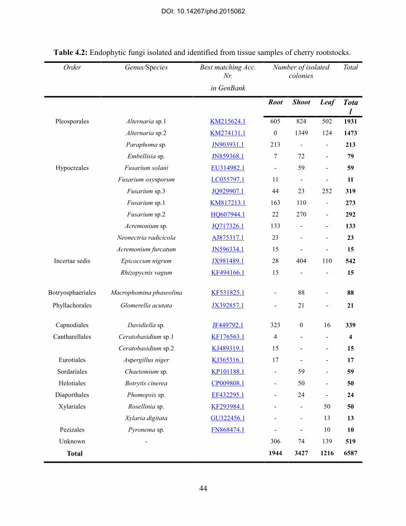
44
Table 4.2: Endophytic fungi isolated and identified from tissue samples of cherry rootstocks.
Order Genus/Species Best matching Acc. Nr.
in GenBank
Number of isolated colonies
Total
Root Shoot Leaf Total
Pleosporales Alternaria sp.1 Alternaria sp.2 Paraphoma sp. Embellisia sp.
KM215624.1
KM274131.1
JN903931.1
JN859368.1
605
0
213
7
824
1349
-
72
502
124
-
-
1931
1473
213
79
Hypocreales Fusarium solani
Fusarium oxysporum Fusarium sp.3 Fusarium sp.1 Fusarium sp.2
Acremonium sp. Neonectria radicicola
Acremonium furcatum
EU314982.1
LC055797.1
JQ929907.1
KM817213.1
HQ607944.1
JQ717326.1
AJ875317.1
JN596334.1
-
11
44
163
22
133
23
15
59
-
23
110
270
-
-
-
-
-
252
-
-
-
-
-
59
11
319
273
292
133
23
15
Incertae sedis Epicoccum nigrum
Rhizopycnis vagum
JX981489.1
KF494166.1
28
15
404
-
110
-
542
15
Botryosphaeriales Macrophomina phaseolina KF531825.1 - 88 - 88
Phyllachorales Glomerella acutata JX392857.1 - 21 - 21
Capnodiales Davidiella sp. JF449792.1 323 0 16 339
Cantharellales Ceratobasidium sp.1 Ceratobasidium sp.2
KF176563.1
KJ489319.1
4
15
-
-
-
-
4
15
Eurotiales Aspergillus niger KJ365316.1 17 - - 17
Sordariales Chaetomium sp. KP101188.1 - 59 - 59
Helotiales Botrytis cinerea CP009808.1 - 50 - 50
Diaporthales Phomopsis sp. EF432295.1 - 24 - 24
Xylariales Rosellinia sp. Xylaria digitata
KF293984.1
GU322456.1
-
-
-
-
50
13
50
13
Pezizales Pyronema sp. FN868474.1 - - 10 10
Unknown - 306 74 139 519
Total 1944 3427 1216 6587
DOI: 10.14267/phd.2015062

45
Figure 4.1: Species richness of endophytic fungi associated with different tissue compartments
of examined cherry rootstocks has been demonstrated in three sampling periods.
02468
1012
Spec
ies R
ichn
ess
Inde
x
Rootstock
Species Richness of Isolated Endophytic Fungi from Cherry Rootstocks During the First Sampling Period (Season I)
Leaf
Twig
Root
0123456789
Spec
ies R
ichn
ess
Inde
x
Rootstock
Species Richness of Isolated Endophytic Fungi from Cherry Rootstocks During the Second Sampling Period (Season II)
Leaf
Twig
Root
0123456789
Spec
ies R
ichn
ess
Inde
x
Rootstock
Species Richness of Isolated Endophytic Fungi from Cherry Rootstocks During the Third Sampling Period (Season III)
Leaf
Twig
Root
DOI: 10.14267/phd.2015062

46
As an average, overall species richness of leaf samples from cherry rootstocks was 2.5 species,
ranged from no isolated fungus in Vadcseresznye rootstock to 4 species in SM11/4 and Érdi V,
during season I. Maximum value of species richness during the second season in leaves was
observed in Magyar, Vadcseresznye and Érdi V rootstocks (3 species), while no isolate was
obtained from leaf samples of Gisela6 rootstock in season II (average species richness= 1.8
species from leaves in season II). As it has been shown in Figure 4.1, species richness in leaf had
no significant change in season III in compare with other seasons but leaf specimens were
significantly more infested during this season. The average species richness of leaf in season III
was 2.6 species while maximum number of species (3 species) was detected on Bogdány, SL64,
SM11/4, Magyar, CEMANY, Gisela6, and Prob rootstocks. Minimum number of species
isolated from leaf samples in season III was observed in Egervár, Korponay, Érdi V, and
Vadcseresznye rootstocks (2 species).
Diversity of endophytic fungi was also determined by Shannon-Weaver (eH‟) and Simpson (D)
diversity indices. Both indices take into account not only the species richness but the abundance
of species in a community. Values of eH‟ and D were calculated separately for each rootstock
according to the total composition of isolated endophyte communities in three seasons. Evenness
(Simpson‟s index) of isolated endophytic fungi from each rootstock has been demonstrated in
Figure 4.2. As a sum, the evenness of different species in all rootstocks was comparatively
higher in season II than that in other seasons. The higher value of eH‟ shows either diversity of
species or equitable distribution of isolated colonies among the species. In this sense, it is
expected to defy Simpson index (D) which measures the probability whether two randomly
selected individual colony in an endophyte community belong to the same species, however they
refer to different concepts of diversity. Accordingly, Simpson index for assessment the diversity
of fungal endophytes in each rootstock verified the occurrence of more heterogeneous endophyte
assemblages on cheryy rootstocks during season II. In general, endophyte communities
associated with rootstocks belonged to Prunus mahaleb were relatively more diverse particularly
during the second sampling period in compare with other cherry rootstock examined in the
present study.
DOI: 10.14267/phd.2015062
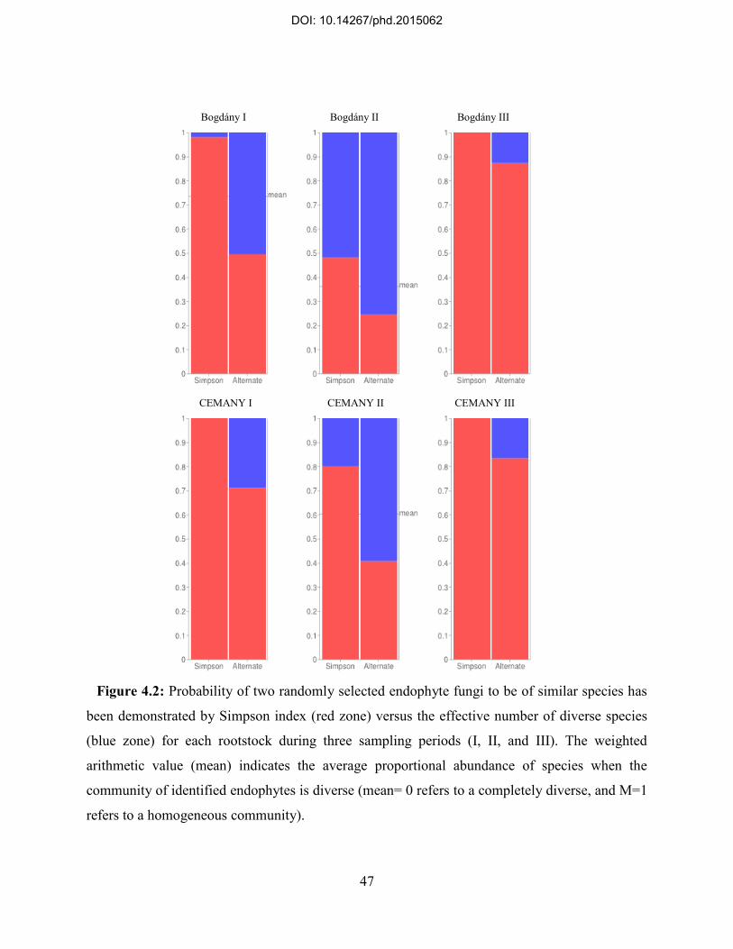
47
Bogdány I Bogdány II Bogdány III
CEMANY I CEMANY II CEMANY III
Figure 4.2: Probability of two randomly selected endophyte fungi to be of similar species has
been demonstrated by Simpson index (red zone) versus the effective number of diverse species
(blue zone) for each rootstock during three sampling periods (I, II, and III). The weighted
arithmetic value (mean) indicates the average proportional abundance of species when the
community of identified endophytes is diverse (mean= 0 refers to a completely diverse, and M=1
refers to a homogeneous community).
DOI: 10.14267/phd.2015062

48
Egervár I Egervár II Egervár III
Érdi V I Érdi V II Érdi V III
Figure 4.2: Continued.
DOI: 10.14267/phd.2015062
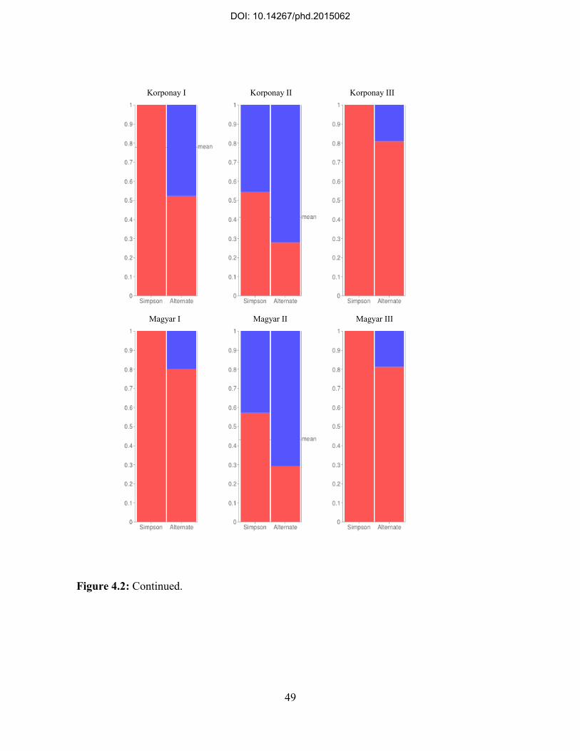
49
Korponay I Korponay II Korponay III
Magyar I Magyar II Magyar III
Figure 4.2: Continued.
DOI: 10.14267/phd.2015062

50
SL64 I SL64 II SL64 III
SM11/4 I SM11/4 II SM11/4 III
Figure 4.2: Continued.
DOI: 10.14267/phd.2015062

51
Vadcseresznye I Vadcseresznye II Vadcseresznye III
Prob I Prob II Prob III
Figure 4.2: Continued.
DOI: 10.14267/phd.2015062

52
Gisela6 I Gisela6 II Gisela6 III
Figure 4.2: Continued.
4.3. Pattern of colonization and distribution of endophytic fungi on examined cherry rootstocks
Although root samples obtained from cherry rootstocks demonstrated the highest average of
species richness in compare with other tissues, twig was found as the most infected histological
compartment in examined host plants. To analyze the rate of infection in specimens, the
proportion of infested inocula in total number of tested tissue explants (CR) and the rate of
isolated colonies from every explant (IR) were applied. Accordingly, twig was found as the most
infected tissue compartment (CR=79%, IR=1.06) while root harbored less endophyte infection
(CR=54%, IR=0.62) than twig. As an overall, leaf samples showed the lowest colonization and
isolation rates in the present study (CR=33%, IR=0.38) (P<0.05). Indices that show the rate of
infection are assumed to be corresponding, but further analysis indicated subsequent changes in
colonization and isolation rates consistency based on the host species and the time periods when
samples were obtained.
DOI: 10.14267/phd.2015062

53
All rootstocks of Prunus mahaleb showed the same pattern regarding the distribution and
colonization rate of endophytic fungi in their roots during three sampling periods. Both
colonization rate and isolation rate were relatively low in season I but had increase in season II
and remained unchanged in season III (P≤0.05). Among other rootstocks, only Vadcseresznye
had the same pattern as Prunus mahaleb rootstocks. The feature of endophyte infection in root,
however, was detected comparatively different in other rootstocks. Root samples obtained from
Prob rootstock had the highest colonization rate in season I, but had a descending rate in season
II and season III (P≤0.05). Such a result for Gisela6 showed a different temporal alteration in
infection rates when no significant change was observed between these rates in season I and
season II, although trees that were sampled from this rootstock showed significantly higher
infection rates during season III (P<0.05). Changes in isolation rate calculated for root samples
had the same pattern as their colonization rate in all rootstocks during three seasons, although
Gisela6 the average number of isolated endophyte colonies from every inocula had a decrease
from season I to season II and again a sudden increase occurred regarding this rate in season III
(P<0.05). Prob rootstock had the highest colonization and isolation rate in root samples during
season I (CR=74%, IR=0.8) and the minimum colonization rate of root was detected in Bogdány
(CR=20%) but isolation rate was the lowest in Vadcsresznye root samples among other
rootstocks (IR=0.2) in this season. During season II, SM11/4 rootstock showed the maximum
infection rate (CR=82%, IR=1.22) in root, while Prob rootstock had the lowest colonization rate
(CR=31%) and Gisela6 had the lowest number of isolated colonies from root (IR=0.06).
Vadcseresznye and Gisela6 had the highest colonization and isolation rates in root samples
obtained from these rootstocks (CR=91%, and IR=1.03, respectively) in season III while the
lowest values of these rates were calculated for root samples of Prob (CR=38%) and Magyar
(IR=0.4) during the last season (Figure 4.3).
Changes in infection burden of twig samples obtained from cherry rootstocks indicated the
same equation according to the colonization and isolation rates in three sampling periods. Both
values for all rootstocks were relatively high in season I and season III (with no significant
difference) and significantly low (P<0.05) in season II. Maximum infection rate of twig samples
was found in Vadcseresznye (CR=100%, IR=1.5) and Korponay rootstock had the minimum rate
(CR=67%, IR=0.95) during season I. A descending change in infection rates of twig samples was
observed in season II when colonization for twig samples obtained from Prob (CR=57%) was the
DOI: 10.14267/phd.2015062

54
highest and Gisela6 (CR=33%) showed the minimum rate. Interestingly, Gisela6 showed the
highest isolation rate (IR=1.03) for twig samples while the lowest rate was calculated for twig
samples from SL64 rootstock (IR=0.54) during season II. CEMANY and SM11/4 (both from
Prunus mahaleb L, rootstocks) had the highest rates of colonization (CEMANY, CR=100%) and
isolation (SM11/4, IR=1.6) from twig samples obtained from these rootstocks in season III.
Moreover, Prob and Magyar rootstocks had the lowest colonization rate (CR=77%) and isolation
rate (IR=1.16) in twigs during the last season, respectively (Figure 4.4).
As it was mentioned previously, leaf samples from cherry rootstocks had the lowest infection
rates in compare with other tissues in all sampling seasons, although these rates showed temporal
changes in leaf according to the rate of fungal endophytes infection in three different sampling
periods of the present study. In brief, colonization and isolation rates had a significant decrease
during season II in compare with season I (P<0.05), but showed a drastic increase in season III
(P<0.05) when both indices were observed remarkably higher than the first two seasons. Such a
pattern was detected in all examined rootstocks. During season I, the maximum colonization and
isolation rates were detected for leaf samples from Érdi V (CR=24%) and Bogdány (IR=0.27),
respectively, while Vadcseresznye rootstock bearded no endophyte infection in its examined leaf
samples (CR=0%, IR=0). Vadcseresznye rootstock showed the highest rates (CR=9%, IR=0.09)
of infection in leaf among other rootstocks in season II and no infection was detected on Gisela6
(CR=0%, IR=0), during the second season. In season III, SL64 rootstock showed the maximum
colonization rate (CR=100%) in the leaf and Érdi V had the highest isolation rate (IR=1.18)
while corresponding values were found the lowest in SM11/4 (CR=63%) and Gisela6 (IR=0.7)
rootstocks during the third sampling season (Figure 4.5).
DOI: 10.14267/phd.2015062

55
Figure 4.3: Colonization and isolation rates of endophytic fungi infection in root samples
obtained from cherry rootstocks in three different sampling periods.
0%
20%
40%
60%
80%
100%C
olon
izat
ion
Rat
e (%
)
Rootstock
Colonization Rate of Isolated Endophytes from the Root of Different Cherry Rootstocks in Three Sampling Periods.
Season I
Season II
Season III
00,20,40,60,8
11,21,4
Isol
atio
n R
ate
Rootstock
Isolation Rate of Endophytes Colonized on the Root of Different Cherry Rootstocks in Three Sampling Periods.
Season I
Season II
Season III
DOI: 10.14267/phd.2015062
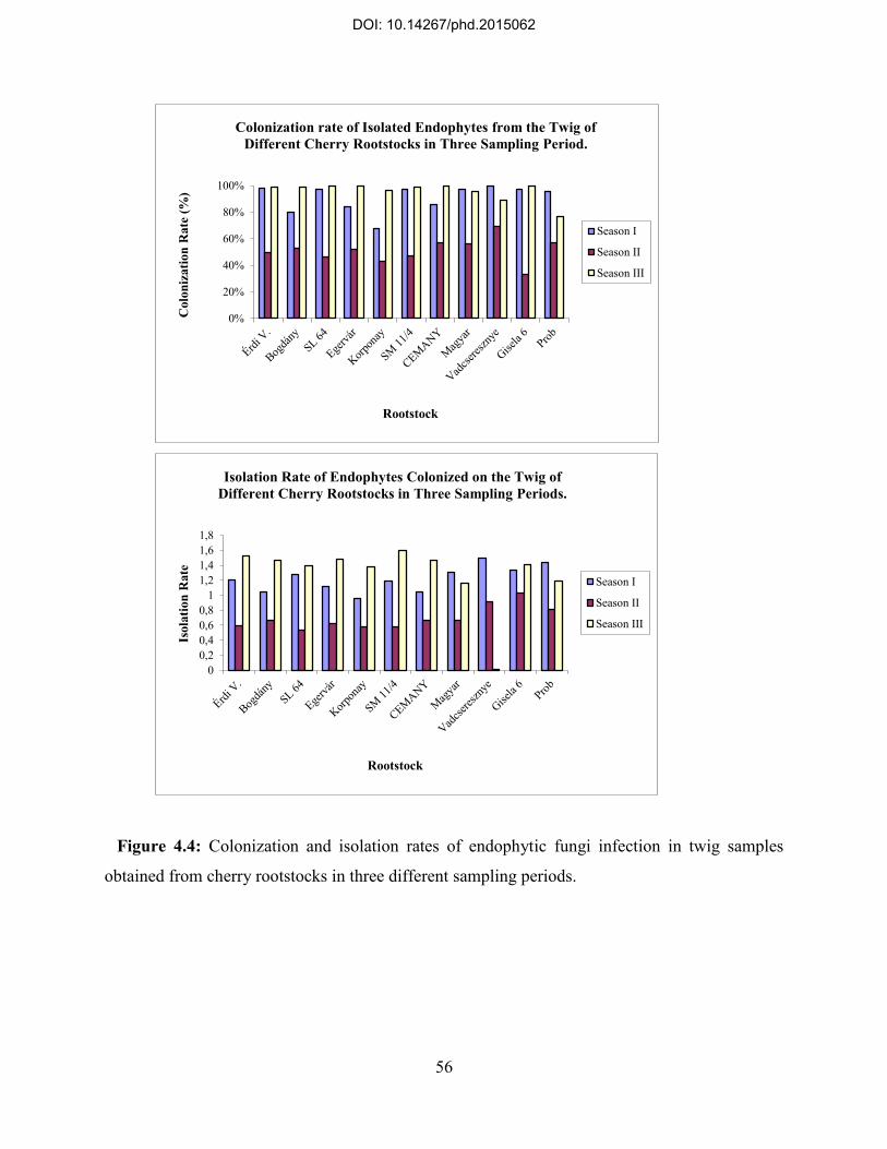
56
Figure 4.4: Colonization and isolation rates of endophytic fungi infection in twig samples
obtained from cherry rootstocks in three different sampling periods.
0%
20%
40%
60%
80%
100%C
olon
izat
ion
Rat
e (%
)
Rootstock
Colonization rate of Isolated Endophytes from the Twig of Different Cherry Rootstocks in Three Sampling Period.
Season I
Season II
Season III
00,20,40,60,8
11,21,41,61,8
Isol
atio
n R
ate
Rootstock
Isolation Rate of Endophytes Colonized on the Twig of Different Cherry Rootstocks in Three Sampling Periods.
Season I
Season II
Season III
DOI: 10.14267/phd.2015062

57
Figure 4.5: Colonization and isolation rates of endophytic fungi infection in leaf samples
obtained from cherry rootstocks in three different sampling periods.
0%
20%
40%
60%
80%
100%C
olon
izat
ion
rate
(%)
Rootstock
Colonization Rate of Isolated Endophytes from the Leaf of Different Cherry Rootstocks in Three Sampling Periods.
Season I
Season II
Season III
00,20,40,60,8
11,21,4
Isol
atio
n R
ate
Rootstock
Isolation Rate of Endophytes Colonized on the Leaf of Different Cherry Rootstocks in Three Sampling Periods.
Season I
Season II
Season III
DOI: 10.14267/phd.2015062

58
4.4: Relative frequency of identified endophytic fungi on cherry rootstocks
Regardless the host species, harboring tissue and the sampling period, overall results achieved
by the present study indicated that Alternaria sp.1 (RF=28.11%) and Alternaria sp.2
(RF=23.65%) were the first and the second most frequent endophytic fungi associated with
examined cherry rootstocks. On the other hand, identified endophytic fungi of the genus
Fusarium contained the most diverse species (5 different species) composition among other taxa.
Ceratobasidium sp.1 had the lowest abundance (RF=0.06%) in general structure of endophytic
fungi communities on cherry rootstocks. Relative frequency of identified fungi that composed
endophyte assemblages on examined cherry rootstocks has been schematically demonstrated in
Figure 4.6.
As the most abundant species, relative frequency of Alternaria sp.1 and Alternaria sp.2 was
assessed for each individual host. According to the results, Korponay rootstock had the most
frequent population of Alternaria sp.1 (RF=32.27%) while the less abundant occurrence of this
species was indicated on Prob (RF=19.9%). Interestingly, Alternaria sp.2 showed reverse pattern
of frequency on recently mentioned hosts, as the maximum relative frequency of this species
was found in Prob (RF=30.71%) and Korponay bearded the less frequent population of
Alternaria sp.2 (RF=18.9%) within the composition of endophytic fungi associated with this
rootstock. The ratio of Alternaria sp.1/Alternaria sp.2 occurrence, however, showed no
difference between examined rootstocks (Figure 4.7).
DOI: 10.14267/phd.2015062

59
Figure 4.6: Overall relative frequency of endophytic fungi on cherry rootstocks.
Rosellinia sp. 0,76%
Chaetomium sp. 0,89% Macrophomina phaseolina
1,33% Pyronema sp.
0,15%
Alternaria sp.1 28,11%
Alternaria sp.2 23,65%
Xylaria digitata 0,20%
Aspergillus niger 0,26%
Acremonium furcatum 1,33%
Rhyzopycnis vagum 0,23%
Ceratobasidium sp.1
0,06%
Ceratobasidium sp.2 0,23%
Neonectria radicicola 0,35%
Acremonium sp. 0,91%
Paraphoma sp. 3,27%
Phomopsis sp. 0,41%
Epicoccum nigrum 8,20%
Embellisia sp. 1,21%
Davidiella sp. 5,20%
Botrytis cinerea 0,67%
Fusarium sp.1 4,14%
Fusarium sp.2 4,43%
Fusarium sp.3 4,84%
Fusarium oxysporum 0,17%
Fusarium solani 0,89%
Glomerella acutata 0,32%
Unknown 7,79%
DOI: 10.14267/phd.2015062

60
Figure 4.7: A Comparison of relative frequency values corresponding occurrence of two
largest populations of identified fungi species composed endophytes assemblages on cherry
rootstocks.
0
5
10
15
20
25
30
35
Rel
ativ
e Fr
eque
ncy
(%)
Rootstocks
Relative Frequency (%) of Alternaria Species Identified on Cherry Rootstocks
Alternaria sp.1
Alternaria sp.2
DOI: 10.14267/phd.2015062

61
According to the results, identified endophytic fungi exhibited some levels of specificity when
their frequency was assessed on different cherry rootstocks. Alternaria sp.1, Alternaria sp.2,
Paraphoma sp., Macrophomina phaseolina, Fusarium sp.1, Fusarium sp.2, Fusarium sp.3,
Fusarium solani, Epicoccum nigrum, and Davidiella sp., were identified on all rootstocks. As
well, Phomopsis sp. and Embellisia sp. were detected on all hosts except on Vadcseresznye
rootstock. Chaetomium sp. also colonized all rootstocks but was not found on Gisela6 and Prob.
Some endophytes were only observed on Prunus mahaleb rootstocks while had no occurrence on
other cherry species. As an instance, Aspergillus niger was detected on Bogdány, SL64, Egervár,
Korponay, and Érdi V, and was not frequent on other rootstocks. Besides, Rhizopycnis vagum
was detected on SL64, Egervár, SM11/4, CEMANY, and Érdi V. Ceratobasidium sp.1 was only
observed on Bogdány, Egervár, CEMANY, and Érdi V. a species identified as Pyronema sp. was
only detected on SM11/4 rootstock. Relative frequency of identified fungal endophytes in
different rootstocks has been shown in Figure 4.8.
Although some endophytic fungi demonstrated tendency to colonize a specific tissue
compartment in examined hosts, but it seemed to be changeable in different sampling periods.
Accordingly, leaf samples of all rootstocks were mostly colonized by Alternaria sp.2
(RF=54.89%) while Alternaria sp.1 had no occurrence in leaf during season I. Rosellinia sp.
(RF=27.17%), Xylaria digitata (RF=1.63%), and Pyronema sp. (RF=5.43%) were exclusively
observed in leaf during season I. Davidiella sp. was also detected in leaves with relatively low
frequency in the first sampling period (RF=5.98%). Such a pattern was slightly different
observed in leaf during season II when Alternaria sp.2 as the largest fungal population
(RF=66%) and Xylaria digitata (RF=20%) and Davidiella sp. (RF=10%) were the only species
identified in this tissue. In season III, Alternaria sp.1 was the predominant species found in
leaves (RF=50.66%) that along with Epicoccum nigrum and Fusarium sp.3 composed the
endophyte communities in this tissue (Figure 4.9).
Twig samples showed the same structure of endophytic fungi community as the most abundant
fungus was Alternaria sp,2 (RF=69.36%) in season I, Alternaria Sp.2 (RF=67.57%) in season II,
and Alternaria sp.1 (RF=56.55%) in season III, identified in this tissue. During season I, other
endophyte as like as Chaetomium sp., Epicoccum nigrum, Fusarium sp.3, Fusarium solani, and
Macrophomina phaseolina were found in twig.
DOI: 10.14267/phd.2015062

62
Figure 4.8: Overall relative frequency (%) of identified endophytic fungi on examined cherry
rootstocks.
0 10 20 30 40 50 60 70 80 90 100
Bogdány
SL64
Egervár
Korponay
SM11/4
CEMANY
Magyar
Érdi.V
Vadcseresznye
Gisela 6
Prob
Relative Frequency (%) of Identified Endophytic Fungi on Examined Cherry Rootstocks
Rosellinia sp.
Chaetomium sp.
Macrophomina phaseolina
Pyronema sp.
Alternaria sp.1
Alternaria sp.2
Xylaria digitata
Aspergillus niger
Acremonium furcatum
Rhyzopycnis vagum
Ceratobasidium sp.1
Ceratobasidium sp.2
Neonectria radicicola
Acremonium sp.
Paraphoma sp.
Phomopsis sp.
Epicoccum nigrum
Embellisia sp.
Davidiella sp.
Botrytis cinerea
Fusarium sp.1
Fusarium sp.2
Fusarium sp.3
Fusarium oxysporum
Fusarium solani
Glomerella acutata
Unknown
DOI: 10.14267/phd.2015062

63
Chaetomium sp. and Macrophomina phaseolina with relatively higher frequency than the
previous season along with Epicoccum nigrum were detected in twig of cherry rootstocks during
season II. Endophytic fungi identified in twig samples from cherry rootstocks were more diverse
and included species such as Phomopsis sp., Epicoccum nigrum, Embellisia sp., Davidiella sp.,
Botrytis cinerea, Fusarium sp.1, Fusarium sp.2, and Glomerella acutata (Figure 4.10).
Endophyte infection of root had different structure in compare with other tissue samples and
showed more divergent composition during three sampling periods. During season I, Davidiella
sp., was the most abundant fungus in the root (RF=18.47%). Although this species had higher
frequency in season II (RF=2638%) than season I, but Alternaria sp.1 was the predominant
species found in roots during the second season (RF=33.06%). Davidiella sp., had no occurrence
in roots during season III. Either Alternaria sp.1 or Alternaria sp.2 was observed in root samples
during season I and season II, although their abundance did not have the same ration in the first
two seasons. During season I, Alternaria sp.2 had higher frequency (RF=14.25%) while in
season II, abundance of this isolate was much lower (RF=2.4%). Alternaria sp.1 was also the
most frequent species in root during season III (RF=29.25%), while Alternaria sp.2 was not
detected in root of cherry rootstocks during this season (Figure 4.11).
DOI: 10.14267/phd.2015062

64
Figure 4.9: Overall relative frequency of endophytic fungi in the leaf of cherry rootstocks in
different sampling periods.
0
20
40
60
Rel
ativ
e Fr
eque
ncy
(%)
Endophytic Fungi
Relative Frequency (%) of Identified Endophytic Fungi in the Leaf of Cherry Rootstocks During Season I
020406080
Rel
ativ
e Fr
eque
ncy
(%)
Endophytic Fungi
Relative Frequency (%) of Identified Endophytic Fungi in the Leaf of Cherry Rootstocks During Season II
0
20
40
60
Rel
ativ
e Fr
eque
ncy
(%)
Endophytic Fungi
Relative Frequency (%) of Identified Endophytic Fungi in the Leaf of Cherry Rootstocks During Season III
DOI: 10.14267/phd.2015062

65
Figure 4.10: Overall relative frequency of endophytic fungi in the twig of cherry rootstocks in
different sampling periods.
020406080
Rel
ativ
e Fr
eque
ncy
(%)
Endophytic Fungi
Relative Frequency (%) of Identified Endophytic Fungi in the Twig of Cherry rootstocks During Season I
020406080
Rel
ativ
e Fr
eque
ncy
(%)
Endophytic Fungi
Relative Frequency (%) of Identified Endophytic Fungi in the Twig of Cherry Rootstocks During Season II
0
20
40
60
Rel
ativ
e Fr
eque
ncy
(%)
Endophytic Fungi
Relative Frequency (%) of Identified Endophytic Fungi in the Twig of Cherry Rootstocks During Season III
DOI: 10.14267/phd.2015062

66
Figure 4.11: Overall relative frequency of endophytic fungi in the root of cherry rootstocks in
different sampling periods.
0
5
10
15
20
Rel
ativ
e Fr
eque
ncy
(%)
Endophytic Fungi
Relative Frequency (%) of Identified Endophytic Fungi in the Root of Cherry rootstocks During Season I
010203040
Rel
ativ
e Fr
eque
ncy
(%)
Endophytic Fungi
Relative Frequency (%) of Identified Endophytic Fungi in the Root of Cherry Rootstocks During Season II
010203040
Rel
ativ
e Fr
eque
ncy
(%)
Endophytic Fungi
Relative Frequency (%) of Identified Endophytic Fungi in the Root of Cherry Rootstocks During Season III
DOI: 10.14267/phd.2015062

67
4.5. Endophytic fungi associated with Capsicum annuum L.
The aim of this project was the identification of endophytic fungi living in pepper to find out
whether significant cultivar dependent differences can be observed. We also investigated the
effect of environment and cultivation conditions, therefore both cultivars, „Hó‟ and „Kárpia‟,
were cultivated in greenhouse as well as in open field. Several students took part in the project, I
was partly involved in starting the fungal cultures, but my main task was to prepare and establish
single spore / single hyphae cultures from selected isolates to have well defined, stable cultures
for DNA ITS-sequence based identification of individual fungal strains. Samples originated from
8 different organs and were collected in April, May, August and October. Cultivation and first
morphological categorization of endophytic fungi was essentially the same as in case of sweet
cherry. Single spore cultures were initiated from 118 isolates selected from the endophytes of
samples collected in August and October. Methodology applied for identification of endophytic
fungi associated with pepper was as the same as that was used for cherry.
I would like to summarize briefly the final results of our team, stating that the quantitative
evaluation of the frequency of endophytic fungi was mainly done by Csaba Borbély and is
described in his diploma thesis (Borbely, 2014). Andras Bärnkopf was mainly responsible for the
ITS sequence based identification of single spore fungal isolates, detailed results were presented
in his diploma thesis (Bärnkopf, 2013). The first question analyzed was whether colonization by
endophytic filamentous microfungi arises during the vegetation period. The data are summarized
in Figure 4.12 (taken von Borbely (2014) with permission) and clearly show that considerably
more endophytes were isolated at the end of the vegetation period, i.e. in August and in October
than in April and May, the difference between the frequencies in May and August or May and
October are significant for both cultivars at 0,1%. Although we isolated more endophytic
fungi from the cultivar „Hó‟ than from „Kárpia‟ (Figure 4.12) at all sampling times, statistically
significant difference (0,1%) was only found in October. When colonization rates of field-
grown plants were compared to those in greenhouse, the former were always significantly higher
for both cultivars (Borbely, 2014).
Fungal genera and species were identified on the basis of their ITS1+5.8S RNA+ITS2
sequences. 50 strains from our collection were identified in this way. As in sweet cherry, we
observed that the potentially pathogenic Alternaria genus occurred most frequently in pepper,
members of this genus were detected in every organ. Figure 4.13 shows the occurrence of
DOI: 10.14267/phd.2015062

68
different morphotypes in the samples collected in October. In addition to Alternaria 4 further
morphotypes were very common: Cladosporium, Verticilium, Acremonium and the yet
unidentified group DB (Figure 4.13); (Bärnkopf, 2013); (Borbély, 2014). From the root we also
isolated Plectosphaerella, Colletotrichum, Paecilomyces, Penicillium and Fusarium strains,
while in the shoot Cladosporium, Acremonium, Chaetomium and Lewia strains were identified.
Figure 4.12: Change of colonization rates during the vegetation period. Colonization rates =
colonized samples/all samples. The results demonstrate enhanced colonization towards the end
of vegetation period, and higher colonization of the cultivar „Hó‟ compared to „Kárpia‟. (Figure
from Borbely, 2014).
0,000
0,100
0,200
0,300
0,400
0,500
0,600
Április Május Augusztus Október
Hó
Kárpia
DOI: 10.14267/phd.2015062

69
Figure 4.13: Colonization by different morphotypes in pepper in October. Of all endophytic
fungi identified Alternaria is by far the most common, followed by Cladosporium. (From:
Borbely, 2014)
DA (Alternaria)
DB
DC (Verticillium)
DE (Acremonium)
DG (Cladosporium)
DH (Aspergillus)
DK Penicillium)
DO (Galactomyces)
DR (Anthirinium)
DU (Cercospora)
DW (Colletotrichum)
Egyéb morfológiaicsoportEgyik morfológiaicsoportba sem besorolt
DOI: 10.14267/phd.2015062

70
Our results indicate that greenhouse-grown and field-grown peppers harbour different
endomycota. We also observed differences between individual plant parts and seasonal
difference in the samples of the same plant. Plant parts of the cultivar „Hó‟ usually showed
higher colonisation rate by endophytic fungi than those of „Kárpia‟. As expected, colonization
rates were higher in field-grown plants and in older organs. Highest colonisation frequency was
found in old leaves and in stalks of fruits. Several strains belonging to the Alternaria genus were
detected; members of this genus occurred in every plant part. From the root we also isolated
Plectosphaerella, Colletotrichum, Paecilomyces, Penicillium, Rhizopycnis, Pyrenochaeta and
Fusarium strains, while in the shoot Cladosporium, Acremonium, Chaetomium, Myrothecium,
and Verticillium strains were identified. Young and older leaves showed Acremonium,
Alternaria, Cercospora, Xylaria and Penicillium strains. Pericarp was colonized mainly with
Alternaria, while Arthrinium, Galactomyces, Penicillium, Acremonium, Cladosporium,
Chaetomium and Lewia were isolated from fruit stalks (Table 4.3).
4.6. The evolutionary relationship of identified fungal endophyte taxa
The evolutionary history was inferred using the UPGMA method (Sneath & Sokal, 1973). The
bootstrap consensus tree inferred from 1000 replicates (Felsenstein, 1987) is taken to represent
the evolutionary history of the taxa analyzed (Felsenstein, 1987). Branches corresponding to
partitions reproduced in less than 50% bootstrap replicates are collapsed. The percentage of
replicate trees in which the associated taxa clustered together in the bootstrap test (1000
replicates) are shown next to the branches (Felsenstein, 1987). The tree is drawn to scale, with
branch lengths in the same units as those of the evolutionary distances used to infer the
phylogenetic tree. The evolutionary distances were computed using the Maximum Composite
Likelihood method (Tamura et al., 2004) and are in the units of the number of base substitutions
per site. The analysis involved 42 nucleotide sequences. Codon positions included were
1st+2nd+3rd+Noncoding. All positions with less than 95% site coverage were eliminated. That
is, fewer than 5% alignment gaps, missing data, and ambiguous bases were allowed at any
position. There were a total of 240 positions in the final dataset. Evolutionary analyses were
conducted in MEGA5 (Tamura et al., 2007). Figures 4.14 and 4.15 demonstrate the overall
evolutionary relationship between isolated morphotaxa from cherry and peppr, repectively.
DOI: 10.14267/phd.2015062

71
Table 4.3: Features of endophytic fungi morphotaxa isolated from pepper (Capsicum annuum) Number Isolated fungi Cultivar Organ Growing site
P4 Alternaria sp. Kárpia leaf greenhouse
P6 Alternaria sp. Kárpia roots greenhouse
P7 Alternaria sp. Kárpia roots greenhouse
P9 Plectosphaerella cucumerina Hó roots greenhouse
P10 Paecilomyces sp. Hó roots greenhouse
P13 Alternaria sp. Hó roots greenhouse
P20 Penicillium adametzii Kárpia roots greenhouse
P22 Cladosporium sp. 1 Hó shoot greenhouse
P28 Xylaria sp. Kárpia young leaf open field
P31 Fusarium nematophilum Kárpia roots open field
P33 Lewia infectoria Kárpia pedicle open field
P34 Acremonium sp. Kárpia young leaf greenhouse
P35 Lewia infectoria Kárpia old leaf greenhouse
P36 Alternaria sp. Kárpia old leaf greenhouse
P37 Cladosporium sp. 2 Kárpia pedicle greenhouse
P38 Penicillium steckii Kárpia roots greenhouse
P40 Rhizopycnis vagum Hó roots open field
P42 Fusarium oxysporum Hó roots open field
P43 Colletotrichum aff. coccodes Hó roots open field
P45 Chaetomium globosum Hó pedicle open field
P49 Alternaria sp. Hó young pericarp open field
P50 Aspergillus sp. Hó young pericarp open field
P54 Cladosporium sphaerospermum Hó pedicle greenhouse
P55 Acremonium sp. Hó old leaf greenhouse
P57 Acremonium sp. Hó pedicle greenhouse
P58 Acremonium sp. Hó pericarp greenhouse
P59 Cladosporium sphaerospermum Hó old pericarp greenhouse
P60 Myrothecium sp. Hó shoot greenhouse
P62 Chaetomium sp. 2 Hó old pericarp greenhouse
P63 Acremonium sp. Hó shoot greenhouse
P64 Pyrenochaeta sp. Kárpia roots open field
P76 Penicillium olsonii Hó pedicle greenhouse
P82 Alternaria sp. Kápia young pericarp greenhouse
P84 Penicillium olsonii Kárpia old leaf greenhouse
P86 Verticillium dahliae Kárpia shoot open field
P87 Acremonium sp. Kárpia old leaf greenhouse
P89 Galactomyces geotrichum Kárpia pedicle greenhouse
P90 Cercospora sp. Kárpia old leaf greenhouse
P93 Arthrinium sp. Kárpia pedicle open field
P95 Alternaria sp. Kárpia young pericarp open field
P96 Alternaria sp. Kárpia old pericarp open field
P97a Alternaria sp. Kárpia old pericarp open field
DOI: 10.14267/phd.2015062

72
Figure 4.14: Phylogeny tree showing the revolutionary relationship between identified fungal
endophytes from cherry.
DOI: 10.14267/phd.2015062

73
Figure 4.15: Phylogeny tree showing the revolutionary relationship between identified fungal
endophytes from pepper.
P82_Alternaria sp.
P97a_Alternaria sp.
P96_Alternaria sp.
P36_Alternaria sp.
P13_Alternaria sp.
P7_Alternaria sp.
P49_Alternaria sp.
P4_Alternaria sp.
P6_Alternaria sp.
P95_Alternaria sp.
P33_Lewia infectoria
P35_Lewia infectoria
P40_Rhizopycnis vagum
P64_Pyrenochaeta sp.
P89_Galactomyces geotrichum
P90_Cercospora sp.
P28_Xylaria sp.
P93_Arthrinium sp.
P31_Fusarium nematophilum
P60_Myrothecium sp.
P20_Penicillium adametzii
P10_Paecilomyces sp.
P50_Aspergillus sp.
P38_Penicillium steckii
P76_Penicillium olsonii
P84_Penicillium olsonii
P9_Plectosphaerella cucumerina
P86_Verticillium dahliae
P42_Fusarium oxysporum
P45_Chaetomium globosum
P62_Chaetomium sp.2
P58_Acremonium sp.
P55_Acremonium sp.
P87_Acremonium sp.
P34_Acremonium sp.
P57_Acremonium sp.
P63_Acremonium sp.
P43_Colletotrichum aff. coccodes
P22_Cladosporium sp. 1
P59_Cladosporium sphaerospermum
P37_Cladosporium sp. 2
P54_Cladosporium sphaerospermum100
100
100
100
90
100
100
100
99
71
49
85
44
74 92
100
62
100
100
79
99
0.000.050.100.150.20
DOI: 10.14267/phd.2015062
74
4.7. Dual-culture of identified endophytic fungi with selected pathogens and potential antagonistic activities
According to the results, endophytic fungi communities isolated from cherry rootstocks
included 27 different species. All identified taxa were subjected to a pilot study to investigate
their potential antagonistic activity against two selected plant pathogens, Agrobacterium
tumefaciens and Monilia laxa using dual-culture method on Malt yeast-Extract Agar (MYEA)
and Potato-Dextrose Agar (PDA) media. Due to the aftermath of MYEA cultures, the pilot study
yielded that examined fungi species have remarkably more vigorous growth on PDA. Thus this
culture medium was chosen for further investigation about anti-pathogenic traits of isolated
fungal endophytes.
Furthermore, amidst 27 taxa included in antagonistic activity test, 7 species showed competent
colony expansion and development in dual-cultures with pathogens: Botrytis cinerea, Alternaria
sp.1, Rhizopycnis vagum, Epicoccum nigrum, Embellisia sp., Fusarium oxysporum and
Ceratobasidium sp1. Therefore, recently mentioned endophytes were applied for dual-culture
with pathogens and other species were omitted from the test. Each endophytic isolate was
cultured separately with Agrobacterium tumefaciens and Monilia laxa in two different Petri
dishes and single cultures of endophyte, Monilia laxa, and Agrobacterium tumefaciens were used
as controls. All cultures were prepared in 6 replicates and radial growth of colonies was
measured within 15 days.
In control plates, Monilia laxa exhibited a competent growth rate on PDA with average vertical
expansion as 0.2 mm/day, and 0.1 mm/day and 0.3 mm/day growth rate in diagonal and
horizontal directions, respectively. Agrobacterium tumefaciens, however, had a relatively less
vigorous growth rate on PDA in control plates where colonies had almost the same expansion
towards each measured directions during 15 days (~0.07 mm/day). Details of radial growth
measurments for all media cultures corresponding to antagonistic tests have been demonstrated
in an Apendix section.
Primary evaluation of antagonistic effect between endophytes and the selected pathogens
demonstrated the potential ability of the candidate fungi species to inhibit the radial growth of
both Monilia laxa and Agrobacterium tumefaciens on PDA cultures. Inhibition of growth was
assayed by calculating growth rate (GR) and growth inhibition (GI) indices and results were
tested regarding the significance of the differences by a one-way ANOVA test. Endophytes
DOI: 10.14267/phd.2015062

75
which were nominated for antagonistic test had the most vigorous growth on PDA, therefore
colonies could last long enough to measure growth factor in dual-cultures with the pathogen. All
values were defined as mean of measurements for six replicates of every dual-culture and single
culture of pathogens and each endophyte were used as controls.
As a conclusion, a decline in growth rates of both Monilia laxa and Agrobacterium tumefaciens
was observed in all dual-cultures after the incubation period (15 days), however neither
endophytic fungus showed any defect in growth rate while confronting the pathogen in compare
with control samples. According to the growth rate, Monilia laxa had comparatively less radial
extension toward horizontal and diagonal dimensions but such a difference was not found in
vertical growth of the pathogen (Figure 4.16). Expansion of Agrobacterium tumefaciens colonies
was dominated in all measured dimensions while confronting the endophyte in dual-cultures
(Figure 4.17).
Radial growth of colonies was applied to assess the growth inhibition value in dual-cultures.
All measurements were recorded from day 0 to day 15, and results were compared. Accordingly,
Alternaria sp.1 had the strongest antagonistic effect on Monilia laxa while Fusarium oxysporum
showed a relatively feeble effect on this pathogen particularly against vertical extension of the
colonies. In compare with other endophytes, Fusarium oxysporum had also weaker antagonistic
effect on growth of Agrobacterium tumefaciens although other fungi showed almost the same
growth inhibitory effect in dual-cultures with this pathogen (Figure 4.18 to Figure 4.24).
DOI: 10.14267/phd.2015062

76
Figure 4.16: Growth rate of Monilia laxa colonies in dual-cultures with endophytic fungi.
0,00
0,04
0,08
0,12
0,16G
row
th r
ate
(mm
/day
)
Culture
Horizontal Growth Rate of Monilia laxa Confronting the Endophytic Fungi in Compare with Control Culture During 15 Days
0,00
0,10
0,20
0,30
0,40
Gro
wth
rat
e (m
m/d
ay)
Culture
Diagonal Growth Rate of Monilia laxa Confronting the Endophytic Fungi in Compare with Control Culture During 15 Days
0,00
0,10
0,20
0,30
Gro
wth
rat
e (m
m/d
ay)
Culture
Vertical Growth Rate of Monilia laxa Confronting the Endophytic Fungi in Compare with Control Culture During 15 Days
DOI: 10.14267/phd.2015062

77
Figure 4.17: Growth rate of Agrobacterium tumefaciens in dual-cultures with endophytes.
0,00
0,03
0,06
0,09
Gro
wth
rat
e (m
m/d
ay)
Culture
Horizontal Growth Rate of Agrobacterium tumefaciens Confronting the Endophytic Fungi in Compare with Control Culture During 15 Days
0,00
0,03
0,06
0,09
Gro
wth
rat
e (m
m/d
ay)
Culture
Diagonal Growth Rate of Agrobacterium tumefaciens Confronting the Endophytic Fungi in Compare with Control Culture During 15 Days
0,00
0,03
0,06
0,09
Gro
wth
rat
e (m
m/d
ay)
Culture
Vertical Growth Rate of Agrobacterium tumefaciens Confronting the Endophytic Fungi in Compare with Control Culture During 15 Days
DOI: 10.14267/phd.2015062

78
Figure 4.18: Growth inhibitory effect of Botrytis cinerea on selected pathogens in dual-
cultures during 15 days.
-20
0
20
40
60
80
100
1 2 3 4 5 6 7 8 9 10 11 12 13 14 15
Gro
wth
inhi
bito
ry (G
I) %
Day after inoculation
Growth Inhibitory Effect of Botrytis cinerea on Monilia laxa in Dual Culture During 15 Days
Horizontal
Diagonal
Vertical
0
20
40
60
1 2 3 4 5 6 7 8 9 10 11 12 13 14 15
Gro
wth
inhi
bito
ry (G
I) %
Day after inoculation
Growth Inhibitory Effect of Botrytis cinerea on Agrobacterium tumefaciens in Dual Culture During 15 Days
Horizontal
Diagonal
Vertical
DOI: 10.14267/phd.2015062

79
Figure 4.19: Growth inhibitory effect of Alternaria sp.1 on selected pathogens in dual-cultures
during 15 days.
0
20
40
60
80
1 2 3 4 5 6 7 8 9 10 11 12 13 14 15
Gro
wth
inhi
bitio
n (G
I) %
Day after inoculation
Growth Inhibitory Effect of Alternaria sp.1 on Monilia laxa in Dual Culture During 15 Days
Horizontal
Diagonal
Vertical
0
20
40
60
1 2 3 4 5 6 7 8 9 10 11 12 13 14
Gro
wth
inhi
bitio
n (G
I) %
Day after inoculation
Growth Inhibitory Effect of Alternaria sp.1 on Agrobacterium tumefaciens in Dual Culture During 15 Days
Horizontal
Diagonal
Vertical
DOI: 10.14267/phd.2015062

80
Figure 4.20: Growth inhibitory effect of Ceratobasidium sp.1 on selected pathogens in dual-
cultures during 15 days.
0
20
40
60
80
1 2 3 4 5 6 7 8 9 10 11 12 13 14 15
Gro
wth
inhi
bitio
n (G
I) %
Day after inoculation
Growth Inhibitory Effect of Ceratobasidium sp.1 on Monilia laxa in Dual Culture During 15 Days
Horizontal
Diagonal
Vertical
0
20
40
60
80
1 2 3 4 5 6 7 8 9 10 11 12 13 14 15
Gro
wth
inhi
bitio
n (G
I) %
Day after inoculation
Growth Inhibitory Effect of Ceratobasidium sp.1 on Argobacterium Tumefaciens in Dual Culture During 15 days
Horizontal
Diagonal
Vertical
DOI: 10.14267/phd.2015062

81
Figure 4.21: Growth inhibitory effect of Embellisia sp. on selected pathogens in dual-cultures
during 15 days.
-20
0
20
40
60
80
1 2 3 4 5 6 7 8 9 10 11 12 13 14 15
Gro
wth
inhi
bitio
n (G
I) %
Day after inoculation
Growth Inhibitory Effect of Embellisia sp. On Monilia laxa in Dual Culture During 15 Days
Horizontal
Diagonal
Vertical
0
20
40
60
1 2 3 4 5 6 7 8 9 10 11 12 13 14 15
Gro
wth
inhi
bitio
n (G
I) %
Day after inoculation
Growth Inhibitory Effect of Embellisia sp. On Agrobacterium tumefaciens in Dual Culture During 15 Days
Horizontal
Diagonal
Vertical
DOI: 10.14267/phd.2015062
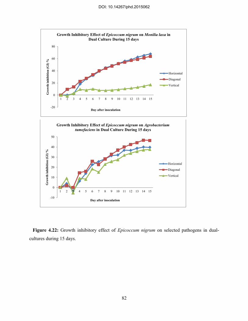
82
Figure 4.22: Growth inhibitory effect of Epicoccum nigrum on selected pathogens in dual-
cultures during 15 days.
-20
0
20
40
60
80
1 2 3 4 5 6 7 8 9 10 11 12 13 14 15
Gro
wth
inhi
bitio
n (G
I) %
Day after inoculation
Growth Inhibitory Effect of Epicoccum nigrum on Monilia laxa in Dual Culture During 15 days
Horizontal
Diagonal
Vertical
-10
0
10
20
30
40
50
1 2 3 4 5 6 7 8 9 10 11 12 13 14 15
Gro
wth
inhi
bitio
n (G
I) %
Day after inoculation
Growth Inhibitory Effect of Epicoccum nigrum on Agrobacterium tumefaciens in Dual Culture During 15 days
Horizontal
Diagonal
Vertical
DOI: 10.14267/phd.2015062

83
Figure 4.23: Growth inhibitory effect of Fusarium oxysporum on selected pathogens in dual-
cultures during 15 days.
-20
0
20
40
60
80
1 2 3 4 5 6 7 8 9 10 11 12 13 14 15
Gro
wth
inhi
bitio
n (G
I) %
Day after inoculation
Growth Inhibitory Effect of Fusarium oxysporum on Monilia laxa in Dual Culture Durin 15 Days
Horizontal
Diagonal
Vertical
0
20
40
60
1 2 3 4 5 6 7 8 9 10 11 12 13 14 15
Gro
wth
inhi
bitio
n (G
I) %
Day after inoculation
Growth Inhibitory Effect of Fusarium oxysporum on Agrobacterium tumefaciens in Dual Culture During 15 Days
Horizontal
Diagonal
Vertical
DOI: 10.14267/phd.2015062

84
Figure 4.24: Growth inhibitory effect of Rhizopycnis vagum on selected pathogens in dual-
cultures during 15 days.
-20
0
20
40
60
80
1 2 3 4 5 6 7 8 9 10 11 12 13 14 15
Gro
wth
inhi
bitio
n (G
I) %
Day after inoculation
Growth Inhibitory Effect of Rhizopycnis vagum on Monilia laxa in Dual Culture During 15 Days
Horizontal
Diagonal
Vertical
0
20
40
60
1 2 3 4 5 6 7 8 9 10 11 12 13 14 15
Gro
wth
inhi
bitio
n (G
I) %
Day after inoculation
Growth Inhibitory Effect of Rhizopycnis vagum on Agrobacterium tumefaciens in Dual Culture During 15 Days
Horizontal
Diagonal
Vertical
DOI: 10.14267/phd.2015062
85
Analytical data confirmed the primary results derived from in vitro assays about growth
inhibitory effects of nominated endophytic fungi on selected pathogens. The mean values of all
measurements were taken into account for calculating the variance (mean square) of radial
growth of an individual colony on PDA in three dimensions (horizontal, diagonal, and vertical).
Achieved data was applied to examine the significance of alterations in radial growth of every
colony on PDA during the incubation time (15 days) in single cultures (control) and in dual-
cultures where endophyte and pathogen confronted.
Collectively, Monilia laxa appeared more competent in dual-cultures with endophytes in
compare with Agrobacterium tumefaciens. In general, restriction in colony growth of Monilia
laxa was significant (P=0) in horizontal and diagonal directions but no difference was found in
vertical growth of the pathogen colonies in deal with endophyte in dual-cultures. On the other
hand, comparative analysis revealed that differences between endophytic fungi regarding their
capability to inhibit horizontal growth of Monilia laxa was significantly higher than that for
diagonal growth of the pathogen. Changes in horizontal growth of this pathogen ranged from
relatively weak when cultured with Rhizopycnis vagum (mean±variance=1.39±0.17 mm) to the
most regressed growth in dual-culture with Botrytis cinerea (mean±variance=0.68±0.01 mm) on
PDA. The inequality in growth inhibition effect between nominated endophytes was less
significant concerning their ability to diminish diagonal growth of Monilia laxa colonies.
Although Botrytis cinerea showed a relatively strong inhibitory effect on diagonal growth
(mean±variance=0.92±0.03 mm) of the pathogen, differences between other endophytes was not
significant. No significant difference was found between endophytic fungi regarding their
capability to inhibit vertical growth of Monilia laxa on PDA (Table 4.4) (Figure 4.25).
All examined endophytes had significant inhibitory effect on Agrobacterium tumefaciens
growth on PDA with no significant difference toward three dimensions. Nonetheless, the
outcome of inhibition was found different between endophyte taxa. According to the results,
horizontal growth of the pathogen had the maximum regression while confronted
Ceratobasidium sp.1, Embellisia sp., and Alternaria sp.1 on the media and the mean value of
horizontal regression of the pathogen colonies was equal in dual-cultures with mentioned fungi
(mean± variance= 0.56± 0.003 mm). In contrast, Rhizopycnis vagum had the weakest effect on
horizontal extension of the pathogen (mean±variance=0.71±0.022 mm). As well, this fungus had
relatively weaker effect on diagonal (mean±variance=0.66±0.006 mm) and vertical
DOI: 10.14267/phd.2015062

86
(mean±variance=0.71±0.01 mm) growth of Agrobacterium tumefaciens in compare with other
fungi, however variance of radial growth inhibition toward diagonal and vertical dimensions had
no significant difference between dual-cultures of this pathogen with selected endophytic fungi
(Table 4.5) (Figure 4.26).
Table 4.4: Two-way ANOVA test results of the average growth inhibition effect studied by
dual-culture of tested endophytic fungi with Monilia laxa.
Radial
Growth
Variance Sum
Square
Df* Mean
Square
Mean
Square
Between**
(F)
P
value
Horizont
al
Within 27.671 112 0.247 16.235 0.000
Between 28.109 7 4.016
Total 55.78 119 -
Diagonal Within 47.909 112 0.428 8.158 0.000
Between 24.428 7 3.49
Total 72.337 119 -
Vertical Within 72.657 112 0.649 0.942 0.477
Between 4.277 7 0.611
Total 76.934 119 -
* df- Degree of freedom
** Mean Square Between shows the mean square (variance) of inhibition effect between different dimensions.
F≥1 when the test is significant.
DOI: 10.14267/phd.2015062

87
Table 4.5: Two-way ANOVA test results of average growth inhibition effect studied by dual-
culture of tested endophytic fungi with Agrobacterium tumefaciens.
Radial
Growth
Variance Sum
Square
Df* Mean
Square
Mean
Square
Between**
(F)
P
value
Horizont
al
Within 1.853 112 0.017 13.415 0.000
Between 1.554 7 0.222
Total 3.407 119 -
Diagonal Within 1.867 112 0.017 14.894 0.000
Between 1.738 7 0.248
Total 3.606 119 -
Vertical Within 1.942 112 0.017 11.246 0.000
Between 1.365 7 0.195
Total 3.307 119 -
* df- Degree of freedom
** Mean Square Between shows the mean square (variance) of inhibition effect between different dimensions.
F≥1 when the test is significant.
DOI: 10.14267/phd.2015062

88
Figure 4.25: Radial growth (mean mm± variance) of Monilia laxa colonies in 1: single culture
(control), and dual-cultures with 2: Epicoccum nigrum, 3: Botrytis cinerea, 4: Fusarium
oxysporum, 5: Rhizopycnis vagum, 6: Ceratobasidium sp.1, 7: Embellisia sp., and 8: Alternaria
sp.1, has been demonstrated in this Figure. Significance of the radial growth change has been
indicated by (*). Bars show the variance of the radial growth of the pathogen in each dual-
culture.
-1
0
1
2
3
4
5
0 1 2 3 4 5 6 7 8Mea
n ra
dial
exte
nsio
n (m
m)
Culture
Horizontal Extension (Radial Growth Mean mm±Variance) of Monilia laxa Colony on PDA Confronting the Endophytic Fungi in Compare with Control
* * * * *
-1
0
1
2
3
4
5
0 1 2 3 4 5 6 7 8
Mea
n ra
dial
exte
nsio
n (m
m)
Culture
Diagonal Extension (Radial Growth Mean mm±Variance) of Monilia laxa Colony on PDA Confronting the Endophytic Fungi in Compare with Control
*
0
1
2
3
4
5
0 1 2 3 4 5 6 7 8
Mea
n ra
dial
exte
nsio
n (m
m)
Culture
Vertical Extension (Radial Growth Mean mm±Variance) of Monilia laxa Colony on PDA Confronting the Endophytic Fungi in Compare with Control
DOI: 10.14267/phd.2015062

89
Figure 4.26: Radial growth (mean mm±variance) of Agrobacterium tumefaciens colonies in 1:
single culture (control), and dual-cultures with 2: Epicoccum nigrum, 3: Botrytis cinerea, 4:
Fusarium oxysporum, 5: Rhizopycnis vagum, 6: Ceratobasidium sp.1, 7: Embellisia sp., and 8:
Alternaria sp.1, has been demonstrated in this Figure. Significance of the radial growth change
has been indicated by (*). Bars show the variance of the radial growth of the pathogen in each
dual-culture.
-1
-0,5
0
0,5
1
0 1 2 3 4 5 6 7 8Mae
n ra
dial
exte
nsio
n (m
m)
Culture
Horizontal Extension (Radial Growth Mean mm±Variance) of Agrobacterium tumefaciens Colony on PDA Confronting the Endophytic Fungi in Compare
with Control
* * *
-1
-0,5
0
0,5
1
1,5
0 1 2 3 4 5 6 7 8
Mea
n ra
dial
exte
nsio
n (m
m)
Culture
Diagonal Extension (Radial Growth Mean mm±Variance) of Agrobacterium tumefaciens Colony on PDA Confronting the Endophytic Fungi in Compare
with Control
-1
-0,5
0
0,5
1
1,5
0 1 2 3 4 5 6 7 8Mea
n ra
dial
exte
nsio
n (m
m)
Cullture
Vertical Extension (Radial Growth Mean mm±Variance) of Agrobacterium tumefaciens Colony on PDA Confronting the Endophytic Fungi in Compare
with Control
DOI: 10.14267/phd.2015062

90
As a sample demonstration figures 4.27 and 4.28 display growth inhibitory effect of Epicoccum nigrum on examined pathogens on PDA medium.
Figure 4.27: Single- culture of Epicoccum nigrum a) after 8 days and b) after 15 days,
Agrobacterium tumefaciens c) after 8 days and d) after 15 days, and Monilia laxa e) after 8 days
and f) after 15 days on PDA.
a b
c d
e f
DOI: 10.14267/phd.2015062

91
Figure 4.28: Dual- culture of Epicoccum nigrum vs Agrobacterium tumefaciens a) after 8 days
and b) after 15 days, and Epicoccum nigrum vs Monilia laxa c) after 8 days and d) after 15 days
on PDA.
a b
c d
DOI: 10.14267/phd.2015062

92
4.8. Novel scientific achievements by the present study
Based on the assigned goals and analytical approaches of the present study, our findings were
entirely unique as there has been no study as comprehensive and descriptive as ours on the
biodiversity of fungal endophytes associated with the examined hosts in Hungary as well as in
the Central Europe yet. The most important highlights of our achieved data are listed as follow:
1. The present study for the first time showed the phenotypic and phylogenic interrelations of
endophytic fungi assemblages grafted on Prunus Sp.
2. Biodiversity of isolated endophytes was analyzed on different rootstocks of Prunus Sp. By
application of statistical indices such as Simpson‟s diversity index and Shannon-Weaver index
which could reliably demonstrate the host-dependent features of identified fungi colonized these
species.
3. Results of the present Study were also analyzed according to the tendency of isolated fungal
endophytes to colonize a particular anatomical compartment in order to represent the tissue-
specific traits of these symbionts on the examined host species.
4. Based on the sampling procedures, we were also able to assess the impact of temporal
changes on the biodiversity of isolated endophytic fungi associated with the examined host
species.
5. All the above mentioned analyses were cumulatively considered to indicate the significant
variations in host-specific and tissue-specific characteristics of isolated fungal endophytes
according to the temporal changes in the sampling area.
6. We also examined the impact of harboring tissue, the host species and cultivation conditions
on fungal endophytes assemblages associated with Capsicum annuum L.
7. Results of this study for the first time showed the antagonistic effects of fungal symbionts
colonizing cherry rootstocks on growth of potential pathogens in vitro. As efficiently applied
criteria, the growth rate toward three dimensions and the inhibitory effect of fungus-pathogen
dual cultures were defined for all assays and the results were comprehensively discussed.
DOI: 10.14267/phd.2015062

93
5. Discussion
The present study provided valuable data regarding biodiversity and impact of determinant
factors on composition of fungal endophyte communities that attribute symbiotic micro-flora of
widely used cherry rootstocks in the region. It has been demonstrated that endophytic fungi can
profoundly influence different aspects of plant pathophysiology and actively participate bio-
mechanisms by which plant growth and regeneration is controlled. Thus understanding the
structure of endophyte assemblages is required for further approaches in order to improve the
agro-economic status of the fruit production and to find novel applications of these
microorganisms and their metabolites in horticultural, or even medicinal, studies. Results
achieved by the present study were applied for an analytical assessment to explicate the
composition of endophytic fungi assemblages on sweet cherry and pepper in Hungary.
Moreover, the antagonistic activity of isolated endophytes with vigorous growth on PDA was
assessed in vitro against to common plant pathogens. Data derived from such a study could be
useful for further investigation about the effect of these symbionts in pathophysiology of the host
and their contribution in host resistance against pathogens.
Biodiversity of endophytic fungi associated with host plant and structure of endophyte
communities are dynamically convertible depending on plant physiology, environmental
stresses, their interplay with other parasitic or pathogenic microorganisms and bio-geographical
factors (Arnold et al., 2007). Endophytes also tend to be host-specific that can change the
prevalence of endophytic taxa in a particular plant genus between different species. As well,
divergent endophyte composition may be harbored in host organs due to the histological
differences and availability of nutrients by which endophyte colonization is conducted in a
tissue-specific manner within a distinctive host species (Arnold et al., 2007). Along with
temporal changes in the niche where both host plant and endophyte inhabit, host species and
harboring tissue are consequently the most important factors modulating endophyte diversity
(Arnold & Herre, 2003) (Arnold et al., 2007). Therefore, the present study was conducted with
defined methodology so that could be applied to obtain such data and infer a comprehensive
view about composition of fungal endophyte communities associated with sweet cherry and
pepper.
DOI: 10.14267/phd.2015062

94
5.1. Characterization of endophytic fungi isolated from cherry rootstocks
As a sum, 26 morpho-taxa were classified among a total of 6587 isolated colonies from
cherry rootstocks. Some species primarily identified based on the morphology were later
subsumed with a different species name due to their phylogeny. Moreover, fungi with unique
ITS sequence which did not have acceptable resemblance to existed data, were identified as
unknown.
Although differences were observed in composition of endophyte assemblages between host
individuals, terrestrial and spatial accumulation of the endophytes and their occurrence due to the
temporal changes, an overall view could be inferred to characterize genera that orchestrate the
endophytic fungi populations associated with cherry rootstocks. The majority of identified
species (24 species) were from the phylum Ascomycota. Only two species of the phylum
Basidiomycota were isolated from cherry rootstocks. From Ascomycota, the most diverse species
belonged to Sordariomycetes while Dothideomycetes were the most frequent fungi isolated from
cherry rootstocks. Sordariomycetes were mostly Hypocreales (5 species), but this class also
included Phyllachorales (1 species), Sordariales (1 species), Diaporthales (1 species), and
Xylariales (2 species). The most abundant fungi (Alternaria sp.1 and Alternaria sp.2)
(Paleosporales) along with other less frequent fungi: Davidiella sp. (Capnodiales),
Macrophomina phaseolina ( Botryosphaeriales), Epicoccum nigrum and Rhizopycnis vagum
belonged to class Dothideomycetes, however taxonomy of the last two species is not thoroughly
assigned yet (Incertae sedis). Such a structure of endophytic fungi assemblages is identical to
formerly suggested endophytic association with woody plants and implies the potential
contribution of class 2 endophytes in symbiotic micro-flora of cherry rootstocks (Faeth & Fagan,
2002). Basidiomycota are reported to have rare evenness in plants. According to the results, two
species of Agaricomycetes (Ceratobasidium sp.1 and Ceratobasidium sp.2) were the less
abundant fungi identified on examined cherry rootstocks (Faeth & Sullivan, 2003).
Class 3 endophytes are usually considered as highly diverse fungi that are harbored in above
ground tissues of the host. Former studies have suggested the community of class 3 endophyte
mostly comprises Agaricomycetes, and with less frequency may contain Eurotiomycetes and
Pezizomycetes (Faeth & Sullivan, 2003). From Eurotiomycetes, Aspergillus niger (Eurotiales)
was identified on cherry rootstocks, whereas had occurrence only in root samples. A species
belonged to Pezizomycetes (Pyronema sp.) was also identified only in leaf with comparatively
DOI: 10.14267/phd.2015062

95
low frequency. Botrytis cinerea was the only genus of Leotiomycetes isolated from cherry
rootstocks and was only observed in twig.
In general, class 2 endophytes are recognized by their broad host range and horizontal
transmission in hosts. Nonetheless, self-experience on isolation of fungi from canola seed and
other studies on different plant species show that air-born fungi may colonize the seed (vertical
transmission) and thus may maintain transmission both within and between plant generations
(Faeth & Sullivan, 2003). Moreover, these endophytes are thought to enhance the host tolerance
against drought, temperature and salt and may help the host to resist against pathogens. the
greatest proportion of fungal metabolites which have industrial or medicinal applications and are
currently isolated and used in biological control, have been found in class 2 endophytes (Faeth &
Sullivan, 2003); (Berdy, 2005). Feature of endophytic fungi assemblages on cherry rootstocks, as
the results showed, highly resembles the association of class 2 and class 3 endophytes that
indicates the transmission mode of the fungi and their impact on biotic and abiotic tolerance of
the host. Nonetheless these endophytes also possess another trait which could be taken into
account to realistically conclude their relationship with host plants. Either class 2 or class 3
endophytic fungi may confer benefits to the host in a habitat-adapted manner, a characteristic
that make the host- endophyte interaction fluctuate in a range of symbiosis to parasitism and they
may also turn to pathogens under particular circumstances (Aly et al., 2011); (Faeth & Sullivan,
2003).
To characterize the nature of host plant-endophyte relationship, it is essential to determine
potential pathogenicity of these symbiotic inhabitants and their role in plant diseases. Nearly all
class 2, as well as class 3 endophytes were primarily identified as plant pathogens. Alternaria
sp., as the most frequent endophytic genus found in cherry rootstocks, is known as a major plant
pathogen with a widespread distribution; however study has documented the endophytic life
style of this genus in different plants. Embellisia sp., has been shown to induce bulb canker in
garlic and causes formation of black soot on alliums (Faeth & Fagan, 2002). Neonectria
radicicola is reported to cause ginseng root rot (Hamilton et al., 2010) but this species has been
also isolated as an endophyte harbored in roots of Pinus sylvestris (Hamilton et al., 2009).
Rhizopycnis vagum, is a recently described coelomycetous fungus that contributes to vine decline
of muskmelone in Central and North America, although this species has been isolated from
healthy tissues of cacao (Theobroma cacao L.) in Brazil (Meijer & Leuchtmann, 2001).
DOI: 10.14267/phd.2015062

96
Macrophomina phaseolina is recognized as an opportunistic fungus that may transform from a
saprophyte to a pathogen particularly in high temperature or water stress and causes root rot in
different plant species worldwide (Faeth & Sullivan, 2003). Glomerella acutata develops on
naturally infected fruit in two forms: conidial (Colletotrichum acutatum) and the ascigerous
(Glomerella acutata), but it endophytic association with plant in tropical and temperate regions
has been reported (Rodriguez et al., 2009 a); (Ernst et al., 2003). Davidiella sp., formerly known
as a pathogen inducing fruit rot in date palm, has been indicated to colonize host plants as an
endophyte from Antarctica to temperate flora (Weber et al., 2005); (Selosse et al., 2004);
(Newsham, 2011). Aspergillus niger and Chaetomium sp., are mainly considered as black mold
or dark-walled mold (dematiaceous) with pathogenic properties for plant and human, but have
been widely used in food industries and producing biocontrol agents due to their metabolites.
Reports are scarce about the endophytic life style of recently mentioned fungi species (Rodriguez
et al., 2008); (Rodriguez et al., 2009 a); (Arnold & Lutzoni, 2007).
Phytopathogens are conventionally categorized into three groups based on their habitat and the
way they obtain their nutrient supply: biotrophic, hemibiotrophic, and necrotrophic. Among
isolated endophytes from cherry rootstocks, Botrytis cinerea is known as a necrotrophic fungus
and usually is attributed with grey mold, although it has been recently demonstrated to have
endophytic traits in different plant species (Gamboa et al., 2002). Phomopsis sp., is also a
facultative plant pathogen which has been recently isolate as an endophyte from different hosts
(Verma et al., 2007); (Arnold & Herre, 2003). From Xylariales, the greatest number of Xylaria
sp., has been found in tropics. This fungus normally lives in dead tropical woody plants and may
colonize healthy tissue (Higgins et al., 2007); (Ganley et al., 2004); (Santamaria & Bayman,
2005). Whereas Xylaria digitata was identified as an endophyte associated with cherry
rootstocks, this species had relatively low frequency in the host. Ceratobasidium sp., was the
only genus of Basidiomycota isolated from cherry rootstocks. These fungi are commonly
facultative pathogens and have also saprotrophic life style. Species belong to this genus have
been isolated as endophytes mainly from host in the temperate regions (Selosse et al., 2009);
(Herre et al., 2007); (Rodriguez et al., 2009 b).
The most diverse fungi in orchestration of endophytic communities associated with cherry
rootstocks were Fusarium sp. Such a result may imply the ability of this genus to colonize host
tissue with no detectable symptom, however these fungi are normally considered as plant
DOI: 10.14267/phd.2015062

97
pathogens. There is a body of evidence that confirms symbiotic life style of Fusarium sp., with
plants (Jumpponen et al., 1998); (Kaplan et al., 2013); (Reininger & Sieber, 2012) that altogether
they highlight the importance of these fungi in bio-ecosystem and their impact on plant
pathophysiology. Some endophytes isolated from cherry rootstocks have formerly been shown to
be beneficial to the host. Acremonium sp., and Epicoccum nigrum, as two widespread fungi, have
been described as endophytes with ability to improve the host resistance against pathogens
(Mandyam et al., 2013); (Paul et al., 2012); (Marasco et al., 2012).
The most striking outcome of this study was the isolation and identification of two fungi
species on the leaf of cherry rootstocks (only during season I) which according to the best of our
knowledge have not been reported so far as resident endophytes of leaves at least in temperate
climate. Rosellinia sp., (Sordariomycetes) and Pyronema sp., (Pezizomycetes) were found only
in leaves in the first sampling period and had no occurrence on other tissues in that season or on
all examined specimens during the next two seasons, suggesting that these fungi may colonize
the host incidentally in a tissue-specific manner in early stages of the foliar growth.
5.2. Impact of host species, colonized tissue, and temporal changes on biodiversity of endophytic fungi
5.2.1. Sweet cherry All cherry trees subjected to the present study, were selected from an orchard nursery. Thus
such a sampling procedure did not allow analyzing the biodiversity of endophytes in different
habitats. Nonetheless, by considering the regional ecosystem of the sampling locality and other
factors like quality of soil and environmental conditions, results of this study can not only
demonstrate the status of endophytic fungi infection in cherry rootstocks throughout the state but
also can be extended to a broader geographical range in Central and Eastern Europe.
According to the results, endophytic communities associated with rootstocks belonged to
Prunus mahaleb L, were comparatively more diverse and distinctive species had higher
occurrence on these rootstocks during a particular sampling period, although the general feature
of endophyte assemblages on cherry rootstocks showed more tendencies to vary depending on
the host organ and seasonal changes than the host species. Perhaps less diverse endophytic
assemblages were observed on some rootstocks like Vadcseresznye or Gisela6, but disparities in
infection rates and species richness in different tissues of these individual hosts during three
sampling periods suggested mild impact of host species in composition of endophyte
DOI: 10.14267/phd.2015062

98
communities and their diversity in cherry rootstocks. Consequently, it remains to be elucidated
whether the association of endophytic fungi can favor vegetative growth, precocity, adaptability
to different environmental conditions, disease tolerance, and productivity of the examined cherry
rootstocks. Furthermore, the probability of host-endophytes interaction to switch from mutualism
to antagonism and changes in transmission mode of the fungi under the influence of host
physiology and other stresses is to be studied.
Infection burden on the host tissues and diversity of endophytic fungi showed more
correlation to the organ where they were colonized and the time when tissue specimens were
obtained. However, some degree of tissue-specificity was detected in distinctive fungi, but
colonization pattern and distribution of endophytes encompassed the pivotal role of temporal
changes in host physiology that more likely can induce alteration in the feature of these
symbionts communities.
The first and the third sampling procedures were fulfilled in early autumn time, with almost
one year interval between two periods, prior to defoliation of the trees, when the second
sampling period was carried on in mid spring during the flowering time. Differences concerning
the infection rate and composition of endophytes assemblages between these two periods perhaps
demonstrate the effect of annual changes and aging process in the host on biodiversity of fungal
endophytes. Nevertheless, host species may have an impact on endophyte divergence, more
probably based on the differences in growth habit, vegetation characteristics and their ability to
maintain efficient adaptivity to environmental stresses.
Results of the present study showed that roots of Prunus mahaleb L, rootstocks were less
infected in season I and then infection rate raised during the next two seasons. This index was
found different in Gisela6 which showed high rate of infection in the root during season III. The
highest infection in the root was detected during season I in Prob but decreased in the last two
seasons. It is generally believed that the root in woody plants is the main habitat of soil-borne
microorganisms and usually harbors more diverse symbiotic fungi and bacteria than spatial
organs of the host. It may explain the higher diversity of endophytic fungi in cherry rootstocks
however decline in number of isolated species and increased infection burden occurred in roots
of examined rootstocks can indicated that endophytic fungi horizontally transmit between
individual hosts and colonize the root while incidental species are depleted through the aging
process and taxa which are capable to adapt their niche perpetuate distribution and dominantly
DOI: 10.14267/phd.2015062

99
amplify the infection rate in the tissue. Seasonal changes in physiological functions of the root
rather affect endophyte continuum toward occurrence of more competent and prevalent fungal
endophyte than infection with more diverse and incidental species. It is not clear that how these
resident symbionts confer benefits (i.e. absorbance of water and nutrients, growth vigor, and
vulnerability to pathogens) to the root when their share of photosynthesis is supplied by the host
plant.
Temporal changes of infection rate in twigs were similar in all rootstocks and exhibited high
colonization and isolation rates in season I and season III, however these indices comparatively
had decline in season II. On the other hands, the number of isolated taxa had no difference
between the first two seasons and rose during the last sampling period. Altogether these findings
indicate that biodiversity of endophyte infection in the twig is more likely under the influence of
annual biochemical and physiological changes in this organ. It can be assumed that aged twigs
are preferentially colonized by endophytes that travel through water and nutrient transportation
system from the root to foliage during the time of plant vegetative growth. Moreover, twigs may
be less vulnerable to endophyte infection probably due to the physiological processes occur
during simultaneous vegetative and reproductive growth of the host.
As for leaves, results showed a remarkable increase in infection burden during season III in
compare with the first two seasons. Perhaps such findings indicate that the infection burden and
diversity of fungal endophyte flora on leaves of cherry rootstocks differs in accordance to the
aging process in foliar tissues but has less correlation to the annual changes. In addition,
tendency of some isolated fungi to exclusively colonize the leaf and occurrence of other taxa that
as well were accommodated in twigs and roots can highlight the existence of air-born fungi in
the studied ecosystem, although soil-born endophytes can infest the leaf via water and nutrient
transportation system.
As a matter of fact, these outcomes can be confirmed by existing knowledge about aspects of
endophytic fungi biodiversity in terrestrial plants. Former studies have also suggested that
species composition of endophyte assemblages and infection frequencies vary according to host
species, site characteristics, such as elevation, exposure, and associated vegetation, tissue type;
and tissue age (Khan,et al., 2012); (Ahlholm et al., 2002). For large woody hosts, growth stage
and position in the canopy also may affect distribution. In addition to the core group of species
consistently isolated as endophytes from any given host, surveys of plant hosts for endophytes
DOI: 10.14267/phd.2015062

100
invariably generate long lists of incidental species that are not known to sporulate on the host
(Aly et al., 2011); (Khan et al., 2012). Each incidental species often is represented only once or
twice in several hundred samples. In general, the number of rare and incidental species isolated
is proportional to the intensity of sampling; distribution of rare species is influenced more by site
than by host (Khan et al., 2012); (Arnold et al., 2007). Variation in species assemblages on the
same host at different sites usually is attributable to recovery of incidental species with more
disjunctive distributions. Pronounced seasonal differences in colonization frequencies might be
predicted for climates with a distinct wet/dry seasonal cycle. Therefore species composition and
relative abundances in endophyte assemblages may reflect spatial distributions as well as
sampling times (Arnold et al., 2007).
Studies on foliar and stem endophytes have been repeatedly verified that overall infection
frequencies increase with the age of host organs or tissues (Parsa et al., 2013); (Arnold et al.,
2007). This is best observed from evergreen plants or plants with long-lived foliage but is also
apparent to a lesser degree in deciduous trees and annuals. In term of methodology, however, it is
essential to distinguish between systemic endophtes and non-systemic epiphytes to assess the
colonization pattern and tissue-specificity of endophytes (Arnold et al., 2007). The infection
domain of endophytes has a profound effect on sampling efficiency for species diversity.
Unfortunately there is a lack of information about non-systemic, non–seed-borne fungi.
Dominant non-systemic endophytes known today are generally familiar epiphytes, such as
Alternaria sp., Cladosporium sp., and Epicoccum sp., or typical pathogen of grass hosts
(Rodriguez et al., 2009 a); (Provorov, 2009); (Porras-Alfaro & Bayman, 2011) . The differences
between systemic infections and those of limited domain should be taken into account for
plotting an efficient sampling strategy particularly when isolation of greater number of species
and/or assessment of tissue-specific biodiversity of endophytes is subjected. No sampling
method has been established yet that would be reliable to apply for detection of systemic and
epiphytic fungi and selection of sample units in majority of published studies is apparently
arbitrary and is highly variable. If sample units are not appropriate to the microscopic scale of
infections, undue bias will be introduced and then inferences regarding species dominance and
diversity drawn from those may be uncertain as a consequence (Arnold & Herre, 2003);
(Rodriguez et al., 2009 a).
DOI: 10.14267/phd.2015062

101
An important notion is also worth considering about protocols are used for collection and
preparation endophyte samples. So far, methods applied to detect and quantify the endophytic
fungi have been mainly based on common isolation from surfaced-sterilized tissue sections and
culture techniques. For inventories of species occurrences and diversity, that is presently the
most practical approach, although some species (e.g., obligate biotrophs) may be undetected or
underrepresented. Isolates that fail to sporulate in culture may need to be characterized by other
means. Presumably smaller sections of plant tissues and multiple cultures yield more accurate
estimation of fungal infection frequencies (Watrud et al., 2006). Also, conversely, larger
sampling units have greater potential to miss rare or slow-growing species and to recover mixed
genotypes of the same species.
5.2.2. Capsicum annuum L. In our experiments 42 endophytic fungal strains were successfully identified in pepper
(Bärnkopf, 2013). They belong to 19 genera: Alternaria, Cladosporium, Penicillium,
Acremonium, Chaetomium, Fusarium, Lewia, Arthirinium, Cercospora, Colletotrichum,
Galactomyces, Myrothecium, Paecilomyces, Plectosphaerella, Pyrenochaeta, Rhizopycnis,
Verticillium and Xylaria. 10 of these genera were also showed to be present in pepper by Paul et
al. (2011): Alternaria, Aspergillus, Bionectria, Chaetomium, Cladosporium, Colletotrichum,
Fusarium, Paecilomyces, Penicillium, Plectosphaerella and Xylaria. We also identified
Acremonium sp. as a quite frequently occurring endophyte, but this was not found by the Korean
research group. On the other hand, 11 of their strains were not positively identified in our
experiments up to now (Bärnkopf, 2013). This discrepancy may originate from the completely
different geographical region of cultivation, but also from the fact that neither Paul and
coworkers‟ studies (Paul et al., 2011) nor ours cover the whole range of the endophytes. The
success of detection of endophytic fungi depends on many factors. This shows the necessity to
use NGS sequencing methods for endophyte identification to gain more detailed information on
the endophyte population present.
Colonization of pepper plants did show several characteristic features. Colonization was
higher at the end of vegetation period and under open field conditions, but we have also observed
cultivar and organ specific differences. Of our cultivars „Hó‟ seemed to be higher colonized than
„Kárpia‟ and the fungi preferred old leaves, fruit stalk and roots in comparison to other organs.
DOI: 10.14267/phd.2015062

102
5.3. Pathogen growth inhibition by endophytic fungi
Recently, many scholars focus on the study of endophytes as biocontrol agent of pathogen and
insects and successfully applied endophytes to plant protection. It is suggested that endophytic
fungi confer resistance to plant pathogens by means of different mechanisms such as direct and
indirect inhibition of pathogenic microorganisms and ecological effects (Li et al., 2007); (Zhi-
Lin et al., 2012). The common method to evaluate antagonistic activity of endophytic fungi
against plant pathogens is the In Vitro co-culture with pathogens and endophytes, or comparison
of the survival rate of plant inoculated with fungal endophytes with endophyte-free plants (In
Planta methods). Despite growing number of studies, existing knowledge about potential
mechanisms by which endophytic fungi limit pathogen damage and protect their hosts from
diseases, is still so poor.
In the present study, seven endophytic species with more vigorous growth on the media were
selected for dual cultures with Monilia laxa and Agrobacterium tumefaciens. As it was
demonstrated by results, all species showed a range of inhibition and growth factors of pathogens
in all plates were collectively impaired in compare with controls. In all plates, endophytic fungi
had higher radial growth rate and their confrontation with pathogens had not only no influence
on colony expansion and development but also in the case of some species induced improved
growth rate to the endophyte. Presumably, being obliged competing other microorganisms
inflicts enhanced growth and intensifies colony development of the endophytic fungi in order to
acquire more nutrient resources and extend the life span of the species. Such an inference may
explain a possible mechanism by which endophytic fungi induce immunity to the host as they
contend other microorganisms in colonized tissues and inhibit the growth of potential pathogens.
These findings remark the ability of endophytic fungi to directly inhibit the pathogen. Many
fungal endophytes produce secondary metabolites and some of these compounds are antifungal
and antibacterial which strongly inhibit the growth of other microorganisms including plant
pathogens. For example, altersetin, a new alkaloid isolated from endophytic Alternaria spp.,
showed antibacterial activity against several pathogenic gram-positive bacteria (Hellwig et al.,
2002). However, as low concentration of these substances are excreted by endophytes in host
plant, the effects of antibiotics to pathogens In Vivo need to be assessed. As well, many
microorganisms produce and release lytic enzymes that can hydrolyze a wide variety of
polymeric compounds, including chitin, proteins, cellulose, hemi-cellulose and DNA
DOI: 10.14267/phd.2015062

103
(Choudhary et al., 2014). When endophytes colonize on the plant surface, they produce enzymes
to hydrolyze plant cell walls. As a result, these enzymes also have the function to suppress plant
pathogen activities directly and have the capability of degrading the cell walls of fungi and
oomycetes. Although, the enzymes may not be of most importance in antagonism, they may
contribute to antagonistic activity through a combination of mechanisms (Choudhary et al.,
2014). Endophytic fungi are also thought to induce host resistance to pathogen by activating the
plant natural defenses. During the long term evolution, plants have developed two types of innate
resistance: non-specific (general) resistance and specific resistance. The first form is effective
against several pathogenic species, while the latter can resist infection of one or a few pathogenic
strains (Barna et al., 2003). Since fungal endophytes may evolve from plant pathogenic fungi,
plant defense could be triggered by fungal endophytes like pathogens. Actually, the defense of
plant associated with endophytes is increased through resistance enhancement and secondary
metabolites production. As an example, Fusarium solani isolated from root tissues of tomato
elicited systemic resistance against the tomato foliar pathogen Septoria lycopersici and triggered
pathogenesis-related proteins (PR) genes, PR5 and PR7 expression in roots (Kavroulakis et al.,
2007). Epicoccum nigrum which was found in cherry rootstocks and had growth inhibitory effect
on examined pathogens in dual-cultures is also reported to contribute enhanced root growth and
generation of antifungal compounds in sugarcane. Antimicrobial activity of Epicoccum sp., is
attributed to production of antibiotics such as epicorazines A-B, epirodines A-B, flavipin,
epicoccines A-D, epipiridones, and epicocarines. In particular, flavipin and epicorazines A-B
have been associated with Epicoccum nigrum biocontrol activity against Monilia sp. (Favaro et
al., 2012).
Furthermore, it has been implied that endophytic fungi association can stimulate plant
resistance more rapidly and enhances the production plant secondary metabolites. These
metabolites have an important role in plants adaptation with their environment. However, studies
refer to the plant secondary metabolism mediated by the fungal endophytes is still on the early
stage. As an instance, a strains of Fusarium oxysporum could up-regulate the expression of
glutelin A (GLUA) and salicylic acid-inducible PR-1a genes in tomato root which promoted
tissue resistance against Fusarium wilt (Aime et al., 2013). It seems that co-culturing with
endophytic elicitor is an alternative way to enhance plant secondary metabolites and increase
plant resistance. Mechanism of endophytic elicitor induced plant secondary metabolites
DOI: 10.14267/phd.2015062

104
production is similar to stimulation of plant resistance. Colonization of fungal endophytes results
in the secretion of hydrolase of plant cell to limit the growth of fungi, thus, fragments of
endophytes, acting as elicitor, are produced by hydrolization. The elicitors such as
lipopolysaccharides, polysaccharides and glycoprotein will stimulate plant defense and plant
secondary metabolites, which would suppress pathogens attack efficiently. However,
determination of how endophytes can survive in high concentration of secondary metabolites in
host plant is still unknown (Ming et al., 2013).
Arguably, fungal endophyte association with host plants can shift between pathogenic and
symbiotic under the particular circumstances (Kaplan et al., 2013). It is assumed that
horizontally transmitted fungi may primarily generate parasitism, but in case emergence of
infection by other microorganisms, apply mechanisms to eliminate the rival and sustain normal
status of the host through life-history trade-offs with growth/survival. Thus they can perpetuate
their dominance in the adapted niche that in turn indirectly confers benefit to the host and
decreases the pathology of diseases (Faeth, 2009); (Arnold et al., 2003). Although screening of
antimicrobial activity of endophytic fungi isolated from cherry rootstocks was performed by
using In Vitro assays, such a life-history could be evolved for some species. For instance,
Botrytis cinerea had a comparatively high antagonistic effect on pathogen growth in dual
cultures, whereas it has been rather introduced as a pathogenic fungus in different hosts. These
types of fungi probably tend to colonize the host tissue as parasites and rapidly occupy the
ecological niche and leave no space for pathogen which would be the main reason that fungal
endophytes inhibit pathogen infection in plants. Such a property of fungal endophyte along with
the host reactions against endophytes (i.e. secretion of lignin and other cell-wall deposits to limit
the endophyte growth which as a shelter protects endophyte from pathogen invasion) and their
capability to parasitize pathogen organisms (hyperparasite endophytes) can be considered as the
ecological effects of endophytic fungi on plant resistance to pathogens (Petrini et al., 1992);
(Grosch et al., 2006).
DOI: 10.14267/phd.2015062

105
Summary
Endophytic symbionts including bacteria and fungi live within plant tissues without causing
any obvious negative effects and have been found in every plant species examined to date. It
became evident that endophytes are rich sources of bioactive natural products with promising
applications in development of pharmaceutical and industrial compounds. In addition studies in
recent decades have suggested all plants maintain associations with endophytes and epibionts
while such a symbiotic relationship is generally a cryptic phenomenon in Nature. Fungal
endophytes may inhabit tissues of roots, stems, branches, twigs, bark, leaves, petioles, flowers,
fruit, and seeds, including xylem of all available plant organs. These fungi are alleged to affect
the ecophysiology of plants, by frequently enhancing the capacity of host plants to tolerate
different environmental stresses through mechanisms that are only partially understood. It is also
believed that endophytes have important roles in plant protection by enhancing the plant
resistance against herbivores, insects and pathogens. Therefore, understanding the pattern of
endophytic infection and diversity of these symbionts in hosts within a certain biogeographical
niche is of a fundamental importance for improving the existing knowledge of plant physiology.
Besides, any attempt to expound the impact of the plant-endophyte interaction on biological
resistance of host plants against biotic and abiotic stresses can lead to apply more efficient
strategies for increasing the quality and quantity of agricultural and horticultural products.
In the current research work, colonization pattern of fungal endophytes on different rootstocks
of sweet cherry (Prunus spp) grafted on cultivar Péter including Érdi V., Bogdány, SL64,
Egervár, Korponay, SM11/4, CEMANY and Magyar rootstocks (of Prunus mahaleb L.,) , two
varieties of P.avium (Vadcseresznye) and P.fruticosa (Prob) and hybrid inbred rootstock of
P.cerasus and P.canescens (Gisela 6), was studied by application of morphological and
molecular phylogeny methods. Three anatomical tissue compartments (roots, twigs and leaves)
of above-mentioned rootstocks were collected during three different sampling periods (autumn
2008, spring 2009 and autumn 2009) and endophytic fungi were isolated by single-spore
methods and primarily identified according to their morphology. Phylogeny of the isolated fungi
was determined by application of PCR using ribosomal internal transcribed spacer (ITS) region
sequence. Furthermore, endophytes with more vigorous growth on culture media were used for
dual-cultures with two selected pathogens (Monilia laxa and Agrobacterium tumefaciens) to
DOI: 10.14267/phd.2015062
106
elucidate their antagonistic effect on one another. The same method was applied for isolation and
identification of endophytic fungi associated with pepper (Capsicum annuum L.) cultivars Hó F1
and Kárpia F1.. Samples were obtained from the root, shoot, leaves, pedicles, pericarps at
different vegetative stages and seed of pepper plants from open field (grown on sandy soil) and
the greenhouse (grown on rockwool). This process was performed in three replicates for four
times during the vegetation period (April, May, August and October, 2013).
As a sum, 26 morpho-taxa were classified among a total of 6587 isolated colonies from cherry
rootstocks. The majority of identified species (24 species) were from the phylum Ascomycota.
Two species of Alternaria spp (Paleosporales) were the most frequent isolates while Fusarium
spp showed the highest diversity in fungal assemblages on cherry rootstocks. The highest species
richness was detected in roots while twig samples showed the the highest infection burden in
cherry rootstocks. This study was the first to report isolation and identification of Rosellinia sp.,
(Sordariomycetes) and Pyronema sp., (Pezizomycetes) from trees in temperate climate only
detected on leaves in the first sampling period and had no occurrence on other tissues in that
season or on all examined specimens during the next two seasons. Collectively, however some
degrees of tissue-specificity was observed in composition of fungal assemblages identified on
cherry rootstocks, results of this study suggested that the biodiversity of endophytes is more
likely affected by annual climate alterations, temporal changes in the host physiology and the
process of aging. As well, species richness and relative frequency of fungi were found
comparatively higher in Prunus mahaleb L, although host-specificity seemed to affect the
biodiversity of endophytes on cherry trees less significantly than seasonal changes. As for the
growth inhibitory test, all examined endophytes showed antagonistic effect on growth and
development of the targeted pathogens while Alternaria sp.1 and Fusarium spp. were the
strongest and the weakest antagonists to pathogen growth, respectively. As an overall, 42 fungal
endophyte strains belonged to 19 genera were isolated from Capsicum annuum. Results showed
the dominant occurrence of Alternaria sp., and as well colonies belonged Acremonium sp., were
also isolated from different tissue compartments of pepper. Colonization rate of endophytic fungi
showed alteration due to the cultivar, harboring tissue and growth condition of the host. Cultivar
Hó F1 showed the higher colonization rate compared to Kárpia F1. The highest infection burden
was found at the end of vegetation period under the open field condition. Endophytic fungi
DOI: 10.14267/phd.2015062

107
showed higher tendency to old leaves, fruit stalks and roots of examined pepper plants. The
inhibitory effect of endophytes on pathogens growth has been discussed in detail.
As a conclusion, fungal endophytes isolated from cherry rootstocks demonstrated a
considerable diversity which is seemingly sustained by the physiological traits of the organ they
inhabit and temporal climate changes that influence the species richness and frequency of these
symbionts in their hosts. Moreover, these endophytes can likely contribute to the host resistance
against potential pathogens.
DOI: 10.14267/phd.2015062

108
Acknowledgement I would like to express my very great gratitude to Professor Dr. Noémi Lokács, my research
supervisor, for her patient guidance, enthusiastic encouragement and useful critiques as well as
her valuable and constructive suggestions during the planning and development of this research
work. Her willingness to give her time so generously has been very much appreciated.
I would also like to express my great appreciation to Dr. Krisztián Halász, for his kind
assistance and advice in keeping my progress on schedule and for his vast help in performing
laboratory experiments and his mentorship for methodology applied in this research work.
My grateful thanks are also extended to Dr. László Gáspár for his marvelous scientific advice
and his kind help in doing the computerized measurements and data analysis, all during this
research work.
I would also like to extend my thanks to all staff and technicians at the Department of Plant
Physiology and Plant Biochemistry for their help in offering me the resources in running the
program.
DOI: 10.14267/phd.2015062

109
Appendix
A1. Biodiversity of endophytic fungi associated with Prunus sp., rootstocks
Composition of endophytic fungi community in different rootstocks of Prunus sp.,
demonstrated various structures either between host species or in temporal and tissue-specific
manner between isolated colonies from an individual host.
A.1.1. Biodiversity of endophytic fungi in Prunus mahaleb L., rootstocks Bogdány, Érdi V, SL64, SM11/4, Egervár, Korponay, CEMANY, and Magyar were
rootstocks of Prunus mahaleb L., examined in the present study.
From Bogdány, a total of 700 colonies were isolated from root, twig and leaf specimens and
the highest infection burden was observed in season III (collectively, 336 colonies were isolated
at this season). During all three sampling periods, from all identified fungal endophyte species in
this study, 22 species were isolated from this rootstock. Maximum species richness in season I
was observed in root samples (8 species), in root samples in season II (7 species) but in twig
samples in season III (7 species) (Table A.1). The evenness of species, indicated by Shannon-
Weaver index was comparatively the highest in Season II (eH‟=1.71), while Simpson index was
expectedly the lowest (D=0.48) in the same season for Bogdány, demonstrating the highest
diversity of the endophytic fungi assemblages on this rootstock during the season II. The highest
abundance was detected for Alternaria sp.1 and Alternaria sp.2 that along with Epicoccum
nigrum (with comparatively lower abundance) were isolated from all tissue compartments of this
rootstock. The largest number of Alternaria sp.1 colonies was found on Twig samples in season
III and Alternaria sp.2; however was the most abundant on twig samples in season I. Rosellinia
sp., (RF=70%), Xylaria digitata (RF=67%) and Alternaria sp.1 (RF=55%) were the most
frequent species on leaf samples in season I, season II and season III, respectively. Rosellinia sp.,
was only isolated from leaf samples of this rootstock. Alternaria sp.2 was the most frequent
species obtained from twig samples in season I (RF=66%) and season II (RF=67%), while this
species was not observed in twigs during season III and instead, Alternaria sp.1 (RF=47%) was
the most frequent fungi in twigs from season III. The pattern of frequency in root samples during
season I was dominated by Alternaria sp.1 (RF=24%) and Paraphoma sp., (RF=24%).
Alternaria sp.1 was also the most frequent fungi in root during season II (RF=34%) but was not
isolated from this tissue in season III. In the last sampling period, Pyronema sp., (RF=28%)
DOI: 10.14267/phd.2015062

110
showed the highest frequency among other isolates in root samples from Bogdány rootstock.
According to the results, during season I and season III, explants collected from twigs of
Bogdány, demonstrated the highest colonization rate (CR=80%, and CR=99%, respectively)
along with the highest isolation rate (IR=1.04, and IR=1.47, respectively). The highest
colonization rate in the root was observed in season II (CR=63%) and in the leaf during season
III (CR=67%). As a sum, leaf samples exhibited the lowest colonization and isolation rates in
compare with other tissue specimens in this rootstock.
In total, 869 fungal endophyte colonies were isolated from Érdi V rootstock. The largest
number of isolated colonies was observed in twig samples during season III (199 colonies
isolated). Leaf samples during the season I showed the lowest number of isolated colonies (6
colonies isolated), but in season III a number of 159 colonies was obtained from this tissue. The
most abundant species in this rootstock were Alternaria sp.1 (280 isolated colonies) and
Alternaria sp.2 (214 isolated colonies) while the first species was obtained from all tissue
compartments in season III (120 isolated colonies from twigs, 82 isolated colonies from leaves
and 28 isolated colonies from roots) (RF=60%, RF=52%, and RF=36%, respectively) and only
from root samples (50 isolated colonies) during season II, but no isolate of Alternaria sp.1 was
observed in tissue samples from this rootstock in season I. on contrary, Alternaria sp.2 was only
isolated from all tissue samples in season I and from leaves and twigs, but not roots, in season II.
No colony of this species was isolated from this rootstock in season III. Among all identified
species, Rosellinia sp., was only isolated from leaf samples during the season I (4 isolated
colonies) (Table A.2). The highest species richness on Érdi V rootstock was observed in root
during the season I (9 species) while leaf samples showed the lowest richness according to the
isolated species and in season III, only two different species were isolated from leaves. The
evenness of identified species in season II was the highest in compare with other seasons
(eH‟=1.69) and the most entropy in endophyte communities was calculated in season III
(eH‟=0.41) when Simpson index showed the highest diversity of endophytic fungi associated with
Érdi V rootstock (D=1.73). During the season III, twigs and leaves showed a comparatively high
colonization rate (CR=99%, and CR=93%, respectively), however this index was much lower in
season II particularly in leaf samples (4.3%). Accordingly, the highest isolation rate was
observed in leaves and twigs during season III (IR=1.18, and IR=1.53, respectively). Root
DOI: 10.14267/phd.2015062

111
samples from this rootstock showed the moderate range of colonization, from CR=28% in season
I to CR=60% in season III, and isolation rate from IR=0.3 in season I to IR=0.7 in season III.
A total number of 898 endophytic fungi colonies were isolated from SL64 rootstock.
Maximmum infestion of this rootstock was found on twigs collected in season III (237 isolated
colonies). The lowest endophyte burden was indicated in leaves collected during season II (7
isolated colonies), although 165 colonies were collectively isolated from leaves in season III.
The most frequent species was Alternaria sp.1 identified in all tissue compartments during
season III: 139 colonies from twigs (RF=59%), 96 colonies from leaves (RF=58%), and 16
colonies from roots (RF=18%), however this species was not observed in all tissue samples from
SL64 during the season I. Alternaria sp.2 also showed a reletively high frequency in this
rootstock, but was mostly isolated from tissue samples in season II: 5 colonies from leaves
(RF=71%), 57 colonies from twigs (RF=77%), and 23 colonies from roots (RF=24%). During
season I, this species was isolated from leaves (16 isolated colonies, RF=57%) and twigs (118
isolated colonies, RF=73%), but was not observed in season III. During the season I, 8 colonies
of Rosellinia sp., were obtained from leaf samples (Table A.3). whereas the infection burden on
root samples was comparatively moderate, endophytes colonized on root samples showed the
highest species richness in all sampling periods (8 species in season I and season II, 6 species in
season III). The highest diversity in endophytic fungi communities was calculated in season II
(eH‟=0.96), but the index for season III showed less diverse endophytic assemblages on this
rootstock (eH‟=0.23). Simpson index for season II and season III was D=1.23 and D=1.86,
respectively. Explants obtained from twigs and leaves from SL64 during the third sampling
periods were entirely infected (CR=100%). However, this pattern was observed in twigs during
season I (CR=98%), but had an increase in season II (CR=46%) and was found relatively low in
leaves during season I and season II (CR=20%, and CR=4%, respectively). Accordingly, the
highest isolation rate was found in twig and leaf samples durin season III (IR=1.4, and IR=1.2,
respectively).
From SM11/4 rootstock, a number of 634 colonies were isolated showing the highest
infection burden on root samples during season II (141 isolated colonies) but less number of
fungi colonies on leaves in that season (5 isolated colonies). During the first season, the largest
number of colonies was isolated from twigs (133 isolated colonies) while this rate was almost
constant during next seasons (73 isolated colonies in season II and 121 isolated colonies in
DOI: 10.14267/phd.2015062

112
season III). Regarding other tissue samples, the number of isolated colonies was relatively
flactuated. For instance, from leaved 32 colonies in season I, 5 colonies in season II and 54
colonies in season III were isolated. The most frequent species were Alternaria sp.1 and
Alternaria sp.2, predominantly composed the endophyte cummunities associated with SM11/4
rootstock. Alternaria sp.1 was isolated from root in season II (60 isolated colonies, RF=43%),
and from leaf (20 isolated colonies, RF=37%), twig (54 isolated colonies, RF=45%), and root
(11 isolated colonies, RF=31%) samples in season III, but was not observed in season I. in
season I, alternaria sp.2 was predominant and found in leaf (16 isolated colonies, RF=50%), twig
(103 isolated colonies, RF=77%), and root (7 isolated colonies, 18%) samples. During season II,
the recent species was only found in leaves (3 isolated colonies, RF=60%) and twigs (50 isolated
colonies, RF=68%) and was not observed in season III. five isolated colonies of Rosellinia sp.,
was observed only in leaf samples during season I (Table A.4). the highest sepecies richness was
found in root during the season I (10 species) while leaf samples in season II beared the lowest
number of species (2 species) in this rootstock. Diversity indices demonstrated the most
divergent endophytic infection in season II (eH‟=1.49, D=0.57) and the most homogenic structure
of endophyte assemblages during season III (eH‟=0.58, D=1.6) associated with SM11/4
rootstock. Twig samples were the most infected tissue compartments of this rootstock as the
highest colonization rate was observed in twigs during the first (CR=97%, IR=1.2) and the third
(CR=99%, IR=1.6) sampling periods, however indices remarking infection rate showed an
decrease in season II (CR=47%, IR=0.6) in twigs. Leaf samples had also the same temporal
infection pattern but the highest colonization rate and isolation rate in roots were measured
during the season II (CR=82%, IR=1.2).
Experiment on Egervár rootstock yielded a total number of 692 endophyte colonies where
the largest number of isolates were detected in twig samples. In total, 125 isolated colonies
during season I, 74 isolated colonies during season II, and 169 isolated colonies during season III
were obtained from twigs. In the first two sampling periods, leaves showed a very low rate of
infection, although in season III, 105 colonies were isolated from leaf samples of this rootstock.
As a consequence, either twigs or leaves showed the maximum infection burden during the third
season, whereas root samples were mostly infested in season II (a total of 95 colonies were
isolated from root samples). The most abundant species on this rootstock was Alternaria sp.1 ( a
total of 195 isolated colonies) which was onserved in all tissue samples during season III: 43
DOI: 10.14267/phd.2015062

113
isolated colonied on leaf (RF=41%), 83 isolated colonies on twig (RF=49%), and 19 isolated
colonies on root (RF=27%), but it was only isolated from root samples during season I and
season II (17 isolated colonies, RF=43%, and 33 isolated colonies, RF=35%, respectively).
Alternaria sp.2 had a slightly lower abundance (a total of 147 isolated colonies), however it was
isolated from examined tissue samples of leaves and twigs in season I: 7 isolated colonies from
leaf (RF=70%), and 84 isolated colonies from twigs (RF=67%), and in season II: 4 isolated
colonies from leaf (RF=80%), and 52 isolated colonies from twig (RF=70%) with relatively high
frequency but was not observed in root samples. The recent species was not detected on tissue
samples during season III. Rosellinia sp., was isolated only from leaf samples (1 colony) during
the season I (Table A.5). As a result, the highest species richness was measured in root during
the first two seasons (8 and 7 sepcies, respectively) but twig samples in season III harbored the
highest number of species (7 species) in compare with other tissue compartments. The most
diverse endophyte communities colonizing Egervár rootstock were detected in season II
(eH‟=1.42, D=0.7), but diversity indices showed that the endophytic fungi associted with this
rootstock had relativley the most homogenous structure in season III, according to the existence
of different taxa (eH‟=0.4, D=1.8). During season I, the highest colonization and isolation rates
were measured in twigs (CR=84%, IR=1.1), while such a pattern shifted on root during season II
when these indices showed the highest values for this tissue (CR=77%, IR=0.8). during the first
two seasons, leaves showed relatively low colonization and isolation rates (CR=7%, IR=0.1, and
CR=3%, IR=0.04, respectively). In season III, however, explants from twigs were the most
infected tissue specimens of this rootstock (CR=100%, IR=1.5) and besides, leaf samples also
exhibited higher colonization and isolation rates (CR=80%, IR=1) in compare with previous
sampling periods. Root samples had the lowest infection rates during the season III (CR=58%,
IR=0.7).
According to the results, 688 fungal endophyte colonies were isolated from Korponay
rootstock. The adundance of isolated colonies was found the highest in twigs during season I
(107 isolated colonies) and season III (154 isolated colonies), but the largest number of colonies
was isolated from roots (102 isolated colonies) in the second sampling period. The infection
burden of leaf in this rootstock had a drastic increase in season III (111 isolated colonies) in
compare with the first two seasons. Collectively, twig was found as the most infected tissue
compartment in Korponay rootstock while leaf samples had the lowest infection burden in this
DOI: 10.14267/phd.2015062
114
host. The most frequent species was Alternaria sp.1 (222 isolated colonies) that was detected on
all samples during season III: 69 colonies isolated from leaf samples (RF=62%), 97 colonies
isolated from twigs (RF=63%), and 18 colonies isolated from roots (RF=21%), but only was
obtained from root samples in season II (38 isolated colonies, RF=37%) and was observed on
neither of the tissue samples during season I. In contrast, Alternaria sp.2 was detected in season I
on leaves (13 isolated colonies, RF=68%), twigs (66 isolated colonies, RF=62%), and on roots (4
isolated colonies, RF=10%), and in season II on leaf (4 isolated colonies, RF=80%) and twig (43
isolated colonies, RF=63%), but had no occurance in season III. Rosellinia sp., was just isolated
from leaf samples during season I (4 isolated colonies) (Table A.6). Root samples of this
rootstock bearded the richest species divesity during season I and season II (10 species and 7
species, respectively) but in season III the highest number of species was identified in twig
samples (8 species). In all seasons, the lowest species richness was detected in leaves. Diversity
indices also showed that the endophytic fungi associated with Korponay rootstoch were
comparatively the most divergent in season II and had the less diversity in season III (eH‟=1.64,
D=0.54, and eH‟=0.53, D=1.62, respectively). During season I and seaon III, the highest
colonization and isolation rates were calculated in twigs (CR=67%, IR=1, and CR=96%, IR=1.4,
respectively) while these values were the highest in root samples in season II (CR=70%, IR=0.9).
Leaves showed comparatively low values of infection in season I and season II (CR=17%,
IR=0.2, and CR=4%, IR=0.04, respectively), but indices had increase (CR=94%, IR=1) during
season III.
From CEMANY rootstock, a total of 592 colonies were isolated and identified during three
sampling periods. In season I and season III, the largest number of isolated colonies was
obtained from twigs (97 isolated colonies and 134 isolated colonies, respectively), while in
season II, root samples had the highest infection burden (91 isolated colonies). In the first two
seasons, leaf samples showed a very low infection burden (7 isolated colonies and 2 isolated
colonies, respectively) but in season III a total of 107 colonies were isolated from leaves of this
rootstock. Alternaria sp.1 was the most frequent species (151 isolated colonies) obtained only
from root samples (26 isolated colonies, RF=29%) in season II, but from all tissue samples in
season III (23 isolated colonies from leaf, RF=21%, 84 isolated colonies from twig, RF=63%,
and 18 isolated colonies from root, RF=37%). This fungus was not observed in season I. the
second most frequent species, Alternaria sp.2 (127 isolated colonies), was detected in leaves (4
DOI: 10.14267/phd.2015062

115
isolated colonies, RF=57%), twigs (67 isolated colonies, RF=69%, and in roots (3 isolated
colonies, RF=37%) during the season I and in leaf (2 isolated colonies, RF=100%) and in twig
(51 isolated colonies, RF=75%) but not from root during the season II. This fungus, however,
was not observed in season III. Rosellinia sp., was detected on leaf samples during season I (2
isolated colonies) (Table A.7). As a sum, the highest species richness was found in root.
Although this value was the highest in root during season I (8 species) and season II (7 species),
but in season III more species were isolated from twigs (5 species) in compare with root samples
(4 species). Leaf samples obtained from CEMANY rootstock showed the lowest species richness
in all seasons. Diversity indices demonstrated the highest diversity in endophyte communities
during season II (eH‟=1.27, D=0.8) and less diverse composition of the identified endophytes
assemblages during season III (eH‟=0.5, D=1.7). Whereas, the rate of infection was
proportionally higher in root samples (CR=66%, IR=0.9) during season II in compare with leaf
and twig, the most remarkable colonization and isolation rates were calculated in twigs during
season I (CR=86%, IR=1.1) and season III (CR=100%, IR=1.5) among all tissue samples.
Infection rates in leaf samples from CEMANY rootstock, although were the lowest in the first
two seasons (CR=7%, IR=0.1, and CR=2%, IR=0.02, respectively), showed a striking increase in
season III (CR=82%, IR=1.2).
A total number of 515 colonies were isolated from Magyar rootstock. Although in general,
twig samples had the highest infection burden but in season II the largest number of colonies was
isolated from root samples (75 isolated colonies). The number of isolated colonies was
comparatively low in leaf samples during the first two seasons (11 isolated colonies and 5
isolated colonies, respectively), while 81 colonies were isolated from this tissue in season III.
Alternaria sp.2 was the most frequent fungus isolated from this rootstock (152 isolated colonies).
This species was found only in root (30 isolated colonies, RF=40%) in season II, but was
isolated from all tissue compartments (35 isolated colonies from leaf, RF=43%, 65 isolated
colonies from twig, RF=68%, and 8 isolated colonies from root, RF=26%) during season III.
Alternaria sp.2 was not detected on Magyar rootstock in season I. with a slightly lower
abundance, Alternaria sp.1 was the second most frequent species on this rootstock isolated in
season I from leaf (7 isolated colonies, RF=64%), twig (87 isolated colonies, RF=67%), and
from root (7 isolated colonies, RF=33%) and in season II from leaf (2 isolated colonies,
RF=40%), and from twig (49 isolated colonies, RF=73%) and had no occurance in root during
DOI: 10.14267/phd.2015062

116
this season. Moreover, the recent species was not detected on Magyar rootstock during season
III. Rosellinia sp., was isolated (3 colonies) from leaf only during the first season (Table A.8).
Species richness was higher in root samples in compare with the other tissues (8 species and 7
species were identified from root samples during season I and season II, respectively), but this
value was the highest in twig samples during season III (5 species). Diversity of endophytic
fungi associated with Magyar rootstock was almost the same in season I and season III, however
indices showed more diversity in season II (eH‟=1.53, D=0.57). In all three seasons, the highest
colonization and isolation rates were found in twig samples. As a sum, 97% of twig explants
(CR) were infected during season I and an avarage of 1.3 fungi colonies (IR) were isolated from
each explant during the season. These values for twig samples in season II (CR=56%, IR=0.7)
and season III (CR=95%, IR=1.2) also were relatively higher than the other tissue compartments
of Magyar rootstock. Colonization rate and isolation rate in leaves were calculated as CR=9.2% ,
IR=0.1 in season I and CR=5%, IR=0.05 in season II, although a drastic increase was detected in
the rate of infection for leaf samples during the third season (CR=78%, IR=1).
DOI: 10.14267/phd.2015062

117
Table A.1: Abundance of isolated colonies from identified fungal endophyte species
associated with Bogdány rootstock in three sampling periods. Fungi Genera Season I Season II Season III Total
Leaf Twig Root Leaf Twig Root Leaf Twig Root Rosellinia sp.
21 - - - - - - - - 21
Chaetomium sp. - 2 - - 8 - - - - 10 Macrophomina phaseolina - - - - 10 - - - - 10
Pyronema sp.
- - - - - - - - - -
Alternaria sp.1 - - 6 - - 37 55 83 17 198 Alternaria sp.2
7 78 - 1 52 - - - - 138
Xylaria digitata
- - - 2 - - - - - 2
Aspergillus niger
- - - - - - - - 2 2
Acremonium furcatum
- - - - - 23 - - - 23
Rhyzopycnis vagum
- - - - - - - - - -
Ceratobasidium sp.1
- - - - - 1 - - - 1
Ceratobasidium sp.2
- - 1 - - - - - - 1
Neonectria radicicola
- - 2 - - - - - - 2
Acremonium sp.
- - - - - - - - - -
Paraphoma sp.
- - 6 - - 11 - - 4 21
Phomopsis sp.
- - - - - - - 3 - 3
Epicoccum nigrum - 32 3 - 7 - 12 9 1 64 Embellisia sp. - - 1 - - - - 4 - 5 Davidiella sp.
1 - - - - 33 - - - 34
Botrytis cinerea
- - - - - - - - - -
Fusarium sp.1
- - - - - - - 23 15 38
Fusarium sp.2 - - - - - 1 - 42 - 43 Fusarium sp.3 - 1 2 - - 1 23 - - 27
Fusarium oxyporum - - 1 - - - - - - 1 Fusarium solani - 5 - - 1 - - - - 6
Glomerella acutata
- - - - - - - 2 - 2
Unknown 1 - 3 - - 3 10 9 22 48 Total 30 118 25 3 78 110 100 175 61 700
DOI: 10.14267/phd.2015062

118
Table A.2: Abundance of isolated colonies from identified fungal endophyte species
associated with Érdi V rootstock in three sampling periods. Fungi Genera Season I Season II Season III Total
Leaf Twig Root Leaf Twig Root Leaf Twig Root Rosellinia sp.
4 - - - - - - - - 4
Chaetomium sp. - - - - 4 - - - - 4 Macrophomina phaseolina - - - - 16 - - - - 16
Pyronema sp. - - - - - - - - - -
Alternaria sp.1 - - - - - 50 82 120 28 280 Alternaria sp.2
25 125 12 3 49 - - - - 214
Xylaria digitata 2 - - 2 - - - - - 4
Aspergillus niger - - - - - - - - 3 3
Acremonium furcatum - - - - - - - - 4 4
Rhyzopycnis vagum - - 1 - - 4 - - - 5
Ceratobasidium sp.1 - - - - - 1 - - - 1
Ceratobasidium sp.2 - - - - - - - - - -
Neonectria radicicola - - 5 - - - - - - 5
Acremonium sp. - - - - - 16 - - - 16
Paraphoma sp. - - 2 - - 5 - - 9 16
Phomopsis sp. - - - - - - - 3 - 3
Epicoccum nigrum - 31 - - 9 1 9 2 52 Embellisia sp. - - 1 - - - - 21 - 22 Davidiella sp.
6 - 3 1 - 26 - - - 36
Botrytis cinerea - - - - - - - 4 - 4
Fusarium sp.1 - - 3 - - - - 7 12 22
Fusarium sp.2 - - - - - - - 24 - 24 Fusarium sp.3 - 2 2 - 2 1 61 - - 68
Fusarium oxyporum - - 2 - - - - - - 2 Fusarium solani - 4 - - 2 - - - - 6
Glomerella acutata - - - - - - - 6 - 6
Unknown 1 - 8 - - 2 16 5 20 52 Total 38 162 39 6 82 106 159 199 78 869
DOI: 10.14267/phd.2015062

119
Table A.3: Abundance of isolated colonies from identified fungal endophyte species
associated with SL64 rootstock in three sampling periods. Fungi Genera Season I Season II Season III Total
Leaf Twig Root Leaf Twig Root Leaf Twig Root Rosellinia sp.
8 - - - - - - - - 8
Chaetomium sp. - - - - 10 - - - - 10 Macrophomina phaseolina - - - - 1 - - - - 1
Pyronema sp. - - - - - - - - - -
Alternaria sp.1 - - 9 - - - 96 139 16 260 Alternaria sp.2
16 118 - 5 57 23 - - - 219
Xylaria digitata - - - - - - - - - -
Aspergillus niger - - - - - - - - 1 1
Acremonium furcatum - - - - - - - - - -
Rhyzopycnis vagum - - - - - 4 - - - 4
Ceratobasidium sp.1 - - - - - - - - - -
Ceratobasidium sp.2 - - 4 - - - - - - 4
Neonectria radicicola - - 1 - - - - - - 1
Acremonium sp. - - 1 - - 4 - - - 5
Paraphoma sp. - - 4 - - 16 - - 6 26
Phomopsis sp. - - - - - - - - - -
Epicoccum nigrum - 37 - - 4 2 35 3 2 83 Embellisia sp. - - - - - - - 6 - 6 Davidiella sp.
2 - 9 1 - 40 - - - 52
Botrytis cinerea - - - - - - - - - -
Fusarium sp.1 - - 2 - - - - 19 15 36
Fusarium sp.2 - - - - 1 - 54 - 55 Fusarium sp.3 - 3 3 - 1 2 17 - - 26
Fusarium oxyporum - - - - - - - - - - Fusarium solani - 2 - - 1 - - - - 3
Glomerella acutata - - - - - - - 7 - 7
Unknown 2 1 7 1 - 5 17 9 49 91 Total 28 161 40 7 74 97 165 237 89 898
DOI: 10.14267/phd.2015062

120
Table A.4: Abundance of isolated colonies from identified fungal endophyte species
associated with SM11/4 rootstock in three sampling periods. Fungi Genera Season I Season II Season III Total
Leaf Twig Root Leaf Twig Root Leaf Twig Root Rosellinia sp.
5 - - - - - - - - 5 Chaetomium sp. - 2 - - 6 - - - - 8
Macrophomina phaseolina - - - - 5 - - - - 5 Pyronema sp.
10 - - - - - - - - 10 Alternaria sp.1 - - - - - 60 20 54 11 145 Alternaria sp.2
16 103 7 3 50 - - - - 179 Xylaria digitata
- - - 2 - - - - - 2 Aspergillus niger
- - - - - - - - - - Acremonium furcatum
- - - - - 27 - - 1 28 Rhyzopycnis vagum
- - 1 - - - - - - 1 Ceratobasidium sp.1
- - - - - - - - - - Ceratobasidium sp.2
- - 1 - - - - - - 1 Neonectria radicicola
- - 4 - - - - - - 4 Acremonium sp.
- - - - - - - - - - Paraphoma sp.
- - 3 - - - - - 1 4 Phomopsis sp.
- - - - - 7 - 3 - 10 Epicoccum nigrum - 19 - - 11 - 22 - 3 55
Embellisia sp. - - - - - - - 14 - 14 Davidiella sp.
1 - 9 - - 36 - - - 46 Botrytis cinerea
- - - - - - - 10 - 10 Fusarium sp.1
- - 6 - - - - 10 - 16 Fusarium sp.2 - - 4 - - - - 24 - 28 Fusarium sp.3 - 1 1 - 1 4 5 - - 12
Fusarium oxyporum - - 1 - - - - - - 1 Fusarium solani - 7 - - - - - - - 7
Glomerella acutata - - - - - - - - - -
Unknown - 1 2 - - 7 7 6 20 43 Total 32 133 39 5 73 141 54 121 36 634
DOI: 10.14267/phd.2015062

121
Table A.5: Abundance of isolated colonies from identified fungal endophyte species
associated with Egervár rootstock in three sampling periods. Fungi Genera Season I Season II Season III Total
Leaf Twig Root Leaf Twig Root Leaf Twig Root Rosellinia sp.
1 - - - - - - - - 1
Chaetomium sp. - - - - 6 - - - - 6 Macrophomina
phaseolina - - - - 3 - - - - 3
Pyronema sp. - - - - - - - - - -
Alternaria sp.1 - - 17 - - 33 43 83 19 195 Alternaria sp.2
7 84 - 4 52 - - - - 147
Xylaria digitata - - - - - - - - - -
Aspergillus niger - - - - - - - - 1 1
Acremonium furcatum
- - - - - - - - - -
Rhyzopycnis vagum - - - - - 4 - - - 4
Ceratobasidium sp.1 - - - - - 1 - - - 1
Ceratobasidium sp.2 - - 2 - - - - - - 2
Neonectria radicicola
- - 5 - - - - - - 5
Acremonium sp. - - - - - 8 - - - 8
Paraphoma sp. - - - - - 9 - - 4 13
Phomopsis sp. - - - - - - - 2 - 2
Epicoccum nigrum - 29 1 - 8 - - 1 1 40 Embellisia sp. - - 1 - - - - 9 - 10 Davidiella sp.
- - 8 1 - 25 - - - 34
Botrytis cinerea - - - - - - - 8 - 8
Fusarium sp.1 - - - - - - - 9 21 30
Fusarium sp.2 - - - - - - - 41 - 41 Fusarium sp.3 - 7 1 - 1 2 38 - - 49
Fusarium oxyporum - - 3 - - - - - - 3 Fusarium solani - 5 - - 3 - - - - 8
Glomerella acutata - - - - - - - - - -
Unknown 2 - 2 - 1 13 24 16 23 81 Total 10 125 40 5 74 95 105 169 69 692
DOI: 10.14267/phd.2015062

122
Table A.6: Abundance of isolated colonies from identified fungal endophyte species
associated with Korponay rootstock in three sampling periods. Fungi Genera Season I Season II Season III Total
Leaf Twig Root Leaf Twig Root Leaf Twig Root Rosellinia sp.
4 - - - - - - - - 4
Chaetomium sp. - 4 - - 6 - - - - 10 Macrophomina phaseolina - 5 - - 7 - - - - 12
Pyronema sp. - - - - - - - - - -
Alternaria sp.1 - - - - - 38 69 97 18 222 Alternaria sp.2
13 66 4 4 43 - - - - 130
Xylaria digitata 1 - - 1 - - - - - 2
Aspergillus niger - - - - - - - - 10 10
Acremonium furcatum - - - - - - - - 1 1
Rhyzopycnis vagum - - - - - - - - - -
Ceratobasidium sp.1 - - - - - - - - - -
Ceratobasidium sp.2 - - - - - - - - - -
Neonectria radicicola - - 2 - - - - - - 2
Acremonium sp. - - 1 - - 11 - - - 12
Paraphoma sp. - - 8 - - 23 - - 7 38
Phomopsis sp. - - - - - - - 3 - 3
Epicoccum nigrum - 23 2 - 8 - - 4 5 42 Embellisia sp. - - 5 - 0 - - 6 - 11 Davidiella sp.
- - 8 - - 12 - - - 20
Botrytis cinerea - - - - - - - 4 - 4
Fusarium sp.1 - - 2 - - 2 - 9 16 29
Fusarium sp.2 - - 2 - - 10 - 25 - 37 Fusarium sp.3 - - 1 - - 5 26 - - 32
Fusarium oxyporum - - - - - - - - - - Fusarium solani - 6 - - 3 - - - - 9
Glomerella acutata - - - - - - - 4 - 4
Unknown 1 3 4 - 1 1 16 2 26 54 Total 19 107 39 5 68 102 111 154 83 688
DOI: 10.14267/phd.2015062
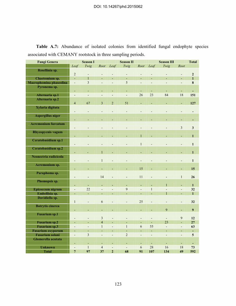
123
Table A.7: Abundance of isolated colonies from identified fungal endophyte species
associated with CEMANY rootstock in three sampling periods. Fungi Genera Season I Season II Season III Total
Leaf Twig Root Leaf Twig Root Leaf Twig Root Rosellinia sp.
2 - - - - - - - - 2 Chaetomium sp. - 1 - - - - - - - 1
Macrophomina phaseolina - 3 - - 5 - - - - 8 Pyronema sp.
- - - - - - - - - - Alternaria sp.1 - - - - - 26 23 84 18 151 Alternaria sp.2
4 67 3 2 51 - - - - 127 Xylaria digitata
- - - - - - - - - - Aspergillus niger
- - - - - - - - - - Acremonium furcatum
- - - - - - - - 3 3 Rhyzopycnis vagum
- - - - - 1 - - - 1 Ceratobasidium sp.1
- - - - - 1 - - - 1 Ceratobasidium sp.2
- - 1 - - - - - - 1 Neonectria radicicola
- - 1 - - - - - - 1 Acremonium sp.
- - - - - 15 - - - 15 Paraphoma sp.
- - 14 - - 11 - - 1 26 Phomopsis sp.
- - - - - - - 1 - 1 Epicoccum nigrum - 22 - - 9 - 1 - - 32
Embellisia sp. - - - - - - - 1 - 1 Davidiella sp.
1 - 6 - - 25 - - - 32 Botrytis cinerea
- - - - - - - 9 - 9 Fusarium sp.1
- - 3 - - - - - 9 12 Fusarium sp.2 - - 4 - - - - 23 - 27 Fusarium sp.3 - - 1 - 1 6 55 - - 63
Fusarium oxyporum - - - - - - - - - - Fusarium solani - 3 - - 2 - - - - 5
Glomerella acutata - - - - - - - - - -
Unknown - 1 4 - - 6 28 16 18 73 Total 7 97 37 2 68 91 107 134 49 592
DOI: 10.14267/phd.2015062

124
Table A.8: Abundance of isolated colonies from identified fungal endophyte species
associated with Magyar rootstock in three sampling periods. Fungi Genera Season I Season II Season III Total
Leaf Twig Root Leaf Twig Root Leaf Twig Root Rosellinia sp.
3 - - - - - - - - 3
Chaetomium sp. - 2 - - 2 - - - - 4 Macrophomina phaseolina - 2 - - 4 - - - - 6
Pyronema sp. - - - - - - - - - -
Alternaria sp.1 - - - - - 30 35 65 8 138 Alternaria sp.2
7 87 7 2 49 - - - - 152
Xylaria digitata - - - 2 - - - - - 2
Aspergillus niger - - - - - - - - -
Acremonium furcatum - - - - - 23 - - 3 26
Rhyzopycnis vagum - - - - - - - - - -
Ceratobasidium sp.1 - - - - - - - - - -
Ceratobasidium sp.2 - - - - - - - - - -
Neonectria radicicola - - - - - - - - - -
Acremonium sp. - - - - - - - - - -
Paraphoma sp. - - 5 - - 4 - - 1 10
Phomopsis sp. - - - - - - - 1 - 1
Epicoccum nigrum - 36 1 - 9 - 20 1 - 67 Embellisia sp. - - - - - - - 1 - 1 Davidiella sp.
- - 6 1 - 13 - - - 20
Botrytis cinerea - - - - - - - 1 - 1
Fusarium sp.1 - - 1 - - - - 13 9 23
Fusarium sp.2 - - - - - - - 14 - 14 Fusarium sp.3 - - - - - 4 22 - - 26
Fusarium oxyporum - - - - - - - - - - Fusarium solani - 2 - - 2 - - - - 4
Glomerella acutata - - - - - - - - - -
Unknown 1 - 1 - 1 1 4 - 9 17 Total 11 129 21 5 67 75 81 96 30 515
DOI: 10.14267/phd.2015062

125
A.1.2. Biodiversity of endophytic fungi associated with other Prunus sp., rootstocks From Prunus avium, a rootstock with high prevalence in Hungarian niche (Vadcseresznye)
was selected for the present study. According to the results, a total number of 372 fungal
endophyte colonies were isolated from tissue samples of Vadcseresznye rootstock. The infection
burden in the first season was relatively low as 30 colonies from twig samples and only 4
colonies from root samples were isolated and leaf samples showed no infection during season I.
The rate of infection had an increase during the second and the third seasons. In season II, the
largest number of colonies was isolated from roots (80 isolated colonies) but this amount reduced
to 65 isolated colonies in season III. During the last sampling period, twig samples were more
infected in compare with the other two tissue compartments as 84 isolated colonies were
obtained from twigs in season III. Although leaf had the lowest infection burden in all seasons,
but the number of isolated endophytes from leaf samples had a significant increase as 7 colonies
in season II and 30 colonies in season III were isolated from these specimens. Alternaria sp.1
and Alternaria sp.2 were the most frequent species among other identified endophytes associated
with Vadcseresznye rootstock; however the pattern of tissue colonization and temporal
occurrence of these species showed a significant difference. Collectively, Alternaria sp.1 was
only isolated from root samples in season II (21 isolated colonies, RF=27%) and from leaf (24
isolated colonies, RF=80%), twig (42 isolated colonies, RF=50%), and from root (25 isolated
colonies, RF=38%) during season III. This species was not detected on Vadcseresznye rootstock
in season I. On the other hand, Alternaria sp.2 was only observed on twigs during season I (20
isolated colonies, RF=67%) and during season II was isolated from leaf (4 isolated colonies,
RF=57%) and from twig (48 isolated colonies, RF=67%), and had no occurrence in season III
(Table A.9). Species richness in this rootstock was comparatively low in compare with other
rootstocks during all seasons as the highest value in season I was demonstrated for twig (2
species) and in season II for root (5 species) and in season III for twig (5 species). Diversity
indices also showed significantly homogeneous endophyte communities particularly in season
III, whereas the infection burden was higher in this season in compare with other sampling
periods (eH‟=0.4, D=1.72). Furthermore, despite a very low infection rate in season I, endophytic
fungi species isolated from Vadcseresznye rootstock appeared to be highly diverse (eH‟=1.1,
D=0). During the season I, no endophyte was found in leaf samples but colonization rate in twig
explants showed a very high value in this season (CR=100%, IR=1.5). In the next two seasons,
DOI: 10.14267/phd.2015062

126
root samples demonstrated the highest colonization and isolation rates (in season II: CR=73%,
IR=1, and in season III: CR=91%, IR=1) while these value for twig were remained almost
unchanged. Infection in leaf had a sever change from uninfected in season I to a high rate of
colonization (CR=78%, IR=0.8) in season III.
A total of 407 colonies were isolated from Prob rootstock (Prunus fruticosa) during three
sampling periods, while the highest infection burden was detected on twig in all seasons (109,
66, and 50 isolated colonies, respectively). In the first season 75 colonies were isolated from root
samples, but the infection burden drastically decreased in root as in season III only 19 colonies
were obtained from this tissue. On contrary, the number of isolated colonies in leaf was the
lowest during the first two seasons (6, and 5 isolated colonies, respectively) but in season III
increased to 37 isolated colonies, showing a temporal change in infection burden of leaves in this
rootstock. The most frequent species was Alternaria sp.2 (125 isolated colonies) which was
observed in season I on leaf (5 isolated colonies, RF=83%), twig (72 isolated colonies,
RF=66%), and on root (4 isolated colonies, RF=5%), and in season II on leaf (5 isolated
colonies, RF=100%) and on twig (39 isolated colonies, RF=59%). This species was not detected
on root during the second season and had no occurrence in season III. Alternaria sp.1 was the
second most frequent species which was found only in root (15 isolated colonies, RF=38%)
during season II, and in all tissue samples: leaf (25 isolated colonies, RF=68%), twig (36 isolated
colonies, RF=72%), and root (5 isolated colonies, RF=26%), in season III, but was not observed
in season I. One colony of Rosellinia sp., was detected in leaf samples from Prob rootstock
during season I (Table A.10). The number of species identified on this rootstock was higher in
the first season in compare with other sampling periods (17 identified species), while 9 species
were harbored on root. In season II, 8 different species were identified on all specimens and the
highest number (4 species) was found in root. During the last season, twig samples showed more
species richness (a total of 6 identified species) in compare with other tissue compartments.
Diversity of endophytic fungi on Prob rootstock was calculated the highest during season II
(eH‟=1.17, D=0.78) and less diversity was observed in season III (eH‟=0.67, D=1.51). Twigs had
the highest colonization and isolation rates during all seasons. In season I, root samples also
showed comparatively high colonization and isolation rates (CR=74%, IR=0.8), however these
value decreased in season II (CR=31%, IR=0.4) and remained almost constant through season III
(CR=38%, IR=0.5). Leaf samples which had the lowest colonization and isolation rates among
DOI: 10.14267/phd.2015062
127
other tissue specimens, (CR=8%, IR=0.1 for season I, and CR=6%, IR=0.1 for season II),
showed a sudden increase in season III (CR=97%, IR=1).
Gisela6 rootstock, known as a hybrid rootstock of Prunus cerasus and Prunus canescens,
was also examined regarding the association of endophytic fungi on different tissue
compartments in the present study. As a sum, 407 colonies were isolated from this rootstock and
the highest infection burden was observed in twig samples during all seasons (109, 66, and 50
isolated colonies, respectively). Infection burden showed a gradual decrease in root and sudden
increase in leaf from season I to season III. In the first season, 75 colonies were isolated from
root samples but this amount was 40 isolated colonies in season II and 19 isolated colonies in
season III. Contrarily, the number of isolated colonies from 6 in season I ascended to 37 in
season III for leaf samples. The most frequent species in Gisela6 rootstock was Alternaria sp.1
(72 isolated colonies) which was detected only in root (7 isolated colonies, RF=32%) during
season II, but had occurrence on all tissues: 30 isolated colonies (RF=71%) in leaf, 21 isolated
colonies (RF=55%) in twig, and 14 isolated colonies (RF=42%) in root, during the third season.
This species was not detected on any specimen from different tissues of Gisela6 rootstock in
season I. The second most frequent species was Alternaria sp.2, with no occurrence in season III,
but was observed in leaf (1 isolated colony, RF=33%), twig (29 isolated colonies, RF=55%), and
in root (17 isolated colonies, RF=85%) in season I and during season II was only isolated from
twig (10 isolated colonies, RF=56%). Rosellinia sp., was isolated only from leaf samples (one
colony) during season I (Table A.11). The endophytes communities associated with this
rootstock showed a comparatively low species richness in all seasons. The highest number of
identified species was found in twigs: 4 species in season I, 4 species in season II, and 5 species
in season III, while leaf had the less species richness: 2 species in season I, no identified species
in season II, and 3 species in season III, among other examined tissues. The most heterogeneous
endophyte assemblage associated with Gisela6 rootstock was observed in season II (eH‟=1.43,
D=0.57), although diversity indices showed slight diversity in fungal endophyte communities
during season I and season III. In season I, the highest values of colonization and isolation rates
were calculated in twig (CR=98%, IR=1.3), but these indices decreased in season II (CR=33%,
IR=1). In season III, 100% of explants obtained from twig were infected (CR=100%) and as an
average at least one endophyte colony (IR=1) was isolated from every explants. Root samples
had almost constant infection rates (CR=51%, IR=0.5 in season I, and CR=53%, IR=0.1 in
DOI: 10.14267/phd.2015062

128
season II), but in season III 81% of explants (CR=81%) were infected and at least one
endophytic colony (IR=1) was isolated from every inocula of this tissue. Leaf samples also had
significant changes according to the colonization and isolation rate values. In season I, these
values were CR=8% and IR=0.1, for leaf samples and in season II were very low (close to 0), but
showed an increase to CR=63%, IR=1 in season III.
DOI: 10.14267/phd.2015062

129
Table A.9: Abundance of isolated colonies from identified fungal endophyte species
associated with Vadcseresznye rootstock in three sampling periods. Fungi Genera Season I Season II Season III Total
Leaf Twig Root Leaf Twig Root Leaf Twig Root Rosellinia sp.
- - - - - - - - - -
Chaetomium sp. - - - - 6 - - - - 6 Macrophomina phaseolina - - - - 6 - - - - 6
Pyronema sp. - - - - - - - - - -
Alternaria sp.1 - - - - - 21 24 42 25 112 Alternaria sp.2
- 20 - 4 48 - - - - 72
Xylaria digitata - - - 1 - - - - - 1
Aspergillus niger - - - - - - - - - -
Acremonium furcatum - - - - - - - - 1 1
Rhyzopycnis vagum - - - - - - - - - -
Ceratobasidium sp.1 - - - - - - - - - -
Ceratobasidium sp.2 - - 4 - - - - - - 4
Neonectria radicicola - - - - - - - - - -
Acremonium sp. - - - - - 3 - - - 3
Paraphoma sp. - - - - - 29 - - - 29
Phomopsis sp. - - - - - - - - - -
Epicoccum nigrum - 10 - - 12 - - - - 22 Embellisia sp. - - - - - - - - - - Davidiella sp.
- - - 1 - 19 - 4 - 24
Botrytis cinerea - - - - - - - 8 - 8
Fusarium sp.1 - - - - - - - 19 24 43
Fusarium sp.2 - - - - - - - 11 - 11 Fusarium sp.3 - - - - - 4 3 - - 7
Fusarium oxyporum - - - - - - - - - - Fusarium solani - - - - - - - - - -
Glomerella acutata - - - - - - - - - -
Unknown - - - 1 - 4 3 - 15 23 Total 0 30 4 7 72 80 30 84 65 372
DOI: 10.14267/phd.2015062

130
Table A.10: Abundance of isolated colonies from identified fungal endophyte species
associated with Prob rootstock in three sampling periods. Fungi Genera Season I Season II Season III Total
Leaf Twig Root Leaf Twig Root Leaf Twig Root Rosellinia sp.
1 - - - - - - - - 1
Chaetomium sp. - - - - - - - - - - Macrophomina phaseolina - 6 - - 14 - - - - 20
Pyronema sp. - - - - - - - - - -
Alternaria sp.1 - - - - - 15 25 36 5 81 Alternaria sp.2
5 72 4 5 39 - - - - 125
Xylaria digitata - - - - - - - - - -
Aspergillus niger - - - - - - - - - -
Acremonium furcatum - - - - - - - - 2 2
Rhyzopycnis vagum - - - - - - - - - -
Ceratobasidium sp.1 - - - - - - - - - -
Ceratobasidium sp.2 - - 2 - - - - - - 2
Neonectria radicicola - - 3 - - - - - - 3
Acremonium sp. - - - - - - - - - -
Paraphoma sp. - - 17 - - 12 - - 3 32
Phomopsis sp. - - - - - - - 1 - 1
Epicoccum nigrum - 24 2 - 13 - 10 1 - 50 Embellisia sp. - - - - - - - 2 - 2 Davidiella sp.
- - 21 - - 10 - - - 31
Botrytis cinerea - - - - - - - - - -
Fusarium sp.1 - - 1 - - - - 1 8 10
Fusarium sp.2 - - - - - - - 8 - 8 Fusarium sp.3 - - 1 - - 3 1 - - 5
Fusarium oxyporum - - 4 - - - - - - 4 Fusarium solani - 6 - - - - - - - 6
Glomerella acutata - - - - - - - - - -
Unknown - 1 20 - - - 1 1 1 24 Total 6 109 75 5 66 40 37 50 19 407
DOI: 10.14267/phd.2015062

131
Table A.11: Abundance of isolated colonies from identified fungal endophyte species
associated with Gisela6 rootstock in three sampling periods. Fungi Genera Season I Season II Season III Total
Leaf Twig Root Leaf Twig Root Leaf Twig Root Rosellinia sp.
1 - - - - - - - - 1
Chaetomium sp. - - - - - - - - - - Macrophomina phaseolina - - - - 1 - - - - 1
Pyronema sp. - - - - - - - - - -
Alternaria sp.1 - - - - - 7 30 21 14 72 Alternaria sp.2
1 29 17 - 10 - - - - 57
Xylaria digitata - - - - - - - - - -
Aspergillus niger - - - - - - - - - -
Acremonium furcatum - - - - - - - - - -
Rhyzopycnis vagum - - - - - - - - - -
Ceratobasidium sp.1 - - - - - - - - - -
Ceratobasidium sp.2 - - - - - - - - - -
Neonectria radicicola - - - - - - - - - -
Acremonium sp. - - - - - 1 - - - 1
Paraphoma sp. - - 1 - - - - - - 1
Phomopsis sp. - - - - - - - 3 - 3
Epicoccum nigrum - 19 - - 4 - 10 - 1 34 Embellisia sp. - - - - - - - 8 - 8 Davidiella sp.
- - - - - 14 - - - 14
Botrytis cinerea - - - - - - - - - -
Fusarium sp.1 - - - - - - - - 14 14
Fusarium sp.2 - - - - - - - 4 - 4 Fusarium sp.3 - 3 - - - - 1 - - 4
Fusarium oxyporum - - - - - - - - - - Fusarium solani - 2 - - 3 - - - - 5
Glomerella acutata - - - - - - - 2 - 2
Unknown 1 - 2 - - - 1 - 4 8 Total 3 53 20 0 18 22 42 38 33 229
DOI: 10.14267/phd.2015062

132
A.2. Radial growth measurements for antagonistic activity of selected fungal endophytes
Two pathogens, Monilia laxa and Agrobacterium tumefaciens, were chosen for examining
the growth inhibitory effect of nominated endophyte in vitro. The growth rate of these pathogens
on PDA while were solely cultured has been shown in Figure A.1.
Ceratobasidium sp.1 was the most vigorous species regarding its higher growth rate among
endophytic fungi examined in this study. In single cultures, colonies of this fungus had averagely
0.7 mm/day, 0.8 mm/day, and 0.8 mm/day expansion to horizontal, diagonal, and vertical
directions, respectively. Nonetheless, a drastic recline in development of endophyte colonies was
observed after day 8. Dual-culture of this endophyte with Monilia laxa significantly (P<0.05)
decreased the pathogen horizontal (0.03 mm/day) and diagonal (0.1 mm/day) growth on PDA
but had no significant effect on vertical growth rate of the pathogen. Accordingly, the growth
inhibition index showed that the pathogen colony expansion toward horizontal and diagonal
directions were 76% and 60% lower than its growth in single culture plates, respectively. In
compare with single cultures, diagonal and vertical growth of the endophyte showed the same
pattern of recline in dual-cultures with Monilia laxa, but the horizontal growth extended even to
day 15 in these plates. Agrobacterium tumefaciens also showed a significant decrease in colony
expansion toward all directions when confronted Ceratobasidium sp.1 in dual cultures
(horizontal GR=0.01 mm/day, diagonal GR=0.02, and vertical GR=0.01, P<0.05). Diagonal
expansion of the bacterial pathogen showed GI=70% decrease in dual cultures while restriction
of pathogen growth in horizontal and vertical directions was calculated as GI=56% and GI=51%
of the observed colony development in single cultures, respectively. No difference was observed
according to features of the endophyte colonies and their growth on PDA between single cultures
of this endophyte and dual-cultures with Agrobacterium tumefaciens (Figure A.2).
Alternaria sp.1 in control cultures showed a medium growth rate (horizontal GR=0.3
mm/day, diagonal GR=0.3 mm/day, and vertical GR=0.2 mm/day), which did not significantly
differ in dual cultures with both pathogens. Growth of Monilia laxa colonies, however,
significantly decreased in horizontal (GR=0.07 mm/day, GI=66%, P<0.05) and diagonal
(GR=0.1 mm/day, GI=59%, P<0.05) when were cultured in the same plate with Alternaria sp.1.
The same pattern of inhibition was detected in dual-cultures of this endophyte with
Agrobacterium tumefaciens. According to the results, growth rate of the bacterium was
weakened significantly in horizontal (GR=0.01 mm/day, GI=54%) and diagonal (GR=0.01
DOI: 10.14267/phd.2015062
133
mm/day, GI=53%) and vertical (GR=0.02 mm/day, GI=46%) (P<0.05) directions when
encountered the endophyte in dual-cultures (Figure A.3).
Botrytis cinerea showed a promising growth rate in single cultures during the first 9 days of
incubation, although colonies had a sudden recline after this period in their vertical extension.
Such a weakened growth was observed in Botrytis cinerea colonies toward other directions after
day 13. Being cultured in the same plate with pathogen organisms, the growth pattern of the
endophyte remained unchanged, whereas decrease in vertical growth of the endophyte colonies
was postponed to day 13-14 and did not occur in diagonal and horizontal directions when
confronted with Monilia laxa and Agrobacterium tumefaciens in dual-cultures. On contrary,
growth factors of both pathogens were significantly lower in dual-cultures in compare with their
controls. Botrytis cinerea had the most sever effect on horizontal and diagonal growth rates of
Monilia laxa among other examined endophytes. According to the results, the recent pathogen
showed restricted growth in dual-cultures with the endophyte as its colonies had GR=0.02
mm/day in horizontal direction (GI=82%), GR=0.03 mm/day in diagonal direction (GI=78%),
and GR=0.1 in vertical direction (GI=40%) in this cultures. The value of P was <0.05 for all
analysis. This endophyte also had strong growth inhibitory effect on Agrobacterium tumefaciens
on PDA and growth indices of the pathogen were GR=0.01 mm/day in horizontal direction
(GI=50%), GR=0.007 mm/day in diagonal direction (GI=57%), and GR=0.01 mm/day in vertical
direction (GI=48%) with acceptable value of significance P<0.05 (Figure A.4).
Although Embellisia sp., showed a decline in vertical growth of the colonies after 15 days in
control cultures, but in dual-cultured plates had almost similar growth rate in all direction during
the incubation period. This fungus had significant growth inhibitory effect on Monilia laxa in
every directions (horizontal GR=0.04 mm/day, GI=76%, diagonal GR=0.1, GI=53%, P<0.05)
except vertical growth of the pathogen in which the decrease in extension of the colonies was not
significant. On the other hand, this endophyte restricted the growth of Agrobacterium
tumefaciens toward all directions: horizontal GR=0.01 mm/day, GI=54%, diagonal GR=0.02
mm/day, GI=50% and vertical GR=0.01, GI=51% (P<0.05) in dual-cultures (Figure A.5).
Epicoccum nigrum had almost the same (~0.3 mm/day) growth rate in all directions in single
cultures, however the vertical expansion of its colonies was impaired at the end of incubation
period. In dual-cultures with Monilia laxa, the endophyte colonies grew vertically as well as to
other directions even after 15 days. Cultured with Agrobacterium tumefaciens, the endophyte,
DOI: 10.14267/phd.2015062

134
however, showed the same growth pattern as that in single cultures. Antagonistic effect of this
fungus on horizontal (GR=0.06 mm/day, GI=68%) and diagonal (GR=0.08, GI=64%) growth
(but not on vertical growth) of Monilia laxa was found significant (P<0.05). Besides, this
endophyte significantly inhibited the growth of Agrobacterium tumefaciens in every direction:
horizontal GR=0.02 mm/day, GI=40%, diagonal GR=0.02 mm/day, GI=46%, and vertical
GR=0.03 mm/day, GI=38%, although the overall growth inhibitory effect of this fungus against
the bacterial pathogen was weaker than other examined endophytic fungi (Figure A.6).
Fusarium oxysporum in single cultures had a dramatic recline between day 9 and day 15
according to the expansion of colonies in every direction. Dual-culture with pathogen
microorganisms only resulted to an amplified horizontal growth of the endophyte colonies, but
impaired development of colonies was observed for other directions. This endophyte had
significant growth inhibitory effect on Monilia laxa as in dual-cultures resulted in decreased
growth factors of the pathogen: horizontal GR=0.04 mm/day, GI=73%, diagonal GR=0.1
mm/day, GI=64%, vertical GR=0.2 mm/day, GI=25% (P<0.05). These values were also
significantly lower than control cultures for Agrobacterium tumefaciens when confronted
Fusarium oxysporum: horizontal GR=0.02 mm/day, GI=47%, diagonal GR=0.02 mm/day,
GI=47%, and vertical GR=0.03 mm/day, GI=51% (P<0.05) (Figure A.7).
No change was observed in growth pattern of Rhizopycnis vagum between dial-cultures with
pathogens and control plates. This fungus had promising growth on PDA, although its expansion
toward vertical direction declined after 11 days of incubation. The antagonistic effect of this
endophyte against Monilia laxa caused significant decrease in growth rates of the pathogen
colonies toward horizontal (GR=0.08 mm/day, GI=60%), and diagonal (GR=0.1 mm/day,
GI=52%, P<0.05), but not vertical direction. Although this endophyte showed the lowest growth
inhibitory effect against horizontal extension of Agrobacteriun tumefaciens in dual-cultures
among other examined endophytes, but significantly restricted the pathogens growth toward all
directions (P<0.05): horizontal GR=0.03 mm/day, GI=34%, diagonal GR=0.02 mm/day,
GI=47%, and vertical GR=0.03 mm/day, GI=51% (Figure A.8).
DOI: 10.14267/phd.2015062

135
Figure A.1: Growth rate (mm/day) of pathogenic organisms applied for antagonistic activity
tests in single culture plates (control samples).
0
1
2
3
4
5
6
7
1 2 3 4 5 6 7 8 9 10 11 12 13 14 15
Exp
ensio
n of
the
Col
ony
(mm
)
Day
Monilia laxa
Horizontal
Diagonal
Vertical
0
1
2
3
4
5
6
7
1 2 3 4 5 6 7 8 9 10 11 12 13 14 15
Exp
ensi
on o
f the
Col
ony
(mm
)
Day
Agrobacterium tumefaciens
Horizontal
Diagonal
Vertical
DOI: 10.14267/phd.2015062

136
Figure A.2: Growth rate of colonies, measured in three directions, in single cultures of
Ceratobasidium sp.1 and dual-cultures of this fungus with selected pathogenic organisms, has
been demonstrated in this Figure.
0
1
2
3
4
5
6
7
1 2 3 4 5 6 7 8 9 10 11 12 13 14 15
Exp
ensi
on o
f the
Col
ony
(mm
)
Day
Ceratobasidium sp.1
Horizontal
Diagonal
Vertical
0
1
2
3
4
5
6
7
1 2 3 4 5 6 7 8 9 10 11 12 13 14 15
Exp
ensi
on o
f the
Col
ony
(mm
)
Day
Ceratobasidium sp.1 vs. Monilia laxa
Horizontal
Diagonal
Vertical
0
1
2
3
4
5
6
7
1 2 3 4 5 6 7 8 9 10 11 12 13 14 15
Exp
ensi
on o
f the
Col
ony
(mm
)
Day
Ceratobasidium sp.1 vs. Agrobacterium tumefaciens
Horizontal
Diagonal
Vertical
0
1
2
3
4
5
6
7
1 2 3 4 5 6 7 8 9 10 11 12 13 14 15
Exp
ensi
on o
f the
Col
ony
(mm
)
Day
Monilia laxa vs. Ceratobasidium sp.1
Horizontal
Diagonal
Vertical
0
1
2
3
4
5
6
7
1 2 3 4 5 6 7 8 9 10 11 12 13 14 15
Exp
ensi
on o
f the
Col
ony
(mm
)
Day
Agrobacterium tumefaciens vs. Ceratobasidium sp.1
Horizontal
Diagonal
Vertical
DOI: 10.14267/phd.2015062

137
.
Figure A.3: Growth rate of colonies, measured in three directions, in single cultures of
Alternaria sp.1 and dual-cultures of this fungus with selected pathogenic organisms, has been
demonstrated in this Figure.
0
1
2
3
4
5
6
7
1 2 3 4 5 6 7 8 9 10 11 12 13 14 15
Exp
ensi
on o
f the
Col
ony
(mm
)
Day
Alternaria sp.1
Horizontal
Diagonal
Vertical
0
1
2
3
4
5
6
7
1 2 3 4 5 6 7 8 9 10 11 12 13 14 15
Exp
ensi
on o
f the
Col
ony
(mm
)
Day
Alternaria sp.1 vs. Monilia laxa
Horizontal
Diagonal
Vertical
0
1
2
3
4
5
6
7
1 2 3 4 5 6 7 8 9 10 11 12 13 14 15
Exp
ensi
on o
f the
Col
ony
(mm
)
Day
Alternaria sp.1 vs. Agrobacterium tumefaciens
Horizontal
Diagonal
Vertical
0
1
2
3
4
5
6
7
1 2 3 4 5 6 7 8 9 10 11 12 13 14 15
Exp
ensi
on o
f the
Col
ony
(mm
)
Day
Monilia laxa vs. Alternaria sp.1
Horizontal
Diagonal
Vertical
0
1
2
3
4
5
6
7
1 2 3 4 5 6 7 8 9 101112131415
Exp
ensi
on o
f the
Col
ony
(mm
)
Day
Agrobacterium tumefaciens vs. Alternaria sp.1
Horizontal
Diagonal
Vertical
DOI: 10.14267/phd.2015062

138
Figure A.4: Growth rate of colonies, measured in three directions, in single cultures of
Botrytis cinerea and dual-cultures of this fungus with selected pathogenic organisms, has been
demonstrated in this Figure.
0
1
2
3
4
5
6
7
1 2 3 4 5 6 7 8 9 10 11 12 13 14 15
Exp
ensi
on o
f the
Col
ony
(mm
)
Day
Botrytis cinerea
Horizontal
Diagonal
Vertical
0
1
2
3
4
5
6
7
1 2 3 4 5 6 7 8 9 10 11 12 13 14 15
Exp
ensi
on o
f the
Col
ony
(mm
)
Day
Botrytis cinerea vs. Monilia laxa
Horizontal
Diagonal
Vertical
0
1
2
3
4
5
6
7
1 2 3 4 5 6 7 8 9 10 11 12 13 14 15
Exp
ensi
on o
f the
Col
ony
(mm
)
Day
Botrytis cinerea vs. Agrobacterium tumefaciens
Horizontal
Diagonal
Vertical
0
1
2
3
4
5
6
7
1 2 3 4 5 6 7 8 9 10 11 12 13 14 15
Exp
ensi
on o
f the
Col
ony
(mm
)
Day
Monilia laxa vs. Botrytis cinerea
Horizontal
Diagonal
Vertical
0
1
2
3
4
5
6
7
1 2 3 4 5 6 7 8 9 10 11 12 13 14 15
Exp
ensi
on o
f the
Col
ony
(mm
)
Day
Agrobacterium tumefaciens vs. Botrytis cinerea
Horizontal
Diagonal
Vertical
DOI: 10.14267/phd.2015062
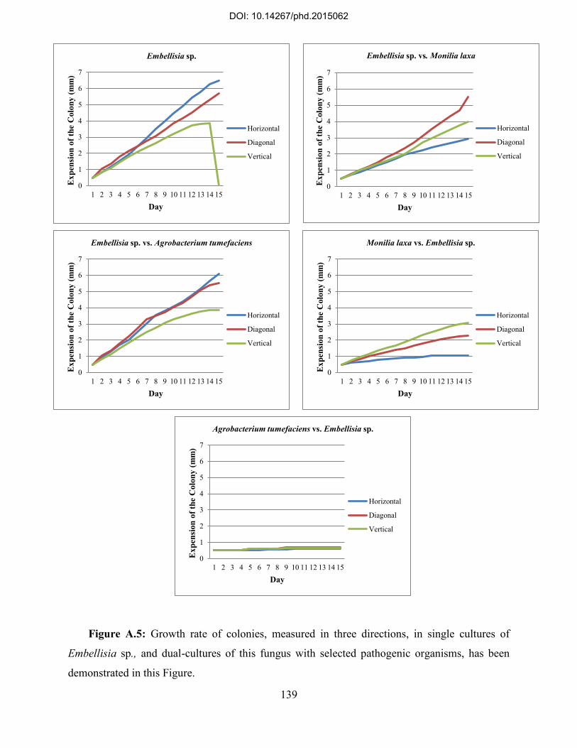
139
Figure A.5: Growth rate of colonies, measured in three directions, in single cultures of
Embellisia sp., and dual-cultures of this fungus with selected pathogenic organisms, has been
demonstrated in this Figure.
0
1
2
3
4
5
6
7
1 2 3 4 5 6 7 8 9 10 11 12 13 14 15
Exp
ensi
on o
f the
Col
ony
(mm
)
Day
Embellisia sp.
Horizontal
Diagonal
Vertical
0
1
2
3
4
5
6
7
1 2 3 4 5 6 7 8 9 10 11 12 13 14 15
Exp
ensi
on o
f the
Col
ony
(mm
)
Day
Embellisia sp. vs. Monilia laxa
Horizontal
Diagonal
Vertical
0
1
2
3
4
5
6
7
1 2 3 4 5 6 7 8 9 10 11 12 13 14 15
Exp
ensi
on o
f the
Col
ony
(mm
)
Day
Embellisia sp. vs. Agrobacterium tumefaciens
Horizontal
Diagonal
Vertical
0
1
2
3
4
5
6
7
1 2 3 4 5 6 7 8 9 10 11 12 13 14 15
Exp
ensi
on o
f the
Col
ony
(mm
)
Day
Monilia laxa vs. Embellisia sp.
Horizontal
Diagonal
Vertical
0
1
2
3
4
5
6
7
1 2 3 4 5 6 7 8 9 10 11 12 13 14 15
Exp
ensi
on o
f the
Col
ony
(mm
)
Day
Agrobacterium tumefaciens vs. Embellisia sp.
Horizontal
Diagonal
Vertical
DOI: 10.14267/phd.2015062

140
Figure A.6: Growth rate of colonies, measured in three directions, in single cultures of
Epicoccum nigrum and dual-cultures of this fungus with selected pathogenic organisms, has been
demonstrated in this Figure.
0
1
2
3
4
5
6
7
1 2 3 4 5 6 7 8 9 10111213141516
Exp
ensi
on o
f the
Col
ony
(mm
)
Day
Epicoccum nigrum
Horizontal
Diagonal
Vertical
0
1
2
3
4
5
6
7
1 2 3 4 5 6 7 8 9 10 11 12 13 14 15
Exp
ensi
on o
f the
Col
ony
(mm
)
Day
Epicoccum nigrum vs. Monilia laxa
Horizontal
Diagonal
Vertical
0
1
2
3
4
5
6
7
1 2 3 4 5 6 7 8 9 10 11 12 13 14 15
Exp
ensi
on o
f the
Col
ony
(mm
)
Day
Epicoccum nigrum vs. Agrobacterium tumefaciens
Horizontal
Diagonal
Vertical
0
1
2
3
4
5
6
7
1 2 3 4 5 6 7 8 9 10 11 12 13 14 15
Exp
ensi
on o
f the
Col
ony
(mm
)
Day
Monilia laxa vs. Epicoccum nigrum
Horizontal
Diagonal
Vertical
0
1
2
3
4
5
6
7
1 2 3 4 5 6 7 8 9 10 11 12 13 14 15
Exp
ensi
on o
f the
Col
ony
(mm
)
Day
Agrobacterium tumefaciens vs. Epicoccum nigrum
Horizontal
Diagonal
Vertical
DOI: 10.14267/phd.2015062

141
Figure A.7: Growth rate of colonies, measured in three directions, in single cultures of
Fusarium oxysporum and dual-cultures of this fungus with selected pathogenic organisms, has
been demonstrated in this Figure.
0
1
2
3
4
5
6
7
1 2 3 4 5 6 7 8 9 10 11 12 13 14 15
Exp
ensi
on o
f the
Col
ony
(mm
)
Day
Fusarium oxysporum
Horizontal
Diagonal
Vertical
0
1
2
3
4
5
6
7
1 2 3 4 5 6 7 8 9 10 11 12 13 14 15
Exp
ensi
on o
f the
Col
ony
(mm
)
Day
Fusarium oxysporum vs. Monilia laxa
Horizontal
Diagonal
Vertical
0
1
2
3
4
5
6
7
1 2 3 4 5 6 7 8 9 10 11 12 13 14 15Exp
ensi
on o
f the
Col
ony
(mm
)
Day
Fusarium oxysporum vs. Agrobacterium tumefaciens
Horizontal
Diagonal
Vertical
0
1
2
3
4
5
6
7
1 2 3 4 5 6 7 8 9 10 11 12 13 14 15
Exp
ensi
on o
f the
Col
ony
(mm
)
Day
Monilia laxa vs. Fusarium oxysporum
Horizontal
Diagonal
Vertical
0
1
2
3
4
5
6
7
1 2 3 4 5 6 7 8 9 10 11 12 13 14 15Exp
ensi
on o
f the
Col
ony
(mm
)
Day
Agrobacterium tumefaciens vs. Fusarium oxysporum
Horizontal
Diagonal
Vertical
DOI: 10.14267/phd.2015062

142
Figure A.8: Growth rate of colonies, measured in three directions, in single cultures of
Rhizopycnis vagum and dual-cultures of this fungus with selected pathogenic organisms, has
been demonstrated in this Figure.
0
1
2
3
4
5
6
7
1 2 3 4 5 6 7 8 9 10 11 12 13 14 15
Exp
ensi
on o
f the
Col
ony
(mm
)
Day
Rhizopycnis vagum
Horizontal
Diagonal
Vertical
0
1
2
3
4
5
6
7
1 2 3 4 5 6 7 8 9 10 11 12 13 14 15
Exp
ensi
on o
f the
Col
ony
(mm
)
Day
Rhizopycnis vagum vs. Monilia laxa
Horizontal
Diagonal
Vertical
0
1
2
3
4
5
6
7
1 2 3 4 5 6 7 8 9 10 11 12 13 14 15
Exp
ensi
on o
f the
Col
ony
(mm
)
Day
Rhizopycnis vagum vs. Agrobacterium tumefaciens
Horizontal
Diagonal
Vertical
0
1
2
3
4
5
6
7
1 2 3 4 5 6 7 8 9 10 11 12 13 14 15
Exp
ensi
on o
f the
Col
ony
(mm
)
Day
Monilia laxa vs. Rhizopycnis vagum
Horizontal
Diagonal
Vertical
0
1
2
3
4
5
6
7
1 2 3 4 5 6 7 8 9 10 11 12 13 14 15
Exp
ensi
on o
f the
Col
ony
(mm
)
Day
Agrobacterium tumefaciens vs. Rhizopycnis vagum
Horizontal
Diagonal
Vertical
DOI: 10.14267/phd.2015062

143
References
Ahlholm, J. U., Helander, M., Henriksson, J., Metzler, M., and Saikkonen, K. (2002).
Environmental conditions and host genotype direct genetic diversity of Venturia ditricha, a
fungal endophyte of birch trees. Evolution, 56(8), 1566-1573.
Aime, S., Alabouvette, C., Steinberg, C., and Olivain, C. (2013). The endophytic strain
Fusarium oxysporum Fo47: a good candidate for priming the defense responses in tomato roots.
Mol Plant Microbe Interact, 26(8), 918-926. doi: 10.1094/MPMI-12-12-0290-R
Alvarez-Loayza, P., White, J. F., Jr., Torres, M. S., Balslev, H., Kristiansen, T., Svenning, J.
C., and Gil, N. (2011). Light converts endosymbiotic fungus to pathogen, influencing seedling
survival and niche-space filling of a common tropical tree, Iriartea deltoidea. PLoS One, 6(1),
e16386. doi: 10.1371/journal.pone.0016386
Aly, A. H., Debbab, A., and Proksch, P. (2011). Fungal endophytes: unique plant inhabitants
with great promises. Appl Microbiol Biotechnol, 90(6), 1829-1845. doi: 10.1007/s00253-011-
3270-y
Arnold, A. E., Henk, D. A., Eells, R. L., Lutzoni, F., and Vilgalys, R. (2007). Diversity and
phylogenetic affinities of foliar fungal endophytes in loblolly pine inferred by culturing and
environmental PCR. Mycologia, 99(2), 185-206.
Arnold, A. E., and Herre, E. A. (2003). Canopy cover and leaf age affect colonization by
tropical fungal endophytes: Ecological pattern and process in Theobroma cacao (Malvaceae).
Mycologia, 95(3), 388-398.
Arnold, A. E., and Lutzoni, F. (2007). Diversity and host range of foliar fungal endophytes: are
tropical leaves biodiversity hotspots? Ecology, 88(3), 541-549.
DOI: 10.14267/phd.2015062

144
Arnold, A. E., Mejia, L. C., Kyllo, D., Rojas, E. I., Maynard, Z., Robbins, N., and Herre, E. A.
(2003). Fungal endophytes limit pathogen damage in a tropical tree. Proc Natl Acad Sci U S A,
100(26), 15649-15654. doi: 10.1073/pnas.2533483100
Bae, H., Roberts, D.P., Lim, H-S., Strem, M.D., Park, S.C., Ryu, C-M., Melnick, R.L. &
Bailey, B.A. (2010). Endophytic Trichoderma isolates from tropical environments delay onset
and induce resistance against Phytophtora capsici in hot pepper using multiple mechanisms.
MPMI, 24(3)336-351
Barna, B., Fodor, J., Pogany, M., and Kiraly, Z. (2003). Role of reactive oxygen species and
antioxidants in plant disease resistance. Pest Manag Sci, 59(4), 459-464. doi: 10.1002/ps.706
Barnkopf András (2013): Paprikában (Capsicum annuum L.) előforduló endofita gombák
izolálása és azonosítása molekuláris módszerekkel (Department of Pland Biology and Plant
Biochemistry with the contribution of the Department of Vegetable Growing). PhD Dissertation.
Berdy, J. (2005). Bioactive microbial metabolites. J Antibiot (Tokyo), 58(1), 1-26. doi:
10.1038/ja.2005.1
Bernardi-Wenzel, J., Garcia, A., Filho, C. J. R., Prioli, A. J., and Pamphile, J. A. (2010).
Evaluation of foliar fungal endophyte diversity and colonization of medicinal plant Luehea
divaricata (Maritus et Zuccarini). Biological Research 43, 375-384.
Borbély Csaba (2014) : A paprikában (Capsicum annuum L.) élő endofita gombák és
gombavírusok jellemzése (Department of Pland Biology and Plant Biochemistry). PhD
Dissertation.
Cannon, P. F., and Simmons, C. M. (2002). Diversity and host preference of leaf endophytic
fungi in the Iwokrama Forest Reserve, Guyana. Mycologia, 94(2), 210-220.
DOI: 10.14267/phd.2015062

145
Chaudhary, K.K., Kaul, R.K. (2011). Compatibility of Pasteuria penetrans with fungal parasite
Paecilomyces lilacinus against root knot nematode on chilli (Capsicum annuum L.). South Asian
Journal of Experimental Biology, 1(1), 36-42
Chaudhari, P., Ahmed, B., Joly, D. L., and Germain, H. (2014). Effector biology during
biotrophic invasion of plant cells. Virulence, 5(7).
Choudhary, B., Nagpure, A., and Gupta, R. K. (2014). Fungal cell-wall lytic enzymes,
antifungal metabolite(s) production, and characterization from Streptomyces exfoliatus MT9 for
controlling fruit-rotting fungi. J Basic Microbiol. doi: 10.1002/jobm.201400380
Clay, K. (1992). Fungal endophytes of plants: biological and chemical diversity. Nat Toxins,
1(3), 147-149.
Davis, E. C., and Shaw, A. J. (2008). Biogeographic and phylogenetic patterns in diversity of
liverwort-associated endophytes. American Journal of Botany, 95(8), 914-924.
Del Olmo-Ruiz, M., and Arnold, A. E. (2014). Interannual variation and host affiliations of
endophytic fungi associated with ferns at La Selva, Costa Rica. Mycologia, 106(1), 8-21. doi:
10.3852/13-098
di Menna, M. E., Finch, S. C., Popay, A. J., and Smith, B. L. (2012). A review of the
Neotyphodium lolii / Lolium perenne symbiosis and its associated effects on animal and plant
health, with particular emphasis on ryegrass staggers. N Z Vet J, 60(6), 315-328. doi:
10.1080/00480169.2012.697429
Eaton, C. J., Cox, M. P., and Scott, B. (2011). What triggers grass endophytes to switch from
mutualism to pathogenism? Plant Sci, 180(2), 190-195. doi: 10.1016/j.plantsci.2010.10.002
DOI: 10.14267/phd.2015062

146
Ernst, M., Mendgen, K. W., and Wirsel, S. G. (2003). Endophytic fungal mutualists: seed-
borne Stagonospora spp. enhance reed biomass production in axenic microcosms. Mol Plant
Microbe Interact, 16(7), 580-587. doi: 10.1094/MPMI.2003.16.7.580
Faeth, S. H. (2009). Asexual fungal symbionts alter reproductive allocation and herbivory over
time in their native perennial grass hosts. Am Nat, 173(5), 554-565. doi: 10.1086/597376
Faeth, S. H., and Fagan, W. F. (2002). Fungal endophytes: common host plant symbionts but
uncommon mutualists. Integr Comp Biol, 42(2), 360-368. doi: 10.1093/icb/42.2.360
Faeth, S. H., and Shochat, E. (2010). Inherited microbial symbionts increase herbivore
abundances and alter arthropod diversity on a native grass. Ecology, 91(5), 1329-1343.
Faeth, S. H., and Sullivan, T. J. (2003). Mutualistic asexual endophytes in a native grass are
usually parasitic. Am Nat, 161(2), 310-325. doi: 10.1086/345937
Favaro, L. C., Sebastianes, F. L., and Araujo, W. L. (2012). Epicoccum nigrum P16, a
sugarcane endophyte, produces antifungal compounds and induces root growth. PLoS One, 7(6),
e36826. doi: 10.1371/journal.pone.0036826
Felsenstein, J. (1985). Confidence limits on phylogenies: An approach using the bootstrap.
Evolution, 39, 783-791.
Gamboa, M. A., Laureano, S., and Bayman, P. (2002). Measuring diversity of endophytic fungi
in leaf fragments: does size matter? Mycopathologia, 156(1), 41-45.
Ganley, R. J., Brunsfeld, S. J., and Newcombe, G. (2004). A community of unknown,
endophytic fungi in western white pine. Proc Natl Acad Sci U S A, 101(27), 10107-10112. doi:
10.1073/pnas.0401513101
DOI: 10.14267/phd.2015062

147
Grosch, R., Scherwinski, K., Lottmann, J., and Berg, G. (2006). Fungal antagonists of the plant
pathogen Rhizoctonia solani: selection, control efficacy and influence on the indigenous
microbial community. Mycol Res, 110(Pt 12), 1464-1474. doi: 10.1016/j.mycres.2006.09.014
Gunatilaka, A. A. (2006). Natural products from plant-associated microorganisms: distribution,
structural diversity, bioactivity, and implications of their occurrence. J Nat Prod, 69(3), 509-526.
doi: 10.1021/np058128n
Guo, B., Wang, Y., Sun, X., and Tang, K. (2008). Bioactive natural products from endophytes:
a review. Prikl Biokhim Mikrobiol, 44(2), 153-158.
Gyeviki, M., Magyar, L., Bujdosó, G., and Hrotkó K. (2008). Cherry rootstock evaluation for
high density cherry orchards in Hungary. Bulletin UASVM Holticulture, 65(1), 231-236.
Hamilton, C. E., Dowling, T. E., and Faeth, S. H. (2010). Hybridization in endophyte
symbionts alters host response to moisture and nutrient treatments. Microb Ecol, 59(4), 768-775.
doi: 10.1007/s00248-009-9606-9
Hamilton, C. E., Faeth, S. H., and Dowling, T. E. (2009). Distribution of hybrid fungal
symbionts and environmental stress. Microb Ecol, 58(2), 408-413. doi: 10.1007/s00248-009-
9504-1
Hawas, U. W., El-Beih, A. A., and El-Halawany, A. M. (2012). Bioactive anthraquinones from
endophytic fungus Aspergillus versicolor isolated from red sea algae. Arch Pharm Res, 35(10),
1749-1756. doi: 10.1007/s12272-012-1006-x
Hellwig, V., Grothe, T., Mayer-Bartschmid, A., Endermann, R., Geschke, F. U., Henkel, T.,
and Stadler, M. (2002). Altersetin, a new antibiotic from cultures of endophytic Alternaria spp.
Taxonomy, fermentation, isolation, structure elucidation and biological activities. J Antibiot
(Tokyo), 55(10), 881-892.
DOI: 10.14267/phd.2015062

148
Hernawati, H.Wiyono, S., Santoso, S. (2011). Leaf endophytic fungi of chili (Capsicum
annuum) and their role in the protection against Aphis gossypii (Homoptera: Aphidiae).
Biodiversitas, 12(4), 187-191
Herre, E. A., Knowlton, N., Mueller, U. G., and Rehner, S. A. (1999). The evolution of
mutualisms: exploring the paths between conflict and cooperation. Trends Ecol Evol, 14(2), 49-
53.
Herre, E. A., Mejia, L. C., Kyllo, D. A., Rojas, E., Maynard, Z., Butler, A., and Van Bael, S. A.
(2007). Ecological implications of anti-pathogen effects of tropical fungal endophytes and
mycorrhizae. Ecology, 88(3), 550-558.
Higginbotham, S. J., Arnold, A. E., Ibanez, A., Spadafora, C., Coley, P. D., and Kursar, T. A.
(2013). Bioactivity of fungal endophytes as a function of endophyte taxonomy and the taxonomy
and distribution of their host plants. PLoS One, 8(9), e73192. doi: 10.1371/journal.pone.0073192
Higgins, K. L., Arnold, A. E., Miadlikowska, J., Sarvate, S. D., and Lutzoni, F. (2007).
Phylogenetic relationships, host affinity, and geoFigureic structure of boreal and arctic
endophytes from three major plant lineages. Mol Phylogenet Evol, 42(2), 543-555. doi:
10.1016/j.ympev.2006.07.012
Hirsch, A. M., and Kapulnik, Y. (1998). Signal Transduction Pathways in Mycorrhizal
Associations: Comparisons with the Rhizobium-Legume Symbiosis. Fungal Genet Biol, 23(3),
205-212. doi: 10.1006/fgbi.1998.1046
Hrotkó, K., and Magyar, L. 2004. Mahaleb Rootstocks From The Department Of Fuit Science,
BUDAPEST. Acta Hort. (ISHS) 658:497-499
DOI: 10.14267/phd.2015062

149
Jumpponen, A., Mattson, K. G., and Trappe, J. M. (1998). Mycorrhizal functioning of
Phialocephala fortinii with Pinus contorta on glacier forefront soil: interactions with soil nitrogen
and organic matter. Mycorrhiza, 7(5), 261-265. doi: 10.1007/s005720050190
Kaplan, D., Maymon, M., Agapakis, C. M., Lee, A., Wang, A., Prigge, B. A., . . . Hirsch, A.
M. (2013). A survey of the microbial community in the rhizosphere of two dominant shrubs of
the Negev Desert highlands, Zygophyllum dumosum (Zygophyllaceae) and Atriplex halimus
(Amaranthaceae), using cultivation-dependent and cultivation-independent methods. Am J Bot,
100(9), 1713-1725. doi: 10.3732/ajb.1200615
Kavroulakis, N., Ntougias, S., Zervakis, G. I., Ehaliotis, C., Haralampidis, K., and
Papadopoulou, K. K. (2007). Role of ethylene in the protection of tomato plants against soil-
borne fungal pathogens conferred by an endophytic Fusarium solani strain. J Exp Bot, 58(14),
3853-3864. doi: 10.1093/jxb/erm230
Khan, A. L., Hamayun, M., Hussain, J., Kang, S. M., and Lee, I. J. (2012). The newly isolated
endophytic fungus Paraconiothyrium sp. LK1 produces ascotoxin. Molecules, 17(1), 1103-1112.
doi: 10.3390/molecules17011103
Khan, A. L., Waqas, M., Hamayun, M., Al-Harrasi, A., Al-Rawahi, A., & Lee, I. J. (2013). Co-
synergism of endophyte Penicillium resedanum LK6 with salicylic acid helped Capsicum
annuum in biomass recovery and osmotic stress mitigation. BMC Microbiol, 13, 51. doi:
10.1186/1471-2180-13-51
Knapp, G. D., and Kovács, G. M. (2010). Study of root colonising endophytic fungi of
Medicago minima in a semiarid sandy grassland area near Fülöpháza. Mikológiai Közlemények –
Clusiana 49: 67-77.
DOI: 10.14267/phd.2015062

150
Knapp, D. G., Kovács, G. M., Zajta, E., Groenewald, J. Z., and Crous, P. W. (2015). Dark
septate endophytic pleosporalean genera from semiarid areas. Persoonia 35, 87-100.
Knapp, G. D., Pintye, A., and Kovács, G. M. (2012). The dark side is not fastidious – dark
septate endophytic fungi of native and invasive plants of semiarid sandy areas. PLoS ONE 7:
e32570.
Knowlton, N., and Rohwer, F. (2003). Multispecies microbial mutualisms on coral reefs: the
host as a habitat. Am Nat, 162(4 Suppl), S51-62. doi: 10.1086/378684
Kock, J. L., Strauss, T., Pohl, C. H., Smith, D. P., Botes, P. J., Pretorius, E. E., . . . Nigam, S.
(2001). Bioprospecting for novel oxylipins in fungi: the presence of 3-hydroxy oxylipins in
Pilobolus. Antonie Van Leeuwenhoek, 80(1), 93-99.
Kogel, K. H., Franken, P., and Huckelhoven, R. (2006). Endophyte or parasite--what decides?
Curr Opin Plant Biol, 9(4), 358-363. doi: 10.1016/j.pbi.2006.05.001
Leuchtmann, A. (1992). Systematics, distribution, and host specificity of grass endophytes. Nat
Toxins, 1(3), 150-162.
Li, C. J., Gao, J. H., and Nan, Z. B. (2007). Interactions of Neotyphodium gansuense,
Achnatherum inebrians, and plant-pathogenic fungi. Mycol Res, 111(Pt 10), 1220-1227. doi:
10.1016/j.mycres.2007.08.012
Mandyam, K., Loughin, T., and Jumpponen, A. (2010). Isolation and morphological and
metabolic characterization of common endophytes in annually burned tallgrass prairie.
Mycologia, 102(4), 813-821.
Mandyam, K. G., Roe, J., and Jumpponen, A. (2013). Arabidopsis thaliana model system
reveals a continuum of responses to root endophyte colonization. Fungal Biol, 117(4), 250-260.
doi: 10.1016/j.funbio.2013.02.001
DOI: 10.14267/phd.2015062

151
Marasco, R., Rolli, E., Ettoumi, B., Vigani, G., Mapelli, F., Borin, S., . . . Daffonchio, D.
(2012). A drought resistance-promoting microbiome is selected by root system under desert
farming. PLoS One, 7(10), e48479. doi: 10.1371/journal.pone.0048479
Martinuz, A., Schouten, A., Menjivar, R.D., Sikora, R.A. (2012). Effectivenes of systemic
resistance toward Aphis gossypii (Hom., Aphididae) as induced by combined applications of the
endophytes Fusarium oxysporum Fo162 and Rhizobium etli G12. Biological Control 62, 206-
212
Meijer, G., and Leuchtmann, A. (2001). Fungal genotype controls mutualism and sex in
Brachypodium sylvaticum infected by epichloe sylvatica. Acta Biol Hung, 52(2-3), 249-263. doi:
10.1556/ABiol.52.2001.2-3.9
Ming, Q., Su, C., Zheng, C., Jia, M., Zhang, Q., Zhang, H., . . . Qin, L. (2013). Elicitors from
the endophytic fungus Trichoderma atroviride promote Salvia miltiorrhiza hairy root growth and
tanshinone biosynthesis. J Exp Bot, 64(18), 5687-5694. doi: 10.1093/jxb/ert342
Moon, C. D., Tapper, B. A., and Scott, B. (1999). Identification of Epichloe endophytes in
planta by a microsatellite-based PCR fingerprinting assay with automated analysis. Appl Environ
Microbiol, 65(3), 1268-1279.
Moricca, S., and Ragazzi, A. (2008). Fungal endophytes in Mediterranean oak forests: a lesson
from Discula quercina. Phytopathology, 98(4), 380-386. doi: 10.1094/PHYTO-98-4-0380
Muller, C. B., and Krauss, J. (2005). Symbiosis between grasses and asexual fungal
endophytes. Curr Opin Plant Biol, 8(4), 450-456. doi: 10.1016/j.pbi.2005.05.007
Newsham, K. K. (2011). A meta-analysis of plant responses to dark septate root endophytes.
New Phytol, 190(3), 783-793. doi: 10.1111/j.1469-8137.2010.03611.x
DOI: 10.14267/phd.2015062

152
Osono, T., and Masuya, H. (2012). Endophytic fungi associated with leaves of Betulaceae in
Japan. Canadian Journal of Microbiology, 58(4), 507-515.
Parsa, S., Ortiz, V., and Vega, F. E. (2013). Establishing fungal entomopathogens as
endophytes: towards endophytic biological control. J Vis Exp(74). doi: 10.3791/50360
Paul, N.C., Deng, J.X., Sang, H.K., Choi, Y.P. & Yu, S.H. (2011). Distribution and antifungal
activity of endophytic fungi in different growth stages of chili pepper (Capsicum annuum L.) in
Korea. Plant Pathol. J., 28(1), 10-19
Paul, N. C., Deng, J. X., Shin, K. S., and Yu, S. H. (2012). Molecular and Morphological
Characterization of Endophytic Heterobasidion araucariae from Roots of Capsicum annuum L. in
Korea. Mycobiology, 40(2), 85-90. doi: 10.5941/MYCO.2012.40.2.85
Pereira, S. I., and Castro, P. M. (2014). Diversity and characterization of culturable bacterial
endophytes from Zea mays and their potential as plant growth-promoting agents in metal-
degraded soils. Environ Sci Pollut Res Int. doi: 10.1007/s11356-014-3309-6
Petrini, O., Sieber, T. N., Toti, L., and Viret, O. (1992). Ecology, metabolite production, and
substrate utilization in endophytic fungi. Nat Toxins, 1(3), 185-196.
Porras-Alfaro, A., and Bayman, P. (2011). Hidden fungi, emergent properties: endophytes and
microbiomes. Annu Rev Phytopathol, 49, 291-315. doi: 10.1146/annurev-phyto-080508-081831
Pressel, S., Ligrone, R., Duckett, J. G., and Davis, E. C. (2008). A novel ascomycetous
endophytic association in the rhizoids of the leafy liverwort family, Schistochilaceae
(Jungermanniidae, Hepaticopsida). Am J Bot, 95(5), 531-541. doi: 10.3732/ajb.2007171
Provorov, N. A. (2009). [Plant-microbe symbioses as an evolutionary continuum]. Zh Obshch
Biol, 70(1), 10-34.
DOI: 10.14267/phd.2015062

153
Reininger, V., and Sieber, T. N. (2012). Mycorrhiza reduces adverse effects of dark septate
endophytes (DSE) on growth of conifers. PLoS One, 7(8), e42865. doi:
10.1371/journal.pone.0042865
Rodriguez, R. J., Freeman, D. C., McArthur, E. D., Kim, Y. O., and Redman, R. S. (2009)a.
Symbiotic regulation of plant growth, development and reproduction. Commun Integr Biol, 2(2),
141-143.
Rodriguez, R. J., Henson, J., Van Volkenburgh, E., Hoy, M., Wright, L., Beckwith, F., . . .
Redman, R. S. (2008). Stress tolerance in plants via habitat-adapted symbiosis. ISME J, 2(4),
404-416. doi: 10.1038/ismej.2007.106
Rodriguez, R. J., White, J. F., Jr., Arnold, A. E., and Redman, R. S. (2009)b. Fungal
endophytes: diversity and functional roles. New Phytol, 182(2), 314-330. doi: 10.1111/j.1469-
8137.2009.02773.x
Rowan, R., and Knowlton, N. (1995). Intraspecific diversity and ecological zonation in coral-
algal symbiosis. Proc Natl Acad Sci U S A, 92(7), 2850-2853.
Saari, S., Helander, M., Faeth, S. H., and Saikkonen, K. (2010). The effects of endophytes on
seed production and seed predation of tall fescue and meadow fescue. Microb Ecol, 60(4), 928-
934. doi: 10.1007/s00248-010-9749-8
Saikkonen, K., Gundel, P. E., and Helander, M. (2013). Chemical ecology mediated by fungal
endophytes in grasses. J Chem Ecol, 39(7), 962-968. doi: 10.1007/s10886-013-0310-3
Saikkonen, K., Ion, D., and Gyllenberg, M. (2002). The persistence of vertically transmitted
fungi in grass metapopulations. Proc Biol Sci, 269(1498), 1397-1403. doi:
10.1098/rspb.2002.2006
DOI: 10.14267/phd.2015062

154
Saikkonen, K., Wali, P., Helander, M., and Faeth, S. H. (2004). Evolution of endophyte-plant
symbioses. Trends Plant Sci, 9(6), 275-280. doi: 10.1016/j.tplants.2004.04.005
Santamaria, J., and Bayman, P. (2005). Fungal epiphytes and endophytes of coffee leaves
(Coffea arabica). Microb Ecol, 50(1), 1-8. doi: 10.1007/s00248-004-0002-1
Schardl, C. L. (2001). Epichloe festucae and related mutualistic symbionts of grasses. Fungal
Genet Biol, 33(2), 69-82. doi: 10.1006/fgbi.2001.1275
Schardl, C. L., and An, Z. (1993). Molecular biology and genetics of protective fungal
endophytes of grasses. Genet Eng (N Y), 15, 191-212.
Schardl, C. L., Leuchtmann, A., and Spiering, M. J. (2004). Symbioses of grasses with
seedborne fungal endophytes. Annu Rev Plant Biol, 55, 315-340. doi:
10.1146/annurev.arplant.55.031903.141735
Schulz, B., and Boyle, C. (2005). The endophytic continuum. Mycol Res, 109(Pt 6), 661-686.
Selosse, M. A., Baudoin, E., and Vandenkoornhuyse, P. (2004). Symbiotic microorganisms, a
key for ecological success and protection of plants. C R Biol, 327(7), 639-648.
Selosse, M. A., Dubois, M. P., and Alvarez, N. (2009). Do Sebacinales commonly associate
with plant roots as endophytes? Mycol Res, 113(Pt 10), 1062-1069. doi:
10.1016/j.mycres.2009.07.004
Sia Ede, F., Marcon, J., Luvizotto, D. M., Quecine, M. C., Tsui, S., Pereira, J. O., Pizzirani-
Kleiner, A. A., and Azevedo, J. L. (2013). Endophytic fungi from the Amazonian plant Paullinia
cupana and from Olea europaea isolated using cassava as an alternative starch media source.
Sprigerplus, 2, 579. doi: 10.1186/2193-1801-2-579
Sneath, P. H. A., and Sokal, R. R. (1973). Numerical taxonomy. Freeman. San Francisco.
DOI: 10.14267/phd.2015062

155
Stadler, M., and Keller, N. P. (2008). Paradigm shifts in fungal secondary metabolite research.
Mycol Res, 112(Pt 2), 127-130. doi: 10.1016/j.mycres.2007.12.002
Stepniewska, Z., and Kuzniar, A. (2013). Endophytic microorganisms--promising applications
in bioremediation of greenhouse gases. Appl Microbiol Biotechnol, 97(22), 9589-9596. doi:
10.1007/s00253-013-5235-9
Strobel, G., and Daisy, B. (2003). Bioprospecting for microbial endophytes and their natural
products. Microbiol Mol Biol Rev, 67(4), 491-502.
Sullivan, T. J., and Faeth, S. H. (2004). Gene flow in the endophyte Neotyphodium and
implications for coevolution with Festuca arizonica. Mol Ecol, 13(3), 649-656.
Tadych, M., Bergen, M. S., and White, J. F., Jr. (2014). Epichloe spp. associated with grasses:
new insights on life cycles, dissemination and evolution. Mycologia, 106(2), 181-201.
Tamura, K., Dudley, J., Nei, M., and Kumar, S. (2007). MEGA4: Molecular evolutionary
genetics analysis (MEGA) software version 4.0. Molecular Biology and Evolution, 24, 1596-
1599.
Tamura, K., Nei, M., and Kumar, S. (2004). Prospects for inferring very large phylogenies by
usinf the neighbor- joining method. Proceedings of the National Academy of Sciences, (USA),
101, 11030- 11035.
Tintjer, T., and Rudgers, J. A. (2006). Grass-herbovore interactions altered by strains of a
native endophyte. New Physiologist, 170(3), 513-521.
Tsai, H. F., Liu, J. S., Staben, C., Christensen, M. J., Latch, G. C., Siegel, M. R., and Schardl,
C. L. (1994). Evolutionary diversification of fungal endophytes of tall fescue grass by
hybridization with Epichloe species. Proc Natl Acad Sci U S A, 91(7), 2542-2546.
DOI: 10.14267/phd.2015062

156
U'Ren J, M., Lutzoni, F., Miadlikowska, J., and Arnold, A. E. (2010). Community analysis
reveals close affinities between endophytic and endolichenic fungi in mosses and lichens.
Microb Ecol, 60(2), 340-353. doi: 10.1007/s00248-010-9698-2
Verma, V. C., Gond, S. K., Kumar, A., Kharwar, R. N., and Strobel, G. (2007). The endophytic
mycoflora of bark, leaf, and stem tissues of Azadirachta indica A. Juss (neem) from Varanasi
(India). Microb Ecol, 54(1), 119-125. doi: 10.1007/s00248-006-9179-9
Watrud, L. S., Martin, K., Donegan, K. K., Stone, J. K., and Coleman, C. G. (2006).
Comparison of taxonomic, colony morphotype and PCR-RFLP methods to characterize
microfungal diversity. Mycologia, 98(3), 384-392.
Weber, D., Gorzalczany, S., Martino, V., Acevedo, C., Sterner, O., and Anke, T. (2005).
Metabolites from endophytes of the medicinal plant Erythrina crista-galli. Z Naturforsch C, 60(5-
6), 467-477.
Westendorf, M. L., Mitchell, G. E., Jr., Tucker, R. E., Bush, L. P., Petroski, R. J., and Powell,
R. G. (1993). In vitro and in vivo ruminal and physiological responses to endophyte-infected tall
fescue. J Dairy Sci, 76(2), 555-563. doi: 10.3168/jds.S0022-0302(93)77375-0
White TJ, Bruns T, Lee S, Taylor JW. Amplification and direct sequencing of fungal ribosomal
RNA genes for phylogenetics. In: Innis MA, Gelfand DH, Sninsky JJ, White TJ, editor. PCR
Protocols: A Guide to Methods and Applications. New York: Academic Press Inc; 1990. pp.
315–322.
White, J. F., Jr., and Torres, M. S. (2010). Is plant endophyte-mediated defensive mutualism
the result of oxidative stress protection? Physiol Plant, 138(4), 440-446. doi: 10.1111/j.1399-
3054.2009.01332.x
DOI: 10.14267/phd.2015062
157
Zhi-Lin, Y., Yi-Cun, C., Bai-Ge, X., and Chu-Long, Z. (2012). Current perspectives on the
volatile-producing fungal endophytes. Crit Rev Biotechnol, 32(4), 363-373. doi:
10.3109/07388551.2011.651429
Zuccaro, A., Lahrmann, U., and Langen, G. (2014). Broad compatibility in fungal root
symbioses. Curr Opin Plant Biol, 20C, 135-145. doi: 10.1016/j.pbi.2014.05.013
DOI: 10.14267/phd.2015062