Expression of Leukocyte Inhibitory Immunoglobulinlike Transcript 3 Receptors by Ovarian Tumors in...
7
Expression of Leukocyte Inhibitory Immunoglobulin- like Transcript 3 Receptors by Ovarian Tumors in Laying Hen Model of Spontaneous Ovarian Cancer 1 Mohammad Faisal Khan*, Janice M. Bahr † , Aparna Yellapa*, Pincas Bitterman ‡ , Jacques S. Abramowicz § , Seby L. Edassery*, Sanjib Basu ¶ , Jacob Rotmensch § and Animesh Barua* ,‡,§ *Department of Pharmacology, Rush University Medical Center, Chicago, IL, USA; † Department of Animal Sciences, University of Illinois at Urbana-Champaign, Champaign, IL, USA; ‡ Department of Pathology, Rush University Medical Center, Chicago, IL, USA; § Department of Obstetrics and Gynecology, Rush University Medical Center, Chicago, IL, USA; ¶ Department of Preventive Medicine (Biostatistics), Rush University Medical Center, Chicago, IL, USA Abstract Attempts to enhance a patient’s immune response and ameliorate the poor prognosis of ovarian cancer (OVCA) have largely been unsuccessful owing to the suppressive tumor microenvironment. Leukocyte immunoglobulin-like tran- script 3 (ILT3) inhibitory receptors have been implicated in immunosuppression in several malignancies. The ex- pression and role of ILT3 in the progression of ovarian tumors are unknown. This study examined the expression and association of ILT3 in ovarian tumors in laying hens, a spontaneous preclinical model of human OVCA. White Leghorn laying hens were selected by transvaginal ultrasound scanning. Serum and normal ovaries or ovarian tumors were collected. The presence of tumors and the expression of ILT3 were examined by routine histology, immuno- histochemistry, Western blot analysis, and reverse transcription–polymerase chain reaction. In addition to stromal immune cell-like cells, the epithelium of the ovarian tumors also expressed ILT3 with significantly high intensity than normal ovaries. Among different subtypes of ovarian carcinomas, serous OVCA showed the highest ILT3 staining intensity, whereas endometrioid OVCA had the lowest intensity. Similar to humans, an immunoreactive protein band of approximately 55 kDa for ILT3 was detected in the ovarian tumors in hens. The patterns of ILT3 protein and messenger RNA expression by ovarian tumors in different subtypes and stages were similar to those of immuno- histochemical staining. The results of this study suggest that laying hens may be useful to generate information on ILT3-associated immunosuppression in OVCA. This animal model also offers the opportunity to develop and test anti-ILT3 immunotherapy to enhance antitumor immunity against OVCA in humans. Translational Oncology (2012) 5, 85–91 Introduction Despite the remarkable improvements in cytoreductive surgeries and chemotherapeutics, ovarian cancer (OVCA) remains one of the most lethal gynecologic malignancies of women with a high death rate [1]. Owing to the lack of an effective early detection test, OVCA in most cases is detected at late stages, and its high recurrence rate (80%-90%) contributes to poor prognosis [2,3]. There is an emerging recognition that tumor growth, in general, elicits specific immune responses me- diated by cell-mediated immunity [4]. As a result, immunotherapies against several cancers are being developed [4–6]. Although recent Address all correspondence to: Animesh Barua, PhD, Laboratory for Translational Re- search on Ovarian Cancer, Department of Pharmacology, Room # 410, Cohn Building, Rush University Medical Center, 1735 W Harrison St, Chicago, IL 60612. E-mail: [email protected] 1 This study was supported by the Idea Development Award from the United States Department of Defense (OCno. 093303), National Cancer Institute Pacific Ovarian Cancer Research Consortium Career Development Program grant P50 CA83636, and the Elmer Sylvia and Sramek Foundation (USA). The authors declare no conflict of interest. Received 16 November 2011; Revised 23 December 2011; Accepted 3 January 2012 Copyright © 2012 Neoplasia Press, Inc. All rights reserved 1944-7124/12/$25.00 DOI 10.1593/tlo.11328 www.transonc.com Translational Oncology Volume 5 Number 2 April 2012 pp. 85–91 85
-
Upload
independent -
Category
Documents
-
view
3 -
download
0
Transcript of Expression of Leukocyte Inhibitory Immunoglobulinlike Transcript 3 Receptors by Ovarian Tumors in...

Trans la t iona l Onco logy Volume 5 Number 2 April 2012 pp. 85–91 85
www.transonc.com
Expression of LeukocyteInhibitory Immunoglobulin-like Transcript 3 Receptorsby Ovarian Tumors in LayingHen Model of SpontaneousOvarian Cancer1
Mohammad Faisal Khan*, Janice M. Bahr†,Aparna Yellapa*, Pincas Bitterman‡,Jacques S. Abramowicz§, Seby L. Edassery*,Sanjib Basu¶, Jacob Rotmensch§
and Animesh Barua*,‡,§
*Department of Pharmacology, Rush University MedicalCenter, Chicago, IL, USA; †Department of Animal Sciences,University of Illinois at Urbana-Champaign, Champaign, IL,USA; ‡Department of Pathology, Rush University MedicalCenter, Chicago, IL, USA; §Department of Obstetrics andGynecology, Rush University Medical Center, Chicago, IL,USA; ¶Department of Preventive Medicine (Biostatistics),Rush University Medical Center, Chicago, IL, USA
AbstractAttempts to enhance a patient’s immune response and ameliorate the poor prognosis of ovarian cancer (OVCA) havelargely been unsuccessful owing to the suppressive tumor microenvironment. Leukocyte immunoglobulin-like tran-script 3 (ILT3) inhibitory receptors have been implicated in immunosuppression in several malignancies. The ex-pression and role of ILT3 in the progression of ovarian tumors are unknown. This study examined the expressionand association of ILT3 in ovarian tumors in laying hens, a spontaneous preclinical model of human OVCA. WhiteLeghorn laying hens were selected by transvaginal ultrasound scanning. Serum and normal ovaries or ovarian tumorswere collected. The presence of tumors and the expression of ILT3 were examined by routine histology, immuno-histochemistry, Western blot analysis, and reverse transcription–polymerase chain reaction. In addition to stromalimmune cell-like cells, the epithelium of the ovarian tumors also expressed ILT3 with significantly high intensity thannormal ovaries. Among different subtypes of ovarian carcinomas, serous OVCA showed the highest ILT3 stainingintensity, whereas endometrioid OVCA had the lowest intensity. Similar to humans, an immunoreactive proteinband of approximately 55 kDa for ILT3 was detected in the ovarian tumors in hens. The patterns of ILT3 proteinand messenger RNA expression by ovarian tumors in different subtypes and stages were similar to those of immuno-histochemical staining. The results of this study suggest that laying hens may be useful to generate information onILT3-associated immunosuppression in OVCA. This animal model also offers the opportunity to develop and testanti-ILT3 immunotherapy to enhance antitumor immunity against OVCA in humans.
Translational Oncology (2012) 5, 85–91
Address all correspondence to: Animesh Barua, PhD, Laboratory for Translational Re-search on Ovarian Cancer, Department of Pharmacology, Room # 410, Cohn Building,Rush University Medical Center, 1735 W Harrison St, Chicago, IL 60612.E-mail: [email protected] study was supported by the Idea Development Award from the United StatesDepartment of Defense (OCno. 093303), National Cancer Institute Pacific OvarianCancer Research Consortium Career Development Program grant P50 CA83636,and the Elmer Sylvia and Sramek Foundation (USA). The authors declare no conflictof interest.Received 16 November 2011; Revised 23 December 2011; Accepted 3 January 2012
Copyright © 2012 Neoplasia Press, Inc. All rights reserved 1944-7124/12/$25.00DOI 10.1593/tlo.11328
IntroductionDespite the remarkable improvements in cytoreductive surgeries andchemotherapeutics, ovarian cancer (OVCA) remains one of the mostlethal gynecologic malignancies of women with a high death rate [1].Owing to the lack of an effective early detection test, OVCA in mostcases is detected at late stages, and its high recurrence rate (80%-90%)contributes to poor prognosis [2,3]. There is an emerging recognitionthat tumor growth, in general, elicits specific immune responses me-diated by cell-mediated immunity [4]. As a result, immunotherapiesagainst several cancers are being developed [4–6]. Although recent
86 ILT3 Expression in Ovarian Tumors Khan et al. Translational Oncology Vol. 5, No. 2, 2012
advances in immunotherapy have been shown to improve the over-all survival ability of patients with hematologic tumors and mela-noma [7], most immunotherapeutic trials have failed to demonstratesuccess in clinical responses [6,8]. Thus, development of new strat-egies to promote immune responses against malignancies is critical inovercoming the limited efficacy of conventional therapies. Despitethe presentation of antigens by ovarian malignant cells, which shouldinduce immune-mediated rejection, spontaneous rejection of an estab-lished tumor is rare [9]. This lack of immune response is not onlybecause of the ignorance of the immune system but also because ofthe tumor-induced immune suppression that protects the tumor fromeradication [9]. Therefore, a better understanding of the mechanismsof tumor-induced immunosuppression will enhance our ability toprevent ovarian tumor progression and to design antitumor interven-tional strategies.
Numerous studies on cancers of several organs have reported severalmechanisms of tumor-induced immune suppression including induc-tion of regulatory T cells [10], expression of immunosuppressive fac-tors (transforming growth factor β, interleukin 10, and chemokineligand 22) [10–12], down-regulation of intracellular adhesion mole-cules [13], and induction of peripheral tolerance [4,14,15]. In contrast,studies on the mechanism of immune suppression in ovarian malig-nancy are very limited. OVCA differs from other malignancies in itsspecific dissemination pattern [9]. The tumor typically spreads in a dif-fuse intra-abdominal fashion rather than through systemic circulation.Thus, antitumor immune response at the tumor environment plays acritical role to ovarian tumor metastasis. Immunosuppressive regulatoryT cells [10], transforming growth factor β [11,12], tolerance-inducingplasmacytoid dendritic cells [16], B7-H4+ macrophages [17], andinterleukin 10 [18] have been reported to be present in the tumormicroenvironment. However, how these immunosuppressive factorsand agents are recruited into the tumor environment is not known.Emerging studies suggest that induction of inhibitory receptor immuno-globulin (Ig)-like transcript 3 (ILT3) expression is one of the mecha-nisms contributing to the tumor-induced immune suppression inseveral malignancies [19].
ILT3 is a member of leukocyte Ig-like receptors family with inhib-itory functions and exists in both membrane and soluble forms [20].Both forms of ILT3s have been suggested to inhibit T-cell prolifera-tion, CD4+ T-cell anergy, suppressing the differentiation of interferonγ–producing CD8+ cytotoxic lymphocytes. In addition, membrane andsoluble ILT3 were also reported to stimulate the differentiation of regu-latory T cells in various cancer patients [4,5,10]. All these findingssuggest that ILT3 may be involved in the immunosuppression againsttumor antigens and prevention or blocking of ILT3 expression mayenhance a patient’s immune responses to malignancies. The expressionof ILT3 in OVCA patients has not yet been reported. Difficulties inidentifying and access to patients at the early stage of OVCA hinderthe ability to study the involvement of ILT3 in OVCA progressionand develop interventional strategies for its prevention. Rodents donot develop OVCA spontaneously, and the histopathologies of inducedOVCA in rodents do not resemble the spontaneous OVCA in humans[21]. Recently, we have shown that laying hens are the only widelyavailable animals that develop OVCA spontaneously with a high inci-dence rate and histopathologies remarkably similar to human OVCA[22]. The expression of leukocyte Ig-like receptors has been reportedin chicken, which are shown to be orthologus to those of mammalsincluding humans [23,24]. Thus, the goal of this study was to examinewhether ILT3 is expressed in ovarian tumors in the laying hen model of
spontaneous OVCA and, if so, whether ILT3 expression is associatedwith the progression of ovarian tumors in hens.
Materials and Methods
AnimalsCommercial strains of approximately 3-year-old white Leghorn
laying hens (Gallus domesticus) were selected from a flock of layersmaintained under standard poultry husbandry practices. The incidenceof OVCA in hens of this age group is approximately 15% to 20%and is associated with low or complete cessation of egg laying [25].Hens (n = 148) were selected on the basis of their egg-laying rates(normal, low, or ceased egg laying) and transvaginal ultrasound scan-ning as reported previously [25]. All experimental procedures were per-formed according to the institutional animal care and use committeeapproved protocol.
Tissue Collection and Processing
Serum samples. Blood was obtained from brachial veins of all hensbefore euthanasia, centrifuged (1000g for 20 minutes), and serumsamples were stored at −80°C until further use.
Gross ovarian morphology and histopathology. Ovarian pathologyand tumor staging were performed by gross and histologic examinationas reported previously [22]. Each normal ovary or ovary with tumor wasdivided into four portions for protein extraction, total RNA collection,paraffin and frozen embedding for routine histology, and immuno-histochemical studies as reported previously [26]. Ovarian surface epi-thelial (OSE) cells from normal or ovaries with tumor were collectedsimilarly as reported earlier [27]. All collected samples were groupedinto three groups including normal-, early-, and late-stage OVCA basedon the diagnosis of the histopathologic ovarian tissue examination asreported previously [22].
Preparation of Ovarian Specimen for Biochemical AnalysisSnap-frozen normal ovaries and ovaries with tumor as well as OSE
from normal ovaries and ovaries with tumor were homogenized with aPolytron homogenizer (Brinkman Instruments, Westbury, NY) as re-ported previously [28] and centrifuged, the supernatant was collected,and the protein content of the extract was measured and stored at−80°C until further use.
Histopathologic Examination and ImmunohistochemistryParaffin or frozen sections from each ovary with tumor or ovaries that
appeared normal without any grossly detectable tumor were stained withhematoxylin and eosin and observed under a light microscope. Presenceor absence of tumors in the section and their histologic types were de-termined as reported earlier [22]. Immunohistochemical detection ofILT3 expression was performed using goat anti-ILT3 (R&D Systems,Inc, Minneapolis, MN) as primary antibodies (n = 15 hens each, fornormal, early, and late stages as reported previously) [26]. The numberof hens for each group was determined based on statistical power analy-sis to achieve significant differences in the intensities of ILT3 immuno-staining among the hens of normal or OVCA groups. These hens wereselected from each group randomly. Briefly, sections were deparaffinized,and antigens on the sections were unmasked by heating the sections withan antigen-unmasking solution (Vector Laboratories, Burlingame, CA)for 20 minutes in a microwave oven. Endogenous peroxidase in the
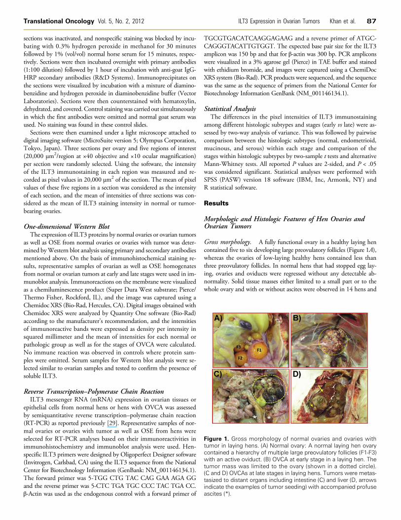
Figure 1. Gross morphology of normal ovaries and ovaries withtumor in laying hens. (A) Normal ovary: A normal laying hen ovarycontained a hierarchy of multiple large preovulatory follicles (F1-F3)with an active oviduct. (B) OVCA at early stage in a laying hen. Thetumor mass was limited to the ovary (shown in a dotted circle).(C and D) OVCAs at late stages in laying hens. Tumors were metas-tasized to distant organs including intestine (C) and liver (D, arrowsindicate the examples of tumor seeding) with accompanied profuseascites (*).
Translational Oncology Vol. 5, No. 2, 2012 ILT3 Expression in Ovarian Tumors Khan et al. 87
sections was inactivated, and nonspecific staining was blocked by incu-bating with 0.3% hydrogen peroxide in methanol for 30 minutesfollowed by 1% (vol/vol) normal horse serum for 15 minutes, respec-tively. Sections were then incubated overnight with primary antibodies(1:100 dilution) followed by 1 hour of incubation with anti-goat IgG-HRP secondary antibodies (R&D Systems). Immunoprecipitates onthe sections were visualized by incubation with a mixture of diamino-benzidine and hydrogen peroxide in diaminobenzidine buffer (VectorLaboratories). Sections were then counterstained with hematoxylin,dehydrated, and covered. Control staining was carried out simultaneouslyin which the first antibodies were omitted and normal goat serum wasused. No staining was found in these control slides.Sections were then examined under a light microscope attached to
digital imaging software (MicroSuite version 5; Olympus Corporation,Tokyo, Japan). Three sections per ovary and five regions of interest(20,000 μm2/region at ×40 objective and ×10 ocular magnification)per section were randomly selected. Using the software, the intensityof the ILT3 immunostaining in each region was measured and re-corded as pixel values in 20,000 μm2 of the section. The mean of pixelvalues of these five regions in a section was considered as the intensityof each section, and the mean of intensities of three sections was con-sidered as the mean of ILT3 staining intensity in normal or tumor-bearing ovaries.
One-dimensional Western BlotThe expression of ILT3 proteins by normal ovaries or ovarian tumors
as well as OSE from normal ovaries or ovaries with tumor was deter-mined byWestern blot analysis using primary and secondary antibodiesmentioned above. On the basis of immunohistochemical staining re-sults, representative samples of ovarian as well as OSE homogenatesfrom normal or ovarian tumors at early and late stages were used in im-munoblot analysis. Immunoreactions on the membrane were visualizedas a chemiluminescence product (Super Dura West substrate; Pierce/Thermo Fisher, Rockford, IL), and the image was captured using aChemidoc XRS (Bio-Rad, Hercules, CA). Digital images obtained withChemidoc XRS were analyzed by Quantity One software (Bio-Rad)according to the manufacturer’s recommendation, and the intensitiesof immunoreactive bands were expressed as density per intensity insquared millimeter and the mean of intensities for each normal orpathologic group as well as for the stages of OVCA were calculated.No immune reaction was observed in controls where protein sam-ples were omitted. Serum samples for Western blot analysis were se-lected similar to ovarian samples and tested to confirm the presence ofsoluble ILT3.
Reverse Transcription–Polymerase Chain ReactionILT3 messenger RNA (mRNA) expression in ovarian tissues or
epithelial cells from normal hens or hens with OVCA was assessedby semiquantitative reverse transcription–polymerase chain reaction(RT-PCR) as reported previously [29]. Representative samples of nor-mal ovaries or ovaries with tumor as well as OSE from hens wereselected for RT-PCR analyses based on their immunoreactivities inimmunohistochemistry and immunoblot analysis were used. Hen-specific ILT3 primers were designed by Oligoperfect Designer software(Invitrogen, Carlsbad, CA) using the ILT3 sequence from the NationalCenter for Biotechnology Information (GenBank: NM_001146134.1).The forward primer was 5-TGG CTG TAC CAG GAA AGA GGand the reverse primer was 5-CTC TGA TGC CCC TAC TGA CC.β-Actin was used as the endogenous control with a forward primer of
TGCGTGACATCAAGGAGAAG and a reverse primer of ATGC-CAGGGTACATTGTGGT. The expected base pair size for the ILT3amplicon was 150 bp and that for β-actin was 300 bp. PCR ampliconswere visualized in a 3% agarose gel (Pierce) in TAE buffer and stainedwith ethidium bromide, and images were captured using a ChemiDocXRS system (Bio-Rad). PCR products were sequenced, and the sequencewas the same as the sequence of primers from the National Center forBiotechnology Information GenBank (NM_001146134.1).
Statistical AnalysisThe differences in the pixel intensities of ILT3 immunostaining
among different histologic subtypes and stages (early vs late) were as-sessed by two-way analysis of variance. This was followed by pairwisecomparison between the histologic subtypes (normal, endometrioid,mucinous, and serous) within each stage and comparison of thestages within histologic subtypes by two-sample t tests and alternativeMann-Whitney tests. All reported P values are 2-sided, and P < .05was considered significant. Statistical analyses were performed withSPSS (PASW) version 18 software (IBM, Inc, Armonk, NY) andR statistical software.
Results
Morphologic and Histologic Features of Hen Ovaries andOvarian Tumors
Gross morphology. A fully functional ovary in a healthy laying hencontained five to six developing large preovulatory follicles (Figure 1A),whereas the ovaries of low-laying healthy hens contained less thanthree preovulatory follicles. In normal hens that had stopped egg lay-ing, ovaries and oviducts were regressed without any detectable ab-normality. Solid tissue masses either limited to a small part or to thewhole ovary and with or without ascites were observed in 14 hens and
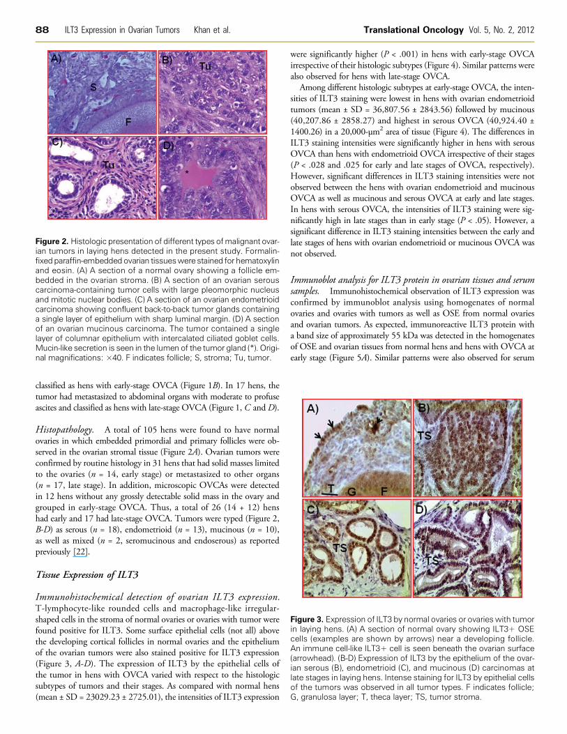
Figure 2. Histologic presentation of different types ofmalignant ovar-ian tumors in laying hens detected in the present study. Formalin-fixedparaffin-embeddedovarian tissueswere stained for hematoxylinand eosin. (A) A section of a normal ovary showing a follicle em-bedded in the ovarian stroma. (B) A section of an ovarian serouscarcinoma-containing tumor cells with large pleomorphic nucleusand mitotic nuclear bodies. (C) A section of an ovarian endometrioidcarcinoma showing confluent back-to-back tumor glands containinga single layer of epithelium with sharp luminal margin. (D) A sectionof an ovarian mucinous carcinoma. The tumor contained a singlelayer of columnar epithelium with intercalated ciliated goblet cells.Mucin-like secretion is seen in the lumen of the tumor gland (*). Origi-nal magnifications: ×40. F indicates follicle; S, stroma; Tu, tumor.
Figure 3. Expression of ILT3 by normal ovaries or ovaries with tumorin laying hens. (A) A section of normal ovary showing ILT3+ OSEcells (examples are shown by arrows) near a developing follicle.An immune cell-like ILT3+ cell is seen beneath the ovarian surface(arrowhead). (B-D) Expression of ILT3 by the epithelium of the ovar-ian serous (B), endometrioid (C), and mucinous (D) carcinomas atlate stages in laying hens. Intense staining for ILT3 by epithelial cellsof the tumors was observed in all tumor types. F indicates follicle;G, granulosa layer; T, theca layer; TS, tumor stroma.
88 ILT3 Expression in Ovarian Tumors Khan et al. Translational Oncology Vol. 5, No. 2, 2012
classified as hens with early-stage OVCA (Figure 1B). In 17 hens, thetumor had metastasized to abdominal organs with moderate to profuseascites and classified as hens with late-stage OVCA (Figure 1, C andD).
Histopathology. A total of 105 hens were found to have normalovaries in which embedded primordial and primary follicles were ob-served in the ovarian stromal tissue (Figure 2A). Ovarian tumors wereconfirmed by routine histology in 31 hens that had solid masses limitedto the ovaries (n = 14, early stage) or metastasized to other organs(n = 17, late stage). In addition, microscopic OVCAs were detectedin 12 hens without any grossly detectable solid mass in the ovary andgrouped in early-stage OVCA. Thus, a total of 26 (14 + 12) henshad early and 17 had late-stage OVCA. Tumors were typed (Figure 2,B-D) as serous (n = 18), endometrioid (n = 13), mucinous (n = 10),as well as mixed (n = 2, seromucinous and endoserous) as reportedpreviously [22].
Tissue Expression of ILT3
Immunohistochemical detection of ovarian ILT3 expression.T-lymphocyte-like rounded cells and macrophage-like irregular-shaped cells in the stroma of normal ovaries or ovaries with tumor werefound positive for ILT3. Some surface epithelial cells (not all) abovethe developing cortical follicles in normal ovaries and the epitheliumof the ovarian tumors were also stained positive for ILT3 expression(Figure 3, A-D). The expression of ILT3 by the epithelial cells ofthe tumor in hens with OVCA varied with respect to the histologicsubtypes of tumors and their stages. As compared with normal hens(mean ± SD = 23029.23 ± 2725.01), the intensities of ILT3 expression
were significantly higher (P < .001) in hens with early-stage OVCAirrespective of their histologic subtypes (Figure 4). Similar patterns werealso observed for hens with late-stage OVCA.
Among different histologic subtypes at early-stage OVCA, the inten-sities of ILT3 staining were lowest in hens with ovarian endometrioidtumors (mean ± SD = 36,807.56 ± 2843.56) followed by mucinous(40,207.86 ± 2858.27) and highest in serous OVCA (40,924.40 ±1400.26) in a 20,000-μm2 area of tissue (Figure 4). The differences inILT3 staining intensities were significantly higher in hens with serousOVCA than hens with endometrioid OVCA irrespective of their stages(P < .028 and .025 for early and late stages of OVCA, respectively).However, significant differences in ILT3 staining intensities were notobserved between the hens with ovarian endometrioid and mucinousOVCA as well as mucinous and serous OVCA at early and late stages.In hens with serous OVCA, the intensities of ILT3 staining were sig-nificantly high in late stages than in early stage (P < .05). However, asignificant difference in ILT3 staining intensities between the early andlate stages of hens with ovarian endometrioid or mucinous OVCA wasnot observed.
Immunoblot analysis for ILT3 protein in ovarian tissues and serumsamples. Immunohistochemical observation of ILT3 expression wasconfirmed by immunoblot analysis using homogenates of normalovaries and ovaries with tumors as well as OSE from normal ovariesand ovarian tumors. As expected, immunoreactive ILT3 protein witha band size of approximately 55 kDa was detected in the homogenatesof OSE and ovarian tissues from normal hens and hens with OVCA atearly stage (Figure 5A). Similar patterns were also observed for serum

Figure 5. Examples of immunoreactive ILT3 protein (A) or mRNA (B)expression in the homogenates of OSE and ovaries from normalhens or hens with OVCA by one-dimensional Western blot (WB)analysis and semiquantitative PCR, respectively. One-dimensionalWB (A): Immunoreactive ILT3 protein of approximately 55-kDa mo-lecular weight was detected in the 1) OSE extract from normal orovarian tumors and 2) homogenates from different histologic sub-types of ovarian tumors at early stage of OVCA. Compared withthe normal OSE or OSE from ovarian tumors at early stage, ILT3protein expression was stronger in the OSE from hens at late-stageOVCA. Serous ovarian tumors at the early stage showed strongILT3 staining followed by mucinous and endometrioid tumors. NoILT3 expression was detected in negative control in which proteinsample was omitted. RT-PCR (B): mRNA expression by the OSEextract from normal or ovarian tumor and from different histologicsubtypes of ovarian tumors at early and late stages of OVCA. Com-pared to the normal OSE, or OSE from ovarian tumor at early stage,a strong expression of ILT3 mRNA was observed in the OSE fromhens with late-stage OVCA. Similarly, strong expression patternswere also observed for the homogenates of ovarian tumors atlate stages than early stages irrespective of tumor subtypes. Asobserved in immunohistochemistry and immunoblot analysis, endo-metrioid tumors at early stage were weaker in ILT3 mRNA expres-sion than their counterpart serous and mucinous tumors. No ILT3mRNA expression was detected in the negative control in whichmRNA sample was omitted.
Figure 4. Changes in the ovarian ILT3 expression are associatedwith ovarian tumor development and progression. The intensityof ILT3 staining is expressed as pixel values (mean ± SD) in a20,000-μm2 area of the ovarian stroma in normal ovaries or ovarieswith tumor. Compared with normal hens, the intensities of theILT3 staining were significantly greater in hens with early-stageOVCA (P < .001) and increased further in hens with late-stageOVCA irrespective of their tumor types. The intensity of ILT3 stain-ing was significantly lower (P < .03) in hens with endometrioidOVCA than hens with serous OVCA. Significant differences werenot observed in the intensity of ILT3 staining between the henswith serous and mucinous OVCA irrespective of their stages. Barswith different letters within the same group indicate significantdifferences in ILT3 staining intensity.
Translational Oncology Vol. 5, No. 2, 2012 ILT3 Expression in Ovarian Tumors Khan et al. 89
samples (data not shown). Compared with the normal OSE or OSEfrom ovarian tumors at early stage, ILT3 protein expression was stron-ger in the OSE from hens at late-stage OVCA (Figure 5A). Conversely,the expression of immunoreactive ILT3 proteins was weaker in thehomogenates of ovarian endometrioid tumors, moderate in mucinous,and stronger in ovarian serous tumors at the early stage (Figure 5A).Similar patterns were also observed for ovarian tumors at late stage (datanot shown). These results confirm the immunohistochemical observa-tion that epithelial cells of ovarian tumors in hens express ILT3.
Expression of ILT3 mRNA. Observed variations in ILT3 proteinexpression among different histologic subtypes of OVCA and theirstages were confirmed by ILT3 mRNA expression. Hens with earlyserous and mucinous ovarian tumors showed strong amplification sig-nal for ILT3, whereas its amplification was low for endometrioid ovar-ian tumors. However, no differences were observed for ILT3 mRNAexpression by different histologic subtypes of late-stage OVCA(Figure 5B). Similar patterns were also observed for the epithelial cellsof ovarian tumors. Thus, the results of gene expression study confirmthe immunohistochemical and immunoblot analysis observations thatthe epithelium of ovarian tumors expresses ILT3 proteins.
DiscussionThis is the first report on the expression of ILT3 by the epitheliumof ovarian tumors in laying hens, an animal model of spontaneousOVCA. This study showed that the expression of ILT3 increases signifi-cantly in association with ovarian tumor development and progression.These results suggest that laying hens can be used to generate informationon the mechanism of spontaneous ovarian tumor–associated immuno-
suppression and may lead to the development of antitumor immuno-therapies and the testing of their efficacies.
Recent progresses in the understanding of tumor-immune interac-tions have led to the successful development of a number of immunother-apeutic approaches. However, tumor escape from immune recognitionis a significant barrier to the success of these immunotherapies. Althoughthe process of escaping immune surveillance by tumors or suppressionof antitumor immune response is not well understood, tumor cells,immune cells, and other stromal cells in the tumor surroundings havebeen reported to interact and create an immunosuppressive micro-environment through a variety of immunosuppressive factors [30]. En-hanced expression of ILT3 by few members of the immune system hasbeen suggested as one of such immunosuppressive factors in the tumormicroenvironment [31]. In the present study, in addition to immunecell-like cells, the epithelium of the ovarian tumors in laying hens also

90 ILT3 Expression in Ovarian Tumors Khan et al. Translational Oncology Vol. 5, No. 2, 2012
expressed ILT3. Under normal physiological condition, ILT3 has beenreported to be expressed selectively by professional antigen-presentingcells including monocytes, macrophages, and dendritic cells [32]. Theexpression of ILT3 on exposure to alloantigen-specific suppressor T cellsor cytokines by nonprofessional antigen-presenting cells, like endo-thelial cells as well as tumor cells from chronic lymphocytic leukemia,was also reported previously [33,34]. Thus, the present results suggestthat ovarian tumors also express immunosuppressive ILT3 as reportedfor few other cancers.
In the present study, compared with normal hens, the intensity of theILT3 expression was significantly high in hens with early-stage OVCAand increased further as the disease progressed to late stages, suggestingthat changes in ILT3 expression may be associated with ovarian tumordevelopment and progression. The presence of active antitumor im-mune responses against ovarian tumors at the early stage has beenreported earlier [9]. However, despite the presence of an antitumor im-mune response, OVCAs in most cases, progress to late stages. Althoughthe precise mechanisms of such immune escaping are not known,our results suggest that the enhanced expression of immune inhibitoryreceptor ILT3 by the ovarian tumors may contribute to the progressionof OVCA.
The results of the present study suggest that the expression patternof ILT3 varies with the different histologic subtypes of ovarian tumors.Significantly high ILT3 staining intensity was observed in serous com-pared with endometrioid ovarian tumors at early as well as late stagesof OVCA. The specific reasons for higher ILT3 expression by serousOVCA are not known. It is possible that higher ILT3 expression willcontribute to the faster progression of OVCA by imparting higherimmune tolerance to tumors. Serous malignant ovarian tumors areconsidered aggressive tumors, and increased ILT3 expression may con-tribute to their faster progression. Similar observations in cancers ofother organs were also reported previously [31,34]. Thus, it is possiblethat ILT3 will contribute to ovarian tumor progression by suppressingantitumor immunity.
In the present study, a portion of the epithelial cells of the normalovarian surface epithelium above the cortical developing follicles (notall epithelial cells) were positive for ILT3 expression albeit with lowerintensity than the epithelium of ovarian tumors. Although the precisereason(s) for such expression is not known, it is possible that ILT3expressed by surface epithelial cells near cortical follicles will protectthem from immune cells by suppression of local antifollicular auto-immune response. Various structures in the developing follicles in-cluding the perivitelline membrane, granulosa and theca layers, aswell as the degenerated cellular components of postovulatory folliclesmay appear as “foreign” to the circulating immune cells because thesestructures were not present during the evolution and maturation of theimmune system.
Our understanding regarding the biology and the role of ILT3 inthe context of OVCA is very limited, in part, because of the lack ofan animal model that develops spontaneous OVCA. Because of thedifficulty of detecting OVCA at an early stage, access to patient speci-mens to study and develop an effective antitumor immunotherapy isdifficult and time consuming. Similarities between the spontaneousOVCA in humans and hens in histologic subtypes, risk factors (e.g.,incessant ovulation), and expression of several molecular makers ofOVCA represent a high probability that results obtained from thisstudy may be translated to clinics. Furthermore, one of the most im-portant advantages of this animal model relative to the possibilitiesof translating current findings to OVCA in humans is the ability to
monitor hens by contrast enhanced ultrasound imaging using equip-ment and mechanical setting similar to those used in clinics [25,26].Thus, there will be less variation in imaging parameters between hensand humans because of the use of similar equipment. With the ad-vancement in in vivo imaging technology, ligands that bind to theepithelial cells of the ovarian tumor are being developed to use as anti-tumor therapies, and their effectiveness in tumor ablation can bemonitored in this animal model using contrast-enhanced ultrasoundscanning [35]. Because the epithelial cells of the tumor in OVCA hensexpress ILT3, laying hens may be used to test the efficacy of anti-ILT3drug by molecular-targeted ultrasound imaging of ovarian tissues.Thus, it will bring a significant change in the development of tumorspecific therapeutics specially immunoenhancing drugs and lead tothe development of treatment modalities for OVCA patients. Takentogether, information on ILT3 expression and its association withOVCA progression will aid in the development of anti-ILT3 immuno-therapies, which will lay the foundation for clinical studies and mayultimately lead to the development of effective therapies for the treat-ment of OVCA.
AcknowledgmentsThe authors thank Chet and Pam Utterback and Doug Hilgendorf, staffof the University of Illinois at Urbana-Champaign Poultry ResearchFarm, for maintenance of the hens. The authors also thank Sergio AbreuMachado and Syed Tahir Abbas Shah, graduate students, Departmentof Animal Sciences, University of Illinois at Urbana-Champaign, forhelping in hen tissue collection.
References[1] Jemal A, Siegel R, Xu J, and Ward E (2010). Cancer statistics, 2010. CA Cancer
J Clin 60, 277–300.[2] Goodman MT, Correa CN, Tung KH, Roffers SD, Cheng Wu X, Young JL Jr,
Wilkens LR, Carney ME, and Howe HL (2003). Stage at diagnosis of ovariancancer in the United States, 1992–1997. Cancer 97, 2648–2659.
[3] Ries LA (1993). Ovarian cancer. Survival and treatment differences by age. Cancer71, 524–529.
[4] Zou W (2006). Regulatory T cells, tumour immunity and immunotherapy. NatRev Immunol 6, 295–307.
[5] Beyer M and Schultze JL (2006). Regulatory T cells in cancer. Blood 108,804–811.
[6] Figdor CG, de Vries IJ, Lesterhuis WJ, and Melief CJ (2004). Dendritic cellimmunotherapy: mapping the way. Nat Med 10, 475–480.
[7] Dougan M and Dranoff G (2009). Immune therapy for cancer. Annu RevImmunol 27, 83–117.
[8] Shu S, Cochran AJ, Huang RR, Morton DL, and Maecker HT (2006). Immuneresponses in the draining lymph nodes against cancer: implications for immuno-therapy. Cancer Metastasis Rev 25, 233–242.
[9] Yigit R, Massuger LF, Figdor CG, and Torensma R (2010). Ovarian cancercreates a suppressive microenvironment to escape immune elimination. GynecolOncol 117, 366–372.
[10] Curiel TJ, Coukos G, Zou L, Alvarez X, Cheng P, Mottram P, Evdemon-Hogan M, Conejo-Garcia JR, Zhang L, Burow M, et al. (2004). Specific recruit-ment of regulatory T cells in ovarian carcinoma fosters immune privilege andpredicts reduced survival. Nat Med 10, 942–949.
[11] Li MO, Wan YY, Sanjabi S, Robertson AK, and Flavell RA (2006). Transform-ing growth factor-β regulation of immune responses. Annu Rev Immunol 24,99–146.
[12] Kriegel MA, Li MO, Sanjabi S, Wan YY, and Flavell RA (2006). Transform-ing growth factor-β: recent advances on its role in immune tolerance. CurrRheumatol Rep 8, 138–144.
[13] Liu Z, Guo B, and Lopez RD (2009). Expression of intercellular adhesionmolecule (ICAM)-1 or ICAM-2 is critical in determining sensitivity of pan-creatic cancer cells to cytolysis by human γδ-T cells: implications in the designof γδ-T-cell–based immunotherapies for pancreatic cancer. J Gastroenterol Hepatol24, 900–911.
Translational Oncology Vol. 5, No. 2, 2012 ILT3 Expression in Ovarian Tumors Khan et al. 91
[14] Roncarolo MG, Bacchetta R, Bordignon C, Narula S, and Levings MK (2001).Type 1 T regulatory cells. Immunol Rev 182, 68–79.
[15] Liu Z, Tugulea S, Cortesini R, and Suciu Foca N (1998). Specific suppression ofT helper alloreactivity by allo MHC class I–restricted CD8+CD28− T cells. IntImmunol 10, 775–783.
[16] Wei S, Kryczek I, Zou L, Daniel B, Cheng P, Mottram P, Curiel T, Lange A,and Zou W (2005). Plasmacytoid dendritic cells induce CD8+ regulatory T cellsin human ovarian carcinoma. Cancer Res 65, 5020–5026.
[17] Kryczek I, Zou L, Rodriguez P, Zhu G, Wei S, Mottram P, Brumlik M, Cheng P,Curiel T, Myers L, et al. (2006). B7-H4 expression identifies a novel suppressivemacrophage population in human ovarian carcinoma. J Exp Med 203, 871–881.
[18] Salazar-Onfray F (1999). Interleukin-10: a cytokine used by tumors to escapeimmunosurveillance. Med Oncol 16, 86–94.
[19] Suciu-Foca N, Feirt N, Zhang QY, Vlad G, Liu Z, Lin H, Chang CC, Ho EK,Colovai AI, Kaufman H, et al. (2007). Soluble Ig-like transcript 3 inhibits tumorallograft rejection in humanized SCIDmice and T cell responses in cancer patients.J Immunol 178, 7432–7441.
[20] Kim-Schulze S, Scotto L, Vlad G, Piazza F, Lin H, Liu Z, Cortesini R, and Suciu-Foca N (2006). Recombinant Ig-like transcript 3-Fc modulates T cell responses viainduction of Th anergy and differentiation of CD8+ T suppressor cells. J Immunol176, 2790–2798.
[21] Rodriguez-Burford C, Barnes MN, Berry W, Partridge EE, and Grizzle WE(2001). Immunohistochemical expression of molecular markers in an avian model:a potential model for preclinical evaluation of agents for ovarian cancer chemo-prevention. Gynecol Oncol 81, 373–379.
[22] Barua A, Bitterman P, Abramowicz JS, Dirks AL, Bahr JM, Hales DB, BradaricMJ, Edassery SL, Rotmensch J, and Luborsky JL (2009). Histopathology ofovarian tumors in laying hens: a preclinical model of human ovarian cancer.Int J Gynecol Cancer 19, 531–539.
[23] Viertlboeck BC, Habermann FA, Schmitt R, Groenen MA, Du Pasquier L, andGobel TW (2005). The chicken leukocyte receptor complex: a highly diverse multi-gene family encoding at least six structurally distinct receptor types. J Immunol175, 385–393.
[24] Dennis G Jr, Kubagawa H, and Cooper MD (2000). Paired Ig-like receptorhomologs in birds and mammals share a common ancestor with mammalianFc receptors. Proc Natl Acad Sci USA 97, 13245–13250.
[25] Barua A, Abramowicz JS, Bahr JM, Bitterman P, Dirks A, Holub KA, Sheiner E,Bradaric MJ, Edassery SL, and Luborsky JL (2007). Detection of ovarian tumors
in chicken by sonography: a step toward early diagnosis in humans? J UltrasoundMed 26, 909–919.
[26] Barua A, Bitterman P, Bahr JM, Bradaric MJ, Hales DB, Luborsky JL, andAbramowicz JS (2010). Detection of tumor-associated neoangiogenesis byDoppler ultrasonography during early-stage ovarian cancer in laying hens: apreclinical model of human spontaneous ovarian cancer. J Ultrasound Med 29,173–182.
[27] Giles JR, Olson LM, and Johnson PA (2006). Characterization of ovarian surfaceepithelial cells from the hen: a unique model for ovarian cancer. Exp Biol Med(Maywood) 231, 1718–1725.
[28] Barua A, Edassery SL, Bitterman P, Abramowicz JS, Dirks AL, Bahr JM, HalesDB, Bradaric MJ, and Luborsky JL (2009). Prevalence of antitumor antibodiesin laying hen model of human ovarian cancer. Int J Gynecol Cancer 19, 500–507.
[29] Hong YH, Lillehoj HS, Lee SH, Dalloul RA, and Lillehoj EP (2006). Analysis ofchicken cytokine and chemokine gene expression following Eimeria acervulinaand Eimeria tenella infections. Vet Immunol Immunopathol 114, 209–223.
[30] Poschke I, Mougiakakos D, and Kiessling R (2011). Camouflage and sabo-tage: tumor escape from the immune system. Cancer Immunol Immunother60, 1161–1171.
[31] Vlad G, Chang CC, Colovai AI, Vasilescu ER, Cortesini R, and Suciu-Foca N(2010). Membrane and soluble ILT3 are critical to the generation of T suppressorcells and induction of immunological tolerance. Int Rev Immunol 29, 119–132.
[32] Cella M, Dohring C, Samaridis J, Dessing M, Brockhaus M, Lanzavecchia A,and Colonna M (1997). A novel inhibitory receptor (ILT3) expressed on mono-cytes, macrophages, and dendritic cells involved in antigen processing. J ExpMed 185, 1743–1751.
[33] Manavalan JS, Kim-Schulze S, Scotto L, Naiyer AJ, Vlad G, Colombo PC,Marboe C, Mancini D, Cortesini R, and Suciu-Foca N (2004). Alloantigen spe-cific CD8+CD28− FOXP3+ T suppressor cells induce ILT3+ ILT4+ tolerogenicendothelial cells, inhibiting alloreactivity. Int Immunol 16, 1055–1068.
[34] Colovai AI, Tsao L, Wang S, Lin H, Wang C, Seki T, Fisher JG, Menes M,Bhagat G, Alobeid B, et al. (2007). Expression of inhibitory receptor ILT3 onneoplastic B cells is associated with lymphoid tissue involvement in chroniclymphocytic leukemia. Cytometry B Clin Cytom 72, 354–362.
[35] Barua A, Bitterman P, Bahr JM, Basu S, Sheiner E, Bradaric MJ, Hales DB,Luborsky JL, and Abramowicz JS (2011). Contrast-enhanced sonography de-picts spontaneous ovarian cancer at early stages in a preclinical animal model.J Ultrasound Med 30, 333–345.