
Progestin-Induced Apoptosis in the Macaque Ovarian Epithelium: Differential Regulation of...
11
Progestin-Induced Apoptosis in the Macaque Ovarian Epithelium: Differential Regulation of Transforming Growth Factor- Gustavo C. Rodriguez, Nimesh P. Nagarsheth, Karen L. Lee, Rex C. Bentley, David K. Walmer, Mark Cline, Regina S. Whitaker, Pam Isner, Andrew Berchuck, Richard K. Dodge, Claude L. Hughes Background: Oral contraceptive (OC) use is associated with a reduced risk of ovarian cancer. An OC component, pro- gestin, induces apoptosis in the primate ovarian epithelium. One regulator of apoptosis is transforming growth factor- (TGF-). We determined the effect of progestin on TGF- expression in the primate ovarian epithelium and examined the relationship between TGF- expression and apoptosis. Methods: Female cynomolgus macaques were randomly as- signed to receive a diet for 35 months containing no hor- mones (n = 20); the OC Triphasil (n = 17); or each of its constituents, ethinyl estradiol (estrogen, n = 20) or levonor- gestrel (progestin, n = 18 ), alone. Ovarian sections were immunostained with monoclonal antibodies against TGF-1 or TGF-2 plus TGF-3 (TGF-2/3) isoforms. The expres- sion of TGF- isoforms in four ovarian compartments (epi- thelium, oocytes, granulosa cells, and hilar vascular endo- thelium) was compared among treatment groups. The association between TGF- expression and apoptosis, as de- termined by morphology and histochemistry, was examined in ovarian epithelium. All statistical tests were two-sided. Results: Compared with ovaries from the control and estro- gen-only-treated monkeys, the ovaries of progestin-treated monkeys showed 1) a marked decrease in the expression of TGF-1 and a concomitant increase in the expression of the TGF-2/3 isoforms in the ovarian epithelium (P<.001), 2) an increase in the expression of TGF-2/3 in the hilar vascular endothelium (P<.001), and 3) a marked decrease in TGF- 2/3 expression in granulosa cells (P<.001). The apoptotic index of the ovarian epithelium was highly associated with the change in expression from TGF-1(P<.001) to TGF- 2/3 (P.002) induced by progestin treatment. Conclusions: Progestin induces differential regulation in the ovarian epi- thelium of TGF-, a change in the expression of which is highly associated with apoptosis. These data suggest a pos- sible biologic mechanism for the protective association be- tween OC use and reduced ovarian cancer risk. [J Natl Can- cer Inst 2002;94:50–60] Epithelial ovarian cancer remains an important public health problem. It is the fourth leading cause of cancer-related deaths among women in the United States and causes over 100 000 deaths annually worldwide (1,2). Despite intensive research ef- forts over the past decade directed toward improved detection and treatment of ovarian cancer, the long-term survival of women with ovarian cancer has improved only modestly. Prog- ress in the fight against ovarian cancer has been hampered by a number of factors, including late diagnosis, the molecular het- erogeneity of ovarian tumors, the absence of highly curative chemotherapy, and the lack of a valid animal model for the disease. The development of effective chemopreventive agents for ovarian cancer may represent our best hope for decreasing the ovarian cancer mortality rate in the future. A potent preventive agent already exists in the estrogen–progestin combination oral contraceptive (OC). Routine use of OCs for as little as 3 years confers as much as a 50% reduction in risk of ovarian cancer. The protective association increases with the duration of use and lasts for as long as 20 years after the discontinuation of use (3–7). It has been our belief that, if the mechanism(s) underlying the remarkable protective effect of the OC can be elucidated, it may be possible to develop a pharmacologic chemopreventive strategy that is even more protective against ovarian cancer than OCs. Moreover, it may be possible to develop a chemopreven- tive strategy that is more broadly applicable than the use of OCs, potentially extending the benefits of chemoprevention beyond the reproductive age group to include those women who are menopausal, a group that currently lacks a nonsurgical approach for ovarian cancer prevention. Although the biologic mechanism underlying the protective association between OC use and reduction in the risk of ovarian cancer remains unproven, two previously cited theories have focused on the known inhibitory effect of OCs on ovulation and on the inhibitory effect of OCs on the secretion of the pituitary gonadotropins follicle-stimulating hormone and luteinizing hor- mone. In the first theory, the inhibition of ovulation is presumed to reduce ovarian surface trauma and thereby to reduce the po- tential for genetic damage in the ovarian epithelium, while the second theory suggests that lowering gonadotropin levels poten- tially decreases a stimulus to proliferation in the ovary (8–11). The ovulation-suppression theory has been challenged because the amount of risk reduction conferred by OCs far exceeds what would be predicted on the basis of the number of ovulations inhibited (12). Similarly, the gonadotropin theory has been criti- cized because of the lack of evidence of an ovarian cancer- Affiliations of authors: G. C. Rodriguez, R. S. Whitaker, P. Isner, A. Berchuck (Division of Gynecologic Oncology, Department of Obstetrics and Gynecology), N. P. Nagarsheth, K. L. Lee (Department of Obstetrics and Gynecology), R. C. Bentley (Department of Pathology), D. K. Walmer (Division of Reproductive Endocrinology, Department of Obstetrics and Gynecology), R. K. Dodge (De- partment of Biostatistics, Duke Comprehensive Cancer Center), Duke University Medical Center, Durham, NC; M. Cline, Section of Comparative Medicine, Wake Forest University School of Medicine, Winston-Salem, NC; C. L. Hughes, Department of Obstetrics and Gynecology, Duke University Medical Center and Avalon Medical Group, Chapel Hill, NC. Correspondence to: Gustavo C. Rodriguez, M.D., Division of Gynecologic Oncology, Department of Obstetrics and Gynecology, Rm. 1315, Evanston Northwestern Healthcare, 2650 Ridge Rd., Evanston, IL 60201 (e-mail: [email protected]). See “Notes” following “References.” © Oxford University Press 50 ARTICLES Journal of the National Cancer Institute, Vol. 94, No. 1, January 2, 2002 by guest on December 3, 2015 http://jnci.oxfordjournals.org/ Downloaded from
-
Upload
independent -
Category
Documents
-
view
0 -
download
0
Transcript of Progestin-Induced Apoptosis in the Macaque Ovarian Epithelium: Differential Regulation of...

Progestin-Induced Apoptosis in the Macaque OvarianEpithelium: Differential Regulation of TransformingGrowth Factor-�
Gustavo C. Rodriguez, Nimesh P. Nagarsheth, Karen L. Lee, Rex C. Bentley,David K. Walmer, Mark Cline, Regina S. Whitaker, Pam Isner, Andrew Berchuck,Richard K. Dodge, Claude L. Hughes
Background: Oral contraceptive (OC) use is associated witha reduced risk of ovarian cancer. An OC component, pro-gestin, induces apoptosis in the primate ovarian epithelium.One regulator of apoptosis is transforming growth factor-�(TGF-�). We determined the effect of progestin on TGF-�expression in the primate ovarian epithelium and examinedthe relationship between TGF-� expression and apoptosis.Methods: Female cynomolgus macaques were randomly as-signed to receive a diet for 35 months containing no hor-mones (n = 20); the OC Triphasil (n = 17); or each of itsconstituents, ethinyl estradiol (estrogen, n = 20) or levonor-gestrel (progestin, n = 18 ), alone. Ovarian sections wereimmunostained with monoclonal antibodies against TGF-�1or TGF-�2 plus TGF-�3 (TGF-�2/3) isoforms. The expres-sion of TGF-� isoforms in four ovarian compartments (epi-thelium, oocytes, granulosa cells, and hilar vascular endo-thelium) was compared among treatment groups. Theassociation between TGF-� expression and apoptosis, as de-termined by morphology and histochemistry, was examinedin ovarian epithelium. All statistical tests were two-sided.Results: Compared with ovaries from the control and estro-gen-only-treated monkeys, the ovaries of progestin-treatedmonkeys showed 1) a marked decrease in the expression ofTGF-�1 and a concomitant increase in the expression of theTGF-�2/3 isoforms in the ovarian epithelium (P<.001), 2) anincrease in the expression of TGF-�2/3 in the hilar vascularendothelium (P<.001), and 3) a marked decrease in TGF-�2/3 expression in granulosa cells (P<.001). The apoptoticindex of the ovarian epithelium was highly associated withthe change in expression from TGF-�1 (P<.001) to TGF-�2/3 (P�.002) induced by progestin treatment. Conclusions:Progestin induces differential regulation in the ovarian epi-thelium of TGF-�, a change in the expression of which ishighly associated with apoptosis. These data suggest a pos-sible biologic mechanism for the protective association be-tween OC use and reduced ovarian cancer risk. [J Natl Can-cer Inst 2002;94:50–60]
Epithelial ovarian cancer remains an important public healthproblem. It is the fourth leading cause of cancer-related deathsamong women in the United States and causes over 100 000deaths annually worldwide (1,2). Despite intensive research ef-forts over the past decade directed toward improved detectionand treatment of ovarian cancer, the long-term survival ofwomen with ovarian cancer has improved only modestly. Prog-ress in the fight against ovarian cancer has been hampered by anumber of factors, including late diagnosis, the molecular het-erogeneity of ovarian tumors, the absence of highly curativechemotherapy, and the lack of a valid animal model for the disease.
The development of effective chemopreventive agents forovarian cancer may represent our best hope for decreasing theovarian cancer mortality rate in the future. A potent preventiveagent already exists in the estrogen–progestin combination oralcontraceptive (OC). Routine use of OCs for as little as 3 yearsconfers as much as a 50% reduction in risk of ovarian cancer.The protective association increases with the duration of use andlasts for as long as 20 years after the discontinuation of use(3–7). It has been our belief that, if the mechanism(s) underlyingthe remarkable protective effect of the OC can be elucidated, itmay be possible to develop a pharmacologic chemopreventivestrategy that is even more protective against ovarian cancer thanOCs. Moreover, it may be possible to develop a chemopreven-tive strategy that is more broadly applicable than the use of OCs,potentially extending the benefits of chemoprevention beyondthe reproductive age group to include those women who aremenopausal, a group that currently lacks a nonsurgical approachfor ovarian cancer prevention.
Although the biologic mechanism underlying the protectiveassociation between OC use and reduction in the risk of ovariancancer remains unproven, two previously cited theories havefocused on the known inhibitory effect of OCs on ovulation andon the inhibitory effect of OCs on the secretion of the pituitarygonadotropins follicle-stimulating hormone and luteinizing hor-mone. In the first theory, the inhibition of ovulation is presumedto reduce ovarian surface trauma and thereby to reduce the po-tential for genetic damage in the ovarian epithelium, while thesecond theory suggests that lowering gonadotropin levels poten-tially decreases a stimulus to proliferation in the ovary (8–11).The ovulation-suppression theory has been challenged becausethe amount of risk reduction conferred by OCs far exceeds whatwould be predicted on the basis of the number of ovulationsinhibited (12). Similarly, the gonadotropin theory has been criti-cized because of the lack of evidence of an ovarian cancer-
Affiliations of authors: G. C. Rodriguez, R. S. Whitaker, P. Isner, A. Berchuck(Division of Gynecologic Oncology, Department of Obstetrics and Gynecology),N. P. Nagarsheth, K. L. Lee (Department of Obstetrics and Gynecology), R. C.Bentley (Department of Pathology), D. K. Walmer (Division of ReproductiveEndocrinology, Department of Obstetrics and Gynecology), R. K. Dodge (De-partment of Biostatistics, Duke Comprehensive Cancer Center), Duke UniversityMedical Center, Durham, NC; M. Cline, Section of Comparative Medicine,Wake Forest University School of Medicine, Winston-Salem, NC; C. L. Hughes,Department of Obstetrics and Gynecology, Duke University Medical Center andAvalon Medical Group, Chapel Hill, NC.
Correspondence to: Gustavo C. Rodriguez, M.D., Division of GynecologicOncology, Department of Obstetrics and Gynecology, Rm. 1315, EvanstonNorthwestern Healthcare, 2650 Ridge Rd., Evanston, IL 60201 (e-mail:[email protected]).
See “Notes” following “References.”
© Oxford University Press
50 ARTICLES Journal of the National Cancer Institute, Vol. 94, No. 1, January 2, 2002
by guest on Decem
ber 3, 2015http://jnci.oxfordjournals.org/
Dow
nloaded from

protective effect associated with noncontraceptive estrogen use(which lowers gonadotropin levels) and because of the absenceof an association between serum levels of follicle-stimulatinghormone and luteinizing hormone and ovarian cancer risk(12,13). Both of these theories fail to consider that the ovarianepithelium contains receptors for estrogen, progesterone, andandrogen and that reproductive factors may affect ovarian can-cer risk via a potent biologic interaction of sex steroid hormoneswith the ovarian epithelium (14).
Recently, we performed a study in primates demonstratingthat a combination estrogen–progestin OC has a potent apoptoticeffect on the ovarian epithelium, mediated by the progestin com-ponent (15). Primates randomly assigned in a 3-year trial toreceive either combination estrogen and progestin or progestinalone had a fourfold to sixfold increase in the proportion ofapoptotic ovarian epithelial cells as compared with control orestrogen-only-treated monkeys. The apoptosis pathway is one ofthe most important in vivo mechanisms that function to eliminatecells that have sustained DNA damage and, thus, are prone tomalignant transformation (16). In addition, a number of well-known chemopreventive agents have been demonstrated to ac-tivate the apoptosis pathway in the target tissues that they protectfrom neoplastic transformation (17–32). The finding that pro-gestins activate this critical pathway in the ovarian epitheliumsuggests that the protective effects afforded by OCs againstovarian cancer may at least in part be caused by progestin-mediated apoptosis. This forms the basis for an investigation ofthe progestin class of drugs as chemopreventive agents for epi-thelial ovarian cancer.
The regulation of apoptosis is complex and is influenced bynumerous families of transcriptional factors, tumor suppressorgenes, oncogenes, and growth factors (33). Among the growthfactors, transforming growth factor-� (TGF-�) has been impli-cated as an important regulator of apoptosis and as a mediator ofthe apoptotic effects of steroid hormones (34–37). An associa-tion between the degree of TGF-� expression and apoptosis hasbeen shown in cells derived from the breast (38) and prostate(39), and the apoptotic activity of hormones such as the retinoidshas been shown to be mediated at least in part by the activity ofTGF-� (18,19,40). Notably, multiple members of the steroidhormone superfamily, including the retinoids, vitamin D, andsex steroids, have been shown to modulate the expression ofTGF-�, and the promoter region for specific TGF-� isotypessuch as TGF-�2 and TGF-�3 contains response elements sug-gesting hormonal and developmental regulation (41–51).
Given the link between TGF-� molecular pathways and ap-optosis and evidence suggesting unique regulation of TGF-� bysteroid hormones, we sought to determine in the current studywhether there is an association between progestin-induced apop-totic effects in the primate ovarian epithelium and expression ofTGF-�.
MATERIALS AND METHODS
Animals/Randomization
As described previously (15), 130 young adult female cyno-molgus macaques (Macaca fascicularis), with an average age of4.75 years, were randomly assigned into a study designed toevaluate the long-term biologic effects of the contraceptive Tri-phasil (Wyeth Ayerst, St. Davids, PA). The cynomolgus ma-caque is an excellent animal model for yielding experimental
results that are pertinent to human reproductive biology. Thisnonhuman primate has a 28-day menstrual cycle that is similarto that of humans (52–54). The study was a prospective, ran-domized, controlled trial designed for the primary endpoint ofevaluating the effects of Triphasil and its individual components(ethinyl estradiol and levonorgestrel) on the cardiovascular sys-tem. Secondary outcomes to be analyzed included the biologiceffects of Triphasil on the reproductive organs and breast. Therandomization process was based on the serum lipid responses(total plasma cholesterol, triglycerides, and high-density lipo-protein-C) of animals to challenge with an atherogenic diet (44%of calories from fat, 0.28 mg of cholesterol per kilocalorie).After randomization, there were no differences between studygroups with regard to body weight or age.
Forty of the animals were killed early in the study for baselinecardiovascular and lipoprotein studies, and an additional 14 ani-mals died during the course of the study, primarily from traumaand diarrheal diseases. One animal was excluded because itsovarian tissue was not available for study. The remaining 75animals were necropsied at the completion of the thirty-fifthmonth of the study and form the basis for this investigation. Thestudy was approved by the Animal Care and Use Committee atthe Wake Forest University School of Medicine, Winston-Salem, NC.
The macaques were prospectively randomly assigned via thelipid response parameters noted above into four groups to re-ceive a diet for 35 months that contained 1) no hormones (con-trol); 2) the oral combination contraceptive Triphasil, which iscomposed of estrogen (ethinyl estradiol) and progestin (levo-norgestrel); 3) the estrogenic component of Triphasil (ethinylestradiol) alone; or 4) the progestin component of Triphasil (le-vonorgestrel) alone. Hormones in the latter two groups wereadministered in the same dosage and schedule that occurs in atypical Triphasil regimen. Doses were scaled on the basis ofcaloric intake, which takes into account species differences inmetabolic rate; this is the generally accepted way to achievedosages comparable to those in women. The human-equivalentdoses were given as follows: 6 days of 0.030 mg ethinyl estra-diol plus 0.050 mg levonorgestrel per day, followed by 5 days of0.040 mg ethinyl estradiol plus 0.075 mg levonorgestrel per day,followed by 10 days of 0.030 mg ethinyl estradiol plus 0.125 mglevonorgestrel per day, followed by 7 days of no hormone treat-ment. This cyclic regimen was repeated every 28 days continu-ously for 35 months. During the third week of the last month ofthe study, the animals were killed and their ovaries were care-fully removed and preserved.
Tissue Preparation and Immunohistochemistry
From each animal in the study, one ovary was flash frozen byimmersion in liquid nitrogen and saved for future molecularstudies, and the other was formalin fixed and paraffin embedded.
Apoptosis. The median proportion of apoptotic ovarian epi-thelial cells associated with each treatment group had been quan-tified previously (15). Briefly, 5-�m sections taken from themiddle of each paraffin-embedded ovary were mounted oncharged slides, and the ovarian epithelium was examined formorphologic and immunohistochemical evidence of apoptosisafter staining with the APOPTAG-plus kit (Oncor, Gaithersburg,MD). Dark-brown, nuclear staining easily identified cells under-going apoptosis. Tonsillar and deoxyribonuclease-digested tis-sue sections were used as positive controls. To calculate the
Journal of the National Cancer Institute, Vol. 94, No. 1, January 2, 2002 ARTICLES 51
by guest on Decem
ber 3, 2015http://jnci.oxfordjournals.org/
Dow
nloaded from

percentage of ovarian epithelial cells undergoing apoptosis, wecounted both the total number of ovarian epithelial cells and thenumber undergoing apoptosis on each 5-�m section. The me-dian proportion of cells undergoing apoptosis was calculated foreach treatment group. At each step in this study, including thehistologic examinations of the ovaries, the investigators wereblinded with regard to the treatment group associated with eachovary.
TGF-� expression. Immunohistochemical expression ofTGF-� was performed as previously described, with slightmodification (55). Briefly, 5-�m sections taken from the middleof each paraffin-embedded ovary were cut and mounted oncharged slides. Two slides from each specimen were placed in a60 °C oven for 1 hour. One slide was used as the negative con-trol, while the other was used as the study specimen. The sec-tions were deparaffinized, immersed in 0.3% hydrogen peroxideto quench endogenous peroxidase, hydrated, placed in AntigenRetrieval Citra solution at pH 6.0 (BioGenex Laboratories, Inc.,San Ramon, CA), and then heated with an electric pressurecooker (Biocare Medical,Walnut Creek, CA) for 5 minutes. Thesections were then cooled and rinsed with three washes of phos-phate-buffered saline, preincubated in Power Block (BioGenexLaboratories, Inc.) for 10 minutes, and then incubated for 18hours (overnight) at 4 °C in a humid chamber with primaryantibody. For TGF-�1 expression, sections were immunostainedwith a monoclonal antibody that reacts with TGF-�1 but notTGF-�2 or TGF-�3 (2.5 �g/mL anti-TGF-�1 monoclonal anti-body, catalog No. MAB 240; Research and Development Sys-tems (Minneapolis, MN). To evaluate TGF-�2 and TGF-�3(TGF-�2/3) expression, we stained sections with a mouse mono-clonal antibody that reacts with the N-terminal region of bothTGF-�2 and TGF-�3 but has no cross-reactivity with TGF-�1(0.25 �g/mL TGF-�3 mouse monoclonal antibody; OncogeneResearch Products, Cambridge, MA). For negative controlspecimens for TGF-�1 and TGF-�2/3 staining, mouse immuno-globulin G antibody (Coulter Corporation, Miami, FL) was ap-plied at concentrations of 2.5 and 0.25 �g/mL, respectively.Slides were then washed three times with phosphate-bufferedsaline for 5 minutes each. Application of a biotinylated second-ary antibody (Multi-Link Super Sensitive Detection System;BioGenex Laboratories) was performed at room temperature ina humid chamber for 20 minutes, then followed by three washesin phosphate-buffered saline for 5 minutes each. Peroxidase-conjugated streptavidin (Multi-Link Super Sensitive DetectionSystem) was applied to sections and allowed to incubate for 20minutes in a humid chamber, then followed by three washes inphosphate-buffered saline for 5 minutes each. Slides were incu-bated with freshly prepared 3,3-diaminobenzidine (D5637;Sigma Chemical Co., St. Louis, MO) chromogen solution (0.5%3,3-diaminobenzidine, 0.6% hydrogen peroxide, and 0.05% Trisbuffer) for 4 minutes and then washed in deionized water for 5minutes to stop the reaction. This was followed by a 5-minuteincubation in a 0.1 M sodium acetate solution and then stainingwith 1.5% methyl green for 5 minutes. Sections were dipped 10times each in a serial fashion in the following solutions: 95%acetone, 95% acetone, 100% acetone, 100% acetone, 100% xy-lene, 100% xylene, and 100% xylene; then coverslips wereplaced on the slides. Umbilical cord sections, stained in a similarfashion, were used as positive control (56).
The ovarian sections were examined by two independent setsof reviewers, all of whom were blinded to the hormone admin-
istration data (R. C. Bentley and K. L. Lee for TGF-�1 staining;R. C. Bentley and N. P. Nagarsheth for TGF-�2/3 staining).Staining for TGF-� was evaluated in four separate ovarian com-partments of each study slide (ovarian surface epithelium, pri-mordial oocyte cytoplasm, granulosa cells of tertiary follicles,and endothelium in ovarian hilar vessels) and graded accordingto the degree of staining intensity from 0 to 3+ (TGF-�1) andfrom 0 to 4+ (TGF-�2/3). High expression of TGF-�1 wasdefined by the slide reviewers as 2+ to 3+ staining intensity,whereas high expression of TGF-�2/3 was defined as 3+ to 4+staining intensity. Three ovarian sections in the TGF-�1 staininggroup and two ovarian sections in the TGF-�2/3 staining groupwere excluded from grading because the samples were techni-cally insufficient for evaluation.
Statistical Analysis
Quantitation and comparison of the median proportion ofapoptotic cells in the ovarian epithelium had been performedpreviously (15). Briefly, the Kruskal–Wallis test was used toperform multiple comparisons of all paired treatments (57), andthe statistical analysis was carried out with the use of the BMDPstatistical software package (Biomathematical Data PackageStatistical Software, Inc., Los Angeles, CA) (58). For this study,the association between expression of the TGF-� isoforms andtreatment was analyzed with the use of an overall approximateexact test for contingency tables (59). In addition, each 2 × 2table involving treatment and control was analyzed by use ofFisher’s two-sided exact test. The relationship between treat-ment, amount of expression of TGF-� in the ovarian epithelium,and the mean proportion of apoptotic ovarian epithelial cells wasanalyzed by use of the general linear model (PROC GLM in theSAS statistical package; SAS Institute, Cary, NC) (60). Multiplecomparisons were performed with the use of Dunnett’s two-sided test for each treatment compared with the control. Therelationship between the proportion of high TGF-� expressionand the mean proportion of apoptotic cells across treatments wasanalyzed by use of standard correlation analysis. The associationbetween the TGF-� isoforms with respect to overexpression wasanalyzed with the use of the � statistic (61). All statistical testswere two-sided.
RESULTS
Effect of Hormone Treatment on Expression of TGF-�
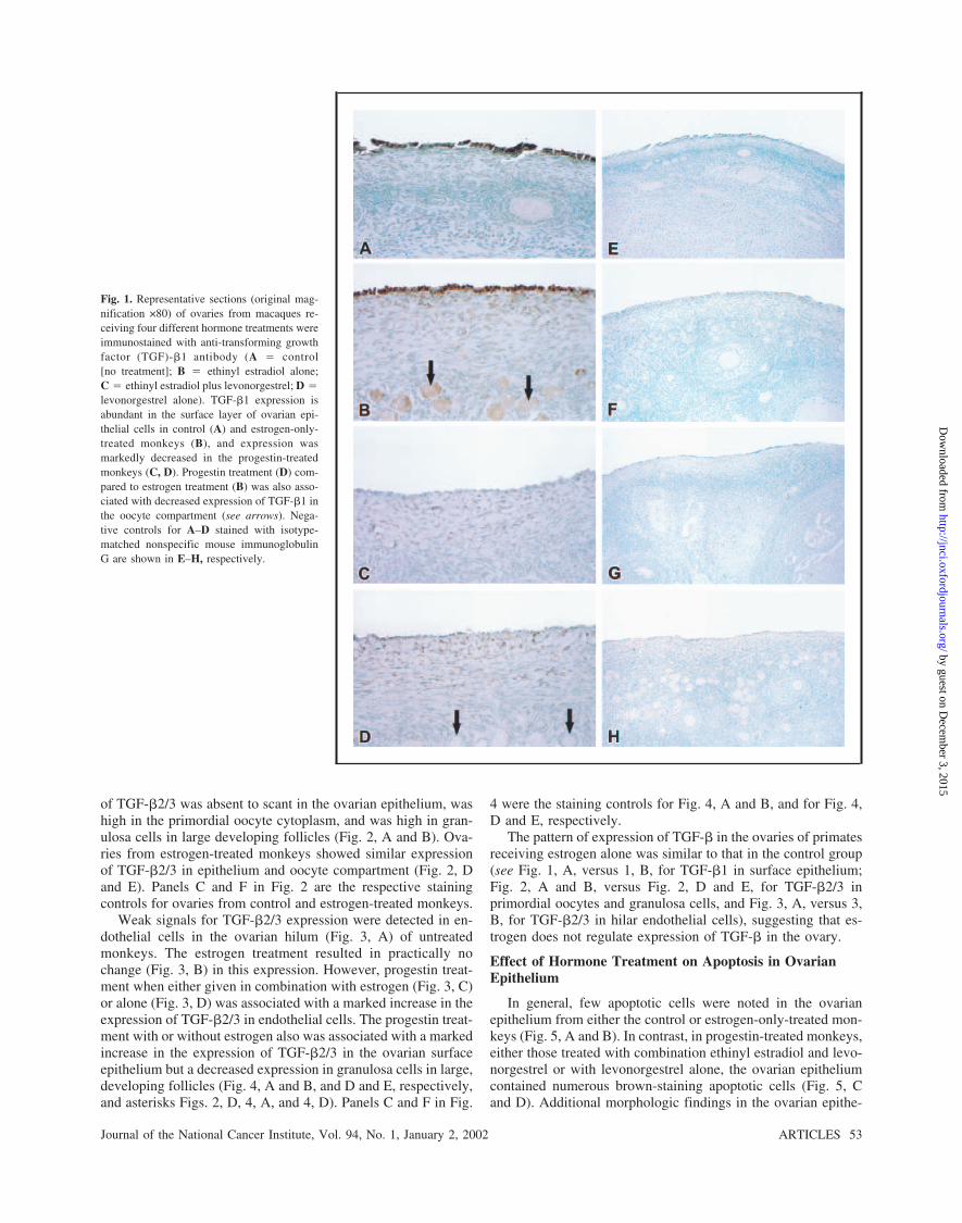
In general, in ovarian sections from the control group ofmonkeys, the pattern of expression of TGF-� was qualitativelysimilar to the pattern described previously in the human ovary(62–65). In untreated monkeys (Fig. 1, A) and in estrogen-treated monkeys (Fig. 1, B), TGF-�1 expression was abundantin the ovarian epithelium and low to moderate in the stroma(structural tissue under epithelium) and the oocyte cytoplasm.Exposure to progestin either with estrogen (Fig. 1, C) or alone(Fig. 1, D) was associated with a marked decrease in the ex-pression of TGF-�1 in the ovarian epithelium and in the oocytecompartment (see arrows in Fig. 1, B and D). The endothelialcells of the vascular hilum had little detectable expression ofTGF-�1 (data not shown). Panels E–H in Fig. 1 represent thestaining controls for monkey ovaries from four treatments, re-spectively.
The pattern of expression of TGF-�2/3 in untreated monkeyovaries was distinctly different from that of TGF-�1. Expression
52 ARTICLES Journal of the National Cancer Institute, Vol. 94, No. 1, January 2, 2002
by guest on Decem
ber 3, 2015http://jnci.oxfordjournals.org/
Dow
nloaded from
of TGF-�2/3 was absent to scant in the ovarian epithelium, washigh in the primordial oocyte cytoplasm, and was high in gran-ulosa cells in large developing follicles (Fig. 2, A and B). Ova-ries from estrogen-treated monkeys showed similar expressionof TGF-�2/3 in epithelium and oocyte compartment (Fig. 2, Dand E). Panels C and F in Fig. 2 are the respective stainingcontrols for ovaries from control and estrogen-treated monkeys.
Weak signals for TGF-�2/3 expression were detected in en-dothelial cells in the ovarian hilum (Fig. 3, A) of untreatedmonkeys. The estrogen treatment resulted in practically nochange (Fig. 3, B) in this expression. However, progestin treat-ment when either given in combination with estrogen (Fig. 3, C)or alone (Fig. 3, D) was associated with a marked increase in theexpression of TGF-�2/3 in endothelial cells. The progestin treat-ment with or without estrogen also was associated with a markedincrease in the expression of TGF-�2/3 in the ovarian surfaceepithelium but a decreased expression in granulosa cells in large,developing follicles (Fig. 4, A and B, and D and E, respectively,and asterisks Figs. 2, D, 4, A, and 4, D). Panels C and F in Fig.
4 were the staining controls for Fig. 4, A and B, and for Fig. 4,D and E, respectively.
The pattern of expression of TGF-� in the ovaries of primatesreceiving estrogen alone was similar to that in the control group(see Fig. 1, A, versus 1, B, for TGF-�1 in surface epithelium;Fig. 2, A and B, versus Fig. 2, D and E, for TGF-�2/3 inprimordial oocytes and granulosa cells, and Fig. 3, A, versus 3,B, for TGF-�2/3 in hilar endothelial cells), suggesting that es-trogen does not regulate expression of TGF-� in the ovary.
Effect of Hormone Treatment on Apoptosis in OvarianEpithelium
In general, few apoptotic cells were noted in the ovarianepithelium from either the control or estrogen-only-treated mon-keys (Fig. 5, A and B). In contrast, in progestin-treated monkeys,either those treated with combination ethinyl estradiol and levo-norgestrel or with levonorgestrel alone, the ovarian epitheliumcontained numerous brown-staining apoptotic cells (Fig. 5, Cand D). Additional morphologic findings in the ovarian epithe-
Fig. 1. Representative sections (original mag-nification ×80) of ovaries from macaques re-ceiving four different hormone treatments wereimmunostained with anti-transforming growthfactor (TGF)-�1 antibody (A � control[no treatment]; B � ethinyl estradiol alone;C � ethinyl estradiol plus levonorgestrel; D �
levonorgestrel alone). TGF-�1 expression isabundant in the surface layer of ovarian epi-thelial cells in control (A) and estrogen-only-treated monkeys (B), and expression wasmarkedly decreased in the progestin-treatedmonkeys (C, D). Progestin treatment (D) com-pared to estrogen treatment (B) was also asso-ciated with decreased expression of TGF-�1 inthe oocyte compartment (see arrows). Nega-tive controls for A–D stained with isotype-matched nonspecific mouse immunoglobulinG are shown in E–H, respectively.
Journal of the National Cancer Institute, Vol. 94, No. 1, January 2, 2002 ARTICLES 53
by guest on Decem
ber 3, 2015http://jnci.oxfordjournals.org/
Dow
nloaded from

lium of progestin-treated monkeys included patches of ovariansurface devoid of epithelium, epithelial cells with sparse cyto-plasm that appeared to be detaching from the surface, areas ofepithelial denudation, and brown-staining apoptotic cells con-taining apoptotic bodies. The apoptotic changes noted in theovarian epithelium of progestin-treated monkeys were not asso-
ciated with any change in the proliferative index of the ovarianepithelium via staining for Ki-67 (data not shown).
Semiquantitative Determination of TGF-� Expression
Tables 1–3 summarize semiquantitative measurements of thehormonal regulation of TGF-�1 and TGF-�2/3 expression in
Fig. 2. Representative ovarian sections fromcontrol (no treatment) (A, original magnifica-tion ×25; B, original magnification ×80) andethinyl estradiol-only treated (D, original mag-nification ×25; E, original magnification ×80)macaques immunostained with anti-transforminggrowth factor (TGF)-�2/3 antibody showingmarked expression of TGF-�2/3 in primordialoocytes and granulosa cells (*) in large, devel-oping follicles and little detectable expressionof TGF-�2/3 in the ovarian surface epitheliallayer. C and F: negative controls for A and Band C and D, respectively, stained with iso-type-matched nonspecific mouse immuno-globulin G.
Fig. 3. Representative sections of the macaqueovarian hilum from the four hormone treatmentgroups immunostained with anti-transforminggrowth factor (TGF)-�2/3 antibody (A � con-trol; B � ethinyl estradiol alone; C � ethinylestradiol plus levonorgestrel; D � levonor-gestrel alone). C and D: progestin treatmentassociated with a marked increase in expres-sion of TGF-�2/3 in endothelial cells.
54 ARTICLES Journal of the National Cancer Institute, Vol. 94, No. 1, January 2, 2002
by guest on Decem
ber 3, 2015http://jnci.oxfordjournals.org/
Dow
nloaded from

vivo. Progestin treatment, either combined with estrogen (Tri-phasil group) or administered alone (levonorgestrel group), wasassociated with a striking and highly statistically significant de-crease in the expression of TGF-�1 in the ovarian epithelium(P<.001) and a moderate decrease in the expression of TGF-�1
in the oocyte cytoplasm (P � .002) (Table 1). In contrast, pro-gestin treatment was associated with a marked increase in theexpression of TGF-�2/3 in the ovarian epithelium (P<.001)(Table 2). Without exception, TGF-�2/3 expression in the ovar-ian epithelium was high (3+ to 4+ staining) in every monkey on
Fig. 4. Representative ovarian sections fromcombination estrogen–progestin-treated (A,original magnification ×25; B, original magni-fication ×80) and levonorgestrel-only treated(D, original magnification ×25; E, originalmagnification ×80) macaques immunostainedwith anti-transforming growth factor (TGF)-�2/3 antibody showing marked expression ofTGF-�2/3 in the ovarian surface epitheliumand decreased expression of TGF-�2/3 in gran-ulosa cells (*) in large developing follicles. Cand F: negative controls for A and B and D andE, respectively, stained with isotype-matchednonspecific mouse immunoglobulin G.
Fig. 5. Apoptag staining of representative ma-caque ovarian sections from the four hormonetreatment groups (A � control; B � ethinylestradiol alone; C � ethinyl estradiol plus le-vonorgestrel; D � levonorgestrel alone) show-ing marked apoptosis in the ovarian epitheliumassociated with progestin treatment (C, D)(original magnification ×80).
Journal of the National Cancer Institute, Vol. 94, No. 1, January 2, 2002 ARTICLES 55
by guest on Decem
ber 3, 2015http://jnci.oxfordjournals.org/
Dow
nloaded from

progestin (n � 34). Similarly, there was a significant increase inTGF-�2/3 expression in the ovarian hilar endothelial cells inmonkeys on progestin (P<.001). In contrast, progestin treatmentwas associated with a marked decrease in TGF-�2/3 expressionin granulosa cells (P<.001) (Table 2).
Within the ovarian epithelial compartment, comparison of theapoptotic index with the degree of change in the expression ofthe TGF-� isoforms revealed a highly significant correlationbetween changes in TGF-� expression and apoptosis (P<.001)(Table 3). With the use of the general linear model, for TGF-�1there was a statistically significant treatment effect (P<.001)with respect to the mean proportion of apoptotic cells. Table 3gives the mean proportion of apoptotic cells for each treatmentgroup and shows that the Triphasil and levonorgestrel groupsdiffer significantly from the control group (P<.001 for both com-parisons with the use of Dunnett’s test). Similarly, for TGF-�2/3there was also a statistically significant treatment effect(P<.001), and Table 3 shows that the Triphasil and levonor-gestrel groups differ statistically significantly from the controlgroup (P � .002, and P<.001, respectively, by Dunnett’s test).The Pearson correlation coefficients between the proportion of
high TGF-� expression and the mean proportion of apoptoticcells across treatments were –0.998 (P �.002) for TGF-�1 and0.973 (P �.03) for TGF-�2/3. Finally, overall, there was anegative association between TGF-�2/3 overexpression andTGF-�1 overexpression (� � –0.62; P<.001). Taken together,these data demonstrate the novel finding that progestin-inducedapoptosis in the ovarian epithelium is associated with an isoformswitch in expression of TGF-�.
DISCUSSION
To the best of our knowledge, this is the first study to dem-onstrate regulation of TGF-� expression in the primate ovarianepithelium in vivo by a contraceptive steroid. We found TGF-�expression to be differentially regulated in the ovarian epithe-lium of primates that received progestin, administered either inthe form of an estrogen–progestin combination pill or alone.Progestin treatment was associated with a marked decrease inexpression of TGF-�1 concomitant with a marked increase inexpression of TGF-�2/3. In addition, the progestin-inducedchange in TGF-� isoform expression was highly correlated with
Table 1. Hormonal regulation of expression of transforming growth factor (TGF)-�1 in the macaque ovary
Treatment group
No. (%) ovaries/treatment group with high TGF-�1 expression (2+ to 3+) in each ovarian compartment
No. Epithelium Granulosa cells Oocytes Endothelium
Control 20 18 (90%) 7 (35%) 7 (35%) 0 (0%)Ethinyl estradiol 19 16 (84%) 4 (21%) 2 (13%) 0 (0%)Triphasil 16 3 (19%) 2 (13%) 0 (0%) 0 (0%)Levonorgestrel 17 1 (6%) 2 (12%) 0 (0%) 0 (0%)
Overall approximate exact test P<.001 P � .31 P � .002 P � 1.00Control vs. ethinyl estradiol P � .66 P � .48 P � .13 P � 1.00Control vs. Triphasil P<.001 P � .24 P � .01 P � 1.00Control vs. levonorgestrel P<.001 P � .14 P � .009 P � 1.00
Table 2. Hormonal regulation of expression of transforming growth factor (TGF)-�2/3 in the macaque ovary
Treatment group
No. (%) ovaries/treatment group with high TGF-�2/3 expression (3+ to 4+) in each ovarian compartment
No. Epithelium Granulosa cells Oocytes Endothelium
Control 19 6 (32%) 12 (63%) 14 (74%) 5 (26%)Ethinyl estradiol 20 2 (10%) 8 (38%) 17 (81%) 3 (23%)Triphasil 17 17 (100%) 1 (6%) 16 (94%) 16 (94%)Levonorgestrel 17 17 (100%) 1 (6%) 14 (82%) 16 (94%)
Overall approximate exact test P<.001 P<.001 P � 0.45 P<.001Control vs. ethinyl estradiol P � .13 P � .11 P � .72 P � .72Control vs. Triphasil P<.001 P<.001 P � .18 P<.001Control vs. levonorgestrel P<.001 P<.001 P � .70 P<.001
Table 3. Relationship between treatment, transforming growth factor (TGF)-� expression, and apoptosis in the macaqueovarian epithelium
Treatment
TGF-�1 TGF-�2/3
No. % 2+ to 3+Mean proportion of apoptotic cellsin ovarian epithelium (95% CI*) No. % 3+ to 4+
Mean proportion of apoptotic cellsin ovarian epithelium (95% CI*)
Control 20 90 6.3 (3.0 to 9.6) 19 32 6.4 (2.8 to 10)Ethinyl estradiol 19 84 6.2 (1.8 to 10.6) 20 10 4.5 (1.2 to 7.8)Triphasil 16 19 22.3 (13.6 to 31.0)† 17 100 21.2 (12.7 to 29.7)‡Levonorgestrel 17 6 25.1 (16.0 to 34.2)† 17 100 26.4 (17.7 to 35.1)†
*CI � confidence interval for the mean.†P<.001 by Dunnett’s test comparing mean apoptotic index seen after treatment with that seen in controls (no treatment).‡P � .002 by Dunnett’s test comparing mean apoptotic index seen after treatment with that seen in controls (no treatment).
56 ARTICLES Journal of the National Cancer Institute, Vol. 94, No. 1, January 2, 2002
by guest on Decem
ber 3, 2015http://jnci.oxfordjournals.org/
Dow
nloaded from

an increase in apoptosis in the ovarian epithelium. Estrogentreatment appeared to have no impact on TGF-� expression inthe ovary.
The mechanism underlying progestin regulation of TGF-�expression in the ovary remains to be determined. It is possiblethat progestins induce factors in the ovarian stroma that regulateTGF-� pathways at sites throughout the ovary via a paracrineeffect. Conversely, it is possible that progestins act directlythrough classic progesterone receptor-mediated pathways to ef-fect TGF-� expression. The results of this study suggest thatprogestin regulation of ovarian TGF-� expression occurs via adirect effect in that changes in TGF-� expression associatedwith progestin treatment were primarily localized to sites in theovary known to express progestin receptors. These include theovarian epithelium (66) and granulosa cells of large follicles(67). In addition, the finding that progestins increase the expres-sion of TGF-�2/3 in the ovarian epithelium while at the sametime decreasing expression of TGF-�2/3 in granulosa cells sup-ports the notion not only that progestin induction of TGF-�2/3 inthe ovarian epithelium is a direct effect but also that the endeffect of progestins in the ovary is site specific. It is interestingthat we also noted increased expression of TGF-�2/3 in theendothelial cells of the vascular hilum in progestin-treated mon-keys. It has been shown recently that endothelial cells containfunctional progesterone receptors and that progesterone inhibitsendothelial proliferation (68).
There is mounting evidence that differential regulation ofpeptide growth factors by steroid hormones contributes to thediverse end effects of these hormones in target tissues. Amongthe growth factors, TGF-� has been shown to be differentiallyregulated by estrogens, retinoids, androgens, and vitamin Dcompounds. In bone, raloxifene increases the expression ofTGF-�3 while having no effect on the expression of TGF-�1and TGF-�2 (69). In cells derived from the breast, estradioldecreases the expression of TGF-�2 and TGF-�3 while havingno effect on the expression of TGF-�1 (70), whereas tamoxifenhas been shown to increase the expression of TGF-�1 (71). Inchondrocytes, vitamin D increases the expression of TGF-�2and decreases the expression of TGF-�1 and TGF-�3 (72). Glu-cocorticoids differentially regulate TGF-� in healing wounds,leading to the suppression of TGF-�1 and TGF-�2 and the in-creased expression of TGF-�3 (73). In the palates of mice, ret-inoids have been shown to decrease the expression of TGF-�1while having no effect on the expression of other TGF-� iso-forms (74), whereas in keratinocytes, induction of TGF-�2 is amajor mechanism underlying the biologic effects of retinoids(51,75). Finally, in the male accessory organs, androgen with-drawal is associated with both apoptosis and differential regu-lation of TGF-� (76). Thus, the TGF-� isotypes appear to bedifferentially regulated in a tissue-specific manner. Although themechanism underlying the complex regulation of TGF-� by hor-mones is not completely understood, differences in the promoterregion among the TGF-� isoforms or in post-transcriptionalevents may be means by which TGF-� is differentially regulated(77–80).
Although the design of our study does not allow us to provethe causal relationship between TGF-� expression and apopto-sis, the finding that changes in TGF-� expression were highlyassociated with apoptosis in the ovarian epithelium in primateson progestin is strongly supportive of the hypothesis that pro-gestin-induced apoptosis may be occurring via a TGF-�-
mediated molecular pathway. In addition to the findings of thisstudy, other lines of evidence support this hypothesis. First, inhormone-responsive tissues, such as the breast and prostate,TGF-� has been shown to mediate the apoptotic effects of ste-roid hormones, including the antiestrogens, retinoids, and vita-min D (40,81,82). Second, in tissues that are progesterone re-ceptor positive, such as the breast and endometrium, progestinshave been shown to be associated with both induction of TGF-�and apoptosis (37,41,83–88). Third, both our group and others(89–91) have shown previously that some ovarian cancer celllines undergo apoptosis when treated with TGF-�. It is interest-ing that, in our laboratory, we were not able to demonstrateinduction of apoptosis in normal ovarian epithelial cells treatedwith TGF-�. It is possible, however, that the assay techniquesused in our study were not sufficiently sensitive to detect apop-tosis in a limited sample of normal human ovarian epithelialcells. Alternatively, it is possible that our in vitro experimentslacked an important cofactor present in vivo that is required forTGF-�-mediated apoptosis to occur or that our in vitro condi-tions failed to simulate the complex interrelationship of TGF-�isoform expression required for apoptosis to occur in nonmalig-nant human ovarian epithelial cells. A fourth line of evidence isthat TGF-� is related to müllerian inhibitory factor, a peptidethat causes complete regression of the müllerian system (theprecursor to the uterus, fallopian tubes, and upper vagina) in thedeveloping male embryo (92–95). In the embryo, the mülleriantract develops from an invagination of the celomic epitheliumand, therefore, is derived from the same embryonic precursortissue as the ovarian epithelium (96). Given the marked inhibi-tory effect that the müllerian inhibitory factor has on the mül-lerian system, it is interesting to speculate that the ovarian epi-thelium may be uniquely susceptible in vivo to undergoingapoptosis in response to TGF-� and that agents that selectivelyregulate TGF-� in the ovarian epithelium may be potent apop-tosis-inducing agents and cancer preventive agents.
A growing body of laboratory and animal evidence has im-plicated TGF-� as a potent tumor suppressor and cancer pre-ventive agent (97–99). Transgenic mice that have a constitu-tively active form of TGF-�1 are resistant to 7,12-dimethylbenz[a]anthracene-induced mammary tumors (100).Conversely, mice with heterozygous deletions of one copy of theTGF-� gene have an increased susceptibility to chemical carci-nogenesis (101). In humans, mutations have been described inthe TGF-� signaling pathway in a variety of tumors, includingcancers of colon, cancers of the gastric, pancreatic, and uterinesystems, and cancers of lymphoid organs (99). Furthermore, anumber of cellular oncogenes are known to inhibit TGF-� ac-tivity. Finally, TGF-� has been implicated as a mediator of thebiologic effects of a number of chemopreventive agents, includ-ing tamoxifen, which induces expression of TGF-�1 in stromalcells in the breast (71), as well as retinoids, which induce TGF-�in the prostate and respiratory tract (98). Taken together, thesedata provide compelling evidence that TGF-� plays an impor-tant role as an inhibitor of carcinogenesis.
In light of the known association between TGF-� and cancerprevention, the observation that OCs markedly alter expressionof TGF-� in the ovarian epithelium implicates TGF-� as possi-bly mediating the ovarian cancer-protective effects of the pill.The finding that OCs induce both apoptosis and TGF-� in theovary suggests that OCs may be acting as true chemopreventiveagents by activating molecular pathways known to arrest or
Journal of the National Cancer Institute, Vol. 94, No. 1, January 2, 2002 ARTICLES 57
by guest on Decem
ber 3, 2015http://jnci.oxfordjournals.org/
Dow
nloaded from

reverse the process of carcinogenesis, rather than simply bylimiting ovulation-induced damage in the ovarian epithelium.Moreover, the finding that activation of apoptosis and differen-tial regulation of TGF-� are related specifically to the progestincomponent of the OC provides strong evidence in support of thenotion that biologic effects produced by the progestin compo-nent may be major mechanisms underlying the marked protec-tion conferred by OCs against ovarian cancer.
The discovery that contraceptive progestins activate cancer-preventive molecular pathways in the ovarian epithelium opensthe door to the development of a highly effective pharmacologicpreventive strategy for ovarian cancer that may be more effec-tive and more broadly applicable than OCs. For example, if theprotective effects of OCs were solely a result of ovulation inhi-bition as previously believed, then there is little potential fordesigning improved OC formulations that have enhanced ovar-ian cancer-protective effects, and the protective effects can onlybe beneficial for premenopausal women who are ovulating.However, if OCs confer marked ovarian cancer protectionthrough a biologic effect unrelated to ovulation inhibition, thenit may be possible to design OC formulations that maximizethese biologic effects to achieve enhanced ovarian cancer pro-tection. In addition, it may be possible to develop a pharmaco-logic preventive strategy that can be applied to all women, in-cluding administration of a pharmacologic regimen that hasovarian cancer-preventive effects in postmenopausal womenwho are anovulatory.
The ideal preventive agent for ovarian cancer may be com-posed of a combination of agents that act in an additive orsynergistic fashion to maximally activate molecular pathwaysthat inhibit carcinogenesis in the ovarian epithelium, therebymaximizing ovarian cancer prevention while minimizing sideeffects. In this regard, agents selected from the steroid hormonesuperfamily on the basis of their ability to activate TGF-� areuniquely attractive. Steroid hormones would be expected to spe-cifically target only cells expressing appropriate steroid ligandreceptor. In addition, given the short half-life of active TGF-� invivo, rapid clearance of TGF-� at target sites would limit thesystemic toxicity associated with chemoprevention (75). It isinteresting to speculate that the combination of a progestin,which regulates TGF-� in the ovarian epithelium, and a retinoidand/or vitamin D might achieve synergistic or additive effects onTGF-� pathways in the ovarian epithelium, leading to a power-ful cancer preventive agent. Synergistic effects on both growthinhibition and apoptosis have been described in vitro in cellsderived from ovarian epithelium with the use of the combinationof vitamin A derivatives and TGF-� (102). Similarly, cross-talkhas been described between vitamin D and TGF-� signalingpathways, and the combination of vitamin D and TGF-� hasbeen shown to have synergistic effects in vitro (103,104). Theseapproaches will be the subject of further investigation as wework toward the development of optimal chemopreventive strat-egies.
REFERENCES
(1) Landis SH, Murray T, Bolden S, Wingo PA. Cancer statistics, 1999. CACancer J Clin 1999;49:8–31.
(2) Parkin DM, Pisani P, Ferlay J. Global cancer statistics. CA Cancer J Clin1999;49:33–64.
(3) Rosenberg L, Palmer JR, Zauber AG, Warshauer ME, Lewis JL Jr, StromBL, et al. A case–control study of oral contraceptive use and invasiveepithelial ovarian cancer. Am J Epidemiol 1994;139:654–61.
(4) Epithelial ovarian cancer and combined oral contraceptives: the WHOcollaborative study of neoplasia and steroid contraceptives. Int J Epide-miol 1989;18:538–45.
(5) The reduction in risk of ovarian cancer associated with oral-contraceptiveuse. The Cancer and Steroid Hormone Study of the Centers for DiseaseControl and the National Institute of Child Health and Human Develop-ment. N Engl J Med 1987;316:650–5.
(6) Gross TP, Schlesselman JJ. The estimated effect of oral contraceptive useon the cumulative risk of epithelial ovarian cancer. Obstet Gynecol 1994;83:419–24.
(7) Franceschi S, Parazzini F, Negri E, Booth M, La Vecchia C, Beral V, etal. Pooled analysis of 3 European case–control studies of epithelial ovar-ian cancer: III. Oral contraceptive use. Int J Cancer 1991;49:61–5.
(8) Wu ML, Whittemore AS, Paffenbarger RS Jr, Sarles DL, Kampert JB,Grosser S, et al. Personal and environmental characteristics related toepithelial ovarian cancer. I. Reproductive and menstrual events and oralcontraceptive use. Am J Epidemiol 1988;128:1216–27.
(9) Greene MH, Clark JW, Blayney DW. The epidemiology of ovarian can-cer. Semin Oncol 1984;11:209–26.
(10) Cramer DW, Welch WR. Determinants of ovarian cancer risk. II. Infer-ences regarding pathogenesis. J Natl Cancer Inst 1983;71:717–21.
(11) Whittemore AS, Harris R, ltnyre J. Characteristics relating to ovariancancer risk: collaborative analysis of 12 U.S. case–control studies. II.Invasive epithelial ovarian cancers in white women. Collaborative Ovar-ian Cancer Group. Am J Epidemiol 1992;136:1184–203.
(12) Risch HA, Weiss NS, Lyon JL, Daling JR, Liff JM. Events of reproduc-tive life and the incidence of epithelial ovarian cancer. Am J Epidemiol1983;117:128–39.
(13) Helzlsouer KJ, Alberg AJ, Gordon GB, Longcope C, Bush TL, HoffmanSC, et al. Serum gonadotropins and steroid hormones and the develop-ment of ovarian cancer. JAMA 1995;274:1926–30.
(14) Risch HA. Hormonal etiology of epithelial ovarian cancer, with a hypoth-esis concerning the role of androgens and progesterone. J Natl Cancer Inst1998;90:1774–86.
(15) Rodriguez GC, Walmer DK, Cline M, Krigman H, Lessey BA, WhitakerRS, et al. Effect of progestin on the ovarian epithelium of macaques:cancer prevention through apoptosis? J Soc Gynecol Investig 1998;5:271–6.
(16) Canman CE, Chen CY, Lee MH, Kastan MB. DNA damage responses:p53 induction, cell cycle pertubations, and apoptosis. Cold Spring HarbSymp Quant Biol 1994;59:277–86.
(17) Chan LN, Zhang S, Cloyd M, Chan TS. N-(4-Hydroxyphenyl) retinamideprevents development of T-lymphomas in AKR/J mice. Anticancer Res1997;17:499–503.
(18) Ponzoni M, Bocca P, Chiesa V, Decensi A, Pistoia V, Raffaghello L, etal. Differential effects of N-(4-hydroxyphenyl)retinamide and retinoicacid on neuroblastoma cells: apoptosis versus differentiation. Cancer Res1995;55:853–61.
(19) Delia D, Aiello A, Lombardi L, Pelicci PG, Grignani F, Grignani F, et al.N-(4-Hydroxyphenyl)retinamide induces apoptosis of malignant hemo-poietic cell lines including those unresponsive to retinoic acid. Cancer Res1993;53:6036–41.
(20) Seewaldt VL, Yim JR, Caldwell LE, Johnson BS, Swisshelm Y, CollinsSJ. All-trans-retinoic acid mediates G1 arrest but not apoptosis of normalhuman mammary epithelial cells. Cell Growth Differ 1995;6:863–9.
(21) Lotan R. Retinoids in cancer chemoprevention. FASEB J 1996;10:1031–9.
(22) Sankaranarayanan R, Mathew B. Retinoids as cancer-preventive agents.IARC Sci Publ 1996;139:47–59.
(23) Toma S, Isnardi L, Raffo P, Dastoli G, De Francisci E, Riccardi L, et al.Effects of all-trans-retinoic acid and 13-cis-retinoic acid on breast-cancercell lines: growth inhibition and apoptosis induction. Int J Cancer 1997;70:619–27.
(24) Oridate N, Lotan D, Mitchell MF, Hong WK, Lotan R. Inhibition ofproliferation and induction of apoptosis in cervical carcinoma cells byretinoids: implications for chemoprevention. J Cell Biochem Suppl 1995;23:80–6.
(25) Dolivet G, Ton Van J, Sarini J, Wattel E, Lagarde P, Chomy F, et al.Current knowledge on the action of retinoids in carcinoma of the head andneck. Rev Laryngol Otol Rhinol (Bord) 1996;117:19–26.
58 ARTICLES Journal of the National Cancer Institute, Vol. 94, No. 1, January 2, 2002
by guest on Decem
ber 3, 2015http://jnci.oxfordjournals.org/
Dow
nloaded from

(26) Kuo SM. Antiproliferative potency of structurally distinct dietary flavo-noids on human colon cancer cells. Cancer Lett 1996;110:41–8.
(27) Thompson HJ, Jiang C, Lu J, Mehta RG, Piazza GA, Paranka NS, et al.Sulfone metabolite of sulindac inhibits mammary carcinogenesis. CancerRes 1997;57:267–71.
(28) Reddy BS, Wang CX, Samaha H, Lubet R, Steele VE, Kelloff GJ, et al.Chemoprevention of colon carcinogenesis by dietary perillyl alcohol.Cancer Res 1997;57:420–5.
(29) Gould MN. Cancer chemoprevention and therapy by monoterpenes. En-viron Health Perspect 1997;105 Suppl 4:977–9.
(30) Pascale RM, Simile MM, De Miglio MR, Nufris A, Daino L, SeddaiuMA, et al. Chemoprevention by S-adenosyl-L-methionine of rat liver car-cinogenesis initiated by 1,2-dimethylhydrazine and promoted by oroticacid. Carcinogenesis 1995;16:427–30.
(31) Thompson HJ, Wilson A, Lu J, Singh M, Jiang C, Upadhyaya P, et al.Comparison of the effects of an organic and an inorganic form of seleniumon a mammary carcinoma cell line. Carcinogenesis 1994;15:183–6.
(32) el-Bayoumy K, Upadhyaya P, Chae YH, Sohn OS, Rao CV, Fiala E, et al.Chemoprevention of cancer by organoselenium compounds. J Cell Bio-chem Suppl 1995;22:92–100.
(33) Franko J, Pomfy M, Prosbova T. Apoptosis and cell death (mechanisms,pharmacology and promise for the future). Acta Medica (Hradec Kralove)2000;43:63–8.
(34) Haufel T, Dormann S, Hanusch J, Schwieger A, Bauer G. Three distinctroles for TGF-� during intercellular induction of apoptosis: a review.Anticancer Res 1999;19:105–12.
(35) Bursch W, Oberhammer F, Jirtle RL, Askari M, Sedivy R, Grasl-KrauppB, et al. Transforming growth factor-�1 as a signal for induction of celldeath by apoptosis. Br J Cancer 1993;67:531–6.
(36) Chiarugi V, Magnelli L, Cinelli M. Complex interplay among apoptosisfactors: RB, p53, E2F, TGF-�, cell cycle inhibitors and the bcl2 genefamily. Pharmacol Res 1997;35:257–61.
(37) Rotello RJ, Lieberman RC, Purchio AF, Gershenson LE. Coordinatedregulation of apoptosis and cell proliferation by transforming growth fac-tor � 1 in cultured uterine epithelial cells. Proc Natl Acad Sci U S A1991;88:3412–5.
(38) Strange R, Li F, Saurer S, Burkhardt A, Friis RR. Apoptotic cell death andtissue remodeling during mouse mammary gland involution. Development1992;115:49–58.
(39) Kyprianou N, Isaacs JT. Expression of transforming growth factor-beta inthe rat ventral prostate during castration-induced programmed cell death.Mol Endocrinol 1989;3:1515–22.
(40) Roberson KM, Penland SN, Padilla GM, Selvan RS, Kim CS, Fine RL, etal. Fenretinide: induction of apoptosis and endogenous transforminggrowth factor � in PC-3 prostate cancer cells. Cell Growth Differ 1997;8:101–11.
(41) Colletta AA, Wakefield LM, Howell FV, Danielpour D, Baum M, SpornMB. The growth inhibition of human breast cancer cells by a novel syn-thetic progestin involves the induction of transforming growth factor beta.J Clin Invest 1991;87:277–83.
(42) Reiss M, Barcellos-Hoff MH. Transforming growth factor-� in breastcancer: a working hypothesis. Breast Cancer Res Treat 1997;45:81–95.
(43) Dannecker C, Possinger K, Classen S. Induction of TGF-� by an anti-progestin in the human breast cancer cell line T-47D. Ann Oncol 1996;7:391–5.
(44) Lucia MS, Sporn MB, Roberts AB, Stewart LV, Danielpour D. The roleof transforming growth factor-beta1, -beta2, and -beta3 in androgen-responsive growth of NRP-152 rat prostatic epithelial cells. J Cell Physiol1998;175:184–92.
(45) Jeng MH, ten Dijke P, Iwata KK, Jordan VC. Regulation of the levels ofthree transforming growth factor � mRNAs by estrogen and their effectson the proliferation of human breast cancer cells. Mol Cell Endocrinol1993;97:115–23.
(46) Heberden C, Denis I, Pointillart A, Mercier T. TGF-� and calcitriol. GenPharmacol 1998;30:145–51.
(47) Gold LI. The role for transforming growth factor-� (TGF-�) in humancancer. Crit Rev Oncog 1999;10:303–60.
(48) Wu Y, Craig TA, Lutz WH, Kumar R. Identification of 1 � 25-dihydroxyvitamin D3 response elements in the human transforminggrowth factor �2 gene. Biochemistry 1999;38: 2654–60.
(49) Roberts AB. Molecular and cell biology of TGF-�. Miner ElectrolyteMetab 1998;24:111–9.
(50) Knabbe C, Lippman ME, Wakefield LM, Flanders KC, Kasid A, DerynckR, et al. Evidence that transforming growth factor-� is a hormonallyregulated negative growth factor in human breast cancer cells. Cell 1987;48:417–28.
(51) Roberts AB, Sporn MB. Mechanistic interrelationships between two su-perfamilies: the steroid/retinoid receptors and transforming growth factor-�. Cancer Surv 1992;14:205–20.
(52) Brenner RM, Slayden OD. Cyclic changes in the primate oviduct andendometrium. In: Knobil E, Neill JD, editors. The physiology of repro-duction. New York (NY): Raven Press; 1994. p. 541–69.
(53) Hotchkiss J, Knobil E. The menstrual cycle and its neuroendocrine con-trol. In: Knobil E, Neill JD, editors. The physiology of reproduction. NewYork (NY): Raven Press; 1994. p. 711–36.
(54) Kaiserman-Abramof IR, Padykula HA. Ultrastructural epithelial zonationof the primate endometrium (rhesus monkey). Am J Anat 1989;184:13–30.
(55) Hurteau J, Rodriguez GC, Whitaker RS, Shah S, Mills G, Bast RC, et al.Transforming growth factor-beta inhibits proliferation of human ovariancancer cells obtained from ascites. Cancer 1994;74:93–9.
(56) Stewart AA, Haley JD, Qu GY, Stam K, Fenyo D, Chait BT, et al.Umbilical cord transforming growth factor-�3: isolation, comparison withrecombinant TGF-�3 and cellular localization. Growth Factors 1996;13:87–98.
(57) Hollander M, Wolfe DA. Nonparametric statistical methods. New York(NY): Wiley; 1973.
(58) BMDP statistical software manual. Los Angeles (CA): University of Cali-fornia Press; 1992. p. 459–61.
(59) Mehta CR, Patel NR. A network algorithm for performing Fisher’s exacttest in r×c contingency tables. J Am Stat Assoc 1983;78:427–34.
(60) SAS Institute Inc. SAS/STAT user’s guide, version 6. Vol 2. 4th ed. Cary(NC): SAS Institute, Inc.; 1990.
(61) Agresti A. Categorical data analysis. New York (NY): Wiley; 1990.(62) Henriksen R, Gobl A, Wilander E, Oberg K, Miyazono K, Funa K. Ex-
pression and prognostic significance of TGF-� isotypes, latent TGF-�1binding protein, TGF-� type I and type II receptors, and endoglin innormal ovary and ovarian neoplasms. Lab Invest 1995;73:213–20.
(63) Schilling B, Yeh J. Expression of transforming growth factor (TGF)-�1,TGF-�2, and TGF-�3 and of type I and II TGF-� receptors during thedevelopment of the human fetal ovary. Fertil Steril 1999;72:147–53.
(64) Chegini N, Flanders KC. Presence of transforming growth factor-� andtheir selective cellular localization in human ovarian tissue of variousreproductive stages. Endocrinology 1992;130:1707–15.
(65) Berchuck A, Rodriguez G, Olt G, Whitaker R, Boente MP, Arrick BA, etal. Regulation of growth of normal ovarian epithelial cells and ovariancancer cell lines by transforming growth factor-beta. Am J Obstet Gynecol1992;166:676–84.
(66) Lau KM, Mok SC, Ho SM. Expression of human estrogen receptor-� and-�, progesterone receptor, and androgen receptor mRNA in normal andmalignant ovarian epithelial cells. Proc Natl Acad Sci U S A 1999;96:5722–27.
(67) Revelli A, Pacchioni D, Cassoni P, Bussolati G, Massobrio M. In situhybridization study of messenger RNA for estrogen receptor and immu-nohistochemical detection of estrogen and progesterone receptors in thehuman ovary. Gynecol Endocrinol 1996;10:177–86.
(68) Vazquez F, Rodriguez-Manzaneque JC, Lydon JP, Edwards DP,O’Malley BW, Iruela-Arispe ML. Progesterone regulates proliferation ofendothelial cells. J Biol Chem 1999;274:2185–92.
(69) Yang NN, Bryant HU, Hardikar S, Sato M, Galvin RJ, Glasebrook AL, etal. Estrogen and raloxifene stimulate transforming growth factor-beta 3gene expression in rat bone: a potential mechanism for estrogen of ral-oxifene-mediated bone maintenance. Endocrinology 1996;137:2075–84.
(70) Arrick BA, Kore M, Derynck R. Differential regulation of expression ofthree transforming growth factor beta species in human breast cancer celllines by estradiol. Cancer Res 1990;50:299–303.
(71) Butta A, MacLennan K, Flanders KC, Sacks NP, Smith I, McKinna A, etal. Induction of transforming growth factor �1 in human breast cancer invivo following tamoxifen treatment. Cancer Res 1992;52:4261–4.
(72) Farquharson C, Law AS, Seawright E, Burt DW, Whitehead CC. The
Journal of the National Cancer Institute, Vol. 94, No. 1, January 2, 2002 ARTICLES 59
by guest on Decem
ber 3, 2015http://jnci.oxfordjournals.org/
Dow
nloaded from

expression of transforming growth factor-beta by cultured chick growthplate chondrocytes: differential regulation by 1,25-dihydroxyvitamin D3.J Endocrinol 1996;149:277–85.
(73) Frank S, Madlener M, Werner S. Transforming growth factors beta1,beta2, and beta3 and their receptors are differentially regulated duringnormal and impaired wound healing. J Biol Chem 1996;271:10188–93.
(74) Degitz SJ, Morris D, Foley GL, Francis BM. Role of TGF-beta in RA-induced cleft palate in CD-1 mice. Teratology 1998;58:197–204.
(75) Wakefield L, Kim SJ, Glick A, Winokur T, Colletta A, Sporn M. Regu-lation of transforming growth factor-� subtypes by members of the stre-roid hormone superfamily. J Cell Sci Suppl 1990;13:139–48.
(76) Desai KV, Kondaiah P. Androgen ablation results in differential regula-tion of transforming growth factor-beta isoforms in rat male accessory sexorgans and epididymis. J Mol Endocrinol 2000;24:253–60.
(77) Geiser AG, Busam KJ, Kim SJ, Lafyatis R, O’Reilly MA, Webbink R, etal. Regulation of the transforming growth factor-beta 1 and -beta 3 pro-moters by transcription factor Sp1. Gene 1993;129:223–8.
(78) Kim SJ, Park K, Koeller D, Kim KY, Wakefield LM, Sporn MB, et al.Post-transcriptional regulation of the human transforming growth factor-beta gene. J Biol Chem 1992;267:13702–7.
(79) Kim SJ, Glick A, Sporn MB, Roberts AB. Characterization of the pro-moter region of the human transforming growth factor-beta 1 gene. J BiolChem 1989;264:402–8.
(80) Noma T, Glick AB, Geiser AG, O’Reilly MA, Miller J, Roberts AB, et al.Molecular cloning and structure of the human transforming growth factor-beta 2 gene promoter. Growth Factors 1991;4:247–55.
(81) El Etreby MF, Liang Y, Wrenn RW, Schoenlein PV. Additive effect ofmifepristone and tamoxifen on apoptotic pathways in MCF-7 humanbreast cancer cells. Breast Cancer Res Treat 1998;51:149–68.
(82) James SY, Mackay AG, Colston KW. Effects of 1,25 dihydroxyvitaminD3 and its analogues on induction of apoptosis in breast cancer cells. JSteroid Biochem Mol Biol 1996;58:395–401.
(83) Formby B, Wiley TS. Progesterone inhibits growth and induces apoptosisin breast cancer cells: inverse effects on Bcl-2 and p53. Ann Clin Lab Sci1998;28:360–9.
(84) Bruner KL, Eisenberg E, Gorstein F, Osteen KG. Progesterone and trans-forming growth factor-beta coordinately regulate suppression of endome-trial matrix metalloproteinases in a model of experimental endometriosis.Steroids 1999;64:648–53.
(85) Casslen B, Sandberg T, Gustavsson B, Willen R, Nilbert M. Transforminggrowth factor �1 in the human endometrium. Cyclic variation, increasedexpression by estradiol and progesterone, and regulation of plasminogenactivators and plasminogen activator inhibitor-1. Biol Reprod 1998;58:1343–50.
(86) Ace CI, Okulicz WC. Differential gene regulation by estrogen and pro-gesterone in the primate endometrium. Mol Cell Endocrinol 1995;115:95–103.
(87) Amezcua CA, Zheng W, Muderspach LI, Felix JC. Down-regulation ofbcl-2 is a potential marker of the efficacy of progestin therapy in thetreatment of endometrial hyperplasia. Gynecol Oncol 1999;73:126–36.
(88) Amezcua CA, Lu JJ, Felix JC, Stanczyk FZ, Zheng W. Apoptosis may bean early event of progestin therapy for endometrial hyperplasia. GynecolOncol 2000;79:169–76.
(89) Havrilesky LJ, Hurteau JA, Whitaker RS, Elbendary A, Wu S, RodriguezGC, et al. Regulation of apoptosis in normal and malignant ovarian epi-thelial cells by transforming growth factor �. Cancer Res 1995;55:944–8.
(90) Lafon C, Mathieu C, Guerrin M, Pierre O, Vidal S, Vallette A. Trans-forming growth factor �1-induced apoptosis in human ovarian carcinomacells: protection by the antioxidant N-acetylcysteine and bcl-2. CellGrowth Differ 1996;7:1095–104.
(91) Mathieu C, Jozan S, Mazars P, Côme MG, Moisand A, Vallette A. Den-sity-dependent induction of apoptosis by transforming growth factor-�1 ina human ovarian carcinoma cell line. Exp Cell Res 1995;216:13–20.
(92) Mishina Y, Whitworth DJ, Racine C, Behringer RR. High specificity ofMüllerian-inhibiting substance signaling in vivo. Endocrinology 1999;140:2084–8.
(93) Voutilainen R, Miller WL. Potential relevance of mullerian-inhibitingsubstance to ovarian physiology. Semin Reprod Endocrinol 1989;7:88–93.
(94) Cate RL, Mattaliano RJ, Hession C, Tizard R, Farber NM, Cheung A, etal. Isolation of the bovine and human genes for Müllerian inhibitingsubstance and expression of the human gene in animal cells. Cell 1986;45:685–98.
(95) Byskov AG. Differentiation of mammalian embryonic gonad. Physiol Rev1986;66:71–117.
(96) Moore KL, Persaud TVN. The developing human: clinically orientedembryology. 6th ed. Philadelphia (PA): Saunders; 1998.
(97) Akhurst RJ, Balmain A. Genetic events and the role of TGF � in epithelialtumour progression. J Pathol 1999;187:82–90.
(98) Reiss M. Transforming growth factor-� and cancer: a love–hate relation-ship? Oncol Res 1997;9:447–57.
(99) Blobe GC, Schiemann WP, Lodish HF. Role of transforming growthfactor � in human disease. N Engl J Med 2000;342:1350–8.
(100) Pierce DF Jr, Gorska AE, Chytil A, Meise KS, Page DL, Coffey RJ Jr, etal. Mammary tumor suppression by transforming growth factor beta 1transgene expression. Proc Natl Acad Sci U S A 1995;92:4254–8.
(101) Tang B, Bottiner E, Bagnall K, Anver M, Wakefield L. Enhanced liverand lung tumorigenesis in TGF-�1 heterozygous knock-out mice follow-ing treatment with diethylnitrosamine and phenobarbital [abstract]. ProcAm Assoc Cancer Res 1997;38:584.
(102) Brewer MA, Mitchell MF, Bast RC. Prevention of ovarian cancer. In Vivo1999;13:99–106.
(103) Guzey M, Sattler C, DeLuca HF. Combinational effects of vitamin D andretinoic acid (all trans and 9 cis) on proliferation, differentiation, andprogrammed cell death in two small cell lung carcinoma cell lines. Bio-chem Biophys Res Commun 1998;249:735–44.
(104) Yanagisawa J, Yanagi Y, Masuhiro Y, Suzawa M, Watanabe M, Kashi-wagi K, et al. Convergence of transforming growth factor-beta and vita-min D signaling pathways on SMAD transcriptional coactivators. Science1999;283:1317–21.
NOTES
Supported in part by Department of Defense grant DAMD 17-98-1-8686.We acknowledge Dr. Mark M. Adams for the generous provision of ovarian
tissues from his primate study, supported by Public Health Service grantR01HL46409 from the National Heart, Lung, and Blood Institute, NationalInstitutes of Health, Department of Health and Human Services.
Manuscript received February 21, 2001; revised October 10, 2001; acceptedOctober 17, 2001.
60 ARTICLES Journal of the National Cancer Institute, Vol. 94, No. 1, January 2, 2002
by guest on Decem
ber 3, 2015http://jnci.oxfordjournals.org/
Dow
nloaded from