Ciliotherapy: a novel intervention in polycystic kidney disease
Upload
independentCategory
view
3download
0
Am. J. Hum. Genet. 72:691–703, 2003
691
Mutations in PRKCSH Cause Isolated Autosomal Dominant PolycysticLiver DiseaseAirong Li,1 Sonia Davila,1 Laszlo Furu,1 Qi Qian,3 Xin Tian,1 Patrick S. Kamath,3Bernard F. King,4 Vicente E. Torres,3 and Stefan Somlo1,2
Departments of 1Internal Medicine and 2Genetics, Yale University School of Medicine, New Haven; and Departments of 3Medicine and4Radiology, Mayo Clinic, Rochester, MN
Autosomal dominant polycystic liver disease (ADPLD) is a distinct clinical and genetic entity that can occurindependently from autosomal dominant polycystic kidney disease (ADPKD). We previously studied two largekindreds and reported localization of a gene for ADPLD to an ∼8-Mb region, flanked by markers D19S586/D19S583 and D19S593/D19S579, on chromosome 19p13.2-13.1. Expansion of these kindreds and identificationof an additional family allowed us to define flanking markers CA267 and CA048 in an ∼3-Mb region containing170 candidate genes. We used a combination of denaturing high-performance liquid chromatography (DHPLC)heteroduplex analysis and direct sequencing to screen a panel of 15 unrelated affected individuals for mutationsin genes from this interval. We found sequence variations in a known gene, PRKCSH, that were not observed incontrol individuals, that segregated with the disease haplotype, and that were predicted to be chain-terminatingmutations. In contrast to PKD1, PKD2, and PKHD1, PRKCSH encodes a previously described human proteintermed “protein kinase C substrate 80K-H” or “noncatalytic beta-subunit of glucosidase II.” This protein is highlyconserved, is expressed in all tissues tested, and contains a leader sequence, an LDLa domain, two EF-hand domains,and a conserved C-terminal HDEL sequence. Its function may be dependent on calcium binding, and its putativeactions include the regulation of N-glycosylation of proteins and signal transduction via fibroblast growth-factorreceptor. In light of the focal nature of liver cysts in ADPLD, the apparent loss-of-function mutations in PRKCSH,and the two-hit mechanism operational in dominant polycystic kidney disease, ADPLD may also occur by a two-hit mechanism.
Introduction
Polycystic liver disease is characterized by the presenceof multiple bile duct–derived epithelial cysts scattered inthe liver parenchyma (Torres 1996). It often occurs inassociation with autosomal dominant polycystic kidneydisease (ADPKD [MIM 173900 and MIM 173910]), butit also exists as a distinct entity (ADPLD [MIM174050]). Polycystic kidneys were detected in only one-half of polycystic liver disease cases in old autopsy orsurgical series (Comfort et al. 1952; Melnick 1955). Alarge and more recent retrospective study of medicolegalautopsies in Finland also found that polycystic liver dis-ease often occurs as an entity separate from ADPKD(Karhunen and Tenhu 1986). Further evidence thatADPLD is a novel inherited disease came from threestudies in which isolated ADPLD was excluded fromgenetic linkage to the PKD1 or PKD2 loci for ADPKD
Received December 10, 2002; accepted for publication December26, 2002; electronically published January 15, 2003.
Address correspondence to: Stefan Somlo, M.D., Section of Neph-rology, Yale University School of Medicine, 295 Congress Avenue,New Haven, CT 06519. E-mail: [email protected]
� 2003 by The American Society of Human Genetics. All rights reserved.0002-9297/2003/7203-0019$15.00
(Somlo et al. 1995; Pirson et al. 1996; Iglesias et al.1999). Finally, we recently identified a locus for ADPLDon chromosome 19p13.2-13.1 (Reynolds et al. 2000).
ADPLD is characterized by an overgrowth of biliaryepithelium and supporting connective tissue (Torres1996). As in polycystic liver disease associated withADPKD, the cysts derive from focal dilatations of smallclusters of intralobular bile ductules surrounded by fi-brous tissue known as biliary microhamartomas (Ra-mos et al. 1990; Qian et al. 2003). Some cysts in poly-cystic liver disease derive by cystic dilatation of theperibiliary glands that surround and communicate withthe large intrahepatic bile ducts (Kida et al. 1992; Qianet al. 2003). Other lesions less consistently found inpolycystic livers include dilatation of the intrahepaticand extrahepatic bile ducts and focal biliary fibroad-enomatosis (Torres 1996). The latter are characterizedby fibrosis and enlargement of the portal tracts andproliferation of the bile ducts.
ADPLD is often asymptomatic (Qian et al. 2003). Asa consequence, the disease may go undetected and islikely to be underdiagnosed in the general population.When symptoms occur, they are usually either due tomass effects from the expanding cyst burden or due to
692 Am. J. Hum. Genet. 72:691–703, 2003
hemorrhage, infection, or rupture of cysts. Liver met-abolic and synthetic functions remain normal. Symp-toms caused by the mass effect of the cysts include ab-dominal distention, early satiety, dyspnea, and backpain. Rarely, ascites can form because of hepatic venousoutflow obstruction by cysts, and lower extremityedema can occur secondary to compression of the in-ferior vena cava (Torres et al. 1994). Most patients withpolycystic liver disease require no treatment. In highlysymptomatic patients, percutaneous cyst aspiration andsclerosis, cyst fenestration, partial hepatectomy, andliver transplantation may be indicated. Determining fac-tors regarding therapy include the extent, distribution,and anatomy of the cysts, as well as the nature andseverity of the symptoms (Que et al. 1995).
Subsequent to our initial discovery of genetic linkage,we narrowed the ADPLD candidate interval to ∼3 Mband focused our mutation screening efforts on ∼70genes in the two-thirds of this interval that is conservedin the mouse. We discovered heterozygous putative loss-of-function mutations in PRKCSH, the gene encodinga protein varyingly called “protein kinase C substrate80K-H” or “the beta-subunit of glucosidase II.” Theprotein is widely expressed in tissues and is highly con-served as a single-copy gene in evolution from fissionyeast to man. The PRKCSH gene product is predictedto be an endoplasmic reticulum luminal protein thatrecycles from the Golgi. It is predicted to contain low-density lipoprotein receptor domain class A (LDLa) andEF-hand domains, suggesting that Ca2� binding may playa role in its function. We report that PRKCSH functionsin cyst formation in lumen-forming epithelial tissues.
Subjects and Methods
Subjects
Blood samples for DNA analysis were obtained fromsubjects belonging to 25 unrelated families with ADPLDafter they signed an informed written consent form, inaccordance with institutional review board–approvedprotocols. Subjects from 19 families were specifically re-cruited for this project and were studied at the GeneralClinical Research Center of the Mayo Clinic (Qian etal. 2003). Six additional patients were referred fromother centers. A panel of DNA samples from 15 of the25 index cases was used for the initial screening formutations in candidate genes. All patients were evalu-ated by abdominal ultrasonography, computed tomog-raphy scan, or magnetic resonance scan. No patients—orfamily members, when available—met the diagnostic cri-teria for ADPKD (Ravine et al. 1994). Subjects aged �40years who had any liver cysts and those aged 140 yearswho had at least four liver cysts were considered affected(Reynolds et al. 2000).
Genotyping and Linkage Analysis
Genomic DNA was extracted from EDTA-treatedblood with the PureGene Kit (Gentra Systems). Linkageanalysis to the disease interval on chromosome 19 wasperformed with 25 microsatellite markers betweenD19S586 (32.94 cM) and D19S593 (45.5 cM) labeledwith HEX or FAM dye (table A1; fig. 1). PCR productswere separated by electrophoresis, using an ABI 3700(Applied Biosystems), and were analyzed by Genescanand Genotyper software (version 3.5; Applied Biosys-tems). Allele calls by Genotyper were checked manuallyfor accuracy. Multipoint LOD scores were computedusing GENEHUNTER (version 2.1); two-point linkageanalysis used the MLINK (version 5.1) program of theLINKAGE package. The ADPLD allele frequency in thepopulation was set at 0.0002, the phenocopy rate wasset to 0.01, and the heterozygous disease penetrance wasestimated to be 95%. Marker allele frequencies were setto 1/n, where n is the number of alleles observed in thepedigrees analyzed.
Identification of Candidate Genes
Genomic sequences were initially retrieved from theJoint Genome Institute database. As genomic sequencesbecame available, each BAC and cosmid clone was an-alyzed by RepeatMasker followed by gene predictionprograms GENSCAN and FGENES. BLAST analysesagainst the GenBank nonredundant (nr) and EST(dbEST) databases via the National Center for Biotech-nology Information (NCBI) Web site were used to iden-tify genes and ESTs mapping to the region of interest.Candidate sequences were further annotated by searchesin a series of databases, including UniGene, PubMed,and GenBank (on the NCBI Web site), as well as Celera.Since the completion of the Human Genome Projectdraft sequence, we have integrated the annotated ge-nome resources from the Human Genome Mapviewer[build 30] and Ensembl Genome Browser into our owngene-prediction data.
Expression of transcripts in liver was confirmed byRT-PCR. Poly-A� RNA from human adult liver, kidney,brain, and testis was obtained from Clontech and cDNAwas synthesized by use of the SUPERSCRIPT Pream-plification System for First Strand cDNA Synthesis (In-vitrogen). RT-PCR was performed by use of primer-spe-cific reaction conditions.
Mutation Screening
Primers were designed using the program Primer 3,on the basis of the annotated genomic sequence acrosseach exon, including regions of at least 20–40 bp offlanking intervening sequence. A list of candidate genesscreened is provided in table A2. The primers for

Figure 1 Refinement of the genetic interval for ADPLD. A, Partial representation of the previously published family 1 (Reynolds et al.2000), showing the disease-associated haplotype (black bar) and critical recombination events in newly recruited individuals (red boxes). Family7 is a newly ascertained kindred with possible linkage and a centromeric recombination. B, Schematic representation of the disease-associatedhaplotypes (all red) in two large kindreds (Reynolds et al. 2000) and family 7, along with critical recombinant chromosomes that define theclosest flanking markers (red boxes). Individual identifications correspond to previously published pedigrees for families 1 and 2 (Reynolds etal. 2000).

694 Am. J. Hum. Genet. 72:691–703, 2003
PRKCSH are given in table A3. In case of exons 1400bp in size, overlapping primer pairs were designed tokeep amplicons !400 bp in size. PCR was performed in30-ml reaction volumes using a GeneAmp PCR System9700 (PE Applied Biosystems). The PCR mixture con-tained 50 ng DNA, 5 mmol/l of each dNTP (BoehringerMannheim), 1 U of AmpliTaq DNA Polymerase (PE Ap-plied Biosystems) and 5 pmol of each sense and antisenseprimer in a reaction buffer (0.5 mmol MgCl2, 10 mmolTris-HCl pH 8.3, 50 mmol KCl). Reactions were firstheated at 95�C for 3 min, followed by 35 cycles of PCRamplifications (at 95�C for 30 s, 60�C for 30 s, and 72�Cfor 30 s). PCR products were directly analyzed by de-naturing high-performance liquid chromatography(DHPLC) on The Wave DNA Fragment Analysis System(Transgenomic). The reference sequences of the ampli-cons (wild-type DNA) are imported to the WAVEMakersoftware version 3.3 (and, subsequently, version 4.1), togenerate a method that gives sequence-specific separa-tion temperature and separation gradient for the ana-lyzed DNA fragment. Eight to 12 ml of each PCR re-action was injected onto the column and was eluted witha linear gradient at a flow rate of 0.9 ml/min. The mobilephase consisted of a mixture of buffer A (0.1 M TEAAand 1 mM EDTA) and buffer B (25% acetonitrile in 0.1M TEAA). If the resolution of the DHPLC profiles wasnot adequate, a second temperature—typically 2�Cabove or below the first—was used to improve the res-olution. Samples displaying altered elution propertieswere sequenced bidirectionally using Big Dye TerminatorCycle Sequencing Reactions on an ABI 3700 (PE AppliedBiosystems). Sequence electropherograms were com-pared with gene sequence from GenBank and controlsamples. Variants identified were tested in 20 normalcontrol subjects; if not present, an additional 66 normalcontrol subjects (total 86 samples, 172 chromosomes)were tested, as well as segregation among family mem-bers, if available.
Expression Analysis
A 386-bp RT-PCR product from exons 10–13 ofPRKCSH was 32P-labeled using the multiprime methodand was hybridized to an adult human MTN blot (Clon-tech), as described elsewhere (Onuchic et al. 2002). Theputative splice-site variant in family 1 was examinedby RT-PCR from mRNA extracted from Epstein-Barr–transformed lymphoblasts of an affected individualin family 1. Primers spanning exons 2–17 of PRKCSHamplified the entire ORF (table A3). Direct sequencingin the reverse direction showed skipping of exon 16.
Results
We had previously studied two large kindreds and re-ported localization of a gene for ADPLD without kidneycysts to an ∼8-Mb region flanked by markers D19S586/D19S583 and D19S593/D19S579 on chromosome19p13.2-13.1 (Reynolds et al. 2000). As the next stepin the identification of the underlying gene defect, werefined the genetic interval by fine genetic mapping andby identification of additional disease chromosomes withinformative recombination events. For fine mapping, weused the available genomic sequence in the region toidentify additional microsatellite markers (table A1). Us-ing the previously reported recombinant chromosomes,we were able to refine the interval to ∼5 Mb flanked byCA802 and CA387 (fig. 1). Recruitment of additionalmembers of family 1 (II:17, III:20, IV:5, and IV:6) per-mitted further refinement of the ADPLD interval, to ∼3Mb flanked by CA267 and CA048 (fig. 1). The closestflanking centromeric marker (CA048) was also sup-ported by a recombination event, in the newly identifiedfamily 7, that showed possible linkage to chromosome19 (multipoint ) (fig. 1).Z p 1.12max
This region of the human genome is very gene richand did not present any particularly enticing positionalcandidates on the basis of known structures or functionsof polycystin-1 and -2 (fig. 2). To aid in disease-geneidentification, we divided the ADPLD genomic regionconceptually into three general regions, each ∼1 Mb inlength. The telomeric portion, bounded by CA267 andthe acid phosphatase 5 gene (ACP5), is syntenic withmouse chromosome 9 and contains ∼25 known genesand ∼10 unknown predicted genes (fig. 2). The cen-tromeric portion, flanked by D19S221 and CA048, issyntenic with mouse chromosome 8 and also contains∼25 known genes and ∼10 unknown predicted genes(fig. 2). The middle of the interval, ∼1 Mb betweenACP5 and D19S221 is not conserved in mouse andcontains a large cluster of zinc finger genes (Bellefroidet al. 1995). The latter region was excluded from mu-tation detection, because we hypothesized that—as isthe case with ADPKD (Wu et al. 1998)—the liver cysticpathway in ADPLD will be conserved in mice. In theremainder of the region, we excluded genes from initialmutation detection on the basis of any of the followingcriteria: (1) the absence of transcript detected by RT-PCR in liver tissue (CNN1, KLF1), (2) that the gene isknown to be mutated in another disease (LDLR,EPOR, MAN2B1, CACNA1A), (3) mouse knockoutwith no liver phenotype (CDKN2D, SMARCA4), (4)genes encoding ribosomal proteins (RPSL30, RPSL18),or (5) enzymes (RNASEH1, TGT, DNASE2, GCDH,FARSL). We sought candidate genes expressed exclu-sively in liver by screening liver, kidney, brain, and testiscDNA by RT-PCR, but none were found. Although fam-

Li et al.: Mutations in PRKCSH cause ADPLD 695
Figure 2 Positional cloning strategy for ADPLD. A, Genetic map position for microsatellite markers used in linkage mapping. Sex-averagedgenetic distances (in cM), from the Marshfield map, are shown. B, Physical map of critical interval, showing newly identified markers includingthe closest flanking microsatellites CA267 and CA048 (bold). C, Partial representation of known genes and their direction of transcription(arrows). ZNF gene region, ∼1 Mb containing zinc finger genes, that is not conserved in the mouse genome. D, Genomic organization ofPRKCSH; exons indicated by vertical bars.
Table 1
Mutations in PRKCSH in Patients with ADPLD
Family Exon Nucleotide Changea ORF Changeb
No. of AffectedIndividualsb
25 4 216insA N72Xfs84 241 9 IVS9�2TrC Splice site 1O-1 13 1168insC I390Xfs400 12 14 1237CrT Q413X 167 15 1266CrG Y422X 31 IVS16 IVS16�1delGT Exon 16 skip; G446Xfs457 13
a Nomenclature for description of sequence variations is from the Leiden Muscular DystrophyPages Web site. None of these variants were observed in 172 normal chromosomes.
b Number of affected individuals in the respective families—all variants segregated with the diseasehaplotype where the haplotype could be established.
ilies 1 and 2 were not known to be related (Reynoldset al. 2000), we tested for a possible founder mutationby haplotype segregation analysis, in an effort to extractadditional genetic information to narrow the candidateregion. Informative sequence variants, SNPs, and small
insertion/deletion polymorphisms discovered duringmutation detection were analyzed by direct sequencingand were tested by segregation. No common haplotypewas observed.
Our mutation screening panel consisted of 15 unre-

Li et al.: Mutations in PRKCSH cause ADPLD 697
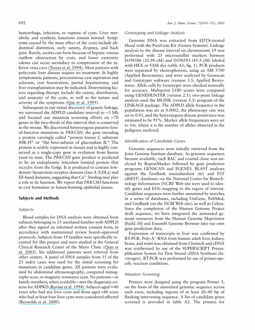
Figure 3 Mutations in PRKCSH in ADPLD. A, Sequence electropherograms showing mutant (affected) and wild-type (control) sequences.All template DNA was genomic except as indicated for the IVS16�1delGT mutation where both genomic (left) and RT-PCR (right) was analyzed.Six mutations are shown. B, Segregation of mutations in families 1 (IVS16�1delGT), 2 (C1237T), and 7 (C1266G), as analyzed by DHPLC.Only selected traces are shown, but the entire family was analyzed. Individual identifications correspond to previously published pedigrees(Reynolds et al. 2000) and figure 1.
lated affected individuals. Of these, 10 were membersof families with at least one other affected member. Al-though some of the families were large, our ability todetermine genetic linkage was hampered by the late on-set of the disease—clinical diagnosis of ADPLD cannotbe excluded in individuals under age 40 years, partic-ularly not in men (Qian et al. 2003). To compensatefor the lack of a large number of individuals with provenlinkage to chromosome 19, mutation detection was car-ried out by a bipartite strategy (table A2). Mutationscreening by DHPLC heteroduplex analysis was carriedout in 15 unrelated affected individuals. In parallel, mu-tation detection by direct sequencing was performed inone affected individual from each of the three familieswith proven or suggestive linkage (families 1, 2 and 7).We chose to screen known genes in the region first (tableA2).
We detected sequence variations in PRKCSH byDHPLC and sequencing that were not observed in 86normal control individuals (172 chromosomes) and thatsegregated with the disease haplotype, when it wasknown (table 1; fig. 3). In addition, we observed severalintragenic polymorphisms (table A4). PRKCSH is an∼15-kb gene encoded in 18 exons, of which the firstand last are untranslated (fig. 2). The predicted effectof all of the pathogenic sequence variants we found ispremature termination of translation. The mutationsoccur throughout the gene, from exon 4 to exon 16.The mutation in family 1 (Reynolds et al. 2000) resultsin deletion of the 5′ splice site in IVS16. We tested theeffect of this mutation on the PRKCSH transcript inpatient lymphoblasts by RT-PCR. We found skippingof exon 16 in a transcript in which exon 15 is splicedto exon 17, causing a frame shift and premature ter-mination (fig. 3A). This mutation segregates on the af-fected haplotype in family 1 (fig. 3B). The mutation infamily 2 (Reynolds et al. 2000) is a nucleotide substi-tution 1237CrT resulting in a premature terminationcodon, Q413X (fig. 3A). This mutation segregates withthe affected haplotype (fig. 3B). The mutation in family7 is also nucleotide substitution producing a stop codonthat segregates in all affected individuals (fig. 3). Twofamilies (25 and O-1) have single-nucleotide insertionsin exons 4 and 13, respectively, that are predicted toresult in premature terminations after frame shifts (fig.3). The mutation in family 41 is predicted to disruptthe 5′ splice site in IVS9 (table 1; fig. 3). The most likely
result of all of these mutations is loss of PRKCSHfunction.
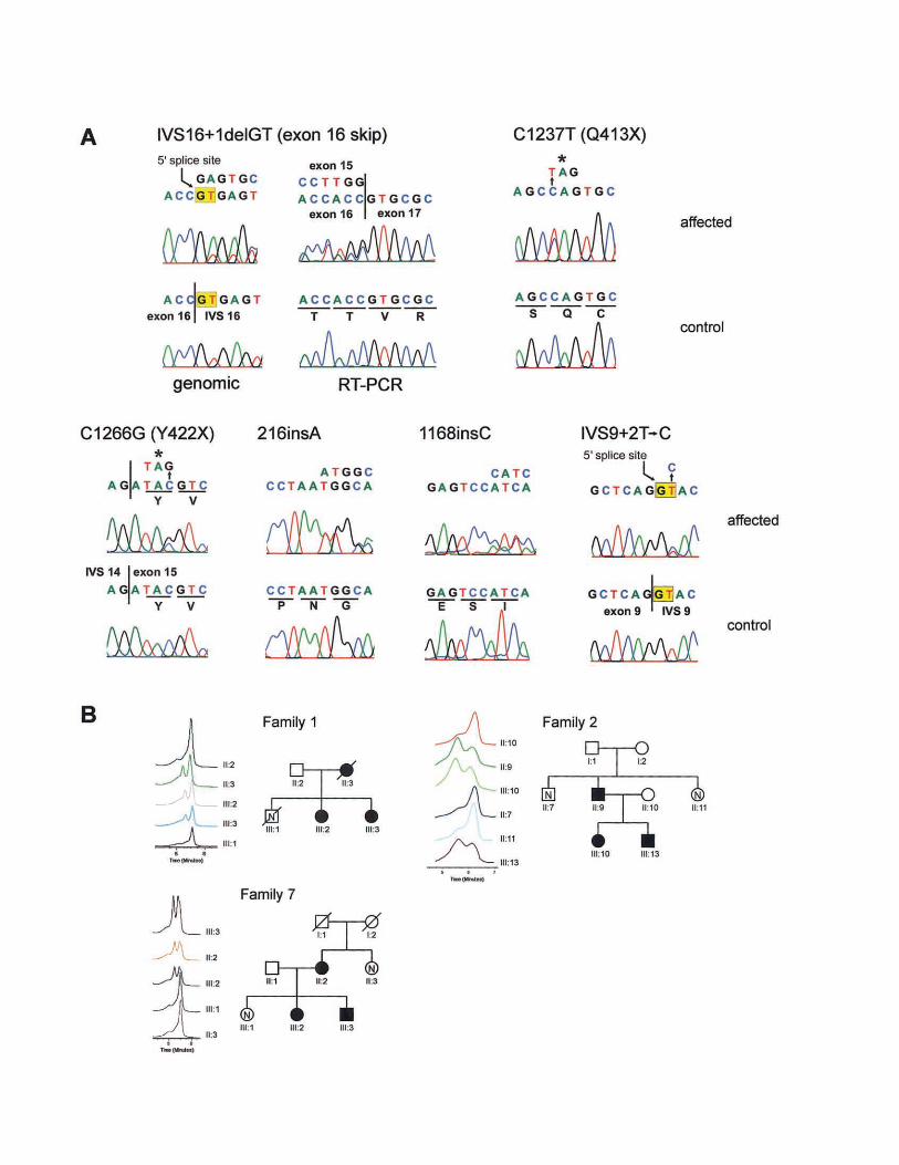
The tissue-expression pattern of PRKCSH has notbeen reported. A 2.5–3.0-kb transcript for this gene isexpressed in all tissues tested (fig. 4). In addition, cardiacmuscle seems to have a slightly larger transcript (∼3.0kb) and skeletal muscle has a 7.0–7.5-kb transcript thatis detected even at high stringency (fig. 4). PRKCSH ispredicted to encode a 527–amino acid protein that isvery highly conserved (fig. 4), even in Schizosaccharo-myces pombe and Arabidopsis thaliana (data notshown). Structural features predicted by SMART andPFAM analysis include a leader sequence, and cysteine-rich LDLa type domain and one or two EF-hand do-mains. The latter two domains likely confer Ca2� depen-dent regulation on the PRKCSH product. The terminalHDEL endoplasmic reticulum retention sequence is con-served throughout evolution (fig. 4). Mutations inPRKCSH cause ADPLD linked to chromosome 19p.
Discussion
We have identified PRKCSH as the gene responsible forADPLD by positional cloning. In contrast to the poly-cystic kidney disease genes PKD1, PKD2, and PKHD1,PRKCSH encodes a previously described human proteincalled either “protein kinase C substrate 80K-H” (80K-H) or “beta-subunit of glucosidase II” (GIIb). Similar tothe other polycystic disease proteins, however, its func-tion remains incompletely understood. The mutationsfound in patients with ADPLD are all predicted to causepremature chain termination. Although most occur inCOOH-terminal exons 13–16, we also found mutationsin exons 4 and 9 in the NH2-terminus half of the protein.In light of the nature and distribution of these mutations,it is most likely that they constitute loss-of-functionchanges, although, in the absence of functional studies,dominant negative or gain-of-function/loss-of-regulationeffects in the residual protein products cannot be ex-cluded. A common feature of all truncating mutationsin 80K-H/GIIb is the loss of the highly conserved ter-minal four amino acids, His-Asp-Glu-Leu (HDEL). Thismotif constitutes an endoplasmic reticulum (ER) luminalretention sequence (Trombetta et al. 1996; Arendt andOstergaard 2000), and its loss presumably would resultin failure to retain 80K-H/GIIb in the ER. This wouldbe significant, because the protein has a leader sequence

698 Am. J. Hum. Genet. 72:691–703, 2003
Figure 4 Expression and conservation of PRKCSH. A, Tissue northern showing expression of PRKCSH in all tissues including liver andkidney. The transcript size is ∼2.5-3.0 kb with skeletal muscle showing evidence of a transcript 17.0 kb and cardiac muscle perhaps having apair of transcripts in the 2.5-3.0 kb range. B, High degree of sequence conservation from C. elegans to humans. S. pombe and A. thaliana alsohave highly conserved PRKCSH homologs (not shown).
and no membrane spans suggesting that without reten-tion by HDEL-mediated binding to the KDEL receptor,it would enter the secretory pathway and be traffickedout of the cell (Arendt and Ostergaard 2000). There isprecedent for reconciling loss-of-function changes withdominant inheritance in polycystic liver and kidney dis-ease. Liver and kidney cyst formation can occur by atwo-hit somatic mutation mechanism resulting in ho-
mozygous inactivation of PKD1 or PKD2 at the cellularlevel (Watnick et al. 1998; Wu et al. 1998). This mech-anism has been invoked to explain the focal nature ofcyst formation in affected organs but may not be theonly factor determining the occurrence of cysts. Evidenceexists for a potential role of compound heterozygousstates and gene dosage (high or low) in cyst formationin ADPKD as well. However, given the similarity of the

Li et al.: Mutations in PRKCSH cause ADPLD 699
clinical presentations of liver disease in ADPLD andADPKD, the two-hit hypothesis is extensible to ADPLDand bears further investigation.
The available functional data do not immediately sug-gest role for PRKCSH in a common pathway with theother polycystic disease genes, but the similarities in theliver phenotype warrant consideration of such a con-vergence of pathways. PRKCSH is broadly expressedin tissues including those with lumen-forming epithelia(e.g., liver, kidney, and pancreas). However, cyst for-mation is confined to the liver, suggesting that there maybe tissue-specific factors required for cyst formation dueto mutations in this gene. The 80K-H/GIIb protein wasinitially identified during a search for protein kinase Csubstrates (Hirai and Shimizu 1990). Although 80K-Hturned out to be a poor substrate for PKC, other pos-sible functions were later suggested by its ability to bindadvanced glycation end products (Li et al. 1996; Thor-nalley 1998) and by its rapid phosphorylation followingactivation of fibroblast growth-factor receptors (Shaoulet al. 1995; Goh et al. 1996; Kanai et al. 1997). Morerecently, homologous proteins were identified as thebeta subunit of glucosidase II (GIIb) (Trombetta et al.1996; Arendt and Ostergaard 1997) and as a vacuolarsystem-associated protein-60 (VASAP-60) (Brule et al.2000).
80K-H/GIIb is an alternatively spliced protein thatmigrates as a doublet at ∼80 kDa on polyacrylamidegels (Arendt and Ostergaard 2000; Trombetta et al.2001). It contains an NH2-terminal cysteine-rich LDLaregion, two EF-hand domains, a highly acidic domainflanked by proline-rich segments presenting putativeGrb2-binding domains, and the aforementioned HDELendoplasmic reticulum luminal retention sequence(Arendt and Ostergaard 2000; Trombetta et al. 2001).An alternatively spliced region and two interaction do-mains responsible for the heterodimerization with thealpha catalytic subunit of glucosidase II (GIIa) havebeen described in the mouse GIIb (Arendt and Oster-gaard 2000). The overall domain structure of 80K-H/GIIb suggests that it may be regulated by Ca2� bind-ing—the LDAa, EF-hand, and acidic regions are allpotential Ca2� interacting motifs. Interestingly, amongthe other polycystic disease gene products, polycystin-1 contains an LDLa motif in its extracellular portionand polycystin-2 is a Ca2� channel and has an EF-handin its COOH terminus.
GIIb does not have enzymatic activity, but it is es-sential for the maturation of GIIa and its retention inthe endoplasmic reticulum (D’Alessio et al. 1999; Tremlet al. 2000). Glucosidase II plays a major role in reg-ulation of proper folding and maturation of glycopro-teins (Ellgaard et al. 1999). A preliminary step to proteinN-glycosylation is the sequential addition of N-acetyl-
glucosamine, mannose, and glucose to a dolichol py-rophosphate lipid carrier to form a mature oligosac-charide composed of two glucosamines, nine mannoses,and three glucoses (Freeze 1998). This preassembledoligosaccharide is transferred to an N-X-S/T sequencein a nascent polypeptide chain in the ER. Cleavage ofthe terminal glucose by glucosidase I and of the middleglucose by glucosidase II generates a monoglucosylatedproduct (Ellgaard et al. 1999). This product is recog-nized by the chaperones calnexin and calreticulin andforms a complex with Erp57, a thiol reductase necessaryfor proper protein folding. By chance, calreticulin alsomaps to the ADPLD candidate region and was excludedby mutation detection (fig. 2; table A2). It is conceivablethat alterations in the folding and maturation of specificglycoproteins results in the development of biliary cysts.Polycystin-1, polycystin-2, and fibrocystin/polyductinare all glycoproteins. It has been suggested that the po-lycystin-1/2 complex forms in the ER (Newby et al.2002) and polycystin-2 is largely retained in the ERmembrane (Cai et al. 1999) and only selectively traf-ficked to the primary cilia (Pazour et al. 2002). Improperassociation and trafficking of polycystins due to defec-tive glycosylation with mutant GIIb may tie ADPLDinto the ADPKD pathway. Direct or indirect interactionof GIIb with the polycystins can also be considered.Since 80K-H/GIIb is an ER luminal protein, it couldinteract with the extracellular domains of either poly-cystin. Interestingly, autosomal recessive carbohydrate-deficient glycoprotein syndrome type Ib is associatedwith congenital hepatic fibrosis and results from nearlycomplete absence of phosphomannose isomerase, theenzyme needed to maintain the dolichol pyrophosphate-oligosaccharide pool (Jaeken et al. 1998; Niehues et al.1998; Westphal et al. 2001). Understanding how mu-tations in PRKCSH lead to cyst formation in the liverand why they do not appear to affect the kidney willprove instructive in understanding all human polycysticdiseases.
Acknowledgments
We thank the family members for their generous partici-pation in this study. We thank Kristin Simonson and PatriciaUrban for help with patient recruitment, Michael Ott and YorkPei for referring patients, Joan Steitz and Richard Lifton forhelpful discussions, and Asghar Rastegar for timely support.DNA sequencing was performed by the Keck BiotechnologyResource at Yale. This work was supported by National In-stitutes of Health (NIH) grant DK51041 (to S.S. and V.E.T.),Mayo Clinic General Clinical Research Center grant M01-RR00585, and Yale Liver Center Training Grant T32DK07356 (to A.L.). A.L., S.D, L.F, X.T., and S.S. are membersof the Yale Center for the Study of Polycystic Kidney Disease(NIH grant P50 DK57328).

Appendix A
Table A1
Primer Sequences for New Microsatellite Markers
MARKER
POSITION
IN BUILD
30(bp)
PRIMERTm
(�C)
PRODUCT
SIZE
(bp)Forward Reverse
CA802 9950900 ACTCTGGCAAACAACTTACAGAT CCTTAATCCTGGCTCCCTTC 60 152–170CA267 10693500 TGCCCTTGGACACAAACATA GAGGGTGGATCAAGCATCTG 60 170–186CA569 11036500 TGCTATGTGCTCATTGTAAACAG CATGGATGTCTGTCTACAAG 58 160–186CAB2 11195400 ATTACAGGAATGAGCCACCAC ATGCAAACCTACATAGGAAGC 60 143–157CA315 13059000 CGGGTTTCTCCATACTGGTC GCAAAATAGGAAGTCCCTGTC 60 164–174CAF1 13230400 TTCTTCCCATTGCAGTTGTG ACACATCCTCATTCAAAGTTC 60 106–118CA246 13656000 AGTTCTGCGTGGAATTGGAAG CAGAGAGCAGTGTGTGGACAA 58 139–149CA048 13850000 TGTGGACTAGAGATGGAGCT GCTGATTTATGTGTCATTTCTCC 60 141–155CA917 14290000 TTGGTCTCAAATTCCTGGCC GCTCTTACAGGCTGTTCTTC 60 148–170CA665 14694900 GGTTCATTTTCTGCCTGGGG GACCAGCAATTCCCCAATTCC 58 191–205CA387 15057620 CCAGGTCTTGTCCCTCCTAC TCTTCCGTGTGTGAATCCAA 60 172–198CA262A 15194650 AGCTGTCCCAAAGCTGAGTC CACACAGACCAGCACACAAA 60 162–178CA327 15256650 CCAGCAGGAAAGCACAATAA AGAGCCACATGGTGGAAACT 60 145–169CA663B 15676310 CCCATTCAGAAATAACCTAGTCAC GCCAAGATTGTGCCACTGTA 60 163–179CA699 16016000 ATTGAACTTGGCCTTGAGGA AAGGGAGAGGGAGCGTATGT 60 144–158CA255 17656190 CGCAAGTCAATGCTTTTTGA ATGATTGCACCACTGTACGC 60 174–188
Table A2
Candidate Genes Excluded by DHPLC and Direct Sequencing
Gene Name Gene Locus DHPLCa Sequencingb
PDE4A Phosphodiesterase 4A, cAMP-specific 5141 �
EDG8 Endothelial differentiation, sphingolipid G-protein-coupled receptor, 8 53637 �
MGC15906 Hypothetical protein MGC15906 84971 �
AK023011 Hypothetical protein FLJ12949 65095 �
AP1M2 Adaptor-related protein complex 1, mu 2 subunit 10053 �
CTL2 CTL2 57153 �
ILF3 (NF90) Interleukin enhancer binding factor 3 3609 �
DNM2 Dynamin 2 1785 �
KIAA1518 KIAA1518 protein 25959 �
AW245557 EST AW245557 AW245557 �
TM4B Tetraspanin TM4-B 26526 � �
KIAA1395 KIAA1395 protein 57572 �
RAB3D Ras-related small GTP binding protein 3D 9545 �
ELAV3 Hu antigen C 1995 �
AK023117 Hypothetical protein FLJ13055 64748 �
BE885128 EST BE995128 126074 �
BC011875 Hypothetical protein DKFZp547J036 84241 �
115950 Hypothetical protein BC016816 115950 �
115948 (MGC20983) Hypothetical protein MGC20983 115948 �
ECSIT Signaling intermediate in Toll pathway-evolutionarily conserved 51295 �
MGC4549 (AK001171) Hypothetical protein MGC4549 84337 �
PTD008 PTD008 protein 51398 � �
DHPS Deoxyhypusine synthase 1725 �
MGC4238 Hypothetical protein MGC4238 84292 �
ASNA1 ArsA arsenite transporter, ATP-binding, homolog 1 439 �
VMD2L1 (AK000139) Vitelliform macular dystrophy 2-like protein 1 54831 �
199695 LOC199695 199695 �
MGC10870 Hypothetical protein MGC10870 84261 �
(continued)

Table A2 (continued)
Gene Name Gene Locus DHPLCa Sequencingb
Hook 2 Hook2 protein 29911 � �
JUNB Jun B proto-oncogene 3726 �
SAST Syntrophin associated serine/threonine kinase 22983 �
CALR Calreticulin 811 �
RAD23A RAD23 homolog A 5886 �
NFIX Nuclear factor I/X (CCAAT-binding transcription factor) 4784 �
LYL1 Lymphoblastic leukemia derived sequence 1 4066 �
FLJ20244 Hypothetical protein FLJ20244 55621 �
NAC1 Transcriptional repressor NAC1 112939 �
STX10 Syntaxin 10 8677 � �
ETR101 Immediate early protein 9592 �
NOTE.—� denotes that the gene was excluded by this method.a DHPLC was performed across all exons under two conditions in 15 affected, unrelated individuals.b Direct sequencing of all exons in one affected individual from families 1, 2, and 7.
Table A3
PCR Primers Used for Mutation Detection in PRKCSH
EXON
OLIGONUCLEOTIDEANNEALING
TEMPERATURE
(�C)
PRODUCT
SIZE
(bp)Sense Antisense
1a GTTCGCGGGCATTTTTCAGGAAC GATCTCCCCAATCCTGGCCA 60 4802a CGGGAAACTGAGTCAAAAGG GTGAAGACACAGCGCATCTC 60 2543a GGCACTGAGCAGTGTTCAATAA ATGGGAGGACAGAGGTGGTA 60 2444a GTGATGGGAGGGGTACTGTC TCTGTGGATGGATGGGACAT 60 3495a GTGATGGGAGGGGTACTGTC TCTGTGGATGGATGGGACAT 60 3496a TTGCAAGCCCACACTATGAG GCAGACCTGGTGGATCCTAA 60 2447a GCAGCATGATCAAAAACCTG AGCTGGTCTCTTGCCTTCTG 60 2748a AAGGAGGATCTGGCTGGTTT GGGTGACAGAGGTGGCTTCT 60 2059a CTCCCTAGAAGTCCCCAACC AGGTCCTGAAGCAAGTTCCA 60 24010a CAACCACTCCAGCCCCTGGT CCAGGTGCCAGAACCAGAGG 61 39011a GCAGGAGGGGCAGAGACACC CCAAGATACTGGGGCTTGTG 60 50012a GCAGGAGGGGCAGAGACACC CCAAGATACTGGGGCTTGTG 60 50013a CTGGGAGTCAAGGAGCAGTC ATGAGGGTATGGGAGCACAC 59 22714a TTCCCCAACCATCAGGAAACTG AGACCCTCCTGTGTCTGTCG 60 26015a TCCCCTGCCTTGCAGGCCT GTTCCCCAAC CCATATGTCCC 60 32016a TCCCCTGCCTTGCAGGCCT GTTCCCCAAC CCATATGTCCC 60 32017a GGTCCATCTTCCTCAGGGCC CGAGCACCCG TCTGCCCATC 60 32018a CTGGTCAACTCCTGGCCTCA CACACCCCAGCAAAGCGAGG 60 48010–13b HAF: ACAGACAGACGCCACCTCTT HAR: TGGACTCCTCCATGTCCTTC 60 3862–17c F2: GTGAAGACACAGCGCATCTC 17R: GGTCCATCTTCCTCAGGGCC 60 174110–17c HAF: ACAGACAGACGCCACCTCTT Ex17R: GGTCCATCTTCCTCAGGGCC 60 8931–4d F1: CTGCTGGACAAGAGGGGTGC SCHR3: GACCCGGTTGGAGGGGATATA 60 4021–8d F1: CTGCTGGACAAGAGGGGTGC HBR: TGTCATCATCCAGCTCCTTG 60 8001–11d F1: CTGCTGGACAAGAGGGGTGC HCR: CTCCTCCTCCTCCTCTGTGG 60 10771–13d F1: CTGCTGGACAAGAGGGGTGC HAR : TGGACTCCTCCATGTCCTTC 60 12902–4d F2: GTGAAGACACAGCGCATCTC SCHR3: GACCCGGTTGGAGGGGATATA 60 3132–8d F2: GTGAAGACACAGCGCATCTC HBR: TGTCATCATCCAGCTCCTTG 60 7112–11d F2: GTGAAGACACAGCGCATCTC HCR: CTCCTCCTCCTCCTCTGTGG 60 9882–13d F2: GTGAAGACACAGCGCATCTC HAR : TGGACTCCTCCATGTCCTTC 60 12012–17d F2: GTGAAGACACAGCGCATCTC 17R: GGTCCATCTTCCTCAGGGCC 60 1741
a PCR primers and condition for exon-by-exon amplification and mutation detection by DHPLC and direct sequencing.b RT-PCR for Northern probe.c RT-PCR for IVS16�1delGT mutation.d RT-PCR IVS2�6delTCC mutation.

702 Am. J. Hum. Genet. 72:691–703, 2003
Table A4
Polymorphisms in PRKCSH
Exon Nucleotide Change ORF Change
2 IVS2�6delTCC No2 IVS2-75CrT No3 IVS3-7CrG No5 IVS5-32ArC No5 IVS5-36GrC No6 IVS6-5CrT No9 IVS9�76insG No11 G871A A291T11 956insGGA 319insE18 C1769T No
Electronic-Database Information
Accession numbers and URLs for data presented herein areas follows:
Celera, http://cds.celera.com/cds/login.cfmEnsembl Genome Browser, http://www.ensembl.org/FGENES, http://genomic.sanger.ac.uk/gf/gf.shtmlGenBank, http://www.ncbi.nlm.nih.gov/Genbank/ (for
PRKCSH homologs PRKCSH [J03075] and Prkcsh[BC009816], Bos taurus [U49178], Caenorhabditis elegans[NM_063672], Drosophila melanogaster [AY058725],Ciona intestinalis [AK112399], S. pombe [D89245], and A.thaliana, [NM_148139])
GENSCAN, http://genes.mit.edu/GENSCAN.htmlHuman Genome Mapviewer, http://www.ncbi.nlm.nih.gov/
cgi-bin/Entrez/map_searchJoint Genome Institute, http://www.jgi.doe.gov/Leiden Muscular Dystrophy pages, http://www.dmd.nl/mut-
nomen.html (for nomenclature for the description of se-quence variations)
Marshfield Medical Research Foundation, Center for MedicalGenetics, http://research.marshifieldclinic.org/genetics
NCBI, http://www.ncbi.nlm.nih.gov/Online Mendelian Inheritance in Man (OMIM), http://www
.ncbi.nlm.nih.gov/Omim/ (for ADPKD [MIM 173900,173910] and ADPLD [MIM 174050])
PFAM Home Page, http://pfam.wustl.edu/index.html (for EFhand [PF00036])
Primer 3, http://www-genome.wi.mit.edu/cgi-bin/primer/primer3_www.cgi
RepeatMasker, http://ftp.genome.washington.edu/cgi-bin/RepeatMasker
SMART, http://dylan.embl-heidelberg.de/ (for LDLa domain[SMART # SM0192])
UniGene, http://www.ncbi.nlm.nih.gov/entrez/query.fcgi?dbpunigene
References
Arendt CW, Ostergaard HL (1997) Identification of the CD45-associated 116-kDa and 80-kDa proteins as the alpha- andbeta-subunits of alpha-glucosidase II. J Biol Chem 272:13117–13125
——— (2000) Two distinct domains of the beta-subunit ofglucosidase II interact with the catalytic alpha-subunit. Gly-cobiology 10:487–492
Bellefroid EJ, Marine JC, Matera AG, Bourguignon C, DesaiT, Healy KC, Bray-Ward P, Martial JA, Ihle JN, Ward DC(1995) Emergence of the ZNF91 Kruppel-associated box-containing zinc finger gene family in the last common an-cestor of anthropoidea. Proc Nat Acad Sci USA 92:10757–10761
Brule S, Rabahi F, Faure R, Beckers JF, Silversides DW, LussierJG (2000) Vacuolar system-associated protein-60: a proteincharacterized from bovine granulosa and luteal cells that isassociated with intracellular vesicles and related to human80K-H and murine beta-glucosidase II. Biol Reprod 62:642–654
Cai Y, Maeda Y, Cedzich A, Torres VE, Wu G, Hayashi T,Mochizuki T, Park JH, Witzgall R, Somlo S (1999) Identi-fication and characterization of polycystin-2, the PKD2 geneproduct. J Biol Chem 274:28557–28565
Comfort MW (1952) Polycystic disease of the liver: a studyof 24 cases. Gastroenterology 20:60–78
D’Alessio C, Fernandez F, Trombetta ES, Parodi AJ (1999)Genetic evidence for the heterodimeric structure of glucos-idase II: the effect of disrupting the subunit-encoding geneson glycoprotein folding. J Biol Chem 274:25899–25905
Ellgaard L, Molinari M, Helenius A (1999) Setting the stan-dards: quality control in the secretory pathway. Science 286:1882–1888
Freeze HH (1998) Disorders in protein glycosylation and po-tential therapy: tip of an iceberg? J Pediatr 133:593–600
Goh KC, Lim YP, Ong SH, Siak CB, Cao X, Tan YH, GuyGR (1996) Identification of p90, a prominent tyrosine-phos-phorylated protein in fibroblast growth factor-stimulatedcells, as 80K-H. J Biol Chem 271:5832–5838
Hirai M, Shimizu N (1990) Purification of two distinct proteinsof approximate Mr 80,000 from human epithelial cells andidentification as proper substrates for protein kinase C.Biochem J 270:583–589
Iglesias DM, Palmitano JA, Arrizurieta E, Kornblihtt AR, Her-rera M, Bernath V, Martin RS (1999) Isolated polycysticliver disease not linked to polycystic kidney disease 1 and2. Dig Dis Sci 44:385–388
Jaeken J, Matthijs G, Saudubray JM, Dionisi-Vici C, BertiniE, de Lonlay P, Henri H, Carchon H, Schollen E, Van Schaf-tingen E (1998) Phosphomannose isomerase deficiency: acarbohydrate-deficient glycoprotein syndrome with hepatic-intestinal presentation. Am J Hum Genet 62:1535–1539
Kanai M, Goke M, Tsunekawa S, Podolsky DK (1997) Signaltransduction pathway of human fibroblast growth factorreceptor 3. Identification of a novel 66-kDa phosphoprotein.J Biol Chem 272:6621–6628
Karhunen PJ, Tenhu M (1986) Adult polycystic liver and kid-ney diseases are separate entities. Clin Genet 30:29–37
Kida T, Nakanuma Y, Terada T (1992) Cystic dilatation of

Li et al.: Mutations in PRKCSH cause ADPLD 703
peribiliary glands in livers with adult polycystic disease andlivers with solitary nonparasitic cysts: an autopsy study. He-patology 16:334–340
Li YM, Mitsuhashi T, Wojciechowicz D, Shimizu N, Li J, StittA, He C, Banerjee D, Vlassara H (1996) Molecular identityand cellular distribution of advanced glycation endproductreceptors: relationship of p60 to OST-48 and p90 to 80K-H membrane proteins. Proc Natl Acad Sci USA 93:11047–11052
Melnick PJ (1955) Polycystic liver: analysis of seventy cases.Arch Pathol 59:162–172
Newby LJ, Streets AJ, Zhao Y, Harris PC, Ward CJ, Ong AC(2002) Identification, characterization, and localization of anovel kidney polycystin-1-polycystin-2 complex. J BiolChem 277:20763–20773
Niehues R, Hasilik M, Alton G, Korner C, Schiebe-SukumarM, Koch HG, Zimmer KP, Wu R, Harms E, Reiter K, vonFigura K, Freeze HH, Harms HK, Marquardt T (1998) Car-bohydrate-deficient glycoprotein syndrome type Ib. Phos-phomannose isomerase deficiency and mannose therapy. JClin Invest 101:1414–1420
Onuchic LF, Furu L, Nagasawa Y, Hou X, Eggermann T, RenZ, Bergmann C, Senderek J, Esquivel E, Zeltner R, Rudnik-Schoneborn S, Mrug M, Sweeney W, Avner ED, Zerres K,Guay-Woodford LM, Somlo S, Germino GG (2002)PKHD1, the polycystic kidney and hepatic disease 1 gene,encodes a novel large protein containing multiple immu-noglobulin-like plexin-transcription-factor domains andparallel beta-helix 1 repeats. Am J Hum Genet 70:1305–1317
Pazour GJ, San Agustin JT, Follit JA, Rosenbaum JL, WitmanGB (2002) Polycystin-2 localizes to kidney cilia and the cil-iary level is elevated in orpk mice with polycystic kidneydisease. Curr Biol 12:R378–R380
Pirson Y, Lannoy N, Peters D, Geubel A, Gigot JF, BreuningM, Verellen-Dumoulin C (1996) Isolated polycystic liver dis-ease as a distinct genetic disease, unlinked to polycystic kid-ney disease 1 and polycystic kidney disease 2. Hepatology23:249–252
Qian Q Li A, King BF, Kamath PS, Lager DJ, Huston J III,Shub C, Davila S, Somlo S, Torres VE (2003) Clinical profileof autosomal dominant polycystic liver disease. Hepatology37:164–171
Que F, Nagorney DM, Gross JB Jr, Torres VE (1995) Liverresection and cyst fenestration in the treatment of severepolycystic liver disease. Gastroenterology 108:487–494
Ramos A, Torres VE, Holley KE, Offord KP, Rakela J, LudwigJ (1990) The liver in autosomal dominant polycystic kidneydisease: implications for pathogenesis. Arch Pathol Lab Med114:180–184
Ravine D, Gibson RN, Walker RG, Sheffield LJ, Kincaid-Smith
P, Danks DM (1994) Evaluation of ultrasonographic diag-nostic criteria for autosomal dominant polycystic kidney dis-ease 1. Lancet 343:824–827
Reynolds DM, Falk CT, Li A, King BF, Kamath PS, Huston J3rd, Shub C, Iglesias DM, Martin RS, Pirson Y, Torres VE,Somlo S (2000) Identification of a locus for autosomal dom-inant polycystic liver disease, on chromosome 19p13.2-13.1.Am J Hum Genet 67:1598–1604
Shaoul E, Reich-Slotky R, Berman B, Ron D (1995) Fibroblastgrowth factor receptors display both common and distinctsignaling pathways. Oncogene 10:1553–1561
Somlo S, Torres VE, Reynolds D, King BF, Nagorney DM(1995) Autosomal dominant polycystic liver disease withoutpolycystic kidney disease is not linked to either the PKD1or PKD2 gene loci. J Amer Soc Nephrol 6:727a
Thornalley PJ (1998) Cell activation by glycated proteins: AGEreceptors, receptor recognition factors and functional clas-sification of AGEs. Cell Mol Biol 44:1013–1023
Torres VE (1996) Polycystic liver disease. In: Watson ML, Tor-res VE (eds) Polycystic kidney disease. Oxford UniversityPress, Oxford
Torres VE, Rastogi S, King BF, Stanson AW, Gross JB Jr, No-gorney DM (1994) Hepatic venous outflow obstruction inautosomal dominant polycystic kidney disease. J Amer SocNephrol 5:1186–1192
Treml K, Meimaroglou D, Hentges A, Bause E (2000) Thealpha- and beta-subunits are required for expression of cat-alytic activity in the hetero-dimeric glucosidase II complexfrom human liver. Glycobiology 10:493–502
Trombetta ES, Fleming KG, Helenius A (2001) Quaternaryand domain structure of glycoprotein processing glucosidaseII. Biochemistry 40:10717–10722
Trombetta ES, Simons JF, Helenius A (1996) Endoplasmic re-ticulum glucosidase II is composed of a catalytic subunit,conserved from yeast to mammals, and a tightly bound non-catalytic HDEL-containing subunit. J Biol Chem 271:27509–27516
Watnick TJ, Torres VE, Gandolph MA, Qian F, Onuchic LF,Klinger KW, Landes G, Germino GG (1998) Somatic mu-tation in individual liver cysts supports a two-hit model ofcystogenesis in autosomal dominant polycystic kidney dis-ease. Mol Cell 2:247–251
Westphal V, Kjaergaard S, Davis JA, Peterson SM, Skovby F,Freeze HH (2001) Genetic and metabolic analysis of the firstadult with congenital disorder of glycosylation type Ib: long-term outcome and effects of mannose supplementation. MolGenet Metab 73:77–85
Wu G, D’Agati V, Cai Y, Markowitz G, Park JH, ReynoldsDM, Maeda Y, Le TC, Hou H Jr, Kucherlapati R, EdelmannW, Somlo S (1998) Somatic inactivation of Pkd2 results inpolycystic kidney disease. Cell 93:177–188
Copyright © 2022 FDOKUMEN