Linkage to chromosome 2q36.1 in autosomal dominant Dandy-Walker malformation with occipital...
16
Linkage to chromosome 2q36.1 in autosomal dominant Dandy- Walker malformation with occipital cephalocele and evidence for genetic heterogeneity Ali Jalali ✉ , Department of Neurology, Northwestern University Feinberg School of Medicine, Chicago, IL, USA Kimberly A. Aldinger, Committee on Neurobiology, The University of Chicago, Chicago, IL, USA Ajit Chary, Department of Pediatrics, Northwestern University Feinberg School of Medicine, Chicago, IL, USA David G. Mclone, Department of Neurosurgery, Northwestern University Feinberg School of Medicine, Chicago, IL, USA Robin M. Bowman, Department of Neurosurgery, Northwestern University Feinberg School of Medicine, Chicago, IL, USA Luan Cong Le, HoChiMinh City Hospital, HoChiMinh City, Vietnam Phillip Jardine, Department of Paediatrics, Bristol Royal Hospital for Children, Bristol, UK Ruth Newbury-Ecob, Department of Clinical Genetics, St Michael's Hospital, Bristol, UK Andrew Mallick, Department of Paediatrics, Yeovil Hospital, Somerset, UK Nadereh Jafari, Center for Genetic Medicine, Northwestern University Feinberg School of Medicine, Chicago, IL, USA Eric J. Russell, Department of Radiology, Northwestern University Feinberg School of Medicine, Chicago, IL, USA John Curran, Department of Radiology, Northwestern University Feinberg School of Medicine, Chicago, IL, USA Pam Nguyen, Department of Radiology, Northwestern University Feinberg School of Medicine, Chicago, IL, USA Karim Ouahchi, Department of Pathology, Brigham and Women's Hospital, Harvard Medical School, Boston, MA, UK Charles Lee, A. Jalali, 303 E. Chicago Ave., Ward 10-233, Chicago, IL 60611, USA, [email protected]. NIH Public Access Author Manuscript Hum Genet. Author manuscript; available in PMC 2010 February 16. Published in final edited form as: Hum Genet. 2008 April ; 123(3): 237. doi:10.1007/s00439-008-0467-y. NIH-PA Author Manuscript NIH-PA Author Manuscript NIH-PA Author Manuscript
-
Upload
independent -
Category
Documents
-
view
0 -
download
0
Transcript of Linkage to chromosome 2q36.1 in autosomal dominant Dandy-Walker malformation with occipital...

Linkage to chromosome 2q36.1 in autosomal dominant Dandy-Walker malformation with occipital cephalocele and evidence forgenetic heterogeneity
Ali Jalali✉,Department of Neurology, Northwestern University Feinberg School of Medicine, Chicago, IL, USA
Kimberly A. Aldinger,Committee on Neurobiology, The University of Chicago, Chicago, IL, USA
Ajit Chary,Department of Pediatrics, Northwestern University Feinberg School of Medicine, Chicago, IL, USA
David G. Mclone,Department of Neurosurgery, Northwestern University Feinberg School of Medicine, Chicago, IL,USA
Robin M. Bowman,Department of Neurosurgery, Northwestern University Feinberg School of Medicine, Chicago, IL,USA
Luan Cong Le,HoChiMinh City Hospital, HoChiMinh City, Vietnam
Phillip Jardine,Department of Paediatrics, Bristol Royal Hospital for Children, Bristol, UK
Ruth Newbury-Ecob,Department of Clinical Genetics, St Michael's Hospital, Bristol, UK
Andrew Mallick,Department of Paediatrics, Yeovil Hospital, Somerset, UK
Nadereh Jafari,Center for Genetic Medicine, Northwestern University Feinberg School of Medicine, Chicago, IL,USA
Eric J. Russell,Department of Radiology, Northwestern University Feinberg School of Medicine, Chicago, IL, USA
John Curran,Department of Radiology, Northwestern University Feinberg School of Medicine, Chicago, IL, USA
Pam Nguyen,Department of Radiology, Northwestern University Feinberg School of Medicine, Chicago, IL, USA
Karim Ouahchi,Department of Pathology, Brigham and Women's Hospital, Harvard Medical School, Boston, MA,UK
Charles Lee,
A. Jalali, 303 E. Chicago Ave., Ward 10-233, Chicago, IL 60611, USA, [email protected].
NIH Public AccessAuthor ManuscriptHum Genet. Author manuscript; available in PMC 2010 February 16.
Published in final edited form as:Hum Genet. 2008 April ; 123(3): 237. doi:10.1007/s00439-008-0467-y.
NIH
-PA Author Manuscript
NIH
-PA Author Manuscript
NIH
-PA Author Manuscript
Department of Pathology, Brigham and Women's Hospital, Harvard Medical School, Boston, MA,UK
William B. Dobyns,Departments of Human Genetics and Neurology, The University of Chicago, Chicago, IL, USA
Kathleen J. Millen,Departments of Human Genetics and Neurology, The University of Chicago, Chicago, IL, USA
Joao M. Pina-Neto,Department of Genetics, School of Medicine, Universidade de São Paolo, Ribeirão Preto, Brazil
John A. Kessler, andDepartment of Neurology, Northwestern University Feinberg School of Medicine, Chicago, IL, USA
Alexander G. BassukDepartment of Neurology, Northwestern University Feinberg School of Medicine, Chicago, IL, USA;Department of Pediatrics, Northwestern University Feinberg School of Medicine, Chicago, IL, USAAli Jalali: ; Kimberly A. Aldinger: ; Ajit Chary: ; David G. Mclone: ; Robin M. Bowman: ; Luan Cong Le: ; Phillip Jardine: ; RuthNewbury-Ecob: ; Andrew Mallick: ; Nadereh Jafari: ; Eric J. Russell: ; John Curran: ; Pam Nguyen: ; Karim Ouahchi: ; CharlesLee: ; William B. Dobyns: ; Kathleen J. Millen: ; Joao M. Pina-Neto: ; John A. Kessler: ; Alexander G. Bassuk: [email protected]
AbstractWe previously reported a Vietnamese-American family with isolated autosomal dominant occipitalcephalocele. Upon further neuroimaging studies, we have recharacterized this condition as autosomaldominant Dandy-Walker with occipital cephalocele (ADDWOC). A similar ADDWOC family fromBrazil was also recently described. To determine the genetic etiology of ADDWOC, we performedgenome-wide linkage analysis on members of the Vietnamese-American and Brazilian pedigrees.Linkage analysis of the Vietnamese-American family identified the ADDWOC causative locus onchromosome 2q36.1 with a multipoint parametric LOD score of 3.3, while haplotype analysis refinedthe locus to 1.1 Mb. Sequencing of the five known genes in this locus did not identify any protein-altering mutations. However, a terminal deletion of chromosome 2 in a patient with an isolated caseof Dandy-Walker malformation also encompassed the 2q36.1 chromosomal region. The Brazilianpedigree did not show linkage to this 2q36.1 region. Taken together, these results demonstrate a locusfor ADDWOC on 2q36.1 and also suggest locus heterogeneity for ADDWOC.
IntroductionWe recently reported a Vietnamese-American kindred with isolated autosomal dominantoccipital cephalocele over three generations (Bassuk et al. 2004). While the original report ofthis kindred focused on the atretic cephalocele, our subsequent review of brain imaging in theproband and his affected second cousin showed Dandy-Walker malformation in addition tothe cephalocele. Three other families with autosomal dominant Dandy-Walker malformationand occipital cephaloceles (ADDWOC) have been reported from Brazil, Spain, and China(Carvalho et al. 2006; Martinez-Lage et al. 1996; Zhao et al. 2007), signifying the worldwideoccurrence of this heritable disorder. Here, we further characterize the phenotype of the originalVietnamese-American kindred, map the locus to 2q36.1 by genome-wide linkage analysis, anddescribe an overlapping phenotype in a child with deletion of this region. We also demonstratelocus heterogeneity by excluding the Brazilian family from this locus.
Jalali et al. Page 2
Hum Genet. Author manuscript; available in PMC 2010 February 16.
NIH
-PA Author Manuscript
NIH
-PA Author Manuscript
NIH
-PA Author Manuscript

Materials and methodsPatient data and DNA gathering
The ADDWOC pedigrees comprised a 3-generation Vietnamese-American family with 16 anda Brazilian family with 6 affected individuals. Positive diagnosis was made by the presence ofone or a combination of an occipital cephalocele or occult Dandy-Walker or cerebellar changes,as described in the results section. We tried to minimize any bias in this process by utilizingthe opinions of multiple experts in the field in a blinded fashion. For the Vietnamese-Americanfamily, the attending neurologist contacted 14 family members, including 13 affectedindividual used in the affected-only linkage analysis, and administered informed consent withthe approval of the local institutional review board. The Brazilian family was recruitedseparately and their clinical phenotype has previously been described (Carvalho et al. 2006).Genomic DNA was extracted from blood or buccal samples collected from family membersusing Gentra Autopure LS (Qiagen). A Siemens Trio 3 Tesla MRI Scanner at the NorthwesternUniversity Magnetic Resonance facility was used for imaging studies.
Microarray SNP analysis and DNA sequencingThe Affymetrix GeneChip Human Mapping 10 K Array (Xba 131) and the GeneChipInstrument System (Hybridization Oven 640, Fluidics Station 450, Scanner 3000) were usedfor single nucleotide polymorphism (SNP) genotyping at the Genomics Core Facility ofNorthwestern University Center for Genetic Medicine following the standard protocol suppliedby the manufacturer. Affymetrix GCOS and GDAS software were used for scanning the arraysand assigning genotype calls. All array call rates were above 95%. Gene sequencing involvedthe exons and flanking 100 bp intronic or 200 bp promoter regions of the proband DNAamplified using the Platinum Pfx PCR kit (Invitrogen). Primer sequences are available uponrequest. Purified PCR amplification products were electrophoresed on ABI 3100 and 3730DNA sequencers by the Genomics Core Facility at Northwestern University. Novelheterozygous SNPs found during gene sequencing of the proband DNA were resequencedusing the same primers in all affected individuals.
Genome-wide linkage analysisOf the 11,560 total SNPs, 662 were excluded from further analysis due to lack of positionalinformation from Affymetrix or location on the X chromosome, as this condition does notfollow an X-linked pattern of inheritance. Additionally, 52 SNPs with Mendelianinconsistencies were identified using PedCheck (O'Connell and Weeks 1998) and excludedfrom further analysis. The remaining 10,846 SNPs were subjected to parametric multipointlinkage analysis assuming a fully penetrant autosomal dominant mode of inheritance, equalSNP allele frequencies, and a disease allele frequency estimated at 1/10,000. easyLINKAGEPlus(Hoffmann and Lindner 2005) v5.02 was used to prepare the data for linkage computationusing Allegro 2.0 f (Gudbjartsson et al. 2005). The haplotype diagram was prepared usingHaploPainter (Thiele and Nurnberg 2005).
CGH and FISH analysisDNA samples from the proband and his maternal grandfather were subjected to comparativegenomic hybridization (CGH) on a 1-Mb resolution genome-wide array CGH platform(Spectral Genomics, Inc.) using manufacturer's recommendation (Iafrate et al. 2004). G-banded karyotype analysis and fluorescent in situ hybridization (FISH) on metaphasechromosomes was performed by the Genetic Services Laboratories at the University ofChicago. BAC clones from the RPCL-11 human library were selected across 2 q. GenomicDNA from BAC clones was isolated using a DNA isolation kit (Qiagen, Valencia, CA, USA)and directly labeled with Spectrum Orange-dUTP (Vysis/Abbot, Abbot Park, IL, USA) or
Jalali et al. Page 3
Hum Genet. Author manuscript; available in PMC 2010 February 16.
NIH
-PA Author Manuscript
NIH
-PA Author Manuscript
NIH
-PA Author Manuscript
Spectrum Green-dUTP (Vysis/Abbott) using a standard nick-translation kit (Vysis/Abbott).Slide, probe preparation, and hybridization were performed using standard methods.Additional information is available upon request.
ResultsBrain imaging features in the expanded Vietnamese-American family
While the initial study of the Vietnamese-American family focused on the occipital cephalocelepresent in seven family members (Fig. 1a), we have now reviewed the brain imaging studiesin the two most severely affected family members (III-3 and III-8 in Fig. 2) in more detail. Inaddition to the cephalocele, III-3 and III-8 demonstrate Dandy-Walker malformationcharacterized by a small and upwardly rotated cerebellar vermis, marked cystic enlargementof the fourth ventricle, and greatly enlarged posterior fossa (Fig. 1b).
We also obtained MRI studies in seven additional family members that lacked an obviouscephalocele including those we originally designated as obligate carriers for the cephaloceletrait (I-2 and II-4). While II-4 lacked an obvious cephalocele, her MRI demonstrated an occultcephalocele in a parasagittal section (Fig. 1c) as well as cerebellar vermal hypoplasia with amildly enlarged fourth ventricle in the mid-sagittal section (Fig. 1d). Similarly, I-2demonstrated cerebellar vermal hypoplasia and a mildly enlarged cisterna magna (Fig. 1e).MRI studies of 4 other family members, II-1, II-2 (Fig. 1f), II-3, and II-6, also demonstratedmild to moderate cerebellar vermal hypoplasia. No anomalies were noted in the MRI study ofII-7. While these family members did not display the full extent of posterior fossadevelopmental anomalies known as Dandy-Walker malformation, the cerebellar changes notedalong with enlargement of the fluid spaces around the cerebellum are considered less severeforms of the same continuum of developmental anomalies. On the basis of these MRI findingsin this family, we recharacterized this inherited condition as ADDWOC. Out of the total of 14family members considered in this study, all except II-7 were included in our affected-onlylinkage analysis.
Interestingly, several family members (II-2, II-3, II-4, III-3) demonstrated mild protrusion ofthe occipital lobes through an apparently enlarged tentorial notch, which can be seen interposedbetween the splenium of the corpus callosum and the top of the vermis (arrowheads in Fig. 1).This anomaly resembles the dysplastic mesial occipital gyri seen in some patients with parietalforamina associated with mutations of ALX4 (Mavrogiannis et al. 2001;Reddy et al.2000;Valente and Valente 2004) and suggests a subtle meningeal deficiency.
Genome-wide linkage analysis and sequencingThe genome-wide parametric LOD scores for this pedigree are shown in Fig. 3a. The mostsignificant score was observed at a single locus spanning 1.5 cM on chromosome 2 (225.3–226.8 cM) with a LOD of 3.3. The maximum LOD score was obtained at rs951997 andrs1374536 (Fig. 3b). Haplotype analysis indicated that the co-segregating segment containingthe ADDWOC locus is flanked proximally by rs724149 and distally by rs1371552 (Fig. 4),demarcating a 1.1 Mb region on chromosome 2q36.1 containing 5 known genes (Fig. 5).Sequencing the genes in this region revealed two previously reported SNPs, rs2166652 andrs2044537, and two novel polymorphisms, S1 and A1. The dbSNP annotated markers,rs2166652 and rs2044537, are located 100 and 173 bp upstream of FARSB exon 1. The novelS1 variation is a G to A transition at the third base position resulting in a synonymous changeof the Ala36 residue of SGPP2, while the A1 variation is an A insertion/deletion in a polyAstretch 85 bp upstream of ACSL3 exon 4. Extending the linkage analysis to include the fouradditional polymorphisms discovered through sequencing did not further delineate theADDWOC locus, though a LOD of 3.3 was extended to include A1 (Fig. 3b). No missense,
Jalali et al. Page 4
Hum Genet. Author manuscript; available in PMC 2010 February 16.
NIH
-PA Author Manuscript
NIH
-PA Author Manuscript
NIH
-PA Author Manuscript

nonsense, frameshift, or splice-site mutations were identified. Comparative genomichybridization of I-8 and III-8 DNA on a 1 Mb resolution BAC array (Iafrate et al. 2004) failedto identify any significant gains or losses.
We also performed a genome-wide linkage analysis on the Brazilian family, which wasgenotyped using the same Affymetrix platform. No evidence for linkage was detected at the2q36.1 locus (maximum LOD of −7.59 at rs1440066), nor at a single locus. Instead, multipleloci with LOD scores of 1.20 occurred across several chromosomes (data not shown). Takentogether, the linkage results for the Vietnamese-American and Brazilian families demonstratelocus heterogeneity in ADDWOC.
Isolated Dandy-Walker patient with 2q36.1 deletionWe evaluated a 4-year-old boy with mild developmental delay who began walking at 14 monthsand uses about 50 words but no sentences at 4 years; formal intelligence testing has not beenperformed. His facial appearance was significant for a relatively prominent forehead, mildlydownturned vermilion border of the upper lip, deep-set eyes and flat philtrum, and he hasminimal high frequency hearing loss. His MRI shows mild cerebellar vermal hypoplasia andenlarged retrocerebellar fluid space (Fig. 6a). G-banded karyotype analysis revealed a smallheterozygous deletion of distal 2q in 12 of 30 cells examined (Fig. 6b). FISH analysis confirmeda 20.5 Mb telomeric deletion encompassing the ADDWOC locus. The position of the deletionbreakpoint was narrowed to a 0.2-Mb region of chromosome 2q36.1 between RP11-1080C21and RP11-794I13 and 0.6 Mb centromeric to the ADDWOC locus. The karyotype is thusdesignated 46,XY,del(2)(q36.1→qter)[12]/46,XY[18]. Both parents had normal karyotypes.
DiscussionThe pathogenesis of Dandy-Walker malformation is unknown but certainly heterogeneous.Most patients are sporadic with an empiric sibling recurrence risk less than 5% for the isolatedform of the disease (Murray et al. 1985). Families with autosomal dominant inheritance oftypical Dandy-Walker malformation without an occipital cephalocele have not been reported,perhaps in part due to the occult nature of some of the weaker variants. In this paper; however,we cite several reports of families that feature a combination of Dandy-Walker malformationand occipital cephaloceles, which have recently been described.
The genetic causes of Dandy-Walker malformation and occipital cephaloceles are not wellunderstood, although the last several years have seen some progress. The genes for severalcongenital anomaly syndromes associated with occipital cephaloceles have been reported;these include Knobloch, Joubert, Meckel-Gruber, Roberts, and Walker-Warburg syndromes(Beltran-Valero de Bernabe et al. 2002; Dixon-Salazar et al. 2004; Ferland et al. 2004; Kyttalaet al. 2006; Parisi et al. 2004; Sertie et al. 2000; Smith et al. 2006; Valente et al. 2006; vanReeuwijk et al. 2005; Vega et al. 2005). Our studies in patients with Dandy-Walkermalformation and overlapping chromosome deletions mapped the first two Dandy-Walker locito 3q24 and 6p25, and identified ZIC1 and ZIC4 as the first causative genes for Dandy-Walkermalformation (Descipio et al. 2005; Grinberg et al. 2004). However, no genes associated withthe novel ADDWOC syndrome have been identified.
In the continuum of posterior fossa developmental anomalies, Dandy-Walker malformation isoften associated with developmental delay, difficulty with balance, spasticity, poor fine-motorcontrol, and seizures. Hearing and/or visual difficulties are also associated with poorintellectual development. Those presenting with milder variants in this continuum can,however, have mild or no symptoms, a normal neurological exam, and normal intellectualdevelopment (Hart et al. 1972; Tal et al. 1980). In the Vietnamese-American pedigree, onlythe third generation (III-3 and III-8) presented with Dandy-Walker malformation and the
Jalali et al. Page 5
Hum Genet. Author manuscript; available in PMC 2010 February 16.
NIH
-PA Author Manuscript
NIH
-PA Author Manuscript
NIH
-PA Author Manuscript

associated symptoms. The affected family members in the first and second generation,however, showed only milder posterior fossa variations while functioning normally as adultsand with no associated signs or symptoms other than the presence of an occipital cephalocele.Although originally our diagnostic criteria were limited to the presence of an occipitalcephalocele or obligate-carrier status, after recharacterization of this condition as ADDWOCwe were able to use the neuroradiological features of Dandy-Walker malformation and milderanomalies in the cerebellum and its surrounding fluid spaces as a more sensitive means ofdesignating the affection status in this pedigree.
A genome-wide linkage screen of ADDWOC in this three-generation Vietnamese-Americanfamily resulted in the most significant evidence for linkage on chromosome 2 with a LODscore of 3.30 at 225.3–226.8 cM. We believe, given our assumptions, the maximum theoreticalLOD score achievable for this pedigree is 3.61. The obtained LOD score is equivalent to thethreshold value of 3.3 for significant linkage of a Mendelian disorder in the presence of locusheterogeneity (Terwilliger and Ott 1994). Haplotype analysis confirmed that this relativelysmall 1.1 Mb locus co-segregated with the ADDWOC phenotype. Additionally, we identifiedan isolated Dandy-Walker patient with a 2q36.1→qter deletion encompassing the 2q36.1ADDWOC locus. Another isolated patient with a 2q35→qter deletion was also reported tohave a Dandy-Walker malformation (Waters et al. 1993), which is consistent with the presenceof a Dandy-Walker locus in this region. A second genome-wide linkage screen of ADD-WOCin a four-generation Brazilian family failed to detect evidence for linkage at 2q36.1, providingevidence for locus heterogeneity of the disorder.
Sequencing the coding exons, immediately flanking intronic regions, and promoter regions ofSGPP2, FARSB, MOGAT1, ACSL3, and KCNE4 in the 2q36.1 locus failed to identify thedisease-causing variant in our Vietnamese-American ADDWOC family. Further, comparativegenomic hybridization using a BAC array failed to identify any significant gains or losses.Nevertheless, a higher resolution BAC array or analysis using multiplex ligation-dependentprobe amplification (MLPA) may be able to uncover deletions or other ploidy variations. It isalso possible that a mutation in an uncharacterized gene in this locus or a mutation in aregulatory region affecting the expression of a gene in this locus is causative for the ADDWOCphenotype. Furthermore, genes outside of this locus may also have enhancers/repressorslocated within the 2q36.1 locus. Regulatory elements have been identified 1 Mb away fromthe gene whose expression they control (Bien-Willner et al. 2007; Jeong et al. 2006). In fact,point mutations in a SHH enhancer located greater than 1 Mb away from the SHH gene havebeen identified as the causative mutations in families with triphalangeal thumb and preaxialpolydactyly (Gurnett et al. 2007). Therefore, it is possible that mutations of regulatory regionswithin the ADDWOC locus may affect the expression of genes located outside of this locus.
The PAX3 transcriptional start site is located 107 kb centromeric to the ADDWOC 2q36.1locus (Fig. 5) and proceeds from the negative strand, suggesting PAX3 cis-regulatory elementsmay lie within the ADDWOC locus. An association between PAX3 mutations and theADDWOC clinical phenotype is suggested by observations in both murine Pax3 mutant lines,and in Waardenburg syndrome, which involves PAX3 mutations in humans (Tassabehji et al.1993). However, it is not clear whether putative changes in PAX3 expression due to disruptionof its regulatory elements can mimic the effects of exon mutations described in mice or humans.Genetic lesions in murine Pax3 lead to developmental abnormalities such as severe defects ofthe neural tube and neural crest derivatives (Franz and Kothary 1993;Mansouri et al. 1996,1994;Moase and Trasler 1989) including absent cerebellum (Keller et al. 2004) and hindbraincranioschisis (Robert 1954). Expression analysis of PAX3 in human fetuses by in situhybridization detects transcripts in the ventricular zone at the mid–hindbrain boundary, and inthe dorsal region of the ventricular zone and the roof plate of the medulla and the spinal cord(Terzic and Saraga-Babic 1999). This posterior neural tube expression pattern is consistent
Jalali et al. Page 6
Hum Genet. Author manuscript; available in PMC 2010 February 16.
NIH
-PA Author Manuscript
NIH
-PA Author Manuscript
NIH
-PA Author Manuscript

with a role for PAX3 in the development of posterior fossa structures, the structures primarilyaffected by ADDWOC. In addition, while individuals with Waardenburg syndrome typicallypresent with abnormalities of eye and nose formation and deafness and pigmentationanomalies, several cases have been described with posterior neural tube defects (Hol et al.1995;Kujat et al. 2007;Lu et al. 2007;Nye et al. 1998;Shim et al. 2004), providing an additionallink between PAX3 and posterior neural tube structures. In silico analysis did not reveal anysignificant homology between PAX3 transcript and the sequence of the identified 2q36.1 locus,ruling out an undiscovered PAX3 homolog in this region.
We should finally point out that the appearance of the most severely affected members of theVietnamese-American pedigree in the third generation (III-3 and III-8) suggests thephenomenon of anticipation in this family. The number and depth of pedigrees is too small todefinitively conclude whether this condition is associated with anticipation. Additionally, wedid not observe any nucleotide repeat expansion in the areas we sequenced (i.e., exons andimmediate intronic or promoter regions). Further, the di-, tri-, tetra- and higher order nucleotiderepeats in this locus, similarly to other parts of the genome, are too numerous to be sequencedentirely using the simple sequencing technique utilized in this study. Nevertheless, we cannotrule out that a nucleotide repeat expansion somewhere in this locus may play a role in thiscondition. Further sequencing in the conserved non-coding or nucleotide repeat regions of the2q36.1 locus may help to uncover the genetic basis for ADDWOC.
AcknowledgmentsWe would like to thank Hoang Le and Dung Tri Pham, HoChiMinh City Hospital, HoChiMinh City, Vietnam, fortheir assistance in patient recruitment. A.J. is supported by NIH pre-doctoral NRSA grant F30-NS51962. K.A.A. issupported by NIH Pre-doctoral grant GM007839–26. A.G.B. is supported by NIH grant K08-NS48174.
ReferencesBassuk AG, McLone D, Bowman R, Kessler JA. Autosomal dominant occipital cephalocele. Neurology
2004;62:1888–1890. [PubMed: 15159504]Beltran-Valero de Bernabe D, Currier S, Steinbrecher A, Celli J, van Beusekom E, van der Zwaag B,
Kayserili H, Merlini L, Chitayat D, Dobyns WB, Cormand B, Lehesjoki AE, Cruces J, Voit T, WalshCA, van Bokhoven H, Brunner HG. Mutations in the O-mannosyltransferase gene POMT1 give riseto the severe neuronal migration disorder Walker-Warburg syndrome. Am J Hum Genet2002;71:1033–1043. [PubMed: 12369018]
Bien-Willner GA, Stankiewicz P, Lupski JR. SOX9cre1, a cisacting regulatory element located 1.1 Mbupstream of SOX9, mediates its enhancement through the SHH pathway. Hum Mol Genet2007;16:1143–1156. [PubMed: 17409199]
Carvalho DR, Giuliani LR, Simao GN, Santos AC, Pina-Neto JM. Autosomal dominant atreticcephalocele with phenotype variability: report of a Brazilian family with six affected in fourgenerations. Am J Med Genet A 2006;140:1458–1462. [PubMed: 16718686]
Descipio C, Schneider L, Young TL, Wasserman N, Yaeger D, Lu F, Wheeler PG, Williams MS, BasonL, Jukofsky L, Menon A, Geschwindt R, Chudley AE, Saraiva J, Schinzel AA, Guichet A, DobynsWE, Toutain A, Spinner NB, Krantz ID. Subtelomeric deletions of chromosome 6p: molecular andcytogenetic characterization of three new cases with phenotypic overlap with Ritscher-Schinzel (3C)syndrome. Am J Med Genet A 2005;134:3–11. [PubMed: 15704124]
Dixon-Salazar T, Silhavy JL, Marsh SE, Louie CM, Scott LC, Gururaj A, Al-Gazali L, Al-Tawari AA,Kayserili H, Sztriha L, Gleeson JG. Mutations in the AHI1 gene, encoding jouberin, cause Joubertsyndrome with cortical polymicrogyria. Am J Hum Genet 2004;75:979–987. [PubMed: 15467982]
Ferland RJ, Eyaid W, Collura RV, Tully LD, Hill RS, Al-Nouri D, Al-Rumayyan A, Topcu M, GasconG, Bodell A, Shugart YY, Ruvolo M, Walsh CA. Abnormal cerebellar development and axonaldecussation due to mutations in AHI1 in Joubert syndrome. Nat Genet 2004;36:1008–1013. [PubMed:15322546]
Jalali et al. Page 7
Hum Genet. Author manuscript; available in PMC 2010 February 16.
NIH
-PA Author Manuscript
NIH
-PA Author Manuscript
NIH
-PA Author Manuscript

Franz T, Kothary R. Characterization of the neural crest defect in Splotch (Sp1H) mutant mice using alacZ transgene. Brain Res Dev Brain Res 1993;72:99–105.
Grinberg I, Northrup H, Ardinger H, Prasad C, Dobyns WB, Millen KJ. Heterozygous deletion of thelinked genes ZIC1 and ZIC4 is involved in Dandy-Walker malformation. Nat Genet 2004;36:1053–1055. [PubMed: 15338008]
Gudbjartsson DF, Thorvaldsson T, Kong A, Gunnarsson G, Ingolfsdottir A. Allegro version 2. Nat Genet2005;37:1015–1016. [PubMed: 16195711]
Gurnett CA, Bowcock AM, Dietz FR, Morcuende JA, Murray JC, Dobbs MB. Two novel point mutationsin the long-range SHH enhancer in three families with triphalangeal thumb and preaxial polydactyly.Am J Med Genet A 2007;143:27–32. [PubMed: 17152067]
Hart MN, Malamud N, Ellis WG. The Dandy-Walker syndrome. A clinicopathological study based on28 cases. Neurology 1972;22:771–780. [PubMed: 4343429]
Hoffmann K, Lindner TH. easyLINKAGE-Plus-automated linkage analyses using large-scale SNP data.Bioinformatics 2005;21:3565–3567. [PubMed: 16014370]
Hol FA, Hamel BC, Geurds MP, Mullaart RA, Barr FG, Macina RA, Mariman EC. A frameshift mutationin the gene for PAX3 in a girl with spina bifida and mild signs of Waardenburg syndrome. J MedGenet 1995;32:52–56. [PubMed: 7897628]
Iafrate AJ, Feuk L, Rivera MN, Listewnik ML, Donahoe PK, Qi Y, Scherer SW, Lee C. Detection oflarge-scale variation in the human genome. Nat Genet 2004;36:949–951. [PubMed: 15286789]
Jeong Y, El-Jaick K, Roessler E, Muenke M, Epstein DJ. A functional screen for sonic hedgehogregulatory elements across a 1 Mb interval identifies long-range ventral forebrain enhancers.Development 2006;133:761–772. [PubMed: 16407397]
Keller C, Hansen MS, Coffin CM, Capecchi MR. Pax3:Fkhr interferes with embryonic Pax3 and Pax7function: implications for alveolar rhabdomyosarcoma cell of origin. Genes Dev 2004;18:2608–2613. [PubMed: 15520281]
Kujat A, Veith VP, Faber R, Froster UG. Prenatal diagnosis and genetic counseling in a case of spinabifida in a family with Waardenburg syndrome type I. Fetal Diagn Ther 2007;22:155–158. [PubMed:17139175]
Kyttala M, Tallila J, Salonen R, Kopra O, Kohlschmidt N, Paavola-Sakki P, Peltonen L, Kestila M. MKS1,encoding a component of the flagellar apparatus basal body proteome, is mutated in Meckelsyndrome. Nat Genet 2006;38:155–157. [PubMed: 16415886]
Lu W, Zhu H, Wen S, Laurent C, Shaw GM, Lammer EJ, Finnell RH. Screening for novel PAX3polymorphisms and risks of spina bifida. Birth Defects Res A Clin Mol Teratol 2007;79:45–49.[PubMed: 17149730]
Mansouri A, Stoykova A, Gruss P. Pax genes in development. J Cell Sci Suppl 1994;18:35–42. [PubMed:7883790]
Mansouri A, Hallonet M, Gruss P. Pax genes and their roles in cell differentiation and development. CurrOpin Cell Biol 1996;8:851–857. [PubMed: 8939674]
Martinez-Lage JF, Martinez Robledo A, Poza M, Sola J. Familial occurrence of atretic cephaloceles.Pediatr Neurosurg 1996;25:260–264. [PubMed: 9309791]
Mavrogiannis LA, Antonopoulou I, Baxova A, Kutilek S, Kim CA, Sugayama SM, Salamanca A, WallSA, Morriss-Kay GM, Wilkie AO. Haploinsufficiency of the human homeobox gene ALX4 causesskull ossification defects. Nat Genet 2001;27:17–18. [PubMed: 11137991]
Moase CE, Trasler DG. Spinal ganglia reduction in the splotch-delayed mouse neural tube defect mutant.Teratology 1989;40:67–75. [PubMed: 2763211]
Murray JC, Johnson JA, Bird TD. Dandy-Walker malformation: etiologic heterogeneity and empiricrecurrence risks. Clin Genet 1985;28:272–283. [PubMed: 4064366]
Nye JS, Balkin N, Lucas H, Knepper PA, McLone DG, Charrow J. Myelomeningocele and Waardenburgsyndrome (type 3) in patients with interstitial deletions of 2q35 and the PAX3 gene: possible digenicinheritance of a neural tube defect. Am J Med Genet 1998;75:401–408. [PubMed: 9482647]
O'Connell JR, Weeks DE. PedCheck: a program for identification of genotype incompatibilities in linkageanalysis. Am J Hum Genet 1998;63:259–266. [PubMed: 9634505]
Jalali et al. Page 8
Hum Genet. Author manuscript; available in PMC 2010 February 16.
NIH
-PA Author Manuscript
NIH
-PA Author Manuscript
NIH
-PA Author Manuscript

Parisi MA, Bennett CL, Eckert ML, Dobyns WB, Gleeson JG, Shaw DW, McDonald R, Eddy A, ChancePF, Glass IA. The NPHP1 gene deletion associated with juvenile nephronophthisis is present in asubset of individuals with Joubert syndrome. Am J Hum Genet 2004;75:82–91. [PubMed: 15138899]
Reddy AT, Hedlund GL, Percy AK. Enlarged parietal foramina: association with cerebral venous andcortical anomalies. Neurology 2000;54:1175–1178. [PubMed: 10720293]
Robert A. Analysis of the developmental effects of a lethal mutation in the house mouse. J Exp Zool1954;127:305–329.
Sertie AL, Sossi V, Camargo AA, Zatz M, Brahe C, Passos-Bueno MR. Collagen XVIII, containing anendogenous inhibitor of angiogenesis and tumor growth, plays a critical role in the maintenance ofretinal structure and in neural tube closure (Knobloch syndrome). Hum Mol Genet 2000;9:2051–2058. [PubMed: 10942434]
Shim SH, Wyandt HE, McDonald-McGinn DM, Zackai EZ, Milunsky A. Molecular cytogeneticcharacterization of multiple intrachromosomal rearrangements of chromosome 2q in a patient withWaardenburg's syndrome and other congenital defects. Clin Genet 2004;66:46–52. [PubMed:15200507]
Smith UM, Consugar M, Tee LJ, McKee BM, Maina EN, Whelan S, Morgan NV, Goranson E, GissenP, Lilliquist S, Aligianis IA, Ward CJ, Pasha S, Punyashthiti R, Malik Sharif S, Batman PA, BennettCP, Woods CG, McKeown C, Bucourt M, Miller CA, Cox P, Algazali L, Trembath RC, Torres VE,Attie-Bitach T, Kelly DA, Maher ER, Gattone VH II, Harris PC, Johnson CA. The transmembraneprotein meckelin (MKS3) is mutated in Meckel-Gruber syndrome and the wpk rat. Nat Genet2006;38:191–196. [PubMed: 16415887]
Tal Y, Freigang B, Dunn HG, Durity FA, Moyes PD. Dandy-Walker syndrome: analysis of 21 cases.Dev Med Child Neurol 1980;22:189–201. [PubMed: 7380119]
Tassabehji M, Read AP, Newton VE, Patton M, Gruss P, Harris R, Strachan T. Mutations in the PAX3gene causing Waardenburg syndrome type 1 and type 2. Nat Genet 1993;3:26–30. [PubMed:8490648]
Terwilliger, JD.; Ott, J. Handbook of human genetic linkage. Johns Hopkins University Press; Baltimore:1994.
Terzic J, Saraga-Babic M. Expression pattern of PAX3 and PAX6 genes during human embryogenesis.Int J Dev Biol 1999;43:501–508. [PubMed: 10610023]
Thiele H, Nurnberg P. HaploPainter: a tool for drawing pedigrees with complex haplotypes.Bioinformatics 2005;21:1730–1732. [PubMed: 15377505]
Valente EM, Silhavy JL, Brancati F, Barrano G, Krishnaswami SR, Castori M, Lancaster MA,Boltshauser E, Boccone L, Al-Gazali L, Fazzi E, Signorini S, Louie CM, Bellacchio E, Bertini E,Dallapiccola B, Gleeson JG. Mutations in CEP290, which encodes a centrosomal protein, causepleiotropic forms of Joubert syndrome. Nat Genet 2006;38:623–625. [PubMed: 16682970]
Valente KD, Valente M. Epilepsy in one family with parietal foramina: an incidental finding? J NeurolNeurosurg Psychiatry 2004;75:1648–1649. [PubMed: 15489411]
van Reeuwijk J, Janssen M, van den Elzen C, Beltran-Valero de Bernabe D, Sabatelli P, Merlini L, BoonM, Scheffer H, Brockington M, Muntoni F, Huynen MA, Verrips A, Walsh CA, Barth PG, BrunnerHG, van Bokhoven H. POMT2 mutations cause alpha-dystroglycan hypoglycosylation and Walker-Warburg syndrome. J Med Genet 2005;42:907–912. [PubMed: 15894594]
Vega H, Waisfisz Q, Gordillo M, Sakai N, Yanagihara I, Yamada M, van Gosliga D, Kayserili H, Xu C,Ozono K, Jabs EW, Inui K, Joenje H. Roberts syndrome is caused by mutations in ESCO2, a humanhomolog of yeast ECO1 that is essential for the establishment of sister chromatid cohesion. Nat Genet2005;37:468–470. [PubMed: 15821733]
Waters BL, Allen EF, Gibson PC, Johnston T. Autopsy findings in a severely affected infant with a 2qterminal deletion. Am J Med Genet 1993;47:1099–1103. [PubMed: 8291531]
Zhao X, Chi L, Zhao Y, Chi Z. A five-generation family with occipital encephalocele. Clin NeurolNeurosurg 2007;109:81–84. [PubMed: 16793200]
Jalali et al. Page 9
Hum Genet. Author manuscript; available in PMC 2010 February 16.
NIH
-PA Author Manuscript
NIH
-PA Author Manuscript
NIH
-PA Author Manuscript
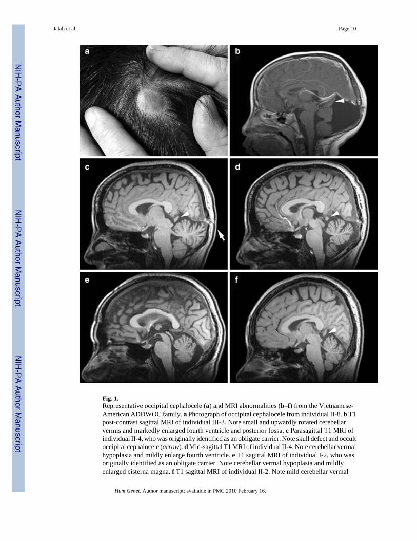
Fig. 1.Representative occipital cephalocele (a) and MRI abnormalities (b–f) from the Vietnamese-American ADDWOC family. a Photograph of occipital cephalocele from individual II-8. b T1post-contrast sagittal MRI of individual III-3. Note small and upwardly rotated cerebellarvermis and markedly enlarged fourth ventricle and posterior fossa. c Parasagittal T1 MRI ofindividual II-4, who was originally identified as an obligate carrier. Note skull defect and occultoccipital cephalocele (arrow). d Mid-sagittal T1 MRI of individual II-4. Note cerebellar vermalhypoplasia and mildly enlarge fourth ventricle. e T1 sagittal MRI of individual I-2, who wasoriginally identified as an obligate carrier. Note cerebellar vermal hypoplasia and mildlyenlarged cisterna magna. f T1 sagittal MRI of individual II-2. Note mild cerebellar vermal
Jalali et al. Page 10
Hum Genet. Author manuscript; available in PMC 2010 February 16.
NIH
-PA Author Manuscript
NIH
-PA Author Manuscript
NIH
-PA Author Manuscript

hypoplasia. Arrowheads in b, c, d, and f point to the protrusion of occipital lobe through thetentorial notch
Jalali et al. Page 11
Hum Genet. Author manuscript; available in PMC 2010 February 16.
NIH
-PA Author Manuscript
NIH
-PA Author Manuscript
NIH
-PA Author Manuscript

Fig. 2.Pedigree of the Vietnamese-American ADDWOC family. Generation number is listed alongthe left margin, and individuals are numbered underneath each symbol. Thus, the proband(indicated by an asterisk) corresponds to individual III-8. Solid-shaded large symbolscorrespond to affected members. The smaller shaded symbol represents a spontaneouslyaborted fetus. Only the affected individuals (except I-3, I-4, and III-7) were included in thelinkage analysis
Jalali et al. Page 12
Hum Genet. Author manuscript; available in PMC 2010 February 16.
NIH
-PA Author Manuscript
NIH
-PA Author Manuscript
NIH
-PA Author Manuscript
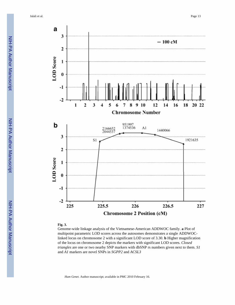
Fig. 3.Genome-wide linkage analysis of the Vietnamese-American ADDWOC family. a Plot ofmultipoint parametric LOD scores across the autosomes demonstrates a single ADDWOC-linked locus on chromosome 2 with a significant LOD score of 3.30. b Higher magnificationof the locus on chromosome 2 depicts the markers with significant LOD scores. Closedtriangles are one or two nearby SNP markers with dbSNP rs numbers given next to them. S1and A1 markers are novel SNPs in SGPP2 and ACSL3
Jalali et al. Page 13
Hum Genet. Author manuscript; available in PMC 2010 February 16.
NIH
-PA Author Manuscript
NIH
-PA Author Manuscript
NIH
-PA Author Manuscript

Fig. 4.Haplotype analysis of the ADDWOC locus in the Vietnamese-American pedigree. SNPmarkers are listed along the left margin, and under each individual the corresponding allelesare listed. The two alleles for each SNP are represented by numbers 1 and 2. A1 allele 2 andS1 allele 2 are the newly discovered variants. The ancestral haplotype containing theADDWOC affection locus is noted by the closed box. Chromosomal recombinations duringmeioses leading to individuals II-3, III-3, and III-8 narrowed the ADD-WOC haplotype, asnoted by the smaller box in those individuals
Jalali et al. Page 14
Hum Genet. Author manuscript; available in PMC 2010 February 16.
NIH
-PA Author Manuscript
NIH
-PA Author Manuscript
NIH
-PA Author Manuscript
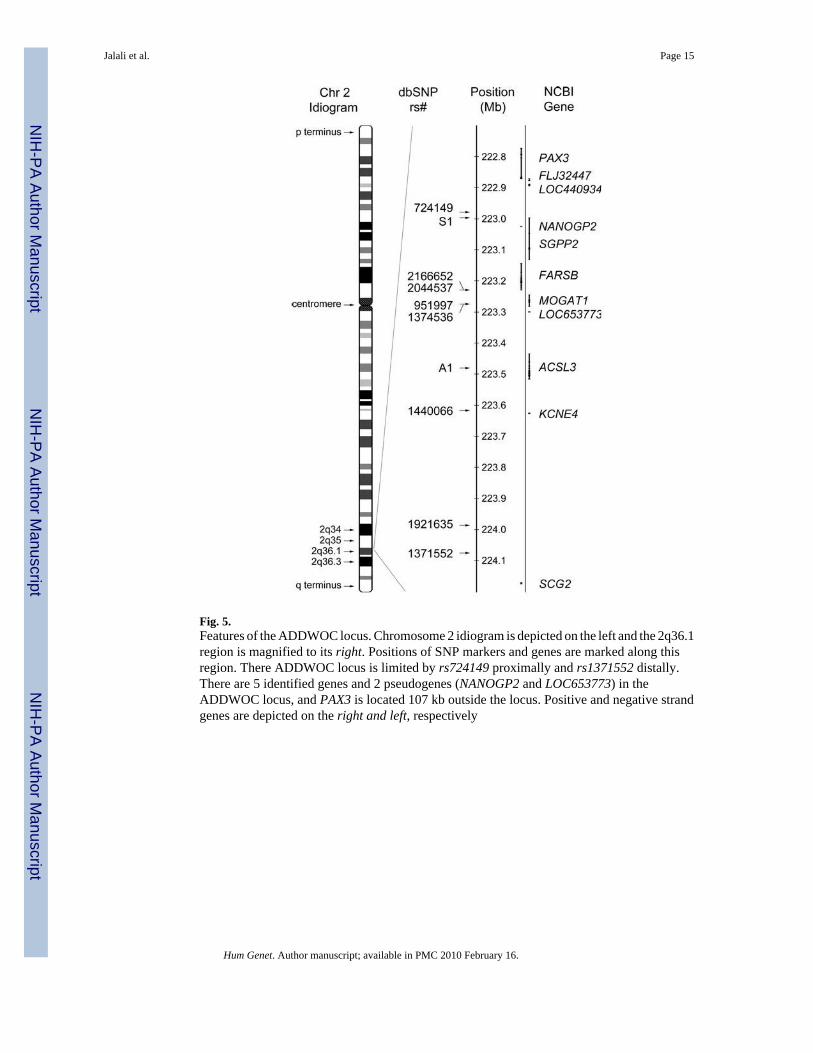
Fig. 5.Features of the ADDWOC locus. Chromosome 2 idiogram is depicted on the left and the 2q36.1region is magnified to its right. Positions of SNP markers and genes are marked along thisregion. There ADDWOC locus is limited by rs724149 proximally and rs1371552 distally.There are 5 identified genes and 2 pseudogenes (NANOGP2 and LOC653773) in theADDWOC locus, and PAX3 is located 107 kb outside the locus. Positive and negative strandgenes are depicted on the right and left, respectively
Jalali et al. Page 15
Hum Genet. Author manuscript; available in PMC 2010 February 16.
NIH
-PA Author Manuscript
NIH
-PA Author Manuscript
NIH
-PA Author Manuscript
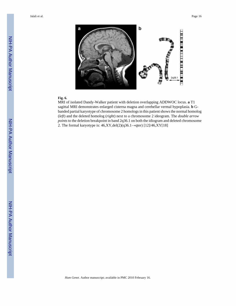
Fig. 6.MRI of isolated Dandy-Walker patient with deletion overlapping ADDWOC locus. a T1sagittal MRI demonstrates enlarged cisterna magna and cerebellar vermal hypoplasia. b G-banded partial karyotype of chromosome 2 homologs in this patient shows the normal homolog(left) and the deleted homolog (right) next to a chromosome 2 ideogram. The double arrowpoints to the deletion breakpoint in band 2q36.1 on both the idiogram and deleted chromosome2. The formal karyotype is: 46,XY,del(2)(q36.1→qter) [12]/46,XY[18]
Jalali et al. Page 16
Hum Genet. Author manuscript; available in PMC 2010 February 16.
NIH
-PA Author Manuscript
NIH
-PA Author Manuscript
NIH
-PA Author Manuscript