Modulation masking in cochlear implant listeners: envelope versus tonotopic components
Upload
independentCategory
view
4download
0
1
A fungal SLMAP homolog plays a fundamental role in development and localizes to the 1
nuclear envelope, ER, and mitochondria 2
3
Steffen Nordziekea, Thomas Zobel
b, Benjamin Fränzel
c, Dirk A. Wolters
c, Ulrich Kück
a#, Ines 4
Teicherta#
5
6
Lehrstuhl für Allgemeine und Molekulare Botanika and Lehrstuhl für Analytische Chemiec, Ruhr-7
Universität Bochum, Bochum, Germany, and Institut für Neurobiologieb, Westfälische Wilhelms-8
Universität Münster, Münster, Germany 9
10
#Address correspondence to: [email protected], [email protected] 11
12
Running Head 13
SLMAP in fungal development 14
EC Accepts, published online ahead of print on 19 December 2014Eukaryotic Cell doi:10.1128/EC.00241-14Copyright © 2014, American Society for Microbiology. All Rights Reserved.
2
Abstract 15
Sarcolemmal membrane-associated protein (SLMAP) is a tail-anchored protein involved in 16
fundamental cellular processes such as myoblast fusion, cell cycle progression, and chromosomal 17
inheritance. Further, SLMAP mis-expression is associated with endothelial dysfunctions in 18
diabetes and cancer. SLMAP is part of the conserved striatin-interacting phosphatase and kinase 19
(STRIPAK) complex required for specific signaling pathways in yeasts, filamentous fungi, 20
insects, and mammals. In filamentous fungi, STRIPAK was initially discovered in Sordaria 21
macrospora – a model system for fungal differentiation. Here, we functionally characterize the 22
STRIPAK subunit PRO45, a homolog of human SLMAP. We show that PRO45 is required for 23
sexual propagation and cell-to-cell fusion, and that its forkhead-associated (FHA) domain is 24
essential for these processes. Protein-protein interaction studies revealed that PRO45 binds to 25
STRIPAK subunits PRO11 and SmMOB3, which are also required for sexual propagation. 26
Super-resolution structured-illumination microscopy (SIM) further established that PRO45 27
localizes to the nuclear envelope, ER, and mitochondria. SIM also showed that localization to the 28
nuclear envelope required STRIPAK subunits PRO11 and PRO22, whereas for mitochondria it 29
did not. Taken together, our study provides important insights into fundamental roles of the 30
fungal SLMAP homolog PRO45 and suggests STRIPAK-related and STRIPAK-unrelated 31
functions. 32
3
Introduction 33
Membrane recruitment of protein complexes, cell signaling modules, and enzymes is a critical 34
step for many cellular functions. The family of tail-anchored proteins is recognized for anchoring 35
proteins and vesicles to specific membranes such as the endoplasmic reticulum (ER) and the 36
outer mitochondrial membrane (1), and tail-anchored proteins are characterized by a C-terminal 37
single transmembrane domain, which is post-translationally inserted into membranes (2, 3). 38
Sarcolemmal membrane-associated protein (SLMAP) is a tail-anchored protein first identified in 39
myocardiac cells (4). In mammals, this protein is known to be involved in myoblast fusion during 40
embryonic development, excitation-contraction coupling in cardiac myocytes, and cell cycle 41
progression (5-8). Furthermore, SLMAP was identified as a disease gene for Brugada syndrome, a 42
cardiac channelopathy (9). The functional diversity of SLMAP is dependent on alternative 43
splicing, leading to at least four different isoforms of the protein (4, 6, 7, 10). Importantly, gene 44
expression analyses have implicated SLMAP mis-expression with endothelial dysfunctions in 45
diabetes, chromosomal aberrations, and cancer (11-14), and currently, SLMAP is the target of 46
lectin-based treatment of drug-resistant cancer cells (15). 47
SLMAP is conserved from yeast to humans, and characterized fungal SLMAP homologs include 48
Neurospora crassa HAM-4 (hyphal anastomosis), Saccharomyces cerevisiae Far9p (factor arrest) 49
and Far10p, as well as Schizosaccharomyces pombe Csc1p (component of SIP complex 1) (16-50
18). HAM-4 is essential for vegetative cell fusion, whereas Far9p and Far10p are required for 51
pheromone-induced cell cycle arrest during yeast mating, and Csc1p acts in cytokinesis. 52
Interestingly, in a genome-wide screen for vacuolar protein sorting-deficient (vps) mutants, Far9p 53
was also identified as Vps64, and vacuolar morphology is altered in N. crassa ham-4 mutants, 54
indicating a role for SLMAP homologs in organelle morphology in fungi (18, 19). 55

4
Recently, SLMAP has been identified as an accessory protein to the human striatin-interacting 56
phosphatase and kinase (STRIPAK) complex, a large multi-protein complex assembled around a 57
core of protein phosphatase 2A (PP2A) subunits (20). In addition to PP2A structural (PP2A A) 58
and catalytic (PP2Ac) subunits, human STRIPAK complex contains striatins (regulatory PP2A 59
B´´´ subunits), striatin-interacting proteins 1 and 2 (STRIP1/2), monopolar spindle-one-binder 60
(MOB) proteins, germinal center kinase (GCK)-III protein kinases, and cerebral cavernous 61
malformation protein 3 (CCM3). This core STRIPAK complex is able to assemble in a mutually 62
exclusive way with other accessory proteins like SLMAP and suppressor of IKK (SIKE) or a 63
cortactin-binding protein 2 family member (CTTNBP2 or CTTNBP2NL) (21). The high diversity 64
of STRIPAK and STRIPAK-like complexes makes estimating the molecular weight of the 65
complex difficult. Human STRIPAK was found to play a role in Golgi polarization and is 66
involved in mitosis by tethering Golgi vesicles to centrosomes and the nuclear membrane in a cell 67
cycle-specific manner (22, 23). 68
STRIPAK-equivalent complexes have been found in a number of diverse organisms from yeasts 69
to humans. The Drosophila melanogaster dSTRIPAK (Drosophila STRIPAK) complex is a 70
negative regulator of the Hippo signaling pathway (24). The S. cerevisiae Far complex plays a 71
role in cell cycle arrest during mating as well as acts in an antagonistic fashion towards TORC 72
(target of rapamycin complex) 2 signaling (17, 25). The S. pombe SIP (SIN (septation initiation 73
network) inhibitory PP2A) complex is required for the coordination of mitosis and cytokinesis by 74
inhibiting SIN (16). The N. crassa STRIPAK complex controls nuclear accumulation of the 75
MAK1 MAP kinase and regulates chemotropic interactions between conidial germlings (26). 76
Moreover, in the fungal model organism Sordaria macrospora (27-29), the STRIPAK complex is 77
required for cell fusion and sexual reproduction, namely the formation of multicellular fruiting 78
bodies (30). Discrete STRIPAK components have been characterized in other filamentous fungi, 79

5
e.g. Aspergillus nidulans, Fusarium graminearum, Magnaporthe oryzae, and Sclerotinia 80
sclerotiorum (31-34); however, a description of the STRIPAK complex in these fungi is still 81
lacking. 82
S. macrospora STRIPAK consists of PP2A A, PP2Ac1, striatin homolog PRO11, STRIP 83
homolog PRO22, and the MOB protein SmMOB3. Strikingly, a mutant lacking the striatin 84
homolog PRO11 can be complemented by mouse striatin cDNA (35), thereby highlighting the 85
suitability of S. macrospora for studying the molecular function of STRIPAK components. 86
Finally, defects in multicellular differentiation can easily be observed in S. macrospora, since the 87
fungus forms complex three-dimensional fruiting bodies (perithecia) within seven days without 88
the need of a mating partner, and early developmental structures (coiled hyphae termed ascogonia 89
and spherical immature fruiting bodies termed protoperithecia) are not masked by any asexual 90
spores (27). 91
The aim of this study was to functionally characterize PRO45, the SLMAP homolog from S. 92
macrospora, and provide insights into its role within the fungal STRIPAK complex. Protein-93
protein interaction studies indeed showed that PRO45 is part of fungal STRIPAK. We further 94
established super-resolution structured-illumination microscopy (SIM) for S. macrospora to 95
distinctly demonstrate PRO45 cellular localization dependent on STRIPAK integrity. Here, we 96
provide evidence that the SLMAP homolog PRO45 plays a fundamental role in fungal 97
development and might have STRIPAK-related and STRIPAK-unrelated functions. 98

6
Materials and Methods 99
100
Strains and growth conditions 101
All S. macrospora strains used in this study (Table 1) were grown under standard conditions and 102
transformed with recombinant plasmids as described previously (36, 37), with the following 103
modifications. Fernbach flasks containing liquid CM were inoculated with S. macrospora strains 104
and incubated for 3 days. The cell wall was degraded using 0.05 g/ml VinoTaste® Pro 105
(Novozymes, Bagsvaerd, Denmark), 0.015 g/ml caylase (Cayla, Toulouse, France), and 27 Units 106
of chitinase (ASA Spezialenzyme GmbH, Wolfenbüttel, Germany). Transformation was 107
performed using 15 - 20 µg of circular (ectopic integration) or linearized (homologous 108
integration) plasmid DNA. 109
Cloning was performed using the homologous recombination system in S. cerevisiae strain PJ69-110
4A as described previously (38, 39). Yeast cells were transformed by electroporation according to 111
the method of Becker and Lundblad (40) in a Multiporator (Eppendorf, Wesseling-Berzdorf, 112
Germany) at 1.5 kV. Transformants were selected by screening for uracil prototrophy. For DNA 113
isolation, yeast cell walls were broken with glass beads (0.5 mm diameter), followed by DNA 114
extraction with the E.N.Z.A. Plasmid Miniprep Kit I (Peqlab Biotechnologie GmbH, Erlangen, 115
Germany). 116
For documentation of vegetative growth rates, S. macrospora strains were grown on corn meal-117
malt fructification medium (BMM) for two days, and standard inoculates were transferred to 118
synthetic Westergaard´s medium (SWG) (41), where growth distance was determined for 2 119
consecutive days. Perithecia formation was determined after 7 days of growth on BMM using 10 120
regions of 37 mm2 from 2 plates per strain. 121
122

7
Construction of plasmids 123
All plasmids and primers used in this study are listed in Tables 2 and 3, respectively. pDS16 was 124
generated by insertion of a hph cassette (1,4 kb EcoRI fragment of pDrivehph) (42) between a 5´ 125
and 3´ region of S. macrospora SMAC_01224 in pRS426 (43). Flanking regions were amplified 126
using SMACG_01224_5F-fw/SMACG_01224_5F-rv (5´) and SMACG_01224_3F-127
fw/SMACG_01224_3F-rv (3´), respectively. 128
For the construction of TAP vectors, fragments were amplified by 45_CTAP-fw x 45_int-bw and 129
45_int-fw x 45_CTAP-bw and transformed into yeast with a BglII-linearized pDS22 130
(Nowrousian, unpublished data). For the construction of p45ΔFHA-TAP, the amplicons of 131
45_int-fw/45ΔFHA_CTAP-bw and 45_int-fw-2/ 45_int-bw-2 were transformed into ClaI-132
linearized p45-CTAP. 133
For construction of GFP vectors, PCR fragments were amplified with 45_NtermGFP-fw/45_int-134
bw and 45_int-fw/45_CtermGFP-bw and co-transformed into yeast with either a SpeI (N-terminal 135
fusion) or a BglII (C-terminal fusion) linearized pDS23 (44) resulting in plasmids pEGFP-45 and 136
p45-EGFP. For the construction of a truncated version of PRO45 lacking the C–terminal 137
transmembrane domain, amplicons of 45_int-fw/45ΔTM_CEGFP-bw and 45_int-fw-3/1757 were 138
transformed into yeast together with a NotI hydrolyzed pEGFP-45 resulting in plasmid p45ΔTM-139
EGFP. 140
141
Generation of a pro45 deletion strain 142
To generate Δpro45 deletion strains by homologous recombination, the 4,9 kb pro45 deletion 143
cassette of pDS16 was transformed into S. macrospora Δku70 strain S96888 (45). Ascospore 144
isolates, in which the pro45 ORF was replaced by the hph cassette and which had the wild type 145
(N161) or the fus1-1 (R7329) genetic background were obtained as described previously by 146
8
crosses to spore color mutant fus 1-1 (45). Strains were analyzed by PCR using primers pairs 147
1224KOvp1neu x hph1MN, 1224KOvp2neu x hph2MN and 1224KOvp1neu x 1224KOvp3 for 148
the validation of the 5´flanking region, the 3´ flanking region and the wild type control, 149
respectively. Southern blotting and hybridization were performed according to standard 150
techniques (46). A 1012-bp (891-1903) pro45-fragment was generated by the digestion of p45-151
CTAP with SacI, labeled with 32
P and used as DNA probe to detect pro45, whereas a 616 bp hph-152
fragment was generated by PCR using the primer pair hph1MN/hph2_2010-bw and used to detect 153
the hph cassette. 154
155
Light and fluorescence microscopy 156
Light and fluorescence microscopic investigations were carried out with an AxioImager.M1 157
microscope (Zeiss, Jena, Germany) using an XBO 75 xenon lamp (LEJ, Jena, Germany) for 158
fluorescence excitation. For detection of EGFP and detection of DsRED and MitoTracker® 159
orange CMTMRos fluorescence, the Chroma filter sets (Chroma Technology Corp, Bellows 160
Falls, Vermont, USA) 49002 (excitation filter HQ470/40, emission filter HQ525/50, beamsplitter 161
T495LPXR) and 49008 (excitation filter HQ560/40, emission filter ET630/75m, beamsplitter 162
T585lp) were used, respectively. For detection of DAPI and ER-Tracker™ Blue-White DPX, 163
Chroma filter set 31000v2 (excitation filter D350/50, emission filter D460/50, beamsplitter 164
400dclp) was used. Images were captured with a Photometrix Cool SnapHQ camera (Roper 165
Scientific, Martinsried, Germany) and MetaMorph (version 7.7.0.0; Universal Imaging, Bedford 166
Hills, New York, USA). 167
To analyze hyphal fusion, strains were inoculated on minimal media containing soluble starch 168
(MMS) on top of a layer of cellophane (BioRad, München, Germany). After incubation for two 169

9
days, hyphal fusion was analyzed by light microscopy (47). Furthermore, microscopy was used to 170
determine sexual development and fluorescence of selected strains on BMM-covered slides (48). 171
To determine differences in sexual development, strains were grown for 2, 4, and 7 days and at 172
each time point, the most advanced stages were detected. For fluorescence microscopy, strains 173
were grown for 2 days on BMM-covered slides. For visualization of nuclei, the mitochondria, 174
and the ER, hyphae were stained with 50 µg/ml DAPI (Life Technologies, Darmstadt, Germany), 175
100 µM MitoTracker orange CMTMRos (Life Technologies, Darmstadt, Germany), and 100 µM 176
ER-Tracker™ Blue-White DPX (Life Technologies, Darmstadt, Germany), respectively. 177
Formation of perithecia was documented using a stereomicroscope (Stemi 2000-C; Zeiss) 178
equipped with a digital camera (AxioCamERc 5s; Zeiss). Recorded images were processed with 179
MetaMorph, Adobe Photoshop, and Adobe Illustrator CS4. 180
181
Super-resolution structured-illumination microscopy (SIM) 182
Strains were grown for 2 days on BMM-covered slides, and fixation was performed with a 0.2% 183
formaldehyde solution for 5 minutes followed by a washing step with phosphate buffered saline 184
(PBS). Staining was performed as described above. 185
SIM images were taken with an ELYRA S.1 Microscope (CellObserver SD, 63×/1.4 oil-186
immersion objective; Zeiss, Jena, Germany) with the software ZEN 2010 D (Zeiss, Jena, 187
Germany). For image acquisition, 5 grid rotations were used with an average of 2. Beam Splitter 188
settings were: GFP (488): BP 495-550 + LP 750; DsRED and MitoTracker® orange CMTMRos 189
(568): BP 570-620 + LP 750; DAPI and ER-Tracker™ Blue-White DPX (405): BP 420-480 + LP 190
750. For SIM calculations of fluorescent hyphae, the following manual settings were used: Noise 191
Filter, -3; SR frequency weighting, 1.0; sectioning, zero order 100 / first order 75 / second order 192

10
75. Recorded images were processed with ZEN 2010 D, Adobe Photoshop, and Adobe Illustrator 193
CS4. 194
195
Tandem affinity purification and mass spectrometry 196
Tandem affinity purification of PRO45-TAP and PRO45FHA-TAP, tryptic digestion of proteins 197
and multi-dimensional protein identification technology (MudPIT; 49, 50) analysis were 198
performed as described previously (30) using an Orbitrap Velos ion trap mass spectrometer 199
coupled to an Accela quaternary U-HPLC pump (Thermo Fisher Scientific, MA, USA). Proteome 200
Discoverer software version 1.2 was used for MS/MS data interpretation, and data were searched 201
against the S. macrospora database (smacrosporapep_v4_110909) with tryptic peptides, mass 202
accuracy of 10 ppm, fragment ion tolerance of 0.8 Da, and with oxidation of methionine as 203
variable modification allowing 4 missed cleavage sites. All accepted results had a high peptide 204
confidence with a score of 10. 205
206
Co-immunoprecipitation 207
Plasmid p45-EGFP was co-transformed with pFLAGMob3 and pHA11 (30) into the S. 208
macrospora wild type strain S91327, resulting in hygromycin- and nourseothrycin resistant 209
strains T105.2, T104.4, and T133-E6 (Table 1). Additionally, pDS23 (44) was co-transformed 210
with pFLAGMob3 and pHA11 to generate T1201 and T1202, respectively, which served as 211
control strains. 212
Purification of FLAG- and GFP- tagged proteins was performed using anti-FLAG M2 affinity gel 213
(Sigma-Aldrich, St. Louis, MO, USA) and GFP-Trap® (Chromotek, Planegg-Martinsried, 214
Germany), whereas the HA-purification was performed using an anti-HA antibody (Sigma-215
Aldrich, St. Louis, MO, USA), which was subsequently recovered by protein A sepharose 216

11
(Amersham GE Healthcare Europe GmbH, Freiburg, Germany). Crude protein extracts and 217
immunopurified complexes were subjected to SDS-PAGE, Western blotting, and 218
immunodetection with anti-FLAG (ANTI-FLAG® M2, Sigma-Aldrich, St. Louis, MO, USA), 219
anti-GFP (Living Colors® JL-8, Takara Bio Europe/Clontech, Saint-Germain-en-Laye, France) 220
and anti-HA (Monoclonal Anti-HA, Sigma-Aldrich, St. Louis, MO, USA) antibodies in 221
combination with an anti-mouse IgG HRP-linked secondary antibody (Cell Signaling). Signal 222
detection was performed using Immun-Star™ WesternC™ (BioRad, München, Germany) or 223
SuperSignal West Femto (Thermo Fisher Scientific, MA, USA) chemiluminescent solutions on a 224
ChemiDoc XRS+ system (BioRad, München, Germany). 225

12
Results 226
227
A pro45 deletion strain is sterile and shows a defect in hyphal fusion 228
Recently, a homolog of the human STRIPAK complex was identified in S. macrospora (30). This 229
fungal complex consists of striatin homolog PRO11, STRIP1/2 homolog PRO22, SmMOB3, and 230
PP2A subunits. STRIPAK complexes from different systems contain further accessory 231
components, amongst them SLMAP, which interacts with distinct STRIPAK subunits and links 232
STRIPAK sub-complexes to the nuclear envelope (22, 51). 233
We used BLAST (52) to search for an S. macrospora homolog of SLMAP (gi|109731644) and 234
identified SMAC_01224, which we termed PRO45 due to the phenotype of the mutant, as 235
described below. Reciprocal BLAST confirmed that PRO45 is an ortholog of SLMAP. Further, 236
PRO45 shows the same domain organization as SLMAP and other fungal SLMAP homologs 237
such as N. crassa HAM-4 and S. cerevisiae Far9/10p, with a forkhead-associated (FHA) domain, 238
two coiled coil domains, and a C-terminal transmembrane (TM) domain typical for tail-anchored 239
proteins (Fig. 1A). The FHA domain is a phosphoprotein-binding domain that specifically 240
recognizes phosphothreonine residues, whereas coiled-coil domains contain α-helices wound 241
around each other and mediating protein-protein interactions (53, 54). PRO45 consists of 796 242
amino acids and shows 10.9% and 92.5% overall amino acid identity to human SLMAP and N. 243
crassa HAM-4, respectively. Amino acid identity between S. macrospora and human SLMAP is 244
increased within conserved domains, as shown by amino acid alignments of the FHA and TM 245
domains (Fig. 1 B and C). Here, amino acid identity is 51.7% and 19.2% for the FHA and the TM 246
domain, respectively. 247
To functionally characterize PRO45, we generated a pro45 deletion strain by homologous 248
recombination of a pro45 deletion cassette using non-homologous end-joining-deficient ku70 as 249

13
a host strain (45). Ascospore isolates carrying the hph marker cassette instead of pro45 and 250
devoid of the ku70 background were obtained from crosses of primary transformants to spore 251
color mutant fus (55). Two randomly selected ascospore isolates, N161 (pro45) and R7329 252
(pro45/fus), were subjected to Southern analysis, confirming the deletion of the pro45 gene 253
(Fig. S1). Vegetative growth was strongly reduced in the mutant compared to wild type, and this 254
defect was restored by transformation with the full-length gene (Fig. S2A). 255
All Sordaria STRIPAK components characterized so far are involved in sexual development, i.e. 256
the formation of sexual fruiting bodies (perithecia) (35, 56, 57). We therefore analyzed the pro45 257
deletion strain for defects in the sexual cycle. Sexual development starts with the formation of a 258
hyphal coil (ascogonium) two days after inoculation. At day four, enveloping hyphae have 259
covered the ascogonium, thereby forming melanized spherical structures (protoperithecia) that 260
develop further into mature, pear-shaped perithecia. As can be seen from Fig. 2A, the wild type 261
forms ascogonia, protoperithecia and mature perithecia within the expected time frame on BMM 262
fructification medium. In contrast, pro45 forms only protoperithecia that do not develop any 263
further. To verify that the observed defect is due to deletion of the pro45 gene, we ectopically 264
integrated pro45 wild type gene copies into pro45. Since RNA-seq analysis showed that pro45 265
transcript levels were very low (58), we expressed pro45 from the Aspergillus nidulans gpd 266
promoter (59). Two different translational fusions of PRO45 with a TAP (tandem affinity 267
purification) tag (pro45-TAP) and GFP (EGFP-pro45) were able to complement the sexual 268
developmental defect of pro45 (Fig. 2A, Fig. S2B). The observed sexual phenotype for S. 269
macrospora pro45 is the most severe developmental phenotype described so far for a fungal 270
SLMAP mutant and clearly differs from the phenotype of a N. crassa ham-4 mutant that is still 271
able to complete the sexual cycle (18). 272

14
Many mutants impaired in sexual development are also impaired in vegetative hyphal fusion and 273
vice versa, although this correlation is not strict (e.g. 27, 60, 61, 62). Mutants lacking Sordaria 274
STRIPAK components PRO11, PRO22, and SmMOB3 are also impaired in vegetative hyphal 275
fusion (30, 56). We thus assessed hyphal fusion in pro45 and complemented strains. Like the 276
other STRIPAK mutants, pro45 is unable to undergo hyphal fusion, although hyphae frequently 277
grow side by side (Fig. 2B, marked by asterisks). In contrast, wild type and pro45 strains 278
harboring an ectopically integrated pro45-TAP or EGFP-pro45 gene are able to undergo hyphal 279
fusion, which is indicated by fusion “bridges” between hyphae (Fig. 2B, marked by arrows). The 280
aforementioned phenotypes for pro45, namely a defect in sexual development and hyphal 281
fusion, have also been described for the S. macrospora STRIPAK mutants pro11, pro22 (30), 282
and Smmob3 (56), and point to a role for PRO45 in the fungal STRIPAK complex. 283
As mentioned earlier, in silico analysis identified a FHA domain, two coiled-coil domains and a 284
C-terminal TM domain in PRO45. To functionally analyze these domains, we generated mutated 285
PRO45 versions lacking either the C-terminal TM domain (PRO45TM-EGFP) or the N-286
terminal FHA domain (PRO45FHA-TAP; Fig. 1D) and expressed them in pro45 as a host 287
strain. Strains carrying the PRO45TM-EGFP version were indistinguishable from wild type 288
with respect to fruiting body formation and hyphal fusion (Fig. 2, Fig. S2B). In contrast, 289
PRO45FHA-TAP was unable to restore sexual development and hyphal fusion in the pro45 290
deletion strain, while a TAP-tagged full-length PRO45 fusion did (Fig. 2). Thus, the FHA domain 291
plays a fundamental role in overall PRO45 function, whereas the TM domain is dispensable for 292
hyphal fusion and sexual development. 293
294
PRO45 interacts with STRIPAK components PRO11 and SmMOB3 295

15
Our incentive to study the PRO45 protein was to establish it as an additional component of the S. 296
macrospora STRIPAK complex. Therefore, we performed tandem affinity purification followed 297
by mass spectrometry (TAP-MS) with a strain carrying a PRO45-TAP protein in the pro45 298
background. This strain is able to form perithecia and to undergo hyphal fusion, proving the 299
functionality of the TAP-fusion construct (Fig. 2). 300
TAP-MS was carried out three times, and all detected proteins are listed in Table S1. All three 301
TAP-MS analyses with PRO45 as bait yielded high amounts of the bait protein (234 spectral 302
counts), as well as peptides from previously identified S. macrospora STRIPAK components 303
PRO11 (164 spectral counts) and SmMOB3 (22 spectral counts, Table 4). The S. macrospora 304
STRIPAK complex contains further subunits, PRO22, PP2AA, and PP2Ac (30). However, these 305
STRIPAK subunits were not detected in our TAP-MS experiments. We performed co-306
immunoprecipitation to confirm the interactions between PRO45 and PRO11 as well as 307
SmMOB3 (Fig. 3). For this experiment, we used S. macrospora strains carrying GFP-tagged 308
PRO45 and either HA-tagged PRO11 or FLAG-tagged SmMOB3, as well as control strains 309
carrying only one tagged protein. Using these tagged versions, we verified the interaction of 310
PRO45 with PRO11 (Fig. 3A) and PRO45 with SmMOB3 (Fig. 3B). 311
We further assessed whether the FHA domain, which is crucial for PRO45 function, mediates 312
protein-protein interaction within STRIPAK. Therefore, TAP-MS was performed with sterile 313
strain N1032 carrying a PRO45FHA-TAP fusion protein in the pro45 background (Fig. 2, Fig. 314
S2). TAP-MS established that both PRO11 and SmMOB3 were still detectable with reasonable 315
spectral counts (Table 4). This result suggests that the FHA domain, which is supposed to 316
mediate phosphoprotein interactions, is dispensable for the interaction of PRO45 with PRO11 317
and SmMOB3. 318
319

16
PRO45 localizes to the nuclear envelope, ER, and mitochondria 320
SLMAP has been described to be targeted to different membrane systems, including the 321
sarcolemma, the sarcoplasmic and endoplasmic reticulum, as well as the outer mitochondrial 322
membrane (4, 7, 22, 63). To assess the localization of PRO45 in S. macrospora, we generated a 323
translational fusion of PRO45 to an N-terminal GFP tag and showed its functionality by 324
complementation of the pro45 phenotype (Fig. 2). Initial microscopic analysis with EGFP- 325
PRO45 suggested that the protein localizes to a membrane. To scrutinize PRO45 localization at a 326
higher resolution beyond the Abbe diffraction limit, we employed super-resolution structured 327
illumination microscopy (SIM). We chose SIM since it allows to double the resolution of a 328
conventional widefield (WF) fluorescence image by a combination of spatially structured 329
illumination and computational three-dimensional reconstruction, using conventional 330
fluorophores and dyes (64). 331
Due to longer acquisition times when using SIM in comparison to conventional fluorescence 332
microscopy, we had to fix fungal samples. To confirm that our microscopic studies did not 333
provide artificial localization data, we first tested the effect of fixation and SIM computational 334
reconstruction. For this purpose, we used N883, a strain carrying histone 2B labeled with 335
tdTomato, and stained ER membranes with ER-Tracker. After fixation, no artefacts were 336
observed at the membrane and nuclear signals in WF images (Fig. 4A). Comparing WF and 337
reconstructed (SIM) images, it appears that computational reconstruction did not lead to artefacts 338
in localization, but to a highly refined membrane structure (Fig. 4B). We further tested organelle 339
markers for usage in SIM (Fig. 4C). Nuclear, ER, and mitochondrial labeling with DAPI, ER-340
Tracker, and MitoTracker, respectively, revealed that SIM is a highly suitable super-resolution 341
microscopy method for filamentous fungi. 342
17
SIM images demonstrated that PRO45 localizes to ring-like structures and to patches. Co-343
localization of EGFP-PRO45 with ER-targeted DsRed (DsRed-KDEL; 41) and simultaneous 344
DAPI staining revealed an association of PRO45 to the ER, mainly to patches at the nuclear 345
envelope (Fig. 5A). This is consistent with previously published data from N. crassa (26). 346
However, some of the PRO45 patches did not co-localize with the ER marker DsRed-KDEL. Co-347
localization of EGFP-PRO45 with MitoTracker showed that non-ER fluorescent patches were 348
closely associated to mitochondria (Fig. 5B). To assess the association of PRO45 with 349
mitochondria and a possible connection between PRO45-containing ER and mitochondrial 350
structures more closely, we analyzed strain N506 carrying EGFP-PRO45, labelled with 351
MitoTracker. Fig. 5C shows PRO45 localization in this strain in high resolution. Clearly, PRO45 352
simultaneously localizes to the nuclear envelope and to closely attached mitochondria. 353
The localization of PRO45 to mitochondria prompted us to look for differences in mitochondrial 354
structure in wild type and pro45. However, MitoTracker staining revealed no differences 355
between both strains, showing filamentous and fragmented mitochondria at the colony periphery 356
and colony interior, respectively (Fig. 6A and B). In SIM images, mitochondria appear mostly 357
fragmented, which might be due to fixation or to the usage of slim optical sections for 358
reconstruction. To assess whether PRO45 co-localizes only with fragmented, possibly stressed 359
mitochondria or also with filamentous mitochondria at the colony periphery, we stained hyphae 360
of a strain carrying EGFP-PRO45 with MitoTracker. As shown in Fig. 6C, WF images show that 361
PRO45 co-localizes with both morphotypes of mitochondria. Human PRO45 homolog SLMAP 362
also localizes to ER structures, primarily the nuclear envelope, and to mitochondria, and this is 363
due to different tail-anchor domains generated by alternative splicing (22, 63) . However, pro45 364
does not possess an intronic sequence in the vicinity of the TM-coding region. Further, no 365
evidence of alternative splicing of the single intron was found in RNA-seq data from a previous 366
18
analysis (58). Hydrophobic profiling of the PRO45 tail anchor revealed that it is highly similar to 367
the SLMAP tail-anchor 2, which targets SLMAP to both, ER structures and mitochondria (Fig. 368
5D) (63). These data are consistent with our microscopic observations. 369
370
PRO45 localization to ER and nuclear envelope requires STRIPAK subunits PRO11 and 371
PRO22 372
Recently, S. cerevisiae PRO45 homologs Far9p and Far10p were reported to remain at the 373
nuclear envelope, even in the absence of all other Far complex components (51). However, in N. 374
crassa, targeting of PRO45 homolog HAM-4 to the nuclear envelope depends on PRO11 375
homolog HAM-3 and PRO22 homolog HAM-2 (26). To assess PRO45 localization in mutants 376
lacking S. macrospora STRIPAK components PRO11 or PRO22, we transformed plasmids 377
encoding EGFP-PRO45 into sterile mutants pro11 and pro22 (30). 378
SIM was performed using transformants (Table 1), and nuclei and mitochondria were stained 379
with DAPI and MitoTracker, respectively (Fig. 7). Localization of EGFP-PRO45 in pro11 is 380
shown in Fig. 7A. PRO45 was absent from the nuclear membrane in pro11. However, 381
mitochondrial localization of PRO45 was not affected in the pro11 deletion strain. This 382
mitochondrial localization pattern was clearly different from the localization pattern of 383
cytoplasmic GFP in SIM images (Fig. S3). Similar to pro11, PRO45 localization to the nuclear 384
envelope, but not to mitochondria, was lost in a pro22 background (Fig. 7B). Our data suggest 385
that STRIPAK subunits PRO11 and PRO22 are required for nuclear envelope localization of 386
PRO45 in S. macrospora, as described for N. crassa (26). Remarkably, the association of PRO45 387
to mitochondria was retained in the STRIPAK mutants. These data suggest that mitochondrial 388
association of PRO45 might be STRIPAK-independent. 389
390

19
Discussion 391
The STRIPAK complex is a highly conserved multi-protein complex containing phosphatases 392
and kinases (21). Recently, a Sordaria STRIPAK complex has been described that controls 393
sexual development and cell fusion (30). In this study, we showed that S. macrospora SLMAP 394
homolog PRO45 is a component of this STRIPAK complex, plays a fundamental role in cell 395
fusion and sexual propagation, and that it localizes to the ER and mitochondria. 396
The composition of STRIPAK complexes in different organisms seems to be diverse, with 397
striatin, MOB3, PP2A scaffolding and catalytic subunits and STRIP proteins being central 398
components (21). For instance, SLMAP has been shown to be an accessory protein to human and 399
N. crassa STRIPAK complexes, but not Drosophila dSTRIPAK (20, 24, 26). However, SLMAP 400
homologs Far9p/Far10p of S. cerevisiae and Csc1p of S. pombe are integral components of the 401
STRIPAK-equivalent Far and SIP complexes (16, 51). Using a TAP-MS approach, we showed 402
that PRO45 is part of the S. macrospora STRIPAK complex and interacts with the striatin 403
homolog PRO11 and SmMOB3. In N. crassa, SLMAP homolog HAM-4 co-precipitated all five 404
STRIPAK components and was itself co-precipitated as prey by all five STRIPAK members (26). 405
Yet, TAP-MS analysis with STRIP homolog PRO22 from S. macrospora did not yield any 406
PRO45 peptides (30). However, this result may be due to the experimental approach that required 407
two subsequent affinity purifications compared to the one-step purifications used for N. crassa 408
(26). In human cells, interaction data were also dependent on the experimental approach. In 409
FLAG pull-down experiments, SLMAP was precipitated by striatins, Mob3, members of the 410
GCKIII kinase family, PP2A subunits, and STRIP1. However, in TAP experiments, SLMAP 411
reciprocally interacted only with MOB3 (20). Thus, the absence of other STRIPAK components 412
in PRO45 TAP-MS data does not necessarily mean that there is no complex formation of PRO45 413
with STRIPAK. In a previous study, for example, we found PRO22 to interact with PRO11, 414

20
generating the link to PRO45 (Fig. 8). Thus, although the general composition of the STRIPAK 415
complex and interactions between core components, i.e. PP2A scaffolding subunit PP2AA, PP2A 416
catalytic subunit PP2Ac1, and PP2A regulatory subunit PRO11, seem to be conserved, 417
interactions to accessory components may be species-specific. 418
The function of SLMAP proteins in fungi seems to be diverse. The paralogs Far9p and Far10p of 419
S. cerevisiae are both required for pheromone-mediated cell cycle arrest (17), and Far9p, also 420
identified as Vps64p, has a defect in α factor secretion (19). S. pombe Csc1p participates in SIN 421
inactivation, and a csc1 deletion mutant shows septation defects (16). In N. crassa, HAM-4 is 422
required for cell-to-cell fusion and possibly vacuolar morphology (18). In this study we showed 423
that S. macrospora pro45 has a severe developmental defect, in addition to the cell fusion 424
phenotype. Like other pro mutants, the pro45 strain is unable to form perithecia. This is clearly 425
different from the phenotype of a N. crassa ham-4 strain that is fertile. However, slight 426
developmental defects have been described, namely a delay in protoperithecia formation and 427
aberrant meiosis and ascospore formation in homozygous crosses (18). 428
Diverse domains have been annotated in SLMAP homologs, among them the FHA domain, a 429
phosphothreonine-specific protein binding motif. The FHA domain of mouse SLMAP has been 430
found to be necessary, but not sufficient, for targeting of SLMAP to centrosomes (6). However, 431
the underlying molecular mechanisms are still unknown. Results from this study show hyphal 432
fusion deficiency and sterility in a S. macrospora strain with an FHA-deleted PRO45, like in the 433
pro45 deletion strain. Interestingly, N. crassa ham-4 repeat-induced point mutation (RIP) mutants 434
harboring a HAM-4 protein with a shortened FHA domain are self-fusion competent, whereas a 435
ham-4 deletion mutant is not (18). In human cells, the FHA domain of SLMAP is required for 436
binding mammalian Hippo homologs MST1 and MST2 (mammalian STE20-like kinases 1 and 2) 437
(65). Taken together, these findings point to multiple important roles of the FHA domain in 438
21
different developmental processes. However, at least in S. macrospora these roles seem to be 439
independent of the interaction of PRO45 with STRIPAK components PRO11 and SmMOB3 440
since both were co-purified with PRO45FHA-TAP. 441
Interestingly, the TM domain of PRO45 seems to have no developmental function. This is 442
surprising, because the TA domain of tail-anchor proteins, consisting of the C-terminal TM 443
domain and the amino acids located at the very C-terminus are responsible for targeting these 444
proteins to specific membranes (1-3). One explanation could be that we did not delete the 445
complete TA, but only the TM domain of PRO45, and that the residual amino acids and/or 446
PRO45 interaction partners are sufficient for targeting. Indeed, many mitochondrial proteins were 447
identified in TAP-MS experiments with PRO45 as bait (Table S1); however, if these candidates 448
are true interaction partners remains to be elucidated. 449
In this study, we analyzed PRO45 localization by super-resolution microscopy. Several super-450
resolution microscopy methods have been developed, such as photoactivation localization 451
microscopy (PALM), SIM, stimulated emission depletion (STED), and stochastic optical 452
reconstruction microscopy (STORM) (reviewed by 66). Although PALM, STED, and STORM 453
give higher optical resolution than SIM, they require photocontrollable fluorescent proteins. 454
Thus, SIM is the only super-resolution microscopy method that can be applied using 455
conventional fluorescence proteins, such as GFP, and dyes (64). In fungi, SIM has been applied 456
e.g. in the yeast S. cerevisiae to analyze ER-mitochondria association in bud tips (67). However, 457
as for the other super-resolution methods, cells need to be fixed. Since fixation might lead to 458
artefacts, localization data have to be carefully re-checked to WF or confocal images. In this 459
study, PRO45 localization to fragmented mitochondria by SIM could imply binding of PRO45 to 460
only stressed mitochondria; however, in WF microscopy we showed co-localization with both, 461
fragmented and filamentous mitochondria (Fig. 6). Thus, here we report application of SIM for 462

22
filamentous fungi and show that it is a highly convenient method to study the localization of a 463
developmental protein with possible functions in organelle connections (see below). 464
A recurring theme is the function of SLMAP homologs as proteins that mediate membrane 465
anchoring to and/or attachment of different membranes. Organelle contact sites have e.g been 466
described between inner nuclear membrane, ER, and vacuoles, between ER and plasma 467
membrane, and between ER and mitochondria (reviewed by 68). Such associations are thought to 468
facilitate communication between organelles as well as lipid and ion transfer. In the yeast S. 469
cerevisiae, the ER mitochondria encounter structure (ERMES) complex localizes at ER-470
mitochondria contact sites, tethering both organelles (69). ERMES, a homolog of which has not 471
yet been identified in higher eukaryotes, was suggested to be involved in phospholipid exchange, 472
calcium signaling, and mitochondrial physiology (70-72). Recently, ER-mitochondria contact 473
sites have been shown to play a role in neurodegenerative diseases like Alzheimer’s disease and 474
amyotrophic lateral sclerosis (73, 74). 475
In this study, we localized PRO45 to the ER and mitochondria. This and other data lead to the 476
hypothesis that the association of different organelles may be one function of STRIPAK. S. 477
cerevisiae Far9p and Far10p are responsible for anchoring the Far complex at the ER (51). 478
Human SLMAP has been described to localize to the sarcolemma and the sarcoplasmic 479
reticulum, and it was proposed that homodimerization of differently localized SLMAP proteins 480
mediates excitation-contraction coupling in mice myocardia (8). Human SLMAP was also 481
implicated in Golgi-centrosome connections (22). Similar to mammalian systems, PRO45 might 482
function as a membrane organizer, bridging two organelles, the nuclear envelope and the outer 483
mitochondrial membrane, to mediate signaling. This membrane bridging may also be required to 484
target different STRIPAK sub-complexes to different cellular locations or to mediate STRIPAK-485
related and STRIPAK-unrelated functions of PRO45 in the same cell (Fig. 8). Of course, 486

23
mitochondrial localization might also hint to a role in mitochondrial respiration; however, we 487
excluded such function for PRO45 by high-resolution respirometry of the deletion mutant (S. 488
Nordzieke, A. Hamann, H.D. Osiewacz, I. Teichert; unpublished results). 489
Similar to N. crassa (26), localization of PRO45 to the nuclear envelope was absent in STRIPAK 490
mutants pro11 and pro22. Further data from N. crassa indicate that localization of STRIPAK 491
components to the nuclear envelope may not even be sufficient to direct cell communication and 492
cell differentiation (26). There, STRIPAK has been shown to be required for nuclear 493
accumulation of MAP kinase MAK1; however, MAK1 nuclear accumulation seems not to be 494
required for STRIPAK-related functions in cell-to-cell communication. It can be hypothesized 495
that a correctly assembled STRIPAK complex tethers PRO45 to the ER, while the localization of 496
PRO45 to mitochondria might be STRIPAK-independent. STRIPAK-independent functions of 497
distinct STRIPAK subunits have been described for other systems (reviewed by 21), and one 498
example is the function of S. macrospora PRO22, but not other STRIPAK subunits, in ascogonial 499
septation (57). However, PRO45 might still need interaction with STRIPAK to carry out its 500
function at mitochondria. The analysis of STRIPAK complexes from both the animal and fungal 501
kingdoms clearly reveals that STRIPAK composition and function are diverse. In fact, Frost et al. 502
(22) referred to these functional differences as a “repurposing” of STRIPAK complexes. 503
Scrutinizing the composition and regulation of different STRIPAK sub-complexes with distinct 504
functions will be one of the major tasks for future studies. 505
In summary, we have characterized PRO45 as a STRIPAK-associated protein and, by 506
establishing SIM, showed its localization at high resolution. Importantly, both fungal PRO45 and 507
mammalian SLMAP are required for cell-to-cell fusion, underlining their high functional 508
conservation. It is therefore of major interest to further scrutinize different STRIPAK sub-509
complexes and to decipher the various roles of PRO45 in a fungal model system – a system that 510

24
is highly suitable for unraveling underlying molecular and cellular mechanisms of key eukaryotic 511
cellular processes. 512

25
Acknowledgements 513
This work was supported by a grant from the Deutsche Forschungsgemeinschaft (Bonn, 514
Germany) to U.K. (PAK489, KU517/11-2). 515
We thank Regina Ricke and Susanne Schlewinski for excellent technical assistance, and Gabriele 516
Frenssen-Schenkel for help with graphical work. We thank Prof. C. Klämbt (Münster) for 517
providing the Zeiss ELYRA S.1 system. We acknowledge Daniel Schindler and Christoph Krisp 518
for experimental help. 519
520
26
REFERENCES 521
1. Wattenberg, B., and T. Lithgow. 2001. Targeting of C-terminal (tail)-anchored proteins: 522
understanding how cytoplasmic activities are anchored to intracellular membranes. 523
Traffic 2:66-71. 524
2. Borgese, N., S. Brambillasca, and S. Colombo. 2007. How tails guide tail-anchored 525
proteins to their destinations. Curr Opin Cell Biol 19:368-375. 526
3. Borgese, N., and E. Fasana. 2011. Targeting pathways of C-tail-anchored proteins. 527
Biochim Biophys Acta 1808:937-946. 528
4. Wigle, J. T., L. Demchyshyn, M. A. Pratt, W. A. Staines, M. Salih, and B. S. Tuana. 529
1997. Molecular cloning, expression, and chromosomal assignment of sarcolemmal-530
associated proteins. A family of acidic amphipathic alpha-helical proteins associated with 531
the membrane. J Biol Chem 272:32384-32394. 532
5. Guzzo, R. M., M. Salih, E. D. Moore, and B. S. Tuana. 2005. Molecular properties of 533
cardiac tail-anchored membrane protein SLMAP are consistent with structural role in 534
arrangement of excitation-contraction coupling apparatus. Am J Physiol Heart Circ 535
Physiol 288:H1810-H1819. 536
6. Guzzo, R. M., S. Sevinc, M. Salih, and B. S. Tuana. 2004. A novel isoform of 537
sarcolemmal membrane-associated protein (SLMAP) is a component of the microtubule 538
organizing centre. J Cell Sci 117:2271-2281. 539
7. Guzzo, R. M., J. Wigle, M. Salih, E. D. Moore, and B. S. Tuana. 2004. Regulated 540
expression and temporal induction of the tail-anchored sarcolemmal-membrane-541
associated protein is critical for myoblast fusion. Biochem J 381:599-608. 542
8. Nader, M., B. Westendorp, O. Hawari, M. Salih, A. F. Stewart, F. H. Leenen, and B. 543
S. Tuana. 2012. Tail-anchored membrane protein SLMAP is a novel regulator of cardiac 544
function at the sarcoplasmic reticulum. Am J Physiol Heart Circ Physiol 302:H1138-545
H1145. 546
9. Ishikawa, T., A. Sato, C. A. Marcou, D. J. Tester, M. J. Ackerman, L. Crotti, P. J. 547
Schwartz, Y. K. On, J. E. Park, K. Nakamura, M. Hiraoka, K. Nakazawa, H. 548
Sakurada, T. Arimura, N. Makita, and A. Kimura. 2012. A novel disease gene for 549
Brugada syndrome: sarcolemmal membrane-associated protein gene mutations impair 550
intracellular trafficking of hNav1.5. Circ Arrhythm Electrophysiol 5:1098-1107. 551
10. Wielowieyski, P. A., S. Sevinc, R. Guzzo, M. Salih, J. T. Wigle, and B. S. Tuana. 552
2000. Alternative splicing, expression, and genomic structure of the 3' region of the gene 553
encoding the sarcolemmal-associated proteins (SLAPs) defines a novel class of coiled-554
coil tail-anchored membrane proteins. J Biol Chem 275:38474-38481. 555
11. Beck, A. H., C. H. Lee, D. M. Witten, B. C. Gleason, B. Edris, I. Espinosa, S. Zhu, R. 556
Li, K. D. Montgomery, R. J. Marinelli, R. Tibshirani, T. Hastie, D. M. Jablons, B. P. 557
Rubin, C. D. Fletcher, R. B. West, and M. van de Rijn. 2010. Discovery of molecular 558
subtypes in leiomyosarcoma through integrative molecular profiling. Oncogene 29:845-559
854. 560
12. Ding, H., A. G. Howarth, M. Pannirselvam, T. J. Anderson, D. L. Severson, W. B. 561
Wiehler, C. R. Triggle, and B. S. Tuana. 2005. Endothelial dysfunction in Type 2 562
diabetes correlates with deregulated expression of the tail-anchored membrane protein 563
SLMAP. Am J Physiol Heart Circ Physiol 289:H206-H211. 564

27
13. Fellenberg, J., H. Saehr, B. Lehner, and D. Depeweg. 2012. A microRNA signature 565
differentiates between giant cell tumor derived neoplastic stromal cells and mesenchymal 566
stem cells. Cancer Lett 321:162-168. 567
14. Demicco, E. G., G. M. Boland, K. J. Brewer Savannah, K. Lusby, E. D. Young, D. 568
Ingram, K. L. Watson, M. Bailey, X. Guo, J. L. Hornick, M. van de Rijn, W. L. 569
Wang, K. E. Torres, D. Lev, and A. J. Lazar. 2014. Progressive loss of myogenic 570
differentiation in leiomyosarcoma has prognostic value. Histopathology in press. 571
15. Chen, K., X. Yang, L. Wu, M. Yu, X. Li, N. Li, S. Wang, and G. Li. 2013. Pinellia 572
pedatisecta agglutinin targets drug resistant K562/ADR leukemia cells through binding 573
with sarcolemmal membrane associated protein and enhancing macrophage phagocytosis. 574
PLoS One 8:e74363. 575
16. Singh, N. S., N. Shao, J. R. McLean, M. Sevugan, L. Ren, T. G. Chew, A. Bimbo, R. 576
Sharma, X. Tang, K. L. Gould, and M. K. Balasubramanian. 2011. SIN-inhibitory 577
phosphatase complex promotes Cdc11p dephosphorylation and propagates SIN 578
asymmetry in fission yeast. Curr Biol 21:1968-1978. 579
17. Kemp, H. A., and G. F. Sprague, Jr. 2003. Far3 and five interacting proteins prevent 580
premature recovery from pheromone arrest in the budding yeast Saccharomyces 581
cerevisiae. Mol Cell Biol 23:1750-1763. 582
18. Simonin, A. R., C. G. Rasmussen, M. Yang, and N. L. Glass. 2010. Genes encoding a 583
striatin-like protein (ham-3) and a forkhead associated protein (ham-4) are required for 584
hyphal fusion in Neurospora crassa. Fungal Genet Biol 47:855-868. 585
19. Bonangelino, C. J., E. M. Chavez, and J. S. Bonifacino. 2002. Genomic screen for 586
vacuolar protein sorting genes in Saccharomyces cerevisiae. Mol Biol Cell 13:2486-2501. 587
20. Goudreault, M., L. M. D'Ambrosio, M. J. Kean, M. J. Mullin, B. G. Larsen, A. 588
Sanchez, S. Chaudhry, G. I. Chen, F. Sicheri, A. I. Nesvizhskii, R. Aebersold, B. 589
Raught, and A. C. Gingras. 2009. A PP2A phosphatase high density interaction network 590
identifies a novel striatin-interacting phosphatase and kinase complex linked to the 591
cerebral cavernous malformation 3 (CCM3) protein. Mol Cell Proteomics 8:157-171. 592
21. Hwang, J., and D. C. Pallas. 2014. STRIPAK complexes: structure, biological function, 593
and involvement in human diseases. Int J Biochem Cell Biol 47:118-148. 594
22. Frost, A., M. G. Elgort, O. Brandman, C. Ives, S. R. Collins, L. Miller-Vedam, J. 595
Weibezahn, M. Y. Hein, I. Poser, M. Mann, A. A. Hyman, and J. S. Weissman. 2012. 596
Functional repurposing revealed by comparing S. pombe and S. cerevisiae genetic 597
interactions. Cell 149:1339-1352. 598
23. Kean, M. J., D. F. Ceccarelli, M. Goudreault, M. Sanches, S. Tate, B. Larsen, L. C. 599
Gibson, W. B. Derry, I. C. Scott, L. Pelletier, G. S. Baillie, F. Sicheri, and A. C. 600
Gingras. 2011. Structure-function analysis of core STRIPAK Proteins: a signaling 601
complex implicated in Golgi polarization. J Biol Chem 286:25065-25075. 602
24. Ribeiro, P. S., F. Josue, A. Wepf, M. C. Wehr, O. Rinner, G. Kelly, N. Tapon, and 603
M. Gstaiger. 2010. Combined functional genomic and proteomic approaches identify a 604
PP2A complex as a negative regulator of Hippo signaling. Mol Cell 39:521-534. 605
25. Pracheil, T., J. Thornton, and Z. Liu. 2012. TORC2 signaling is antagonized by protein 606
phosphatase 2A and the Far complex in Saccharomyces cerevisiae. Genetics 190:1325-607
1339. 608
26. Dettmann, A., Y. Heilig, S. Ludwig, K. Schmitt, J. Illgen, A. Fleissner, O. Valerius, 609
and S. Seiler. 2013. HAM-2 and HAM-3 are central for the assembly of the Neurospora 610
28
STRIPAK complex at the nuclear envelope and regulate nuclear accumulation of the 611
MAP kinase MAK-1 in a MAK-2-dependent manner. Mol Microbiol 90:796-812. 612
27. Engh, I., M. Nowrousian, and U. Kück. 2010. Sordaria macrospora, a model organism 613
to study fungal cellular development. Eur J Cell Biol 89:864-872. 614
28. Kück, U., S. Pöggeler, M. Nowrousian, N. Nolting, and I. Engh. 2009. Sordaria 615
macrospora, a model system for fungal development, p. 17-39. In T. Anke (ed.), The 616
Mycota XV. Springer, Heidelberg, New York, Tokyo. 617
29. Teichert, I., M. Nowrousian, S. Pöggeler, and U. Kück. 2014. The filamentous fungus 618
Sordaria macrospora as a genetic model to study fruiting body development. Adv Genet 619
87:199-244. 620
30. Bloemendal, S., Y. Bernhards, K. Bartho, A. Dettmann, O. Voigt, I. Teichert, S. 621
Seiler, D. A. Wolters, S. Pöggeler, and U. Kück. 2012. A homolog of the human 622
STRIPAK complex controls sexual development in fungi. Mol Microbiol 84:310-323. 623
31. Du, Y., Y. Shi, J. Yang, X. Chen, M. Xue, W. Zhou, and Y. L. Peng. 2013. A 624
serine/threonine-protein phosphatase PP2A catalytic subunit is essential for asexual 625
development and plant infection in Magnaporthe oryzae. Curr Genet 59:33-41. 626
32. Erental, A., A. Harel, and O. Yarden. 2007. Type 2A phosphoprotein phosphatase is 627
required for asexual development and pathogenesis of Sclerotinia sclerotiorum. Mol Plant 628
Microbe Interact 20:944-954. 629
33. Shim, W. B., U. S. Sagaram, Y. E. Choi, J. So, H. H. Wilkinson, and Y. W. Lee. 2006. 630
FSR1 is essential for virulence and female fertility in Fusarium verticillioides and F. 631
graminearum. Mol Plant Microbe Interact 19:725-733. 632
34. Wang, C. L., W. B. Shim, and B. D. Shaw. 2010. Aspergillus nidulans striatin (StrA) 633
mediates sexual development and localizes to the endoplasmic reticulum. Fungal Genet 634
Biol 47:789-799. 635
35. Pöggeler, S., and U. Kück. 2004. A WD40 repeat protein regulates fungal cell 636
differentiation and can be replaced functionally by the mammalian homologue striatin. 637
Eukaryot Cell 3:232-240. 638
36. Engh, I., M. Nowrousian, and U. Kück. 2007. Regulation of melanin biosynthesis via 639
the dihydroxynaphthalene pathway is dependent on sexual development in the 640
ascomycete Sordaria macrospora. FEMS Microbiol Lett 275:62-70. 641
37. Walz, M., and U. Kück. 1995. Transformation of Sordaria macrospora to hygromycin B 642
resistance: characterization of transformants by electrophoretic karyotyping and tetrad 643
analysis. Curr Genet 29:88-95. 644
38. Colot, H. V., G. Park, G. E. Turner, C. Ringelberg, C. M. Crew, L. Litvinkova, R. L. 645
Weiss, K. A. Borkovich, and J. C. Dunlap. 2006. A high-throughput gene knockout 646
procedure for Neurospora reveals functions for multiple transcription factors. Proc Natl 647
Acad Sci U S A 103:10352-10357. 648
39. James, P., J. Halladay, and E. A. Craig. 1996. Genomic libraries and a host strain 649
designed for highly efficient two-hybrid selection in yeast. Genetics 144:1425-1436. 650
40. Becker, D. M., and V. Lundblad. 2001. Introduction of DNA into yeast cells. Current 651
protocols in molecular biology Chapter 13:Unit13 17. 652
41. Nowrousian, M., S. Frank, S. Koers, P. Strauch, T. Weitner, C. Ringelberg, J. C. 653
Dunlap, J. J. Loros, and U. Kück. 2007. The novel ER membrane protein PRO41 is 654
essential for sexual development in the filamentous fungus Sordaria macrospora. Mol 655
Microbiol 64:923-937. 656

29
42. Nowrousian, M., and P. Cebula. 2005. The gene for a lectin-like protein is 657
transcriptionally activated during sexual development, but is not essential for fruiting 658
body formation in the filamentous fungus Sordaria macrospora. BMC Microbiol 5:64. 659
43. Christianson, T. W., R. S. Sikorski, M. Dante, J. H. Shero, and P. Hieter. 1992. 660
Multifunctional yeast high-copy-number shuttle vectors. Gene 110:119-122. 661
44. Schindler, D., and M. Nowrousian. 2014. The polyketide synthase gene pks4 is essential 662
for sexual development and regulates fruiting body morphology in Sordaria macrospora. 663
Fungal Genet Biol 68:48-59. 664
45. Pöggeler, S., and U. Kück. 2006. Highly efficient generation of signal transduction 665
knockout mutants using a fungal strain deficient in the mammalian ku70 ortholog. Gene 666
378:1-10. 667
46. Sambrook, J., and D. W. Russel. 2001. Molecular Cloning: A Laboratory Manual, vol. 668
3. Cold Spring Harbor Laboratory Press, Cold Spring Harbor, NY. 669
47. Rech, C., I. Engh, and U. Kück. 2007. Detection of hyphal fusion in filamentous fungi 670
using differently fluorescence-labeled histones. Curr Genet 52:259-266. 671
48. Engh, I., C. Würtz, K. Witzel-Schlömp, H. Y. Zhang, B. Hoff, M. Nowrousian, H. 672
Rottensteiner, and U. Kück. 2007. The WW domain protein PRO40 is required for 673
fungal fertility and associates with Woronin bodies. Eukaryot Cell 6:831-843. 674
49. Washburn, M. P., D. Wolters, and J. R. Yates, 3rd. 2001. Large-scale analysis of the 675
yeast proteome by multidimensional protein identification technology. Nat Biotechnol 676
19:242-247. 677
50. Wolters, D. A., M. P. Washburn, and J. R. Yates, 3rd. 2001. An automated 678
multidimensional protein identification technology for shotgun proteomics. Anal Chem 679
73:5683-5690. 680
51. Pracheil, T., and Z. Liu. 2013. Tiered assembly of the yeast Far3-7-8-9-10-11 complex 681
at the endoplasmic reticulum. J Biol Chem 288:16986-16997. 682
52. Altschul, S. F., T. L. Madden, A. A. Schäffer, J. Zhang, Z. Zhang, W. Miller, and D. 683
J. Lipman. 1997. Gapped BLAST and PSI-BLAST: a new generation of protein database 684
search programs. Nucleic Acids Res 25:3389-3402. 685
53. Mahajan, A., C. Yuan, H. Lee, E. S. Chen, P. Y. Wu, and M. D. Tsai. 2008. Structure 686
and function of the phosphothreonine-specific FHA domain. Sci Signal 1:re12. 687
54. Walshaw, J., and D. N. Woolfson. 2001. Socket: a program for identifying and 688
analysing coiled-coil motifs within protein structures. J Mol Biol 307:1427-1450. 689
55. Nowrousian, M., I. Teichert, S. Masloff, and U. Kück. 2012. Whole-genome 690
sequencing of Sordaria macrospora mutants identifies developmental genes. G3 691
(Bethesda) 2:261-270. 692
56. Bernhards, Y., and S. Pöggeler. 2011. The phocein homologue SmMOB3 is essential 693
for vegetative cell fusion and sexual development in the filamentous ascomycete Sordaria 694
macrospora. Curr Genet 57:133-149. 695
57. Bloemendal, S., K. M. Lord, C. Rech, B. Hoff, I. Engh, N. D. Read, and U. Kück. 696
2010. A mutant defective in sexual development produces aseptate ascogonia. Eukaryot 697
Cell 9:1856-1866. 698
58. Teichert, I., G. Wolff, U. Kück, and M. Nowrousian. 2012. Combining laser 699
microdissection and RNA-seq to chart the transcriptional landscape of fungal 700
development. BMC Genomics 13:511. 701

30
59. Punt, P. J., C. Kramer, A. Kuyvenhoven, P. H. Pouwels, and C. A. van den Hondel. 702
1992. An upstream activating sequence from the Aspergillus nidulans gpdA gene. Gene 703
120:67-73. 704
60. Lichius, A., and K. M. Lord. 2014. Chemoattractive mechanisms in filamentous fungi. 705
The Open Mycology Journal 8:28-57. 706
61. Fu, C., P. Iyer, A. Herkal, J. Abdullah, A. Stout, and S. J. Free. 2011. Identification 707
and characterization of genes required for cell-to-cell fusion in Neurospora crassa. 708
Eukaryot Cell 10:1100-1109. 709
62. Leeder, A. C., W. Jonkers, J. Y. Li, and N. L. Glass. 2013. Early colony establishment 710
in Neurospora crassa requires a MAP kinase regulatory network. Genetics 195:883-898. 711
63. Byers, J. T., R. M. Guzzo, M. Salih, and B. S. Tuana. 2009. Hydrophobic profiles of 712
the tail anchors in SLMAP dictate subcellular targeting. BMC Cell Biol 10:48. 713
64. Gustafsson, M. G., L. Shao, P. M. Carlton, C. J. Wang, I. N. Golubovskaya, W. Z. 714
Cande, D. A. Agard, and J. W. Sedat. 2008. Three-dimensional resolution doubling in 715
wide-field fluorescence microscopy by structured illumination. Biophys J 94:4957-4970. 716
65. Couzens, A. L., J. D. Knight, M. J. Kean, G. Teo, A. Weiss, W. H. Dunham, Z. Y. 717
Lin, R. D. Bagshaw, F. Sicheri, T. Pawson, J. L. Wrana, H. Choi, and A. C. Gingras. 718
2013. Protein interaction network of the mammalian Hippo pathway reveals mechanisms 719
of kinase-phosphatase interactions. Sci Signal 6:rs15. 720
66. Galbraith, C. G., and J. A. Galbraith. 2011. Super-resolution microscopy at a glance. J 721
Cell Sci 124:1607-1611. 722
67. Swayne, T. C., C. Zhou, I. R. Boldogh, J. K. Charalel, J. R. McFaline-Figueroa, S. 723
Thoms, C. Yang, G. Leung, J. McInnes, R. Erdmann, and L. A. Pon. 2011. Role for 724
cER and Mmr1p in anchorage of mitochondria at sites of polarized surface growth in 725
budding yeast. Curr Biol 21:1994-1999. 726
68. Elbaz, Y., and M. Schuldiner. 2011. Staying in touch: the molecular era of organelle 727
contact sites. Trends Biochem Sci 36:616-623. 728
69. Kornmann, B., E. Currie, S. R. Collins, M. Schuldiner, J. Nunnari, J. S. Weissman, 729
and P. Walter. 2009. An ER-mitochondria tethering complex revealed by a synthetic 730
biology screen. Science 325:477-481. 731
70. Kornmann, B., and P. Walter. 2010. ERMES-mediated ER-mitochondria contacts: 732
molecular hubs for the regulation of mitochondrial biology. J Cell Sci 123:1389-1393. 733
71. Michel, A. H., and B. Kornmann. 2012. The ERMES complex and ER-mitochondria 734
connections. Biochem Soc Trans 40:445-450. 735
72. Rowland, A. A., and G. K. Voeltz. 2012. Endoplasmic reticulum-mitochondria contacts: 736
function of the junction. Nat Rev Mol Cell Biol 13:607-625. 737
73. Stoica, R., K. J. De Vos, S. Paillusson, S. Mueller, R. M. Sancho, K. F. Lau, G. 738
Vizcay-Barrena, W. L. Lin, Y. F. Xu, J. Lewis, D. W. Dickson, L. Petrucelli, J. C. 739
Mitchell, C. E. Shaw, and C. C. Miller. 2014. ER-mitochondria associations are 740
regulated by the VAPB-PTPIP51 interaction and are disrupted by ALS/FTD-associated 741
TDP-43. Nat Commun 5:3996. 742
74. Schon, E. A., and E. Area-Gomez. 2010. Is Alzheimer's disease a disorder of 743
mitochondria-associated membranes? J Alzheimers Dis 20 Suppl 2:S281-292. 744
75. Dinkel, H., K. Van Roey, S. Michael, N. E. Davey, R. J. Weatheritt, D. Born, T. 745
Speck, D. Krüger, G. Grebnev, M. Kuban, M. Strumillo, B. Uyar, A. Budd, B. 746
Altenberg, M. Seiler, L. B. Chemes, J. Glavina, I. E. Sanchez, F. Diella, and T. J. 747

31
Gibson. 2014. The eukaryotic linear motif resource ELM: 10 years and counting. Nucleic 748
Acids Res 42:D259-266. 749
76. Hofmann, K., and W. Stoffel. 1993. TMbase - A database of membrane spanning 750
protein segments. Biol. Chem. Hoppe-Seyler 374:166. 751
77. Klix, V., M. Nowrousian, C. Ringelberg, J. J. Loros, J. C. Dunlap, and S. Pöggeler. 752
2010. Functional characterization of MAT1-1-specific mating-type genes in the 753
homothallic ascomycete Sordaria macrospora provides new insights into essential and 754
nonessential sexual regulators. Eukaryot Cell 9:894-905. 755
756
757

32
Figure legends 758
759
FIG 1 SLMAP homologs. (A) Domain structure of SLMAP and fungal homologs. Light gray, 760
white, and black boxes represent FHA, coiled-coil, and TM domains, respectively. Domains were 761
analyzed by ELM (75) and TMPred (76). (B, C) Amino acid alignment of FHA (B) and TM 762
domains (C) from indicated SLMAP homologs. Amino acid identities in % are given at the end 763
of the alignments in relation to S. macrospora PRO45. SLMAP, human SLMAP (gi|109731644); 764
PRO45, S. macrospora PRO45 (CCC07657.1); HAM-4, N. crassa HAM-4 (gb|EAA34844.2); 765
Csc1p, S. pombe Csc1p (ref|NP_595762.1); Far9p, S. cerevisiae Far9p (ref|NP_010486.3), 766
Far10p, S. cerevisiae Far10p (gb|EGA73760.1). (D) Domain structure of S. macrospora PRO45 767
derivatives PRO45ΔFHA and PRO45ΔTM. Numbers give the amino acid positions of the 768
domains depicted as in (A). 769
770
FIG 2 Microscopic investigation of sexual development and hyphal fusion in wild type as well as 771
in Δpro45 and different transformants. (A) Strains were grown for 7 days on BMM-covered glass 772
slides (36), and sexual structures were analyzed after 2, 4, and 7 days to document development 773
of ascogonia, protoperithecia and mature perithecia, respectively. When possible, macroscopic 774
images were taken of mature perithecia growing on solid BMM plates (7d, insets). Scale bars 775
represent 20 µm (white) and 100 µm (black). (B) Subperipheral regions 5 to 10 mm from the 776
colony edges were investigated for hyphal fusion. Hyphal fusion events are indicated by arrows, 777
hyphal contacts without fusion are marked by asterisks. Scale bar, 20 µm. 778
779
FIG 3 PRO45 interacts with PRO11 and SmMOB3. Strains carrying GFP-tagged PRO45, HA-780
tagged PRO11 or FLAG-tagged MOB3 as indicated were subjected to immunoprecipitation (IP) 781
33
with anti-HA, anti-FLAG, and anti-GFP antibodies. Subsequent Western analysis detected 782
epitope-tagged proteins. (A) Interaction of PRO45 and PRO11. IP-GFP shows data from two 783
different experiments. (B) Interaction of PRO45 and SmMOB3. GFP in cell extracts was detected 784
in two different experiments with different chemiluminescence solutions. 785
786
FIG 4 Establishment of super-resolution structured-illumination microscopy (SIM) for S. 787
macrospora. (A) Hyphae of S. macrospora N883 carrying a tdTomato-tagged histone H2B were 788
stained with ER-Tracker. Unfixed cells (left) were compared to cells fixed with 0.2% 789
formaldehyde (right). Scale bar, 5 µm. (B) To determine the effect of Fourier transformation, 790
strain N883 was stained with ER-Tracker and micrographs were taken before (WF) and after 791
(SIM) computational reconstruction. Scale bar, 5 µm, and 1 µm for inset. (C) Different organelle 792
dyes were tested for usage in SIM. Scale bar, 5 µm. WF, widefield microscopy. 793
794
FIG 5 Localization of PRO45 by SIM. (A) Strain N861 (pEGFP-45, pDsREDKDEL) was stained 795
with DAPI, indicating localization of PRO45 to the nuclear envelope. (B) Strain N506 (pEGFP-796
45) was co-stained with MitoTracker and ER-Tracker, revealing simultaneous association of 797
PRO45 with the nuclear envelope and mitochondria. Scale bar is 5 µm in A and B; scale bar for 798
the insets is 1 µm. (C) Association of EGFP-PRO45 with ER, nuclear envelope, and 799
mitochondria at high resolution in strain N506. Note the close association of the mitochondrion 800
with the nuclear envelope. (D) Hydrophobicity of tail-anchors (TA) of human SLMAP isoforms 801
and S. macrospora PRO45. The graph represents the last 36 amino acids of each protein, 802
including the transmembrane domains (bold) and the very C-terminal amino acids. SLMAP_TA1 803
and SLMAP_TA2 represent two alternative splice isoforms of SLMAP showing different 804
subcellular localizations to the ER (SLMAP_TA1) and to both, ER and mitochondria 805

34
(SLMAP_TA2) (63). Hydrophobicity was calculated using the Eisenberg normalized scale, with 806
a window size of 9; the relative weight for window edges was 100. 807
808
FIG 6 Mitochondrial morphology in wild type and pro45 deletion and complemented strains. 809
Hyphae of wild type (WT), pro45, and strain N508 (pro45::EGFP-PRO45) were stained with 810
MitoTracker. Mitochondrial morphology and mitochondrial localization of PRO45 were assessed 811
at the colony periphery (filamentous mitochondria) and the colony interior (fragmented 812
mitochondria). Scale bar, 5 µm. 813
814
FIG 7 Localization of PRO45 in Δpro11 and Δpro22 by SIM. Δpro11 and Δpro22 were 815
transformed with pEGFP-45 and stained with either DAPI or MitoTracker. (A) SIM shows that 816
EGFP-PRO45 is absent from the nuclear envelope in the pro11 mutant, but is still associated 817
with mitochondria. (B) Similarly, PRO45 can only be found at mitochondria, but not at the 818
nuclear envelope, in pro22. Scale bar is 5 µm; scale bar for the insets is 1 µm. 819
820
FIG 8 Model of the S. macrospora STRIPAK complex. PRO45 and PRO22 associate STRIPAK 821
components to the nuclear envelope, mitochondria, and other internal membrane systems (30), 822
with PRO11 bridging both proteins. SmMOB3 interacts with both, PRO45 and PRO11. 823
Phosphatases and kinases in the fungal STRIPAK complex promote dephosphorylation and 824
phosphorylation of target proteins, thereby regulating vegetative growth, hyphal fusion, and 825
sexual propagation. 826
827

35
Table 1: Strains used in this study. 828
Strain Relevant genotype and phenotypea
Reference or source
S91327 Wild type, fertile Culture collectionb
S84595 Spore color mutant fus1-1, fertile Culture collectionb
S96888 ku70, recipient strain for homologous
recombination
(45)
N161 Single spore isolate; sterile
Δpro45::hphr
this study
R7329 Single spore isolate; sterile
Δpro45::hphr; fus1-1
this study
N401 Single spore isolate, ectopic integration of p45-
CTAP into N161; fertile
Δpro45; gpd(p)::pro45::ctap::trpC(t); trpC(p)::nat
this study
N506, N508 Single spore isolate, ectopic integration of pEGFP-45
into N161; fertile
Δpro45; gpd(p) ::egfp::pro45::trpC(t); trpC(p)::nat
this study
N861 Single spore isolate, ectopic integration of pEGFP-45
and pDsREDKDEL into N161; fertile
Δpro45; gpd(p)::egfp::pro45::trpC(t); trpC(p)::nat
gpd(p)::Spro41::DsRED::KDEL::trpC(t);
trpC(p)::nat
this study
N883 Single spore isolate, ectopic integration of pEGFP-45
and pRH2B into N161; fertile
this study
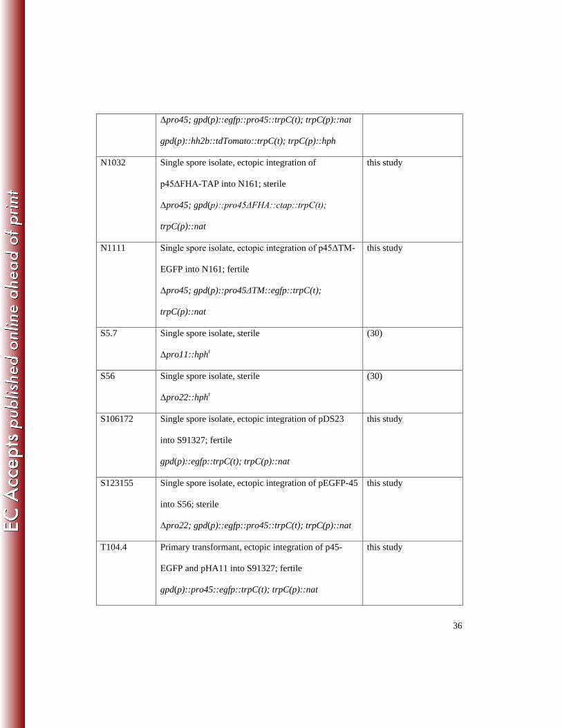
36
Δpro45; gpd(p)::egfp::pro45::trpC(t); trpC(p)::nat
gpd(p)::hh2b::tdTomato::trpC(t); trpC(p)::hph
N1032 Single spore isolate, ectopic integration of
p45ΔFHA-TAP into N161; sterile
Δpro45; gpd(p)::pro45ΔFHA::ctap::trpC(t);
trpC(p)::nat
this study
N1111 Single spore isolate, ectopic integration of p45ΔTM-
EGFP into N161; fertile
Δpro45; gpd(p)::pro45ΔTM::egfp::trpC(t);
trpC(p)::nat
this study
S5.7 Single spore isolate, sterile
Δpro11::hphr
(30)
S56 Single spore isolate, sterile
Δpro22::hphr
(30)
S106172 Single spore isolate, ectopic integration of pDS23
into S91327; fertile
gpd(p)::egfp::trpC(t); trpC(p)::nat
this study
S123155 Single spore isolate, ectopic integration of pEGFP-45
into S56; sterile
Δpro22; gpd(p)::egfp::pro45::trpC(t); trpC(p)::nat
this study
T104.4 Primary transformant, ectopic integration of p45-
EGFP and pHA11 into S91327; fertile
gpd(p)::pro45::egfp::trpC(t); trpC(p)::nat
this study

37
ccg1(p)::HA::pro11; trpC(p)::nat
T105.2 Primary transformant, ectopic integration of p45-
EGFP and pFLAGMob3 into S91327; fertile
gpd(p)::pro45::egfp::trpC(t); trpC(p)::nat
ccg1(p)::3xflag::Smmob3; trpC(p)::hph
this study
T1201-A Primary transformant, ectopic integration of pHA11
into S91327; fertile
ccg1(p)::ha::pro11; trpC(p)::nat
this study
T1202-A2 Primary transformant, ectopic integration of
pFLAGMob3 into S91327; fertile
ccg1(p)::3xflag::Smmob3; trpC(p)::hph
this study
T131-D5 Primary transformant, ectopic integration of p45-
EGFP into S91327; fertile
gpd(p)::pro45::egfp::trpC(t); trpC(p)::nat
this study
T133-E6 Primary transformant, ectopic integration of p45-
EGFP and pHA11 into S91327; fertile
gpd(p)::pro45::egfp::trpC(t); trpC(p)::nat
ccg1(p)::ha::pro11; trpC(p)::nat
this study
T135-B2 Primary transformant, ectopic integration of pEGFP-
45 into S5.7; sterile
Δpro11; gpd(p)::egfp::pro45::trpC(t); trpC(p)::nat
this study
ahph
r, hygromycin resistant; nat
r, nourseothricin resistant 829
bCulture collection, Lehrstuhl für Allgemeine und Molekulare Botanik, Ruhr-Universität 830
Bochum, Bochum, Germany 831

38
Table 2: Plasmids used in this study. 832
Plasmid Characteristics Reference or
source
p45-CTAP pro45 cDNA fused to a C-terminal TAP tag in
pDS22
gpd(p)::pro45::ctap::trpC(t), trpC(p)::nat
this study
p45-EGFP pro45 cDNA in pDS23
gpd(p):: pro45::egfp::trpC(t), trpC(p)::nat
this study
p45ΔFHA-TAP pro45 cDNA (Δbp682-855) fused to a C-terminal
TAP tag in pDS22
gpd(p)::pro45ΔFHA::ctap::trpC(t), trpC(p)::nat
this study
p45ΔTM-EGFP pro45 cDNA (Δbp2302-2370) in pDS23
gpd(p)::pro45ΔTM::egfp::trpC(t), trpC(p)::nat
this study
pDrivehph Hygromycin resistance cassette consisting of the
hph gene from Escherichia coli and the trpC
promoter from Aspergillus nidulans in pDrive
(42)
pDS16 hph gene with 5´ and 3´ flanking regions of
SMAC_01224 (pro45) in pRS426
pro45_5´::trpC(p)::hph::pro45_3´, trpC(p)::nat
this study
pDS22 C-terminal TAP tag in pRSnat
gpd(p)::ctap::trpC(t), trpc(p)::nat
Nowrousian,
unpublished data
pDS23 egfp in pRSnat
gpd(p)::egfp::trpC(t), trpc(p)::nat
(44)
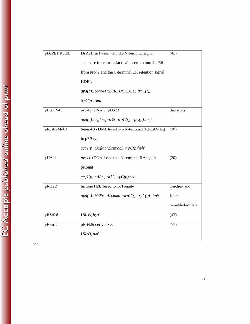
39
pDsREDKDEL DsRED in fusion with the N-terminal signal
sequence for co-translational insertion into the ER
from pro41 and the C-terminal ER retention signal
KDEL
gpd(p)::Spro41::DsRED::KDEL::trpC(t);
trpC(p)::nat
(41)
pEGFP-45 pro45 cDNA in pDS23
gpd(p):: egfp::pro45::trpC(t), trpC(p)::nat
this study
pFLAGMob3 Smmob3 cDNA fused to a N-terminal 3xFLAG tag
in pRShyg
ccg1(p)::3xflag::Smmob3, trpC(p)hphr
(30)
pHA11 pro11 cDNA fused to a N-terminal HA tag in
pRSnat
ccg1(p)::HA::pro11, trpC(p)::nat
(30)
pRH2B histone H2B fused to TdTomato
gpd(p)::hh2b::tdTomato::trpC(t); trpC(p)::hph
Teichert and
Kück,
unpublished data
pRS426 URA3, hygr
(43)
pRSnat pRS426 derivative;
URA3, natr
(77)
833

40
Table 3: Oligonucleotides used in this study. 834
Oligonucleotide Sequence (5´- 3´)
45_start-fw ATGACGGCTGTCGCGAATC
1224KOvp1neu GAGGTAAGAGCAAGCTTGTC
1224KOvp2neu GTACCAATGGCAAGATACGC
1224KOvp3 GACGACGACGAATTGGATCG
1757 AGCTGACATCGACACCAACG
45_CTAP-bw AAATTCTTTTTCCATCTTCTCTTTTCGTTCTTCGCCTGCGGCT
GCCACCC
45_CTAP-fw CTTCATCGCAGCTTGACTAACAGCTACATGACGGCTGTCGC
GAATCCC
45_CtermGFP-bw CAGCTCCTCGCCCTTGCTCACCATGTTCTTCGCCTGCGGCTG
CCACCC
45_int-bw GCTTGGCATTTCGCATTTCC
45_int-bw-2 GGTCGCTCGGTGATTCCTC
45_int-fw CCCTGTCACGACTGAACATATTG
45_int-fw-2 CGCCAGAAAATCGCGAGTC
45_int-fw-3 CAGCCGCAGGCGAAGAAC
45_NtermGFP-fw CTCTCGGCATGGACGAGCTGTACAAGATGACGGCTGTCGCG
AATCCCC
45ΔFHA_CTAP-
bw
CGTGGGGTTCCGACTCGCGATTTTCTGGCGAAATGTCGGGG
TAAAAGG
45ΔTM_NEGFPCE CTCACCATGTTCTTCGCCTGCGGCTGGGCCTGTATCAACGC

41
GFP-bw ACGATCCTGGTTAAGTGGATCTAGTTCTTCGCCTGCGGCTG
GGCCTGTATCAACGCACGATC
hph1MN CGATGGCTGTGTAGAAGTACTCGC
hph2_2010-bw GCCTCCAGAAGAAGATGTTG
hph2MN ATCCGCCTGGACGACTAAACCAA
SMACG_01224_3F
-rv
GCGGATAACAATTTCACACAGGAAACAGCCCGTAACTTTCC
ATACGTAAATACC
SMACG_01224_3F
-fw
GCCCAAAAATGCTCCTTCAATATCAGTTGCCAGTCTCTGTCT
TTCTCATACCACA
SMACG_01224_5F
-rv
CGAGGGCAAAGGAATAGGGTTCCGTTGAGGGTTCTCTTGAG
ATTGTTCCTTTGC
SMACG_01224_5F
-fw
GTAACGCCAGGGTTTTCCCAGTCACGACGATCCAGATCTTC
CATCTCAACAAG
835

42
Table 4: STRIPAK components detected by TAP-MS with PRO45 derivatives as bait. Numbers 836
give spectral counts; numbers in parentheses give peptide counts. 837
S. macrospora
Locus Tag
PRO45
MS1
PRO45
MS2
PRO45
MS3
PRO45
MS1-3
PRO45Δ
FHA
STRIPAK
componenta
SMAC_00877 3 (3) 16 (4) 3 (3) 22 8 (4) SmMOB3 / MOB-
3 / MOB3
SMAC_01224 18 (4) 206 (17) 10 (7) 234 48 (20) PRO45 / HAM-4 /
SLMAP
SMAC_08794 23 (9) 134 (19) 7 (7) 164 42 (16) PRO11 / HAM-3 /
striatin
a gives designations for S. macrospora / N. crassa / human proteins. 838


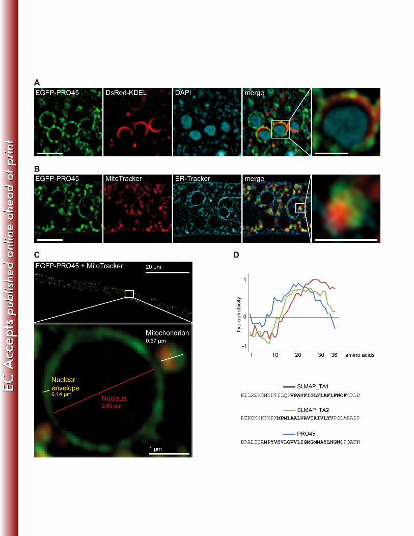

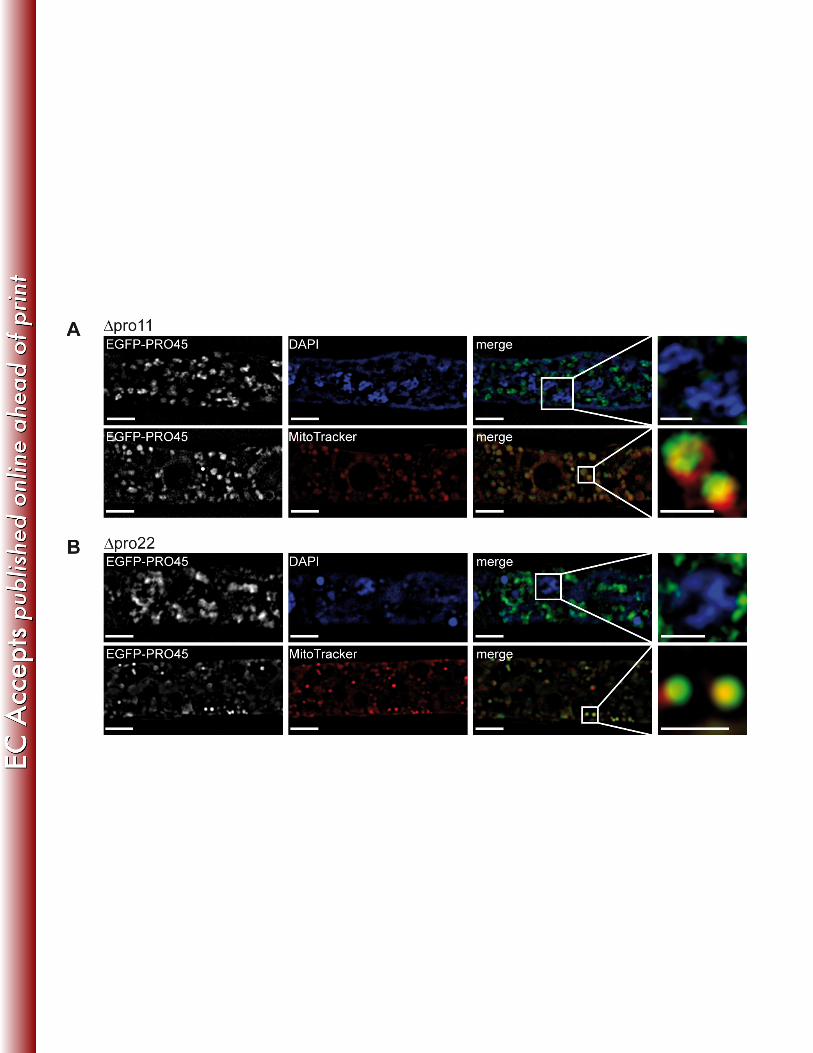
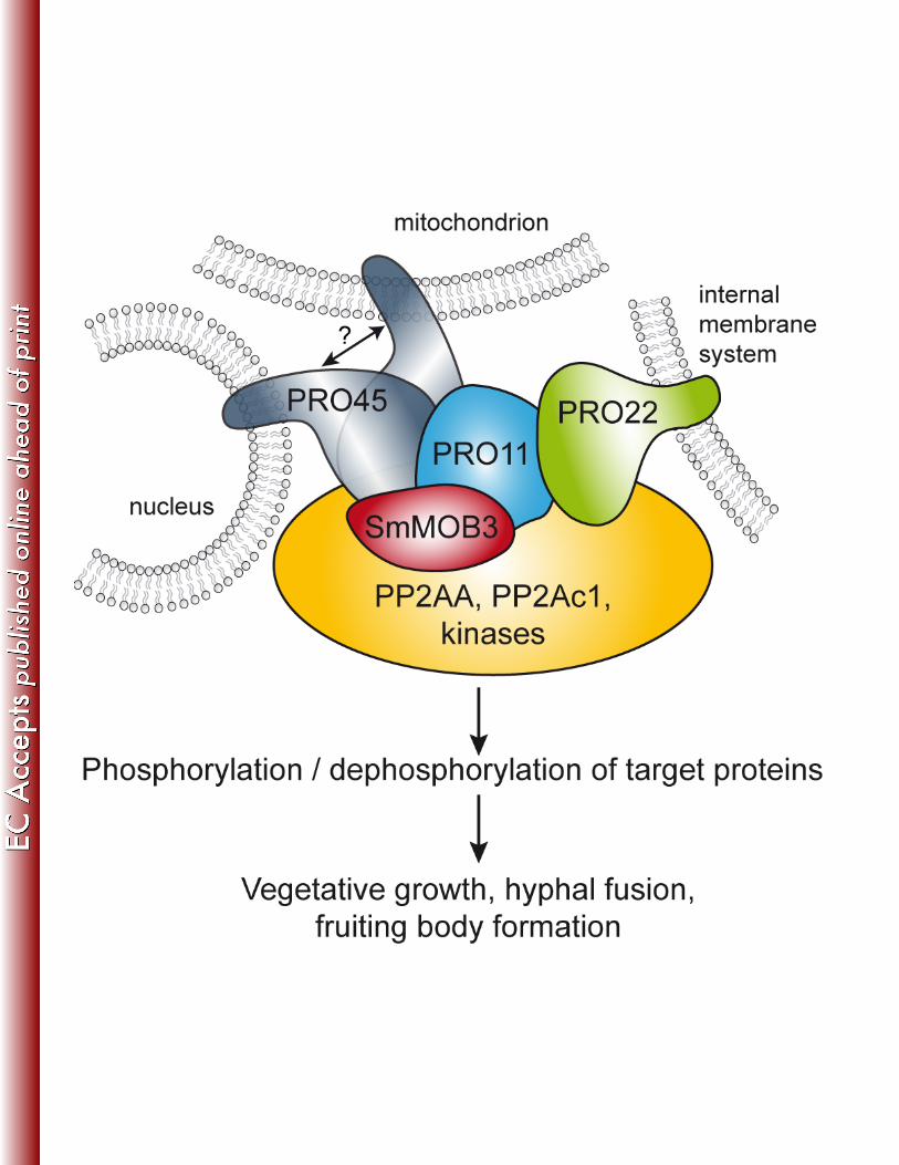
Copyright © 2022 FDOKUMEN