
Essential role of the chaperonin folding compartment in vivo

Monitoring the Systemic Human Memory B CellCompartment of Melanoma Patients for Anti-Tumor IgGAntibodiesAmy E. Gilbert1, Panagiotis Karagiannis1, Tihomir Dodev2, Alexander Koers3, Katie Lacy1, Debra H.
Josephs1, Pooja Takhar2, Jenny L. C. Geh4, Ciaran Healy4, Mark Harries5, Katharine M. Acland4, Sarah M.
Rudman6, Rebecca L. Beavil2, Philip J. Blower3, Andrew J. Beavil2, Hannah J. Gould2, James Spicer6,
Frank O. Nestle1*, Sophia N. Karagiannis1*
1 Cutaneous Medicine and Immunotherapy Unit, Division of Genetics and Molecular Medicine, NIHR Biomedical Research Centre at Guy’s and St. Thomas’s Hospitals and
King’s College London, King’s College London School of Medicine, St. John’s Institute of Dermatology, Guy’s Hospital, King’s College London, London, United Kingdom,
2 Randall Division of Cell and Molecular Biophysics and Division of Asthma, Allergy and Lung Biology, MRC and Asthma UK Centre for Allergic Mechanisms of Asthma,
King’s College London, London, United Kingdom, 3 Division of Imaging Sciences, King’s College London School of Medicine, Rayne Institute, St. Thomas’s Hospital, King’s
College London, London, United Kingdom, 4 Skin Tumour Unit, Guy’s and St. Thomas’s NHS Trust, St. John’s Institute of Dermatology, Guy’s Hospital, London, United
Kingdom, 5 Clinical Oncology, Guy’s and St. Thomas’s NHS Foundation Trust, London, United Kingdom, 6 Division of Cancer Studies, Department of Academic Oncology,
King’s College London, Guy’s Hospital, London, United Kingdom
Abstract
Melanoma, a potentially lethal skin cancer, is widely thought to be immunogenic in nature. While there has been muchfocus on T cell-mediated immune responses, limited knowledge exists on the role of mature B cells. We describe anapproach, including a cell-based ELISA, to evaluate mature IgG antibody responses to melanoma from human peripheralblood B cells. We observed a significant increase in antibody responses from melanoma patients (n = 10) to primary andmetastatic melanoma cells compared to healthy volunteers (n = 10) (P,0.0001). Interestingly, we detected a significantreduction in antibody responses to melanoma with advancing disease stage in our patient cohort (n = 21) (P,0.0001).Overall, 28% of melanoma patient-derived B cell cultures (n = 1,800) compared to 2% of cultures from healthy controls(n = 600) produced antibodies that recognized melanoma cells. Lastly, a patient-derived melanoma-specific monoclonalantibody was selected for further study. This antibody effectively killed melanoma cells in vitro via antibody-mediatedcellular cytotoxicity. These data demonstrate the presence of a mature systemic B cell response in melanoma patients,which is reduced with disease progression, adding to previous reports of tumor-reactive antibodies in patient sera, andsuggesting the merit of future work to elucidate the clinical relevance of activating humoral immune responses to cancer.
Citation: Gilbert AE, Karagiannis P, Dodev T, Koers A, Lacy K, et al. (2011) Monitoring the Systemic Human Memory B Cell Compartment of Melanoma Patients forAnti-Tumor IgG Antibodies. PLoS ONE 6(4): e19330. doi:10.1371/journal.pone.0019330
Editor: Shan Lu, University of Massachusetts Medical Center, United States of America
Received January 29, 2011; Accepted March 26, 2011; Published April 29, 2011
Copyright: � 2011 Gilbert et al. This is an open-access article distributed under the terms of the Creative Commons Attribution License, which permitsunrestricted use, distribution, and reproduction in any medium, provided the original author and source are credited.
Funding: This work was supported by the Department of Health via the National Institute for Health Research (NIHR) comprehensive Biomedical Research Centreaward to Guy’s, and St. Thomas’s NHS Foundation Trust in partnership with King’s College London and King’s College Hospital NHS Foundation Trust, UK (AEG, PK,TD, AK, KL, PT, FON, SNK) (http://www.guysandstthomas.nhs.uk/healthprof/researchanddevelopment/biomedicalresearch/biomedhome.aspx); CR UK/EPSRC/MRC/NIHR KCL/UCL Comprehensive Cancer Imaging Centre (C1519/A10331) (DHJ, PJB, JS, SNK) (http://www.cancerimagingcentre.org.uk/); Cancer Research UK(C30122/A11527) (DHJ) (http://www.cancerresearchuk.org/); Mary Dunhill Trust (FON) (http://www.dunhillmedical.org.uk/page_viewer.asp?page = welcome&pid = 8); CR UK/NIHR in England/DoH for Scotland, Wales and Northern Ireland Experimental Cancer Medicine Centre (FON) (http://www.ecmcnetwork.org.uk/network-centres/london-kcl/); and the Overseas Research Students Award Scheme (AEG) (http://www.orsas.ac.uk/). The funders had no role in study design, datacollection, analysis, decision to publish, or preparation of the manuscript.
Competing Interests: The authors have declared that no competing interests exist.
* E-mail: [email protected] (SNK); [email protected] (FON)
Introduction
Malignant melanoma, the most fatal form of skin cancer, arises
from malignantly-transformed melanocytes in the basal layer of
the epidermis. The incidence of melanoma has been increasing at
an accelerated rate in the past few decades amongst fair skinned
populations [1] and advanced forms of the disease are highly
resistant to treatment [2,3]. Thus, an urgent need exists for novel
therapies and earlier diagnosis.
Melanoma is widely thought to be immunogenic, supported by
clinical observations such as the frequency of spontaneous tumor
regressions, the prevalence of melanoma in immunosuppressed
patients, and the partial success of clinically-available immune
modulatory therapies such as the polyclonal immune activating
cytokines IFNa-2b and IL- 2 [4,5,6,7]. Host adaptive immune
responses have been described in melanoma with a main focus on
melanoma specific T cell responses [8,9], and supported by
successful case scenarios using immunotherapeutic strategies such
as dendritic cell vaccines, adoptive T cell therapies, and CTLA4
monoclonal antibodies [7,10,11,12,13].
Limited research has focused on B cells and the specificity of
antibodies they produce in cancer. Promotion of cancer
development by the creation of a pro-inflammatory environment
[14,15] and anti-tumor functions by activating mature T cell
PLoS ONE | www.plosone.org 1 April 2011 | Volume 6 | Issue 4 | e19330

responses [16] have been proposed as potential roles for B cells in
animal models of cancer. While there may be host immune
responses to malignancy following immunization [17], a variety of
mechanisms involved in tumor escape have been described and
understanding this complex relationship between immunosurveil-
lance and tumor escape in patients is key to the design of effective
immunotherapies [18,19,20,21].
Despite well-characterized tumor-induced immunomodulation,
immunotherapies such as monoclonal antibodies are emerging as
key diagnostic and therapeutic modalities and are now standard of
care for the treatment of various cancers. Antibodies for the
treatment of melanoma aimed at enhancing key pathways of T cell
activation (Cytotoxic T Lymphocyte-Associated Antigen 4, e.g.
Ipilimumab), targeting tumor vasculature (e.g. Bevacizumab), or
tumor-associated antigens (e.g. High Molecular Weight-Melano-
ma Associated Antigen, HMW-MAA) have demonstrated promise
in clinical studies [13,22,23,24]. Antibodies therefore represent an
attractive approach for the treatment of melanoma.
Reports of tumor-specific antibodies in the sera of melanoma
patients date back over forty years [25] and have so far provided
valuable insight into immune responses to cancer. Serological
studies of individuals with melanoma have shown that patients
expressing certain tumor-associated antigens have antibodies
against these antigens, conversely, patients without the antibodies
also lack the corresponding tumor antigens [26]. These studies
have been restricted to few antibodies in sera against known
tumor-associated antigens. Serological studies reported IgG
antibodies recognizing intracellular melanocyte and melanoma-
associated antigens such as tyrosinase, tyrosinase-related protein
(TRP)-1, TRP-2, and melanoma-associated glycoprotein antigen
family (gp100/pmel17) in patients with melanoma. Serum-
resident antibodies to some of these antigens were enhanced
following polyvalent melanoma cell vaccine immunization in
patients with melanoma, suggesting that melanoma-associated
antigens may be immunogenic and that humoral responses to
melanocyte and melanoma antigens may constitute potential
targets for immunotherapy [27]. New antigens, such as the NY-
ESO-1, with restricted expression in normal tissues and wide
distribution in various cancers including melanoma have been
discovered using serological analysis of recombinant cDNA
expression libraries (SEREX) techniques tested against tumor
mRNA and autologous patient sera [28]. SEREX studies from
human melanomas [29] and from one cell line [30] have led to the
discovery of the human testis antigen HOM-MEL-40. Many of
these antigens are primarily intracellular, making them less
attractive targets as monoclonal antibodies. Furthermore, serolog-
ical screens may also be limited by the temporal dynamics of sera
antibodies. Evaluating the reactivity of antibodies secreted by
circulating B cells may therefore provide additional insight to
serological evaluations by interrogating the long-term memory
anti-tumor systemic mature humoral response to cancer.
The production of tumor-specific antibodies in melanoma from
patient-derived B cells in the peripheral blood and tumors has
been reported and has yielded a few antibodies of the IgM and
IgG class [31,32,33,34]. In the past, such studies have been limited
by poor EBV transformation efficiency of human B cells, low
production of immunoglobulin, evaluation of few patients, and
lack of effective, reproducible methods to rapidly screen for tumor-
specific antibodies. To address some of these limitations, we took
advantage of recent advances in growing and immortalizing
memory B cells in culture [35,36], increased the number of
patients evaluated, and developed a novel screening tool to
specifically detect tumor-reactive antibodies against cell surface
antigens on melanoma cells. Our approach entails culture of
patient-derived circulating B cells and screening of the antibodies
they secrete for their reactivity and specificity to melanoma cells
versus melanocytes. Our strategy does not screen for antibodies
against known antigens or evaluate antibodies secreted or
sequestered in the serum at discrete times, but rather uniquely,
the aim here is to monitor tumor cell-reactive IgG antibodies
produced by B cell cultures, elucidating the breadth of the long-
term mature B cell repertoire recognizing melanoma antigens
expressed on the surface of cancer cells.
In this study, we screen for tumor-reactive and tumor-specific
IgG antibodies produced by patient and healthy individual B cell
cultures. This allowed characterization, beyond phenotype, of the
circulating B cell repertoire of individuals with melanoma and
clinical correlations of mature humoral responses and disease
progression. We also provide an example demonstrating that this
screen may facilitate the identification of antibodies able to target
cancer cells.
Results
Detection of Tumor-specific IgG Antibodies Using aNovel Cell-based ELISA
We developed and optimized a cell-based ELISA for specific
detection of tumor-reactive antibodies in order to obtain a robust
and optimized system for the detection of anti-tumor antibodies
from patients (Figure S1). We first evaluated the sensitivity and
specificity of an IgG antibody against a melanoma cell surface
antigen (HMW-MAA), expressed on A-375 melanoma cells using
immunocytochemistry (cytospins) and live cell flow cytometry. An
anti-HMW-MAA antibody was observed to bind to A-375 cells,
but not melanocytes over a range of concentrations as low as
20 ng/mL using both immunocytochemistry (Figure 1, A) and
flow cytometry (Figure 1, B). Next, we compared the detection of
this antibody bound to melanoma cells in our cell-based ELISA to
the above methods.
Utilizing our novel ELISA, we detected tumor-specific antibod-
ies at concentrations as low 10 ng/mL (Figure 1, C), demonstrat-
ing comparable sensitivity to flow cytometric or immunocyto-
chemical methods. Additionally, we validated our ability to
identify tumor-reactive antibodies from our patient cultures,
compared to equal amounts of non-specific IgG and culture
media (Figure 1, D). We also examined the potential applicability
of this method to identify tumor-specific antibodies in other
cancers using the mammary carcinoma cell line SK-BR-3, which
highly expresses the cell surface tumor-associated antigen HER2/
neu [37]. Trastuzumab (HerceptinTM), a humanized antibody
specific for HER2/neu, was specifically detected compared to an
equal amount of a control IgG employing our method (Figure 1,
D). Thus, we demonstrate that we can detect antibodies against
tumor cell antigens in a sensitive, specific and reproducible
manner.
Melanoma-reactive Antibodies are More Prevalent inMelanoma Patients than Healthy Volunteers
We first established B cell cultures from the peripheral blood of
melanoma patients to study antibody responses to cancer (Figure
S1). In agreement with a previously published report, we detected
a reduced memory B cell subset in melanoma patients. Melanoma
patient and healthy volunteer B cells were cultured with B cell
purity greater than 90%. Following EBV transformation and
activation with a TLR9 agonist, patient B cells were observed to
proliferate in culture for over eight weeks and 80% of the cells in
these cultures were IgG positive. B cell cultures derived from
healthy volunteers (n = 5) and melanoma patients (n = 5) had
Monitoring B Cell Memory in Melanoma
PLoS ONE | www.plosone.org 2 April 2011 | Volume 6 | Issue 4 | e19330

Figure 1. Development of a cell-based ELISA to detect tumor-specific antibodies. (A) Immunocytochemistry of cytospin preparationsdemonstrates that an antibody against the melanoma-associated antigen HMW-MAA, expressed on the surface of A-375 metastatic melanoma cells,can detect the antigen at 30 mg/mL (top left), 200 ng/mL (top middle) and 20 ng/mL (top right), while no binding to melanocytes was seen (bottomleft). IgG from 5% human serum (bottom right) did not bind to A-375 cells. (B) Flow cytometry analysis demonstrates specific binding of the HMW-MAA antibody to A-375 cells (left) at 30 mg/mL (dashed line), 200 ng/mL (dotted line) and 20 ng/mL (solid line), but not to melanocytes (30 mg/mLHMW-MAA antibody). Isotype control is shown in shaded grey histogram. (C) Detection of anti-HMW-MAA antibody binding to melanoma cells over arange of antibody concentrations compared to melanocytes by cell-based ELISA. (D) Melanoma-specific antibody HMW-MAA binding to A-375 cellscompared to an IgG isotype control or to culture media (left) utilizing the cell-based ELISA. Breast cancer-specific antibody Trastuzumab binding toSK_BR-3 cells compared to an IgG isotype control or to culture media (right). Error bars in figures represent 95% confidence intervals.doi:10.1371/journal.pone.0019330.g001
Monitoring B Cell Memory in Melanoma
PLoS ONE | www.plosone.org 3 April 2011 | Volume 6 | Issue 4 | e19330

comparable mean antibody titers after 18 days, ranging from 1 to
7 mg from each individual, with an overall mean of 2.5 mg (95%
CI = 2.3 to 2.7) per culture arising from 500 B cells per well. We
therefore established antibody-secreting cultures from melanoma
patients with comparable rates of IgG secretion to healthy
volunteers.
We next investigated whether specific antibody responses to
melanoma could be detected from circulating B cells of patients
and healthy volunteers utilizing our cell-based ELISA. Antibody-
secreting B cell cultures from 10 healthy volunteers and 10 patients
(n = 4 stage II, n = 4 stage III, and n = 2 stage IV) were evaluated
for reactivity to both metastatic and primary melanoma cells
relative to non-specific human IgG control (calculated as fold
increase above the non-specific human IgG control) employing
our cell-based ELISA. We found a significant (P,0.0001) increase
in the mean reactivity (fold increase) of patient-derived antibody
cultures (n = 600) to metastatic melanoma cells (2.5 fold increase,
95% CI = 2.4 to 2.6) compared to antibody cultures (n = 600)
derived from healthy volunteers (1.1 fold increase, 95% CI = 1.1
to 1.2) (Figure 2, A). A significant (P,0.0001) increase was also
seen in the mean reactivity of patient-derived antibodies to
primary melanoma cells (2.3 fold increase, 95% CI = 2.2 to 2.4)
compared to antibodies from healthy volunteers (1.0 fold increase,
95% CI = 1.0 to 1.1) (Figure 2, B). From this patient cohort, we
thus observed a significantly increased reactivity to primary and
metastatic melanoma cells, compared to healthy volunteers.
Antibody Response to Melanoma Decreases with DiseaseProgression
To examine if antibody responses differ according to disease
stage, we studied a cohort of 21 patients diagnosed with stage I, II,
III and IV melanoma (Table 1) and evaluated the reactivity of
antibody cultures (n = 1,800) from these patients to the metastatic
melanoma cell line A-375 utilizing the cell-based ELISA. This
patient cohort was almost exclusively Caucasian. Antibody
reactivity against melanoma cells was quantified relative to a
non-specific human IgG control and measured as fold increase
above this negative control. Patients with local (non-metastatic,
stages I and II) disease had a significantly (P,0.0001) higher mean
antibody response (2.6 fold increase, 95% CI = 2.4 to 2.8)
compared to those with confirmed metastatic disease (stages III
and IV, 1.7 fold increase, 95% CI = 1.7 to 1.8) (Figure 3, A). We
also found an overall significant reduction (P,0.0001) in the mean
reactivity of antibodies secreted in B cell cultures against
melanoma cells from stage II (2.8 fold increase, 95% CI = 2.6
to 3.0, n = 660) to stage III (1.9 fold increase, 95% CI = 1.8 to
2.0, n = 540) and to stage IV patients (1.5 fold increase, 95% CI
1.5 to 1.6, n = 480) (Figure 3, B). The stage I patient was not
evaluated since samples from only one patient (n = 120 B cell
cultures) from the cohort was available to include in this group.
The highest mean antibody reactivity against melanoma cells was
observed in Patients 5 and 6 diagnosed with stage II and III
melanoma, respectively (Table 1). However, we observed variation
in the antibody response among individual patients, with 19 out of
21 patients in our cohort having at least one antibody-producing
culture with optical density values 2.5-fold above the negative IgG
control (Table 1). These findings suggest that despite the
significant reduction in the proportion of tumor-reactive antibody
cultures as a function of disease progression, patients from each of
stage groups had B cells with antibodies that recognized tumor
cells.
Estimations of Melanoma-reactive Antibody Frequenciesfrom Patient Peripheral Blood B Cell Cultures
We screened for tumor-reactive antibodies from patient B cell
cultures and approximated frequency and specificity of selected
Figure 2. The reactivity of antibodies derived from melanoma patient B cells to primary and metastatic melanoma cells iscompared to healthy volunteers. B cell cultures from melanoma patients secreted antibodies that were significantly more reactive againstmelanoma cell lines compared to healthy volunteers using the cell-based ELISA. Mean reactivity of antibody cultures (n = 600) from 10 melanomapatients to A-375 metastatic melanoma cells (A) and WM-115 primary melanoma cells (B), was compared to 10 healthy volunteer cultures (n = 600)(P ,0.0001). Fold increase values represent the optical density of each B cell culture relative to the mean optical density of a negative control humanIgG antibody. *** = P,0.001. Error bars in figures represent 95% confidence intervals.doi:10.1371/journal.pone.0019330.g002
Monitoring B Cell Memory in Melanoma
PLoS ONE | www.plosone.org 4 April 2011 | Volume 6 | Issue 4 | e19330

cultures to tumor cells. For this we screened B cell culture
supernatants against a stringent comparator using a positive
control monoclonal antibody, Trastuzumab, that recognizes the
HER2/neu tumor antigen, expressed on breast cancer cells and on
some melanoma cells [38]. Trastuzumab was selected as a positive
control because of comparable binding across melanoma cell lines
and melanocytes, as shown by mean fluorescence intensities of
antibody binding against a range of these cells (Figure 4, A).
Previous studies screening for tumor-specific antibodies have
selected wells greater than the mean negative control optical
density (OD) + three standard deviations as criteria for positive
tumor-reactive antibodies [39]. Due to the inherent variability of
cell-based assays, and the potential identification of false positive
cultures, we chose more stringent criteria for antibody screening,
by comparing antibodies produced by B cells to a positive control
antibody (. 75% OD of positive control). Based on this antibody
selection criteria (.75% OD of positive control), we estimate that
28% of B cell cultures (n = 1,800) derived from 21 patients, each
arising from 500 B cells, produced antibodies that recognized
metastatic melanoma cells, compared to 2% (n = 600) of cultures
derived from 10 healthy volunteers (Figure 4, B). From these 10
healthy volunteers, 2 individuals had one reactive culture (out of
60 cultures), 1 individual had 10 reactive cultures, and the rest of
the cohort had no reactive cultures.
From our patient cohort, we can roughly estimate the frequency
of B cells that produce an antibody that recognizes melanoma cells
under the assumption that a reactive antibody culture, defined as
having an OD .75% of the positive control, arises from only a
single B cell (1 out of 500 plated per culture). By dividing the total
number positive antibody cultures from the patient cohort by the
total number of B cells evaluated from the patient cohort by the
number of positive antibody cultures, we roughly approximate that
from our patient cohort one out of 1,765 B cells produce an
antibody that may recognize melanoma cells.
To estimate the frequency of melanoma-reactive antibody-
producing B cells in melanoma patients we performed limiting
dilution analysis. We selected a stage II patient, who, we predict,
may have a high antibody response to melanoma, based on our
findings that the antibody responses were highest in this group
(Figure 3, B). For this stage II patient (Patient 15, see Table 1)
from our limiting dilution analysis assays using the cell-based
ELISA, we estimate that one out of 1,790 peripheral blood B cells
produces antibodies that bind to A-375 melanoma cells (Figure 4,
C). For this same patient, the frequency of B cells producing
antibodies that react with melanocytes was also evaluated at the
same B cell densities as melanoma cells. We did not observe a
comparable patient antibody response to melanocytes as we did
to melanoma cells, suggesting a much lower frequency of
Table 1. Reactivity of patient-derived antibody-producing B cell cultures to A-375 metastatic melanoma cells.
Stage Patient ID* Age Sex EthnicityMean fold increase overnegative control{ 95% CI of mean
Maximum fold increaseover negative control
% Mean reactivecultures{{
I 9 51 F Caucasian 1.1 1 to 1 2.8 2
II 4 75 M Caucasian 2.7 2.6 to 3 4.2 38
II 5 70 M Caucasian 6.1 5.4 to 7 17 63
II 7 69 M Caucasian 1.6 1.4 to 2 4.8 3
II 15 49 F Caucasian 2.6 2.5 to 3 6.2 22
II 1 66 F Caucasian 1.5 1.4 to 2 6.8 5
II 19 63 F Caucasian 1.6 1.5 to 2 3.5 82
II 21 38 M Caucasian 1.3 1.1 to 2 7.7 2
II 20 81 M Caucasian 2 1.9 to 2 3.7 50
2.4 2.2 to 3 6.7 33
III 6 67 M Caucasian 3.2 3 to 3 5.8 14
III 10 54 M Caucasian 1.8 1.6 to 2 3.6 13
III 16 77 M Caucasian 1.8 1.6 to 2 3.8 97
III 17 88 M Caucasian 1.8 1.7 to 2 2.8 40
III 18 68 M Caucasian 1.3 1.2 to 1 2.8 6
III 8 23 M Asian 1 0.8 to 1 2.9 8
1.8 1.7 to 2 3.6 30
IV 11 77 F Caucasian 1.7 1.5 to 2 3.3 92
IV 12 72 F Caucasian 1 0.8 to 1 3.5 10
IV 2 66 M Caucasian 1.8 1.7 to 2 3.1 19
IV 13 55 F Caucasian 1.5 1.4 to 2 2.6 2
IV 3 51 M Caucasian 1.8 1.7 to 2 2.7 8
IV 14 31 F Caucasian 0.7 0.6 to 1 1.3 12
Mean1.4 1.3 to 1.5 24
*Patient ID corresponds to patient number in all figures.{Fold increases values were calculated by dividing the optical density of B cell culture supernatants by the optical density of a non-specific IgG negative control using acell-based ELISA.{{% of cultures with absorbance values greater than 75% of a positive control antibody using a cell-based ELISA.doi:10.1371/journal.pone.0019330.t001
Monitoring B Cell Memory in Melanoma
PLoS ONE | www.plosone.org 5 April 2011 | Volume 6 | Issue 4 | e19330

antibodies that bind to normal cells of the same origin (Figure 4,
C left).
Limiting dilution analysis against two additional metastatic (SK-
MEL-28, A-2058) and one primary (WM-115) melanoma cell lines
for the same patient yielded different but comparable frequencies
to A-375 for the metastatic cell lines (SK-MEL-28, 1 out of 1,650
B cells; A-2058, 1 out of 1,170 cells), and a much lower frequency
of antibodies that bind to the primary melanoma line WM-115
which was similar to that observed with primary melanocytes
(Figure 4, C right). For this patient, the data suggest detectable
circulating B cell humoral response frequency against metastatic
melanoma cells and lower frequency for normal human
melanocytes or primary melanoma cells. To further confirm the
frequency observations for the patient-derived circulating B cell
repertoire, we performed additional limiting dilution assays for
another stage II patient (Patient 21, Table 1). For Patient 21, we
estimate 1 out of 2,430 B cells that produces antibodies bind to the
same melanoma cell line tested for Patient 15 (Figure 4, D),
suggesting lower but comparable frequency to those estimated for
B cells from Patient 15. In summary, applying the above
methodology, these results suggest that tumor-reactive antibodies
from circulating B cells are more frequent in melanoma patients
than healthy volunteers and more frequent against a range of
metastatic melanoma cells compared to normal melanocytes.
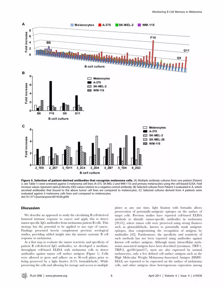
Screening for Tumor-specific Antibodies and Selection ofa Patient-derived Monoclonal Antibody with In VitroCytotoxicity against Melanoma Cells
We then selected patient-derived, tumor-specific antibodies in
order to further evaluate their reactivity to melanoma cells, and
conducted a preliminary assessment of the potential functional
capabilities of a patient-derived antibody from this screen. B cell
culture wells were selected based on stringent criteria (OD . 75%
positive control antibody), using the cell-based ELISA. Tumor
specificity of antibody cultures was evaluated by comparing
binding of antibodies from these cultures against multiple
melanoma cells (A-375, SK-MEL-2, WM-115) versus normal cells
(Figure 5, A). We observed multiple antibody cultures with a
higher degree of binding to some melanoma cells compared to
melanocytes from the same patient (Patient 3, Figure 5, A; a
selection of five of these cultures is shown on Figure 5, B). Similar
results were obtained when we screened for tumor-specific cultures
from different patients against melanoma cells and melanocytes.
Positive cultures with different binding patterns against four
melanoma cell lines (A-375, SK-MEL-2, SK-MEL-28, WM-115)
and primary human melanocytes were detected (selected cultures
derived from Patients 2, 3, 4 and 6 are shown as examples in
Figure 5, C), reflecting specificity and reactivity of different
antibodies to a range of antigens expressed at different levels in a
number of melanoma cell lines, and some reactivity to antigens
lowly expressed on human melanocytes. Selection of a tumor-
positive antibody culture for sub-cloning and limiting dilution was
based on degree of reactivity to melanoma cells relative to
melanocytes (Figure 5, C).
One B cell culture from Patient 6 was selected for further
evaluation since cell culture supernatants were observed to have a
higher degree of binding to A-375 and SK-MEL-28 cells
compared to melanocytes by ELISA (Figure 5, C; right). After
limiting dilution of this melanoma-reactive B cell culture, a
monoclonal antibody (6_2G3) was further assessed for specificity
to 6 melanoma cell lines, melanocytes and fibroblasts by live cell
flow cytometry (Figure 6). Since more antibody was available after
monoclonal dilution, 2 additional melanoma cell lines along with
dermal fibroblasts were evaluated. In concordance with the cell-
based ELISA findings (Figure 5, C), the 6_2G3 clone bound to a
range of melanoma cell lines, but not to melanocytes (Figure 6).
The antibody had no reactivity against primary human dermal
Figure 3. Prevalence of melanoma-reactive antibodies derived from melanoma patient B cell cultures is reduced in advanceddisease stages. (A) Comparison of mean antibody culture reactivity to A-375 cells (fold increase relative to negative control human IgG antibody)for patients with localized (non-metastatic, n = 9) and metastatic (n = 12) disease, P,0.0001. (B) Mean reactivity to A-375 cells (fold increase relative tonegative control antibody) of cultures from patients with stage II (n = 8), III (n = 6) and IV (n = 6) melanoma. Antibody reactivity was determined usinga cell-based ELISA. Fold increase values in panels A and B were determined relative to the mean absorbance to a negative control human IgGantibody. ***P,0.001 and ** P = 0.001 to 0.01. Error bars in figures represent 95% confidence intervals.doi:10.1371/journal.pone.0019330.g003
Monitoring B Cell Memory in Melanoma
PLoS ONE | www.plosone.org 6 April 2011 | Volume 6 | Issue 4 | e19330

fibroblasts. In summary, by evaluating the specificity of antibodies
to melanoma cells versus melanocytes and fibroblasts we could
identify a melanoma-specific monoclonal antibody clone 6_2G3.
While we had limited amounts of monoclonal antibodies our B cell
culture supernatants after evaluating melanoma-cell specificity, we
were able to conduct a limited functional investigation of this
antibody.
Using clone 6_2G3, we wished to assess whether a patient-
derived antibody has potential cytotoxic activity against tumor
cells. We tested the tumor cell killing potential of this antibody
using a real-time live-dead cell cytotoxicity assay using as targets
metastatic melanoma cells recognized by this clone (Figure 7 &
Supporting Videos S1 and S2). In these experiments, U-937
human monocytic cells which express Fcc receptors served as
effector cells [40] and A-375 melanoma cells were used as target
cells to evaluate antibody-dependent cellular cytotoxicity (ADCC)
of tumor cells mediated by patient-derived IgG antibodies. We
tested two monoclonal antibodies, both derived from Patient 6
(Table 1): (1) the 6_2G3 antibody, which bound to A-375 cells and
not melanocytes and (2) the 6_2D10 antibody, which did not bind
to A-375 cells or melanocytes in the cell based ELISA prior to
limiting dilution, which served as a non-tumor-reactive control
(Figure 7).
After 2 hours in culture, 18% (95% CI = -5 to 41%) of tumor
cells given the melanoma-specific antibody were viable, compared
to 95% (95% CI = 86 to 104%) of the tumor cells given the non-
melanoma specific antibody (P,0.0001) (Figure 7, A, left).
Relative to tumor cell fluorescence at the start of the assay, mean
green/live tumor cell fluorescent intensity was reduced to 64% for
the non-tumor specific antibody (6_2D10) compared to 18% for
the tumor-specific antibody (6_2G3) (Figure 7, A, right). These
results highlight the potential of a patient derived tumor-specific
antibody to kill tumor cells by antibody-dependent cell cytotoxicity
(Figure 7, B and see Videos S1 and S2). For tumor cells treated
with the tumor-specific 6_2G3 antibody, we also observed a
significant (P = 0.0002) reduction in the movement of monocytic
effector cells in contact with tumor (13 mm, 95% CI = 10 to 17
mm) compared to effector cells not in contact with tumor (26 mm,
95% CI = 21 to 31 mm) (Figure 7, C and D). Using the 6_2D10
non-specific antibody, no significant (P = 0.3) difference was
observed for the movement of effector cells not in contact with
tumor cells (20 mm, 95% CI = 15 to 25 mm) compared to those in
contact with tumor cells (25 mm, 95% CI = 18 to 31). With this
example, we demonstrate that a patient-derived tumor-specific
antibody is capable of engaging immune effector cells in antibody-
dependent cellular cytotoxicity against tumor cells. Taken
together, these data suggest that systemic melanoma-specific
mature B cell responses may be present in patients with melanoma
and may harbor the potential to be activated against cancer
cells.
Figure 4. Estimations of the frequency of circulating B cells producing melanoma-reactive antibodies relative to a positive controlantibody. (A) Binding of Trastuzumab is consistent across melanoma cell lines and primary human melanocytes evaluated by mean fluorescenceintensity (MFI) above isotype control antibody binding for each cell line. (B) Proportion of B cell cultures (n = 1,800) from 21 melanoma patients arisingfrom 500 B cells each that produced antibodies that reacted to metastatic melanoma cells compared to cultures (n = 600) from 10 healthy volunteers.(C) Frequency of B cells producing IgG antibodies able to bind to melanoma cells estimated by limiting dilution analysis for Patient 15 (see Table 1).The frequency of B cells producing antibodies reactive to the cells of interest was approximated according to Poisson distribution, the number of Bcells at which 37% of the cultures were non-reactive (dotted horizontal line). Frequencies of tumor-reactive B cells against A-375 melanoma cellsversus melanocytes evaluated at the same B cell densities (left) and against four melanoma cell lines (primary WM-115, and metastatic cell linesderived from different anatomic locations, right). (D) Comparison of the frequency of B cells that react to metastatic melanoma cells between twostage II patients, estimated by limiting dilution analysis. For these two patients, frequency was estimated to be 1 in 1,790 B cells (Patient 15) and 1 in2,430 B cells (Patient 21).doi:10.1371/journal.pone.0019330.g004
Monitoring B Cell Memory in Melanoma
PLoS ONE | www.plosone.org 7 April 2011 | Volume 6 | Issue 4 | e19330
Discussion
We describe an approach to study the circulating B cell-derived
humoral immune response to cancer and apply this to detect
tumor-specific IgG antibodies from melanoma patient B cells. This
strategy has the potential to be applied to any type of cancer.
Findings presented herein complement previous serological
studies, providing added insight into the mature systemic B cell
response to melanoma.
As a first step to evaluate the tumor reactivity and specificity of
patient B cell-derived IgG antibodies, we developed a medium-
throughput cell-based ELISA with melanoma cells to detect
antibodies against tumor cell surface antigens (Figure 1). Cells
were allowed to grow and adhere on to 96-well plates prior to
being preserved by a light fixative (0.5% formaldehyde). While
preserving the cells and allowing for storage and access to multiple
plates at any one time, light fixation with formalin allows
preservation of potentially-antigenic epitopes on the surface of
target cells. Previous studies have reported cell-based ELISA
methods to identify tumor-specific antibodies in melanoma
[39,41], where tumor cells were preserved using strong fixatives
such as glutaraldehyde, known to potentially mask antigenic
epitopes, thus compromising the recognition of antigens by
antibodies [42]. Furthermore, the specificity and sensitivity of
such methods has not been reported using antibodies against
known cell surface antigens. Although many intracellular mela-
noma associated antigens have been described (tyrosinase, TRP-1,
TRP-2, gp100/pmel17), most are also expressed by normal
melanocytes, only a few defined cell surface antigens such as the
High Molecular Weight Melanoma-Associated Antigen (HMW-
MAA) are reported to be expressed on the surface of melanoma
cells, and other antigens show heterogeneous expression among
Figure 5. Selection of patient-derived antibodies that recognize melanoma cells. (A) Multiple antibody cultures from one patient (Patient3, see Table 1) were screened against 3 melanoma cell lines (A-375, SK-MEL-2 and WM-115) and primary melanocytes using the cell-based ELISA. Foldincrease values represent optical density (OD) values relative to a negative control antibody. (B) Selected cultures from Patient 3 evaluated in A, whichsecreted antibodies that bound to the above tumor cell lines are compared to melanocytes. (C) Selected cultures derived from 4 patients wereevaluated against 4 melanoma cells lines and compared to melanocytes.doi:10.1371/journal.pone.0019330.g005
Monitoring B Cell Memory in Melanoma
PLoS ONE | www.plosone.org 8 April 2011 | Volume 6 | Issue 4 | e19330

patients [43,44]. Thus, this ELISA constitutes an attractive tool to
evaluate broad responses to any naturally-expressed antigens on
the surface of melanoma cells and melanocytes in this context.
This screening methodology has additional potential advantages.
Unlike assays screening against a single recombinant antigen or
antigenic epitope, our method enables the evaluation of antibody
repertoires of patients against a multitude of cell surface antigens
in their native confirmation on the surface of both primary and
metastatic melanoma cells and also melanocytes, providing more
comprehensive information on the broad prevalence of tumor-
reactive and tumor-specific antibodies. Previous studies have
shown concordance of cell line-associated antigens with antigens
expressed on corresponding tumors, making them a suitable
platform for tumor-reactive antibody screening [26,45]. Thus, cell
lines provide a promising alternative source of multiple tumor
antigens in the absence of multiple well-defined, highly expressed,
and readily available recombinant antigens. Unlike flow cytomet-
ric evaluations, the cell-based ELISA does not require the use of
proteolytic enzymes such as trypsin, therefore better preserving
cell surface antigens. Plates of target cells can be prepared, fixed
and frozen in batches, thus allowing for higher throughput
screening for tumor cell-reactive antibodies. It can be applied to
evaluate . 300 culture supernatants against cell lines within a few
hours. In principle, numerous ELISA plates for screening a range
of cell lines with multiple supernatant samples can be processed
simultaneously. Additionally, this methodology may be a potential
tool for immunomonitoring tumor-specific humoral responses to
therapies; selecting patients most likely to benefit from immuno-
therapy; or as a prognostic factor in linking tumor-reactive
humoral responses to clinical outcomes. This assay may also be
utilized to detect surface antigens in a range of cell types, and thus
may be adapted to monitor the B cell-derived antibody repertoire
in different disease contexts.
In agreement with a recent report [46], we also observed a
reduction in the peripheral blood memory B cell compartment of
metastatic melanoma patients. We measured a reduction of the
CD27+ subset of memory B cells in patients with both metastatic
and non-metastatic melanoma compared to healthy volunteers
(Figure S2). Despite the reduction of circulating memory B cells in
our cohort, patient-derived B cells were capable of secreting high
amounts of IgG antibodies when activated in vitro with a TLR 9
agonist, with comparable antibody production to B cells from
healthy individuals, and a high percentage of patient- and healthy
volunteer-derived B cells expressed IgG antibodies within a few
days in culture (80% of B cells from three patients with melanoma,
Figure S2). Thus, while a reduced memory B compartment has
been reported in cancer patients, we show that a melanoma-
reactive portion of this compartment remains in our patient
cohort.
We demonstrated a high prevalence of melanoma patient-
derived antibodies produced by circulating B cells in cancer
patients that recognize melanoma cell lines (Figure 2). We
observed that B cell culture supernatants from different patients
displayed differential binding to each cell line, which reflects
specificity and reactivity of different antibodies to a range of
antigens expressed at different levels in a number of melanoma cell
lines; these may also reflect binding to some antigens lowly
expressed on human melanocytes (Figure 4 and Table 1).
Melanoma patients had a high percentage of melanoma-reactive
antibody-producing B cell cultures, significantly higher than those
from healthy volunteer-derived B cell cultures (Figure 2), with 28%
of melanoma patient-derived B cell cultures recognizing melano-
ma cells, compared to 2% of cultures from healthy volunteers
(Figure 4). Limiting dilution analyses of reactivity against melanoma
cells versus normal melanocytes provided further evidence in
support of the presence and frequency of tumor-reactive B cells in
Figure 6. Selection of a patient derived B antibody that recognizes melanoma cells but not melanocytes. A selected tumor-reactiveculture from a stage III patient (Patient 6) which secreted antibodies that bound to tumor cell lines and compared to melanocytes by ELISA (Figure 5,C) was sub-cloned, and a monoclonal antibody 6_2G3 was selected and evaluated on live cells by flow cytometry (solid black line histograms) forreactivity to fibroblasts, melanocytes, and 6 melanoma cell lines. IgG isotype controls are shown in shaded grey histograms.doi:10.1371/journal.pone.0019330.g006
Monitoring B Cell Memory in Melanoma
PLoS ONE | www.plosone.org 9 April 2011 | Volume 6 | Issue 4 | e19330

Figure 7. A tumor-specific antibody derived from a patient with melanoma is able to induce tumor cell cytotoxicity. Two monoclonalantibodies were evaluated in vitro using a live cell imaging assay: 6_2G3 clone bound to A-375 cells compared to melanocytes; and 6_2D10, a clonealso from Patient 6 did not, and served as a negative control antibody for these experiments. (A) Cell viability of A-375 melanoma cells incubated with
Monitoring B Cell Memory in Melanoma
PLoS ONE | www.plosone.org 10 April 2011 | Volume 6 | Issue 4 | e19330

patient blood (Figure 4). For one stage II patient evaluated, the data
indicate that metastatic melanoma cells are recognized by a higher
proportion of B cells (estimated on average , 1 in 2,000 mature B
cells) compared to primary melanoma cells or melanocytes,
although in a cohort of 10 patients we measured reactivity of B
cell cultures to both metastatic and primary melanoma cell lines
(Figure 2). Taking into consideration the expected variability in
immune responses among patients, and the array of tumor antigens
these patients may be exposed to, the observations that B cells from
two patients with stage II melanoma yielded comparable reactivity
to metastatic melanoma cells (estimated 1 in 1,790 for Patient 15,
and 1 in 2,430 B cells for Patient 21) indicate the presence and
support the prevalence of a circulating melanoma-reactive B cell
compartment.
While tumor-reactive antibodies were detected from most
melanoma patients studied, antibody responses derived from
circulating B cells against melanoma cells decreased with more
advanced disease stages (Figure 3). Previous serological studies
report serum-resident antibodies against tumor cells in melano-
ma patients, with some evidence that serum antibodies are
diminished in patients with advanced disease [25,47]. It was
unclear whether this was a consequence of the sequestering of
antibodies into tumors with increasing tumor burden in these
patients. Our findings provide further insight by demonstrating
the presence of a circulating long-term mature B cell response to
cancer at all disease stages, against a broad range of naturally
expressed antigens on the surface of primary and metastatic
tumor cells. We also report decreased frequency of tumor-
reactive antibody-producing B cells with advanced disease, thus
supporting the premise that mechanisms of immune tolerance
rather than adsorption of antibodies into tumors in advanced
disease setting may also explain these reductions. One limitation
may arise from screening for antibodies against mostly metastatic
melanoma cells. It is possible that our observations may reflect
reactivity to antigens present in primary disease, which may be
preserved or upregulated in advanced disease setting. While our
findings may not account for reactivity to tumor antigens that
are lost with disease progression, this reduced reactivity to
melanoma we observed may imply weakened immune responses
to a subset of antigens on the surface of melanoma cells. Another
explanation for these observations may be that with advanced
disease, mature circulating B cells home into increasing tumor
sites, thus reducing the circulating tumor-reactive B cell
compartment in these patients. Future studies aimed at
monitoring local B cell responses in tumors may provide further
clues into the dynamics of mature B cell responses at the systemic
and local levels in cancer. Thus, despite well-known weakened
host immune response with disease progression [48], we were
able to detect melanoma-reactive antibodies from patient
circulating B cells, implying that although mature humoral
immune responses are weakened, responses in the form of
mature memory B cells may persist. However, further work
elucidating potential immunomodulatory roles of B cells and
other immune cells in cancer, including the production of IL-10
by B cell population subsets [49], merits consideration.
Although we report the presence of anti-tumor antibodies
produced by patient memory B cells, and these cells were
stimulated ex vivo to secrete antibodies, it is not clear whether
tumor antigen-reactive B cells are activated in patients to secrete
antibodies or whether these humoral responses are capable of
exerting any beneficial anti-tumoral activities in the same patients
in vivo. In the 21 patient cohort at different disease stages in this
study, we were not able to draw any conclusions regarding the
relationship between tumor cell reactivity and clinical disease
progression in the short term (6 months to 2 year follow up) or
associations with any particular disease treatment regimes.
However, monitoring mature memory B cells and their antibody
repertoires together with clinical outcomes in patients over a long
period of time may help identify any correlations between
melanoma-reactive mature memory B cell responses and disease
progression. Additionally, future studies may help identify
particular components of the humoral response which may hold
clinical relevance, and elucidate the potential merits of monitoring
these responses in relation to therapies, or of evaluating humoral
responses as a prognostic factor to clinical outcomes.
An important question therefore relates to whether patient-
derived mature B cell responses have any functional capability to
potently activate immune effector cells against cancer. For this, we
measured the capacity of one antibody clone to kill tumor cells.
Antibody clone 6_2G3 derived from a patient with stage III
disease (Patient 6, Table 1) was not observed to bind to fibroblasts
or melanocytes, but bound to a proportion of melanoma cell lines
tested (Figure 5). Antibodies against tumor-associated antigens can
attack tumor cells via a number of mechanisms including
induction of apoptosis in tumor cells and engaging Fc receptors
on immune cells [50,51,52,53]. Antibodies approved for the
treatment of cancer have been shown to function through one or
more of these mechanisms [54,55]. While our strategy yields fully
human monoclonal antibodies in a matter of a few months, we
were limited in the amount of antibody we could produce from the
B cells to perform functional studies and evaluate reactivity to
patient-derived melanoma tumors. However, we had sufficient
quantity to evaluate whether a patient-derived melanoma tumor-
specific monoclonal antibody could mediate antibody dependent
cellular cytotoxicity (ADCC) in the presence of monocytic effector
cells and tumor cells using a real-time live cell imaging assay. We
show that the tumor-specific 6_2G3 clone is capable of mediating
ADCC in vitro and additionally measured the restricted movement
of monocytic effector cells once in contact with tumor-specific
antibody-coated tumor cells, providing further evidence of ADCC
(Figure 6). These preliminary assessments provide a promising clue
that a potentially active mature B cell response against melanoma
may be present in patients. An example of this possibility was
recently reported by Yuan et al. who demonstrated that
administration of the anti-CTLA-4 antibody ipilimumab led to
serological enhancement of antibodies to the testis antigen NY
human U-937 monocytic cells was compared between samples treated with 6_2G3 or 6_2D10 antibody after 2 hours at 37uC (*** = P,0.001) (left).Error bars represent 95% confidence intervals. Mean fluorescence intensity of A-375 tumor cells pre-labeled with the live cell dye Calcein AM, andincubated with U-937 cells and antibody 6_2G3 or 6_2D10 was measured at 0 and 120 min time points (right). (B) Fluorescent images of the live cellcytotoxicity assays at 30 minute intervals. Live Calcein AM-labeled melanoma tumor cells (green) were incubated with 6_2G3 or 6_2D10 antibody andU-937 cells (blue) and cell death was evaluated (red). Incorporation of Ethidium homodimer-1 (incorporation of red into tumor cells) was observedwith 6_2G3 but not 6_2D10 (magnification 20x, Scale bar: 100 mm). (C) Movement of U-937 cells tracked and measured over two hours was comparedfor cells in contact and those not in contact with tumor cells (*** = P,0.001for 6_2G3 and P = 0.3 for 6_2D10 antibody) (upper panel). (D) Images ofU-937 movement in tumor cell cultures treated with 6_2G3, tracked for cells in contact (left) and cells not in contact with tumor cells (right).Movement is indicated by tracking lines (red to yellow) from the original position of U-937 cells at t = 0 to t = 2 hours (magnification 20x, Scale bar:50 mm).doi:10.1371/journal.pone.0019330.g007
Monitoring B Cell Memory in Melanoma
PLoS ONE | www.plosone.org 11 April 2011 | Volume 6 | Issue 4 | e19330

-ESO-1 in patients who responded to the antibody therapy [56]. It
is therefore conceivable that the mature B cell compartment could
be enhanced with immunotherapeutic approaches, and that
monitoring humoral responses to therapeutics may have clinical
relevance.
Harnessing the cancer-specific antibody repertoire of cancer
patients using the methodology described herein may also
potentially offer an alternate strategy to yield IgG antibodies
against cancer antigens. Recent advances reported by Traggiai
et al., evaluating monoclonal antibodies from human memory B
cells have yielded fully-human virus-neutralizing antibodies of
therapeutic relevance for infectious diseases and have contributed
to the dissection of humoral memory responses to vaccinations
[35,57,58]. Here, we focus on B cells from cancer patients such as
melanoma patients, analyze systemic humoral responses to cancer
and demonstrate the presence of tumor-reactive and tumor-
specific antibodies. This approach may offer an advantage over
other approaches such as phage display in that it yields in vivo
affinity-matured human antibodies with naturally paired heavy
and light chains. The patient-derived monoclonal antibody 6_2G3
bound to 2 out of 6 of the melanoma cell lines evaluated compared
to melanocytes, suggesting that this antibody may be against a
protein over-expressed or mutated on the surface of cancer cells.
In light of the efficacy of Trastuzumab, against the HER2/neu
antigen expressed on 20–30% of breast cancers, as a clinically-
validated therapeutic tool for the treatment of an equivalent
proportion of breast cancer patients [59], selection of antibodies
that bind to a portion of cell lines may merit further
characterization. Although the clinical significance of mature
memory B cells expressing antibodies that recognize tumor cells in
patients remains to be elucidated, antibodies derived from these
cells, introduced by passive immunotherapy in therapeutically-
relevant doses, such as those used for Trastuzumab to patients
with breast cancer, merit investigation for any potential relevance
in melanoma. Other potential future benefits of screening patient-
derived B cells from tumor-reactive antibodies may be identifica-
tion of novel cell surface tumor antigens. Future evaluations of
clone 6_2G3 will include sequence analysis and expression cloning
to allow for further analyses of specificity to melanoma tumors,
antigen identification, and for thorough functional assessments.
These data provide additional understanding of the mature B
cell response to melanoma by evaluating antibodies derived from
circulating B cells of cancer patients. The prevalence of mature
humoral responses against cancer cells in patients, as well as the
capacity of a patient-derived antibody to activate effector cells
against melanoma cells indicate the potential functional signifi-
cance of the humoral immune response against cancer.
Materials and Methods
Ethics StatementSpecimens from patients and healthy volunteers were collected
with informed written consent. The work was conducted in strict
accordance with study design approved by the Guy’s Research
Ethics Committee, St. Thomas’ Hospital, London, UK.
Study Subjects and Isolation and Culture of PeripheralBlood Human B Cells
After obtaining informed consent, peripheral blood was isolated
from healthy volunteers (n = 10) and from patients with melanoma
(n = 21). Patients were staged and classified according to the
American Joint Committee on Cancer Melanoma Staging and
Classification criteria [60]. B cells were isolated by negative
selection using RosetteSepH B cell enrichment cocktail (Stem Cell
Technologies, Vancouver, Canada) according to the manufactur-
er’s instructions. B cell purity was assessed by flow cytometry by
staining for mature B cells (CD22), T cells (CD3), monocytes
(CD14) and plasmacytoid dendritic cells (BDCA3) using fluores-
cently-labeled monoclonal antibodies, all from BD Biosciences,
Oxford, UK (Figure S1). Flow cytometry experiments were
conducted with either the FACSAria or FACSCanto (BD
Biosciences) and flow cytometric data were analyzed using Flow
Jo (Tree Star, Ashland, OR).
B cells were plated at 500 cells per well on 96 well U-bottom
microplates (Nunc, Rochester, NY) along with 3x10⁴ cells per well
of irradiated (30 Gy) autologous PBMCs, obtained by Ficoll
centrifugation, as feeder cells. B cells were grown in RPMI-1640
medium obtained from Gibco (Invitrogen, Carlsbad, CA)
supplemented with 10% fetal calf serum, 1% penicillin-strepto-
mycin, 2.5 ng/mL TLR9 ligand CpG 2006 ODN (Operon,
Ebersberg, Germany), and 30% supernatant of Epstein Barr Virus
(EBV) producing B95-8 cells [35]. For each patient evaluated, 60-
120 B cell cultures originating from 500 B cells each were
established, and cultures were grown in 200 mL per well volumes.
After 18 days, supernatant (40 mL) from each culture well was
screened individually for tumor-specific antibodies and selected B
cultures were sub-cloned by limiting dilution to derive monoclonal
cultures. We plated B cells at 1 cell/well in the presence of 3x10⁴
autologous 30 Gy irradiated autologous PBMC stimulated with
2.5 ng/mL CpG 2006 ODN.
Cell Lines and CultureHuman dermal fibroblasts were a gift from Dr. Christian
Hundhausen, King’s College London, UK. All other cell lines
used were obtained from the American Type Culture Collection
[ATCC] (Manassas, VA). Cell lines were used to identify tumor-
reactive antibodies and to test for cytotoxic activity of antibodies.
Media used for cell lines A-375 (CRL-1619), A-2058 (CRL-
11147), G-361 (CRL-1424), SK-MEL-2 (HTB-68), SK-MEL-28
(HTB-72), SK-BR-3 (HTB-30), U-937 (CRL-1593.2) and WM-
115 (CRL-1675) were obtained from Gibco and supplemented
with 10% fetal calf serum and 1% penicillin-streptomycin. The
human metastatic melanoma cell lines A-375 and A-2058 were
grown in Dulbecco’s Modified Eagle’s Medium. The human
melanoma cell line derived from primary melanoma tissue, WM-
115, and the metastatic melanoma cell lines SK-MEL-2 and SK-
MEL-28 were grown in Eagle’s Minimum Essential Medium. The
human metastatic melanoma cell line G-361, and the human
mammary carcinoma cell line SK-BR-3, which expresses the
Human Epidermal Growth Factor Receptor 2 (HER2/neu), were
grown in McCoy’s medium. The Fc receptor-expressing mono-
cytic-like U-937 cell line was grown in RMPI-1640 medium.
Primary human melanocytes (ATCC, PCS-2000-012) were grown
in Dermal Cell Basal Medium (ATCC) and supplemented with the
Melanocyte Growth Kit (ATCC). Human fibroblasts were grown
in Medium 106 (Invitrogen) and supplemented with Low Serum
Growth Supplement (Invitrogen).
Detection of Antibodies Bound to Tumor Cell SurfaceProteins by Immunocytochemistry and Flow Cytometry
Qualitative detection of tumor-specific antibodies by immuno-
cytochemistry was performed by centrifugation of 26105 cells at
300g using a Shandon CytospinH 4 Cytocentrifuge (Thermo
Fisher Scientific, Waltham, MA) onto glass slides. Cells were fixed
in 0.5% formalin and antibodies, such as those recognizing the
human High Molecular Weight Melanoma-Associated Antigen
(anti-HMW-MAA clone LHM2, Invitrogen, Carlsbad, CA), were
incubated overnight at 4uC and detected following a 2 hour
Monitoring B Cell Memory in Melanoma
PLoS ONE | www.plosone.org 12 April 2011 | Volume 6 | Issue 4 | e19330

incubation at 4uC with a horseradish peroxidase-conjugated anti-
IgG Fc-specific antibody (1:100 dilution in Tris Buffered Saline,
Sigma, Dorset, UK). Slides were stained with DAB chromogenic
substrate (DAKO, Ely, UK) for 5 minutes, washed and
counterstained with Mayer’s hematoxlin (Merck, Darmstadt,
Germany) for one minute, dehydrated and mounted in DPX
mountant (Sigma) prior to assessments.
Antibodies bound to cell surface antigens were also detected on
live cells by flow cytometry. Adherent cells were detached using
StemProH AccutaseH cell disassociation solution (Gibco) and
incubated at 26105 cells per sample with antibody, isotype control
or cell culture supernatants for 30 minutes at 4uC. Antibodies
bound to cells were detected using a FITC-conjugated anti-IgG
Fc-specific antibody (Jackson ImmunoResearch). The binding of
tumor-specific antibodies to cells was compared to an excess of
isotype control IgG1 antibody (Jackson ImmunoResearch).
Binding of Trastuzumab across melanoma cell lines and primary
human melanocytes was evaluated by subtracting the mean
fluorescence intensity (MFI) values of equal amounts of isotype
control. Evaluations are representative of three experiments.
Development of a Cell-based ELISA to Detect Tumor-specific Antibodies
We developed and employed a novel cell-based ELISA to
identify melanoma-reactive antibodies. Adherent cells of interest
were plated at 36105 cells per in 200 mL of appropriate media well
on 96-well flat bottom tissue culture plates (Corning, Corning, NY)
and were grown in a monolayer at 37uC and 5% CO2 to 80-100%
confluence. Cells were then lightly fixed in 0.5% formaldehyde/
Hank’s Buffered Salt Solution. Plates were then wrapped in foil
and placed in a -80uC freezer until the day of the assay. On the
day of the assay, plates were thawed for 30 minutes, washed 3
times with PBS and then blocked with a 5% non-fat milk/PBS
solution for 2 hours. After removal of the blocking solution, 50 mL
of culture supernatants or tumor-specific antibodies were diluted
1:2 in 1% non-fat milk/PBS solution and then added to each well,
and plates were incubated for 90 minutes at room temperature on
an orbital shaker. Plates were then washed 4 times with PBS/
0.05%Tween (PBS-T). The binding of antibodies to cell surface
proteins was detected following a 45 minute incubation with a goat
anti-human horseradish peroxidase-labeled F(ab)’2 Fc-specific
antibody (Jackson ImmunoResearch, West Grove, PA) diluted
1:250 in 1% milk/PBS-T at room temperature on an orbital
shaker. Wells were then washed 4 times with PBS-T. The color
reaction was developed for 15 minutes with OPD (Sigma) and OD
was measured in an ELISA reader (BMG Labtech, Offenbury,
Germany) at 492 nm (reference wavelength, 650 nm). Each plate
contained triplicate wells of a positive control antibody, Trastu-
zumab (Genentech, South San Francisco, CA), and a negative
control antibody, non-specific human IgG1 (Jackson Immunor-
esearch) at a concentration of 250 ng/mL both diluted in RPMI-
1640 media supplemented with 10% fetal calf serum. Binding of
Trastuzumab to cells and background OD values for the negative
non-specific human IgG control antibody formed the criteria for
inclusion of readouts in the study. Since we were limited by the
volume of culture supernatants for each culture, assays were
repeated only when sufficient culture supernatants were available
to confirm reproducibility of readouts.
Criteria for Evaluating Antibody Responses to MelanomaUsing the Cell-based ELISA
Patient and healthy volunteer antibody responses were assessed
using the cell-based ELISA. We evaluated the reactivity of the
supernatant from each B cell culture to tumor cells relative to
negative and positive control antibodies. In order to compare anti-
tumor antibody responses to metastatic and primary melanoma
cells between patients and healthy volunteers, and among patient
groups, optical densities (OD) were normalized using the following
formula:
Fold increase~Optical density of B cell culture supernatant
Mean optical density of non{specific IgG1
Additionally, this calculation was used to normalize ELISA results
among multiple melanoma cell lines and primary melanocytes in
order to evaluate the tumor specificity of antibodies.
To evaluate the presence and estimate the frequency of tumor-
reactive antibodies, we selected wells with OD values above 75% of
the OD of the positive control antibody. To compare the percentage
of positive cultures across patients, OD values were normalized
against the positive control. For these evaluations, the mean positive
control OD was assigned a relative absorbance of 1 for each plate
and B cell cultures were converted from OD units to relative
absorbance, and culture wells with relative absorbance values
greater than 0.75 to melanoma cells but not melanocytes were
selected. These criteria were also applied in limiting dilution assays
to estimate the percentage of non-reactive B cell culture well. In
these limiting dilution assays, B cells were plated at different
densities (ranging from 125 to 2,500 B cells) and the percentage of
non-reactive cultures was calculated for different patients and cell
lines as a way to approximate the frequency of B cells producing
melanoma-reactive antibodies using Poisson distribution.
Live Cell Imaging Assays to Measure Antibody-Dependent Cellular Cytotoxicity
The tumor-killing potential of 2 patient-derived monoclonal
antibodies was assessed: one tumor-specific antibody (6_2G3), and
another antibody that did not recognize tumor cells (6_2D10),
both derived from the same patient (Patient 6). Both antibodies
were simultaneously evaluated using a three-color fluorescent live
cell imaging cytotoxicity assay. A-375 cells were plated overnight
at 2x10⁵ cells per well on 6-well culture plates (Corning). Using a
LIVE/DEADH Viability/Cytotoxicity kit (Molecular Probes,
Eugene, OR) live tumor cells were labeled with 2mM of Calcein
AM 30 minutes prior to cytotoxicity assays, washed in RPMI 1640
supplemented with 10% FCS and 1% penicillin streptomycin, and
re-suspended in media containing 4 mM Ethidium homodimer-1.
Ethidium homodimer-1 incorporates into the DNA of dead cells
and served as a label for cell death in this assay. U-937 monocytic
cells expressing Fcc receptors were used as immune effector cells at
a ratio of 3:1 (effectors: tumor cells) [40]. U-937 monocytes were
incubated with the 6_2G3 or 6_2D10 antibody for 30 minutes,
stained with the CellTrackerTM Blue dye (4-chloromethyl-7-
hydroxycoumarin) (Molecular Probes), washed and added to the
Calcein AM-labelled tumor cell cultures containing Ethidium
homodimer-1. Samples were incubated and images were captured
every 5 minutes for two hours in a humidified temperature
controlled chamber using a Zeiss Axiovert microscope equipped
with a LD-Plan-Neofluar 20x/0.4 Korr/Ph2 objective and
AxioVision software system (Carl Zeiss, Jena, Germany). Follow-
ing incubation, fluorescent intensities of Calcein AM-positive live
tumor cells, as well as incorporation of Ethidium homodimer-1
into cells were measured and cell death was assessed with NIS-
Elements BR 3 software (Nikon). The movement of effector cells in
the cultures was tracked and analyzed using IMARIS software
(Bitplane, Zurich, Switzerland).
Monitoring B Cell Memory in Melanoma
PLoS ONE | www.plosone.org 13 April 2011 | Volume 6 | Issue 4 | e19330

Statistical MethodsDescriptive statistics were generated to examine the distribution
of melanoma-reactive B cell cultures from each patient including
the mean, 95% confidence interval and maximum reactivity to
melanoma cells. A two-sided Student’s t test was used to compare
the mean reactivity of antibody cultures derived from melanoma
patients to healthy volunteers to primary or metastatic melanoma
cell lines and to compare antibody responses between patients with
non-metastatic and metastatic disease. A one-way ANOVA was
used to compare antibody reactivity to a metastatic melanoma cell
line among B cell cultures derived from patients with stage II, III
and IV disease with a Tukey’s post hoc comparison test. A two-
sided Student’s t test was used to compare antibody-mediated
tumor cell killing between tumor-specific and non-specific
monoclonal antibodies derived from the same patient. A two-
sided Student’s t test was also employed to compare the movement
of immune effector cells, pre-incubated with antibodies, in contact
with tumor cells to the movement of immune cells not in contact
with tumor cells. All statistical analyses were performed using
GraphPad Prism software (version 5.03, GraphPad, San Diego,
CA) and error bars in all figures represent 95% confidence
intervals.
Supporting Information
Figure S1 Schematic of cell-based ELISA used to detect
antibodies against tumor cell antigens.
(TIF)
Figure S2 Secretion of IgG antibodies from peripheral blood B
cells derived from patients and healthy volunteers.
(TIF)
Video S1 Real-time live-cell cytotoxicity assay for the 6_2G3
melanoma-specific antibody.
(AVI)
Video S2 Real-time live-cell cytotoxicity assay for the 6_2D10
non-melanoma-specific antibody (negative control).
(AVI)
Videos S1 and S2
These video files show our real-time cytotoxic assays. Video S1
shows real-time functional data of the 6_2G3 melanoma-specific
patient derived antibody which was observed to kill melanoma
cells. Video S2 shows the identical assays shown in Video S1 using
a non-melanoma specific antibody derived from the same patient,
as a negative control. In these assays, live tumor cells are labeled in
green (live cell dye), U-937 monocytic cells are labeled in blue, and
cell death is indicated by the incorporation of red (Ethidium
homodimer-1 incorporation). Frames from these videos are also
displayed in Figure 7, B.
Acknowledgments
The authors thank Mrs. Angela Clifford and Mrs. Sharon Jones for
recruitment of volunteers, Ms. Isabella Tosi and Ms. Katazryna Grys for
sample provision and Mrs. Lynda Miles for critical comments. We thank
all patients and healthy volunteers who participated in this study. This
manuscript is dedicated to the memory of Mrs. Kate Kirwan and Mr.
David King.
Author Contributions
Conceived and designed the experiments: AEG PK SNK FON. Performed
the experiments: AEG PK. Analyzed the data: AEG PK SNK FON.
Contributed reagents/materials/analysis tools: DHJ KL TD PT RLB AJB
HJG SMR JS MH JLCG CH KMA AK PJB. Wrote the paper: AEG PK
SNK FON DHJ KL TD PT RLB AJB HJG SMR JS MH JLCG KMA
AK PJB.
References
1. Lens MB, Dawes M (2004) Global perspectives of contemporary epidemiological
trends of cutaneous malignant melanoma. British Journal of Dermatology 150:
179–185.
2. Cummins DL, Cummins JM, Pantle H, Silverman MA, Leonard AL, et al.
(2006) Cutaneous Malignant Melanoma. Mayo Clinic Proceedings 81: 500–
507.
3. Nestle FO, Halpern AC (2007) Melanoma. In: JL B, JL J, RP R, eds.
Dermatology. 2 ed. St. Louis: Mosby Elsevier.
4. Kalialis LV, Drzewiecki KT, Klyver H (2009) Spontaneous regression of
metastases from melanoma: review of the literature. [Review]. Melanoma
Research October 19: 275–282.
5. Schadendorf D, Algarra SM, Bastholt L, Cinat G, Dreno B, et al. (2009)
Immunotherapy of distant metastatic disease. Annals of Oncology 20: vi41–vi50.
6. Vajdic CM, van Leeuwen MT, Webster AC, McCredie MRE, Stewart JH, et al.
(2009) Cutaneous Melanoma Is Related to Immune Suppression in Kidney
Transplant Recipients. Cancer Epidemiology Biomarkers & Prevention 18:
2297–2303.
7. Kirkwood JM, Tarhini AA, Panelli MC, Moschos SJ, Zarour HM, et al. (2008)
Next Generation of Immunotherapy for Melanoma. J Clin Oncol 26:
3445–3455.
8. Lee PP, Yee C, Savage PA, Fong L, Brockstedt D, et al. (1999) Characterization
of circulating T cells specific for tumor-associated antigens in melanoma
patients. Nat Med 5: 677–685.
9. Vence L, Palucka AK, Fay JW, Ito T, Liu Y-J, et al. (2007) Circulating tumor
antigen-specific regulatory T cells in patients with metastatic melanoma.
Proceedings of the National Academy of Sciences 104: 20884–20889.
10. Rosenberg SA, Restifo NP, Yang JC, Morgan RA, Dudley ME (2008) Adoptive
cell transfer: a clinical path to effective cancer immunotherapy. Nat Rev Cancer
8: 299–308.
11. Schadendorf D, Ugurel S, Schuler-Thurner B, Nestle FO, Enk A, et al. (2006)
Dacarbazine (DTIC) versus vaccination with autologous peptide-pulsed
dendritic cells (DC) in first-line treatment of patients with metastatic melanoma:
a randomized phase III trial of the DC study group of the DeCOG. Annals of
Oncology 17: 563–570.
12. Besser MJ, Shapira-Frommer R, Treves AJ, Zippel D, Itzhaki O, et al. (2010)
Clinical Responses in a Phase II Study Using Adoptive Transfer of Short-term
Cultured Tumor Infiltration Lymphocytes in Metastatic Melanoma Patients.
Clinical Cancer Research 16: 2646–2655.
13. Hodi FS, O’Day SJ, McDermott DF, Weber RW, Sosman JA, et al. Improved
Survival with Ipilimumab in Patients with Metastatic Melanoma. N Engl J Med
363(8): 711–723.
14. Andreu P, Johansson M, Affara NI, Pucci F, Tan T, et al. (2010) FcR[gamma]
Activation Regulates Inflammation-Associated Squamous Carcinogenesis.
Cancer Cell 17: 121–134.
15. Qin Z, Richter G, Schuler T, Ibe S, Cao X, et al. (1998) B cells inhibit induction
of T cell-dependent tumor immunity. Nat Med 4: 627–630.
16. DiLillo DJ, Yanaba K, Tedder TF (2010) B Cells Are Required for Optimal
CD4+ and CD8+ T Cell Tumor Immunity: Therapeutic B Cell Depletion
Enhances B16 Melanoma Growth in Mice. J Immunol 184: 4006–4016.
17. Takahashi T, Johnson TD, Nishinaka Y, Morton DL, Irie RF (1999) IgM Anti-
Ganglioside Antibodies Induced by Melanoma Cell Vaccine Correlate with
Survival of Melanoma Patients. 112: 205–209.
18. Restifo NP, Marincola FM, Kawakami Y, Taubenberger J, Yannelli JR, et al.
(1996) Loss of Functional Beta2-Microglobulin in Metastatic Melanomas From
Five Patients Receiving Immunotherapy. J Natl Cancer Inst 88: 100–108.
19. Dunn GP, Bruce AT, Ikeda H, Old LJ, Schreiber RD (2002) Cancer
immunoediting: from immunosurveillance to tumor escape. Nat Immunol 3:
991–998.
20. Houghton AN, Gold JS, Blachere NE (2001) Immunity against cancer: lessons
learned from melanoma. Current Opinion in Immunology 13: 134–140.
21. Marincola FM, Wang E, Herlyn M, Seliger B, Ferrone S (2003) Tumors as
elusive targets of T-cell-based active immunotherapy. Trends in Immunology
24: 334–341.
22. Kirkwood JM, Lorigan P, Hersey P, Hauschild A, Robert C, et al. Phase II Trial
of Tremelimumab (CP-675,206) in Patients with Advanced Refractory or
Relapsed Melanoma. Clinical Cancer Research 16: 1042–1048.
23. Mittelman A, Chen ZJ, Yang H, Wong GY, Ferrone S (1992) Human high
molecular weight melanoma-associated antigen (HMW-MAA) mimicry by
mouse anti-idiotypic monoclonal antibody MK2-23: induction of humoral anti-
HMW-MAA immunity and prolongation of survival in patients with stage IV
melanoma. Proceedings of the National Academy of Sciences of the United
States of America 89: 466–470.
Monitoring B Cell Memory in Melanoma
PLoS ONE | www.plosone.org 14 April 2011 | Volume 6 | Issue 4 | e19330

24. Perez DG, Suman VJ, Fitch TR, III TA, Morton RF, et al. (2009) Phase 2 trial
of carboplatin, weekly paclitaxel, and biweekly bevacizumab in patients withunresectable stage IV melanoma. Cancer 115: 119–127.
25. Lewis MG, Ikonopisov RL, Nairn RC, Phillips TM, Fairley GH, et al. (1969)
Tumour-specific Antibodies in Human Malignant Melanoma and theirRelationship to the Extent of the Disease. Br Med J 3: 547–552.
26. Stockert E, Jager E, Chen Y-T, Scanlan MJ, Gout I, et al. (1998) A Survey of theHumoral Immune Response of Cancer Patients to a Panel of Human Tumor
Antigens. J Exp Med 187: 1349–1354.
27. Huang SKS, Okamoto T, Morton DL, Hoon DSB (1998) Antibody Responsesto Melanoma//Melanocyte Autoantigens in Melanoma Patients. 111: 662–667.
28. Chen Y-T, Scanlan MJ, Sahin U, Tureci O, Gure AO, et al. (1997) A testicularantigen aberrantly expressed in human cancers detected by autologous antibody
screening. Proceedings of the National Academy of Sciences 94: 1914–1918.29. Sahin U, Tureci O, Schmitt H, Cochlovius B, Johannes T, et al. (1995) Human
neoplasms elicit multiple specific immune responses in the autologous host.
Proceedings of the National Academy of Sciences of the United States ofAmerica 92: 11810–11813.
30. Chen Y-T, Gure AO, Tsang S, Stockert E, Jager E, et al. (1998) Identification ofmultiple cancer/testis antigens by allogeneic antibody screening of a melanoma
cell line library. Proceedings of the National Academy of Sciences of the United
States of America 95: 6919–6923.31. Yamaguchi H, Furukawa K, Fortunato SR, Livingston PO, Lloyd KO, et al.
(1987) Cell-surface antigens of melanoma recognized by human monoclonalantibodies. Proceedings of the National Academy of Sciences of the United
States of America 84: 2416–2420.32. Kirkwood JM, Robinson JE (1990) Human IgG and IgM monoclonal antibodies
against autologous melanoma produced by Epstein-Barr-virus-transformed B
lymphocytes. Cancer Immunology, Immunotherapy 32: 228–234.33. Yeilding NM, Gerstner C, Kirkwood JM (1992) Analysis of two human
monoclonal antibodies against melanoma. International Journal of Cancer 52:967–973.
34. Punt CJA, Barbuto JAM, Zhang H, Grimes WJ, Hatch KD, et al. (1994) Anti-
tumor antibody produced by human tumor-infiltrating and peripheral blood Blymphocytes. Cancer Immunology, Immunotherapy 38: 225–232.
35. Traggiai E, Becker S, Subbarao K, Kolesnikova L, Uematsu Y, et al. (2004) Anefficient method to make human monoclonal antibodies from memory B cells:
potent neutralization of SARS coronavirus. Nat Med 10: 871–875.36. Lanzavecchia A, Corti D, Sallusto F (2007) Human monoclonal antibodies by
immortalization of memory B cells. Current Opinion in Biotechnology 18:
523–528.37. Gril B, Palmieri D, Bronder JL, Herring JM, Vega-Valle E, et al. (2008) Effect of
Lapatinib on the Outgrowth of Metastatic Breast Cancer Cells to the Brain.J Natl Cancer Inst 100: 1092–1103.
38. Stove C, Stove V, Derycke L, Van Marck V, Mareel M, et al. (2003) The
Heregulin//Human Epidermal Growth Factor Receptor as a New GrowthFactor System in Melanoma with Multiple Ways of Deregulation. 121: 802–812.
39. Zhang H, Lake DF, Barbuto JAM, Bernstein RM, Grimes WJ, et al. (1995) AHuman Monoclonal Antimelanoma Single-Chain Fv Antibody Derived from
Tumor-infiltrating Lymphocytes. Cancer Res 55: 3584–3591.40. Karagiannis P, Singer J, Hunt J, Gan S, Rudman S, et al. (2009)
Characterisation of an engineered trastuzumab IgE antibody and effector cell
mechanisms targeting HER2/ neu -positive tumour cells. Cancer Immunology,Immunotherapy 58: 915–930.
41. Cai X, Garen A (1995) Anti-melanoma antibodies from melanoma patientsimmunized with genetically modified autologous tumor cells: selection of specific
antibodies from single-chain Fv fusion phage libraries. Proceedings of the
National Academy of Sciences of the United States of America 92: 6537–6541.
42. Wewetzer K, Heiniger C, Seilheimer B (1996) An improved cell-ELISA for the
differential screening of antibodies against cell surface molecules of viable
adherent Schwann cells. Journal of Immunological Methods 191: 171–178.
43. Hodi FS (2006) Well-Defined Melanoma Antigens as Progression Markers for
Melanoma: Insights into Differential Expression and Host Response Based on
Stage. Clinical Cancer Research 12: 673–678.
44. Campoli MR, Chang C-C, Kageshita T, Wang X, McCarthy JB, et al. (2004)
Human High Molecular Weight-Melanoma-Associated Antigen (HMW-MAA):
A Melanoma Cell Surface Chondroitin Sulfate Proteoglycan (MSCP) with
Biological and Clinical Significance. Critical Reviews Immunology 24: 30.
45. Wistuba II, DavidBryant, Behrens C, Milchgrub S, Virmani AK, et al. (1999)
Comparison of Features of Human Lung Cancer Cell Lines and Their
Corresponding Tumors. Clinical Cancer Research 5: 991–1000.
46. Carpenter EL, Mick R, Rech AJ, Beatty GL, Colligon TA, et al. (2009) Collapse
of the CD27+ B-cell compartment associated with systemic plasmacytosis in
patients with advanced melanoma and other cancers. Clin Cancer Res 15:
4277–4287.
47. Shimbo T, Tanemura A, Yamazaki T, Tamai K, Katayama I, et al. (2010)
Serum Anti-BPAG1 Auto-Antibody Is a Novel Marker for Human Melanoma.
PLoS One 5: e10566.
48. Swann JB, Smyth MJ (2007) Immune surveillance of tumors. The Journal of
Clinical Investigation 117: 1137–1146.
49. Bouaziz J-D, Calbo S, Maho-Vaillant M, Saussine A, Bagot M, et al. IL-10
produced by activated human B cells regulates CD4+ T-cell activation in vitro.
European Journal of Immunology 40: 2686–2691.
50. Weiner LM, Surana R, Wang S (2010) Monoclonal antibodies: versatile
platforms for cancer immunotherapy. Nat Rev Immunol 10: 317–327.
51. Clynes R, Towers T, Presta L, Ravetch J (2000) Inhibitory Fc receptors
modulate in vivo cytoxicity against tumor targets. Nature Med 6: 443–446.
52. Karagiannis SN, Bracher MG, Hunt J, McCloskey N, Beavil RL, et al. (2007)
IgE-Antibody-Dependent Immunotherapy of Solid Tumors: Cytotoxic and
Phagocytic Mechanisms of Eradication of Ovarian Cancer Cells. J Immunol
179: 2832–2843.
53. Karagiannis Sophia N, Wang Q, East N, Burke F, Riffard S, et al. (2003)
Activity of human monocytes in IgE antibody-dependent surveillance and killing
of ovarian tumor cells. European Journal of Immunology 33: 1030–1040.
54. Hudis CA (2007) Trastuzumab -- Mechanism of Action and Use in Clinical
Practice. N Engl J Med 357: 39–51.
55. Glennie MJ, French RR, Cragg MS, Taylor RP (2007) Mechanisms of killing by
anti-CD20 monoclonal antibodies. Molecular Immunology 44: 3823–3837.
56. Yuan J, Gnjatic S, Li H, Powel S, Gallardo HF, et al. (2008) CTLA-4 blockade
enhances polyfunctional NY-ESO-1 specific T cell responses in metastatic
melanoma patients with clinical benefit. Proceedings of the National Academy of
Sciences 105: 20410–20415.
57. DeboraPinna, DavideCorti, DavidJarrossay, FedericaSallusto, Lanzavecchia A
(2009) Clonal dissection of the human memory B-cell repertoire following
infection and vaccination. European Journal of Immunology 39: 1260–1270.
58. Kwakkenbos MJ, Diehl SA, Yasuda E, Bakker AQ, van Geelen CMM, et al.
Generation of stable monoclonal antibody-producing B cell receptor-positive
human memory B cells by genetic programming. Nat Med 16: 123–128.
59. Slamon D, Clark G, Wong S, Levin W, Ullrich A, et al. (1987) Human breast
cancer: correlation of relapse and survival with amplification of the HER-2/neu
oncogene. Science 235: 177–182.
60. Balch CM, Gershenwald JE, Soong S-J, Thompson JF, Atkins MB, et al. (2009)
Final Version of 2009 AJCC Melanoma Staging and Classification. J Clin Oncol
27: 6199–6206.
Monitoring B Cell Memory in Melanoma
PLoS ONE | www.plosone.org 15 April 2011 | Volume 6 | Issue 4 | e19330
Copyright © 2022 FDOKUMEN