HIV1 Vpr-Induced Apoptosis Is Cell Cycle Dependent and Requires Bax but Not ANT
Upload
independentCategory
view
0download
0
The Rockefeller University Press, 0021-9525/99/11/809/14 $5.00The Journal of Cell Biology, Volume 147, Number 4, November 15, 1999 809–822http://www.jcb.org 809
The Pro-apoptotic Proteins, Bid and Bax, Cause a Limited Permeabilization of the Mitochondrial Outer Membrane that Is Enhanced by Cytosol
Ruth M. Kluck,* Mauro Degli Esposti,
¶
Guy Perkins,
‡
Christian Renken,
§
Tomomi Kuwana,*
Ella Bossy-Wetzel,* Martin Goldberg,
i
Terry Allen,
i
Michael J. Barber,
¶
Douglas R. Green,*and Donald D. Newmeyer*
*Division of Cellular Immunology, La Jolla Institute for Allergy and Immunology, San Diego, California 92121;
‡
Department of Neurosciences, University of California San Diego, San Diego, California 92093;
§
Biology Department, San Diego State
University, San Diego, California 92182;
i
Paterson Institute, Christie Hospital NHS Trust, Manchester M20 9BX, United
Kingdom; and
¶
Department of Biochemistry and Molecular Biology, University of South Florida, College of Medicine, Tampa, Florida 33612
Abstract.
During apoptosis, an important pathway leading to caspase activation involves the release of cy-tochrome
c
from the intermembrane space of mito-chondria. Using a cell-free system based on
Xenopus
egg extracts, we examined changes in the outer mito-chondrial membrane accompanying cytochrome
c
ef-flux. The pro-apoptotic proteins, Bid and Bax, as well as factors present in
Xenopus
egg cytosol, each induced cytochrome
c
release when incubated with isolated mi-tochondria. These factors caused a permeabilization of the outer membrane that allowed the corelease of mul-tiple intermembrane space proteins: cytochrome
c,
ad-enylate kinase and sulfite oxidase. The efflux process is thus nonspecific. None of the cytochrome
c-
releasing factors caused detectable mitochondrial swelling, argu-
ing that matrix swelling is not required for outer mem-brane permeability in this system. Bid and Bax caused complete release of cytochrome
c
but only a limited permeabilization of the outer membrane, as measured by the accessibility of inner membrane-associated res-piratory complexes III and IV to exogenously added cytochrome
c
.
However, outer membrane permeability was strikingly increased by a macromolecular cytosolic factor, termed PEF (permeability enhancing factor). We hypothesize that PEF activity could help determine whether cells can recover from mitochondrial cyto-chrome
c
release.
Key words: apoptosis • Bid • Bax • cytochrome
c
• mitochondrial outer membrane
T
HE
translocation of cytochrome
c
from the mito-chondrial intermembrane space to the cytoplasmplays a crucial role in apoptosis (reviewed by Reed,
1997). In cell-free systems derived from mammalian so-matic cells (Liu et al., 1996) or
Xenopus
eggs (Kluck et al.,1997a,b), and in whole cells (Liu et al., 1996; Kluck et al.,1997a; Yang et al., 1997; Bossy-Wetzel et al., 1998), cyto-chrome
c
is released from mitochondria during apoptosis.Once in the cytoplasm, cytochrome
c
switches on the cell-dismembering caspases by complexing with Apaf-1, pro-caspase-9, and dATP (Li et al., 1997b). In some cell types
the cytochrome
c
molecule can be both necessary and suf-ficient for the activation of an apoptotic pathway, as theintroduction of cytochrome
c
into cells by microinjection
(Li et al., 1997a; Zhivotovsky et al., 1998)
is capable of ini-tiating apoptosis, and microinjection of cytochrome
c
neu-tralizing antibody abrogates apoptosis (Neame et al., 1998;Juin et al., 1999). Furthermore, in knockout mice lackingeither caspase-9 (Hakem et al., 1998; Kuida et al., 1998) orApaf-1 (Cecconi et al., 1998; Yoshida et al., 1998), reducedapoptosis in the brain can be linked to the absence of aprogram normally activated by cytosolic cytochrome
c
.A major unanswered question concerns how cyto-
chrome
c
leaves mitochondria during apoptosis. One po-tential mechanism involves mitochondrial swelling, eitherdue to opening of the permeability transition pore in theinner membrane (Bernardi, 1996; Susin et al., 1997, 1999b)or to mitochondrial hyperpolarization (Vander Heiden etal., 1999, 1997). However, it is questionable whether such
Dr. Degli Esposti is on leave from the Department of Biochemistry andMolecular Biology, Monash University, Melbourne, Vic 3169, Australia.
Address correspondence to Donald D. Newmeyer, Division of CellularImmunology, La Jolla Institute of Allergy/Immunology, 10355 ScienceCenter Drive, San Diego, CA 92121. Tel.: (858) 558-3539. Fax: (858) 558-3526. E-mail: [email protected]
on March 18, 2013
jcb.rupress.orgD
ownloaded from
Published November 15, 1999
The Journal of Cell Biology, Volume 147, 1999 810
mitochondrial swelling occurs in most instances of apopto-sis or is required for cytochrome
c
release. Electron micro-graphs of apoptotic cells frequently contain apparently in-tact unswollen mitochondria (Searle et al., 1975; Manciniet al., 1997; Zhuang et al., 1998; Martinou et al., 1999).Further, it has been reported that the pro-apoptotic pro-teins Bid (Luo et al., 1998) and Bax (Eskes et al., 1998; Jur-gensmeier et al., 1998; Finucane et al., 1999) can releasecytochrome
c
from isolated mitochondria in the absence ofdetectable mitochondrial swelling.
Exactly how Bid, Bax, and related proteins function tocause cytochrome
c
release is unclear. In cells given cer-tain apoptotic stimuli, Bid or Bax can translocate to mito-chondria (Wolter et al., 1997; Luo et al., 1998; Desagher etal., 1999) to initiate the release of cytochrome
c
(Jurgens-meier et al., 1998; Li et al., 1998; Luo et al., 1998; Narita etal., 1998; Rosse et al., 1998). In some cases, translocationof these proteins may require changes in their conforma-tion (Desagher et al., 1999; Nechushtan et al., 1999). Theformation of ion channels in synthetic lipid bilayers bysome members of the Bcl-2 family such as Bax (Antonssonet al., 1997; Schlesinger et al., 1997), Bcl-x
L
(Minn et al.,1997), and Bcl-2 (Schendel et al., 1997), suggests that pro-apoptotic members of this family might interact directlywith the outer mitochondrial membrane to allow efflux ofcytochrome
c
.
Ion channel formation by anti-apoptoticmembers of the Bcl-2 family has been proposed to preventhyperpolarization (Vander Heiden et al., 1997) and thusindirectly counteract closure of the voltage-dependent an-ion channel (VDAC)
1
on the outer membrane (VanderHeiden et al., 1999), although other studies report that theouter membrane is freely permeable to small ions (Man-nella, 1992). Alternatively, Bax has recently been reportedto interact directly with VDAC on the outer membrane torelease cytochrome
c
(Narita et al., 1998; Shimizu et al.,1999) or with ANT on the inner membrane to initiate thepermeability transition, indirectly leading to cytochrome
c
release (Marzo et al., 1998). More recently, intriguingstudies report that Bax may cause instability in artificiallipid membranes, suggesting another mechanism by whichBax may permeabilize the outer mitochondrial membrane(Basanez et al., 1999). An important caveat in this regardis that Bcl-x
L
did not prevent these effects, whereas it isknown to block cytochrome
c
release from intact mito-chondria. The three-dimensional structures of Bid (Chouet al., 1999; McDonnell et al., 1999) and Bcl-x
L
(Much-more et al., 1996; Sattler et al., 1997) are similar, suggest-ing that Bid may also form ion channels. However, studiesfrom Desagher et al. (1999) have suggested that Bid re-quires the cooperation of relatives such as Bax for its ef-fect.
Here we examine the mitochondrial changes associatedwith apoptotic cytochrome
c
efflux in the cytosol-based
Xenopus
cell-free system. Bid and Bax, as well as pro-apop-totic factors endogenous to the
Xenopus
system, initiatedthe release of multiple intermembrane space proteins,namely cytochrome
c
,
adenylate kinase (AK) and sulfite
oxidase (SOx). Despite these alterations in outer mem-brane permeability, both the size and ultrastructural mor-phology of mitochondria remained unchanged, results in-consistent with a mechanism for release of cytochrome
c
requiring swelling of the mitochondrial matrix and ruptureof the outer membrane.
Interestingly, we found that Bid and Bax by themselvesproduced only a small permeabilization of the outer mito-chondrial membrane. However, a much greater permeabi-lization occurred when mitochondria were incubated withBid or Bax in cytosol. This secondary permeability changeis the result of a novel cytosolic activity, termed perme-ability enhancing factor (PEF). We propose that the pres-ence or regulation of PEF activity may help decidewhether mitochondria can recover from the activation ofan apoptotic program.
Materials and Methods
Materials
Horse heart cytochrome
c
was obtained from Sigma Chemical Co. or fromAmersham. SOx was purified from rat and chicken and anti–rat SOx anti-bodies raised in rabbits as previously described (Barber and Neame, 1990;Neame et al., 1998). Ac-DEVD-CHO and zVAD-fmk were purchasedfrom Biomol and dissolved in DMSO.
Xenopus Egg Extracts and Incubations
Xenopus
egg extracts were prepared as described (Newmeyer, 1998; New-meyer et al., 1994). The final egg cytosolic extracts were approximatelytwo parts cytoplasm and one part buffer A (250 mM sucrose, 20 mMHepes/KOH, pH 7.5, 50 mM KCl, 2.5 mM MgCl
2
, 1 mM DTT, 5
m
g/ml cy-tochalasin B, and 50
m
g/ml cycloheximide). Incubations were comprisedof either crude egg extract that contained mitochondria, or of isolated
Xe-nopus
egg mitochondria (1 mg/ml as protein) incubated in either buffer or
Xenopus
egg cytosol. All cell-free incubations, including those with bufferonly, were supplemented with an ATP regenerating system (10 mM phos-phocreatine, 2 mM ATP, and 150
m
g/ml creatine phosphokinase).The buffer used in incubations was buffer A supplemented to 100 mM
KCl (buffer B), as we found that 80 mM KCl was required for completedissociation of cytochrome
c
(but not AK) from mitochondrial mem-branes after hypotonic lysis or exposure to either Bid or Bax (not shown;Margoliash and Bosshard, 1983; Gaikwad et al., 1991; Eskes et al., 1998).
Measurement of DEVDase Activity
To measure caspase-3-like activation, extract aliquots (2
m
l) were incu-bated with DEVD-pNA (
N
-acetyl-Asp-Glu-Val-Asp-
p
-nitroanilide, 40
m
M; Biomol) in 200
m
l of a buffer (250 mM sucrose, 20 mM Hepes/KOHpH 7.5, 50 mM KCl, 2.5 mM MgCl
2
, and 1 mM DTT) similar to that usedto make the egg extracts. Incubations were kept at 22
8
C and A
405
develop-ment monitored over 30 min (SPECTRAmax 250 microplate spectropho-tometer).
Isolation and Incubation of Rat Liver Mitochondria
Male Sprague Dawley rats (300–500 gm body weight) were killed with car-bon dioxide and their livers were removed and placed in ice-cold sucrosesolution (300 mM sucrose, 10 mM Tris-HCl pH 7.4, and 1 mM EDTA) fortransport. All subsequent procedures were performed at 0 or 4
8
C. Tissuewas placed in ice-cold buffer
C
(60 mM sucrose, 210 mM mannitol, 10 mMKCl, 0.5 mM DTT, 10 mM succinate, 10 mM Hepes/KOH, pH 7.5, 5 mMEGTA, 1 mM PMSF, 0.1% BSA, 10
m
g/ml aprotinin, and 10
m
g/ml leu-peptin), diced, and aliquots transferred to fresh buffer in a 10-ml WheatonPotter-Elvehjem tissue grinder. Using a drill-driven Teflon-coated pestle,aliquots were homogenized by 3–5 strokes. Total homogenate was dilutedto 100 ml with buffer
C
and centrifuged twice at 2,000
g
for 5 min to re-move unbroken cells. Mitochondria were then pelleted from the superna-tant by 10 min at 8,500
g
(Sorvall RC5C), and washed twice before a final
1.
Abbreviations used in this paper:
AK, adenylate kinase; FEISEM, fieldemission in-lens scanning electron microscopy; PEF, permeability enhanc-ing factor; SOx, sulfite oxidase; tBid, truncated Bid; VDAC, voltage-dependent anion channel.
on March 18, 2013
jcb.rupress.orgD
ownloaded from
Published November 15, 1999
Kluck et al.
Mitochondrial Permeabilization by Apoptotic Factors
811
wash in buffer
C
without PMSF or BSA, then resuspension in a minimalvolume. Protein concentration was estimated by the Biuret method.
Light Scatter
Mitochondrial swelling was assessed by measuring the decrease in lightscattering at 520 nm (see Fig. 1). Light scatter measurements of reconsti-tuted extracts needed to be performed on aliquots diluted with buffer A(which already constitutes 1/3 of the extract), as A
520
of undiluted extractwas in the nonlinear range (
.
3). Aliquots (40
m
l) of reconstituted extractswere diluted with 260
m
l buffer B
and added to a 1-ml cuvette for mea-surement of A
520
in a Hitachi 2000 spectrophotometer. As a positive con-trol, mitochondria were swollen by incubation with the membrane dis-rupting peptide mastoparan from wasp venom (Sigma Chemical Co.),sometimes used as an inducer of permeability transition (Pfeiffer et al.,1995).
Transmission Electron Microscopy
Samples of extract containing mitochondria (5
m
l) were placed in an Ep-pendorf tube and fixative (5
m
l of 4% glutaraldehyde in 0.1 M cacodylatebuffer, pH 7.4) layered on top, so that after a few minutes on ice the pel-lets formed a congealed plug. After full fixation (2 h), samples werewashed in cacodylate buffer, followed by secondary fixation in 4% os-mium tetroxide in 0.1 M cacodylate buffer pH 7.4. Samples were thenwashed in double distilled water and stained en bloc with 2% aqueousuranyl acetate overnight. Dehydration was carried out stepwise in 30, 50,70, 90, 95, and 100% acetone/water solutions before embedment in Dur-copan (EMS). Thin sections
z
100 nm thick were cut and imaged withtransmission electron microscopy on either a JEOL 100CX or Philips 410microscope. Cross-sectional area measurements were performed usingNIH Image program (1.61) and presented as mean
6
SD, with statisticalsignificance determined using the paired Student’s
t
test.
Field Emission In-Lens Scanning Electron Microscopy
High resolution scanning electron microscopy was performed using the re-cent technology afforded by high-brightness sources used in in-lens fieldemission instruments (FEISEM), which provide resolution to 1 nm (Allenet al., 1996). Reconstituted extracts were incubated at 22
8
C with Bax
D
TMor alamethicin (2
m
g/ml; Sigma Chemical Co.) to induce cytochrome
c
re-lease (confirmed by DEVDase activation and Western blot). Aliquots (10
m
l) were mixed with buffer B (400
m
l) to wash the mitochondria, and thenplaced on 5-mm-square silicon chips in chambers constructed from Ep-pendorf tubes. Mitochondria were spun onto the chips (5 min, 14,000
g
),followed by removal of supernatant, and fixation in 100
m
l 150 mM su-crose, 80 mM Pipes-KOH, pH 6.8, 1 mM MgCl
2
, 2% paraformaldehyde,0.25% glutaraldehyde on ice, followed by postfixation in OsO
4
in 0.2 Mcacodylate buffer, pH 7.4. After 10 min in 1% aqueous uranyl acetate, thespecimens were dehydrated through an ethanol series, and critical pointdried from CO
2
using Arklone (Freon112; ICI), as intermediate solvent.The chips were then sputter coated with 4 nm chromium, and examined ina Topcon ISI DS 130F field emission, in-lens SEM at 30 kV acceleratingvoltage, with digital image acquisition.
Western Blotting
To determine mitochondrial and cytosolic content of various proteins, in-cubation aliquots were centrifuged (12,000
g
) to pellet mitochondria andthe supernatant carefully removed. For SDS-PAGE, supernatant samples(10
m
l) were mixed with 2
3
loading buffer (10
m
l) and mitochondrial pel-lets were resuspended in a volume of 1
3
loading buffer equivalent to theinitial volume of the centrifuged aliquot. Supernatant (20
m
l) and mito-chondrial (10
m
l) samples were heated at 95
8
C for 5 min, loaded onto 12%SDS–polyacrylamide gels for electrophoresis, and then transferred to ni-trocellulose (BioRad) membranes. Membranes were then blocked for 1 hin TBST (25 mM Tris-HCl, pH 7.4, 140 mM NaCl, 27 mM KCl, and 0.02%Tween 20) containing 5% nonfat dried milk. Membranes were probedwith monoclonal anti–cytochrome
c
antibody (clone 7H8.2C12; PharMin-gen), rabbit antiserum to rat SOx, mouse anti-mtHsp70 antibody (kindlyprovided by Richard Morimoto), or rabbit anti-serum against bovineheart Rieske’s iron-sulfur protein (MW 21406; kindly provided by DiegoGonzalez, Northwestern University, Evanston, IL). Recognized proteinswere detected using horseradish peroxidase-labeled secondary antibodies(Amersham) and enhanced chemiluminescence (Amersham).
Adenylate Kinase Activity
Mitochondrial AK activity was measured by a modification of the methodof Schmidt et al. (1984). Extract aliquots (50
m
l) containing
z
0.05 mg mi-tochondrial protein were pelleted at 12,000
g
for 3 min, and the pelletwashed twice in 800
m
l buffer D (60 mM sucrose, 210 mM mannitol, 10mM KCl, 0.5 mM DTT, 10 mM succinate, 10 mM Hepes/KOH, pH 7.5,and 5 mM EGTA) to remove contaminating cytosolic AKs. The mito-chondrial pellets were lysed with 50
m
l of 1% Triton X-100 in buffer D torelease remaining AK and the sample stored at –80
8
C. AK activity wasmeasured in a mixture composed of 1 ml of 130 mM KCl, 6 mM MgSO
4
,100 mM Tris-HCl, pH 7.5, 15
m
l of 0.1 M NADH, and 5
ml each of 0.1 MATP, 100 mM phosphoenol pyruvate, 1 mM rotenone, 1.5 mM oligomy-cin, a mixture of pyruvate kinase and lactate dehydrogenase (80 U/mleach), and 0.15 M AMP. 200 ml of buffer mix was added to 6 ml of samplein a microtiter plate. The absorbance decrease of NADH was measured at366 nm in a microtiter plate reader (SPECTRAmax 250) for 10 min at228C. The rates were calculated (SOFTmax PRO) and calibrated againstchicken muscle myokinase (Sigma) activity. Rates obtained in the pres-ence of the inhibitor di-adenosine pentaphosphate (400 mM; Sigma) wereminimal and subtracted as background.
Complex IV Accessibility (Cytochrome c Oxidase Latency Assay)To examine the intactness of the outer mitochondrial membrane we mea-sured the accessibility of cytochrome c oxidase to exogenous cytochromec. The assay (modified from Wojtczak et al., 1972) involved the additionof extract (2 ml) containing mitochondria (final protein concentration z9mg/ml) to 450 ml assay solution in a 1-ml cuvette. The assay solution con-sisted of 100 mM horse heart cytochrome c, 60 mM KCl, 125 mM sucrose,20 mM Tris-HCl, pH 7.4, 1 mM each of carbonyl cyanide m-chlorophenyl-hydrazone (CCCP), rotenone and antimycin, and sufficient sodiumdithionite crystals to obtain near fully reduced cytochrome c (A550/A565 be-tween 6 and 10). On addition of mitochondria, samples were quicklymixed and A550 recorded every 6 s (Hitachi 2000). The rate of cytochromec oxidation at 550 nm integrated over 30 s reflected complex IV accessibil-ity to the solution. Maximal complex IV accessibility was obtained by pre-mixing 2 ml extract with 2 ml Triton X-100 (2%) and used to calculate per-cent complex IV accessibility.
Complex III Accessibility (Quinol–Cytochrome c Assay)An additional method used to assess permeability of the mitochondrialouter membrane was the reduction of exogenous cytochrome c by com-plex III (cytochrome bc1; Degli Esposti et al., 1982). Mitochondria fromtreated extracts (250 ml) were washed by dilution in 1.25 ml buffer E (125mM sucrose, 60 mM KCl, 20 mM Tris-HCl, pH 7.4), followed by pelletingfor 5 min at 12,000 g. The mitochondrial pellet was resuspended in 250 mlbuffer E and 25 ml (final concentration 0.15 mg/ml) added to 300 ml bufferE containing 2 mM potassium cyanide (to block oxidation by complexIV). Decyl benzoquinol (DBH2, 5 ml of 3.5 mM stock in ethanol) wasadded and the sample sealed with parafilm and mixed. Finally, 10 ml of 2.5mM ferricytochrome c (horse heart; Sigma Chemical Co.) was added witha Hamilton syringe and the rate of cytochrome c reduction at 550 nm wasintegrated over 30 s.
Production of Recombinant Bcl-2 Family ProteinsBcl-2 and the control b-galactosidase were expressed in baculovirus-infected Sf-9 cells, and lysates were prepared as described (Newmeyer etal., 1994). Human Bid (hBid) cDNA was amplified with PCR and sub-cloned into the BamHI and NotI sites in pGEX4T-1 (Pharmacia).BL21(DE3) cells were transformed and GST-hBid induced with 1 mMisopropyl β-D-thiogalactopyranoside (IPTG) at 378C for 4 h. The inducedprotein was incubated with glutathione sepharose (Pharmacia) and elutedwith 20 mM reduced glutathione. Eluted GST-hBid was digested withthrombin (Pharmacia) to cleave GST, which was subsequently removedby incubation with a new batch of glutathione sepharose. The hBid prod-uct was .90% pure with minor contamination of GST. Human caspase-8lacking the prodomain in PET15b vector (Novagen) was expressed inBL21(DE3) cells by induction with 1 mM IPTG at 378C for 4 h and affin-ity-purified with Ni11-NTA (Qiagen). The protein was eluted with 50 mMand 250 mM imidazole, and the 250 mM imidazole eluate further purifiedon a 1 ml Q-Sepharose FF HiTrap column (Pharmacia) using a 0–0.6 MKCl linear gradient. The product was fully processed and electrophoreti-
on March 18, 2013
jcb.rupress.orgD
ownloaded from
Published November 15, 1999
The Journal of Cell Biology, Volume 147, 1999 812
cally .90% pure. tBid was made by incubation of recombinant humanBid with active recombinant caspase-8 at 378C for 2–4 h in 25 mM Hepes-KOH, pH 7.5, 80 mM KCl, and 10 mM DTT. (The tBid preparation wasused without removing caspase-8; however, control experiments showedthat caspase-8 alone did not release cytochrome c from mitochondria.)COOH-terminally deleted Bax (BaxDTM) was produced as previouslydescribed (Finucane et al., 1999). Bak BH3 domain (NH2-GQVGRQ-LAIIGDDINR-COOH) and mutant (NH2-GQVGRQAAIIGDDINR-COOH) peptides were kindly provided by S. Kataoka and Y. Tokoro (Ki-rin Brewery Co., Japan).
Characterization of Cytosolic PEFCytosol was size-fractionated using Millipore Ultrafree-MC centrifugal ul-trafiltration membranes. Cytosol was dialyzed against 4 changes of bufferB (500 ml each) over 24 h at 48C in 3500 MW cutoff dialysis tubing (Spec-tra/Por). Cytosolic proteins were hydrolyzed by incubation with 0.1 mg/mlproteinase K or 1.0 mg/ml trypsin (Sigma Chemical Co.) for 30 min at228C, followed by addition of phenylmethylsulfonyl fluoride (1 mM).Heat-denatured cytosol was prepared by heating at 508C for 15 min or958C for 1 min, followed by clearing at 12,000 g for 5 min.
Results
Absence of Gross Mitochondrial Swelling Accompanying Cytochrome c Release fromXenopus Mitochondria
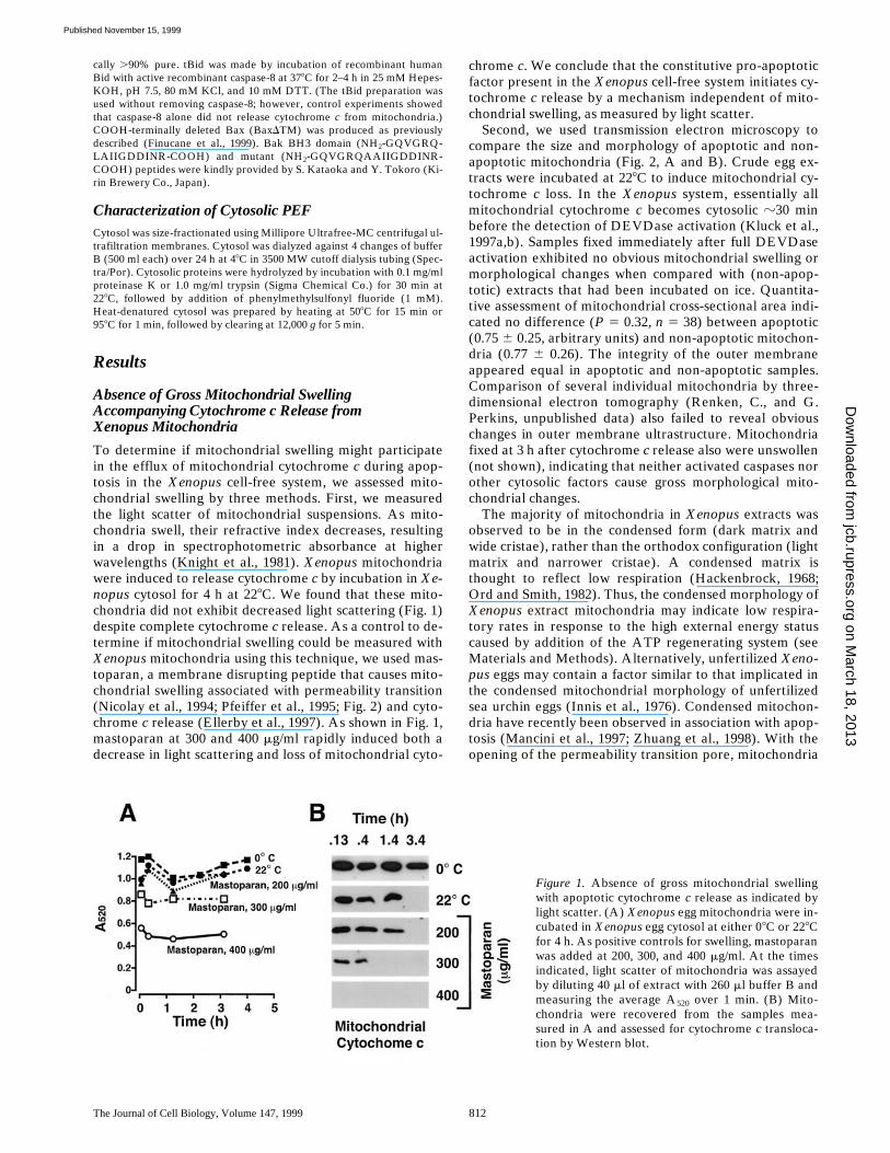
To determine if mitochondrial swelling might participatein the efflux of mitochondrial cytochrome c during apop-tosis in the Xenopus cell-free system, we assessed mito-chondrial swelling by three methods. First, we measuredthe light scatter of mitochondrial suspensions. As mito-chondria swell, their refractive index decreases, resultingin a drop in spectrophotometric absorbance at higherwavelengths (Knight et al., 1981). Xenopus mitochondriawere induced to release cytochrome c by incubation in Xe-nopus cytosol for 4 h at 228C. We found that these mito-chondria did not exhibit decreased light scattering (Fig. 1)despite complete cytochrome c release. As a control to de-termine if mitochondrial swelling could be measured withXenopus mitochondria using this technique, we used mas-toparan, a membrane disrupting peptide that causes mito-chondrial swelling associated with permeability transition(Nicolay et al., 1994; Pfeiffer et al., 1995; Fig. 2) and cyto-chrome c release (Ellerby et al., 1997). As shown in Fig. 1,mastoparan at 300 and 400 mg/ml rapidly induced both adecrease in light scattering and loss of mitochondrial cyto-
chrome c. We conclude that the constitutive pro-apoptoticfactor present in the Xenopus cell-free system initiates cy-tochrome c release by a mechanism independent of mito-chondrial swelling, as measured by light scatter.
Second, we used transmission electron microscopy tocompare the size and morphology of apoptotic and non-apoptotic mitochondria (Fig. 2, A and B). Crude egg ex-tracts were incubated at 228C to induce mitochondrial cy-tochrome c loss. In the Xenopus system, essentially allmitochondrial cytochrome c becomes cytosolic z30 minbefore the detection of DEVDase activation (Kluck et al.,1997a,b). Samples fixed immediately after full DEVDaseactivation exhibited no obvious mitochondrial swelling ormorphological changes when compared with (non-apop-totic) extracts that had been incubated on ice. Quantita-tive assessment of mitochondrial cross-sectional area indi-cated no difference (P 5 0.32, n 5 38) between apoptotic(0.75 6 0.25, arbitrary units) and non-apoptotic mitochon-dria (0.77 6 0.26). The integrity of the outer membraneappeared equal in apoptotic and non-apoptotic samples.Comparison of several individual mitochondria by three-dimensional electron tomography (Renken, C., and G.Perkins, unpublished data) also failed to reveal obviouschanges in outer membrane ultrastructure. Mitochondriafixed at 3 h after cytochrome c release also were unswollen(not shown), indicating that neither activated caspases norother cytosolic factors cause gross morphological mito-chondrial changes.
The majority of mitochondria in Xenopus extracts wasobserved to be in the condensed form (dark matrix andwide cristae), rather than the orthodox configuration (lightmatrix and narrower cristae). A condensed matrix isthought to reflect low respiration (Hackenbrock, 1968;Ord and Smith, 1982). Thus, the condensed morphology ofXenopus extract mitochondria may indicate low respira-tory rates in response to the high external energy statuscaused by addition of the ATP regenerating system (seeMaterials and Methods). Alternatively, unfertilized Xeno-pus eggs may contain a factor similar to that implicated inthe condensed mitochondrial morphology of unfertilizedsea urchin eggs (Innis et al., 1976). Condensed mitochon-dria have recently been observed in association with apop-tosis (Mancini et al., 1997; Zhuang et al., 1998). With theopening of the permeability transition pore, mitochondria
Figure 1. Absence of gross mitochondrial swellingwith apoptotic cytochrome c release as indicated bylight scatter. (A) Xenopus egg mitochondria were in-cubated in Xenopus egg cytosol at either 08C or 228Cfor 4 h. As positive controls for swelling, mastoparanwas added at 200, 300, and 400 mg/ml. At the timesindicated, light scatter of mitochondria was assayedby diluting 40 ml of extract with 260 ml buffer B andmeasuring the average A520 over 1 min. (B) Mito-chondria were recovered from the samples mea-sured in A and assessed for cytochrome c transloca-tion by Western blot.
on March 18, 2013
jcb.rupress.orgD
ownloaded from
Published November 15, 1999

Kluck et al. Mitochondrial Permeabilization by Apoptotic Factors 813
in the condensed state are converted irreversibly (in thepresence of sucrose) to the orthodox conformation. Thus,simply on ultrastructural grounds, as both apoptotic andnon-apoptotic Xenopus mitochondria are condensed, it isunlikely that a permeability transition had taken place.
To examine further the ultrastructure of apoptotic mito-chondria, we used a third approach, a high-resolution formof scanning electron microscopy (FEISEM; Allen andGoldberg, 1993; Allen et al., 1996). As Fig. 2 shows,FEISEM revealed that mitochondria depleted of cyto-chrome c were of similar size and appearance to controlmitochondria. Outer membrane discontinuities were notobserved. This technique did reveal outer membranepores, 10–13 nm in diameter, in mitochondria treated withthe antibiotic peptide alamethicin (Fig. 2 F), which also in-duced complete release of cytochrome c (Kluck and New-meyer, unpublished). However, no such pores were ob-served in mitochondria depleted of cytochrome c by thefactors present in Xenopus cytosol, or by Bid or Bax, sug-gesting that if these factors form pores, they are eithertransient or considerably smaller than those formed byalamethicin (Woolley and Wallace, 1992). In summary,multiple approaches (Figs. 1 and 2) demonstrated the ab-sence of gross ultrastructural changes in mitochondria ac-companying cytochrome c release in the Xenopus cell-freesystem.
Adenylate Kinase Loss from the Intermembrane Space Precedes Caspase Activation
To continue investigating the mitochondrial events lead-ing to cytochrome c release, we next determined whether
other soluble proteins present in the intermembrane spacewere released during apoptosis. One such protein is ade-nylate kinase (25,200 Da). The amount of mitochondrialadenylate kinase (AK) released into the cytosol is difficultto measure because cytosol already contains z50% of thecell’s AK activity (not shown). However, the AK activityremaining in the mitochondrial pellet can readily be as-sayed. We incubated mitochondria in cytosol for varioustimes, washed them free of cytosol, and measured adenyl-ate kinase content in the final pellet. As Fig. 3 shows, withapoptosis induced by Xenopus cytosol, almost all AK ac-tivity was lost from mitochondria before DEVDase activa-tion, with kinetics similar to those observed for cyto-chrome c.
Sulfite Oxidase Is Coreleased with Cytochrome c from Rat Liver Mitochondria
Another mitochondrial protein, sulfite oxidase (SOx), isfound only in the intermembrane space. As antisera raisedto rat SOx failed to recognize the Xenopus protein, we de-termined whether rat liver mitochondria incubated in Xe-nopus extracts coreleased SOx with cytochrome c. Manyof the apoptotic features of the Xenopus egg cell-free sys-tem were duplicated if rat liver mitochondria were incu-bated in Xenopus cytosol. That is, mitochondrial cyto-chrome c was released after a period of incubation at 228C,but not at 08C (Fig. 3). (It should be noted that, for reasonsunknown, some preparations of rat liver mitochondriafailed to exhibit cytochrome c release in response to in-cubation with Bid, Bax or Xenopus egg cytosol.) Nearthe time of rat cytochrome c release, Xenopus cytosolic
Figure 2. Apoptotic mito-chondria exhibit normal sizeand morphology on electronmicroscopy. (A–C) Crude ex-tracts were incubated at 08Cas a non-apoptotic control(A) or incubated at 228C toinduce spontaneous cyto-chrome c release (B). Imme-diately after cytochrome c re-lease in the sample taken at2.75 h, aliquots were fixedand examined by transmis-sion electron microscopy. (C)A swollen mitochondriontaken from a further sampletreated with mastoparan (400mg/ml). (D–F) FEISEM ofreconstituted extracts incu-bated at 08C for 3 h, 40 min(D, non-apoptotic) or 228C inthe presence of 35 mg/mlBaxDTM for 1 h, 40 min (E).(F) Alamethicin addition (2mg/ml, 40 min) caused bothcytochrome c release and theformation of 10–13-nm pores(arrows). Bar, (D–F) 200 nm.
on March 18, 2013
jcb.rupress.orgD
ownloaded from
Published November 15, 1999
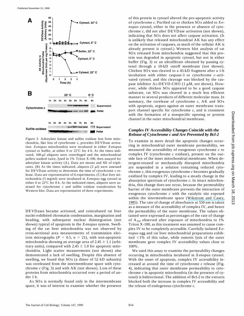
The Journal of Cell Biology, Volume 147, 1999 814
DEVDases became activated, and coincubated rat livernuclei exhibited chromatin condensation, margination andbeading, with subsequent nuclear disintegration (notshown) typical of apoptosis in the Xenopus system. Swell-ing of the rat liver mitochondria was not observed bycross-sectional area measurements of transmission elec-tron micrographs (P 5 0.5, n 5 21), with non-apoptoticmitochondria showing an average area of 2.45 6 1.1 (arbi-trary units), compared with 2.45 6 1.0 for apoptotic mito-chondria. Light scatter measurements (not shown) alsodemonstrated a lack of swelling. Despite this absence ofswelling, we found that SOx (a dimer of 52 kD subunits)was coreleased from the intermembrane space with cyto-chrome c (Fig. 3) and with AK (not shown). Loss of theseproteins from mitochondria occurred over a period of un-der 1 h.
As SOx is normally found only in the intermembranespace, it was of interest to examine whether the presence
of this protein in cytosol altered the pro-apoptotic activityof cytochrome c. Purified rat or chicken SOx added to Xe-nopus cytosol, either in the presence or absence of cyto-chrome c, did not alter DEVDase activation (not shown),indicating that SOx does not affect caspase activation. (Itis unlikely that released mitochondrial AK has any effecton the activation of caspases, as much of the cellular AK isalready present is cytosol.) Western blot analysis of ratSOx released from mitochondria suggested that this pro-tein was degraded in apoptotic cytosol, but not in eitherbuffer (Fig. 3) or an ultrafiltrate obtained by passing cy-tosol through a 10-kD cutoff membrane (not shown).Chicken SOx was cleaved to a 45-kD fragment after a 1-hincubation with either caspase-3 or cytochrome c–acti-vated cytosol, and this cleavage was blocked by the cas-pase inhibitor Ac-DEVD-CHO (1 mM, not shown). How-ever, while chicken SOx appeared to be a good caspasesubstrate, rat SOx was cleaved in a much less efficientmanner to several products of different molecular mass. Insummary, the corelease of cytochrome c, AK and SOxwith apoptosis, argues against an outer membrane trans-port channel specific for cytochrome c, and is consistentwith the formation of a nonspecific opening or proteinchannel in the outer mitochondrial membrane.
Complex IV Accessibility Changes Coincide with the Release of Cytochrome c and Are Prevented by Bcl-2
To examine in more detail the apoptotic changes occur-ring in mitochondrial outer membrane permeability, wemeasured the accessibility of exogenous cytochrome c tocomplex IV (cytochrome c oxidase), present on the out-side face of the inner mitochondrial membrane. When de-tergent-treated or mechanically disrupted mitochondriaare suspended in a solution containing reduced cyto-chrome c, this exogenous cytochrome c becomes graduallyoxidized by complex IV, leading to a steady change in theabsorption spectrum of cytochrome c. In intact mitochon-dria, this change does not occur, because the permeabilitybarrier of the outer membrane prevents the interaction ofexogenous cytochrome c with the catalytic site exposedwithin the intermembrane space (Wikstrom and Casey,1985). The rate of change of absorbance at 550 nm is takenas a measure of the accessibility of complex IV, and hencethe permeability of the outer membrane. The values ob-tained were expressed as percentages of the rate of changeof A550 observed after exposure of mitochondria to 1%Triton X-100, as this treatment was assumed to cause com-plex IV to be completely accessible. Carefully isolated Xe-nopus egg and rat liver mitochondrial preparations exhib-ited ,1% of this value, while osmotic lysis of the outermembrane gave complex IV accessibility values close to100%.
We used this assay to examine the permeability changesoccurring in mitochondria incubated in Xenopus cytosol.With the onset of apoptosis, complex IV accessibility in-creased at around the time of cytochrome c release (Fig.4), indicating that outer membrane permeability to cyto-chrome c in apoptotic mitochondria (in the presence of cy-tosol) is bidirectional. The addition of Bcl-2 to the extractsblocked both the increase in complex IV accessibility andthe release of endogenous cytochrome c.
Figure 3. Adenylate kinase and sulfite oxidase loss from mito-chondria, like loss of cytochrome c, precedes DEVDase activa-tion. Xenopus mitochondria were incubated in either Xenopuscytosol or buffer, at either 0 or 228C for 4 h. At the times indi-cated, 100-ml aliquots were centrifuged and the mitochondrialpellets washed twice, lysed in 1% Triton X-100, then assayed foradenylate kinase activity (A). Data are means and SD of tripli-cates. (B) At the times indicated, aliquots (2 ml) were assessedfor DEVDase activity to determine the time of cytochrome c re-lease. Data are representative of 6 experiments. (C) Rat liver mi-tochondria (5 mg/ml) were incubated in Xenopus egg cytosol ateither 0 or 228C for 8 h. At the indicated times, aliquots were as-sessed for cytochrome c and sulfite oxidase translocation byWestern blot. Data are representative of three experiments.
on March 18, 2013
jcb.rupress.orgD
ownloaded from
Published November 15, 1999
Kluck et al. Mitochondrial Permeabilization by Apoptotic Factors 815
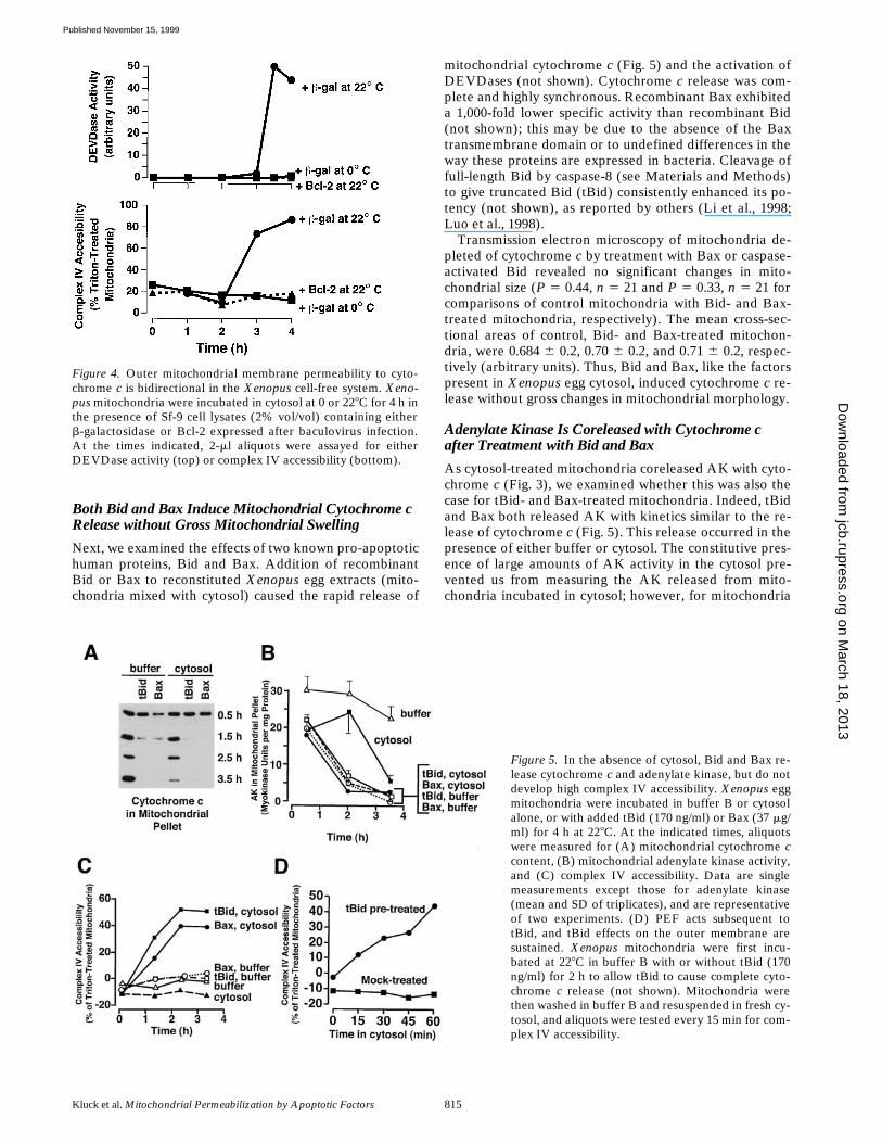
Both Bid and Bax Induce Mitochondrial Cytochrome c Release without Gross Mitochondrial Swelling
Next, we examined the effects of two known pro-apoptotichuman proteins, Bid and Bax. Addition of recombinantBid or Bax to reconstituted Xenopus egg extracts (mito-chondria mixed with cytosol) caused the rapid release of
mitochondrial cytochrome c (Fig. 5) and the activation ofDEVDases (not shown). Cytochrome c release was com-plete and highly synchronous. Recombinant Bax exhibiteda 1,000-fold lower specific activity than recombinant Bid(not shown); this may be due to the absence of the Baxtransmembrane domain or to undefined differences in theway these proteins are expressed in bacteria. Cleavage offull-length Bid by caspase-8 (see Materials and Methods)to give truncated Bid (tBid) consistently enhanced its po-tency (not shown), as reported by others (Li et al., 1998;Luo et al., 1998).
Transmission electron microscopy of mitochondria de-pleted of cytochrome c by treatment with Bax or caspase-activated Bid revealed no significant changes in mito-chondrial size (P 5 0.44, n 5 21 and P 5 0.33, n 5 21 forcomparisons of control mitochondria with Bid- and Bax-treated mitochondria, respectively). The mean cross-sec-tional areas of control, Bid- and Bax-treated mitochon-dria, were 0.684 6 0.2, 0.70 6 0.2, and 0.71 6 0.2, respec-tively (arbitrary units). Thus, Bid and Bax, like the factorspresent in Xenopus egg cytosol, induced cytochrome c re-lease without gross changes in mitochondrial morphology.
Adenylate Kinase Is Coreleased with Cytochrome c after Treatment with Bid and Bax
As cytosol-treated mitochondria coreleased AK with cyto-chrome c (Fig. 3), we examined whether this was also thecase for tBid- and Bax-treated mitochondria. Indeed, tBidand Bax both released AK with kinetics similar to the re-lease of cytochrome c (Fig. 5). This release occurred in thepresence of either buffer or cytosol. The constitutive pres-ence of large amounts of AK activity in the cytosol pre-vented us from measuring the AK released from mito-chondria incubated in cytosol; however, for mitochondria
Figure 4. Outer mitochondrial membrane permeability to cyto-chrome c is bidirectional in the Xenopus cell-free system. Xeno-pus mitochondria were incubated in cytosol at 0 or 228C for 4 h inthe presence of Sf-9 cell lysates (2% vol/vol) containing eitherb-galactosidase or Bcl-2 expressed after baculovirus infection.At the times indicated, 2-ml aliquots were assayed for eitherDEVDase activity (top) or complex IV accessibility (bottom).
Figure 5. In the absence of cytosol, Bid and Bax re-lease cytochrome c and adenylate kinase, but do notdevelop high complex IV accessibility. Xenopus eggmitochondria were incubated in buffer B or cytosolalone, or with added tBid (170 ng/ml) or Bax (37 mg/ml) for 4 h at 228C. At the indicated times, aliquotswere measured for (A) mitochondrial cytochrome ccontent, (B) mitochondrial adenylate kinase activity,and (C) complex IV accessibility. Data are singlemeasurements except those for adenylate kinase(mean and SD of triplicates), and are representativeof two experiments. (D) PEF acts subsequent totBid, and tBid effects on the outer membrane aresustained. Xenopus mitochondria were first incu-bated at 228C in buffer B with or without tBid (170ng/ml) for 2 h to allow tBid to cause complete cyto-chrome c release (not shown). Mitochondria werethen washed in buffer B and resuspended in fresh cy-tosol, and aliquots were tested every 15 min for com-plex IV accessibility.
on March 18, 2013
jcb.rupress.orgD
ownloaded from
Published November 15, 1999

The Journal of Cell Biology, Volume 147, 1999 816
incubated in buffer, the amount of AK released intothe supernatant of tBid and Bax-treated mitochondriaaccounted for that lost from the mitochondrial pellet(not shown), indicating that the protein was translocated,rather than inactivated. These data support the hypothesisthat similar cytochrome c–releasing mechanisms are usedby various cytochrome c–releasing proteins.
Cytosol Greatly Increases Complex IV Accessibility Induced by Bid and Bax
Cytochrome c release from Xenopus mitochondria incu-bated in Xenopus cytosol is associated with high accessibil-ity of the inner membrane protein cytochrome c oxidase(complex IV) to exogenous cytochrome c (Fig. 4). Thishigh accessibility is also seen in Xenopus mitochondriatreated with tBid and Bax in the presence of Xenopus cy-tosol (Fig. 5 C). It was surprising to note, however, thatmitochondria treated with tBid and Bax in buffer dis-played an almost undetectable level of accessibility ofcomplex IV to exogenous cytochrome c (Fig. 5 C), despitetheir complete loss of endogenous cytochrome c (Fig. 5A). These results imply that a novel cytosolic activity,which we call Permeability Enhancing Factor, or PEF, isresponsible for a striking increase in complex IV accessi-bility beyond that produced by factors such as tBid or Bax.
PEF Can Act Downstream of tBid
PEF might function either simultaneously with factors liketBid or subsequent to their action. To address this ques-tion, we did the following experiment. Mitochondria werefirst depleted of cytochrome c by an initial incubation withtBid in buffer, and then washed and resuspended in cyto-sol for a second incubation. As Fig. 5 D shows, complex IVaccessibility rapidly increased during this second incuba-tion in cytosol. This is consistent with data showing thattBid stays strongly associated with mitochondria afterwashing (Kuwana, O., T. von Ahsen, and D.D. Newmeyer,unpublished results), but further implies that the effect oftBid is sustained. Some samples were incubated in freshbuffer before reincubation with cytosol, in an attempt toencourage maximum repair of the tBid-induced perme-ability. However, this intermediate incubation had no ef-fect (not shown). We conclude from this experiment thatPEF can function subsequent to tBid-induced cytochromec translocation. This experiment also demonstrates thatPEF activity is not dependent on downstream effects ofcytochrome c release, because the cytosol used in Fig. 5 Dcontained neither cytochrome c (nor any other solubleproteins from the mitochondrial intermembrane space)nor active caspases.
Cytosol Increases Complex III Accessibility
It could have been argued that the quantitative differencesin accessibility of complex IV reflected some effect spe-cific for that enzyme complex, rather than changes in thepermeability of the outer membrane. To rule out this pos-sibility, we measured the accessibility of another mito-chondrial inner membrane component, complex III. Thisassay is similar in principle to the complex IV assay, exceptthat cytochrome c reduction rather than oxidation is mea-
sured, and an electron donor (DBH2) is added to ensuresaturating substrate (see Materials and Methods). Theseexperiments showed that cytosol-induced cytochrome crelease was accompanied by an increase in the accessibilityof both complexes III and IV (Fig. 6, lane 4), confirmingthe bidirectional permeability of the outer membrane un-der these conditions. Again, however, in the presence ofbuffer alone, tBid induced a nearly undetectable accessi-bility of complexes III and IV (Fig. 6, lane 2), which wasgreatly enhanced in the presence of cytosol (Fig. 6, lanes 6and 7). The ability of cytosol to increase outer membranepermeability was not due to a feedback effect of cyto-chrome c–activated caspases, as Ac-DEVD-CHO (10 mM)did not prevent the development of high complex III andIV accessibility (not shown).
In summary, the enhancing effect of cytosol on accessi-bility of complexes III and IV (Figs. 4–6) is explained bythe presence of a factor in Xenopus cytosol, which we havenamed permeability enhancing factor (PEF). PEF doesnot release cytochrome c by itself, but rather enhancesouter membrane permeability after a pro-apoptotic factorhas caused the release of endogenous cytochrome c.
Figure 6. Cytosol (PEF) increases both complex III and IV ac-cessibility after tBid-induced cytochrome c release. Mitochondriawere incubated with or without tBid in either buffer B or cytosol,at 0 or 228C. At suitable times (1.7, 2.7, or 3.7 h), mitochondriawere pelleted and analyzed by Western blot for cytochrome ctranslocation (bottom) or washed and resuspended in buffer Efor measurement of complex III (top) and IV (middle) accessibil-ity (arbitrary units). For comparison between assays, mitochon-dria were lysed by incubation in water for 30 min on ice. Theseosmotically lysed mitochondria retain their cytochrome c due tothe low salt content (see Materials and Methods). Data are meanand SD for duplicate measurements and are representative ofthree experiments. Note: The time course of permeabilization byPEF is slower in Fig. 6 than Fig. 5; this merely reflects a normalvariation between Xenopus extracts in time of onset of apoptoticchanges (Newmeyer et al., 1994; Farschon et al., 1997).
on March 18, 2013
jcb.rupress.orgD
ownloaded from
Published November 15, 1999

Kluck et al. Mitochondrial Permeabilization by Apoptotic Factors 817
tBid and PEF Do Not Permeabilize the Mitochondrial Inner Membrane
To address the possibility that PEF increases accessibilityof complexes III and IV by grossly perturbing the integrityof mitochondria, we examined whether a soluble mito-chondrial matrix protein, mtHsp70, is released after theactions of tBid and PEF. First, to show that mtHsp70 canbe released if the inner mitochondrial membrane is per-meabilized, we treated mitochondria with increasing con-centrations of digitonin and followed the redistribution ofmtHsp70 from the mitochondrial pellet fraction to the sol-uble supernatant. Fig. 7 A shows that cytochrome c is re-leased at a low concentration (0.1%) of digitonin, but thatmtHsp70 becomes soluble at higher digitonin concentra-tions (0.2%). Only at still higher concentrations (0.6%)did the membrane-associated Rieske protein become solu-bilized, showing that at the intermediate concentrationssufficient to release mtHsp70, digitonin did not solubilizethe mitochondrial membranes, but merely permeabilizedthem. Next, we examined the behavior of mtHsp70 in mi-tochondria induced to release cytochrome c by cytosol orby tBid (in the presence or absence of cytosol). As seen inFig. 7 B, mtHsp70 remained in the mitochondrial pelletfraction under all conditions, even in the presence of cyto-sol containing active PEF. We conclude that cytosolic fac-tors and tBid can permeabilize the outer mitochondrialmembrane while leaving the inner membrane intact.
PEF Characteristics
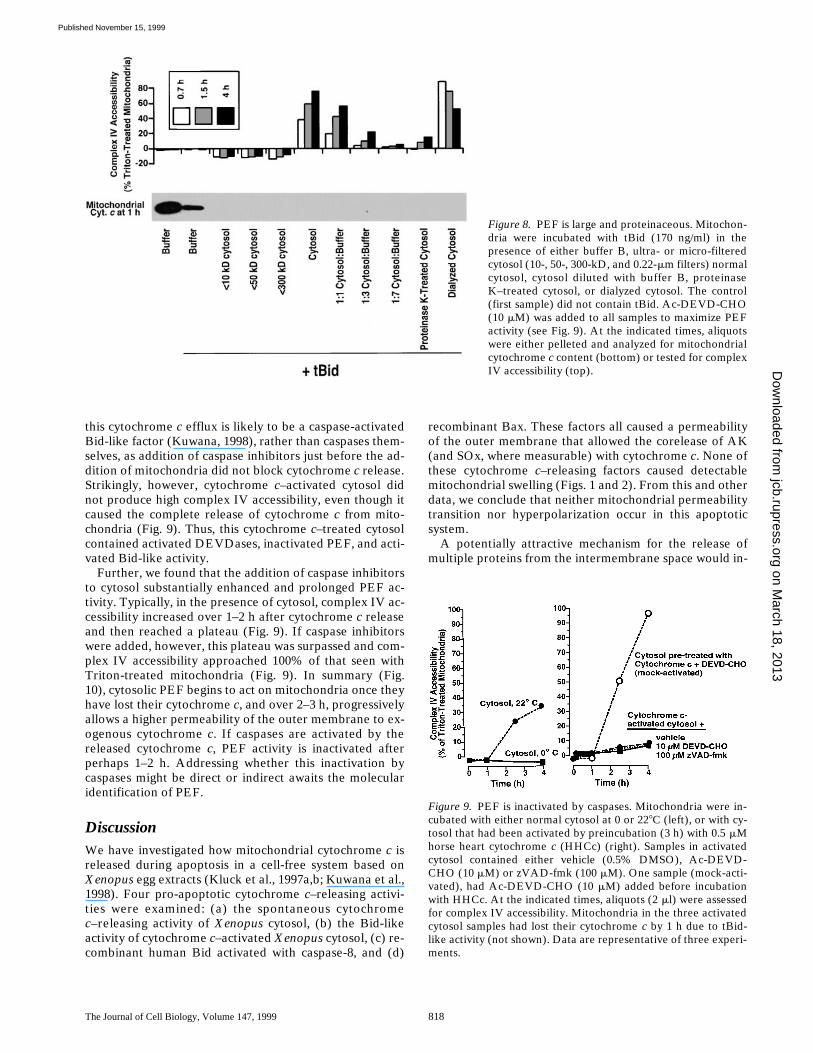
To begin characterizing PEF, we treated cytosol in various
ways before incubating it with Xenopus mitochondriaand tBid (Fig. 8). Treatment with proteinase K (Fig. 8),trypsin, or heat (508C, 15 min, or 958C, 1 min; not shown)greatly diminished or abolished cytosol’s ability to in-crease complex IV accessibility, indicating that PEF is pro-teinaceous. PEF activity was not observed in the ultrafil-trate obtained by passing cytosol through a 300-kD cutoffmembrane, and was not lost on dialysis, suggesting thatPEF is macromolecular and possibly present in a largecomplex. The complex IV accessibility produced by PEFaccumulates over a few hours to approach that seen in mito-chondria treated with Triton X-100 (see Materials and Meth-ods) and digitonin (not shown). PEF activity is reduced sub-stantially on dilution of cytosol. A PEF-like activity mayalso be present in mammalian cells, as a lysate (40 mg/ml)of human Jurkat cells caused an increase in complex IV ac-cessibility of isolated Xenopus mitochondria (not shown).This Jurkat cell PEF-like activity was observed in the pres-ence or absence of tBid, suggesting that the Jurkat cell ly-sate contained endogenous cytochrome c–releasing factors.
PEF Is Destroyed by Caspases
To address the possibility that PEF is a substrate ofcaspases, we examined the effect of cytochrome c–acti-vated cytosol on fresh Xenopus mitochondria. Fresh cyto-sol was first incubated for 3 h with a low level (0.5 mM) ofcytochrome c to activate cytosolic caspases (DEVDases).Subsequent addition of mitochondria to this activated cy-tosol resulted in rapid loss of mitochondrial cytochrome cand AK (by 1 h, not shown). The factor responsible for
Figure 7. tBid and PEF preserve the integrity of themitochondrial matrix compartment, as measured byretention of a soluble mitochondrial matrix protein,mtHsp70. (A) mtHsp70 is released when mitochon-dria are treated with intermediate concentrations ofdigitonin. Xenopus egg mitochondria were treatedwith increasing concentrations of digitonin to selec-tively permeabilize the outer, then the inner mito-chondrial membranes. After two hours incubation,the contents of the pellet and supernatant fractionswere analyzed by immunoblotting (left panel) withantibodies to cytochrome c, mtHsp70, and the innermembrane-associated Rieske protein (part of com-plex III). Also, accessibility of complex IV was mea-sured as described above (right panel). Note thatmtHsp70 is released by digitonin at 0.2%, a concen-tration insufficient to solubilize the Rieske protein,but more than sufficient to release cytochrome c. (B)mtHsp70 is not released by tBid and PEF, despitefull release of cytochrome c and full accessibility ofcomplex IV. Xenopus mitochondria were treatedwith 170 ng/ml tBid, in either the presence or ab-sence of cytosol, and either at 0 or 228C, as indicated.DEVD-CHO (10 mM) was added to cytosol incuba-tions to minimize PEF degradation and thus maxi-mize its activity. After 3 h of incubation, the contentsof the mitochondrial pellet were analyzed by immu-
noblotting with antibodies to cytochrome c, mtHsp70 and Rieske protein (left), and the accessibility of complex IV (right). The sampleswere taken well after full cytochrome c release from mitochondria, which occurred in this experiment at 1 h in the tBid-treated sampleand at 2 h in cytosol at 228C (not shown). Note that mtHsp70 is retained in the pellet fraction despite full cytochrome c release and in-creased complex IV accessibility (reflecting PEF activity). (The bands stained by anti-Rieske antibody in cytosol are nonmitochondrialcross-reacting proteins.)
on March 18, 2013
jcb.rupress.orgD
ownloaded from
Published November 15, 1999
The Journal of Cell Biology, Volume 147, 1999 818
this cytochrome c efflux is likely to be a caspase-activatedBid-like factor (Kuwana, 1998), rather than caspases them-selves, as addition of caspase inhibitors just before the ad-dition of mitochondria did not block cytochrome c release.Strikingly, however, cytochrome c–activated cytosol didnot produce high complex IV accessibility, even though itcaused the complete release of cytochrome c from mito-chondria (Fig. 9). Thus, this cytochrome c–treated cytosolcontained activated DEVDases, inactivated PEF, and acti-vated Bid-like activity.
Further, we found that the addition of caspase inhibitorsto cytosol substantially enhanced and prolonged PEF ac-tivity. Typically, in the presence of cytosol, complex IV ac-cessibility increased over 1–2 h after cytochrome c releaseand then reached a plateau (Fig. 9). If caspase inhibitorswere added, however, this plateau was surpassed and com-plex IV accessibility approached 100% of that seen withTriton-treated mitochondria (Fig. 9). In summary (Fig.10), cytosolic PEF begins to act on mitochondria once theyhave lost their cytochrome c, and over 2–3 h, progressivelyallows a higher permeability of the outer membrane to ex-ogenous cytochrome c. If caspases are activated by thereleased cytochrome c, PEF activity is inactivated afterperhaps 1–2 h. Addressing whether this inactivation bycaspases might be direct or indirect awaits the molecularidentification of PEF.
DiscussionWe have investigated how mitochondrial cytochrome c isreleased during apoptosis in a cell-free system based onXenopus egg extracts (Kluck et al., 1997a,b; Kuwana et al.,1998). Four pro-apoptotic cytochrome c–releasing activi-ties were examined: (a) the spontaneous cytochromec–releasing activity of Xenopus cytosol, (b) the Bid-likeactivity of cytochrome c–activated Xenopus cytosol, (c) re-combinant human Bid activated with caspase-8, and (d)
recombinant Bax. These factors all caused a permeabilityof the outer membrane that allowed the corelease of AK(and SOx, where measurable) with cytochrome c. None ofthese cytochrome c–releasing factors caused detectablemitochondrial swelling (Figs. 1 and 2). From this and otherdata, we conclude that neither mitochondrial permeabilitytransition nor hyperpolarization occur in this apoptoticsystem.
A potentially attractive mechanism for the release ofmultiple proteins from the intermembrane space would in-
Figure 8. PEF is large and proteinaceous. Mitochon-dria were incubated with tBid (170 ng/ml) in thepresence of either buffer B, ultra- or micro-filteredcytosol (10-, 50-, 300-kD, and 0.22-mm filters) normalcytosol, cytosol diluted with buffer B, proteinaseK–treated cytosol, or dialyzed cytosol. The control(first sample) did not contain tBid. Ac-DEVD-CHO(10 mM) was added to all samples to maximize PEFactivity (see Fig. 9). At the indicated times, aliquotswere either pelleted and analyzed for mitochondrialcytochrome c content (bottom) or tested for complexIV accessibility (top).
Figure 9. PEF is inactivated by caspases. Mitochondria were in-cubated with either normal cytosol at 0 or 228C (left), or with cy-tosol that had been activated by preincubation (3 h) with 0.5 mMhorse heart cytochrome c (HHCc) (right). Samples in activatedcytosol contained either vehicle (0.5% DMSO), Ac-DEVD-CHO (10 mM) or zVAD-fmk (100 mM). One sample (mock-acti-vated), had Ac-DEVD-CHO (10 mM) added before incubationwith HHCc. At the indicated times, aliquots (2 ml) were assessedfor complex IV accessibility. Mitochondria in the three activatedcytosol samples had lost their cytochrome c by 1 h due to tBid-like activity (not shown). Data are representative of three experi-ments.
on March 18, 2013
jcb.rupress.orgD
ownloaded from
Published November 15, 1999

Kluck et al. Mitochondrial Permeabilization by Apoptotic Factors 819
volve swelling of the mitochondrial matrix, leading to me-chanical disruption of the outer mitochondrial membrane.A single break in the outer mitochondrial membrane istheoretically sufficient to allow cytochrome c to diffuserapidly out of mitochondria down its concentration gradi-ent (10 mM in intermembrane space and cristae). Rup-tures in the outer membrane ensue if the matrix swellspast the point that the outer membrane can expand. Suchmatrix swelling is proposed to occur during apoptosis inresponse either to the formation of a 1.5-kD permeabilitytransition pore in the inner membrane (Bernardi, 1996;Petit et al., 1996; Susin et al., 1997) or to hyperpolarizationof the inner membrane (Vander Heiden et al., 1997, 1999).
However, mitochondrial swelling has not been observedas a frequent accompaniment to apoptosis. In the studiesreported here, light scatter measurements (Fig. 1) andtransmission and scanning electron microscopy (Fig. 2) allfailed to provide evidence of mitochondrial swelling in as-sociation with apoptotic cytochrome c efflux from Xeno-pus mitochondria. Another group has reported that, inwhole cells of Xenopus embryos treated with gamma irra-diation, apoptotic mitochondria display normal size andmorphology (Hensey and Gautier, 1997). In other systemsalso, mitochondrial swelling has not been observed withapoptosis (Searle et al., 1975; Mancini et al., 1997; Zhuanget al., 1998), and in some apoptotic cells mitochondriaeven shrink (Martinou et al., 1999). Previously, Bid was re-ported to release cytochrome c from isolated mitochon-dria without swelling (Luo et al., 1998). Bax-mediated cy-tochrome c release has been associated in some reports
with mitochondrial swelling (Narita et al., 1998) or perme-ability transition (Pastorino et al., 1998), but in other re-ports with an absence of swelling (Jurgensmeier et al.,1998; Martinou et al., 1999). The presence of swollen mito-chondria in some apoptotic systems, including whole cells,may reflect either secondary necrosis or a fundamentallydistinct apoptotic cytochrome c release mechanism.
Cytochrome c efflux is not a specific export process, asAK (25,200 Da) was also released by each of the four pro-apoptotic factors we studied. Furthermore, SOx (104 kD)was released from rat liver mitochondria incubated in cy-tosol (Fig. 3). Several intermembrane space proteins, in-cluding AK (Kohler et al., 1999; Samali et al., 1999; Singleet al., 1998), a cytochrome c–GFP fusion protein (Heis-kanen et al., 1999), AIF (Susin et al., 1997, 1999b), andcertain caspases (Mancini et al., 1998; Susin et al., 1999a)have been reported to be coreleased with cytochrome c inother systems, often in the absence of a permeability tran-sition.
Whereas proteins such as Bid, Bax, and certain endoge-nous Xenopus egg cytosolic factors produced only a lim-ited outer membrane permeability, we detected anothercytosolic activity that caused a much stronger permeabili-zation effect. We termed this novel cytosolic activity PEF,as it enhanced, rather than initiated, outer membrane per-meabilization (Figs. 5–10). The initial permeability causedby cytochrome c releasing factors is limited as it allows arelatively small cytochrome c flux across the outer mem-brane. This flux is sufficient to allow the loss of endo-genous intermembrane space proteins, but not the rapidexchange of exogenous cytochrome c measured as accessi-bility of complexes III and IV (Fig. 5). However, if freshcytosol (PEF) is present when cytochrome c–releasing fac-tors act on mitochondria, an additional mitochondrialchange occurs which strikingly enhances the permeabilityof the outer membrane to exogenous cytochrome c. Frommeasurements of complex IV accessibility such as thosepresented in Fig. 4, we calculate that 1 ml of apoptotic mi-tochondria can oxidize z60 nmol of cytochrome c perminute. As the same volume of Xenopus egg mitochondriacontains z12 pmol of cytochrome c, we conclude that therate of cytochrome c exchange across the outer membraneproduced by PEF is at least 5,000-fold greater than thatneeded for complete efflux of endogenous cytochrome cper minute.
A potential explanation for the PEF activity we ob-served is that PEF could be an analogue of Bax or Bid thatis present in cytosol at higher concentrations than thosewe used with recombinant Bid and Bax. However, this no-tion can be ruled out for the following reasons: (a) whencytosol activated by pretreatment with cytochrome c issubsequently mixed with mitochondria, rapid release ofcytochrome c is observed, likely due to the cleavage andactivation of endogenous Xenopus Bid. This activated cy-tosol, however, has lost PEF activity rather than gained it;(b) recombinant tBid was added at five-fold higher con-centrations (170 ng/ml) than those required for cyto-chrome c release (not shown); and (c) (most compelling)PEF-containing cytosol does not possess the ability tocause rapid release of cytochrome c, and therefore cannotcontain Bid or Bax activity greater than the amounts of re-combinant protein we added. Further dilution of PEF-con-
Figure 10. Model of two types of mitochondrial outer membranepermeability observed in Xenopus extracts during apoptosis. Mi-tochondria treated with tBid or Bax in buffer undergo efflux ofendogenous cytochrome c (12 pmol/ml mitochondria) across theouter membrane, with limited permeability to exogenous cyto-chrome c. Additional exposure to cytosol (PEF) allows high per-meability to exogenous cytochrome c (60 nmol/ml mitochondriaper min, as measured by the rate of the complex IV reaction).Thus, this increased exchange of endogenous cytochrome c is atleast z5,000 times the rate of cytochrome c efflux after tBid orBax treatment, assuming that all of the mitochondrial cyto-chrome c is released over 1 min. PEF begins to act immediatelyafter tBid–induced permeabilization, but is inactivated over 1–2 hby caspase activation. The inner membrane appears unaffected.
on March 18, 2013
jcb.rupress.orgD
ownloaded from
Published November 15, 1999

The Journal of Cell Biology, Volume 147, 1999 820
taining cytosol results in diminution of PEF activity, butrapid cytochrome c release is never seen. Thus, PEF doesnot behave like Bax and Bid.
The high permeability caused by PEF is consistent witha major rearrangement of the outer mitochondrial mem-brane, as a similar permeability (complex III or IV accessi-bility) was obtained with Triton X-100 (Figs. 7–9), osmoticbreakage of the outer membrane (Fig. 6), and with digito-nin (Fig. 7) in the absence of cytosol. The permeability in-crease by PEF was not, however, accompanied by observ-able changes in mitochondrial ultrastructure (Fig. 2, B andE). Furthermore, evidence suggests that the inner mito-chondrial membrane remains intact even after PEF hasacted to permeabilize the outer membrane. We observedthat the mtHsp70 protein was released from the matrix ofXenopus mitochondria treated with certain concentrationsof the detergent digitonin but was not released with apop-tosis (Fig. 7), in agreement with another report that the in-ner membrane remains impermeable to proteins (Kohleret al., 1999). In addition, our earlier data (Kluck et al.,1997a) show that inner membrane potential (as measuredby mitochondrial retention of the dye, DiOC6) remainshigh for at least 3 h after cytochrome c release, a findinginconsistent with inner membrane permeabilization. Thus,the effects of tBid, Bax, and PEF are confined to the outermembrane, rather than reflecting a global alteration in mi-tochondrial integrity.
The outer membrane permeability changes we observedwere not due to a feedback effect of cytochrome c–acti-vated caspases. Caspase inhibitors failed to block cyto-chrome c and AK efflux induced by Bid, Bax and the pro-apoptotic proteins in Xenopus cytosol. Moreover, Bid andBax induced cytochrome c and AK release from mito-chondria incubated in buffer alone (Fig. 5). Caspase inhib-itors also did not block the permeabilizing activity of PEF,but rather enhanced it (Fig. 9).
Preliminary characterization showed that PEF is pro-teinaceous and apparently large (.300 kD). While theslow kinetics of PEF action may suggest enzymatic behav-ior, PEF is not a caspase, as it was not inhibited by caspaseinhibitors. Indeed, PEF is inactivated by caspases, becausecytosol in which caspases had been activated by the addi-tion of cytochrome c contained no PEF activity, andcaspase inhibitors prolonged the effects of PEF in fresh cy-tosol. A PEF-like activity was also detected in human Jur-kat cell extracts (not shown). It is possible that PEF is ac-tive in whole cells, as a similar assay monitoring oxygenconsumption (complex IV activity) in permeabilized cellsfound that apoptosis induced high oxygen consumption inthe presence of exogenous reduced cytochrome c (VanderHeiden et al., 1997).
There are several mechanisms by which pro-apoptoticBcl-2 family members might induce outer membrane per-meability without affecting the inner membrane. Thesemembrane-associated proteins may form channels largeenough for the passage of soluble proteins (reviewed byReed, 1997) or cooperate with channels already present(Mannella, 1992) such as VDAC (Shimizu et al., 1999;Vander Heiden et al., 1999). Alternate mechanisms mightbe similar to the manner in which 15-lipoxygenase can in-teract specifically with organellar membranes to cause vis-ible changes (3–10 nm) in the lipid structure and perme-
ability to high molecular mass proteins (van Leyen et al.,1998), or to that in which Bax causes instabilities in artifi-cial membranes (Basanez et al., 1999). Also unclear is themechanism of PEF action, in particular how PEF is per-mitted to function only after a prior permeabilization ofthe outer membrane by factors such as Bid and Bax. Onepossibility is that PEF could interact with a target locatedon the lumenal side of the outer membrane. In this case,PEF would only gain access to its sites of action when theouter membrane becomes permeable to some degree. Al-ternatively, PEF could require the cooperation of a non-soluble cofactor that is located within the intermembranespace (the experiment in Fig. 5 D rules out the involve-ment of a soluble intermembrane space protein). Yet an-other possibility is that PEF interacts directly with chan-nels produced by tBid or Bax.
It is thought that eukaryotic mitochondria originatefrom a symbiotic relationship between primitive host cellsand primitive gram-negative bacteria, which allowed thehost cells to use oxygen to produce energy (Brown andDoolittle, 1997; Martin and Muller, 1998). It can be arguedthat pressures to maintain this symbiotic relationship mayhave led to the utilization of death pathways involving mi-tochondria, i.e., apoptosis via cytochrome c or necrosis viareactive oxygen species (Kobayashi, 1998; Blackstone andGreen, 1999). From this perspective, it is interesting toconsider whether the Bcl-2 family might have evolvedfrom primitive antibiotics produced by the host cell. Allknown effects of the Bcl-2 family members, with the ex-ception of Bcl-2 cell cycle effects (Huang et al., 1997), canbe linked to their regulation of cell death, often via effectson mitochondria. Indeed, the structures of Bcl–xL (Much-more et al., 1996; Sattler et al., 1997) and Bid (Chou et al.,1999; McDonnell et al., 1999) reveal similarities to those ofthe pore-forming domains of bacterial toxins. Moreover,like bacterial toxins, Bcl-2 proteins form channels in syn-thetic lipid membranes.
Other factors, derived from the host cell, might alsohave evolved to destroy mitochondrial integrity. Suchproducts include defensins (White et al., 1995), granulysin(Stenger et al., 1998), bactericidal permeability-increasingproteins (Beamer et al., 1998), and channel-forming pep-tides (Nicolay et al., 1994; Bechinger, 1997), such as masto-paran and alamethicin, both of which we have shown canrelease mitochondrial cytochrome c. PEF can now beadded to this list of molecules affecting mitochondrial per-meability.
What are the implications of PEF activity for a cell? IfPEF is present, attempts to rescue doomed cells by usingcaspase inhibitors would likely be futile, and may even en-hance mitochondrial damage by leaving PEF fully active.Some cells given an apoptotic stimulus undergo necroticdeath instead of apoptosis when caspase inhibitors arepresent (Green and Reed, 1998), and in such cases PEFmight be responsible for ensuring that mitochondria are ir-reversibly damaged. On the other hand, in Apaf-1 andcaspase-9 knockout mice, where the cytochrome c–initi-ated caspase activation pathway is missing, many neuronssurvive despite likely mitochondrial cytochrome c release(Cecconi et al., 1998; Hakem et al., 1998; Kuida et al.,1998; Yoshida et al., 1998). Moreover, in the presence ofcaspase inhibitors, trophic factor-deprived primary sympa-
on March 18, 2013
jcb.rupress.orgD
ownloaded from
Published November 15, 1999
Kluck et al. Mitochondrial Permeabilization by Apoptotic Factors 821
thetic neurons survive for many days despite mitochon-drial cytochrome c translocation (Deshmukh and Johnson,1998; Martinou et al., 1999). In such saved cells, neuronalmitochondria can survive the loss of cytochrome c and,upon restoration of trophic support, actually recover theircontent of cytochrome c after 24 h (Martinou et al., 1999).It has not yet been determined whether the recovery ofmitochondrial cytochrome c in these neurons is due tomultiplication of a few intact mitochondria, or to the reim-port of (apo-)cytochrome c into mitochondria that recoverfrom their permeabilization. We hypothesize that in mostnonneural tissues, PEF provides a backup mechanism forkilling the cell in the event of a disabled caspase pathway.This kind of redundancy in death pathways might be desir-able in lymphocytes and Xenopus eggs, but not in irre-placeable cells such as neurons in the adult organism.
We thank Mike Yaffe for advice on the complex IV accessibility assay andBeni Wolf for Jurkat extract. Caspase-8 cDNA was a gift from V. Dixit(Genentech Inc., San Francisco) and hBid cDNA from H. Yoshida (KirinBrewery Co., Japan).
This work was supported by grants from the National Institutes ofHealth (NIH) to D.D. Newmeyer (GM50284), M.J. Barber (GM32696),and D.R. Green (AI40646, CA69381). Some of the work included herewas conducted at the National Center for Microscopy and Imaging Re-search, San Diego, which is supported by NIH grant RR04050 to M.H. El-lisman. E. Bossy-Wetzel was supported by the Swiss National ScienceFoundation (823A-046638) and subsequently by NIH training grant (CI-2-05999). C. Renken acknowledges support from Grant-in-Aid (98-256A)from the Western States Affiliate of the American Heart Association toT.G. Frey. This is publication no. 311 from La Jolla Institute for Allergyand Immunology.
Submitted: 5 August 1999Revised: 28 September 1999Accepted: 12 October 1999
References
Allen, T.D., and M.W. Goldberg. 1993. High-resolution SEM in cell biology.Trends. Cell Biol. 3:205–208.
Allen, T.D., G.R. Bennion, S.A. Rutherford, S. Reipert, A. Ramalho, E. Kisel-eva, and M.W. Goldberg. 1996. Accessing nuclear structure for field emis-sion, in lens, scanning electron microscopy (FEISEM). Scanning Microsc.Suppl. 10:149–163.
Antonsson, B., F. Conti, A. Ciavatta, S. Montessuit, S. Lewis, I. Martinou, L.Bernasconi, A. Bernard, J.J. Mermod, G. Mazzei, et al. 1997. Inhibition ofBax channel-forming activity by Bcl-2. Science. 277:370–372.
Barber, M.J., and P.J. Neame. 1990. A conserved cysteine in molybdenum oxo-transferases. J. Biol. Chem. 265:20912–20915.
Basanez, G., A. Nechushtan, O. Drozhinin, A. Chanturiya, E. Choe, S. Tutt,K.A. Wood, Y. Hsu, J. Zimmerberg, and R.J. Youle. 1999. Bax, but not Bcl-xL, decreases the lifetime of planar phospholipid bilayer membranes at sub-nanomolar concentrations. Proc. Natl. Acad. Sci. USA. 96:5492–5497.
Beamer, L.J., S.F. Carroll, and D. Eisenberg. 1998. The BPI/LBP family of pro-teins: a structural analysis of conserved regions. Protein Sci. 7:906–914.
Bechinger, B. 1997. Structure and functions of channel-forming peptides: ma-gainins, cecropins, melittin and alamethicin. J. Membr. Biol. 156:197–211.
Bernardi, P. 1996. The permeability transition pore. Control points of a cy-closporin A-sensitive mitochondrial channel involved in cell death. Biochim.Biophys. Acta. 1275:5–9.
Blackstone, N.W., and D.R. Green. 1999. The evolution of a mechanism of cellsuicide. Bioessays. 21:84–88.
Bossy-Wetzel, E., D.D. Newmeyer, and D.R. Green. 1998. Mitochondrial cyto-chrome c release in apoptosis occurs upstream of DEVD-specific caspase ac-tivation and independently of mitochondrial transmembrane depolarization.EMBO (Eur. Mol. Biol. Organ.) J. 17:37–49.
Brown, J.R., and W.F. Doolittle. 1997. Archaea and the prokaryote-to-eukary-ote transition. Microbiol. Mol. Biol. Rev. 61:456–502.
Cecconi, F., G. Alvarez-Bolado, B.I. Meyer, K.A. Roth, and P. Gruss. 1998.Apaf1 (CED-4 homolog) regulates programmed cell death in mammaliandevelopment. Cell. 94:727–737.
Chou, J.J., H. Li, G.S. Salvesen, J. Yuan, and G. Wagner. 1999. Solution struc-ture of BID, an intracellular amplifier of apoptotic signaling. Cell. 96:615–
624.Degli Esposti, M., G. Lenaz, G. Izzo, and S. Papa. 1982. Kinetics and sidedness
of ubiquinol-cytochrome c reductase in beef-heart mitochondria. FEBS Lett.146:101–105.
Desagher, S., A. Osen-Sand, A. Nichols, R. Eskes, S. Montessuit, S. Lauper, K.Maundrell, B. Antonsson, and J.C. Martinou. 1999. Bid-induced conforma-tional change of Bax is responsible for mitochondrial cytochrome c releaseduring apoptosis. J. Cell Biol. 144:891–901.
Deshmukh, M., and E.M. Johnson, Jr. 1998. Evidence of a novel event duringneuronal death: development of competence-to-die in response to cytoplas-mic cytochrome c. Neuron. 21:695–705.
Ellerby, H.M., S.J. Martin, L.M. Ellerby, S.S. Naiem, S. Rabizadeh, G.S. Salve-sen, C.A. Casiano, N.R. Cashman, D.R. Green, and D.E. Bredesen. 1997.Establishment of a cell-free system of neuronal apoptosis: comparison ofpremitochondrial, mitochondrial, and postmitochondrial phases. J. Neurosci.17:6165–6178.
Eskes, R., B. Antonsson, A. Osen-Sand, S. Montessuit, C. Richter, R. Sadoul,G. Mazzei, A. Nichols, and J.C. Martinou. 1998. Bax-induced cytochrome Crelease from mitochondria is independent of the permeability transitionpore but highly dependent on Mg21 ions. J. Cell Biol. 143:217–224.
Farschon, D.M., C. Couture, T. Mustelin, and D.D. Newmeyer. 1997. Temporalphases in apoptosis defined by the actions of Src Homology 2 domains, cer-amide, Bcl-2, interleukin-1b converting enzyme family proteases, and adense membrane fraction. J. Cell Biol. 137:1117–1125.
Finucane, D.M., E. Bossy-Wetzel, N.J. Waterhouse, T.G. Cotter, and D.R.Green. 1999. Bax-induced caspase activation and apoptosis via cytochrome crelease from mitochondria is inhibitable by Bcl-xL. J. Biol. Chem. 274:2225–2233.
Gaikwad, A.S., T. Ramasarma, and C.K. Ramakrishna Kurup. 1991. Brown ad-ipose tissue mitochondria are cytochrome c-subsaturated. Mol. Cell. Bio-chem. 105:119–125.
Green, D.R., and J.C. Reed. 1998. Mitochondria and Apoptosis. Science. 281:1309–1312.
Hackenbrock, C.R. 1968. Ultrastructural bases for metabolically linked me-chanical activity in mitochondria. II. Electron transport-linked ultrastruc-tural transformations in mitochondria. J. Cell Biol. 37:345–369.
Hakem, R., A. Hakem, G.S. Duncan, J.T. Henderson, M. Woo, M.S. Soengas,A. Elia, J.L. de la Pompa, D. Kagi, W. Khoo, et al. 1998. Differential re-quirement for caspase 9 in apoptotic pathways in vivo. Cell. 94:339–352.
Heiskanen, K.M., M.B. Bhat, H.W. Wang, J. Ma, and A.L. Nieminen. 1999. Mi-tochondrial depolarization accompanies cytochrome c release during apop-tosis in PC6 cells. J. Biol. Chem. 274:5654–5658.
Hensey, C., and J. Gautier. 1997. A developmental timer that regulates apopto-sis at the onset of gastrulation. Mech. Dev. 69:183–195.
Huang, D.C., L.A. O’Reilly, A. Strasser, and S. Cory. 1997. The anti-apoptosisfunction of Bcl-2 can be genetically separated from its inhibitory effect oncell cycle entry. EMBO (Eur. Mol. Biol. Organ.) J. 16:4628–4638.
Innis, M.A., T.R. Beers, and S.P. Craig. 1976. Mitochondrial regulation in seaurchins. I. Mitochondrial ultrastructure transformations and changes in theADP:ATP ratio at fertilization. Exp Cell Res. 98:47–56.
Juin, P., A.O. Hueber, T. Littlewood, and G. Evan. 1999. c-myc-induced sensiti-zation to apoptosis is mediated through cytochrome c release. Genes Dev.13:1367–1381.
Jurgensmeier, J.M., Z. Xie, Q. Deveraux, L. Ellerby, D. Bredesen, and J.C.Reed. 1998. Bax directly induces release of cytochrome c from isolated mito-chondria. Proc. Natl. Acad. Sci. USA. 95:4997–5002.
Kluck, R.M., E. Bossy-Wetzel, D.R. Green, and D.D. Newmeyer. 1997a. Therelease of cytochrome c from mitochondria: a primary site for Bcl-2 regula-tion of apoptosis. Science. 275:1132–1136.
Kluck, R.M., S.J. Martin, B.M. Hoffman, J.S. Zhou, D.R. Green, and D.D.Newmeyer. 1997b. Cytochrome c activation of CPP32-like proteolysis playsa critical role in a Xenopus cell-free apoptosis system. EMBO (Eur. Mol.Biol. Organ.) J. 16:4639–4649.
Knight, V.A., P.M. Wiggins, J.D. Harvey, and J.A. O’Brien. 1981. The relation-ship between the size of mitochondria and the intensity of light that theyscatter in different energetic states. Biochim. Biophys. Acta. 637:146–151.
Kobayashi, I. 1998. Selfishness and death: raison d’être of restriction, recombi-nation and mitochondria. Trends Genet. 14:368–374.
Kohler, C., A. Gahm, T. Noma, A. Nakazawa, S. Orrenius, and B. Zhivotovsky.1999. Release of adenylate kinase 2 from the mitochondrial intermembranespace during apoptosis. FEBS Lett. 447:10–12.
Kuida, K., T.F. Haydar, C.Y. Kuan, Y. Gu, C. Taya, H. Karasuyama, M.S. Su,P. Rakic, and R.A. Flavell. 1998. Reduced apoptosis and cytochromec-mediated caspase activation in mice lacking caspase 9. Cell. 94:325–337.
Kuwana, T., J.J. Smith, M. Muzio, V. Dixit, D.D. Newmeyer, and S. Kornbluth.1998. Apoptosis induction by caspase-8 is amplified through the mitochon-drial release of cytochrome c. J. Biol. Chem. 273:16589–16594.
Liu, X., C.N. Kim, J. Yang, R. Jemmerson, and X. Wang. 1996. Induction ofapoptotic program in cell-free extracts: requirement for dATP and cyto-chrome c. Cell. 86:147–157.
Li, F., A. Srinivasan, Y. Wang, R.C. Armstrong, K.J. Tomaselli, and L.C. Fritz.1997a. Cell-specific induction of apoptosis by microinjection of cytochromec. Bcl-xL has activity independent of cytochrome c release. J. Biol. Chem.272:30299–30305.
Li, P., D. Nijhawan, I. Budihardjo, S.M. Srinivasula, M. Ahmad, E.S. Alnemri,
on March 18, 2013
jcb.rupress.orgD
ownloaded from
Published November 15, 1999
The Journal of Cell Biology, Volume 147, 1999 822
and X. Wang. 1997b. Cytochrome c and dATP-dependent formation ofApaf-1/caspase-9 complex initiates an apoptotic protease cascade. Cell. 91:479–489.
Li, H., H. Zhu, C.J. Xu, and J. Yuan. 1998. Cleavage of BID by caspase 8 medi-ates the mitochondrial damage in the Fas pathway of apoptosis. Cell. 94:491–501.
Luo, X., I. Budihardjo, H. Zou, C. Slaughter, and X. Wang. 1998. Bid, a Bcl2 in-teracting protein, mediates cytochrome c release from mitochondria in re-sponse to activation of cell surface death receptors. Cell. 94:481–490.
Mancini, M., B.O. Anderson, E. Caldwell, M. Sedghinasab, P.B. Paty, and D.M.Hockenbery. 1997. Mitochondrial proliferation and paradoxical membranedepolarization during terminal differentiation and apoptosis in a human co-lon carcinoma cell line. J. Cell Biol. 138:449–469.
Mancini, M., D.W. Nicholson, S. Roy, N.A. Thornberry, E.P. Peterson, L.A.Casciola-Rosen, and A. Rosen. 1998. The caspase-3 precursor has a cytosolicand mitochondrial distribution: implications for apoptotic signaling. J. CellBiol. 140:1485–1495.
Mannella, C.A. 1992. The ‘ins’ and ‘outs’ of mitochondrial membrane channels.Trends Biochem. Sci. 17:315–320.
Margoliash, E., and E.R. Bosshard. 1983. Guided by electrostatics, a textbookprotein comes of age. Trends Biochem. Sci. 8:316–320.
Martin, W., and M. Muller. 1998. The hydrogen hypothesis for the first eukary-ote. Nature. 392:37–41.
Martinou, I., S. Desagher, R. Eskes, B. Antonsson, E. André, S. Fakan, andJ.-C. Martinou. 1999. The release of cytochrome c from mitochondria duringapoptosis of NGF-deprived sympathetic neurons is a reversible event. J. CellBiol. 144:883–889.
Marzo, I., C. Brenner, N. Zamzami, J.M. Jurgensmeier, S.A. Susin, H.L. Vieira,M.C. Prevost, Z. Xie, S. Matsuyama, J.C. Reed, and G. Kroemer. 1998. Baxand adenine nucleotide translocator cooperate in the mitochondrial controlof apoptosis. Science. 281:2027–2031.
McDonnell, J.M., D. Fushman, C.L. Milliman, S.J. Korsmeyer, and D. Cow-burn. 1999. Solution structure of the proapoptotic molecule BID: a struc-tural basis for apoptotic agonists and antagonists. Cell. 96:625–634.
Minn, A.J., P. Velez, S.L. Schendel, H. Liang, S.W. Muchmore, S.W. Fesik, M.Fill, and C.B. Thompson. 1997. Bcl-x(L) forms an ion channel in syntheticlipid membranes. Nature. 385:353–357.
Muchmore, S.W., M. Sattler, H. Liang, R.P. Meadows, J.E. Harlan, H.S. Yoon,D. Nettesheim, B.S. Chang, C.B. Thompson, S.-L. Wong, S.-C. Ng, and S.W.Fesik. 1996. X-ray and NMR structure of human Bcl-xL, an inhibitor of pro-grammed cell death. Nature. 381:335–341.
Narita, M., S. Shimizu, T. Ito, T. Chittenden, R.J. Lutz, H. Matsuda, and Y.Tsujimoto. 1998. Bax interacts with the permeability transition pore to in-duce permeability transition and cytochrome c release in isolated mitochon-dria. Proc. Natl. Acad. Sci. USA. 95:14681–14686.
Neame, S.J., L.L. Rubin, and K.L. Philpott. 1998. Blocking cytochrome c activ-ity within intact neurons inhibits apoptosis. J. Cell Biol. 142:1583–1593.
Nechushtan, A., C.L. Smith, Y.T. Hsu, and R.J. Youle. 1999. Conformation ofthe Bax C-terminus regulates subcellular location and cell death. EMBO(Eur. Mol. Biol. Organ.) J. 18:2330–2341.
Newmeyer, D.D. 1998. Analysis of apoptotic events using a cell-free systembased on Xenopus laevis eggs. In Weir’s Handbook of Experimental Immu-nology. Vol. III. L.A. Herzenberg, C. Blackwell, and D.M. Weir, editors.Blackwell Scientific. 127.121–127.126.
Newmeyer, D.D., D.M. Farschon, and J.C. Reed. 1994. Cell-free apoptosis inXenopus egg extracts: inhibition by Bcl-2 and requirement for an organellefraction enriched in mitochondria. Cell. 79:353–364.
Nicolay, K., F.D. Laterveer, and W.L. van Heerde. 1994. Effects of amphipathicpeptides, including presequences, on the functional integrity of rat liver mi-tochondrial membranes. J. Bioenerg. Biomembr. 26:327–334.
Ord, M.J., and R.A. Smith. 1982. Correlation of mitochondrial form and func-tion in vivo: microinjection of substrate and nucleotides. Cell Tissue Res.227:129–137.
Pastorino, J.G., S.T. Chen, M. Tafani, J.W. Snyder, and J.L. Farber. 1998. Theoverexpression of Bax produces cell death upon induction of the mitochon-drial permeability transition. J. Biol. Chem. 273:7770–7775.
Petit, P.X., S.A. Susin, N. Zamzami, B. Mignotte, and G. Kroemer. 1996. Mito-chondria and programmed cell death: back to the future. FEBS Lett. 396:7–13.
Pfeiffer, D.R., T.I. Gudz, S.A. Novgorodov, and W.L. Erdahl. 1995. The pep-tide mastoparan is a potent facilitator of the mitochondrial permeabilitytransition. J. Biol. Chem. 270:4923–4932.
Reed, J.C. 1997. Double identity for proteins of the Bcl-2 family. Nature. 387:773–776.
Rosse, T., R. Olivier, L. Monney, M. Rager, S. Conus, I. Fellay, B. Jansen, andC. Borner. 1998. Bcl-2 prolongs cell survival after Bax-induced release of cy-tochrome c. Nature. 391:496–499.
Samali, A., J. Cai, B. Zhivotovsky, D.P. Jones, and S. Orrenius. 1999. Presence
of a pre-apoptotic complex of pro-caspase-3, Hsp60 and Hsp10 in the mito-chondrial fraction of Jurkat cells. EMBO (Eur. Mol. Biol. Organ.) J. 18:2040–2048.
Sattler, M., H. Liang, D. Nettesheim, R.P. Meadows, J.E. Harlan, M. Eberstadt,H.S. Yoon, S.B. Shuker, B.S. Chang, A.J. Minn, et al. 1997. Structure of Bcl-xL-Bak peptide complex: recognition between regulators of apoptosis. Sci-ence. 275:983–986.
Schendel, S.L., Z. Xie, M.O. Montal, S. Matsuyama, M. Montal, and J.C. Reed.1997. Channel formation by antiapoptotic protein Bcl-2. Proc. Natl. Acad.Sci. USA. 94:5113–5118.
Schlesinger, P.H., A. Gross, X.M. Yin, K. Yamamoto, M. Saito, G. Waksman,and S.J. Korsmeyer. 1997. Comparison of the ion channel characteristics ofproapoptotic BAX and antiapoptotic BCL-2. Proc. Natl. Acad. Sci. USA. 94:11357–11362.
Schmidt, B., E. Wachter, W. Sebald, and W. Neupert. 1984. Processing pepti-dase of neurospora mitochondria. Two-step cleavage of imported ATPasesubunit 9. Eur. J. Biochem. 144:581–588.
Searle, J., T.A. Lawson, P.J. Abbott, B. Harmon, and J.F. Kerr. 1975. An elec-tron-microscope study of the mode of cell death induced by cancer-chemo-therapeutic agents in populations of proliferating normal and neoplasticcells. J. Pathol. 116:129–138.
Shimizu, S., M. Narita, and Y. Tsujimoto. 1999. Bcl-2 family proteins regulatethe release of apoptogenic cytochrome c by the mitochondrial channelVDAC. Nature. 399:483–487.
Single, B., M. Leist, and P. Nicotera. 1998. Simultaneous release of adenylatekinase and cytochrome c in cell death. Cell Death Differ. 5:1001–1003.
Stenger, S., D.A. Hanson, R. Teitelbaum, P. Dewan, K.R. Niazi, C.J. Froelich,T. Ganz, S. Thoma-Uszynski, A. Melian, C. Bogdan, et al. 1998. An antimi-crobial activity of cytolytic T cells mediated by granulysin. Science. 282:121–125.
Susin, S.A., N. Zamzami, M. Castedo, E. Daugas, H.G. Wang, S. Geley, F.Fassy, J.C. Reed, and G. Kroemer. 1997. The central executioner of apopto-sis: multiple connections between protease activation and mitochondria inFas/APO-1/CD95- and ceramide-induced apoptosis. J. Exp. Med. 186:25–37.
Susin, S.A., H.K. Lorenzo, N. Zamzami, I. Marzo, C. Brenner, N. Larochette,M.C. Prévost, P.M. Alzari, and G. Kroemer. 1999a. Mitochondrial release ofCaspase-2 and -9 during the apoptotic process. J. Exp. Med. 189:381–394.
Susin, S.A., H.K. Lorenzo, N. Zamzami, I. Marzo, B.E. Snow, G.M. Brothers, J.Mangion, E. Jacotot, P. Costantini, M. Loeffler, N. Larochette, D.R.Goodlett, R. Aebersold, D.P. Siderovski, J.M. Penninger, and G. Kroemer.1999b. Molecular characterization of mitochondrial apoptosis-inducing fac-tor. Nature. 397:441–446.
van Leyen, K., R.M. Duvoisin, H. Engelhardt, and M. Wiedmann. 1998. Afunction for lipoxygenase in programmed organelle degradation. Nature.395:392–395.
Vander Heiden, M.G., N.S. Chandel, E.K. Williamson, P.T. Schumacker, andC.B. Thompson. 1997. Bcl-xL regulates the membrane potential and volumehomeostasis of mitochondria. Cell. 91:627–637.
Vander Heiden, M.G., N.S. Chandel, P.T. Schumacker, and C.B. Thompson.1999. Bcl-xL prevents cell death following growth factor withdrawal by facil-itating mitochondrial ATP/ADP exchange. Mol. Cell. 3:159–167.
White, S.H., W.C. Wimley, and M.E. Selsted. 1995. Structure, function, andmembrane integration of defensins. Curr. Opin. Struct. Biol. 5:521–527.
Wikstrom, M., and R. Casey. 1985. The oxidation of exogenous cytochrome cby mitochondria. Resolution of a long-standing controversy. FEBS Lett. 183:293–298.
Wojtczak, L., H. Zaluska, A. Wroniszewska, and A.B. Wojtczak. 1972. Assayfor the intactness of the outer membrane in isolated mitochondria. Acta Bio-chim. Pol. 19:227–234.
Wolter, K.G., Y.T. Hsu, C.L. Smith, A. Nechushtan, X.G. Xi, and R.J. Youle.1997. Movement of Bax from the cytosol to mitochondria during apoptosis.J. Cell Biol. 139:1281–1292.
Woolley, G.A., and B.A. Wallace. 1992. Model ion channels: gramicidin andalamethicin. J. Membr. Biol. 129:109–136.
Yang, J., X. Liu, K. Bhalla, C.N. Kim, A.M. Ibrado, J. Cai, T.-I. Peng, D.P.Jones, and X. Wang. 1997. Prevention of apoptosis by Bcl-2: release of cyto-chrome c from mitochondria blocked. Science. 275:1129–1132.
Yoshida, H., Y.Y. Kong, R. Yoshida, A.J. Elia, A. Hakem, R. Hakem, J.M.Penninger, and T.W. Mak. 1998. Apaf1 is required for mitochondrial path-ways of apoptosis and brain development. Cell. 94:739–750.
Zhivotovsky, B., S. Orrenius, O.T. Brustugun, and S.O. Doskeland. 1998. In-jected cytochrome c induces apoptosis. Nature. 391:449–450.
Zhuang, J., D. Dinsdale, and G.M. Cohen. 1998. Apoptosis, in human mono-cytic THP.1 cells, results in the release of cytochrome c from mitochondriaprior to their ultracondensation, formation of outer membrane discontinui-ties and reduction in inner membrane potential. Cell Death Differ. 5:953–962.
on March 18, 2013
jcb.rupress.orgD
ownloaded from
Published November 15, 1999
Copyright © 2022 FDOKUMEN


![Ca]i elevation and oxidative stress induce KCNQ1 translocation from cytosol to cell surface and increase IKs in cardiac myocytes](https://static.fdokumen.com/doc/165x107/6313ba673ed465f0570ace55/cai-elevation-and-oxidative-stress-induce-kcnq1-translocation-from-cytosol-to-cell.jpg)