Mitochondrial calcium homeostasis as potential target for mitochondrial medicine
Upload
independentCategory
view
2download
0
MiniReview
Yeast as a tool to study Bax/mitochondrial interactions in cell death
Muriel Priault a, Nadine Camougrand a, Kathleen W. Kinnally b,Franc"ois M. Vallette c, Ste¤phen Manon a;�
a IBGC/CNRS, 1 Rue Camille Saint-Sae«ns, Universite¤ de Bordeaux 2, F-33077 Bordeaux, Franceb Basic Sciences, NYU CD, New York, NY, USA
c UR419, INSERM/Universite¤ de Nantes, Nantes, France
Received 31 December 2002; received in revised form 16 April 2003; accepted 17 April 2003
First published online 17 May 2003
Abstract
The budding yeast Saccharomyces cerevisiae has proven to be a powerful tool in investigations of the molecular aspects of the eventsinvolved in apoptosis, particularly the steps implicating mitochondria. Yeast does not have obvious homologs of the proteins involved inthe regulation of apoptosis, and provides a simplified model system in which the function of these proteins can be unraveled. This reviewfocuses on the interactions of two of the major pro-apoptotic Bcl-2 family members, Bax and Bid, with mitochondria. It is shown thatyeast has allowed questioning of several crucial aspects of the function of these two proteins, namely the molecular mechanisms drivingtheir insertion into the mitochondrial outer membrane and those leading to the permeabilization to cytochrome c. More recently, signalingpathways leading to Bax-induced cell death, as well as other forms of cell death, have been identified in yeast. Both ‘apoptosis-like’ andautophagy-related forms of cell degradation are involved, and mitochondria play a central role in these two signaling pathways.: 2003 Federation of European Microbiological Societies. Published by Elsevier B.V. All rights reserved.
Keywords: Apoptosis ; Autophagy; Bax; Cytochrome c ; Mitochondrion; Yeast
1. Introduction
Apoptosis is the best-described form of programmedcell death and is a highly regulated process. Apoptosis isunder the control of numerous proteins, including proteinsof the Bcl-2 family ([1,2] for reviews). Since the ¢ndingthat these proteins can be localized, at least in part, inmitochondria, the molecular mechanisms underlying theinteraction between Bcl-2 family members and mitochon-dria has become the focus of many investigations ([3^5]for reviews). After a decade of intensive research, a con-sensus has emerged on the hypothesis that the mitochon-drial localization of the Bcl-2 family proteins is a regulatedprocess and that, once in mitochondria, these proteins
modulate the permeability of mitochondrial membrane(s)and, possibly, their bioenergetic function. But the natureof these e¡ects and, further, the molecular events involvedremain highly controversial. Major questions arise fromthe peculiar properties of the Bcl-2 family proteins:b These proteins exhibit a remarkable degree of similarity,not only in their primary structure ([1] for review), buteven more in their tertiary structure [6^8] ;
b Despite these similarities, they may have opposite func-tions. The Bcl-2 family includes both pro-apoptotic andanti-apoptotic proteins. In some cases, alternative splic-ing of the mRNA, or cleavage of the mature protein byspeci¢c proteases, converts anti-apoptotic proteins topro-apoptotic proteins, such as for Bcl-2 [9,10] or Bcl-xL [11,12] ;
b Within a pro- or anti-apoptotic functional family, theseproteins are potentially able to replace the function ofeach other, e.g. Bcl-2 with Bcl-xL and Bax with Bak;
b Subtle conformational changes are the basis of theirlocalization and, further, of their functions (see reviewscited above).This complex behavior may lead to apparently contra-
dictory results, from experiments performed on mamma-
1567-1356 / 03 / $22.00 : 2003 Federation of European Microbiological Societies. Published by Elsevier B.V. All rights reserved.doi :10.1016/S1567-1356(03)00143-0
* Corresponding author. Tel. : +33 (5) 56 99 90 45;Fax: +33 (5) 56 99 90 51.
E-mail address: [email protected] (S. Manon).
Abbreviations: AIF, apoptosis-inducing factor; ANT, adeninenucleotide translocator; MAC, mitochondrial apoptosis-induced channel;PTP, permeability transition pore; ROS, reactive oxygen species; VDAC,voltage-dependent anion channel
FEMSYR 1576 23-9-03 Cyaan Magenta Geel Zwart
FEMS Yeast Research 4 (2003) 15^27
www.fems-microbiology.org

lian cells, due to the presence of the whole apoptotic net-work. This can be illustrated by a simple example where amutant of pro-apoptotic Bax is expressed in a mammaliancell, which expresses its own endogenous native Bax. Oli-gomerization of Bax is now known to be a crucial step forits e¡ect on mitochondrial permeability [13,14] and furthere¡ects. This experiment will obviously lead to heterogene-ity of oligomers, which will contain various ratios of mu-tant and native Bax. This bias, which frequently appearsin the literature, can be overcome by working in knockoutcells (bax3/bax3), but this does not eliminate possiblecompensatory e¡ects by other pro-apoptotic members ofthe family.The budding yeast Saccharomyces cerevisiae is used as a
tool to understand the complexities of the function of Bcl-2 family members. Since the ¢rst report of an unexpectede¡ect of the heterologous expression of these proteins inyeast [15] (using yeast as a basic tool in a two-hybridsystem), investigators have now reached a level wheredata obtained in yeast provide new information aboutthe function of apoptosis-regulating proteins. In parallel,a new research ¢eld has emerged that is aimed at under-standing the mechanisms of cell death in S. cerevisiae (re-viewed in [16^18]) and, more generally, in other microor-ganisms (reviewed in [19]). This review covers theknowledge of the molecular mechanisms of apoptosis ob-tained via the utilization of yeast, speci¢cally concerningthe action of the two major pro-apoptotic proteins, Baxand Bid, on mitochondria.
2. Bax-induced yeast death involves mitochondria
The idea to use yeast as an alternative system to studythe molecular aspects of the function of these proteinsarose accidentally. The yeast two-hybrid system was usedto test possible interactions between Bcl-2 family mem-bers, namely anti-apoptotic Bcl-2 and Bcl-xL and pro-ap-optotic Bax. Unexpectedly, the chimeric protein LexA-Baxwas able to kill yeast, and this death was prevented by theco-expression of native and chimeric proteins derived fromBcl-2 or Bcl-xL [15]. This ¢rst report suggested that theseproteins were able to keep at least part of their function,when expressed in a heterologous cellular system devoid ofendogenous homologous proteins.Further studies showed that at least some apoptotic cell
death hallmarks could also be identi¢ed in Bax-expressingyeast. Zha et al. [20] and Greenhalf et al. [21] reported thatBax-induced cell death probably involves mitochondria.The chimera LexA-Bax localizes mostly to mitochondria,and morphological alterations of mitochondria are ob-served early after Bax expression [20]. In addition, aC-terminal c-myc-tagged Bax variant requires di¡erenti-ated mitochondria for optimal killing e⁄ciency; it is lesse⁄cient when expressed in b0 cells lacking mitochondrialDNA [21]. This role of mitochondria in Bax-induced ef-
fects in yeast is emphasized by the crucial observation thatexpression of the Bax-c-myc variant leads to a massiverelease of cytochrome c from mitochondria to the cytosol[22], similar to the cytochrome c relocalization that hap-pens early during mammalian apoptosis [23,24]. This ob-servation provided the foundation for the idea that yeastcould serve as a tool to study the interaction of Bax withmitochondria [25]. It should be noted that, in all thesereports, the e¡ects of Bax variants in yeast are fully pre-vented by the co-expression of anti-apoptotic Bcl-2 and/orBcl-xL, showing the speci¢city of the killing e¡ect.
3. Molecular mechanisms underlying Bax e¡ects onmitochondrial permeability
3.1. The C-terminus of Bax is not essential formitochondrial interaction and cell death in yeast
The role of the C-terminus of Bax in its interaction withmitochondria will be detailed later in this review. Variantsof Bax, and not the native protein, are typically used instudies of the role of Bax in mitochondrial membranepermeability for several reasons. Bax is essentially a cyto-solic protein in healthy mammalian cells. However, Bax istranslocated to mitochondria after induction of apoptosis[26]. Interaction assays of full-length unmodi¢ed Bax pro-tein with mitochondria show that Bax does not spontane-ously insert in mitochondrial membrane(s) [27]. Like mostBcl-2 family proteins, the C-terminus of Bax bears a hy-drophobic K-helix that is predicted to behave as a poten-tial transmembrane segment [7]. Actually, early experi-ments have demonstrated that the C-terminal K-helicesof anti-apoptotic Bcl-2 or Bcl-xL are responsible for theirinteraction with mitochondria, since deletion of this seg-ment impairs both their mitochondrial localization andfurther anti-apoptotic function [28,29]. Since the predictedsecondary structure of Bax is very homologous to that ofBcl-2 (or Bcl-xL), it has been suggested that Bax wouldbehave like anti-apoptotic proteins, and that its C-termi-nus would play a crucial role in its interaction with mito-chondria. This hypothesis is reinforced by the observationthat the C-terminus of Bax, like that of Bcl-2 or Bcl-xL, isable to drive the mitochondrial localization of reporterproteins, such as DHFR or GFP [30]. However, this hy-pothesis does not explain why Bcl-2 and Bcl-xL interactspontaneously with mitochondria in vitro, but native Baxdoes not [27].When expressed in yeast cells, like in healthy mamma-
lian cells, full-length unmodi¢ed human Bax is not able toinsert into mitochondria and does not induce cell death[31]. However, addition of a c-myc tag to the C-terminusof the protein induces a strong interaction of Bax withmitochondria and cell death [22]. On the basis of thisc-myc-tagged human Bax variant, a mutant lacking theC-terminus was constructed and assayed in yeast. Quite
FEMSYR 1576 23-9-03 Cyaan Magenta Geel Zwart
M. Priault et al. / FEMS Yeast Research 4 (2003) 15^2716

surprisingly, this variant is not impaired in its ability tointeract with mitochondria, to promote the release of cy-tochrome c, and to induce cell death [32,33]. Moreover, itis more e⁄cient than c-myc-tagged full-length human Bax,since its e¡ects are only partially prevented by the co-ex-pression of Bcl-xL [33] or Bcl-2 [32]. These data appear tocontradict the hypothesis that the C-terminus of Bax con-tains a mitochondrial targeting sequence. Similarly, Trem-blais et al. [27] have demonstrated that C-terminally de-leted Bax is able to underlie apoptosis as e⁄ciently as full-length Bax in a mammalian model system. This last reportis complicated since these mammalian cells contain endog-enous Bax, as well as closely related proteins, such as Bak.However, strong support is provided by the fact that thesame observations are made in yeast which obviously doesnot contain Bax or any other related protein.
3.2. Bax-induced cell death in yeast is accompanied by therelease of cytochrome c
The mitochondria’s principal responsibility in early ap-optosis is to release several proteins from the mitochon-drial intermembrane space into the cytosol. At least fourproteins have been identi¢ed so far: cytochrome c [23,24],apoptosis-inducing factor (AIF) [34], smac/diablo [35,36]and endonuclease G [37]. Each of these proteins plays adi¡erent crucial role in later apoptotic steps. Cytochromec associates with Apaf1 and procaspase 9 to initiate cas-pase activation [38]. Smac/diablo inhibits XIAP (inhibitorsof apoptosis proteins) allowing further caspase activation[36]. Finally, endonuclease G and AIF directly participatein nuclear degradation [37,39]. Di¡erent hypotheses havebeen proposed to explain this release (reviewed by Desa-gher and Martinou [5]).
3.2.1. The ‘permeability transition pore’ hypothesisUnder certain conditions, isolated mammalian mito-
chondria are able to undergo a permeability transitionfollowing the opening of a channel in the mitochondrialinner membrane, termed the permeability transition pore(PTP) (reviewed in [40,41]). The regulation of PTP hasbeen intensively studied, and a model of regulation hasbeen proposed in which Ca2þ [42], thiol oxidation [43],and matrix pH [42] act as the main modulators. The actualstructure of the PTP is still a matter of debate. PTP be-haves as a voltage-dependent channel [44], which supportsthe hypothesis that the outer membrane voltage-dependentanion channel (VDAC) is part of it. The inner membraneadenine nucleotide translocator (ANT) is also thought tobe involved in PTP [45]. Several observations support thenotion that PTP opening is involved in the release of in-termembrane space proteins during apoptosis:b Cyclosporin A, the widely used inhibitor of the PTP, isalso a potent blocker of apoptosis [46] ;
b The ANT inhibitors carboxyatractyloside and bong-krekic acid modulate PTP opening and apoptosis in
the same way [47]. It should be noted, however, thatthe concentrations needed for modulation of PTP andapoptosis are several orders of magnitude greater thanthat required to inhibit the ANT;
b PTP opening is modulated positively by pro-apoptoticBax [48] and negatively by anti-apoptotic Bcl-2 [49];
b PTP opening leads to matrix swelling and cytochrome crelease by isolated mitochondria [49].Although considered by many investigators to be the
main event causing the release of intermembrane spaceproteins during apoptosis, mitochondrial swelling follow-ing PTP opening does not account for numerous crucialobservations, and strong arguments have also been devel-oped against this hypothesis.b Atractyloside and bongkrekic acid are not speci¢c to theANT at the concentrations used. While the induction ofthe release of cytochrome c and AIF by atractyloside isconsidered a strong argument supporting the involve-ment of PTP, contamination of the mitochondrial prep-arations by the lysosomal protease cathepsin B under-mines these results. Cathepsin B is able to initiatecaspase activation [50] and to stimulate cytochrome crelease [51]. In addition, the widely used inhibitors ofcaspases also inhibit cathepsin B [52];
b There are numerous observations that PTP opening in-duces the swelling of isolated mitochondria under stan-dard osmolarity conditions (around 200 mosm). How-ever, the high osmotic pressure of the cytosol is nottaken into account in these experiments and the unam-biguous demonstration that swelling also occurs in situfollowing PTP opening is still lacking. In fact, recentwork clearly demonstrates the opposite [53] ;
b Most importantly, the kinetics for the release of inter-membrane space proteins often do not correlate withPTP opening. (1) Cytochrome c release occurs beforethe collapse of mitochondrial transmembrane potentialattributed to PTP opening [23,53,54] or even withoutany collapse [55^57]. (2) All the proteins may not bereleased by the same mechanism. Cytochrome c releasedoes not depend on caspase activity, while smac/diablorelease does [58]. More recently, strong support for asequential release of cytochrome c and AIF was alsoprovided [59].These observations do not contradict the hypothesis
that PTP opening and matrix swelling, leading to outermitochondrial membrane rupture and a massive one-timerelease of all the intermembrane space content, may occurat the end of mitochondrial steps of apoptosis [58,59] ;however, an early and more speci¢c mechanism of perme-abilization is also likely to occur.Following its expression in yeast, the tagged Bax-c-myc
variant induces the release of cytochrome c from mito-chondria to the cytosol [22]. This was the ¢rst demonstra-tion that this pro-apoptotic protein had this function, andprovided a tool to study the molecular mechanisms under-lying this release. According to the PTP hypothesis, Bax
FEMSYR 1576 23-9-03 Cyaan Magenta Geel Zwart
M. Priault et al. / FEMS Yeast Research 4 (2003) 15^27 17

would induce the opening of a ‘PTP-like’ channel, leadingto the loss of homeostasis across the mitochondrial innermembrane, loss of bioenergetic capacity, and swelling ofmitochondria. Measurements of bioenergetic parametersof mitochondria isolated from Bax-c-myc-expressing yeastsunambiguously demonstrate that the integrity of the mi-tochondrial inner membrane is conserved [60]. State 4 res-piration, which is kinetically controlled by the permeabil-ity properties of the inner membrane, remained un-changed. State 3 respiration, like uncoupled respiration,is decreased following the release of cytochrome c, whichis no longer available to function as a mobile electroncarrier. The activities of functionally isolated complex III(bc1 complex) and ATP synthase are unchanged. The ac-tivity of complex IV (cytochrome c oxidase) is decreased[22], as a secondary consequence of the release of cyto-chrome c, which leads to the proteolysis of cytochrome coxidase subunit 2 by the protease Yme1p [61]. Direct mea-surements of matrix volumes show no swelling and theosmotic properties of mitochondria are not a¡ected [60].The activity of the yeast mitochondrial unselective chan-nel, sometimes considered to be the yeast counterpart ofPTP [62,63], is not modi¢ed [60]. All of these data unam-biguously demonstrate that Bax-c-myc expression is ableto induce a massive release of cytochrome c without theinvolvement of any permeability transition of the mito-chondrial inner membrane.Possible involvement of individual major components of
the PTP, namely VDAC and ANT, was examined by ex-pressing Bax-c-myc in VDAC-less and ANT-less strains.In both cases, the absence of these proteins does not im-pair the e¡ects of Bax-c-myc in yeast [64]. These observa-tions were con¢rmed independently by two groups whoused hemagglutinin-tagged versions of human [65] andmouse [66] Bax. Their results strikingly contradict otherdata obtained with the chimeric protein LexA-Bax assayedon an ANT-less strain [47] and data obtained with re-combinant Bax assayed on isolated VDAC-less mitochon-dria [67].In summary, these data clearly demonstrate that c-myc-
tagged Bax is able to release cytochrome c independent ofthe PTP or any similar system in a heterologous systemwhen it is expressed in the absence of any other Bcl-2family member.
3.2.2. The ‘Bax channel’ hypothesisThe ¢rst report of the 3D structure of a Bcl-2 family
member was on Bcl-xL deprived of its hydrophobic C-ter-minus [6]. This report revealed the presence of two amphi-pathic K-helices, namely K5 and K6, that form a structureresembling that found in bacterial toxins capable of poreformation. This observation led to the proposal that Bcl-2family members could form channels which are able topermeabilize membranes to ions or to proteins. Bcl-xL,Bcl-2 and Bax form ionic channels when assayed onpure synthetic systems [68,69]. The conductance properties
of those channels support the hypothesis that these pro-teins may function as ionic channels, but do not have thesize required for the passage of proteins, including a smallprotein like cytochrome c. However, this did not precludethe possibility that these proteins could form bigger chan-nels when present in natural membranes, particularly themitochondrial outer membrane.Electrophysiological investigation of the mitochondrial
outer membrane is limited by the presence of largeamounts of VDAC, which usually masks any other chan-nel activity. This problem is overcome by expressing Bax-c-myc in a VDAC-less yeast strain [60]. Outer membranespuri¢ed from VDAC-less mitochondria of yeast expressingand not expressing Bax-c-myc were examined with patch-clamp techniques. A novel channel activity is present inthe Bax-containing mitochondria, and absent in the mito-chondria without Bax [70]. This new channel is detectedwith a frequency correlating with the level of Bax expres-sion. This channel displays a very high maximal conduc-tance (3^4 nS), several substrates, no voltage dependencein a 320/+20 mV range, and a slight cation selectivity(PK/PCl = 4). A channel with similar properties has alsobeen found in mammalian apoptotic cells [70], showingthat it is relevant to an actual function of Bax duringapoptosis. This channel, termed mitochondrial apoptosis-induced channel (MAC), exhibits the characteristics ex-pected of a system able to transport small proteins.On the basis of its conductance, and assuming a cylin-
drical aqueous pore, one can calculate that the diameter ofMAC is about 4 nm. Cytochrome c is a globular proteinwith a diameter size of V3 nm (12.5 kDa), and could thusbe theoretically transported through MAC. In addition,the Kþ/Cl3 selectivity of MAC makes it able to discrim-inate between a positively charged protein like cytochromec (pI=9.2) and a neutral or negatively charged protein.From these characteristics, MAC ful¢lls the requirementsof a channel exhibiting a relatively high selectivity forsmall positively charged proteins, like cytochrome c. Itssize makes it unlikely to transport larger proteins releasedduring apoptosis, such as smac/diablo (48 kDa for thenative dimer) and AIF (65 kDa). It is noteworthy that,under conditions where Bax-c-myc induces the release ofcytochrome c in yeast, it does not induce the release of acytochrome c-GFP fusion protein [71], suggesting that thelatter (40 kDa) cannot be transported by this channel.
3.2.3. A ‘two-step’ hypothesis for the release ofmitochondrial proteins
The existence of MAC provides a basis for the molec-ular mechanism underlying the release of mitochondrialintermembrane space proteins during the early steps ofapoptosis. Recent reports strongly support the view thatcytochrome c is released before other proteins, such assmac/diablo and AIF [58,59]. These ¢ndings correlatewith our observations that Bax is able to create a channelthat would potentially be able to transport cytochrome c,
FEMSYR 1576 23-9-03 Cyaan Magenta Geel Zwart
M. Priault et al. / FEMS Yeast Research 4 (2003) 15^2718

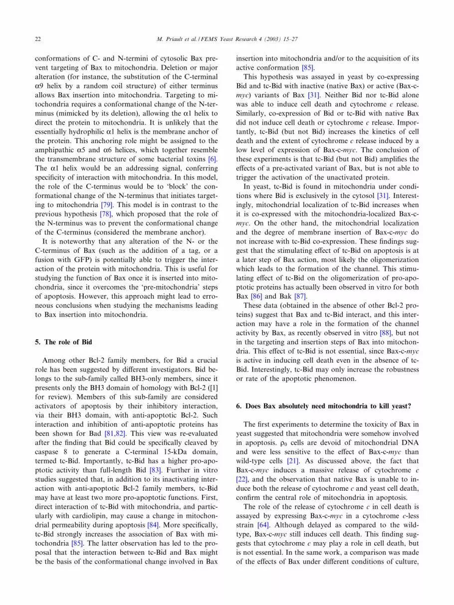
but not smac/diablo or AIF. On the basis of these obser-vations, we propose a two-step mechanism for the releaseof mitochondrial apoptogenic proteins (Fig. 1). In thismodel, Bax would participate in the formation of MAC,facilitating the release of cytochrome c. This step can bereproduced in yeast expressing c-myc-tagged Bax. In ac-cordance with the observations of Adrain et al. [58], oncecytosolic cytochrome c is able to activate caspases, theseproteases would attack mitochondria and allow furtherpermeabilization of the outer membrane [72]. This actionleads to the release of smac/diablo, AIF and endonucleaseG, by a mechanism that may be related to PTP opening[59]. Actually, the e¡ects of activated caspases on mito-chondrial permeability have often been observed [73].In this model, yeast is a powerful tool to investigate the
¢rst step (Bax-induced release of cytochrome c), indepen-dent of later steps linked to caspase activation and thepermeability transition, which never occurs in yeast [60].Work is under way, on the same heterologous system, toinvestigate di¡erent aspects of MAC function:b Is MAC composed of Bax alone, or does it includeother mitochondrial proteins?
b Which residues of Bax are involved in the channel ac-tivity?
b Is MAC activity su⁄cient to induce the release of cyto-chrome c, or are other phenomena, such as the destabi-lization of the interaction of cytochrome c with mito-chondrial inner membrane components, required?This latter aspect was recently highlighted by electron
tomography and reconstruction experiments [74] stronglysuggesting that remodeling of cristae might be a majorregulatory step of the release of cytochrome c. Therefore,MAC formation and activation might still not be enoughto induce the release of cytochrome c. It should be noted
that, although MAC does not require VDAC, since it wasevidenced in a VDAC-less strain, VDAC might play a rolein Bax e¡ects in yeast [75], although it is not mandatoryfor Bax-induced cell death [60]. Since VDAC is stronglypresent at the contact sites between outer and inner mem-branes, it is likely to play a role in the remodeling ofmitochondrial membranes. The comparison of Bax-in-duced morphological alterations of mitochondria fromwild-type and VDAC-less strains, as well as from mutantshaving alterations of their cristae [76], will help to inves-tigate this problem.
4. Addressing and insertion of Bax in the outermitochondrial membrane
4.1. Bcl-2, but not Bax, interacts with mitochondria via itsC-terminus
The 3D structure of Bax has been determined by nu-clear magnetic resonance spectroscopy [7]. Like othermembers of the Bcl-2 family, Bax is formed by K-helicesconnected by loops. The last K-helix, K9, has a markedlyhydrophobic nature, like the homologous helix of Bcl-2 orBcl-xL. In contrast to Bax, Bcl-2 (as well as Bcl-xL) spon-taneously inserts in membranes. It is hypothesized that theC-terminal K-helix of Bcl-2 serves both as a membrane-targeting signal and as a membrane anchor. This possibil-ity has been veri¢ed often, by the observation that C-ter-minally truncated variants of Bcl-2 (and Bcl-xL) lose theirability to insert into membranes in vitro and in vivo, con-comitantly with impairment of their anti-apoptotic activity[28,29]. The same is true in yeast. C-terminally truncatedvariants of Bcl-2 and Bcl-xL are unable to prevent the
Fig. 1. A two-step model for Bax-induced mitochondrial permeabilization. Bax inserts into the outer mitochondrial membrane and forms the MACwhich catalyzes the release of cytochrome c, through selective permeabilization of the outer mitochondrial membrane [70]. These steps can be repro-duced in yeast with a C-terminally altered Bax variant [70]. In mammalian cells, cytochrome c activates subsequent steps of apoptosis which, in return,induce the opening of the PTP, which allows the release of other regulators of apoptosis, such as AIF or smac/diablo [58,59]. This full permeabilizationof mitochondria is never observed in yeast [60].
FEMSYR 1576 23-9-03 Cyaan Magenta Geel Zwart
M. Priault et al. / FEMS Yeast Research 4 (2003) 15^27 19

e¡ects of the expression of Bax-c-myc on the release ofcytochrome c [32] and cell death [32,33].The well-accepted conclusion that the C-terminal hydro-
phobic K-helical domain of anti-apoptotic Bcl-2 (or Bcl-xL) is its membrane-targeting/insertion signal led to theextension of this hypothesis for all Bcl-2 family mem-bers. However, this e¡ect of the C-terminus has neverbeen demonstrated for Bax, and is not likely to be cor-rect.
4.2. The C-terminal K-helix of Bax is not a targeting signal,nor a membrane anchor
A Bax-c-myc variant was built lacking amino acids 172^192 and, hence, deprived of its C-terminal K-helix (termedBaxvC-c-myc). Following expression in yeast, this variantinduced the same e¡ects as full-length Bax-c-myc, namelyrelease of cytochrome c and cell death [32,33]. The mito-chondrial localization of the truncated protein was identi-cal to that of the full-length Bax.Since the addition of a c-myc already represented a
modi¢cation of the native structure of Bax, a new seriesof variants was built, on the basis of the untagged humanprotein (Bax). A ¢rst variant corresponded to a deletion ofthe C-terminal K-helix (BaxvC). In a second variant, theC-terminal K-helix was replaced by the correspondingsequence of anti-apoptotic Bcl-xL, which, as discussedabove, is a true membrane anchor (BaxXL). In a thirdvariant, the C-terminal K-helix was replaced by a hydro-philic sequence, predicted to acquire a random coiledstructure (BaxRC). Bax and its three variants were ex-pressed in yeast. As expected, unmodi¢ed Bax was notlethal in yeast [31]. As in healthy mammalian cells, Baxdid not localize to mitochondria, showing that this proteindoes not spontaneously interact with mitochondria inyeast or mammalian cells. However, all three Bax variantslocalized to mitochondria. This ¢nding is in striking con-tradiction to the hypothesis that the C-terminal K-helix isa membrane anchor. On the contrary, when the C-termi-nal K-helix is deleted or replaced, the protein gains mito-chondrial localization. Comparison of the predicted sec-ondary structure of the C-terminal end of Bax, BaxXLand BaxRC suggests that both the hydrophobicity andthe dipole moment may play a role in the ability of theprotein to interact with mitochondria. The hydrophobicityof the C-terminal end of Bax is higher than that ofBaxXL, which is higher than that of BaxRC. Also, thenet positive charge of the C-terminal end of Bax is slightlyhigher than that of BaxXL, which is considerably higherthan that of BaxRC. Both parameters correlate with theextent of the interaction of the Bax variant with mitochon-dria: the more hydrophobic and the more positivelycharged, the lower the interaction.The release of cytochrome c follows the extent of the
interaction with mitochondria for variants BaxvC andBaxRC. The relationship is less obvious for the variant
BaxXL, which induces a marginal release. The killing e⁄-ciency follows the extent of cytochrome c release, with astrong e¡ect of variants BaxvC and BaxRC, but no e¡ectof variant BaxXL or native Bax. BaxXL interacts withmitochondria but does not induce a signi¢cant release ofcytochrome c and does not kill yeast. It should be notedthat the same variant was assayed in mammalian cells,with a similar conclusion. This variant inserts into mito-chondria but does not have pro-apoptotic activity [73].This observation is crucial and shows that the address-ing/insertion of Bax into the mitochondrial outer mem-brane is a required but not a su⁄cient step to induce theapoptotic process.Taken together, these data suggest that the C-terminal
K-helix of Bax might play a role in its localization but,unlike the C-terminus of Bcl-xL, is not an addressing sig-nal or a membrane anchor. On the contrary, conforma-tional changes, mimicked by deletion or substitution,might be crucial for the addressing/insertion process.This conclusion is in striking contradiction with pre-
vious reports which concluded that the C-terminal K-helixof Bax was required for its insertion into the mitochon-drial membrane [26,77]. These data had been obtainedwith a GFP-Bax fusion protein, instead of a native Bax,like we report here. The fact that the role of the C-termi-nus in addressing/insertion is di¡erent for GFP-Bax andnative Bax supports the hypothesis that, in addition toconformational changes of the C-terminus, conformation-al changes of the N-terminus are involved in the trans-location of Bax to mitochondria.
4.3. Role of the N-terminus of Bax
Goping et al. [78] observed that deletion of the N-ter-minus of Bax (amino acids 1^20) increases the localizationof a Bax-GFP fusion protein to mitochondria. They con-cluded that this part of the protein inhibited Bax targetingto mitochondria and suggested that movement of theN-terminus would be required to allow the C-terminalK-helix to act as a membrane anchor.Two new variants were constructed and assayed in
yeast. A variant deprived of the N-terminus (amino acids1^20: BaxvN) and a variant deprived of both N- andC-termini (amino acids 1^20 and 176^192: BaxvNvC).Both variants have a mitochondrial localization, high cy-tochrome c release activity and strong killing e⁄ciency[79]. A similar behavior was found in mammalian cells[79,80]. These ¢ndings con¢rm the hypothesis that theN-terminus of Bax plays an inhibitory role, but, in accor-dance with the results observed on C-terminus variants,contradicts the hypothesis that the C-terminus is the mem-brane anchor.
4.4. The K1 helix may be the addressing signal
The observation that deletion of the N-terminus of Bax
FEMSYR 1576 23-9-03 Cyaan Magenta Geel Zwart
M. Priault et al. / FEMS Yeast Research 4 (2003) 15^2720

increases mitochondrial localization suggests that the ad-dressing sequence might lie in the N-terminus of BaxvN,corresponding to the K1 helix of Bax (amino acids 24^37).To verify this hypothesis, variants of BaxvN containingmutations in the K1 helix were constructed and assayed inyeast and in mammalian cells [79]. Two BaxvN variantsbearing a single mutation A24R or a double mutationL26G/L27V fail to localize to mitochondria in yeast. Athird variant, bearing the single mutation L26G, still ex-hibits mitochondrial localization [79].These three variants were also assayed in mammalian
cells. As in yeast, the variant BaxvN-A24R failed to local-ize to mitochondria. The results were slightly di¡erent forthe other mutants. The variant BaxvN-L26G was alteredin its interaction with mitochondria, whereas the doublemutant BaxvN-L26G/L27V was not, as compared toBaxvN [79,80]. Although the di¡erent variants behaveslightly di¡erently in yeast and mammals, these resultsstrongly suggest that this part of the protein has a functionin mitochondrial localization, both in mammalian and inyeast mitochondria.
4.5. A ‘prey-bird’ model
Taken together, all these results lead to the followingconclusions.b Unlike that of anti-apoptotic Bcl-2 and Bcl-xL, theC-terminal K9 helix of Bax is not a membrane-address-ing signal or a membrane anchor.
b Deletion or alteration of this C-terminal K9 helix in-creases the mitochondrial localization of the protein,suggesting that a conformational change of this partof the protein is required for mitochondrial targeting.
b Deletion of the N-terminus increases the mitochondriallocalization of the protein, suggesting that a conforma-tional change of this part of the protein is required formitochondrial addressing, independent of modi¢cationsof the C-terminus.
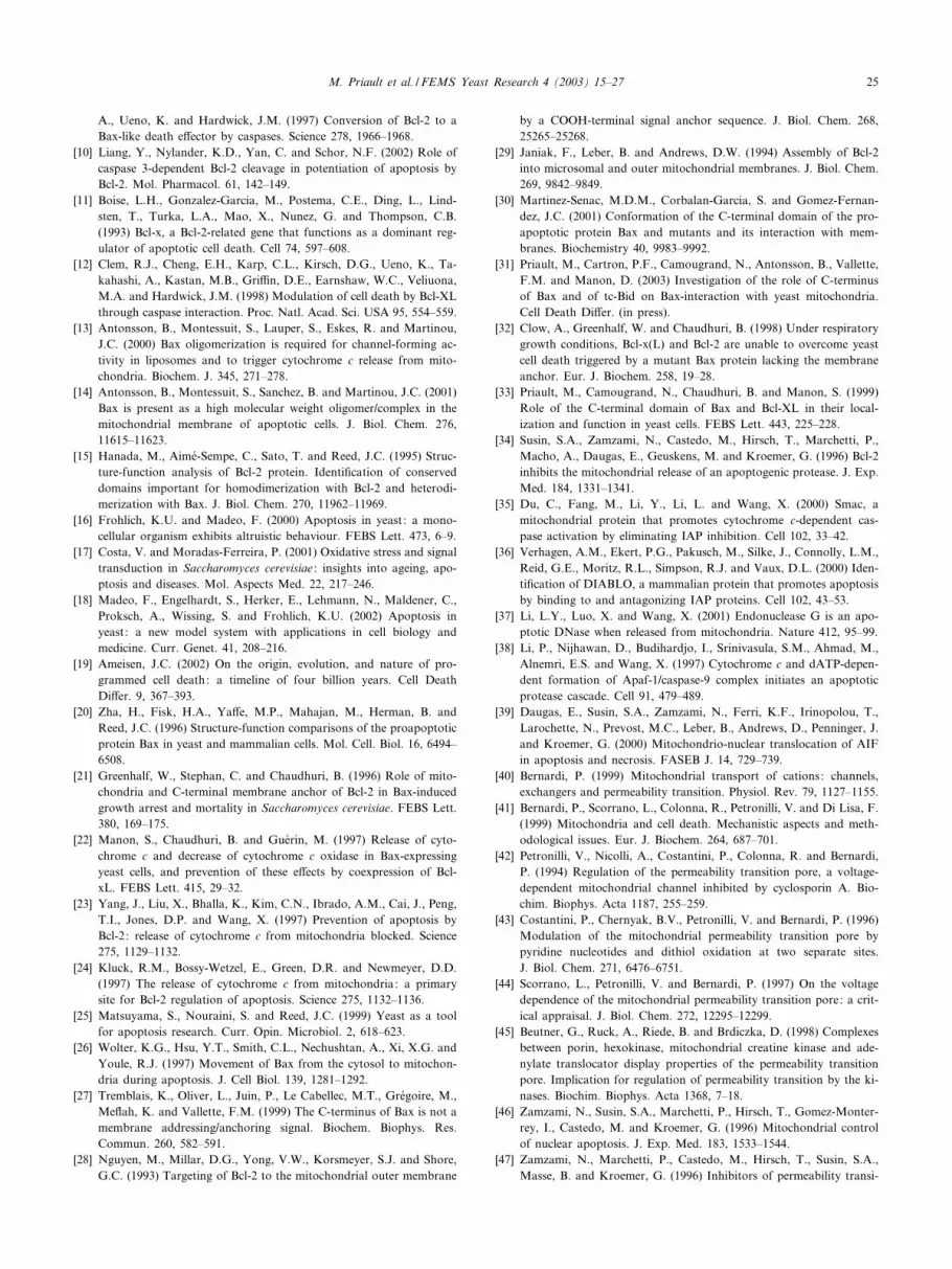
b Once the N-terminus is deleted (thus mimicking a prob-able conformational change), the K1 helix serves as amitochondrial target signal.A model for the targeting and insertion of Bax into
mitochondria is based on these data (Fig. 2). The native
Fig. 2. Models for the insertion of Bax in the outer mitochondrial membrane. In both hypotheses, the ¢rst step involves a movement of the ApoptoticRegulation of Targeting (ART) (blue) freeing the N- and C-termini. In the ¢rst model (top), the movement of ART allows the K9 helix (red) to insertinto the membrane, followed by the K5 (yellow) and K6 (orange) helices [78]. In the second model (bottom), the movement of both ART and K9 allowsthe K1 helix (green) to interact with a mitochondrial component, followed by the insertion of K5 and K6 helices [79]. In this model, the behavior of K9after the insertion of K5/K6 is not determined. Note that addition of a tag or a fusion protein at either end of Bax would force the movement of ARTand/or K9 thus favoring the insertion process.
FEMSYR 1576 23-9-03 Cyaan Magenta Geel Zwart
M. Priault et al. / FEMS Yeast Research 4 (2003) 15^27 21
conformations of C- and N-termini of cytosolic Bax pre-vent targeting of Bax to mitochondria. Deletion or majoralteration (for instance, the substitution of the C-terminalK9 helix by a random coil structure) of either terminusallows Bax insertion into mitochondria. Targeting to mi-tochondria requires a conformational change of the N-ter-minus (mimicked by its deletion), allowing the K1 helix todirect the protein to mitochondria. It is unlikely that theessentially hydrophilic K1 helix is the membrane anchor ofthe protein. This anchoring role might be assigned to theamphipathic K5 and K6 helices, which together resemblethe transmembrane structure of some bacterial toxins [6].The K1 helix would be an addressing signal, conferringspeci¢city of interaction with mitochondria. In this model,the role of the C-terminus would be to ‘block’ the con-formational change of the N-terminus that initiates target-ing to mitochondria [79]. This model is in contrast to theprevious hypothesis [78], which proposed that the role ofthe N-terminus was to prevent the conformational changeof the C-terminus (considered the membrane anchor).It is noteworthy that any alteration of the N- or the
C-terminus of Bax (such as the addition of a tag, or afusion with GFP) is potentially able to trigger the inter-action of the protein with mitochondria. This is useful forstudying the function of Bax once it is inserted into mito-chondria, since it overcomes the ‘pre-mitochondria’ stepsof apoptosis. However, this approach might lead to erro-neous conclusions when studying the mechanisms leadingto Bax insertion into mitochondria.
5. The role of Bid
Among other Bcl-2 family members, for Bid a crucialrole has been suggested by di¡erent investigators. Bid be-longs to the sub-family called BH3-only members, since itpresents only the BH3 domain of homology with Bcl-2 ([1]for review). Members of this sub-family are consideredactivators of apoptosis by their inhibitory interaction,via their BH3 domain, with anti-apoptotic Bcl-2. Suchinteraction and inhibition of anti-apoptotic proteins hasbeen shown for Bad [81,82]. This view was re-evaluatedafter the ¢nding that Bid could be speci¢cally cleaved bycaspase 8 to generate a C-terminal 15-kDa domain,termed tc-Bid. Importantly, tc-Bid has a higher pro-apo-ptotic activity than full-length Bid [83]. Further in vitrostudies suggested that, in addition to its inactivating inter-action with anti-apoptotic Bcl-2 family members, tc-Bidmay have at least two more pro-apoptotic functions. First,direct interaction of tc-Bid with mitochondria, and partic-ularly with cardiolipin, may cause a change in mitochon-drial permeability during apoptosis [84]. More speci¢cally,tc-Bid strongly increases the association of Bax with mi-tochondria [85]. The latter observation has led to the pro-posal that the interaction between tc-Bid and Bax mightbe the basis of the conformational change involved in Bax
insertion into mitochondria and/or to the acquisition of itsactive conformation [85].This hypothesis was assayed in yeast by co-expressing
Bid and tc-Bid with inactive (native Bax) or active (Bax-c-myc) variants of Bax [31]. Neither Bid nor tc-Bid alonewas able to induce cell death and cytochrome c release.Similarly, co-expression of Bid or tc-Bid with native Baxdid not induce cell death or cytochrome c release. Impor-tantly, tc-Bid (but not Bid) increases the kinetics of celldeath and the extent of cytochrome c release induced by alow level of expression of Bax-c-myc. The conclusion ofthese experiments is that tc-Bid (but not Bid) ampli¢es thee¡ects of a pre-activated variant of Bax, but is not able totrigger the activation of the unactivated protein.In yeast, tc-Bid is found in mitochondria under condi-
tions where Bid is exclusively in the cytosol [31]. Interest-ingly, mitochondrial localization of tc-Bid increases whenit is co-expressed with the mitochondria-localized Bax-c-myc. On the other hand, the mitochondrial localizationand the degree of membrane insertion of Bax-c-myc donot increase with tc-Bid co-expression. These ¢ndings sug-gest that the stimulating e¡ect of tc-Bid on apoptosis is ata later step of Bax action, most likely the oligomerizationwhich leads to the formation of the channel. This stimu-lating e¡ect of tc-Bid on the oligomerization of pro-apo-ptotic proteins has actually been observed in vitro for bothBax [86] and Bak [87].These data (obtained in the absence of other Bcl-2 pro-
teins) suggest that Bax and tc-Bid interact, and this inter-action may have a role in the formation of the channelactivity by Bax, as recently observed in vitro [88], but notin the targeting and insertion steps of Bax into mitochon-dria. This e¡ect of tc-Bid is not essential, since Bax-c-mycis active in inducing cell death even in the absence of tc-Bid. Interestingly, tc-Bid may only increase the robustnessor rate of the apoptotic phenomenon.
6. Does Bax absolutely need mitochondria to kill yeast?
The ¢rst experiments to determine the toxicity of Bax inyeast suggested that mitochondria were somehow involvedin apoptosis. b0 cells are devoid of mitochondrial DNAand were less sensitive to the e¡ect of Bax-c-myc thanwild-type cells [21]. As discussed above, the fact thatBax-c-myc induces a massive release of cytochrome c[22], and the observation that native Bax is unable to in-duce both the release of cytochrome c and yeast cell death,con¢rm the central role of mitochondria in apoptosis.The role of the release of cytochrome c in cell death is
assayed by expressing Bax-c-myc in a cytochrome c-lessstrain [64]. Although delayed as compared to the wild-type, Bax-c-myc still induces cell death. This ¢nding sug-gests that cytochrome c may play a role in cell death, butis not essential. In the same work, a comparison was madeof the e¡ects of Bax under di¡erent conditions of culture,
FEMSYR 1576 23-9-03 Cyaan Magenta Geel Zwart
M. Priault et al. / FEMS Yeast Research 4 (2003) 15^2722

from strict respiratory conditions (lactate as a carbonsource) to fermentative conditions (concentrated glucoseas a carbon source) and intermediate respiro-fermentativeconditions (galactose or mannose as carbon source). Asexpected, the killing e⁄ciency of Bax increased under res-piratory conditions, as compared to fermentative condi-tions [64]. Moreover, oxygen is absolutely required forkilling e¡ects since cell death did not occur under anaer-obic conditions [89].
6.1. The role of oxygen
On the basis of the observations that oxygen is abso-lutely required for Bax e¡ects and that H2O2 treatmentinduces the same apoptosis hallmarks as Bax expression,Madeo et al. [87] proposed a crucial role for reactive oxy-gen species (ROS). Actually, it is known that Bax expres-sion induces a massive increase in intracellular ROS con-centration. The main production site of ROS in the cell isthe mitochondrial respiratory chain. A simple hypothesiscan be proposed, on the basis of the release of cytochromec, to explain the massive ROS production which occursfollowing Bax expression. That is, partial blockade of thetransfer of electrons at the bc1 complex (caused by cyto-chrome c release and loss of this electron acceptor) leadsto an increase in the reduced state of quinones and partialreduction of molecular oxygen to superoxide ion by semi-ubiquinones, as happens for example in the presence ofantimycin A. The hypothesis of a role for ROS thus pro-vides a valuable explanation for the important, but notessential, roles of the release of cytochrome c and therequirement of a fully functional respiratory chain.However, ROS are not the only factor explaining the
role of oxygen. Preventing ROS production by chemicalreducers or overexpression of the scavenging enzymessuperoxide dismutase or catalase does not result in a sig-ni¢cant attenuation of the e¡ects of Bax [90]. Mitochon-drial lipid oxidation was also identi¢ed as an oxygen-de-pendent e¡ect of Bax, and preventing Bax-induced lipidoxidation actually delayed Bax-induced cell death [90].
From these data, the role of oxygen in Bax toxicityseems to be crucial at least in two ways. First, ROS pro-duction is linked to cytochrome c release, and second,lipid oxidation occurs, possibly caused by still unidenti¢edlipoxygenase activities.
6.2. Yeast cell death: apoptosis and/or autophagy?
Bax-induced cell death is accompanied by the hallmarksof apoptosis including cytochrome c release [22], DNAfragmentation and phosphatidylserine exposure [89,91].More recently, an additional apoptotic hallmark was ob-served under peculiar conditions. Bax expression is able tokill yeast under fermentative conditions (concentrated glu-cose as a carbon source) but dying cells have a plasmamembrane which remains impermeable to propidium io-dide [92], like mammalian apoptotic cells.At the molecular level, apoptosis is typically character-
ized by the activation of speci¢c proteases, particularlycaspases ([93,94] for reviews). Sequence homology search-ing does not reveal any obvious caspase homologs in theyeast genome. However, recent work supports a crucialrole for a protease having a caspase-like activity inH2O2-induced cell death [95], which exhibits features sim-ilar to Bax-induced cell death [89]. Other proteases mightalso play a function in Bax-induced cell death, such as theproteasome [96] and the mitochondrial AAA-type proteaseYme1p, which is activated by cytochrome c release [61,97].The mechanisms underlying the activation of these pro-teases and the way in which this activation is related tomitochondria remain to be determined.Alternatively, like all eukaryotic cells, yeast contains
another degradation pathway termed autophagy ([98] forreview). This pathway is activated by nitrogen starvationand is characterized by the formation of autophagosomescontaining intracellular structures, including mitochon-dria, which are ¢nally carried to the vacuole and degraded.In yeast, this pathway has been particularly well describedfrom genetic studies, and most of the proteins regulatingthe formation of autophagosomes have been identi¢ed.
Fig. 3. Bax e¡ects on yeast mitochondria. After addressing and insertion in the outer mitochondrial membrane, Bax is involved in MAC formation andcytochrome c release. Bax also activates a pre-existing pathway of cell death, involving Uth1p, which is physiologically activated by oxidative stress, se-nescence [107,108] and the TOR-dependent autophagy pathway [106] (reprinted from [106], with permission from the editor).
FEMSYR 1576 23-9-03 Cyaan Magenta Geel Zwart
M. Priault et al. / FEMS Yeast Research 4 (2003) 15^27 23

Experimentally, autophagy can also be activated by theinhibition of two kinases, Tor1p and Tor2p, by rapamycin[99].The literature suggests some relationship between apo-
ptosis and autophagy. For instance, the protein Apg5p,which is involved in the formation of autophagosomes[100], is overexpressed early during apoptosis [101]. Also,the Bcl-2-interacting protein Beclin-1 is the mammalianhomolog of yeast Apg6p, which is involved in autophagy[102]. A parallel regulation of autophagy and apoptosis isalso supported by their function in two cases [103,104] : inyeast, the protein Arl1p, which participates in the forma-tion of vacuoles, is involved in autophagy and modulatesBax-induced cell death [105]. A genetic screen of yeastmutations leading to Bax resistance identi¢ed a role forthe mitochondrial protein Uth1p in Bax-induced cell death[106]. This protein is also involved in mitochondrial auto-phagy ([106] and I. Kissova et al., unpublished data). Thisprovides an additional link between Bax and autophagysignaling. It should be noted that Uth1p is also involved instress responses and senescence [107^111]. Bax thereforeseems to be able to activate a signaling pathway whichleads to mitochondrial autophagy in response to di¡erentstimuli (Fig. 3).
6.3. One mitochondrion, two roads to cell death
Data reported above indicate that Bax can lead to yeastcell death by two di¡erent pathways. The ¢rst one mayinvolve the activation of proteases, particularly caspase-like Yca1p [95], by mitochondrial factors. As in mamma-lian apoptosis, cytochrome c may play a major role, but itis not obvious that this role is direct in yeast. The releaseof cytochrome c likely alters redox potentials, both inmitochondria and in the cytosol, and this alteration mightbe the factor activating Yca1p, which is involved in yeastresponse to moderate oxidative stress.Alternatively, there is a second pathway, which involves
the outer membrane protein Uth1p. This protein plays acrucial role in mitochondrial autophagy, suggesting astrong link between Bax-induced cell death and autopha-gy.
7. Conclusion
The central role of mitochondria has been largely docu-mented since the discovery of Bcl-2 family members and oftheir regulatory role in apoptosis. However, because of thecomplexity of the system, experiments done on mamma-lian cells have generated some perplexing data. Yeastpresents a less complex model system than mammaliancells and is therefore useful in gaining unambiguous an-swers concerning the function of individually expressedproteins. This review focuses on pro-apoptotic proteins,but it should be noted that similar experiments have pro-
vided knowledge of anti-apoptotic proteins [112]. Apartfrom contributing to understanding fundamental molecu-lar aspects of the function of Bcl-2 family members, theheterologous expression of these proteins in yeast has alsoprovided a tool to screen gene libraries, allowing identi¢-cation of new candidate proteins for the modulation ofBcl-2 family members [113,114].In addition to these aspects, relevant to the molecular
function of individual proteins, the heterologous expres-sion of Bax allows investigation of yeast death pathways.Bax expression clearly exhibits similar characteristics as‘physiological’ death triggers in yeast, such as moderateoxidative stress [89], acetic acid stress [97], and senescence[115]. Two works have recently established a connectionbetween this form of death and autophagy [105,106]. Thishas revealed a novel possible function for Bax in the au-tophagic process, in accordance with observations provid-ing a link between apoptosis and autophagy in mamma-lian cells.
Acknowledgements
Research in the laboratory of S.M. was supported bythe Centre National de la Recherche Scienti¢que, the As-sociation pour la Recherche contre le Cancer, the ConseilRe¤gional d’Aquitaine and the Universite¤ de Bordeaux 2.Research was also supported by the Ligue Nationalecontre le Cancer and the Conseil Re¤gional Bretagne-Paysde Loire to F.M.V. and by National Institute of HealthGrant GM57249 to K.W.K.
References
[1] Gross, A., McDonnell, J.M. and Korsmeyer, S.J. (1999) Bcl-2 familymembers and the mitochondria in apoptosis. Genes Dev. 13, 1899^1911.
[2] Adams, J.M. and Cory, S. (2001) Life-or-death decisions by the Bcl-2protein family. Trends Biochem. Sci. 26, 61^66.
[3] Mignotte, B. and Vayssie're, J.L. (1998) Mitochondria and apoptosis.Eur. J. Biochem. 252, 1^15.
[4] Cai, J., Yang, J. and Jones, D.P. (1998) Mitochondrial control ofapoptosis : the role of cytochrome c. Biochim. Biophys. Acta 1366,139^149.
[5] Desagher, S. and Martinou, J.C. (2000) Mitochondria as the centralcontrol point of apoptosis. Trends Cell Biol. 10, 369^377.
[6] Muchmore, S.W., Sattler, M., Liang, H., Meadows, R.P., Harlan,J.E., Yoon, H.S., Nettesheim, D.G., Chang, B.S., Thompson, C.B.,Wong, S.L., Ng, S.L. and Fesik, F.W. (1996) X-ray and NMR struc-ture of human Bcl-xL, an inhibitor of programmed cell death. Nature381, 335^341.
[7] Suzuki, M., Youle, R.J. and Tjandra, N. (2000) Structure of Bax:coregulation of dimer formation and intracellular localization. Cell103, 645^654.
[8] Petros, A.M., Medek, A., Nettesheim, D.G., Kim, D.H., Yoon, H.S.,Swift, K., Matayoshi, E.D., Oltersdorf, T. and Fesik, F.W. (2001)Solution structure of the antiapoptotic protein Bcl-2. Proc. Natl.Acad. Sci. USA 98, 3012^3017.
[9] Cheng, E.H., Kirsch, D.G., Clem, R.J., Ravi, R., Kastan, M.B., Bedi,
FEMSYR 1576 23-9-03 Cyaan Magenta Geel Zwart
M. Priault et al. / FEMS Yeast Research 4 (2003) 15^2724
A., Ueno, K. and Hardwick, J.M. (1997) Conversion of Bcl-2 to aBax-like death e¡ector by caspases. Science 278, 1966^1968.
[10] Liang, Y., Nylander, K.D., Yan, C. and Schor, N.F. (2002) Role ofcaspase 3-dependent Bcl-2 cleavage in potentiation of apoptosis byBcl-2. Mol. Pharmacol. 61, 142^149.
[11] Boise, L.H., Gonzalez-Garcia, M., Postema, C.E., Ding, L., Lind-sten, T., Turka, L.A., Mao, X., Nunez, G. and Thompson, C.B.(1993) Bcl-x, a Bcl-2-related gene that functions as a dominant reg-ulator of apoptotic cell death. Cell 74, 597^608.
[12] Clem, R.J., Cheng, E.H., Karp, C.L., Kirsch, D.G., Ueno, K., Ta-kahashi, A., Kastan, M.B., Gri⁄n, D.E., Earnshaw, W.C., Veliuona,M.A. and Hardwick, J.M. (1998) Modulation of cell death by Bcl-XLthrough caspase interaction. Proc. Natl. Acad. Sci. USA 95, 554^559.
[13] Antonsson, B., Montessuit, S., Lauper, S., Eskes, R. and Martinou,J.C. (2000) Bax oligomerization is required for channel-forming ac-tivity in liposomes and to trigger cytochrome c release from mito-chondria. Biochem. J. 345, 271^278.
[14] Antonsson, B., Montessuit, S., Sanchez, B. and Martinou, J.C. (2001)Bax is present as a high molecular weight oligomer/complex in themitochondrial membrane of apoptotic cells. J. Biol. Chem. 276,11615^11623.
[15] Hanada, M., Aime¤-Sempe, C., Sato, T. and Reed, J.C. (1995) Struc-ture-function analysis of Bcl-2 protein. Identi¢cation of conserveddomains important for homodimerization with Bcl-2 and heterodi-merization with Bax. J. Biol. Chem. 270, 11962^11969.
[16] Frohlich, K.U. and Madeo, F. (2000) Apoptosis in yeast: a mono-cellular organism exhibits altruistic behaviour. FEBS Lett. 473, 6^9.
[17] Costa, V. and Moradas-Ferreira, P. (2001) Oxidative stress and signaltransduction in Saccharomyces cerevisiae : insights into ageing, apo-ptosis and diseases. Mol. Aspects Med. 22, 217^246.
[18] Madeo, F., Engelhardt, S., Herker, E., Lehmann, N., Maldener, C.,Proksch, A., Wissing, S. and Frohlich, K.U. (2002) Apoptosis inyeast: a new model system with applications in cell biology andmedicine. Curr. Genet. 41, 208^216.
[19] Ameisen, J.C. (2002) On the origin, evolution, and nature of pro-grammed cell death: a timeline of four billion years. Cell DeathDi¡er. 9, 367^393.
[20] Zha, H., Fisk, H.A., Ya¡e, M.P., Mahajan, M., Herman, B. andReed, J.C. (1996) Structure-function comparisons of the proapoptoticprotein Bax in yeast and mammalian cells. Mol. Cell. Biol. 16, 6494^6508.
[21] Greenhalf, W., Stephan, C. and Chaudhuri, B. (1996) Role of mito-chondria and C-terminal membrane anchor of Bcl-2 in Bax-inducedgrowth arrest and mortality in Saccharomyces cerevisiae. FEBS Lett.380, 169^175.
[22] Manon, S., Chaudhuri, B. and Gue¤rin, M. (1997) Release of cyto-chrome c and decrease of cytochrome c oxidase in Bax-expressingyeast cells, and prevention of these e¡ects by coexpression of Bcl-xL. FEBS Lett. 415, 29^32.
[23] Yang, J., Liu, X., Bhalla, K., Kim, C.N., Ibrado, A.M., Cai, J., Peng,T.I., Jones, D.P. and Wang, X. (1997) Prevention of apoptosis byBcl-2: release of cytochrome c from mitochondria blocked. Science275, 1129^1132.
[24] Kluck, R.M., Bossy-Wetzel, E., Green, D.R. and Newmeyer, D.D.(1997) The release of cytochrome c from mitochondria: a primarysite for Bcl-2 regulation of apoptosis. Science 275, 1132^1136.
[25] Matsuyama, S., Nouraini, S. and Reed, J.C. (1999) Yeast as a toolfor apoptosis research. Curr. Opin. Microbiol. 2, 618^623.
[26] Wolter, K.G., Hsu, Y.T., Smith, C.L., Nechushtan, A., Xi, X.G. andYoule, R.J. (1997) Movement of Bax from the cytosol to mitochon-dria during apoptosis. J. Cell Biol. 139, 1281^1292.
[27] Tremblais, K., Oliver, L., Juin, P., Le Cabellec, M.T., Gre¤goire, M.,Me£ah, K. and Vallette, F.M. (1999) The C-terminus of Bax is not amembrane addressing/anchoring signal. Biochem. Biophys. Res.Commun. 260, 582^591.
[28] Nguyen, M., Millar, D.G., Yong, V.W., Korsmeyer, S.J. and Shore,G.C. (1993) Targeting of Bcl-2 to the mitochondrial outer membrane
by a COOH-terminal signal anchor sequence. J. Biol. Chem. 268,25265^25268.
[29] Janiak, F., Leber, B. and Andrews, D.W. (1994) Assembly of Bcl-2into microsomal and outer mitochondrial membranes. J. Biol. Chem.269, 9842^9849.
[30] Martinez-Senac, M.D.M., Corbalan-Garcia, S. and Gomez-Fernan-dez, J.C. (2001) Conformation of the C-terminal domain of the pro-apoptotic protein Bax and mutants and its interaction with mem-branes. Biochemistry 40, 9983^9992.
[31] Priault, M., Cartron, P.F., Camougrand, N., Antonsson, B., Vallette,F.M. and Manon, D. (2003) Investigation of the role of C-terminusof Bax and of tc-Bid on Bax-interaction with yeast mitochondria.Cell Death Di¡er. (in press).
[32] Clow, A., Greenhalf, W. and Chaudhuri, B. (1998) Under respiratorygrowth conditions, Bcl-x(L) and Bcl-2 are unable to overcome yeastcell death triggered by a mutant Bax protein lacking the membraneanchor. Eur. J. Biochem. 258, 19^28.
[33] Priault, M., Camougrand, N., Chaudhuri, B. and Manon, S. (1999)Role of the C-terminal domain of Bax and Bcl-XL in their local-ization and function in yeast cells. FEBS Lett. 443, 225^228.
[34] Susin, S.A., Zamzami, N., Castedo, M., Hirsch, T., Marchetti, P.,Macho, A., Daugas, E., Geuskens, M. and Kroemer, G. (1996) Bcl-2inhibits the mitochondrial release of an apoptogenic protease. J. Exp.Med. 184, 1331^1341.
[35] Du, C., Fang, M., Li, Y., Li, L. and Wang, X. (2000) Smac, amitochondrial protein that promotes cytochrome c-dependent cas-pase activation by eliminating IAP inhibition. Cell 102, 33^42.
[36] Verhagen, A.M., Ekert, P.G., Pakusch, M., Silke, J., Connolly, L.M.,Reid, G.E., Moritz, R.L., Simpson, R.J. and Vaux, D.L. (2000) Iden-ti¢cation of DIABLO, a mammalian protein that promotes apoptosisby binding to and antagonizing IAP proteins. Cell 102, 43^53.
[37] Li, L.Y., Luo, X. and Wang, X. (2001) Endonuclease G is an apo-ptotic DNase when released from mitochondria. Nature 412, 95^99.
[38] Li, P., Nijhawan, D., Budihardjo, I., Srinivasula, S.M., Ahmad, M.,Alnemri, E.S. and Wang, X. (1997) Cytochrome c and dATP-depen-dent formation of Apaf-1/caspase-9 complex initiates an apoptoticprotease cascade. Cell 91, 479^489.
[39] Daugas, E., Susin, S.A., Zamzami, N., Ferri, K.F., Irinopolou, T.,Larochette, N., Prevost, M.C., Leber, B., Andrews, D., Penninger, J.and Kroemer, G. (2000) Mitochondrio-nuclear translocation of AIFin apoptosis and necrosis. FASEB J. 14, 729^739.
[40] Bernardi, P. (1999) Mitochondrial transport of cations: channels,exchangers and permeability transition. Physiol. Rev. 79, 1127^1155.
[41] Bernardi, P., Scorrano, L., Colonna, R., Petronilli, V. and Di Lisa, F.(1999) Mitochondria and cell death. Mechanistic aspects and meth-odological issues. Eur. J. Biochem. 264, 687^701.
[42] Petronilli, V., Nicolli, A., Costantini, P., Colonna, R. and Bernardi,P. (1994) Regulation of the permeability transition pore, a voltage-dependent mitochondrial channel inhibited by cyclosporin A. Bio-chim. Biophys. Acta 1187, 255^259.
[43] Costantini, P., Chernyak, B.V., Petronilli, V. and Bernardi, P. (1996)Modulation of the mitochondrial permeability transition pore bypyridine nucleotides and dithiol oxidation at two separate sites.J. Biol. Chem. 271, 6476^6751.
[44] Scorrano, L., Petronilli, V. and Bernardi, P. (1997) On the voltagedependence of the mitochondrial permeability transition pore: a crit-ical appraisal. J. Biol. Chem. 272, 12295^12299.
[45] Beutner, G., Ruck, A., Riede, B. and Brdiczka, D. (1998) Complexesbetween porin, hexokinase, mitochondrial creatine kinase and ade-nylate translocator display properties of the permeability transitionpore. Implication for regulation of permeability transition by the ki-nases. Biochim. Biophys. Acta 1368, 7^18.
[46] Zamzami, N., Susin, S.A., Marchetti, P., Hirsch, T., Gomez-Monter-rey, I., Castedo, M. and Kroemer, G. (1996) Mitochondrial controlof nuclear apoptosis. J. Exp. Med. 183, 1533^1544.
[47] Zamzami, N., Marchetti, P., Castedo, M., Hirsch, T., Susin, S.A.,Masse, B. and Kroemer, G. (1996) Inhibitors of permeability transi-
FEMSYR 1576 23-9-03 Cyaan Magenta Geel Zwart
M. Priault et al. / FEMS Yeast Research 4 (2003) 15^27 25

tion interfere with the disruption of the mitochondrial transmem-brane potential during apoptosis. FEBS Lett. 384, 53^57.
[48] Marzo, I., Brenner, C., Zamzami, N., Jurgensmeier, J.M., Susin,S.A., Vieira, H.L., Prevost, M.C., Xie, Z., Matsuyama, S., Reed,J.C. and Kroemer, G. (1998) Bax and adenine nucleotide translocatorcooperate in the mitochondrial control of apoptosis. Science 281,2027^2031.
[49] Zamzami, N., Marzo, I., Susin, S.A., Brenner, C., Larochette, N.,Marchetti, P., Reed, J.C., Ko£er, R. and Kroemer, G. (1998) Thethiol crosslinking agent diamide overcomes the apoptosis-inhibitorye¡ect of Bcl-2 by enforcing mitochondrial permeability transition.Oncogene 16, 1055^1063.
[50] Vancompernolle, K., Van Herreweghe, F., Pynaert, G., Van deCraen, M., De Vos, K., Totty, N., Sterling, A., Fiers, W., Vanden-abeele, P. and Grooten, J. (1998) Atractyloside-induced release ofcathepsin B, a protease with caspase-processing activity. FEBSLett. 438, 150^158.
[51] Guicciardi, M.E., Deussing, J., Miyoshi, H., Bronk, S.F., Svingen,P.A., Peters, C., Kaufmann, S.H. and Gores, G.J. (2001) Cathepsin Bcontributes to TNF-alpha-mediated hepatocyte apoptosis by promot-ing mitochondrial release of cytochrome c. J. Clin. Invest. 107, 21^22.
[52] Schotte, P., Declercq, W., Van Hu¡el, S., Vandenabeele, P. andBeyaert, R. (1999) Non-speci¢c e¡ects of methyl ketone peptide in-hibitors of caspases. FEBS Lett. 442, 117^121.
[53] Wigdal, S.S., Kirkland, R.A., Franklin, J.L. and Haak-Frendscho,M. (2002) Cytochrome c release precedes mitochondrial membranepotential loss in cerebellar granule neuron apoptosis : lack of mito-chondrial swelling. J. Neurochem. 82, 1029^1038.
[54] Vander Heiden, M.G., Chandel, N.S., Williamson, E.K., Schumack-er, P.T. and Thompson, C.B. (1997) Bcl-xL regulates the membranepotential and volume homeostasis of mitochondria. Cell 91, 627^637.
[55] Eskes, R., Antonsson, B., Osen-Sand, A., Montessuit, S., Richter, C.,Sadoul, R., Mazzei, G., Nichols, A. and Martinou, J.C. (1998) Bax-induced cytochrome c release from mitochondria is independent ofthe permeability transition pore but highly dependent on Mg2þ ions.J. Cell Biol. 143, 217^224.
[56] Kluck, R.M., Esposti, M.D., Perkins, G., Renken, C., Kuwana, T.,Bossy-Wetzel, E., Goldberg, M., Allen, T., Barber, M.J., Green, D.R.and Newmeyer, D.D. (1999) The pro-apoptotic proteins, Bid andBax, cause a limited permeabilization of the mitochondrial outermembrane that is enhanced by cytosol. J. Cell Biol. 147, 809^822.
[57] Appaix, F., Guerrero, K., Rampal, D., Izikki, M., Kaambre, T.,Sikk, P., Brdiczska, D., Riva-Lavieille, C., Olivares, J., Longuet,M., Antonsson, B. and Saks, V.A. (2002) Bax and heart mitochon-dria: uncoupling and inhibition of respiration without permeabilitytransition. Biochim. Biophys. Acta 1556, 155^167.
[58] Adrain, C., Creagh, E.M. and Martin, S.J. (2001) Apoptosis-associ-ated release of Smac/DIABLO from mitochondria requires activecaspases and is blocked by Bcl-2. EMBO J. 20, 6627^6636.
[59] Arnoult, D., Parone, P., Martinou, J.C., Antonsson, B., Estaquier, J.and Ameisen, J.C. (2002) Mitochondrial release of apoptosis-induc-ing factor occurs downstream of cytochrome c release in response toseveral proapoptotic stimuli. J. Cell Biol. 159, 923^929.
[60] Priault, M., Chaudhuri, B., Clow, A., Camougrand, N. and Manon,S. (1999) Investigation of Bax-induced release of cytochrome c fromyeast mitochondria permeability of mitochondrial membranes, role ofVDAC and ATP requirement. Eur. J. Biochem. 260, 684^691.
[61] Manon, S., Priault, M. and Camougrand, N. (2001) MitochondrialAAA-type protease Yme1p is involved in Bax e¡ects on cytochrome coxidase. Biochem. Biophys. Res. Commun. 289, 1314^1319.
[62] Jung, D.W., Bradshaw, P.C. and Pfei¡er, D.R. (1997) Properties of acyclosporin-insensitive permeability transition pore in yeast mito-chondria. J. Biol. Chem. 272, 21104^21112.
[63] Manon, S., Roucou, X., Gue¤rin, M., Rigoulet, M. and Gue¤rin, B.(1998) Characterization of the yeast mitochondria unselective chan-nel: a counterpart to the mammalian permeability transition pore?J. Bioenerg. Biomembr. 30, 419^429.
[64] Priault, M., Camougrand, N., Chaudhuri, B., Schae¡er, J. and Ma-non, S. (1999) Comparison of the e¡ects of Bax-expression in yeastunder fermentative and respiratory conditions: investigation of therole of adenine nucleotides carrier and cytochrome c. FEBS Lett. 456,232^238.
[65] Gross, A., Pilcher, K., Blachly-Dyson, E., Basso, E., Jockel, J., Bas-sik, M.C., Korsmeyer, S.J. and Forte, M. (2000) Biochemical andgenetic analysis of the mitochondrial response of yeast to BAX andBCL-XL. Mol. Cell. Biol. 20, 3125^3136.
[66] Kissova, I., Polcic, P., Kempna, P., Zeman, I., Sabova, L. and Ko-larov, J. (2000) The cytotoxic action of Bax on yeast cells does notrequire mitochondrial ADP/ATP carrier but may be related to itsimport to the mitochondria. FEBS Lett. 471, 113^118.
[67] Shimizu, S., Narita, M. and Tsujimoto, Y. (2000) Bcl-2 family pro-teins regulate the release of apoptogenic cytochrome c by the mito-chondrial channel VDAC. Nature 399, 483^487.
[68] Antonsson, B., Conti, F., Ciavatta, A., Montessuit, S., Lewis, S.,Martinou, I., Bernasconi, L., Bernard, A., Mermod, J.J., Mazzei,G., Maundrell, K., Gambale, F., Sadoul, R. and Martinou, J.C.(1997) Inhibition of Bax channel-forming activity by Bcl-2. Science277, 370^372.
[69] Schlesinger, P.H., Gross, A., Yin, X.M., Yamamoto, K., Saito, M.,Waksman, G. and Korsmeyer, S.J. (1997) Comparison of the ionchannel characteristics of proapoptotic BAX and antiapoptoticBCL-2. Proc. Natl. Acad. Sci. USA 94, 11357^11362.
[70] Pavlov, E.V., Priault, M., Pietkiewicz, D., Cheng, E.H., Antonsson,B., Manon, S., Korsmeyer, S.J., Mannella, C.A. and Kinnally, K.W.(2001) A novel, high-conductance channel of mitochondria linked toapoptosis in mammalian cells and Bax expression in yeast. J. CellBiol. 155, 725^731.
[71] Roucou, X., Prescott, M., Devenish, R.J. and Nagley, P. (2000) Acytochrome c-GFP fusion is not released from mitochondria into thecytoplasm upon expression of Bax in yeast cells. FEBS Lett. 471,235^239.
[72] Lassus, P., Opitz-Araya, X. and Lazebnik, Y. (2002) Requirement forcaspase-2 in stress-induced apoptosis before mitochondrial permeabi-lization. Science 297, 1352^1354.
[73] Oliver, L., Priault, M., Tremblais, L., Le Cabellec, M.T., Me£ah, K.,Manon, S. and Vallette, F.M. (2000) The substitution of the C-ter-minus of Bax by that of Bcl-XL does not a¡ect its subcellular local-ization but abrogates its pro-apoptotic properties. FEBS Lett. 487,161^165.
[74] Scorrano, L., Ashiya, M., Buttle, K., Weiler, S., Oakes, S.A., Man-nella, C.A. and Korsmeyer, S.J. (2002) A distinct pathway remodelsmitochondrial cristae and mobilizes cytochrome c during apoptosis.Dev. Cell 2, 55^67.
[75] Shimizu, S., Shinohara, Y. and Tsujimoto, Y. (2000) Bax and Bcl-xLindependently regulate apoptotic changes of yeast mitochondria thatrequire VDAC but not adenine nucleotide translocator. Oncogene 19,4309^4318.
[76] Paumard, P., Vaillier, J., Coulary, B., Schae¡er, J., Soubannier, V.,Mueller, D.M., Brethes, D., Di Rago, J.P. and Velours, J. (2002) TheATP synthase is involved in generating mitochondrial cristae mor-phology. EMBO J. 21, 221^230.
[77] Nechushtan, A., Smith, C.L., Hsu, Y.T. and Youle, R.J. (1999) Con-formation of the Bax C-terminus regulates subcellular location andcell death. EMBO J. 18, 2330^2341.
[78] Goping, I.S., Gross, A., Lavoie, J.N., Nguyen, M., Jemmerson, R.,Roth, K., Korsmeyer, S.J. and Shore, G.C. (1998) Regulated target-ing of BAX to mitochondria. J. Cell Biol. 143, 207^215.
[79] Cartron, P.F., Priault, M., Oliver, L., Me£ah, K., Manon, S. andVallette, F.M. (2003) The N-terminal end of Bax contains a mito-chondrial-targeting signal. J. Biol. Chem. 278, 11633^11641.
[80] Cartron, P.F., Moreau, C., Oliver, L., Mayat, E., Me£ah, K. andVallette, F.M. (2002) Involvement of the N-terminus of Bax in itsintracellular localization and function. FEBS Lett. 512, 95^100.
[81] Zha, J., Harada, H., Osipov, K., Jockel, J., Waksman, G. and Kors-
FEMSYR 1576 23-9-03 Cyaan Magenta Geel Zwart
M. Priault et al. / FEMS Yeast Research 4 (2003) 15^2726

meyer, S.J. (1997) BH3 domain of BAD is required for heterodimer-ization with BCL-XL and pro-apoptotic activity. J. Biol. Chem. 272,24101^24104.
[82] Ottilie, S., Diaz, J.L., Horne, W., Chang, J., Wang, Y., Wilson, G.,Chang, S., Weeks, S., Fritz, L.C. and Oltersdorf, T. (1997) Dimeri-zation properties of human BAD. Identi¢cation of a BH-3 domainand analysis of its binding to mutant BCL-2 and BCL-XL proteins.J. Biol. Chem. 272, 30866^30972.
[83] Li, H., Zhu, H., Xu, C.J. and Yuan, J. (1998) Cleavage of BID bycaspase 8 mediates the mitochondrial damage in the Fas pathway ofapoptosis. Cell 94, 491^501.
[84] Lutter, M., Fang, M., Luo, X., Nishijima, M., Xie, X. and Wang, X.(2002) Cardiolipin provides speci¢city for targeting of tBid to mito-chondria. Nat. Cell Biol. 2, 754^761.
[85] Eskes, R., Desagher, S., Antonsson, B. and Martinou, J.C. (2000) Bidinduces the oligomerization and insertion of Bax into the outer mi-tochondrial membrane. Mol. Cell. Biol. 20, 929^935.
[86] Roucou, X., Rostovtseva, T., Montessuit, S., Martinou, J.C. andAntonsson, B. (2002) Bid induces cytochrome c-impermeable Baxchannels in liposomes. Biochem. J. 363, 547^552.
[87] Wei, M.C., Lindsten, T., Mootha, V.K., Weiler, S., Gross, A.,Ashiya, M., Thompson, C.B. and Korsmeyer, S.J. (2000) tBid, amembrane-targeted death ligand, oligomerizes BAK to release cyto-chrome c. Genes Dev. 14, 2060^2071.
[88] Kuwana, T., Mackey, M.R., Perkins, G., Ellisman, M.H., Latterich,M., Schneiter, R., Green, D.R. and Newmeyer, D.D. (2002) Bid, Baxand lipids cooperate to form supramolecular openings in the outermitochondrial membrane. Cell 111, 331^342.
[89] Madeo, F., Frohlich, E., Ligr, M., Grey, M., Sigrist, S.J., Wolf, D.H.and Frohlich, K.U. (1999) Oxygen stress: a regulator of apoptosis inyeast. J. Cell Biol. 145, 757^767.
[90] Priault, M., Bessoule, J.J., Grelaud-Coq, A., Camougrand, N. andManon, S. (2002) Bax-induced cell death in yeast depends on mito-chondrial lipid oxidation. Eur. J. Biochem. 269, 5440^5450.
[91] Ligr, M., Madeo, F., Frohlich, E., Hilt, W., Frohlich, K.U. andWolf, D.H. (1998) Mammalian Bax triggers apoptotic changes inyeast. FEBS Lett. 438, 61^65.
[92] Marza, E., Camougrand, N. and Manon, S. (2002) Bax expressionprotects yeast plasma membrane against ethanol-induced permeabili-zation. FEBS Lett. 521, 47^52.
[93] Nun‹ez, G., Benedict, M.A., Hu, Y. and Inohara, N. (1998) Cas-pases: the proteases of the apoptotic pathway. Oncogene 17, 3237^3245.
[94] Stennicke, H.R. and Salvesen, G.S. (1999) Catalytic properties of thecaspases. Cell Death Di¡er. 6, 1054^1059.
[95] Madeo, F., Herker, E., Maldener, C., Wissing, S., Lachelt, S., Her-lan, M., Fehr, M., Lauber, K., Sigrist, S.J., Wesselborg, S. and Froh-lich, K.U. (2002) A caspase-related protease regulates apoptosis inyeast. Mol. Cell 9, 911^917.
[96] Ligr, M., Velten, I., Frohlich, E., Madeo, F., Ledig, M., Frohlich,K.U., Wolf, D.H. and Hilt, W. (2001) The proteasomal substrateStm1 participates in apoptosis-like cell death in yeast. Mol. Biol.Cell 12, 2422^2432.
[97] Ludovico, P., Rodrigues, F., Almeida, A., Silva, M.T., Barrientos, A.and Co“rte-Real, M. (2002) Cytochrome c release and mitochondriainvolvement in programmed cell death induced by acetic acid in Sac-charomyces cerevisiae. Mol. Biol. Cell 13, 2598^2606.
[98] Abeliovich, H. and Klionsky, D.J. (2001) Autophagy in yeast: mech-anistic insights and physiological function. Microbiol. Mol. Biol.Rev. 65, 463^479.
[99] Beck, T. and Hall, M.N. (1999) The TOR signalling pathway con-trols nuclear localization of nutrient-regulated transcription factors.Nature 402, 689^692.
[100] George, M.D., Baba, M., Scott, S.V., Mizushima, M., Garrisson,B.S., Ohsumi, Y. and Kionsky, D.J. (2000) Apg5p functions in thesequestration steps in the cytoplasm-to-vacuole targeting and mac-roautophagy pathways. Mol. Biol. Cell 11, 969^982.
[101] Hammond, E.M., Brunet, C.L., Johnson, G.D., Parkhill, J., Milner,A.E., Brady, G., Gregory, C.D. and Grand, R.J. (1998) Homologybetween a human apoptosis-speci¢c protein and the product ofAPG5, a gene involved in autophagy in yeast. FEBS Lett. 425,391^395.
[102] Liang, X.H., Jackson, S., Seaman, M., Brown, K., Kempkes, B.,Hibshoosh, H. and Levine, B. (1999) Induction of autophagy andinhibition of tumorigenesis by beclin-1. Nature 402, 672^676.
[103] Xue, L., Fletcher, G.C. and Tolkovsky, A.M. (1999) Autophagy isactivated by apoptotic signalling in sympathetic neurons: an alter-native mechanism of death execution. Mol. Cell. Neurosci. 14, 180^198.
[104] Petiot, A., Ogier-Denis, E., Blommaart, F., Meijer, A.J. and Codog-no, P. (2000) Distinct classes of phosphatidylinositol 3P-kinases areinvolved in signalling pathways that control macroautophagy inHT-29 cells. J. Biol. Chem. 275, 992^998.
[105] Abudugupur, A., Mitsui, K., Yokota, S. and Tsurugi, K. (2002) AnARL1 mutation a¡ected autophagic cell death in yeast, causing adefect in central vacuole formation. Cell Death Di¡er. 9, 158^168.
[106] Camougrand, N., Grelaud-Coq, A., Marza, E., Priault, M., Bes-soule, J.J. and Manon, S. (2003) The product of the UTH1 gene,required for Bax-induced cell death in yeast, is involved in the re-sponse to rapamycin. Mol. Microbiol. 47, 495^506.
[107] Bandara, P.D., Flattery-O’Brien, J.A., Grant, C.M. and Dawes,I.W. (1998) Involvement of the Saccharomyces cerevisiae UTH1gene in the oxidative-stress response. Curr. Genet. 34, 259^268.
[108] Camougrand, N., Mouassite, M., Velours, G. and Gue¤rin, M.(2000) The ‘SUN’ family: UTH1, an ageing gene, is also involvedin the regulation of mitochondria biogenesis in Saccharomyces cere-visiae. Arch. Biochem. Biophys. 375, 154^160.
[109] Mouassite, M., Gue¤rin, M. and Camougrand, N. (2000) The SUNfamily of Saccharomyces cerevisiae : the double knock-out of UTH1and SIM1 promotes defects in nucleus migration and increased drugsensitivity. FEMS Microbiol. Lett. 182, 137^141.
[110] Camougrand, N. and Rigoulet, M. (2001) Studies of some genesinvolved both in aging and in response to oxidative stress. Respir.Physiol. 128, 393^401.
[111] Velours, G., Boucheron, C., Manon, S. and Camougrand, N. (2002)Dual cell wall/mitochondria localization of the ‘SUN’ family pro-teins. FEMS Microbiol. Lett. 207, 165^172.
[112] Longo, V.D., Ellerby, L.M., Bredesen, D.E., Valentine, J.S. andGralla, E.B. (1997) Human Bcl-2 reverses survival defects in yeastlacking superoxide dismutase and delays death of wild-type yeast.J. Cell Biol. 137, 1581^1588.
[113] Greenhalf, W., Lee, J. and Chaudhuri, B. (1999) A selection systemfor human apoptosis inhibitors using yeast. Yeast 15, 1307^1321.
[114] Xu, Q. and Reed, J.C. (1998) Bax inhibitor-1, a mammalian apo-ptosis suppressor identi¢ed by functional screening in yeast. Mol.Cell 1, 337^346.
[115] Laun, P., Pichova, A., Madeo, F., Fuchs, J., Ellinger, A., Kohlwein,S., Dawes, I., Frohlich, K.U. and Breitenbach, M. (2001) Agedmother cells of Saccharomyces cerevisiae show markers of oxidativestress and apoptosis. Mol. Microbiol. 39, 1166^1173.
FEMSYR 1576 23-9-03 Cyaan Magenta Geel Zwart
M. Priault et al. / FEMS Yeast Research 4 (2003) 15^27 27
Copyright © 2022 FDOKUMEN