Stretch-mediated release of angiotensin II induces myocyte apoptosis by activating p53 that enhances...
17
1326 Leri et al. J. Clin. Invest. © The American Society for Clinical Investigation, Inc. 0021-9738/98/04/1326/17 $2.00 Volume 101, Number 7, April 1998, 1326–1342 http://www.jci.org Stretch-mediated Release of Angiotensin II Induces Myocyte Apoptosis by Activating p53 That Enhances the Local Renin-Angiotensin System and Decreases the Bcl-2-to-Bax Protein Ratio in the Cell Annarosa Leri,* Pier Paolo Claudio, ‡ Qiong Li,* Xiaowei Wang,* Krzysztof Reiss,* Shenglun Wang,* Ashwani Malhotra,* Jan Kajstura,* and Piero Anversa* *Department of Medicine, New York Medical College, Valhalla, New York 10595; and ‡ Departments of Pathology, Anatomy, and Cell Biology and Institute for Cancer Research and Molecular Medicine, Jefferson Medical College, Philadelphia, Pennsylvania 19107 Abstract Physical forces activate apoptosis and gene expression, but the mechanism is unknown. For this purpose, adult myo- cytes were stretched in an equibiaxial stretch apparatus and the magnitude of cell death was examined 4 and 24 h later. The possibility of stretch-mediated activation of p53 and p53-dependent genes was evaluated at 30 min, 2, 4, 8, and 24 h. Myocyte apoptosis increased by 4.4- and 7.6-fold at 4 and 24 h after stretch. p53 binding to the promoter of angio- tensinogen, AT 1 receptor, and Bax also increased. Expres- sion of angiotensinogen, AT 1 receptor, p53, and Bax in- creased and Bcl-2 decreased in stretched myocytes. The changes in AT 1 receptor, p53, Bax, and Bcl-2 became more apparent with the duration of stretch. Angiotensin II con- centration in the medium increased at 10 min, reaching maximal levels at 1 and 20 h. The AT 1 blocker, losartan, abolished apoptosis in stretched myocytes. Myocyte volume was not influenced by stretch. In conclusion, stretch-medi- ated release of angiotensin II is coupled with apoptosis and the activation of p53 which may be responsible for the pro- longed upregulation of the local renin-angiotensin system and the increased susceptibility of myocytes to undergo apop- tosis. (J. Clin. Invest. 1998. 101:1326–1342.) Key words: me- chanical stress • cell death • renin-angiotensin system • pro- tein p53 • gene expression regulation Introduction Heart disease of ischemic and nonischemic origin is character- ized by abnormal changes in myocardial loading that activate a variety of cellular responses (1, 2), including apoptotic myo- cyte cell death (3–5). Ventricular decompensation and failure impose an elevated diastolic load on myocytes, resulting in stretching of sarcomeres (6, 7) and the stimulation of multiple second messenger systems which have been linked to the initi- ation of myocyte reactive hypertrophy in the pathologic heart (8–12). Although not all studies are in agreement (13, 14), the autocrine release of angiotensin II (Ang II) 1 has been pro- posed as the biochemical signal responsible for the translation of physical forces into molecular events and the modulation of myocyte growth (15–18). Ang II triggers apoptosis of neonatal (19) and adult (20) ventricular myocytes in vitro, suggesting that abnormal levels of resting tension may lead to the local re- lease of Ang II and the induction of programmed cell death in the myocardium (21). This hormone may affect cell size and number, the principal determinants of wall and chamber re- modeling in the overloaded ventricle (22). Since myocyte apop- tosis occurs in the severely impaired heart acutely and chroni- cally (3–5, 23–26), the question concerns the identification of the mechanism of prolonged stimulation of the cellular renin- angiotensin system (RAS). Recent observations indicate that overexpression of p53 in myocytes, induced by a replication- deficient adenoviral vector, upregulates transcription of angio- tensinogen (Aogen) and AT 1 receptors, leading to the forma- tion of Ang II and apoptotic cell death (27). Importantly, the quantity of p53 in infected myocytes increased with time in the absence of the adenovirus but in the presence of Ang II, rais- ing the possibility that this peptide may positively regulate p53 (27). On this basis, the hypothesis was advanced that sarco- mere elongation in vitro results in Ang II release and activa- tion of p53 and p53-dependent genes. Since imperfect p53 binding sites are present in the promoter of Aogen and AT 1 receptor genes (27), p53 may enhance the myocyte RAS and the generation of Ang II. The newly formed Ang II may reacti- vate the system, sustaining Ang II synthesis and programmed cell death. Additionally, p53 is a transcriptional modulator of the Bax gene which promotes apoptosis (28), and a p53 nega- tive response element has been identified in the Bcl-2 gene which protects from apoptosis (29). Therefore, adult ventricu- lar myocytes were stretched in an equibiaxial stretch apparatus for periods varying from 10 min to 24 h and the concentration of Ang II in the medium, magnitude of apoptosis, p53 binding to the promoter of Aogen, AT 1 receptor, and Bax genes, as well as the expression of Aogen, AT 1 receptor, p53, Bcl-2, and Bax in the cells were determined. Finally, the occurrence of myocyte hypertrophy was evaluated by measuring protein content per cell by confocal microscopy. Methods Myocyte isolation. Hearts from 3-mo-old Sprague-Dawley rats (Charles River Breeding Laboratories, North Wilmington, MA) were Address correspondence to Piero Anversa, M.D., Department of Medicine, Vosburgh Pavilion, Room 302, New York Medical Col- lege, Valhalla, NY 10595. Phone: 914-594-4168; FAX: 914-594-4406. Received for publication 3 April 1997 and accepted in revised form 24 January 1998. 1. Abbreviations used in this paper: ANF, atrial natriuretic factor; Ang II, angiotensin II; Aogen, angiotensinogen; CM, conditioned medium; HEDAF, 5-hexadecanoylaminofluorescein; RAS, renin- angiotensin system; SFM, serum-free medium; TdT, terminal deoxy- nucleotidyl transferase.
Transcript of Stretch-mediated release of angiotensin II induces myocyte apoptosis by activating p53 that enhances...

1326
Leri et al.
J. Clin. Invest.© The American Society for Clinical Investigation, Inc.0021-9738/98/04/1326/17 $2.00Volume 101, Number 7, April 1998, 1326–1342http://www.jci.org
Stretch-mediated Release of Angiotensin II Induces Myocyte Apoptosis byActivating p53 That Enhances the Local Renin-Angiotensin System andDecreases the Bcl-2-to-Bax Protein Ratio in the Cell
Annarosa Leri,* Pier Paolo Claudio,
‡
Qiong Li,* Xiaowei Wang,* Krzysztof Reiss,* Shenglun Wang,* Ashwani Malhotra,*Jan Kajstura,* and Piero Anversa*
*
Department of Medicine, New York Medical College, Valhalla, New York 10595; and
‡
Departments of Pathology, Anatomy, and Cell Biology and Institute for Cancer Research and Molecular Medicine, Jefferson Medical College, Philadelphia, Pennsylvania 19107
Abstract
Physical forces activate apoptosis and gene expression, butthe mechanism is unknown. For this purpose, adult myo-cytes were stretched in an equibiaxial stretch apparatus andthe magnitude of cell death was examined 4 and 24 h later.The possibility of stretch-mediated activation of p53 andp53-dependent genes was evaluated at 30 min, 2, 4, 8, and24 h. Myocyte apoptosis increased by 4.4- and 7.6-fold at 4and 24 h after stretch. p53 binding to the promoter of angio-tensinogen, AT
1
receptor, and Bax also increased. Expres-sion of angiotensinogen, AT
1
receptor, p53, and Bax in-creased and Bcl-2 decreased in stretched myocytes. Thechanges in AT
1
receptor, p53, Bax, and Bcl-2 became moreapparent with the duration of stretch. Angiotensin II con-centration in the medium increased at 10 min, reachingmaximal levels at 1 and 20 h. The AT
1
blocker, losartan,abolished apoptosis in stretched myocytes. Myocyte volumewas not influenced by stretch. In conclusion, stretch-medi-ated release of angiotensin II is coupled with apoptosis andthe activation of p53 which may be responsible for the pro-longed upregulation of the local renin-angiotensin systemand the increased susceptibility of myocytes to undergo apop-tosis. (
J. Clin. Invest.
1998. 101:1326–1342.) Key words: me-chanical stress
•
cell death
•
renin-angiotensin system
•
pro-tein p53
•
gene expression regulation
Introduction
Heart disease of ischemic and nonischemic origin is character-ized by abnormal changes in myocardial loading that activate avariety of cellular responses (1, 2), including apoptotic myo-cyte cell death (3–5). Ventricular decompensation and failureimpose an elevated diastolic load on myocytes, resulting instretching of sarcomeres (6, 7) and the stimulation of multiplesecond messenger systems which have been linked to the initi-ation of myocyte reactive hypertrophy in the pathologic heart(8–12). Although not all studies are in agreement (13, 14), the
autocrine release of angiotensin II (Ang II)
1
has been pro-posed as the biochemical signal responsible for the translationof physical forces into molecular events and the modulation ofmyocyte growth (15–18). Ang II triggers apoptosis of neonatal(19) and adult (20) ventricular myocytes in vitro, suggestingthat abnormal levels of resting tension may lead to the local re-lease of Ang II and the induction of programmed cell death inthe myocardium (21). This hormone may affect cell size andnumber, the principal determinants of wall and chamber re-modeling in the overloaded ventricle (22). Since myocyte apop-tosis occurs in the severely impaired heart acutely and chroni-cally (3–5, 23–26), the question concerns the identification ofthe mechanism of prolonged stimulation of the cellular renin-angiotensin system (RAS). Recent observations indicate thatoverexpression of p53 in myocytes, induced by a replication-deficient adenoviral vector, upregulates transcription of angio-tensinogen (Aogen) and AT
1
receptors, leading to the forma-tion of Ang II and apoptotic cell death (27). Importantly, thequantity of p53 in infected myocytes increased with time in theabsence of the adenovirus but in the presence of Ang II, rais-ing the possibility that this peptide may positively regulate p53(27). On this basis, the hypothesis was advanced that sarco-mere elongation in vitro results in Ang II release and activa-tion of p53 and p53-dependent genes. Since imperfect p53binding sites are present in the promoter of Aogen and AT
1
receptor genes (27), p53 may enhance the myocyte RAS andthe generation of Ang II. The newly formed Ang II may reacti-vate the system, sustaining Ang II synthesis and programmedcell death. Additionally, p53 is a transcriptional modulator ofthe Bax gene which promotes apoptosis (28), and a p53 nega-tive response element has been identified in the Bcl-2 genewhich protects from apoptosis (29). Therefore, adult ventricu-lar myocytes were stretched in an equibiaxial stretch apparatusfor periods varying from 10 min to 24 h and the concentrationof Ang II in the medium, magnitude of apoptosis, p53 bindingto the promoter of Aogen, AT
1
receptor, and Bax genes, aswell as the expression of Aogen, AT
1
receptor, p53, Bcl-2, andBax in the cells were determined. Finally, the occurrence ofmyocyte hypertrophy was evaluated by measuring proteincontent per cell by confocal microscopy.
Methods
Myocyte isolation.
Hearts from 3-mo-old Sprague-Dawley rats(Charles River Breeding Laboratories, North Wilmington, MA) were
Address correspondence to Piero Anversa, M.D., Department ofMedicine, Vosburgh Pavilion, Room 302, New York Medical Col-lege, Valhalla, NY 10595. Phone: 914-594-4168; FAX: 914-594-4406.
Received for publication 3 April 1997 and accepted in revised form24 January 1998.
1.
Abbreviations used in this paper:
ANF, atrial natriuretic factor;Ang II, angiotensin II; Aogen, angiotensinogen; CM, conditionedmedium; HEDAF, 5-hexadecanoylaminofluorescein; RAS, renin-angiotensin system; SFM, serum-free medium; TdT, terminal deoxy-nucleotidyl transferase.

p53 and Myocyte Apoptosis
1327
excised and myocytes were enzymatically dissociated (20, 23, 25, 30).Rectangular, Trypan blue–excluding cells constituted nearly 80% ofall myocytes. The number of viable myocytes obtained from the leftventricle was 6
3
10
6
.
Cell culture and equibiaxial stretch apparatus.
Myocytes were platedat a density of 2
3
10
4
cells/cm
2
in a device that results in homogenousequibiaxial strains of 0–20% to a culture rubber substrate (31) coatedwith 0.5
m
g/cm
2
laminin (Sigma Chemical Co., St. Louis, MO). Cellswere incubated in serum-free medium (SFM) (20) for 24 h to adhere tothe substrate before stretching. Stretching corresponded to a 9% in-crease in sarcomere length, measured at a magnification of 1,000 by av-eraging groups of 10 sarcomeres each in 300 cells in each preparation.To evaluate whether stretching per se was associated with cell injury,cultures were exposed to 5-hexadecanoylaminofluorescein (HEDAF)(Molecular Probes, Inc., Eugene, OR). HEDAF is integrated exclu-sively in the plasma membrane of intact cells, diffusing to the cyto-plasm in the presence of membrane breakage (32). The percentage ofdamaged myocytes was measured by confocal microscopy after incu-bation with 5
m
M HEDAF in PBS for 1 min at room temperature.This parameter was evaluated in nonstretched and stretched myo-cytes at 30 min, 4, 12, and 24 h. For the actual study, nonstretched and
stretched myocytes were examined at 10 and 30 min, 1, 2, 4, 8, 12, 16,20, and 24 h. In some cultures, the AT
1
receptor antagonist, losartan(Merck, Rahway, NJ), was added to myocytes at 10
2
7
M (20), 30 minbefore stretch, and kept for the period of observation. In other exper-iments, cultures of nonstretched myocytes were exposed to 10
2
9
MAng II. The peptide was added twice over a period of 3.5 h and cellswere harvested at 4 and 24 h after the first administration of Ang II.For histochemistry, cells were washed with cold HBSS, fixed on ice in1% formaldehyde for 20 min, and stored in 70% ethanol at
2
20
8
C for1–3 d. For molecular determinations, cells were collected in cold PBS,centrifuged at 12,000
g
, and stored at
2
75
8
C.
In situ terminal deoxynucleotidyl transferase (TdT) assay.
Cultureswere covered with 50
m
l of solution containing 5 U of TdT, 1.5 mMCoCl
2
, 0.2 M potassium cacodylate, 25 mM Tris-HCl, 0.25% BSA,and 0.5 mM biotin-16-dUTP. After exposure to a solution containing5
m
g/ml FITC labeled Extravidin (Sigma Chemical Co.), nuclei werevisualized with bisbenzimide (25). The fraction of myocytes withDNA cleavage was determined by examining by light microscopy2,000 myocytes in each culture and counting nuclei with green fluo-rescence (19–21, 25). This analysis was confirmed in 300 cells perpreparation by confocal microscopy.
Figure 1. Effects of stretch on myocytes cultured in SFM for 24 h. Arrowheads in A and B illustrate the same myocyte before (A) and after (B) stretching. The changes in sarcomere length in the same cell be-fore (C) and after (D) stretching are shown by ar-rows. The average increase in sarcomere length in this preparation was nearly 10%. Phase-contrast micros-copy: A and B, 3180; C and D, 3750.

1328
Leri et al.
In situ ligation.
Double strand DNA fragments for in situ ligationto 3
9
overhangs were prepared using primers 5
9
-ATGCTCTTCAGT-TCGTGTGT-3
9
and 5
9
-CTGACTTGGCAGGCTTGAGG-3
9
com-plementary to pOCME1B plasmid (33, 34). The reaction included50 mM Tris-HCl, pH 8.3, 10 mM KCl, 1.5 mM MgCl
2
, 16.6
m
M tet-ramethylrhodamine-6-dUTP (Boehringer Mannheim, Indianapolis,IN), 16.6
m
M TTP, 50
m
M of dATP, dCTP, and dGTP, 100 pmol ofeach primer, and 10 pg of plasmid.
Taq
polymerase (2.5 U) was addedto the reaction after heating to 80
8
C. PCR was performed with 35 cyclesof 20 s at 95
8
C, 20 s at 61
8
C, and 120 s at 74
8
C, the final cycle havingan extension time of 4 min. The product was precipitated with 2.5 Mammonium acetate and 70% ethanol. Tetramethylrhodamine-6-dUTP–labeled fragments were ligated to DNA using T4 DNA ligase(33, 35). A mixture of 50 mM Tris-HCl, pH 7.8, 10 mM MgCl
2
, 10 mMDTT, 1 mM ATP, 25
m
g/ml BSA, 15% polyethylene glycol 8,000, 1
m
g/ml probe, and 25 U/ml DNA T4 ligase was applied for 1 h and washedwith water at 70
8
C. An analysis identical to the TdT assay was used.
DNA gel electrophoresis.
Myocytes, 1.5
3
10
6
, were fixed in 70%ethanol and incubated in 40
m
l of phosphate-citrate buffer (pH 7.8)for 1 h (36). The supernatant was concentrated by vacuum and di-gested with RNase (1 mg/ml) and proteinase K (1 mg/ml) (19–21, 25).
RIA.
Ang II was measured in conditioned medium (CM) at 10min, 1, 4, 8, 12, 16, and 20 h. Losartan, 10
2
7
M, was added 1 h beforeCM collection. At 10 min and 1 h, losartan was added before stretch-ing. RIA was done with a Peninsula kit (Belmont, CA).
Ang II antibody labeling.
Cultures, fixed in 3.7% formaldehyde,were incubated with rabbit antiserum to Ang II (Peninsula) diluted1:20 in PBS and with rhodamine-labeled goat anti–rabbit IgG. Speci-ficity was determined by preabsorption of 10
m
l of antibody with 0.05mg of antigen for 2 h at 37
8
C. Nonimmune rabbit serum was also usedas a control (37).
Mobility shift assay of p53 binding activity.
To prepare a double-stranded probe for bax, oligonucleotides 5
9
-AGCTTGCTCAC-AAGTTAGAGACAAGCCTGGGCGTGGCTATATTGA-3
9
and5
9
- AGCTTCAATATAGCCCACGCCCAGGCTTGTCTCTAACT-TGTGAGCA-3
9
(38), which contain a perfect and three imperfectconsensus motifs of p53 from the human bax gene promoter (28),were annealed and labeled with [
g
-
32
P]ATP and T4 polynucleotidekinase (Boehringer Mannheim). The sequence corresponds to
2
492to
2
447 bp located 70 bp 5
9
of the TATAA bax (GenBank U17193).To prepare probe for AT
1
, oligonucleotides 5
9
-ATTTAATTAA-CATGCCTGTGACTTT-3
9
and 5
9
-AAAGTCACAGGCATGT-
TAATTAAAT-3
9
which correspond to rat AT
1
sequence from
2
1862 to
2
1838 bp located 1813 bp 5
9
of the TATAA box (GenBankS66402) were used. To prepare probe for Aogen, oligonucleotides5
9
-CTTCCATCCACAAGCCCAGAACATT-3
9
and 5
9
-AATGTTC-TGGGCTTGTGGATGGAAG-3
9
which correspond to rat Aogensequence from
2
599 to
2
575 bp located 568 bp 5
9
of the TATAAbox (GenBank M31673) were used (27). Nuclear extracts were ob-tained by incubation of myocytes and SV-T2 cells overexpressing p53(American Type Culture Collection, Rockville, MD) with hypotonicbuffer (27). Cells were mixed with 10% NP-40 and centrifuged, nu-clear pellets were incubated in high-salt buffer and centrifuged, andthe supernatant was collected (27). Nuclear extracts (20–40
m
g of pro-tein) were incubated in 10% glycerol, 20 mM MgCl
2
, 10 mM DTT,200 mM NaCl, 200 mM Hepes, pH 7.9, 1.0 mM PMSF, for 10 min onice. 2
m
l of [
32
P]-labeled probe was added and the reaction mixturewas incubated at room temperature. In some experiments, nuclear ex-tracts were incubated with anti-p53 antibodies (0.5
m
g of PAb240;Santa Cruz Biotechnology, Santa Cruz, CA; or PAb122; BoehringerMannheim). Samples were subjected to electrophoresis in 4% poly-acrylamide gel. Controls for specificity included unlabeled bax, AT
1
,and Aogen probes as competitors and unlabeled mutated bax probe(5
9
-AAGTTAGAGATAATGCTGGGCGAG-3
9
and 5
9
-CTCGC-CCAGCATTATCTCTAACTT-3
9
) as noncompetitor.
Northern blot analysis.
Total RNA (20
m
g) was size-separated byelectrophoresis in 1.0% agarose-formaldehyde gel, transferred to ni-trocellulose membrane, and cross-linked (27, 30). Atrial natriureticfactor (ANF) cDNA (600 bp) was released from pBF/ANF plasmid(obtained from Dr. Cricket Seidman, Brigham and Women’s Hospi-tal, Boston, MA) by PstI digestion. Aogen cDNA (1.6 kb) was re-leased from AGTE plasmid (American Type Culture Collection,18710) by digestion with EcoR1 and HindIII. Radioactive probeswere prepared by random priming using the multiprime DNA-label-ing system and [
a
-
32
P]dCTP (300 Ci/mmol; Amersham, ArlingtonHeights, IL). The constancy in amounts of RNA was determined bycomparison with 18S rRNA.
Western blot of AT
1
receptors, p53, Bax, and Bcl-2.
For immuno-blot assay of AT
1
receptors, p53, Bax, and Bcl-2 gene products, myo-cytes were lysed with 150–200
m
l of lysis buffer containing the pro-tease inhibitors 2 mM PMSF, 1
m
g/ml aprotinin, 5 mM DTT, and 1 mMNa
3
VO
4
(5, 27). Equivalents of 50–120
m
g of protein were separatedby 12% SDS-PAGE. Proteins were transferred on nitrocellulose fil-ters and exposed to rabbit polyclonal anti–human AT
1
receptors (306;
Figure 2. HEDAF labeling of myocytes stretched for 4 h in SFM. Green fluorescence is re-stricted to the sarcolemma in in-tact myocytes (A), whereas HEDAF staining involves the entire cell body in a damaged myocyte (B). Confocal micros-copy: A and B, 3400. Quanti-tative results: 30 min: non-stretched 5 0.7560.15% (n 5 4); stretched 5 0.6160.21% (n 5 4); 4 h: nonstretched 5 0.5260.11% (n 5 4); stretched 5 0.6860.19% (n 5 4); 12 h: nonstretched 5
0.706 0.14% (n 5 4); stretched 5 0.4260.18% (n 5 4); 24 h: nonstretched 5 0.5960.17%(n 5 4); stretched 5 0.6460.09% (n 5 4).

p53 and Myocyte Apoptosis 1329
Santa Cruz Biotechnology), to mouse monoclonal anti–human p53(PAb240; Santa Cruz Biotechnology), to rabbit polyclonal anti–humanBcl-2 (DC21; Santa Cruz Biotechnology), and anti–human Bax (P19;Santa Cruz Biotechnology) at a concentration of 1 mg/ml in TBST.Bound antibodies were detected by peroxidase-conjugated anti–mouse or anti–rabbit IgG and ECL reagents (Amersham). AT1 re-ceptor was detected as a 41-kD band, p53 as a 53-kD band, Bcl-2 as a29-kD band, and Bax as a 21-kD band.
Measurement of protein content per cell. Formalin-fixed myocyteswere incubated in PBS containing 0.1 mg/ml FITC, 10 mg/ml propid-ium iodide, and 1 mg/ml RNase A. Total fluorescence, which cor-responded to the protein content per cell (39), was determined byconfocal microscopy (MCR-1000; Bio-Rad, Richmond, CA). The in-tensity of FITC fluorescence was measured by optical sectioning theentire thickness of each myocyte and recording the intensity of fluo-rescence in each of these sections. These intensities were added toyield the total fluorescence in each myocyte. 50 binucleated myocytesin each culture were measured in this manner. This analysis includedfour nonstretched and four stretched myocyte cultures at 24 h.
Data analysis. Measurements are means6SD; n values are listedin the text and correspond to independent cultures. Comparisons be-tween two values were done by Student’s t test and multiple compari-sons by the Bonferroni method (40). P , 0.05 was significant.
Results
Myocyte stretching. Fig. 1 illustrates the effects of an equibiax-ial strain of 20% applied to a substrate in an attempt to trans-mit this deformation to a culture of adult ventricular myocytes.This device resulted in a 9% (P , 0.0001) increase in sarco-mere length from 1.8160.02 (n 5 30) to 1.9860.04 mm (n 540). Changes in sarcomere length varied minimally among dif-ferent preparations, but corresponded to , 50% of the degreeof strain imposed on the distensible membrane. However, sar-comere elongation was uniform in cells distributed in the cen-ter and periphery of the substrate, and was not influenced bycircumferential or radial orientation. Measurements of 50 myo-cytes each near the edge and the center of 14 separate cul-tures showed sarcomere lengths of 1.9660.04 and 2.0060.05 mm,respectively. This small difference, analyzed by paired Stu-dent’s t test, was not statistically significant (P 5 0.13). Theconsequences of stretch on myocyte integrity were establishedby confocal microscopy after HEDAF labeling (Fig. 2). Ineach preparation of nonstretched and stretched myocytes,800–1,000 cells were examined. The percentage of myocytesdamaged was not influenced by stretching since similar valueswere obtained in the absence of sarcomere elongation at thevarious intervals studied (Fig. 2). In summary, the equibiaxialstrain system resulted in moderate and consistent myocytestretching without affecting membrane integrity.
Stretch and myocyte apoptosis. Myocyte cell death was eval-uated by two independent histochemical methods: TdT assayand in situ ligation. This approach was followed because theTdT procedure may overestimate the extent of cell death (33,41, 42). The localization of DNA strand breaks evidenced bythe TdT technique in nuclei of myocytes stretched for 4 and 24 his illustrated by confocal microscopy in Fig. 3. Positive andnegative controls for this assay included, respectively, thetreatment of cultures with DNase I and the omission of biotin-16-dUTP or TdT during the enzymatic reaction (21, 23, 25). Inthe former condition, essentially all nuclei were stained, whilein the latter no labeling was observed (data not shown).
The identification of apoptotic nuclei in stretched myo-cytes by in situ ligation of 39 overhang fragments labeled by
Figure 3. A shows by red fluorescence nuclei stained by propidium iodide and B by green fluorescence DNA strand breaks in nuclei by the TdT assay. C depicts by red fluorescence a-sarcomeric actin anti-body staining of the myocyte cytoplasm and by yellow fluorescence the combination of propidium iodide and TdT labeling of nuclei. Ar-rows indicate apoptosis 4 h after stretching in a mononucleated myo-cyte and arrowheads indicate two aggregated binucleated myocytes. D–F illustrate by higher magnification two nuclei by the red fluores-cence of propidium iodide and the cytoplasm by the red fluorescence of a-sarcomeric actin antibody staining (D); a positive TdT reaction in these nuclei is shown by green fluorescence in E. The combination of these two stainings of nuclei with the red fluorescence of a-sarco-meric actin labeling is depicted in F. This myocyte was stretched for24 h. Confocal microscopy: A–C, 3150; D–F, 3750.

1330 Leri et al.
rhodamine is shown in Fig. 4, A and B. The specificity of thismethod to detect DNA strand breaks was documented by theaddition of DNase I to the culture. This procedure induced adiffuse positive staining (Fig. 4 C). Conversely, when DNA li-gase was not included, DNA fragmentation was not seen (Fig.4 D). Myocyte apoptosis increased after stretch, from 4 to 24 h,with both techniques and the degree of labeling did not appearto differ between the TdT assay and the in situ ligation. How-ever, quantitative data were collected independently withthese two methods and compared. Myocyte apoptosis was notobserved at 10 and 30 min and 2 h after stretching.
At 4 h, TdT labeling involved 1.5960.44% (n 5 4) of con-
trol cells and 7.0261.59% (n 5 7) of stretched myocytes. Cor-responding values at 24 h were 2.3360.55% (n 5 4) and17.664.0% (n 5 5). The 4.4- and 7.6-fold increases in apopto-sis with stretch at 4 and 24 h were statistically significant (P ,0.001; P , 0.001). Moreover, from 4 to 24 h after stretch, therewas a 2.5-fold (P , 0.001) increase in apoptosis. These deter-minations included fluorescence and confocal microscopy.Confocal microscopy allowed also the recognition of chroma-tin alterations in TdT positive nuclei. The typical half-moonappearance of the apoptotic nucleus is shown (Fig. 5). In non-stretched myocytes at 4 and 24 h, 70614% of labeled nucleihad chromatin damage and 30614% did not show morpho-
Figure 3 (Continued)

p53 and Myocyte Apoptosis 1331
logic changes. However, sampling was small and included a to-tal of 46 myocytes and 86 nuclei. In stretched myocytes at 4 h,77611% exhibited nuclear modifications and 23611% hadnormal chromatin appearance. Sampling involved 138 myo-cytes and 255 nuclei. In myocytes stretched for 24 h, 7967%and 2167% showed chromatin abnormalities and intact struc-ture, respectively. Sampling consisted of 255 myocytes and 468nuclei.
In situ ligation at the earlier time point stained 1.4060.25%(n 5 3) of nonstretched cells and 6.6561.94% (n 5 4) ofstretched myocytes. At the later period, values of 2.1060.40%(n 5 3) and 16.663.3% (n 5 3) were found in control andstretched myocytes. With this technique, stretching was char-acterized by a 4.8-fold (P , 0.001) and 7.9-fold (P , 0.001) in-crease in myocyte apoptosis at 4 and 24 h, respectively. From 4to 24 h, stretch augmented myocyte apoptosis by 2.5-fold (P ,0.001). Importantly, none of the small differences between
TdT and in situ ligation experiments were statistically signifi-cant. In summary, 9% sarcomere stretching induced apoptosisin myocytes which increased with time.
Stretch and DNA laddering. DNA agarose gel electro-phoresis of low molecular weight DNA fragments was evaluated(36). Fig. 6 illustrates that DNA fragments of size equivalent tothe mono- and oligonucleosomes were barely detectable in ex-tracts from control myocytes at 4 h. However, they were morevisible in nonstretched cells at 24 h. This confirmed that lowlevels of apoptosis occurred in myocytes in culture in the ab-sence of any intervention. Stretching of sarcomeres was char-acterized by a marked increase in DNA laddering which wasgreater at the later than at the earlier interval examined. DNAfragments of z 200, 400, and 600 bp were the most abundant.In summary, 9% sarcomere stretching produced a DNA elec-trophoretic pattern typical of apoptosis that increased from 4to 24 h.
Figure 4. Detection of DNA strand breaks by in situ ligation at 4 h after stretching. Red fluorescence (arrows) illustrates positive labeling of a binucleated myocyte (A). The localization of nuclei is documented by bisbenzimide staining (B). Myocytes exposed to DNase I (positive con-trol) showed labeling of all nuclei (C). In contrast, the omission of ligase (negative control) resulted in lack of staining of myocyte nuclei (D). A, B, and D, 3400; C, 3200.
1332 Leri et al.
Stretch and Ang II release. To determine whether sarco-mere stretching was associated with the secretion of Ang II inthe medium, Ang II was measured in CM collected from cul-tures of control and stretched myocytes at 10 min, 1, 4, 8, 12,16, and 20 h. Fig. 7 illustrates that the generation of Ang II in
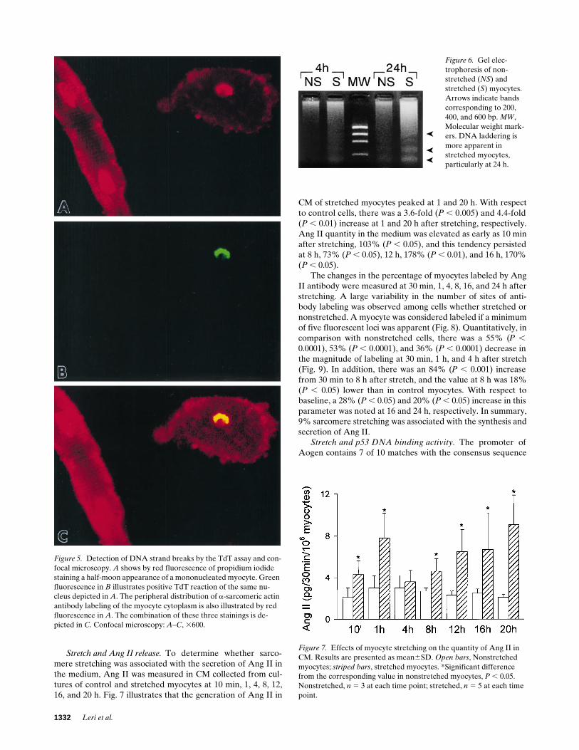
CM of stretched myocytes peaked at 1 and 20 h. With respectto control cells, there was a 3.6-fold (P , 0.005) and 4.4-fold(P , 0.01) increase at 1 and 20 h after stretching, respectively.Ang II quantity in the medium was elevated as early as 10 minafter stretching, 103% (P , 0.05), and this tendency persistedat 8 h, 73% (P , 0.05), 12 h, 178% (P , 0.01), and 16 h, 170%(P , 0.05).
The changes in the percentage of myocytes labeled by AngII antibody were measured at 30 min, 1, 4, 8, 16, and 24 h afterstretching. A large variability in the number of sites of anti-body labeling was observed among cells whether stretched ornonstretched. A myocyte was considered labeled if a minimumof five fluorescent loci was apparent (Fig. 8). Quantitatively, incomparison with nonstretched cells, there was a 55% (P ,0.0001), 53% (P , 0.0001), and 36% (P , 0.0001) decrease inthe magnitude of labeling at 30 min, 1 h, and 4 h after stretch(Fig. 9). In addition, there was an 84% (P , 0.001) increasefrom 30 min to 8 h after stretch, and the value at 8 h was 18%(P , 0.05) lower than in control myocytes. With respect tobaseline, a 28% (P , 0.05) and 20% (P , 0.05) increase in thisparameter was noted at 16 and 24 h, respectively. In summary,9% sarcomere stretching was associated with the synthesis andsecretion of Ang II.
Stretch and p53 DNA binding activity. The promoter ofAogen contains 7 of 10 matches with the consensus sequence
Figure 5. Detection of DNA strand breaks by the TdT assay and con-focal microscopy. A shows by red fluorescence of propidium iodide staining a half-moon appearance of a mononucleated myocyte. Green fluorescence in B illustrates positive TdT reaction of the same nu-cleus depicted in A. The peripheral distribution of a-sarcomeric actin antibody labeling of the myocyte cytoplasm is also illustrated by red fluorescence in A. The combination of these three stainings is de-picted in C. Confocal microscopy: A–C, 3600.
Figure 6. Gel elec-trophoresis of non-stretched (NS) and stretched (S) myocytes. Arrows indicate bands corresponding to 200, 400, and 600 bp. MW, Molecular weight mark-ers. DNA laddering is more apparent in stretched myocytes, particularly at 24 h.
Figure 7. Effects of myocyte stretching on the quantity of Ang II in CM. Results are presented as mean6SD. Open bars, Nonstretched myocytes; striped bars, stretched myocytes. *Significant difference from the corresponding value in nonstretched myocytes, P , 0.05. Nonstretched, n 5 3 at each time point; stretched, n 5 5 at each time point.

p53 and Myocyte Apoptosis 1333
of p53. An oligonucleotide of 25 bp including the ACAAGCCregion was radiolabeled and used as a probe in a gel retarda-tion assay. The radioactive probe was incubated with nuclearextracts prepared from control cells and myocytes stretchedfor 30 min, 2, 4, 8, and 24 h. One complex with shifted gel mo-bility was detected at all time points (Fig. 10 A). Moreover, theresults obtained at different intervals in nonstretched myo-cytes are shown in Fig. 10 B. In comparison with nonstretched
myocytes, the optical density of the p53 shifted band instretched cells was elevated at 30 min and 2 h. DNA bindingactivity increased further at 4 and remained increased at 8 and24 h (Fig. 10 B). The validity of the assay was established bysubjecting p53-specific bands to competition with an excess ofunlabeled self oligonucleotide. Moreover, the position of thep53 band was compared with that detected in SV-T2 cells (Fig.10 A). Subsequently, incubation of nuclear extracts with two
Figure 8. Immunocytochemical detection of Ang II in myocytes stretched for 16 h (A). B illus-trates by phase-contrast micros-copy the same field shown in A. Lack of staining in C was ob-tained after preabsorption of the primary antibody with Ang II. D shows by phase-contrast micros-copy the same field illustrated in C. A–D, 3700.

1334 Leri et al.
different anti-p53 antibodies failed to reveal mobility-shiftedcomplexes (Fig. 11), confirming the specificity of this gel retar-dation assay.
In a manner comparable to Aogen, the AT1 receptor pro-moter shares 7 of 10 matches with the consensus sequence ofp53. Thus, an oligonucleotide probe of 25 bp was prepared.This probe that contained the ACATGCC sequence was end-labeled and used in a gel shift analysis (Fig. 10 C). With respectto nonstretched cells (Fig. 10 D), stretched myocytes showedone shifted complex that increased 30 min after the impositionof stretch. The optical density of the shifted band was also in-creased at 2, 4, 8, and 24 h (Fig. 10 D). The specificity of the as-say was established as described above for Aogen and is illus-trated in Figs. 10 C and 11.
Since the bax promoter possesses a perfect p53 binding siteand three imperfect ones (28), a segment of this promoter,containing 46-bp sequence, was radiolabeled and used as aprobe. When nuclear extracts from stretched myocytes (Fig.10 E) and nonstretched myocytes (not shown) were incubatedwith the DNA probe, a complex with shifted gel mobility wasdetected. p53 DNA binding activity was increased in stretchedmyocytes at 30 min, 2, 4, 8, and 24 h (Fig. 10 E). Controls forthe gel mobility assay are shown in Fig. 10 E and 11. In thiscase, a mutated form of bax was used to confirm further thespecificity of the assay (Fig. 11). Finally, consistency in proteinloading, lack of protein degradation, and uniformity in the rel-ative purity of nuclear extracts at various time points afterstretch are shown in Fig. 10 F. The optical density of the actinband was significantly reduced (see Fig. 13 for comparison)and was consistent throughout, confirming that the level of cy-toplasmic contamination affected in a similar manner the nu-clear preparations. In summary, 9% sarcomere stretching wascoupled with enhanced p53 binding to the promoter of Aogen,AT1 receptor, and bax genes.
Ang II and p53 DNA binding activity. To establish whetherthe changes in DNA binding of p53 described in Figs. 10 and11 were influenced by stretch-mediated release of Ang II frommyocytes, nonstretched myocytes were treated with Ang II at1029 M in the presence of protease inhibitors and the activa-tion of p53-dependent genes was examined. The concentrationof Ang II was 125-fold higher than that detected by RIA afterstretch. Ang II stimulation for a period of 4 h (the last additionof Ang II was 30 min earlier) resulted in a marked increase inp53 binding to the promoter of Aogen. The optical density ofthe shifted complex decreased significantly at 24 h, i.e., 20.5 hafter the last administration of Ang II (Fig. 12 A). However,the intensity of the p53 band at this later time point was stillhigher than at baseline (Fig. 12 A, fourth lane). The 25-fold in-crease at 4 h and the 4-fold increase at 24 h after Ang II stimu-lation were statistically significant (P , 0.0001; P , 0.0001).Similarly, the 78% reduction in this parameter from 4 to 24 hwas significant (P , 0.005). Additionally, p53 binding to thepromoter of AT1 receptor increased 32-fold (P , 0.0001) at 4 hafter Ang II. A subsequent decrease, 93% (P , 0.0001), wasnoted at 24 h (Fig. 12 B). Finally, Ang II stimulation enhancedp53 binding to the consensus sequence in the bax promoter at4 h, 14-fold (P , 0.0001), and the p53 band at 24 h, 241% (P ,0.0001), was reduced (Fig. 12 C). In summary, Ang II stimula-tion was characterized by enhanced p53 binding to the pro-moter of Aogen, AT1 receptor, and bax genes in nonstretchedmyocytes.
Stretch, Aogen, AT1-receptor, p53, Bcl-2, and Bax expres-sion. Fig. 13 A illustrates the effects of stretch on the expres-sion of Aogen in myocytes by Northern blot analysis. Incomparison with nonstretched control myocytes, sarcomereelongation resulted in an increase in the quantity of AogenmRNA at 1 h. At 8 h, the expression of Aogen was still ele-vated, although the amount of Aogen mRNA was lower thanat the earlier time point. At 24 h the transcription of this genewas less than at 1 and 8 h. Densitometric data were obtainedby dividing the signals for Aogen mRNA by the signals for 18SrRNA. However, the increases in Aogen expression at thesethree intervals after stretch (Fig. 13 A) were statistically signif-icant (P , 0.0001; P , 0.0001; P , 0.0001).
The changes in AT1 receptor protein with stretch were de-termined by Western blot (Fig. 13 B). In comparison with non-stretched cells at 4, 8, and 24 h, stretched myocytes showed a25% (NS), 138% (P , 0.0001), and 200% (P , 0.0001) in-crease in the quantity of this protein. In addition, the conse-quences of stretch on p53 expression are illustrated in Fig.13 C. p53 protein increased progressively after stretch, from 4to 24 h. Densitometrically there was a 138% increase at 4 h(P , 0.05), 444% at 8 h (P , 0.0001), and 1,171% at 24 h (P ,0.0001).
Since p53 is a transcriptional modulator of the bcl-2 andbax genes (28), the quantity of these proteins was determinedby Western blot in nonstretched and stretched myocytes. Fig.13 D illustrates that Bax protein in stretched myocytes in-creased 47% (P , 0.0001) at 2 h, returned to baseline at 8 h,increased again 44% (P , 0.01) at 16 h, and returned to con-trol values at 24 h. In contrast, the amount of Bcl-2 (Fig. 13 E)decreased 28% (P , 0.0001), 57% (P , 0.0001), 75% (P ,0.0001), and 53% (P , 0.0001) at 2, 8, 16, and 24 h, respec-tively. These changes resulted in a reduction of the Bcl-2-to-Bax ratio in stretched myocytes. In summary, 9% sarcomerestretching enhanced the expression of Aogen, AT1 receptors,
Figure 9. Effects of stretch on the percentage of myocytes labeled by Ang II. Results are presented as mean6SD. Open bars, Nonstretched myocytes; striped bars, stretched myocytes. *Difference from the cor-responding value in nonstretched myocytes, P , 0.05. **Difference from the corresponding value in stretched myocytes at 30 min, P ,
0.05. ***Difference from the corresponding value in stretched myo-cytes at 1 h, P , 0.05. †Difference from the corresponding values in stretched myocytes at 8 h, P , 0.05. Nonstretched, n 5 3 at each time point; Stretched, n 5 5 in each time point.

p53 and Myocyte Apoptosis 1335
Figure 10. (A) Gel mobility as-say showing the interaction of p53 with its consensus sequence in the Aogen promoter. Nuclear extracts were obtained from nonstretched myocytes (NS) at 24 h and myocytes stretched for 30 min, 2, 4, 8, and 24 h. p53-spe-cific band (4 h after stretching) was subject to competition with an excess of unlabeled self oligo-nucleotide, competitor (C). Ao, Aogen probe in the absence of nuclear extract. SV-T2, nuclear extract from SV-T2 cells. The ar-row indicates the position of the p53 shifted band. (B) Gel mobil-ity assay showing the interaction of p53 with its consensus se-quence in the Aogen promoter. Nuclear extracts were obtained from freshly isolated myocytes (F) and nonstretched myocytes kept in culture for 30 min, 2, 4, 8, and 24 h. p53-specific band (4 h in culture) was subject to compe-tition with an excess of unla-beled self oligonucleotide, com-petitor (C). Ab, Nuclear extract from nonstretched myocytes at4 h incubated with p53 antibody PAb240. Ao, Aogen probe in the absence of nuclear extracts. The arrow indicates the position of the p53 shifted band. Optical density results in nonstretched and stretched myocytes were: 30 min: nonstretched 5 4.061.3,n 5 5, stretched 5 24612, n 5 5, P , 0.005; 2 h: nonstretched 5
4.461.6, n 5 5, stretched 5
4668, n 5 5, P , 0.001; 4 h: non-stretched 5 5.261.6, n 5 5, stretched 5 84616, n 5 5, P ,
0.0001; 8 h: nonstretched 5
4.861.3, n 5 5, stretched 5
51617, n 5 5, P , 0.0001; 24 h: nonstretched 5 4.461.2, n 5 5, stretched 5 73620, n 5 5, P ,
0.0001. (C) Gel mobility assay showing the interaction of p53 with its consensus sequence in the AT1 promoter. Nuclear ex-tracts were obtained from non-stretched myocytes (NS) at 24 h and myocytes stretched for 30 min, 2, 4, 8, and 24 h. p53-spe-cific band (4 h after stretching)
was subject to competition with an excess of unlabeled self oligonucleotide, competitor (C). AT1, AT1 probe in the absence of nuclear extract. SV-T2, Nuclear extract from SV-T2 cells. The arrow indicates the position of the p53 shifted band. (D) Gel mobility assay showing the interac-tion of p53 with its consensus sequence in the AT1 promoter. Nuclear extracts were obtained from freshly isolated myocytes (F) and non-stretched myocytes kept in culture for 30 min, 2, 4, 8, and 24 h. p53-specific band (4 h in culture) was subject to competition with an excess of un-labeled self oligonucleotide, competitor (C). Ab, Nuclear extract from nonstretched myocytes at 4 h incubated with p53 antibody PAb240. AT1, AT1 probe in the absence of nuclear extract. SV-T2, nuclear extract from SV-T2 cells. The arrow indicates the position of the p53 shifted band. Optical density results in nonstretched and stretched myocytes were: 30 min: nonstretched 5 7.062.3, n 5 5, stretched 5 67625, n 5 5, P ,
0.0001; 2 h: nonstretched 5 4.661.9, n 5 5, stretched 5 2868, n 5 5, P , 0.0001; 4 h: nonstretched 5 4.462.0, n 5 5, stretched 5 45616, n 5 5, P , 0.0001; 8 h: nonstretched 5 4.862.6, n 5 5, stretched 5 52612, n 5 5, P , 0.0001; 24 h: nonstretched 5 4.862.4, n 5 5, stretched 5 65617, n 5 5, P , 0.0001. (E) Gel mobility assay showing the interaction of p53 with its consensus sequence in the bax promoter. Nuclear extracts were

1336 Leri et al.
p53, and Bax and decreased the expression of Bcl-2 in thecells.
Stretch, AT1 antagonist, and apoptosis. To determine whetherligand binding to surface AT1 receptors was involved instretch-mediated apoptosis, myocytes were exposed to losar-tan and, 30 min later, sarcomere elongation was induced. Con-trol myocytes were similarly treated, but stretch was notapplied. The selective AT1 blocker, losartan, prevented pro-grammed cell death measured by the TdT assay at both 4(nonstretched: 1.6360.42%, n 5 4; stretched: 1.3660.57%, n 54; not significant) and 24 h (nonstretched: 2.1860.81%, n 5 4;stretched: 2.0260.47, n 5 4; not significant) after stretching.Moreover, a gel mobility assay was performed using nuclearextracts from control and stretched myocytes exposed to losar-tan. The 4-h interval after stretching was examined becausep53 binding activity in the Aogen and bax promoters was ele-vated at this time. As illustrated in Fig. 14, the AT1 receptorblocker reduced the optical density of the p53 shifted band.This determination was repeated three times. In summary,losartan inhibited stretching-induced apoptosis in myocytesand p53 binding activity.
p53 and activation of AT1 and Aogen genes. We have shownpreviously that infection of adult rat ventricular myocytes witha replication-deficient adenoviral vector containing wild-type
human p53 was characterized by enhanced expression of Bax,Aogen, and AT1 receptor (27). p53 DNA binding activity inthese experiments was assessed by oligonucleotide probes forAogen and AT1 identical to those used in this study. More-over, Fig. 11 A illustrated that the addition of a mutated formof bax, characterized by the substitution of two nucleotides inthe perfect consensus motif for p53 binding, had no effecton the interaction between wild bax and p53. This indicatesthat the sequence of the bax oligonucleotide used was criticalfor the detection of p53 binding activity in rat myocytes. Similarresults have been obtained in dog ventricular myocytes (43). Inthis early study, the same bax oligonucleotide was shown tobind to p53 in SV-T2 cells which overexpress p53. The specific-ity of the shifted complex in this cell system was demonstratedby using the mutated form of bax and anti-p53 antibody. Onthis basis, SV-T2 cells were used here to document whetherthe imperfect p53 consensus sequences in the AT1 and Aogenpromoters were capable of competing with the p53 binding siteof bax and between themselves. Such an approach was used todetermine whether the sequences selected were critical for theresponsiveness of the AT1 and Aogen genes to p53.
Fig. 15 A illustrates that, in the presence of nuclear extractfrom SV-T2 cells, the AT1 oligonucleotide resulted in the for-mation of two shifted complexes (second lane) which were
obtained from nonstretched myocytes (NS) at 24 h and myocytes stretched for 30 min, 2, 4, 8, and 24 h. p53-specific band (4 h after stretching) was subject to competition with an excess of unlabeled self oligonucleotide, competitor (C), and with p53 antibody PAb240 (Ab). Bax, Bax probe in the absence of nuclear extract. SV-T2, Nuclear extract from SV-T2 cells. The arrow indicates the position of the p53 shifted band. Opti-cal density results in nonstretched and stretched myocytes were: 30 min: nonstretched 5 2.061.1, n 5 5, stretched 5 45613, n 5 5, P , 0.0001;2 h: nonstretched 5 2.761.2, n 5 5, stretched 5 7267, n 5 5, P , 0.0001; 4 h: nonstretched 5 1.760.7, n 5 5, stretched 5 84619, n 5 5, P ,
0.0001; 8 h: nonstretched 5 2.260.9, n 5 5, stretched 5 73620, n 5 5, P , 0.0001; and 24 h: nonstretched 5 2.360.4, n 5 5, stretched 5 70619, n 5 5, P , 0.0001. (F) The pattern of proteins corresponding to the nuclear preparations used for mobility shift assays is illustrated by Coo-massie blue staining. The actin band, 42 kD, is markedly reduced and rather uniform in all samples. Protein degradation is not apparent. MW, Molecular weight markers.
Figure 11. (A) Gel mobility assay showing the inter-action of p53 with its consensus sequence in the bax promoter. Nuclear extracts were obtained from stretched myocytes at 4 h (S4h). p53-specific band was subject to competition with p53 antibodies PAb240 (Ab1) and PAb122 (Ab2). Bax mut, Unla-beled mutated Bax probe. Bax, Bax probe in the ab-sence of nuclear extract. SV-T2, Nuclear extract from SV-T2 cells. The arrow indicates the position of p53 shifted band. Addition of p53 antibodies opposed the appearance of a p53 shifted complex in both cases. (B) Gel mobility assay showing the interaction of p53 with its consensus sequence in the Aogen promoter. Nuclear extracts were obtained from stretched myo-cytes at 4 h (S4h). p53-specific band was subject to competition with p53 antibodies PAb240 (Ab1) and PAb122 (Ab2). Ao, Aogen probe in the absence of nuclear extract. SV-T2, Nuclear extract from SV-T2 cells. The arrow indicates the position of p53 shifted band. Addition of p53 antibodies opposed the ap-
pearance of a p53 shifted complex in both cases. (C) Gel mobility assay showing the interaction of p53 with its consensus sequence in the AT1 promoter. Nuclear extracts were obtained from stretched myocytes at 4 h (S4h). p53-specific band was subject to competition with p53 antibod-ies PAb240 (Ab1) and PAb122 (Ab2). AT1, AT1 probe in the absence of nuclear extract. SV-T2, Nuclear extract from SV-T2 cells. The arrow in-dicates the position of p53 shifted band. Addition of p53 antibodies opposed the appearance of a p53 shifted complex in both cases.
Figure 10 legend (Continued)

p53 and Myocyte Apoptosis 1337
markedly attenuated by competition with the unlabeled AT1
probe (third lane). Preincubation with unlabeled bax (fourthlane) and unlabeled Aogen (fifth lane) significantly reducedthe p53 bands. Similarly, the single shifted complex obtainedwith the Aogen probe (Fig. 15 B, second lane) essentially dis-appeared with preexposure of nuclear extracts to unlabeledAogen (Fig. 15 B, fourth lane). Preincubation with unlabeledbax (Fig. 15 B, third lane) and unlabeled AT1 (Fig. 15 B, fifthlane) decreased the intensity of the p53 complex. In summary,the oligonucleotide sequences used were critical for the bind-ing of p53 to bax, Aogen, and AT1 receptor genes.
Stretch, ANF expression, and myocyte protein content.Northern blot analysis of nonstretched and stretched myocytesshowed that ANF mRNA increased at 30 min after sarcomereelongation. However, the quantity of ANF mRNA decreasedmarkedly at 4 and 8 h, returning to baseline value at 24 h afterstretch (Fig. 16). Densitometric data were as follows: non-stretched at 30 min 5 1.760.4 (n 5 3), and 24 h 5 1.460.9 (n 53); stretched at 30 min 5 151638 (n 5 3), 4 h 5 2865 (n 5 3),8 h 5 1664 (n 5 3), and 24 h 5 362 (n 5 3). The 89-fold in-crease in ANF mRNA at 30 min in stretched myocytes was sta-tistically significant (P , 0.0001).
To establish whether stretch for 24 h was associated with an
increase in myocyte volume, the amount of protein per cellwas determined by confocal microscopy after staining with flu-orescein isothiocyanate. This parameter was found to be com-parable in nonstretched and stretched myocytes. Total pixelintensity per cell was 185,00069,000 in control myocytes (n 54 separate cultures) and 182,000613,500 in myocytes exposedto sarcomere elongation for 24 h (n 5 4 separate cultures).This small difference was not statistically significant. In sum-mary, myocyte stretch resulted in an acute transient increase inANF expression and in no cellular hypertrophy at 24 h.
Discussion
The results of this study indicate that moderate sarcomerestretching of adult ventricular myocytes was associated withthe release of Ang II and the activation of p53 which were fol-lowed by programmed cell death shortly after the impositionof the mechanical stimulus. Stretch increased p53 binding tothe promoter of Aogen, AT1 receptor, and Bax as well as thecellular formation of Ang II, suggesting that this transcriptionfactor may modulate not only the local RAS but also potenti-ate the susceptibility of myocytes to undergo apoptosis. TheAT1 blocker losartan inhibited apoptosis and p53 DNA bind-
Figure 12. (A) Gel mobility assay showing the interaction of p53 with its consensus sequence in the Aogen promoter. Nuclear extracts were ob-tained from nonstretched myocytes kept in culture for 4 h and from nonstretched myocytes stimulated with Ang II (A-II) for 4 and 24 h. p53-spe-cific band at 4 h after Ang II was subject to competition with an excess of unlabeled self oligonucleotide, competitor (C), and with the p53 anti-body PAb122 (Ab). Ao, Aogen probe in the absence of nuclear extract. SV-T2, Nuclear extract from SV-T2 cells. The arrow indicates the position of the p53 shifted band. Optical density values were: nonstretched, 4 h 5 462 (n 5 4), 24 h 5 562 (n 5 4); nonstretched 1 Ang II,4 h 5 101625 (n 5 4), 24 h 5 2266 (n 5 4). (B) Gel mobility assay showing the interaction of p53 with its consensus sequence in the AT1 pro-moter. Nuclear extracts were obtained from nonstretched myocytes kept in culture for 4 h and from nonstretched myocytes stimulated withAng II (A-II) for 4 and 24 h. p53-specific band at 4 h after Ang II was subject to competition with the p53 antibody PAb122 (Ab). AT1, AT1 probe in the absence of nuclear extract. SV-T2, Nuclear extract from SV-T2 cells. The arrow indicates the position of the p53 shifted band. Opti-cal density values were: nonstretched, 4 h 5 361 (n 5 4), 24 h 5 461 (n 5 4); nonstretched 1 Ang II, 4 h 5 95616 (n 5 4), 24 h 5 765 (n 5 4). (C) Gel mobility assay showing the interaction of p53 with its consensus sequence in the bax promoter. Nuclear extracts were obtained from nonstretched myocytes kept in culture for 4 h and from nonstretched myocytes stimulated with Ang II (A-II) for 4 and 24 h. p53-specific band at 4 h after Ang II was subject to competition with an excess of unlabeled self oligonucleotide, competitor (C), and with the p53 antibody PAb122 (Ab). Bax, Bax probe in the absence of nuclear extract. SV-T2, nuclear extract from SV-T2 cells. The arrow indicates the position of the p53 shifted band. Optical density values were: nonstretched, 4 h 5 562 (n 5 4), 24 h 5 461 (n 5 4); nonstretched 1 Ang II, 4 h 5 7168 (n 5 4),24 h 5 4266 (n 5 4).

1338 Leri et al.
ing activity, raising the possibility that the AT1 effector path-way may be critical in regulating p53 function and cell death.Ligand binding to surface AT1 receptors may be linked to thephosphorylation of the tumor suppressor protein p53 that mayinfluence myocyte death by upregulating the synthesis and se-cretion of Ang II and by decreasing the Bcl-2-to-Bax proteinratio in the cell.
Stretch and myocyte apoptosis. Mechanical stimuli have
multiple effects on myocytes in vivo (1, 2) and in vitro (9).Physical forces cause rapid induction of immediate early genes(8, 11), and expression of skeletal a-actin, ANF, and b-myosinheavy chain (10). They activate a number of second messengerpathways (9, 12) and lead to myocyte hypertrophy (18). Cellsperceive the external load and this recognition has been associ-ated with the release of Ang II which functions as the initial ac-tivator of myocyte growth (18). The growth-promoting influ-
Figure 13. (A) Detection of Aogen mRNA by Northern blot analysis (top) in non-stretched (N) myocytes at1, 8, and 24 h, and stretched (S) myocytes at 1, 8, and 24 h. Equal loading conditions are indicated in the bottom panel by ethidium bromide staining of 18S rRNA. Densi-tometric data were obtained by dividing the signals for Aogen mRNA by the signals for 18S rRNA. Optical den-sity values were: non-stretched, 1 h 5 161 (n 5 3), 8 h 5 262 (n 5 3), 24 h 5
261 (n 5 3); stretched, 1 h 5
147617 (n 5 3), 8 h 5 53615 (n 5 3), 24 h 5 2966 (n 5 3). (B) Detection by Western blot of AT1 receptor protein (top) in nonstretched (NS) myocytes at 24 h and stretched myocytes at 4, 8, and 24 h. Loading of proteins is illustrated by Coomassie blue staining (bottom). Opti-cal density values were: non-stretched, 4 h 5 2464, n 5 4, 8 h 5 2663, n 5 4, 24 h 5
2465, n 5 4; stretched, 4 h 5
3066, n 5 4, 8 h: 62611, n 5
4, 24 h: 7267, n 5 4. (C)Detection by Western blotof p53 protein (top) in non-stretched (NS) myocytes at 24 h and stretched myocytes at 4, 8, and 24 h. Proteins from SV-T2 cells were used
as a positive control. Protein loading consisted of 120 mg for myocytes and 30 mg for SV-T2 cells. Loading of proteins is illustrated by Coomassie blue staining (bottom). Optical density values were: 4 h, nonstretched 5 866, n 5 4; stretched 5 1967, n 5 4; 8 h, nonstretched 5
964, n 5 4; stretched 5 49613, n 5 4; 24 h, nonstretched 5 764, n 5 4; stretched 5 89610,n 5 4. (D) Detection by Western blot of Bax protein (top) in nonstretched (NS) myocytes at 24 h and stretched myocytes at 2, 8, 16, and 24 h. Loading of proteins is illustrated by Coo-massie blue staining (bottom). Optical density values were: 2 h, nonstretched 5 5768, n 5 7; stretched 5 84612, n 5 5; 8 h, nonstretched 5 5567, n 5 5; stretched 5 61613, n 5 8; 16 h, nonstretched 5 57611, n 5 5; stretched 5 82615, n 5 7; 24 h, nonstretched 5 5469, n 5 5; stretched 5 6468, n 5 5. (E) Detection by Western blot of Bcl-2 protein (top) in non-stretched (NS) and stretched myocytes at 2, 8, 16, and 24 h. Loading of proteins is illustrated by Coomassie blue staining (bottom). Optical density values were: 2 h, nonstretched 5
9669, n 5 7; stretched 5 69612, n 5 7; 8 h, nonstretched 5 101612, n 5 5; stretched 5
43619, n 5 6; 16 h, nonstretched 5 94618, n 5 5; stretched 5 24611, n 5 5; 24 h, non-stretched 5 103616, n 5 5; stretched 5 48610, n 5 6.

p53 and Myocyte Apoptosis 1339
ence of Ang II has been shown in neonatal (18) and adult (44)myocytes, although a more attenuated response has beenclaimed in the matured fully differentiated cell (44). More re-cently, stretch has been implicated in the stimulation of pro-grammed myocyte cell death (21) and Ang II in the nanomolar
range has been seen to trigger myocyte apoptosis (19, 20). Thecurrent results provide the first documentation that sarcomereelongation of adult ventricular myocytes was coupled with therelease of Ang II and the activation of the endogenous celldeath pathway. Apoptosis was not observed up to 2 h afterstretch, but involved 7% of myocytes at 4 h and 17% at 24 h.Abnormal increases in preload on the myocardium in vivo andin vitro are characterized by scattered cell death, architecturalrearrangement of myocytes, mural thinning, and cavitary dila-tion (21–24, 45–47). These observations suggest that mechani-cal forces generated in vivo in the pathologic heart may lead towall restructuring through the release of Ang II, apoptosis, andside by side translocation of cells. However, understanding ofthe dual role of stretch on cell growth and cell death is com-plex. The question why apoptosis or cellular hypertrophy oc-curs in certain cells more than in others is important but re-mains to be answered.
Double-strand DNA cleavage in myocyte nuclei was de-
Figure 14. Gel mobility assay showing the effects of losartan (Los) on the interaction of p53 with its consensus sequence in the Aogen (A) and bax (B) promoters. Nuclear extracts were obtained from stretched (S) myocytes for 4 h, in the presence or absence of losartan. p53-specific band was subject to competition with an excess of unlabeled self oli-gonucleotide, competitor (C), and with the p53 an-tibody PAb240 (Ab). Ao, Aogen probe in the ab-sence of nuclear extract. Bax, Bax probe in the absence of nuclear extract. Bax mut, unlabeled mu-tated Bax probe. SV-T2, nuclear extract from SV-T2 cells. The arrow indicates the position of the p53 shifted band.
Figure 15. (A) Gel mobility assay showing the interaction of p53 with its consensus sequence in the AT1 promoter. Nuclear extracts were obtained from SV-T2 cells. p53-specific bands were subject to compe-tition with an excess of unlabeled self oligonucleotide, competitor (C), with unlabeled Bax probe (Bax 1 AT1) and with unlabeled Ao-gen probe (Ao 1 AT1). AT1 corresponds to the AT1 probe in the ab-sence of nuclear extract. Arrows indicate the position of the p53 shifted bands. (B) Gel mobility assay showing the interaction of p53 with its consensus sequence in the Aogen (Ao) promoter. Nuclear ex-tracts were obtained from SV-T2 cells. p53-specific band was subject to competition with an excess of unlabeled self oligonucleotide, com-petitor (C), with unlabeled Bax probe (Bax 1 Ao) and with unla-beled AT1 probe (AT1 1 Ao). Ao corresponds to the Aogen probe in the absence of nuclear extract. The arrow indicates the position of the p53 shifted band.
Figure 16. Detection of ANF mRNA by Northern blot analysis (top) in nonstretched and stretched myocytes. Equal loading conditions are indicated in the bottom panel by ethidium bromide staining of 18S rRNA.

1340 Leri et al.
tected by the TdT assay and the in situ ligation technique. Thisapproach was introduced because the TdT reaction may over-estimate the degree of apoptosis (33, 41, 42). A large variabil-ity in the extent of myocyte cell death has been reported by theTUNEL test and values up to 35% have been claimed in thefailing human myocardium (3, 5, 25, 48). Measurements ofapoptosis here were essentially comparable with the two meth-odologies since similar data were obtained with TdT and insitu ligation. Additionally, as previously performed (5), evalu-ations of cell death by TdT labeling were confirmed by theanalysis of chromatin alterations by confocal microscopy.However, the in situ ligation procedure identifies double-strand DNA cleavage with single base 39 overhangs which oc-cur exclusively during apoptosis (33). Blunt-ended products inDNA damage may be present in advanced stages of cell necro-sis but are not stained by this technique. The presence of singlebase 39 overhangs in nuclei has been linked to the activation ofCa21-dependent DNase I (33) that is implicated in Ang II–mediated myocyte apoptosis (19). The nearly threefold in-crease in myocyte apoptosis from 4 to 24 h after stretchingmost likely reflects the upregulation in proapoptotic geneproducts and in the density of AT1 receptors that together mayhave contributed to enhance the proficiency of cells to die dur-ing this period.
Stretch and Ang II. Neonatal ventricular myocytes containAng II that is released by mechanical stimuli (16). Our obser-vations indicate that a similar phenomenon occurred in adultcells. Ang II antibody labeling of the myocyte cytoplasm de-creased at 30 min and 1 and 4 h after stretching but a patternsimilar to nonstretched cells was detected at 8 h and valuesgreater than baseline were seen at 16 and 24 h. Moreover, theconcentration of Ang II in the CM was elevated at 10 min and1 h, decreased at 4 h, and increased progressively from 8 to 20 h.These results are consistent with an immediate release of AngII and the chronic synthesis and secretion of this peptide fromstretched myocytes. Although the release of Ang II from myo-cyte stores may be the immediate consequence of stretching,more difficult is the identification of the mechanism which ac-tivates chronically the cellular RAS, sustaining the formationof Ang II. In neonatal myocytes in vitro as well as in this studyon adult ventricular myocytes, stretch is characterized by en-hanced transcription of Aogen mRNA and this response hasbeen considered critical for the continuous local production ofAng II (18). A similar effect has been observed by cyclicalstretch (49). However, the molecular bases of the stimulationof the myocyte RAS and Aogen in particular were not identi-fied (18, 49).
Stretch and p53 activation. The tumor suppressor proteinp53 can produce cell cycle arrest and facilitate apoptosis, butthese actions are not interdependent (50). In response to DNAdamage, there is an accumulation of endogenous p53 that ex-erts its growth-suppressive effect by preventing proliferation(51). p53 can also activate transcription when it binds to spe-cific DNA sequences in the promoter region of various genes(50, 51). To date, multiple genes containing p53 binding siteshave been identified. In the heart, p53 transcripts appear to de-crease rapidly during early postnatal development, becomingalmost undetectable in the adult fully differentiated myocytes(52). However, the levels of p53 mRNA and protein may notchange in cells, whereas both DNA binding and transcriptionalactivity may increase severalfold (53).
The results of this study demonstrate that rat ventricular
myocytes subjected to sarcomere stretching were character-ized by enhanced p53 binding to the promoter of Aogen, AT1
receptor, and the proapoptotic gene bax. These observationssuggest that the initial release of Ang II from myocytes and thestimulation of the AT1 receptor effector pathway were the me-diators of the upregulation in p53 function. This contention isconsistent with the capacity of the AT1 receptor blocker losar-tan to attenuate markedly stretch-mediated p53 DNA bindingactivity. Conversely, Ang II increased p53 binding to Aogen,AT1, and bax promoters in nonstretched myocytes. The car-boxy terminal of the p53 protein possesses two distinct regula-tory sites which are phosphorylated, respectively, by proteinkinase C and casein kinase II (54). Sarcomere stretching andthe secretion of Ang II may be coupled with protein kinase Ctranslocation (20) and phosphorylation of p53. This may acti-vate the cellular RAS and the continuous generation of AngII. The relationship between physical forces, on the one hand,and the local RAS and p53, on the other, is consonant with theability of losartan to prevent stretch-mediated apoptosis.
Binding of p53 to Aogen, AT1, and bax promoters instretched myocytes increased as early as 30 min after the impo-sition of the mechanical stimulus and remained elevated up to24 h. Such a response indicates that stretch led to a consistenttransactivation of these genes because only small variations inp53 DNA binding activity were noted at the various intervals.The induction of the Aogen, AT1 receptor, and bax genes byp53 with sarcomere elongation was coupled with increasedAogen mRNA level and AT1 receptor and Bax proteins. Thedecrease in Bcl-2 expression was also indicative of p53 activa-tion. Changes in the proportion of members of the Bcl-2 pro-tein family suggest that the sensitivity of myocytes to undergoapoptosis was increased (55). Conversely, the activation of p53per se and the alterations in the relative quantities of p53-inducible genes cannot be interpreted as indices of apoptosis(28). Upregulation of Bax or attenuation of Bcl-2 alone cannotinitiate apoptosis in myocytes (27). Similarly, p53 may potenti-ate apoptotic signals but cannot by itself trigger apoptosis (27,28). However, the accumulation of Bax and the decrease ofBcl-2 with the duration of stretch, in combination with the in-crease in surface AT1 receptors, provided the molecular basis
Figure 17. Proposed scheme for stretch-induced apoptosis in myo-cytes.
p53 and Myocyte Apoptosis 1341
for the enhanced ability of Ang II to trigger the cell deathpathway in myocytes. Thus, the hypothesis is advanced thatstretch-mediated release of Ang II results in p53 phosphoryla-tion and upregulation of the myocyte RAS which may sustainp53 function and the formation of Ang II. The continuous syn-thesis of this hormone, in combination with p53-induced de-crease in Bcl-2-to-Bax protein ratio, leads to the increase inmyocyte apoptosis with time (Fig. 17). Although the extrapo-lation of in vitro observations to the in vivo state requires ex-treme caution, the recognition that stretch may be connectedto cell death is critical for the heart. Diastolic loads are abnor-mal in all forms of cardiac failure (56–59) and myocyte apopto-sis occurs in the severely impaired human heart (3, 5, 48, 60).Myocyte death facilitates ventricular dilation, counteractscompensatory hypertrophy, and exacerbates the magnitude ofloading on the remaining viable cells (1, 2, 22).
Acknowledgments
The expert technical assistance of Maria Feliciano is greatly appreci-ated.
This work was supported by grants HL-38132, HL-39902, HL-43023, and AG-15756 from the National Institutes of Health, and by aGrant-in-Aid from the American Heart Association (950321).
References
1. Katz, A.M. 1995. Cell death in the failing heart: role of an unnaturalgrowth response to overload. Clin. Cardiol. 18(Suppl. IV):IV-36–IV-44.
2. Cohn, J.N. 1996. The management of chronic heart failure. N. Engl. J.Med. 335:490–498.
3. Narula, J., N. Haider, R. Virmani, T.G. DiSalvo, F.D. Kolodgie, R.J. Haj-jar, U. Schmidt, M.J. Semigran, G.W. Dec, and B.-A. Kjaw. 1996. Apoptosis inmyocytes in end-stage heart failure. N. Engl. J. Med. 335:1182–1189.
4. Colucci, W.S. 1996. Apoptosis in the heart. N. Engl. J. Med. 335:1224–1226.
5. Olivetti, G., R. Abbi, F. Quaini, J. Kajstura, W. Cheng, J.A. Nitahara, E.Quaini, C. Di Loreto, C.A. Beltrami, S. Krajewski, et al. 1997. Apoptosis in thefailing human heart. N. Engl. J. Med. 336:1131–1141.
6. Ross, J., E.H. Sonnenblick, R.R. Taylor, H.M. Spotnitz, and J.W. Covell.1971. Diastolic geometry and sarcomere lengths in the chronically dilated ca-nine left ventricle. Circ. Res. 28:49–61.
7. Vitali-Mazza, L., P. Anversa, F. Tedeschi, R. Mastandrea, V. Mavilla,and O. Visioli. 1972. Ultrastructural basis of acute left ventricular failure fromsevere acute aortic stenosis in the rabbit. J. Mol. Cell. Cardiol. 4:661–671.
8. Komuro, I., T. Kaida, Y. Shibazaki, M. Kurabayashi, Y. Katoh, E. Hoh,F. Takaku, and Y. Yazaki. 1990. Stretching cardiac myocytes stimulates pro-tooncogene expression. J. Biol. Chem. 265:3595–3598.
9. Schneider, M.D., R. Roberts, and T.G. Parker. 1991. Modulation of car-diac genes by mechanical stress. The oncogene signaling hypothesis. Mol. Biol.Med. 8:167–183.
10. Sadoshima, J., L. Jahn, T. Takashashi, T.J. Kulik, and S. Izumo. 1992.Molecular characterization of the stretch-induced adaptation of cultured car-diac cells. An in vitro model of load-induced cardiac hypertrophy. J. Biol.Chem. 267:10551–10660.
11. Sadoshima, J., T. Takahashi, L. Jahn, and S. Izumo. 1992. Roles ofmechano-sensitive ion channels, cytoskeleton, and contractile activity instretch-induced immediate-early gene expression and hypertrophy of cardiacmyocytes. Proc. Natl. Acad. Sci. USA. 89:9905–9909.
12. Sadoshima, J., and S. Izumo. 1993. Mechanical stretch rapidly activatesmultiple signal transduction pathways in cardiac myocytes: potential involve-ment of an autocrine/paracrine mechanism. EMBO (Eur. Mol. Biol. Organ.) J.12:1681–1692.
13. Pennica, D., K.L. King, K.J. Shaw, E. Luis, J. Rullamas, S.-M. Luoh,W.C. Darbonne, D.S. Knutzon, R. Yen, K.R. Chien, et al. 1995. Expressioncloning of cardiotrophin 1, a cytokine that induces cardiac myocyte hypertro-phy. Proc. Natl. Acad. Sci. USA. 92:1142–1146.
14. Kent, R.L., and P.J. McDermott. 1996. Passive load and angiotensin IIevoke differential responses of gene expression and protein synthesis in cardiacmyocytes. Circ. Res. 78:829–838.
15. Baker, K.M., G.W. Booz, and D.E. Dostal. 1992. Cardiac actions of an-giotensin II: role of an intracardiac renin-angiotensin system. Annu. Rev. Phys-iol. 54:227–241.
16. Sadoshima, J., and S. Izumo. 1993. Molecular characterization of angio-tensin II-induced hypertrophy of cardiac myocytes and hyperplasia of cardiacfibroblasts. Critical role of the AT1 receptor subtype. Circ. Res. 73:413–423.
17. Sadoshima, J., and S. Izumo. 1993. Signal transduction pathways of an-giotensin II-induced c-fos gene expression in cardiac myocytes in vitro. Roles ofphospholipid-derived second messengers. Circ. Res. 73:424–438.
18. Sadoshima, J., Y. Xu, H.S. Slayter, and S. Izumo. 1993. Autocrine re-lease of angiotensin II mediates stretch-induced hypertrophy of cardiac myo-cytes in vitro. Cell. 75:977–984.
19. Cigola, E., J. Kajstura, B. Li, L.G. Meggs, and P. Anversa. 1997. Angio-tensin II activates programmed myocyte cell death in vitro. Exp. Cell Res. 231:363–371.
20. Kajstura, J., E. Cigola, A. Malhotra, P. Li, W. Cheng, L.G. Meggs, andP. Anversa. 1997. Angiotensin II induces apoptosis of adult ventricular myo-cytes in vitro. J. Mol. Cell. Cardiol. 29:859–870.
21. Cheng, W., B. Li, J. Kajstura, P. Li, M.S. Wolin, E.H. Sonnenblick, T.H.Hintze, G. Olivetti, and P. Anversa. 1995. Stretch-induced programmed myo-cyte cell death. J. Clin. Invest. 96:2247–2259.
22. Anversa, P., G. Olivetti, L.G. Meggs, E.H. Sonnenblick, and J.M. Ca-passo. 1993. Cardiac anatomy and ventricular loading after myocardial infarc-tion. Circulation. 87:VII22–VII27.
23. Cheng, W., J. Kajstura, J.A. Nitahara, B. Li, K. Reiss, Y. Liu, W.A.Clark, S. Krajewski, J.C. Reed, G. Olivetti, and P. Anversa. 1996. Programmedcell death contributes to ventricular remodeling after myocardial infarction inrats. Exp. Cell Res. 226:316–327.
24. Olivetti, G., F. Quaini, R. Sala, C. Lagrasta, D. Corradi, E. Bonacina,S.R. Gambert, E. Cigola, and P. Anversa. 1996. Acute myocardial infarction inhumans is associated with activation of programmed myocyte cell death in thesurviving portion of the heart. J. Mol. Cell. Cardiol. 28:2005–2016.
25. Kajstura, J., W. Cheng, R. Sarangarajan, P. Li, B. Li, J.A. Nitahara, S.Chapnick, K. Reiss, G. Olivetti, and P. Anversa. 1996. Necrotic and apoptoticmyocyte cell death in the aging heart of Fischer 344 rats. Am. J. Physiol. 217:H1215–H1228.
26. Bardales, R.H., S. Hailey, S.S. Xie, R.F. Schaefer, and S.-M. Hsu. 1996.In situ apoptosis assay for the detection of early acute myocardial infarction.Am. J. Pathol. 149:821–829.
27. Pierzchalski, P., K. Reiss, W. Cheng, C. Cirielli, J. Kajstura, J.A. Ni-tahara, M. Rizk, M.C. Capogrossi, and P. Anversa. 1997. p53 induces myocyteapoptosis via the activation of the renin-angiotensin system. Exp. Cell Res. 234:57–65.
28. Miyashita, T., and J. Reed. 1995. Tumor suppressor p53 is a direct tran-scriptional activator of the human bax gene. Cell. 80:293–299.
29. Miyashita, T., M. Harigai, M. Hanada, and J.C. Reed. 1994. Identifica-tion of a p53-dependent negative response element in the bcl-2 gene. CancerRes. 54:3131–3135.
30. Cheng, W., K. Reiss, P. Li, M. Chun, J. Kajstura, G. Olivetti, and P. An-versa. 1996. Aging does not affect the activation of the myocyte insulin-like growthfactor-1 autocrine system after infarction and ventricular failure in Fischer 344rats. Circ. Res. 78:536–546.
31. Lee, A.A., T. Delhaas, L.K. Waldman, D.A. MacKenna, F.J. Villarreal,and A.D. McCulloch. 1996. An equibiaxial strain system for cultured cells. Am.J. Physiol. 271:1400–1408.
32. Tournier, J.F., A. Lopez, and J.F. Tocanne. 1989. Effect of cell substra-tum on lateral mobility of lipids in the plasma membrane of vascular endothe-lial cells. Exp. Cell Res. 181:105–115.
33. Didenko, V.V., and P.J. Hornsby. 1996. Presence of double-strandedbreaks with single-base 39 overhangs in cells undergoing apoptosis but not ne-crosis. J. Cell Biol. 135:1369–1376.
34. Reiss, K., W. Cheng, A. Ferber, J. Kajstura, P. Li, B. Li, G. Olivetti, C.J.Homcy, R. Baserga, and P. Anversa. 1996. Overexpression of insulin-likegrowth factor-1 in the heart is coupled with myocyte proliferation in transgenicmice. Proc. Natl. Acad. Sci. USA. 93:8630–8635.
35. Maunders, M.J. 1993. DNA and RNA ligases (EC 6.6.1.1, EC 6.5.1.2and EC 6.5.1.3). In Methods in Molecular Biology. Vol. 16. Enzymes of Molec-ular Biology. M.M. Burrell, editor. Humana Press, Totowa, NJ. 213–230.
36. Gong, J., F. Traganos, and Z. Darzynkiewicz. 1994. A selective proce-dure for DNA extraction from apoptotic cells applicable for gel electrophoresisand flow cytometry. Anal. Biochem. 218:314–319.
37. Zhang, X., D.E. Dostal, K. Reiss, W. Cheng. J. Kajstura, P. Li, H.Huang, E.H. Sonnenblick, L.G. Meggs, K.M. Baker, and P. Anversa. 1995.Identification and activation of autocrine renin-angiotensin system in adult ven-tricular myocytes. Am. J. Physiol. 269:H1791–H1802.
38. Hecker, D., G. Page, M. Lohrum, S. Weiland, and K.H. Scheidtmann.1996. Complex regulation of the DNA-binding activity of p53 by phosphoryla-tion: differential effects of individual phosphorylation sites on the interactionwith different binding motifs. Oncogene. 12:953–961.
39. Crissman, H.A., Z. Darzynkiewicz, R.A. Tobey, and J.A. Steinkamp.1985. Correlated measurements of DNA, RNA, and protein in individual cellsby flow cytometry. Science. 228:1321–1324.
40. Wallenstein, S., C.L. Zucker, and J.L. Fleiss. 1980. Some statisticalmethods useful in circulation research. Circ. Res. 47:1–9.
41. Gold, R., M. Schmied, G. Giegerich, H. Breitschopf, H.P. Hartung, K.V.

1342 Leri et al.
Toyka, and H. Lassmann. 1994. Differentiation between cellular apoptosis andnecrosis by the combined use of in situ tailing and nick translation techniques.Lab. Invest. 71:219–225.
42. Mundle, S.D., and A. Raza. 1995. The two in situ techniques do not dif-ferentiate between apoptosis and necrosis but rather reveal distinct patterns ofDNA fragmentation in apoptosis. Lab. Invest. 72:611–612.
43. Leri, A., Y. Liu, A. Malhotra, Q. Li, P. Stiegler, P.P. Claudio, A. Gior-dano, J. Kajstura, T.H. Hintze, and P. Anversa. 1997. Pacing-induced heart fail-ure in dogs enhances the expression of p53 and p53-dependent genes in ventric-ular myocytes. Circulation. 97:194–207.
44. Wada, H., M.R. Zile, C.T. Ivester, G. Cooper IV, and P.J. McDermott.1996. Comparative effects of contraction and angiotensin II on growth of adultfeline cardiocytes in primary culture. Am. J. Physiol. 271:H29–H37.
45. Warren, S.E., H.D. Royal, J.E. Markis, W. Grossman, and R.G. McKay.1988. Time course of left ventricular dilation after myocardial infarction: influ-ence of infarct-related artery and success of coronary thrombolysis. J. Am. Coll.Cardiol. 11:12–19.
46. Weisman, H.F., D.E. Bush, J.A. Mannisi, M.L. Weisfeldt, and B. Healy.1988. Cellular mechanisms of myocardial infarct expansion. Circulation. 78:186–201.
47. Olivetti, G., J.M. Capasso, E.H. Sonnenblick, and P. Anversa. 1990.Side-to-side slippage of myocytes participates in ventricular wall remodelingacutely after myocardial infarction in rats. Circ. Res. 67:23–34.
48. Mallat, Z., A. Tedgui, F. Fontaliran, R. Frank, M. Durigon, and G. Fon-taine. 1996. Evidence of apoptosis in arrhythmogenic right ventricular dyspla-sia. N. Engl. J. Med. 335:1190–1196.
49. Shyu, K.G., J.J. Chen, N.L. Shih, H. Chang, D.L. Wang, W.P. Lien, andC.C. Liew. 1995. Angiotensinogen gene expression is induced by cyclical me-chanical stretch in cultured rat cardiomyocytes. Biochem. Biophys. Res. Com-mun. 211:241–248.
50. Hansen, K.S., and A.W. Braithwaite. 1996. The growth-inhibitory func-tion of p53 is separable from transactivation, apoptosis and suppression oftransformation by E1a and Ras. Oncogene. 13:995–1007.
51. Chernova, O.B., M.V. Chernov, M.L. Agarwal, W.R. Taylor, and G.R.Stark. 1995. The role of p53 in regulating genomic stability when DNA andRNA synthesis are inhibited. TIBS (Trends Biochem. Sci.). 20:431–434.
52. Kim, K.K., M.H. Soonpaa, A.I. Daud, G.Y. Koh, J.S. Kim, and L.J.Field. 1994. Tumor suppressor gene expression during normal and pathologicmyocardial growth. J. Biol. Chem. 269:22607–22613.
53. Atadja, P., H. Wong, I. Garkavtsev, C. Veillette, and K. Riabowol. 1995.Increased activity of p53 in senescing fibroblasts. Proc. Natl. Acad. Sci. USA.92:8348–8352.
54. Hupp, T.R., and D.P. Lane. 1994. Regulation of the cryptic sequence-specific DNA-binding function of p53 by protein kinases. Cold Spring HarborQuant. Biol. 59:195–206.
55. Oltvai, Z.N., and S.J. Korsmeyer. 1994. Checkpoints of dueling dimersfoil death wishes. Cell. 79:189–192.
56. McKay, R.G., M.A. Pfeffer, R.C. Pasternak, J.E. Markis, P.C. Come, S.Nakao, J.D. Alderman, J.J. Ferguson, R.D. Safian, and W. Grossman. 1986.Left ventricular remodeling following myocardial infarction: a corollary to in-farct expansion. Circulation. 74:693–702.
57. Pfeffer, M.A., G.A. Lamas, D.E. Vaughan, A.F. Parisi, and E. Braun-wald. 1988. Effects of captopril on progressive ventricular dilation after anteriormyocardial infarction. N. Engl. J. Med. 319:80–86.
58. Pfeffer, M.A., E. Braunwald, L. Moyé, L. Basta, E.J. Brown, Jr., T.E.Cuddy, B.R. Davis, E.M. Geltman, S. Goldman, G.C. Flaker, et al. 1992. Effectof captopril on mortality and morbidity in patients with left ventricular dysfunc-tion after myocardial infarction: results of the survival and ventricular enlarge-ment trial: the SAVE Investigators. N. Engl. J. Med. 327:669–677.
59. Grossman, W., and B.H. Lorell. 1993. Hemodynamic aspects of left ven-tricular remodeling after myocardial infarction. Circulation. 87:VII28–VII30.
60. Itoh, G., J. Tamura, M. Suzuki, Y. Suzuki, H. Ikeda, M. Koike, M. No-mura, T. Jie, and K. Ito. 1995. DNA fragmentation of human infarcted myocar-dial cells demonstrated by the nick end labeling method and DNA agarose gelelectrophoresis. Am. J. Pathol. 146:1325–1331.