Mitochondria primed by death signals determine cellular addiction to antiapoptotic BCL2 family...
15
Mitochondria primed by death signals determine cellular addiction to antiapoptotic BCL-2 family members Michael Certo, 1 Victoria Del Gaizo Moore, 1 Mari Nishino, 2 Guo Wei, 1 Stanley Korsmeyer, 2 Scott A. Armstrong, 3 and Anthony Letai 1, * 1 Department of Medical Oncology, Dana-Farber Cancer Institute, 44 Binney Street, Boston, Massachusetts 02115 2 Department of Cancer Immunology and AIDS, Dana-Farber Cancer Institute, 44 Binney Street, Boston, Massachusetts 02115 3 Department of Hematology and Oncology, Children’s Hospital, 300 Longwood Avenue, Boston, Massachusetts 02115 *Correspondence: [email protected] Summary We show that the antiapoptotic proteins BCL-2, BCL-XL, MCL-1, BFL-1, and BCL-w each bear a unique pattern of interaction with a panel of peptides derived from BH3 domains of BH3-only proteins. Cellular dependence on an antiapoptotic protein for survival can be decoded based on the pattern of mitochondrial sensitivity to this peptide panel, a strategy that we call BH3 profiling. Dependence on antiapoptotic proteins correlates with sequestration of activator BH3-only proteins like BID or BIM by antiapoptotic proteins. Sensitivity to the cell-permeable BCL-2 antagonist ABT-737 is also related to priming of BCL-2 by activator BH3-only molecules. Our data allow us to distinguish a cellular state we call ‘‘primed for death,’’ which can be determined by BH3 profiling and which correlates with dependence on antiapoptotic family members for survival. Introduction The BCL-2 family of proteins contains key regulators of the mi- tochondrial (also called ‘‘intrinsic’’) pathway of apoptosis (Danial and Korsmeyer, 2004). The family may be subdivided into three main groups based on regions of BCL-2 homology (BH domains) and function: multidomain antiapoptotic (BCL-2, BCL-XL, BCL-w, MCL-1, BFL-1/A1), multidomain proapoptotic (BAX, BAK), and BH3-only proapoptotic (BID, BIM, BAD, BIK, NOXA, PUMA, BMF, HRK). Proapoptotic function of BH3-only proteins requires BAX or BAK (Cheng et al., 2001; Wei et al., 2001; Zong et al., 2001) and an intact BH3 domain (Chittenden et al., 1995; O’Connor et al., 1998; Wang et al., 1996; Zha et al., 1997), the only BCL-2 homology (BH) region this class of protein possesses. The BH3 domain is an amphipathic a helix that interacts with multidomain family members via the hydro- phobic cleft formed by their BH1, BH2, and BH3 domains (Cheng et al., 1996; Kelekar et al., 1997; Kelekar and Thompson, 1998; Muchmore et al., 1996; Sattler et al., 1997). BH3-only family members serve as sentinels for cellular derangement pri- marily by modulating function of the multidomain proteins (Huang and Strasser, 2000; Kelekar and Thompson, 1998; Wei et al., 2000). In response to a wide variety of damage signals, in- cluding DNA damage, growth factor withdrawal, or oncogene activation, BH3-only family members are activated by transcrip- tion or posttranslational modification (Nakano and Vousden, 2001; Oda et al., 2000; Puthalakath et al., 1999; Zha et al., 1996). Certain of these proteins (which we named activators, including BID and BIM [Letai et al., 2002]) induce the oligomer- ization of BAX and/or BAK resulting in mitochondrial outer mem- brane permeabilization (MOMP) (Cartron et al., 2004; Desagher et al., 1999; Kuwana et al., 2005; Kuwana et al., 2002; Letai et al., 2002; Luo et al., 1998; Marani et al., 2002; Wei et al., 2000), allowing the release of proapoptotic factors including SMAC, AIF, and cytochrome c into the cytoplasm (Wang, 2001). Cyto- chrome c forms a complex with APAF-1 and caspase-9 known as the apoptosome; this holoenzyme then cleaves and activates caspase-3, resulting in widespread proteolysis and cell death. Antiapoptotic family members prevent death by interrupting signaling upstream of BAX/BAK oligomerization, largely by binding and sequestering activator BH3 domains and prevent- ing their interaction with BAX/BAK (Cheng et al., 1996, 2001; Kuwana et al., 2005; Letai et al., 2002). Another class of BH3- only proteins, which we term sensitizers (Letai et al., 2002), induce BAX/BAK oligomerization indirectly, by binding anti- apoptotic proteins and displacing activator BH3-only proteins. Another perspective suggests that interactions among BH3-only proteins and BAX/BAK, if they occur at all, are of little SIGNIFICANCE With the advent of effective antagonists of antiapoptotic protein BCL-2, it is vital to understand the mechanism underlying cellular ‘‘addiction’’ to antiapoptotic proteins in the BCL-2 family. Using a panel of peptides that selectively antagonize the individual BCL-2 family members BCL-2, BCL-XL, BCL-w, MCL-1, and BFL-1, we show that cellular ‘‘addiction’’ to individual antiapoptotic proteins may be diagnosed based on mitochondrial response to these peptides. We show that not all cells are sensitive to antagonism of antiapop- totic proteins. Sensitive cells are ‘‘primed for death’’ with death signals carried by a select subset of proapoptotic proteins of the BCL-2 family. Some cancer cells may be tonically primed for death, and thus selectively susceptible to agents that provoke or mimic sensitizer BH3-only domains. A R T I C L E CANCER CELL 9, 351–365, MAY 2006 ª2006 ELSEVIER INC. DOI 10.1016/j.ccr.2006.03.027 351
Transcript of Mitochondria primed by death signals determine cellular addiction to antiapoptotic BCL2 family...
A R T I C L E
Mitochondria primed by death signals determine cellularaddiction to antiapoptotic BCL-2 family members
Michael Certo,1 Victoria Del Gaizo Moore,1 Mari Nishino,2 Guo Wei,1 Stanley Korsmeyer,2
Scott A. Armstrong,3 and Anthony Letai1,*
1 Department of Medical Oncology, Dana-Farber Cancer Institute, 44 Binney Street, Boston, Massachusetts 021152 Department of Cancer Immunology and AIDS, Dana-Farber Cancer Institute, 44 Binney Street, Boston, Massachusetts 021153 Department of Hematology and Oncology, Children’s Hospital, 300 Longwood Avenue, Boston, Massachusetts 02115*Correspondence: [email protected]
Summary
We show that the antiapoptotic proteins BCL-2, BCL-XL, MCL-1, BFL-1, and BCL-w each bear a unique pattern of interactionwith a panel of peptides derived from BH3 domains of BH3-only proteins. Cellular dependence on an antiapoptotic protein forsurvival can be decoded based on the pattern of mitochondrial sensitivity to this peptide panel, a strategy that we call BH3profiling. Dependence on antiapoptotic proteins correlates with sequestration of activator BH3-only proteins like BID or BIMby antiapoptotic proteins. Sensitivity to the cell-permeable BCL-2 antagonist ABT-737 is also related to priming of BCL-2 byactivator BH3-only molecules. Our data allow us to distinguish a cellular state we call ‘‘primed for death,’’ which can bedetermined by BH3 profiling and which correlates with dependence on antiapoptotic family members for survival.
Introduction
The BCL-2 family of proteins contains key regulators of the mi-tochondrial (also called ‘‘intrinsic’’) pathway of apoptosis (Danialand Korsmeyer, 2004). The family may be subdivided into threemain groups based on regions of BCL-2 homology (BHdomains) and function: multidomain antiapoptotic (BCL-2,BCL-XL, BCL-w, MCL-1, BFL-1/A1), multidomain proapoptotic(BAX, BAK), and BH3-only proapoptotic (BID, BIM, BAD, BIK,NOXA, PUMA, BMF, HRK). Proapoptotic function of BH3-onlyproteins requires BAX or BAK (Cheng et al., 2001; Wei et al.,2001; Zong et al., 2001) and an intact BH3 domain (Chittendenet al., 1995; O’Connor et al., 1998; Wang et al., 1996; Zhaet al., 1997), the only BCL-2 homology (BH) region this classof protein possesses. The BH3 domain is an amphipathic a helixthat interacts with multidomain family members via the hydro-phobic cleft formed by their BH1, BH2, and BH3 domains(Cheng et al., 1996; Kelekar et al., 1997; Kelekar and Thompson,1998; Muchmore et al., 1996; Sattler et al., 1997). BH3-onlyfamily members serve as sentinels for cellular derangement pri-marily by modulating function of the multidomain proteins(Huang and Strasser, 2000; Kelekar and Thompson, 1998; Weiet al., 2000). In response to a wide variety of damage signals, in-cluding DNA damage, growth factor withdrawal, or oncogene
CANCER CELL 9, 351–365, MAY 2006 ª2006 ELSEVIER INC. DOI 10.10
activation, BH3-only family members are activated by transcrip-tion or posttranslational modification (Nakano and Vousden,2001; Oda et al., 2000; Puthalakath et al., 1999; Zha et al.,1996). Certain of these proteins (which we named activators,including BID and BIM [Letai et al., 2002]) induce the oligomer-ization of BAX and/or BAK resulting in mitochondrial outer mem-brane permeabilization (MOMP) (Cartron et al., 2004; Desagheret al., 1999; Kuwana et al., 2005; Kuwana et al., 2002; Letai et al.,2002; Luo et al., 1998; Marani et al., 2002; Wei et al., 2000),allowing the release of proapoptotic factors including SMAC,AIF, and cytochrome c into the cytoplasm (Wang, 2001). Cyto-chrome c forms a complex with APAF-1 and caspase-9 knownas the apoptosome; this holoenzyme then cleaves and activatescaspase-3, resulting in widespread proteolysis and cell death.
Antiapoptotic family members prevent death by interruptingsignaling upstream of BAX/BAK oligomerization, largely bybinding and sequestering activator BH3 domains and prevent-ing their interaction with BAX/BAK (Cheng et al., 1996, 2001;Kuwana et al., 2005; Letai et al., 2002). Another class of BH3-only proteins, which we term sensitizers (Letai et al., 2002),induce BAX/BAK oligomerization indirectly, by binding anti-apoptotic proteins and displacing activator BH3-only proteins.Another perspective suggests that interactions amongBH3-only proteins and BAX/BAK, if they occur at all, are of little
S I G N I F I C A N C E
With the advent of effective antagonists of antiapoptotic protein BCL-2, it is vital to understand the mechanism underlying cellular‘‘addiction’’ to antiapoptotic proteins in the BCL-2 family. Using a panel of peptides that selectively antagonize the individual BCL-2family members BCL-2, BCL-XL, BCL-w, MCL-1, and BFL-1, we show that cellular ‘‘addiction’’ to individual antiapoptotic proteins maybe diagnosed based on mitochondrial response to these peptides. We show that not all cells are sensitive to antagonism of antiapop-totic proteins. Sensitive cells are ‘‘primed for death’’ with death signals carried by a select subset of proapoptotic proteins of the BCL-2family. Some cancer cells may be tonically primed for death, and thus selectively susceptible to agents that provoke or mimic sensitizerBH3-only domains.
16/j.ccr.2006.03.027 351
A R T I C L E
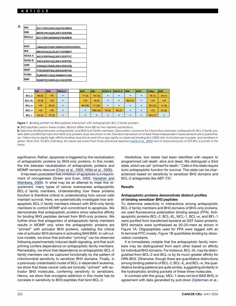
Figure 1. Binding pattern for BH3 peptide interaction with antiapoptotic BCL-2 family proteins
A: BH3 peptides used in these studies. BIDmut differs from BID by two alanine substitutions.B: Selective binding between antiapoptotic and BH3-only family members. Dissociation constants for interactions between antiapoptotic BCL-2 family pro-teins (left) and BH3 domains from BH3-only proteins (top) are shown in nM. Standard deviations of at least three independent measurements are in parenthe-ses. Yellow blocks signify high-affinity binding; blue blocks and minus sign signify no observed binding (Kd >2500 nM). Activators are in purple, and sensitizers ingreen. Note that, for BCL-2 binding, Kd values are lower than those previously reported (Letai et al., 2002) due to improved purity of GST-BCL-2 protein in thisassay.
significance. Rather, apoptosis is triggered by the neutralizationof antiapoptotic proteins by BH3-only proteins. In this model,the link between neutralization of antiapoptotic proteins andMOMP remains obscure (Chen et al., 2005; Willis et al., 2005).
It has been postulated that inhibition of apoptosis is a require-ment of oncogenesis (Green and Evan, 2002; Hanahan andWeinberg, 2000). In what may be an attempt to meet this re-quirement, many types of cancer overexpress antiapoptoticBCL-2 family members. Understanding how these proteinsfunction is therefore critical to understanding how cancer cellsmaintain survival. Here, we systematically investigate how anti-apoptotic BCL-2 family members interact with BH3-only familymembers to control MOMP and commitment to apoptosis. Wedemonstrate that antiapoptotic proteins show selective affinityfor binding BH3 peptides derived from BH3-only proteins. Wefurther show that antagonism of antiapoptotic family membersresults in MOMP only when the antiapoptotic proteins are‘‘primed’’ with activator BH3 proteins, validating the criticalrole of activator BH3 domains in activating BAX/BAK. In cell cul-ture models, we show that activator ‘‘priming’’ can be observedfollowing experimentally induced death signaling, and that suchpriming confers dependence on antiapoptotic family members.Remarkably, we show that dependence on antiapoptotic BCL-2family members can be captured functionally by the pattern ofmitochondrial sensitivity to sensitizer BH3 domains. Finally, ina previously credentialed model of BCL-2-dependent leukemia,we show that these cancer cells are tonically ‘‘primed’’ with ac-tivator BH3 molecules, conferring sensitivity to sensitizers.Hence, we show that oncogene addiction in this model has itscorrelate in sensitivity to BH3 peptides that bind BCL-2.
352
Heretofore, two states had been identified with respect toprogrammed cell death: alive and dead. We distinguish a thirdstate, which we call ‘‘primed for death.’’ Cells in this state requiretonic antiapoptotic function for survival. This state can be char-acterized based on sensitivity to sensitizer BH3 domains andBCL-2 antagonists like ABT-737.
Results
Antiapoptotic proteins demonstrate distinct profilesof binding sensitizer BH3 peptidesTo determine selectivity in interactions among antiapoptoticBCL-2 family members and BH3 domains of BH3-only proteins,we used fluorescence polarization binding assays (FPA). Anti-apoptotic proteins BCL-2, BCL-XL, MCL-1, BCL-w, and BFL-1were purified from transfected bacteria as GST fusion proteins.BH3 domains were synthesized as 20–25-mers as shown inFigure 1A. Oligopeptides used for FPA were tagged with anN-terminal FITC moiety. Figure 1B quantitates binding by disso-ciation constants.
It is immediately notable that the antiapoptotic family mem-bers may be distinguished from each other based on affinityfor individual BH3 domains. For instance, BCL-XL may be distin-guished from BCL-2 and BCL-w by its much greater affinity forHRK BH3. Otherwise, though there are quantitative distinctionsamong binding patterns of BCL-2, BCL-XL and BCL-w, the qual-ititative binding patterns are quite similar, suggesting similarity inthe hydrophobic binding pockets of these three molecules.
In contrast with this group, MCL-1 does not bind BAD BH3, inagreement with data generated by pull-down (Opferman et al.,
CANCER CELL MAY 2006
A R T I C L E
2003), yeast two-hybrid (Leo et al., 1999), and surface plasmonresonance (Chen et al., 2005) assays. Murine NOXA is uniqueamong the known BH3-only proteins in that it possesses twoputative BH3 domains (Oda et al., 2000). It is significant that,while the other four proteins interact with neither of the NOXABH3 domains tested, MCL-1 interacts with both. This suggeststhat the interaction between NOXA and MCL-1 is indeed biolog-ically significant, congruent with prior findings (Chen et al.,2005). The ability to bind both BH3 domains suggests the pos-sibility of multimeric interactions between MCL-1 and murineNOXA, or alternatively, differential control over exposure of thetwo BH3 domains in NOXA.
Also distinct is BFL-1. While it binds BID and BIM, it binds onlyPUMA among the sensitizers tested. It is also notable that theactivators BID and BIM BH3 are bound by all of the antiapo-ptotics tested, distinguishing them from the sensitizers, which,except PUMA, show a more selective pattern of binding. Of ad-ditional note is that the BH3 domain obtained from BNIP-3abinds to none of the proteins tested and does not activateBAX or BAK. While we cannot exclude the possibility thatBNIP BH3 interacts with an untested multidomain pro- or anti-apoptotic BCL-2 family member, it is also possible that BNIP-3adoes not function as a BH3 family member at all (Ray et al.,2000).
Dependence on individual antiapoptotic proteinsmay be deduced by pattern of sensitivity to sensitizerBH3 peptides; Inhibition of antiapoptotic proteinis insufficient for MOMP unless activator tBID is presentWe have previously shown that the BH3 domains of BID and BIMpossess the ability to induce BAX and BAK oligomerization andcytochrome c release in a purified mitochondrial system (Letaiet al., 2002). We termed this class of BH3 domain ‘‘activators.’’BH3 domains from BAD and BIK (which we termed ‘‘sensi-tizers’’) were unable to induce cytochrome c release on theirown. However, when an activator was bound and sequesteredby BCL-2, preventing interaction of the activator with BAX orBAK, sensitizers could provoke mitochondrial apoptosis bycompetitively inhibiting BCL-2’s binding of the activator, freeingthe activator to oligomerize BAX or BAK and induce cytochromec release. Thus, the two sensitizer BH3 domains were shown tobe antagonists of BCL-2 antiapoptotic function. The ability toantagonize BCL-2 function correlated with high-affinity bindingto BCL-2.
In Figure 1B, the expanded range of BH3 domains tested inthe present study demonstrate distinct patterns of binding toantiapoptotic proteins. To test if selective binding correspondedto ability of individual BH3 domains to selectively antagonizeantiapoptotic function, we constructed a purified mitochondrialsystem in which we reconstituted the critical apoptosis deci-sion-making molecular machinery. For the activator function,we used caspase-8 cleaved BID protein, tBID. tBID is an arche-typical activator protein, capable of inducing BAX/BAK oligo-merization and cytochrome c release in purified mitochondria(Wei et al., 2000) and synthetic liposomes (Kuwana et al.,2002, 2005). tBID’s induction of cytochrome c release andapoptosis requires BAX or BAK (Cheng et al., 2001; Wei et al.,2001). The multidomain proapoptotic function was providedby the BAK that resides in mouse liver mitochondria; mouse livermitochondria contain no detectable BAX protein (Letai et al.,2002). The dominant antiapoptotic function was provided by
CANCER CELL MAY 2006
one of the five different recombinant antiapoptotic proteinsused in the binding assays. BH3 peptides provided the sensi-tizer function.
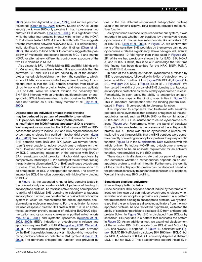
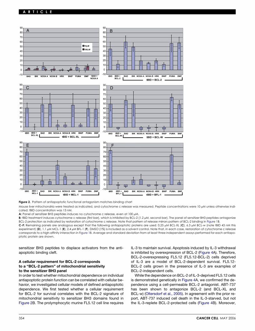
As cytochrome c release is the readout for our system, it wasimportant to test whether our peptides by themselves releasecytochrome c in mouse liver mitochondria like activators BIDor BIM BH3 (Letai et al., 2002). In Figure 2A, we confirm thatnone of the sensitizer BH3 peptides by themselves can inducecytochrome c release significantly above background, even atconcentrations 10-fold higher than those used in Figures 2B–2F. While we had previously shown this for BAD, BIK, NOXAA, and NOXA B BH3s, this is to our knowledge the first timethis finding has been described for the HRK, BNIP, PUMA,and BMF BH3 domains.
In each of the subsequent panels, cytochrome c release bytBID is demonstrated, followed by inhibition of cytochrome c re-lease by addition of either BCL-2 (Figure 2B), BCL-XL (Figure 2C),BCL-w (Figure 2D), MCL-1 (Figure 2E), or BFL-1 (Figure 2F). Wethen tested the ability of our panel of BH3 domains to antagonizeantiapoptotic protection as measured by cytochrome c release.Remarkably, in each case, the ability to antagonize antiapo-ptotic function maps to the binding specificities in Figure 1B.This is important confirmation that the binding pattern eluci-dated in Figure 1B corresponds to biological function.
It is important to emphasize that treatment with sensitizerpeptides alone, even those that bind and antagonize all the anti-apoptotics tested, such as PUMA BH3, or the combination ofNOXA and BAD BH3 is insufficient to cause cytochrome c re-lease (Figure 2A). Furthermore, when the panel of sensitizerBH3 peptides was tested in the presence of the antiapoptoticprotein BCL-XL, there was still no cytochrome c release, for-mally ruling out the possibility that the BH3 peptides were some-how directly converting antiapoptotic proteins to a proapoptoticfunction (Figure S1 in the Supplemental Data available with thisarticle online). To induce MOMP and cytochrome c release,there appears to be an absolute requirement for an activatorfunction, here provided by the tBID protein.
These data critically demonstrate that our panel of peptidescan determine whether a mitochondrion depends on an anti-apoptotic protein to maintain integrity. Furthermore, the identityof the critical antiapoptotic protein can be deduced based onthe pattern of sensitivity to our panel of sensitizer BH3 peptides.We call this strategy BH3 profiling.
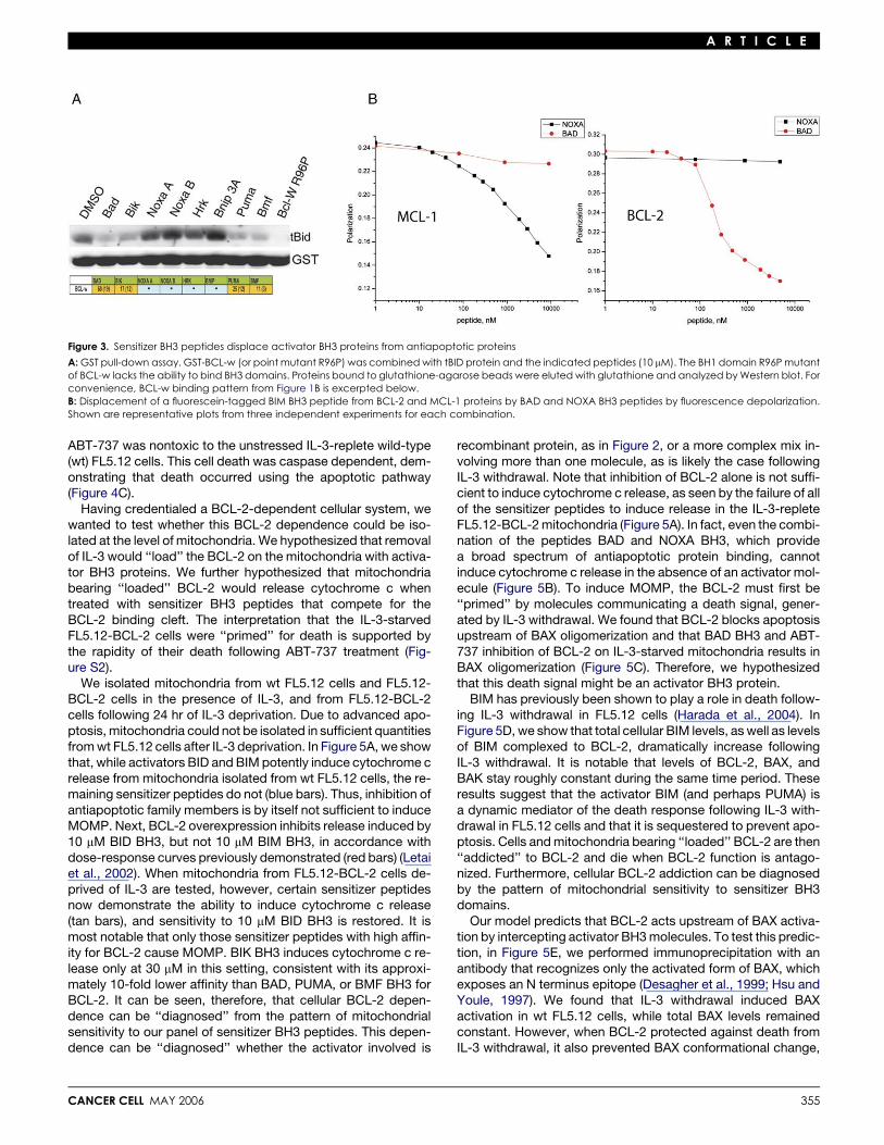
Sensitizers displace activatorsfrom antiapoptotic proteinsSince sensitizer BH3 peptides cannot induce cytochrome c re-lease on their own but can induce cytochrome c release whenactivator and antiapoptotic proteins are present, in a patternthat mirrors their binding to antiapoptotic proteins, we hypothe-sized that the sensitizers are displacing activators from the anti-apoptotic proteins. As one test of this hypothesis, we tested theability of sensitizer peptides to displace tBID from antiapoptoticprotein Bcl-w. In Figure 3A, tBID is displaced from BCL-w bysensitizer BH3 peptides in a pattern that replicates the patternin Figure 2D. As an additional test, we examined displacementof the activator BIM BH3 peptide from BCL-2 and MCL-1 byBAD and NOXA BH3 peptides. In Figure 3B, consistent with Fig-ure 1B, BAD BH3 efficiently displaces BIM BH3 from BCL-2, butnot MCL-1, whereas NOXA A BH3 efficiently displaces BIM fromMCL-1, but not BCL-2. These experiments support the ability of
353
A R T I C L E
Figure 2. Pattern of antiapoptotic functional antagonism matches binding chart
Mouse liver mitochondria were treated as indicated, and cytochrome c release was measured. Peptide concentrations were 10 mM unless otherwise indi-cated; tBID concentration was 13 nM.A: Panel of sensitizer BH3 peptides induces no cytochrome c release, even at 100 mM.B: tBID treatment induces cytochrome c release (first bar), which is inhibited by BCL-2 (1.2 mM, second bar). The panel of sensitizer BH3 peptides antagonizeBCL-2 protection as indicated by restoration of cytochrome c release. Note that pattern of release mirrors pattern of BCL-2 binding in Figure 1B.C–F: Remaining panels are analogous except that the following antiapoptotic proteins are used: 0.25 mM BCL-XL (C), 6.3 mM BCL-w (note tBID 43 nM thisexperiment) (D), 1.1 mM MCL-1 (E), 2.4 mM BFL-1 (F). DMSO (1%) is included as a solvent control. Note that, in each case, restoration of cytochrome c releasecorresponds to a high-affinity interaction in Figure 1B. Average and standard deviation from at least three independent assays performed for each antiapo-ptotic protein are shown.
sensitizer BH3 peptides to displace activators from the anti-apoptotic binding cleft.
A cellular requirement for BCL-2 correspondsto a ‘‘BCL-2 pattern’’ of mitochondrial sensitivityto the sensitizer BH3 panelIn order to test whether mitochondrial dependence on individualantiapoptotic protein function can be correlated with cellular be-havior, we investigated cellular models of defined antiapoptoticdependence. We first tested whether a cellular requirementfor BCL-2 for survival correlates with the BCL-2 signature ofmitochondrial sensitivity to sensitizer BH3 domains found inFigure 2B. The prolymphocytic murine FL5.12 cell line requires
354
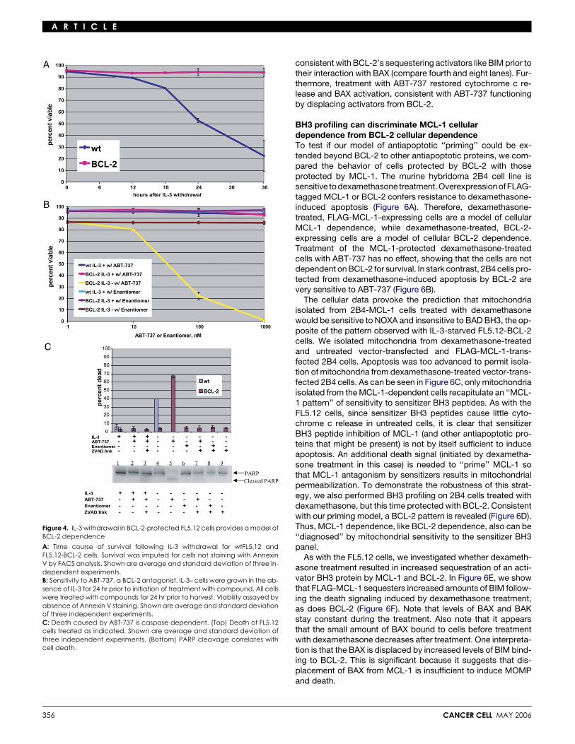
IL-3 to maintain survival. Apoptosis induced by IL-3 withdrawalis inhibited by overexpression of BCL-2 (Figure 4A). Therefore,BCL-2-overexpressing FL5.12 (FL5.12-BCL-2) cells deprivedof IL-3 are a model of BCL-2-dependent survival. FL5.12-BCL-2 cells grown in the presence of IL-3 are examples ofBCL-2-independent cells.
While the dependence on BCL-2 of IL-3-deprived FL5.12 cellsis demonstrated genetically in Figure 4A, we confirmed the de-pendence using a cell-permeable BCL-2 antagonist. ABT-737has been shown to antagonize BCL-2 (and BCL-XL andBCL-w) (Oltersdorf et al., 2005). In agreement with the prior re-port, ABT-737 induced cell death in the IL-3-starved, but notthe IL-3-replete BCL-2-protected cells (Figure 4B). Moreover,
CANCER CELL MAY 2006
A R T I C L E
Figure 3. Sensitizer BH3 peptides displace activator BH3 proteins from antiapoptotic proteins
A: GST pull-down assay. GST-BCL-w (or point mutant R96P) was combined with tBID protein and the indicated peptides (10 mM). The BH1 domain R96P mutantof BCL-w lacks the ability to bind BH3 domains. Proteins bound to glutathione-agarose beads were eluted with glutathione and analyzed by Western blot. Forconvenience, BCL-w binding pattern from Figure 1B is excerpted below.B: Displacement of a fluorescein-tagged BIM BH3 peptide from BCL-2 and MCL-1 proteins by BAD and NOXA BH3 peptides by fluorescence depolarization.Shown are representative plots from three independent experiments for each combination.
ABT-737 was nontoxic to the unstressed IL-3-replete wild-type(wt) FL5.12 cells. This cell death was caspase dependent, dem-onstrating that death occurred using the apoptotic pathway(Figure 4C).
Having credentialed a BCL-2-dependent cellular system, wewanted to test whether this BCL-2 dependence could be iso-lated at the level of mitochondria. We hypothesized that removalof IL-3 would ‘‘load’’ the BCL-2 on the mitochondria with activa-tor BH3 proteins. We further hypothesized that mitochondriabearing ‘‘loaded’’ BCL-2 would release cytochrome c whentreated with sensitizer BH3 peptides that compete for theBCL-2 binding cleft. The interpretation that the IL-3-starvedFL5.12-BCL-2 cells were ‘‘primed’’ for death is supported bythe rapidity of their death following ABT-737 treatment (Fig-ure S2).
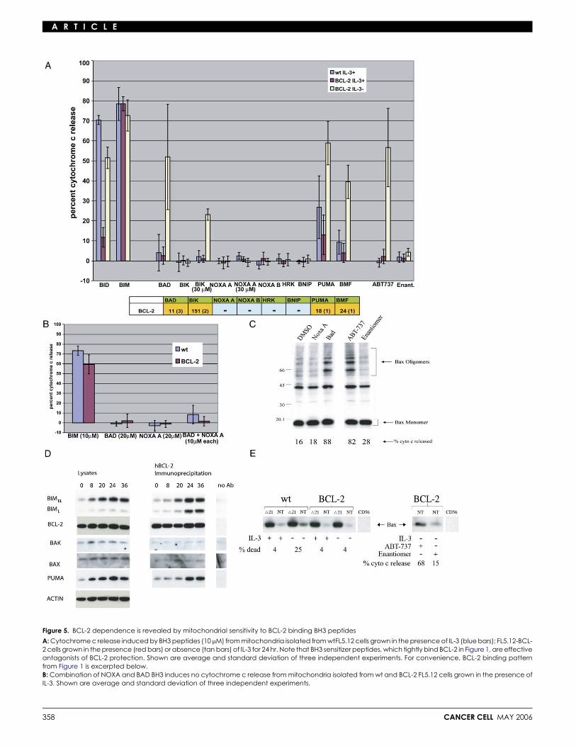
We isolated mitochondria from wt FL5.12 cells and FL5.12-BCL-2 cells in the presence of IL-3, and from FL5.12-BCL-2cells following 24 hr of IL-3 deprivation. Due to advanced apo-ptosis, mitochondria could not be isolated in sufficient quantitiesfrom wt FL5.12 cells after IL-3 deprivation. In Figure 5A, we showthat, while activators BID and BIM potently induce cytochrome crelease from mitochondria isolated from wt FL5.12 cells, the re-maining sensitizer peptides do not (blue bars). Thus, inhibition ofantiapoptotic family members is by itself not sufficient to induceMOMP. Next, BCL-2 overexpression inhibits release induced by10 mM BID BH3, but not 10 mM BIM BH3, in accordance withdose-response curves previously demonstrated (red bars) (Letaiet al., 2002). When mitochondria from FL5.12-BCL-2 cells de-prived of IL-3 are tested, however, certain sensitizer peptidesnow demonstrate the ability to induce cytochrome c release(tan bars), and sensitivity to 10 mM BID BH3 is restored. It ismost notable that only those sensitizer peptides with high affin-ity for BCL-2 cause MOMP. BIK BH3 induces cytochrome c re-lease only at 30 mM in this setting, consistent with its approxi-mately 10-fold lower affinity than BAD, PUMA, or BMF BH3 forBCL-2. It can be seen, therefore, that cellular BCL-2 depen-dence can be ‘‘diagnosed’’ from the pattern of mitochondrialsensitivity to our panel of sensitizer BH3 peptides. This depen-dence can be ‘‘diagnosed’’ whether the activator involved is
CANCER CELL MAY 2006
recombinant protein, as in Figure 2, or a more complex mix in-volving more than one molecule, as is likely the case followingIL-3 withdrawal. Note that inhibition of BCL-2 alone is not suffi-cient to induce cytochrome c release, as seen by the failure of allof the sensitizer peptides to induce release in the IL-3-repleteFL5.12-BCL-2 mitochondria (Figure 5A). In fact, even the combi-nation of the peptides BAD and NOXA BH3, which providea broad spectrum of antiapoptotic protein binding, cannotinduce cytochrome c release in the absence of an activator mol-ecule (Figure 5B). To induce MOMP, the BCL-2 must first be‘‘primed’’ by molecules communicating a death signal, gener-ated by IL-3 withdrawal. We found that BCL-2 blocks apoptosisupstream of BAX oligomerization and that BAD BH3 and ABT-737 inhibition of BCL-2 on IL-3-starved mitochondria results inBAX oligomerization (Figure 5C). Therefore, we hypothesizedthat this death signal might be an activator BH3 protein.
BIM has previously been shown to play a role in death follow-ing IL-3 withdrawal in FL5.12 cells (Harada et al., 2004). InFigure 5D, we show that total cellular BIM levels, as well as levelsof BIM complexed to BCL-2, dramatically increase followingIL-3 withdrawal. It is notable that levels of BCL-2, BAX, andBAK stay roughly constant during the same time period. Theseresults suggest that the activator BIM (and perhaps PUMA) isa dynamic mediator of the death response following IL-3 with-drawal in FL5.12 cells and that it is sequestered to prevent apo-ptosis. Cells and mitochondria bearing ‘‘loaded’’ BCL-2 are then‘‘addicted’’ to BCL-2 and die when BCL-2 function is antago-nized. Furthermore, cellular BCL-2 addiction can be diagnosedby the pattern of mitochondrial sensitivity to sensitizer BH3domains.
Our model predicts that BCL-2 acts upstream of BAX activa-tion by intercepting activator BH3 molecules. To test this predic-tion, in Figure 5E, we performed immunoprecipitation with anantibody that recognizes only the activated form of BAX, whichexposes an N terminus epitope (Desagher et al., 1999; Hsu andYoule, 1997). We found that IL-3 withdrawal induced BAXactivation in wt FL5.12 cells, while total BAX levels remainedconstant. However, when BCL-2 protected against death fromIL-3 withdrawal, it also prevented BAX conformational change,
355
A R T I C L E
Figure 4. IL-3 withdrawal in BCL-2-protected FL5.12 cells provides a model ofBCL-2 dependence
A: Time course of survival following IL-3 withdrawal for wtFL5.12 andFL5.12-BCL-2 cells. Survival was imputed for cells not staining with AnnexinV by FACS analysis. Shown are average and standard deviation of three in-dependent experiments.B: Sensitivity to ABT-737, a BCL-2 antagonist. IL-3– cells were grown in the ab-sence of IL-3 for 24 hr prior to initiation of treatment with compound. All cellswere treated with compounds for 24 hr prior to harvest. Viability assayed byabsence of Annexin V staining. Shown are average and standard deviationof three independent experiments.C: Death caused by ABT-737 is caspase dependent. (Top) Death of FL5.12cells treated as indicated. Shown are average and standard deviation ofthree independent experiments. (Bottom) PARP cleavage correlates withcell death.
356
consistent with BCL-2’s sequestering activators like BIM prior totheir interaction with BAX (compare fourth and eight lanes). Fur-thermore, treatment with ABT-737 restored cytochrome c re-lease and BAX activation, consistent with ABT-737 functioningby displacing activators from BCL-2.
BH3 profiling can discriminate MCL-1 cellulardependence from BCL-2 cellular dependenceTo test if our model of antiapoptotic ‘‘priming’’ could be ex-tended beyond BCL-2 to other antiapoptotic proteins, we com-pared the behavior of cells protected by BCL-2 with thoseprotected by MCL-1. The murine hybridoma 2B4 cell line issensitive to dexamethasone treatment.Overexpression of FLAG-tagged MCL-1 or BCL-2 confers resistance to dexamethasone-induced apoptosis (Figure 6A). Therefore, dexamethasone-treated, FLAG-MCL-1-expressing cells are a model of cellularMCL-1 dependence, while dexamethasone-treated, BCL-2-expressing cells are a model of cellular BCL-2 dependence.Treatment of the MCL-1-protected dexamethasone-treatedcells with ABT-737 has no effect, showing that the cells are notdependent on BCL-2 for survival. In stark contrast, 2B4 cells pro-tected from dexamethasone-induced apoptosis by BCL-2 arevery sensitive to ABT-737 (Figure 6B).
The cellular data provoke the prediction that mitochondriaisolated from 2B4-MCL-1 cells treated with dexamethasonewould be sensitive to NOXA and insensitive to BAD BH3, the op-posite of the pattern observed with IL-3-starved FL5.12-BCL-2cells. We isolated mitochondria from dexamethasone-treatedand untreated vector-transfected and FLAG-MCL-1-trans-fected 2B4 cells. Apoptosis was too advanced to permit isola-tion of mitochondria from dexamethasone-treated vector-trans-fected 2B4 cells. As can be seen in Figure 6C, only mitochondriaisolated from the MCL-1-dependent cells recapitulate an ‘‘MCL-1 pattern’’ of sensitivity to sensitizer BH3 peptides. As with theFL5.12 cells, since sensitizer BH3 peptides cause little cyto-chrome c release in untreated cells, it is clear that sensitizerBH3 peptide inhibition of MCL-1 (and other antiapoptotic pro-teins that might be present) is not by itself sufficient to induceapoptosis. An additional death signal (initiated by dexametha-sone treatment in this case) is needed to ‘‘prime’’ MCL-1 sothat MCL-1 antagonism by sensitizers results in mitochondrialpermeabilization. To demonstrate the robustness of this strat-egy, we also performed BH3 profiling on 2B4 cells treated withdexamethasone, but this time protected with BCL-2. Consistentwith our priming model, a BCL-2 pattern is revealed (Figure 6D).Thus, MCL-1 dependence, like BCL-2 dependence, also can be‘‘diagnosed’’ by mitochondrial sensitivity to the sensitizer BH3panel.
As with the FL5.12 cells, we investigated whether dexameth-asone treatment resulted in increased sequestration of an acti-vator BH3 protein by MCL-1 and BCL-2. In Figure 6E, we showthat FLAG-MCL-1 sequesters increased amounts of BIM follow-ing the death signaling induced by dexamethasone treatment,as does BCL-2 (Figure 6F). Note that levels of BAX and BAKstay constant during the treatment. Also note that it appearsthat the small amount of BAX bound to cells before treatmentwith dexamethasone decreases after treatment. One interpreta-tion is that the BAX is displaced by increased levels of BIM bind-ing to BCL-2. This is significant because it suggests that dis-placement of BAX from MCL-1 is insufficient to induce MOMPand death.
CANCER CELL MAY 2006
A R T I C L E
To further demonstrate that the mitochondrial assays reflecttrue cellular dependence, we transfected peptides via electro-poration into FLAG-MCL-1-transfected 2B4 cells that hadbeen treated with dexamethasone, putatively priming MCL-1with death signals, carried at least in part by BIM. Supportingthe cellular relevance of our mitochondrial BH3 profiling assays,an MCL-1 pattern of response to sensitizer peptides was ob-served (Figure 6G; compare with Figures 1B, 2E, and 6C).
Dependence on BCL-2 in a leukemia correspondsto mitochondrial sensitivity to sensitizersin a ‘‘BCL-2 pattern’’ and sequestration of BIMDependence on antiapoptotic proteins is perhaps of greatestimportance in the context of cancer, in which antiapoptoticBCL-2 family proteins are subjects of intense investigation astherapeutic targets. While the concept of oncogene addictionhas received attention recently (Jonkers and Berns, 2004; Wein-stein, 2002), the molecular details of the addiction to spe-cific oncogenes is poorly understood. We therefore turned toa validated model of oncogene addiction, a BCL-2-dependentmurine leukemia, to examine the molecular basis for BCL-2‘‘addiction.’’
We have previously described a mouse acute lymphocyticleukemia model in which c-myc is constitutively expressedand human BCL-2 is repressibly expressed. In this model,when BCL-2 transgene expression is eliminated by administra-tion of doxycycline, the leukemic cells undergo apoptosis, re-sulting in rapid resolution of the leukemia (Letai et al., 2004).This provides us with an ideal in vivo model of a BCL-2-depen-dent cancer. We wondered if the dependence on BCL-2 wasdue to a similar mechanism to that of the IL-3-deprivedFL5.12-BCL-2 cells—that is, a death signal was being initiatedand carried by an activator BH3 molecule, but BCL-2 was bind-ing it and preventing its interaction with multidomain proapop-totic proteins.
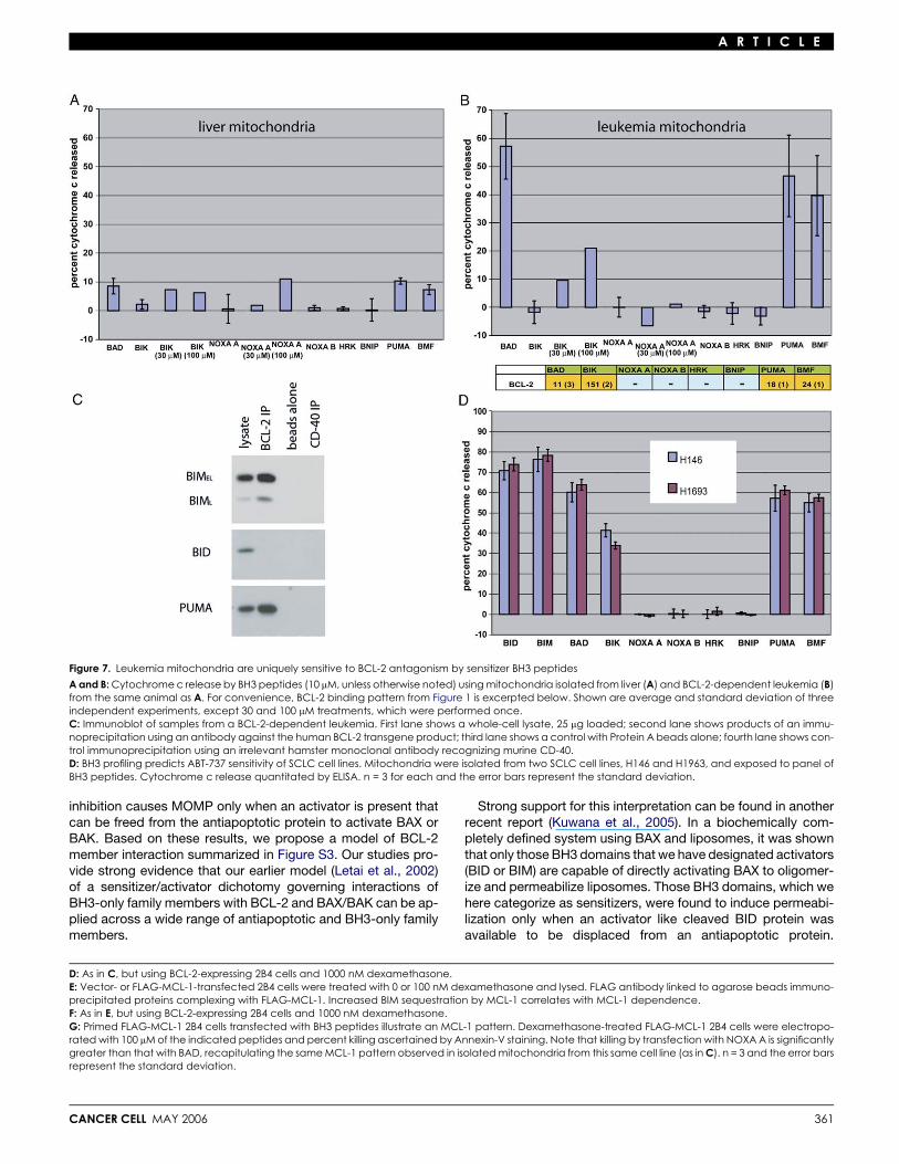
We isolated mitochondria from leukemia cells and exposedthem to sensitizer BH3 peptides and measured release of cyto-chrome c. As an internal control, mitochondria were isolatedfrom liver from the leukemic mice in parallel (Figure 7A). The sen-sitizer BH3 peptides were unable to induce cytochrome c re-lease from nonmalignant hepatocyte mitochondria from the leu-kemic mice, just as they were unable to induce cytochrome crelease from nonmalignant liver mitochondria from normalmice (Figure 2A) or from nonmalignant FL5.12 (Figure 5A) or2B4 mitochondria (Figure 6C). Intriguingly, certain sensitizerBH3 peptides were capable of inducing near total cytochromec release from the leukemic mitochondria (Figure 7B). Signifi-cantly, the pattern of peptides that induced release corre-sponded exactly to those peptides that bind with high affinityto BCL-2 (Figure 1B), namely BAD, BIK, PUMA, and BMF.Note that, consistent with its approximately 10-fold lower affinitythan BAD BH3 for BCL-2, BIK BH3 requires a 10-fold higherconcentration to demonstrate cytochrome c release. A 10-foldincrease in NOXA A peptide concentration has no effect, consis-tent with the extremely low affinity NOXA A has for BCL-2.
These results suggest that, in this leukemia model, deathsignals are being continually initiated, and BCL-2 is required tosequester the activator BH3 molecule to prevent apoptosis. Incontrast to the nonmalignant systems tested above, leukemiccell BCL-2 behaves as if already ‘‘primed’’ with activator pro-tein(s) without any further intervention, such as growth factor
CANCER CELL MAY 2006
withdrawal or dexamethasone treatment. In Figure 7C, weshow that BIM is expressed in the leukemia cells, and it is boundby BCL-2. Supporting the signal importance of BIM in transmit-ting death signals in this model, BID is also present in the lysatebut is not bound by BCL-2. Note that PUMA is also found to bebound by BCL-2, consistent with a report showing that PUMAdeficiency could accelerate myc-induced lymphomagenesis(Hemann et al., 2004). Since in our hands the PUMA BH3 lacksthe ability to directly activate BAX or BAK, we hypothesize thatPUMA is acting as a sensitizer in this context, in effect decreas-ing the amount of BCL-2 available to bind BIM and possibly BAXor BAK.
If BCL-2 maintains survival of this leukemia cell primarily bysequestering BIM, then one would predict that BIM loss of func-tion could substitute for BCL-2 overexpression to cooperatewith c-myc in leukemogenesis. In fact, this experiment has al-ready been performed. It was found that BIM deficiency canindeed cooperate with c-myc to produce a pre-B lymphocyticleukemia like the one produced here by the cooperation ofBCL-2 overexpression with c-myc (Egle et al., 2004). These re-sults support a model in which BCL-2 is necessary for survivalof our leukemia largely because it is required to sequesterBIM, preventing activation of BAX/BAK and subsequentMOMP. The leukemia cells are therefore neither normal andhealthy, nor dead, but rather primed for death.
BH3 profiling predicts sensitivity to ABT-737As another test of the ability of BH3 profiling to detect in vivoBCL-2 dependence, we turned to two small cell lung cancer(SCLC) cell lines that were sensitive to treatment with ABT-737in vitro and in an in vivo murine xenograft model (Oltersdorfet al., 2005). Both H146 and H1963 demonstrate a pattern ofsensitivity diagnostic of BCL-2 sensitivity (Figure 7D). This pro-vides further support, in addition to the results of Figures 4, 5,and 6, that mitochondrial BH3 profiling is a powerful predictorof what cells are sensitive to BH3 mimetic drugs in vitro andin vivo.
Discussion
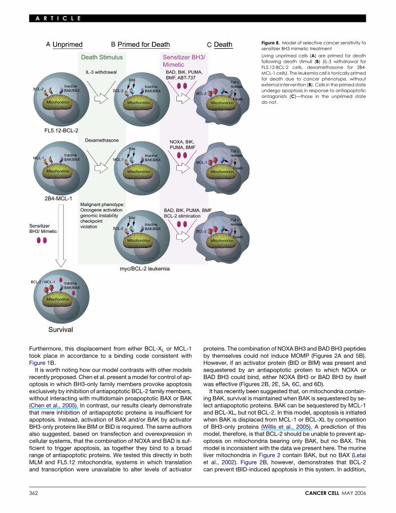
Life on the edge: ‘‘Primed for death’’Conventionally, study of the apoptotic machinery has been ableto discriminate cells into two states: alive or dead. Here, weshow that certain cells live in a state that can be distinguishedby dependence on antiapoptotic proteins for survival. We de-scribe these cells as being ‘‘primed for death,’’ as death signal-ing has caused their antiapoptotic family members to sequestersignificant quantities of proapoptotic BH3 proteins. Inhibition ofthe antiapoptotic proteins in these cells, but not unprimed cells,results in BAX/BAK oligomerization and MOMP. We suggest,therefore, that there are three functionally distinguishable stateswith respect to programmed cell death: unprimed, primed fordeath, and dead. We suggest that the state of being primedfor death is a continuum, as the magnitude of BH3 proteins prim-ing the mitochondrion can, of course, vary continuously until theantiapoptotic reserve is overwhelmed and the cell commits toprogrammed cell death. A summary of this model with referenceto the data in this paper can be found in Figure 8.
We probed mitochondria to determine a cell’s state using ourpanel of sensitizer BH3 peptides, selective antagonists of anti-apoptotic BCL-2 family members. Mitochondria that are primed
357
A R T I C L E
Figure 5. BCL-2 dependence is revealed by mitochondrial sensitivity to BCL-2 binding BH3 peptides
A: Cytochrome c release induced by BH3 peptides (10 mM) from mitochondria isolated from wtFL5.12 cells grown in the presence of IL-3 (blue bars); FL5.12-BCL-2 cells grown in the presence (red bars) or absence (tan bars) of IL-3 for 24 hr. Note that BH3 sensitizer peptides, which tightly bind BCL-2 in Figure 1, are effectiveantagonists of BCL-2 protection. Shown are average and standard deviation of three independent experiments. For convenience, BCL-2 binding patternfrom Figure 1 is excerpted below.B: Combination of NOXA and BAD BH3 induces no cytochrome c release from mitochondria isolated from wt and BCL-2 FL5.12 cells grown in the presence ofIL-3. Shown are average and standard deviation of three independent experiments.
358 CANCER CELL MAY 2006

A R T I C L E
for death are dependent on antiapoptotic protein function toprevent MOMP, so that they release cytochrome c when ex-posed to sensitizer BH3 peptides (Figures 2B–2F, 5A, 6C, 6D,7B, and 7D). In contrast, unprimed cells do not release cyto-chrome c when exposed to sensitizer BH3 peptides. In theory,any cell from which mitochondria can be isolated can thereforebe so tested and categorized as being primed or unprimed.Testing of mitochondria directly has the advantage of eliminat-ing any contribution of transcription, translation, or posttransla-tional modification events that might be triggered by transfec-tion of peptide, protein, or expression vector into a living cell.A ‘‘snapshot’’ of the apoptotic state at a given time may be takenwith minimal perturbation of the extant apoptotic machinery. Insummary, we were able to capture information about a funda-mental aspect of cellular physiology in an assay that can be per-formed using primary or cultured cells in a single day.
Importantly, we link mitochondrial behavior to whole-cellbehavior in several models. Mitochondria were primed whencells were enduring a physiologic challenge, and BH3 profilingrevealed a dependence on antiapoptotic proteins only whena cellular dependence was also demonstrated. In the case ofFL5.12 cells, cells and mitochondria became primed for deathonly after IL-3 withdrawal. For 2B4 cells, cells and mitochondriawere primed for death only after dexamethasone treatment. Forthe primary BCL-2-dependent leukemia cells, the genomicinstability, myc oncogene activation, and checkpoint violationinherent to the cancer phenotype were sufficient to induce mito-chondrial priming without further external intervention. TheSCLC cell lines H164 and H1963 that revealed a BCL-2 patternof sensitivity to BH3 profiling likewise are sensitive to the BCL-2antagonist ABT-737. In each case, mitochondrial studies cor-rectly diagnosed the cellular dependence on an antiapoptoticBCL-2 family member. Furthermore, the identity of the individualfamily member could be decoded based on the pattern of mito-chondrial sensitivity to our peptide panel.
An implication of our results is that in some cells, like IL-3-replete FL5.12-BCL-2 cells, BCL-2 overexpression providesextra antiapoptotic reserve. In others, like the murine leukemias,high levels of BCL-2 are present, but the BCL-2 is so highlyoccupied by activator BH3 proteins that the cell has very poorantiapoptotic reserve and is actually primed for death.
The binding codeAn important property of our panel of sensitizer BH3 peptides isits ability to distinguish among the antiapoptotic proteins basedon binding specificity. Others have recently demonstrated sim-ilar selectivity in interaction among BH3 peptides and antiapo-ptotic proteins using a different technique, surface plasmon res-onance (SPR) (Chen et al., 2005). While dissociation constantswere not explicitly determined, the overall pattern of bindingwas similar, with some notable differences. The binding ofHRK BH3 peptide, restricted to BCL-XL in our work, was
CANCER CELL MAY 2006
promiscuous throughout the antiapoptotic proteins tested bySPR. BFL1/A1, which bound only PUMA among the sensitizerBH3 peptides in our work, bound BIK, HRK, and NOXA as wellin the work by Chen et al. There are several possible explana-tions for these differences. First, the peptides are not identical.Their peptides were 26 amino acids in length, while those weuse are shorter (Figure 1A). In addition, they do not test directbinding of the peptides to the antiapoptotic proteins, as wedid by fluorescence polarization, but rather test the ability ofeach of the peptides to displace a BIM BH3 peptide from theantiapoptotic protein by SPR. We find our peptide panel partic-ularly useful, as it allows us to distinguish BCL-XL protectionfrom the rest due to its selective binding of HRK BH3. Further-more, our demonstration that MCL-1 selectively binds bothmurine NOXA BH3 peptides suggests biological relevance tothe binding pattern observed.
Do these interactions between BH3 peptides and antiapo-ptotic proteins mirror protein-protein interactions in actual cellu-lar systems? Consistent with our binding chart, we have shownthat MCL-1 preferentially binds BIM but not BAD, governing mu-rine lymphocyte dependence on MCL-1 for survival (Opfermanet al., 2003). A yeast two-hybrid interaction study supports thespecificity we found for BAD’s binding of antiapoptotic familymembers in Figure 1B (Bae et al., 2001). Others (Chen et al.,2005) showed that interactions of proteins overexpressed incells showed fidelity with the patterns found in both our systems.Since BH3 domains are the critical ligands for the bindingpockets of antiapoptotic proteins, we expect our BH3 peptidebinding patterns to be consistent with in vivo protein-proteininteraction specificity. Since length of peptide might affect func-tion, a longer, 26 amino acid HRK peptide was tested. Whilelengthening the peptide increased slightly its ability to antago-nize BCL-2 and MCL-1 in cytochrome c release assays, it stilldid not demonstrate activator function (data not shown). It isnonetheless possible that different conformations or posttrans-lational modifications play a role in vivo that was not evaluated inour binding assays. Regardless, our peptide panel capturesbinding differences that allow it to function as a diagnostic ofantiapoptotic protein dependence. BCL-2 and BCL-w aremost similar, with BCL-XL distinguished by its binding to HRKBH3. MCL-1 is quite different, binding both NOXA peptidesbut not BAD, and BFL-1 binds none of the sensitizers exceptPUMA. These binding specificities suggest, moreover, that itis possible for cells to adjust sensitivity to distinct apoptoticinsults by adjusting levels of individual BCL-2 family memberexpression.
The basic apoptotic paradigmOur results shed light on a controversial issue—how interactionsamong BCL-2 family members control MOMP. Our results con-sistently show that ligation of antiapoptotic family membersalone is insufficient to induce MOMP. Rather, antiapoptotic
C: Mitochondria isolated from FL5.12-BCL-2 cells grown in the absence of IL-3 for 24 hr were treated with NOXA A or BAD peptides (30 mM) or ABT-737 or controlenantiomer at 10 mM for 35 min. Mitochondrial pellets were subjected to chemical crosslinking as previously described (Letai et al., 2002).D: Immunoblots of FL5.12-BCL-2 whole-cell lysates (left) and samples immunoprecipitated by an antibody directed against the human BCL-2 transgene prod-uct (right). Numbers at top refer to hours after IL-3 withdrawal. Control lane performed without anti-human BCL-2 antibody in pull-down at right. Negativecontrol immunoprecipitation using anti-human BCL-2 antibody on lysates from IL-3-starved FL5.12-BCL-XL cells yielded no bands (data not shown).E: BCL-2 blocks apoptosis upstream of BAX activation. wt or BCL-2-expressing FL5.12 cells were exposed to IL-3 withdrawal as indicated. BAX was immunopre-cipitated using an antibody recognizing all BAX conformations (D21) or only the activated conformation with N-terminal exposure (NT). Death induced in thecells is indicated below. At right, mitochondria isolated from IL-3-starved cells were treated with ABT-737 or control enantiomer, and immunoprecipitation withNT performed as indicated. CD56 indicates control immunoprecipitation by an irrelevant antibody recognizing CD56.
359

A R T I C L E
Figure 6. BH3 profiling discriminates BCL-2 and MCL-1 dependence
A: MCL-1 and BCL-2 inhibit cell death induced by dexamethasone in 2B4 cells. 2B4 cells transfected with Flag-MCL-1, BCL-2, or empty vector constructs werecultured for 24 hr in the presence of the indicated concentration of dexamethasone. Viability determined by absence of Annexin V staining by FACS. Shownare average and standard deviation of three independent experiments.B: BCL-2 antagonist ABT-737 has no effect on wt or MCL-1-dependent 2B4 cells but kills BCL-2-dependent cells. 2B4 cells were incubated for 24 hr with dexa-methasone and either ABT-737 or enantiomer for 24 hr. Shown are average and standard deviation of three independent experiments.C: Mitochondria isolated from 2B4 cells treated as indicated were exposed to BH3 peptides at the concentrations indicated. Cytochrome c release is inducedby MCL-1 binding sensitizer BH3 peptides only when mitochondria are derived from MCL-1-dependent cells. Shown are average and standard deviation ofthree independent experiments. For convenience, MCL-1 binding pattern from Figure 1 is excerpted below.
360 CANCER CELL MAY 2006
A R T I C L E
Figure 7. Leukemia mitochondria are uniquely sensitive to BCL-2 antagonism by sensitizer BH3 peptides
A and B: Cytochrome c release by BH3 peptides (10 mM, unless otherwise noted) using mitochondria isolated from liver (A) and BCL-2-dependent leukemia (B)from the same animal as A. For convenience, BCL-2 binding pattern from Figure 1 is excerpted below. Shown are average and standard deviation of threeindependent experiments, except 30 and 100 mM treatments, which were performed once.C: Immunoblot of samples from a BCL-2-dependent leukemia. First lane shows a whole-cell lysate, 25 mg loaded; second lane shows products of an immu-noprecipitation using an antibody against the human BCL-2 transgene product; third lane shows a control with Protein A beads alone; fourth lane shows con-trol immunoprecipitation using an irrelevant hamster monoclonal antibody recognizing murine CD-40.D: BH3 profiling predicts ABT-737 sensitivity of SCLC cell lines. Mitochondria were isolated from two SCLC cell lines, H146 and H1963, and exposed to panel ofBH3 peptides. Cytochrome c release quantitated by ELISA. n = 3 for each and the error bars represent the standard deviation.
inhibition causes MOMP only when an activator is present thatcan be freed from the antiapoptotic protein to activate BAX orBAK. Based on these results, we propose a model of BCL-2member interaction summarized in Figure S3. Our studies pro-vide strong evidence that our earlier model (Letai et al., 2002)of a sensitizer/activator dichotomy governing interactions ofBH3-only family members with BCL-2 and BAX/BAK can be ap-plied across a wide range of antiapoptotic and BH3-only familymembers.
CANCER CELL MAY 2006
Strong support for this interpretation can be found in anotherrecent report (Kuwana et al., 2005). In a biochemically com-pletely defined system using BAX and liposomes, it was shownthat only those BH3 domains that we have designated activators(BID or BIM) are capable of directly activating BAX to oligomer-ize and permeabilize liposomes. Those BH3 domains, which wehere categorize as sensitizers, were found to induce permeabi-lization only when an activator like cleaved BID protein wasavailable to be displaced from an antiapoptotic protein.
D: As in C, but using BCL-2-expressing 2B4 cells and 1000 nM dexamethasone.E: Vector- or FLAG-MCL-1-transfected 2B4 cells were treated with 0 or 100 nM dexamethasone and lysed. FLAG antibody linked to agarose beads immuno-precipitated proteins complexing with FLAG-MCL-1. Increased BIM sequestration by MCL-1 correlates with MCL-1 dependence.F: As in E, but using BCL-2-expressing 2B4 cells and 1000 nM dexamethasone.G: Primed FLAG-MCL-1 2B4 cells transfected with BH3 peptides illustrate an MCL-1 pattern. Dexamethasone-treated FLAG-MCL-1 2B4 cells were electropo-rated with 100 mM of the indicated peptides and percent killing ascertained by Annexin-V staining. Note that killing by transfection with NOXA A is significantlygreater than that with BAD, recapitulating the same MCL-1 pattern observed in isolated mitochondria from this same cell line (as in C). n = 3 and the error barsrepresent the standard deviation.
361
A R T I C L E
Figure 8. Model of selective cancer sensitivity tosensitizer BH3 mimetic treatment
Living unprimed cells (A) are primed for deathfollowing death stimuli (B) (IL-3 withdrawal forFL5.12-BCL-2 cells, dexamethasone for 2B4-MCL-1 cells). The leukemia cell is tonically primedfor death due to cancer phenotype, withoutexternal intervention (B). Cells in the primed stateundergo apoptosis in response to antiapoptoticantagonists (C)—those in the unprimed statedo not.
Furthermore, this displacement from either BCL-XL or MCL-1took place in accordance to a binding code consistent withFigure 1B.
It is worth noting how our model contrasts with other modelsrecently proposed. Chen et al. present a model for control of ap-optosis in which BH3-only family members provoke apoptosisexclusively by inhibition of antiapoptotic BCL-2 family members,without interacting with multidomain proapoptotic BAX or BAK(Chen et al., 2005). In contrast, our results clearly demonstratethat mere inhibition of antiapoptotic proteins is insufficient forapoptosis. Instead, activation of BAX and/or BAK by activatorBH3-only proteins like BIM or BID is required. The same authorsalso suggested, based on transfection and overexpression incellular systems, that the combination of NOXA and BAD is suf-ficient to trigger apoptosis, as together they bind to a broadrange of antiapoptotic proteins. We tested this directly in bothMLM and FL5.12 mitochondria, systems in which translationand transcription were unavailable to alter levels of activator
362
proteins. The combination of NOXA BH3 and BAD BH3 peptidesby themselves could not induce MOMP (Figures 2A and 5B).However, if an activator protein (BID or BIM) was present andsequestered by an antiapoptotic protein to which NOXA orBAD BH3 could bind, either NOXA BH3 or BAD BH3 by itselfwas effective (Figures 2B, 2E, 5A, 6C, and 6D).
It has recently been suggested that, on mitochondria contain-ing BAK, survival is maintained when BAK is sequestered by se-lect antiapoptotic proteins. BAK can be sequestered by MCL-1and BCL-XL, but not BCL-2. In this model, apoptosis is initiatedwhen BAK is displaced from MCL-1 or BCL-XL by competitionof BH3-only proteins (Willis et al., 2005). A prediction of thismodel, therefore, is that BCL-2 should be unable to prevent ap-optosis on mitochondria bearing only BAK, but no BAX. Thismodel is inconsistent with the data we present here. The murineliver mitochondria in Figure 2 contain BAK, but no BAX (Letaiet al., 2002). Figure 2B, however, demonstrates that BCL-2can prevent tBID-induced apoptosis in this system. In addition,
CANCER CELL MAY 2006
A R T I C L E
in Figure S1, inhibition of antiapoptotic function of BCL-XL bysensitizer BH3 domains is unable to induce apoptosis; activatortBID is required. Our data are more consistent with a model inwhich BCL-2 and other antiapoptotic BCL-2 family membersinhibit BAK-dependent apoptosis by sequestering activatorslike tBID and preventing their activation of BAK. Our data donot exclude, however, the possibility that in some systems tonicsequestration of BAK by MCL-1 or BCL-XL might play a role inpreventing apoptosis. Further studies will be necessary to deter-mine the relative relevance of each of these models acrossa range of biological systems.
We have here classified PUMA BH3 as a sensitizer. WhilePUMA BH3 sometimes induced low levels of cytochrome c re-lease in the absence of exogenous death stimuli, the levelswere always significantly lower than those found for BID andBIM BH3. Additionally, in these cases it is possible that thelow levels of BIM already present in these cells provided thekey activator function, as can be seen in Figure 6. Some studieshave provided evidence that PUMA can indeed interact directlywith BAX (Cartron et al., 2004; Liu et al., 2003), while others sug-gest that PUMA lacks this property (Kuwana et al., 2005). Leftuntested by these prior studies is the ability of PUMA to interactwith BAK. It is notable that PUMA shares with activators BID andBIM the ability to interact with all five antiapoptotics tested.However, we showed that, under conditions where PUMABH3 could not induce cytochrome c release by itself, it couldnonetheless indirectly provoke cytochrome c release by antag-onism of antiapoptotic proteins occupied by an activator. Whilewe cannot rule out that PUMA may have significant activatorfunction in other conditions, under the conditions tested hereits BH3 domain did not, and we were able to evaluate the spec-trum of its sensitizer function in isolation.
An important consequence of our results is that interpretationof experiments in apoptosis in vivo, on cells, and mitochondriamust consider whether the extent of priming of the system isaffecting results. This consideration may clarify some discrep-ancies present in the field of apoptosis research.
Antiapoptotic oncogene addiction in cancerIt is noteworthy that, while our nonmalignant models like mouseliver (Figure 2) and FL5.12 (Figures 4 and 5) require exogenousdeath signaling to acquire a requirement for antiapoptotic pro-teins, the cancer cells tested in these studies (like the murineleukemia and SCLC cell lines in Figure 7) are apparently alreadyenduring death signaling that renders them dependent onBCL-2.
In our murine leukemia model, BCL-2 sequesters BIM (Fig-ure 7C), the likely explanation for BCL-2’s requirement for leuke-mia maintenance in vivo (Letai et al., 2004). When these mito-chondria were treated with sensitizer antagonists of BCL-2,therefore, they required no additional activator function, asone was already present and bound to BCL-2. Cancer cells vio-late many normal rules, including oncogene activation (c-myc inthis case), cell cycle checkpoint violation, genomic instability,etc. These and other abnormal properties of cancer cells pro-voke death signals, likely producing a requirement for apoptoticdeficiency in cancer cells (Fanidi et al., 1992; Green and Evan,2002; Hanahan and Weinberg, 2000; Schmitt, 2003). Relevantto this model, c-myc has been shown to induce BIM expression(Egle et al., 2004; Hemann et al., 2005). Our experiments laya biochemical framework for studying the widely discussed
CANCER CELL MAY 2006
notion that cancer cells may teeter on the brink between survivaland death.
These experiments suggest an intriguing dichotomy with po-tentially profound therapeutic significance in cancer. It will be in-teresting to explore the extent to which tonic activator BH3priming of antiapoptotic proteins contributes to the preferentialsensitivity of many cancer cells to standard cytotoxic chemo-therapy. This model suggests that there may be a natural thera-peutic window provided between unprimed normal cells andBH3-only protein-primed cancer cells. This is of therapeuticsignificance, since our data suggest that primed cells wouldlikely be selectively sensitive both to sensitizer BH3 mimeticslike ABT-737 as well as to agents that provoke sensitizer BH3-only protein expression. Since conventional chemotherapeuticagents do activate or induce sensitizer BH3-only proteins, itmay be that the priming by death signals we identify may under-lie many instances of cancer sensitivity to cytotoxic chemo-therapy.
Sensitizer BH3 mimetic small molecule inhibitors of antiapo-ptotic proteins are currently in preclinical and clinical develop-ment (Oltersdorf et al., 2005). Our results shed light on howsuch agents might selectively case death in ‘‘primed’’ cells,a state that may be preferentially occupied by cancer cells. Fur-thermore, the binding code presented here suggests that thebinding pockets of the antiapoptotic proteins are structurallyand functionally distinct. Antiapoptotic proteins may thereforebe susceptible to individual targeting by drugs developed asselective mimetics of sensitizer BH3 domains.
Experimental procedures
Reagents
ABT-737 and its negative control enantiomer, which has lower affinity for
BCL-2 family members, were obtained from Abbott Laboratories (Oltersdorf
et al., 2005).
GST pull-downTen micrograms GST-BCL-w (or BH3 binding-defective R96P point mutant)
were incubated with glutathione-agarose beads for 1 hr at 4ºC in binding
buffer (140 mM NaCl, 10 mM Tris [pH 7.4]). Beads were rinsed and incubated
with approximately 0.2 mg tBID for 1 hr at 4ºC. Beads were washed again and
incubated with peptides for 1 hr at 4ºC. tBID protein was eluted from beads
with 50 mM glutathione and loaded on a denaturing NuPAGE gel.
Cytochrome c releaseMitochondria were purified from liver and FL5.12 cells as previously de-
scribed (Letai et al., 2002). Mitochondria were purified from leukemia cells
and 2B4 cells as previously described for FL5.12 cells. Mitochondria were in-
cubated with treatments for 45 (mouse liver mitochondria) or 35 min (FL5.12,
2B4, and leukemic mitochondria). Release of cytochrome c was determined
by a comparison of cytochrome c in the pellet and supernatant following
treatment, quantitated by ELISA (R&D systems). When results of multiple ex-
periments were averaged, results from solvent-only (DMSO) treatments
values were subtracted from each, so that 0 release reflects that observed
in solvent-only treatments.
Other procedures are in the Supplemental Data.
Supplemental data
The Supplemental Data include Supplemental Experimental Procedures and
three supplemental figures and can be found with this article online at http://
www.cancercell.org/cgi/content/full/9/5/351/DC1/.
Acknowledgments
We wish to thank Jill Fisher for mouse husbandry; Mia Sorcinelli for technical
assistance; Joon Oh for tBID protein; Eric Smith for graphical and editorial
363
A R T I C L E
assistance; Joe Opferman for the MCL-1 overexpression construct; Tilman
Oltersdorf for the BCL-w, BFL-1, and MCL-1 bacterial expression con-
structs; Ruth Craig for MCL-1 His construct; and Saul Rosenberg at Abbott
Laboratories for ABT-737 and NCE compounds. This work was supported
by NIH grants KO8 CA10254 and P01 CA068484; a Kimmel Scholar Award;
the Dunkin Donuts Rising Stars Program; and the Smith Family Foundation,
Chestnut Hill, MA.
Received: November 2, 2005Revised: February 6, 2006Accepted: March 31, 2006Published: May 15, 2006
References
Bae, J., Hsu, S.Y., Leo, C.P., Zell, K., and Hsueh, A.J. (2001). Underphos-phorylated BAD interacts with diverse antiapoptotic Bcl-2 family proteinsto regulate apoptosis. Apoptosis 6, 319–330.
Cartron, P.F., Gallenne, T., Bougras, G., Gautier, F., Manero, F., Vusio, P.,Meflah, K., Vallette, F.M., and Juin, P. (2004). The first a helix of Bax playsa necessary role in its ligand-induced activation by the BH3-only proteinsBid and PUMA. Mol. Cell 16, 807–818.
Chen, L., Willis, S.N., Wei, A., Smith, B.J., Fletcher, J.I., Hinds, M.G., Colman,P.M., Day, C.L., Adams, J.M., and Huang, D.C. (2005). Differential targetingof prosurvival Bcl-2 proteins by their BH3-only ligands allows complemen-tary apoptotic function. Mol. Cell 17, 393–403.
Cheng, E.H., Levine, B., Boise, L.H., Thompson, C.B., and Hardwick, J.M.(1996). Bax-independent inhibition of apoptosis by Bcl-XL. Nature 379,554–556.
Cheng, E.H., Wei, M.C., Weiler, S., Flavell, R.A., Mak, T.W., Lindsten, T., andKorsmeyer, S.J. (2001). BCL-2, BCL-X(L) sequester BH3 domain-only mole-cules preventing BAX- and BAK-mediated mitochondrial apoptosis. Mol. Cell8, 705–711.
Chittenden, T., Flemington, C., Houghton, A.B., Ebb, R.G., Gallo, G.J., Elan-govan, B., Chinnadurai, G., and Lutz, R.J. (1995). A conserved domain in Bak,distinct from BH1 and BH2, mediates cell death and protein binding func-tions. EMBO J. 14, 5589–5596.
Danial, N.N., and Korsmeyer, S.J. (2004). Cell death: critical control points.Cell 116, 205–219.
Desagher, S., Osen-Sand, A., Nichols, A., Eskes, R., Montessuit, S., Lauper,S., Maundrell, K., Antonsson, B., and Martinou, J.C. (1999). Bid-induced con-formational change of Bax is responsible for mitochondrial cytochrome c re-lease during apoptosis. J. Cell Biol. 144, 891–901.
Egle, A., Harris, A.W., Bouillet, P., and Cory, S. (2004). Bim is a suppressor ofMyc-induced mouse B cell leukemia. Proc. Natl. Acad. Sci. USA 101, 6164–6169.
Fanidi, A., Harrington, E.A., and Evan, G.I. (1992). Cooperative interaction be-tween c-myc and bcl-2 proto-oncogenes. Nature 359, 554–556.
Green, D.R., and Evan, G.I. (2002). A matter of life and death. Cancer Cell 1,19–30.
Hanahan, D., and Weinberg, R.A. (2000). The hallmarks of cancer. Cell 100,57–70.
Harada, H., Quearry, B., Ruiz-Vela, A., and Korsmeyer, S.J. (2004). Survivalfactor-induced extracellular signal-regulated kinase phosphorylates BIM, in-hibiting its association with BAX and proapoptotic activity. Proc. Natl. Acad.Sci. USA 101, 15313–15317.
Hemann, M.T., Zilfou, J.T., Zhao, Z., Burgess, D.J., Hannon, G.J., and Lowe,S.W. (2004). Suppression of tumorigenesis by the p53 target PUMA. Proc.Natl. Acad. Sci. USA 101, 9333–9338.
Hemann, M.T., Bric, A., Teruya-Feldstein, J., Herbst, A., Nilsson, J.A.,Cordon-Cardo, C., Cleveland, J.L., Tansey, W.P., and Lowe, S.W. (2005).Evasion of the p53 tumour surveillance network by tumour-derived MYCmutants. Nature 436, 807–811.
364
Hsu, Y.T., and Youle, R.J. (1997). Nonionic detergents induce dimerizationamong members of the Bcl-2 family. J. Biol. Chem. 272, 13829–13834.
Huang, D.C., and Strasser, A. (2000). BH3-only proteins—essential initiatorsof apoptotic cell death. Cell 103, 839–842.
Jonkers, J., and Berns, A. (2004). Oncogene addiction: sometimes a tempo-rary slavery. Cancer Cell 6, 535–538.
Kelekar, A., and Thompson, C.B. (1998). Bcl-2-family proteins: the role of theBH3 domain in apoptosis. Trends Cell Biol. 8, 324–330.
Kelekar, A., Chang, B.S., Harlan, J.E., Fesik, S.W., and Thompson, C.B.(1997). Bad is a BH3 domain-containing protein that forms an inactivatingdimer with Bcl-XL. Mol. Cell. Biol. 17, 7040–7046.
Kuwana, T., Mackey, M.R., Perkins, G., Ellisman, M.H., Latterich, M.,Schneiter, R., Green, D.R., and Newmeyer, D.D. (2002). Bid, Bax, and lipidscooperate to form supramolecular openings in the outer mitochondrial mem-brane. Cell 111, 331–342.
Kuwana, T., Bouchier-Hayes, L., Chipuk, J.E., Bonzon, C., Sullivan, B.A.,Green, D.R., and Newmeyer, D.D. (2005). BH3 domains of BH3-only proteinsdifferentially regulate Bax-mediated mitochondrial membrane permeabiliza-tion both directly and indirectly. Mol. Cell 17, 525–535.
Leo, C.P., Hsu, S.Y., Chun, S.Y., Bae, H.W., and Hsueh, A.J. (1999). Charac-terization of the antiapoptotic Bcl-2 family member myeloid cell leukemia-1(Mcl-1) and the stimulation of its message by gonadotropins in the rat ovary.Endocrinology 140, 5469–5477.
Letai, A., Bassik, M.C., Walensky, L.D., Sorcinelli, M.D., Weiler, S., and Kors-meyer, S.J. (2002). Distinct BH3 domains either sensitize or activate mito-chondrial apoptosis, serving as prototype cancer therapeutics. Cancer Cell2, 183–192.
Letai, A., Beard, C., Sorcinelli, M., and Korsmeyer, S.J. (2004). Anti-apoptoticBCL-2 is required for maintenance of a model leukemia. Cancer Cell 6, 241–249.
Liu, F.T., Newland, A.C., and Jia, L. (2003). Bax conformational change isa crucial step for PUMA-mediated apoptosis in human leukemia. Biochem.Biophys. Res. Commun. 310, 956–962.
Luo, X., Budihardjo, I., Zou, H., Slaughter, C., and Wang, X. (1998). Bid, a Bcl2interacting protein, mediates cytochrome c release from mitochondria inresponse to activation of cell surface death receptors. Cell 94, 481–490.
Marani, M., Tenev, T., Hancock, D., Downward, J., and Lemoine, N.R. (2002).Identification of novel isoforms of the BH3 domain protein Bim which directlyactivate Bax to trigger apoptosis. Mol. Cell. Biol. 22, 3577–3589.
Muchmore, S.W., Sattler, M., Liang, H., Meadows, R.P., Harlan, J.E., Yoon,H.S., Nettesheim, D., Chang, B.S., Thompson, C.B., Wong, S.L., et al. (1996).X-ray and NMR structure of human Bcl-xL, an inhibitor of programmed celldeath. Nature 381, 335–341.
Nakano, K., and Vousden, K.H. (2001). PUMA, a novel proapoptotic gene, isinduced by p53. Mol. Cell 7, 683–694.
O’Connor, L., Strasser, A., O’Reilly, L.A., Hausmann, G., Adams, J.M., Cory,S., and Huang, D.C. (1998). Bim: a novel member of the Bcl-2 family that pro-motes apoptosis. EMBO J. 17, 384–395.
Oda, E., Ohki, R., Murasawa, H., Nemoto, J., Shibue, T., Yamashita, T.,Tokino, T., Taniguchi, T., and Tanaka, N. (2000). Noxa, a BH3-only memberof the Bcl-2 family and candidate mediator of p53-induced apoptosis. Sci-ence 288, 1053–1058.
Oltersdorf, T., Elmore, S.W., Shoemaker, A.R., Armstrong, R.C., Augeri, D.J.,Belli, B.A., Bruncko, M., Deckwerth, T.L., Dinges, J., Hajduk, P.J., et al.(2005). An inhibitor of Bcl-2 family proteins induces regression of solid tu-mours. Nature 435, 677–681.
Opferman, J.T., Letai, A., Beard, C., Sorcinelli, M.D., Ong, C.C., and Kors-meyer, S.J. (2003). Development and maintenance of B and T lymphocytesrequires antiapoptotic MCL-1. Nature 426, 671–676.
Puthalakath, H., Huang, D.C., O’Reilly, L.A., King, S.M., and Strasser, A.(1999). The proapoptotic activity of the Bcl-2 family member Bim is regulatedby interaction with the dynein motor complex. Mol. Cell 3, 287–296.
CANCER CELL MAY 2006
A R T I C L E
Ray, R., Chen, G., Vande Velde, C., Cizeau, J., Park, J.H., Reed, J.C., Gietz,R.D., and Greenberg, A.H. (2000). BNIP3 heterodimerizes with Bcl-2/Bcl-X(L)and induces cell death independent of a Bcl-2 homology 3 (BH3) domain atboth mitochondrial and nonmitochondrial sites. J. Biol. Chem. 275, 1439–1448.
Sattler, M., Liang, H., Nettesheim, D., Meadows, R.P., Harlan, J.E., Eber-stadt, M., Yoon, H.S., Shuker, S.B., Chang, B.S., Minn, A.J., et al. (1997).Structure of Bcl-xL-Bak peptide complex: recognition between regulatorsof apoptosis. Science 275, 983–986.
Schmitt, C.A. (2003). Senescence, apoptosis and therapy—cutting the life-lines of cancer. Nat. Rev. Cancer 3, 286–295.
Wang, X. (2001). The expanding role of mitochondria in apoptosis. GenesDev. 15, 2922–2933.
Wang, K., Yin, X.M., Chao, D.T., Milliman, C.L., and Korsmeyer, S.J. (1996).BID: a novel BH3 domain-only death agonist. Genes Dev. 10, 2859–2869.
Wei, M.C., Lindsten, T., Mootha, V.K., Weiler, S., Gross, A., Ashiya, M.,Thompson, C.B., and Korsmeyer, S.J. (2000). tBID, a membrane-targeteddeath ligand, oligomerizes BAK to release cytochrome c. Genes Dev. 14,2060–2071.
CANCER CELL MAY 2006
Wei, M.C., Zong, W.X., Cheng, E.H., Lindsten, T., Panoutsakopoulou, V.,Ross, A.J., Roth, K.A., MacGregor, G.R., Thompson, C.B., and Korsmeyer,S.J. (2001). Proapoptotic BAX and BAK: a requisite gateway to mitochondrialdysfunction and death. Science 292, 727–730.
Weinstein, I.B. (2002). Cancer. Addiction to oncogenes—the Achilles heal ofcancer. Science 297, 63–64.
Willis, S.N., Chen, L., Dewson, G., Wei, A., Naik, E., Fletcher, J.I., Adams,J.M., and Huang, D.C. (2005). Proapoptotic Bak is sequestered by Mcl-1and Bcl-xL, but not Bcl-2, until displaced by BH3-only proteins. GenesDev. 19, 1294–1305.
Zha, J., Harada, H., Yang, E., Jockel, J., and Korsmeyer, S.J. (1996). Serinephosphorylation of death agonist BAD in response to survival factor results inbinding to 14-3-3 not BCL-X(L). Cell 87, 619–628.
Zha, J., Harada, H., Osipov, K., Jockel, J., Waksman, G., and Korsmeyer,S.J. (1997). BH3 domain of BAD is required for heterodimerization withBCL-XL and pro-apoptotic activity. J. Biol. Chem. 272, 24101–24104.
Zong, W.X., Lindsten, T., Ross, A.J., MacGregor, G.R., and Thompson, C.B.(2001). BH3-only proteins that bind pro-survival Bcl-2 family members fail toinduce apoptosis in the absence of Bax and Bak. Genes Dev. 15, 1481–1486.
365