Sequential activation of class IB and class IA PI3K is important for the primed respiratory burst of...
39
doi:10.1182/blood-2005-03-0944 Prepublished online May 5, 2005; Stephens and Phillip T Hawkins Thomas Ruckle, Montserrat Camps, Christian Rommel, Shaun P Jackson, Edwin R Chilvers, Len R Okkenhaug, Bart Vanhaesebroeck, Martin Turner, Louise Webb, Matthias P Wymann, Emilio Hirsch, Alison M Condliffe, Keith Davidson, Karen E Anderson, Chris D Ellson, Tom Crabbe, Klaus primed respiratory burst of human but not murine neutrophils Sequential activation of Class IB and Class IA PI3Ks is important for the (1930 articles) Signal Transduction (973 articles) Phagocytes (1730 articles) Free Research Articles Articles on similar topics can be found in the following Blood collections http://bloodjournal.hematologylibrary.org/site/misc/rights.xhtml#repub_requests Information about reproducing this article in parts or in its entirety may be found online at: http://bloodjournal.hematologylibrary.org/site/misc/rights.xhtml#reprints Information about ordering reprints may be found online at: http://bloodjournal.hematologylibrary.org/site/subscriptions/index.xhtml Information about subscriptions and ASH membership may be found online at: articles must include the digital object identifier (DOIs) and date of initial publication. priority; they are indexed by PubMed from initial publication. Citations to Advance online prior to final publication). Advance online articles are citable and establish publication yet appeared in the paper journal (edited, typeset versions may be posted when available Advance online articles have been peer reviewed and accepted for publication but have not Copyright 2011 by The American Society of Hematology; all rights reserved. Washington DC 20036. by the American Society of Hematology, 2021 L St, NW, Suite 900, Blood (print ISSN 0006-4971, online ISSN 1528-0020), is published weekly For personal use only. by guest on June 4, 2013. bloodjournal.hematologylibrary.org From
Transcript of Sequential activation of class IB and class IA PI3K is important for the primed respiratory burst of...
doi:10.1182/blood-2005-03-0944Prepublished online May 5, 2005;
Stephens and Phillip T HawkinsThomas Ruckle, Montserrat Camps, Christian Rommel, Shaun P Jackson, Edwin R Chilvers, Len ROkkenhaug, Bart Vanhaesebroeck, Martin Turner, Louise Webb, Matthias P Wymann, Emilio Hirsch, Alison M Condliffe, Keith Davidson, Karen E Anderson, Chris D Ellson, Tom Crabbe, Klaus primed respiratory burst of human but not murine neutrophilsSequential activation of Class IB and Class IA PI3Ks is important for the
(1930 articles)Signal Transduction � (973 articles)Phagocytes �
(1730 articles)Free Research Articles �Articles on similar topics can be found in the following Blood collections
http://bloodjournal.hematologylibrary.org/site/misc/rights.xhtml#repub_requestsInformation about reproducing this article in parts or in its entirety may be found online at:
http://bloodjournal.hematologylibrary.org/site/misc/rights.xhtml#reprintsInformation about ordering reprints may be found online at:
http://bloodjournal.hematologylibrary.org/site/subscriptions/index.xhtmlInformation about subscriptions and ASH membership may be found online at:
articles must include the digital object identifier (DOIs) and date of initial publication. priority; they are indexed by PubMed from initial publication. Citations to Advance online prior to final publication). Advance online articles are citable and establish publicationyet appeared in the paper journal (edited, typeset versions may be posted when available Advance online articles have been peer reviewed and accepted for publication but have not
Copyright 2011 by The American Society of Hematology; all rights reserved.Washington DC 20036.by the American Society of Hematology, 2021 L St, NW, Suite 900, Blood (print ISSN 0006-4971, online ISSN 1528-0020), is published weekly
For personal use only. by guest on June 4, 2013. bloodjournal.hematologylibrary.orgFrom
1
Sequential activation of Class IB and Class IA PI3Ks is important for the primed
respiratory burst of human but not murine neutrophils
Alison M. Condliffe1, Keith Davidson1, Karen E. Anderson1, Chris D. Ellson1, Tom Crabbe3,
Klaus Okkenhaug2, Bart Vanhaesebroeck4, Martin Turner2, Louise Webb2, Matthias P.
Wymann5, Emilio Hirsch6, Thomas Ruckle7, Montserrat Camps7, Christian Rommel7, Shaun
P. Jackson8, Edwin R. Chilvers9, Len R. Stephens1, Phillip T. Hawkins1
1The Inositide Laboratory and 2Laboratory of Lymphocyte Signalling and Development,
Babraham Institute, Babraham Research Campus, Cambridge CB2 4AT, UK
3 Celltech R&D Ltd, 208 Bath Road, Slough, Berkshire SL1 3WE, UK
4 Ludwig Institute for Cancer Research, 91 Riding House Street, London W1W 7BS, UK and
Dept. Biochemistry and Molecular Biology, Gower Street, London WC1E 6BT, UK
5 Centre of Biomedicine, Dept. Clinical & Biological Sci., University of Basel, Mattenstrasse
28, CH-4058 Basel, Switzerland
6 Dept Genetics, Biology and Biochemistry, University of Torino, via Santena 5bis, Torino,
Italy
7 Serono Pharmaceutical Research Institute, 14 Chemin des Aulx, Plan-les-Ouates, Geneva,
Switzerland
8 Australian Centre for Blood Diseases, Monash University, 6th Floor Burnet Building, Alfred
Medical Research and Education Precinct, 89 Commercial Road, Prahran, Victoria-3181,
Australia
9 Respiratory Medicine Division, Department of Medicine, University of Cambridge School
of Clinical Medicine, Addenbrooke’s and Papworth Hospitals, Cambridge CB2 2QQ, UK
Blood First Edition Paper, prepublished online May 5, 2005; DOI 10.1182/blood-2005-03-0944
Copyright © 2005 American Society of Hematology
For personal use only. by guest on June 4, 2013. bloodjournal.hematologylibrary.orgFrom
2
This work was supported by Grants from the BBSRC and ARC (S0679). CDE is a Beit
Memorial Fellow.
Correspondence to Phillip T. Hawkins; The Inositide Laboratory, Babraham Institute,
Babraham Research Campus, Cambridge CB2 4AT, UK
Phone: (44)-1223-496598; FAX: (44)-1223-496043; email: [email protected]
Short title: Regulation of the neutrophil oxidase by PI3Ks
Word count abstract: 197
Word count document: 5,150
Section: Phagocytes
Footnote: Several of the authors (TR, MC, CR) are employed by a company (Serono
Pharmaceutical Research Institute) whose potential product (AS-252424) was sudied in the
present work. Additionally, several authors are employed by companies (TR, MC, CR of
Serono Pharmaceutical Research Institute and TC of Celltech R&D Ltd) that are competitors
of a separate company (ICOS corporation) whose potential product (IC87114) was studied in
the present work; none of the authors are employed by ICOS.
For personal use only. by guest on June 4, 2013. bloodjournal.hematologylibrary.orgFrom
3
ABSTRACT
It is well established that pre-exposure of human neutrophils to pro-inflammatory cytokines
markedly augments production of reactive oxygen species (ROS) to subsequent stimuli.
This priming event is thought to be critical for localising ROS to the vicinity of the
inflammation, maximising their role in the resolution of the inflammation and minimising
damage to surrounding tissue. We have used a new generation of isoform-selective
phosphoinositide 3-kinase (PI3K) inhibitors to show that ROS production under these
circumstances is regulated by temporal control of Class I PI3K activity. Stimulation of TNF-
α primed human neutrophils with fMLP results in biphasic activation of PI3K; the first phase
is largely dependent on PI3Kγ and the second phase is largely dependent on PI3Kδ. The
second phase of PI3K activation requires the first phase and it is this second phase which is
augmented by TNF-α priming and which regulates parallel activation of ROS production.
Surprisingly, although TNF-α primed mouse bone-marrow derived neutrophils exhibit
superficially similar patterns of PI3K activation and ROS production in response to fMLP,
these responses are substantially lower and largely dependent on PI3Kγ alone. These results
start to define which PI3K isoforms are responsible for modulating neutrophil responsiveness
to infection and inflammation.
For personal use only. by guest on June 4, 2013. bloodjournal.hematologylibrary.orgFrom
4
INTRODUCTION
Neutrophils are critical components of the immune system, with a vital role in combating
bacterial and fungal infections.1 A key weapon in the neutrophil’s armoury is the so-called
‘respiratory burst’, the generation of reactive oxygen species (ROS) by a multi-component
oxidase complex.2, 3 Patients with chronic granulomatous disease (CGD) caused by defective
expression of active oxidase components suffer recurrent, life-threatening infections.4 The
role of ROS in fighting infections is complex. ROS are involved in the killing process
directly, through the damaging actions of oxygen radicals and their halogenated derivatives,
and indirectly, via the activation of phagosomal proteases.5-7 It is also becoming apparent that
ROS may regulate neutrophil lifespan, modify the extracellular matrix through which the
neutrophils migrate and modulate the function of other cells participating in the inflammatory
response.8-12
Given the potential for self-damage, a key feature of the inflammatory response is
to confine ROS generation in space and time to areas where it is required. One way in which
this is thought to occur is through a form of signal integration in which prior exposure to
local, pro-inflammatory factors is necessary for maximal activation by subsequent oxidase-
triggering signals.13-17 One of best studied examples of this ‘priming’ phenomenon is the
ability of TNF-α, a cytokine released primarily by macrophages, to dramatically augment the
oxidase response to bacterially-derived peptides (e.g fMLP), or components of the
complement cascade (C5a). Indeed it is thought that failure to limit priming of the oxidase
may play a key role in pathologies where inflammation is not effectively resolved – e.g. the
acute respiratory distress syndrome (ARDS), or joint involvement in rheumatoid arthritis.
The neutrophil oxidase is regulated by the combined action of several intracellular
signalling pathways, including those driven by PI3K, PLC/Ca2+/PKC, PLD, PLA2 and
p38/Erk.2, 3 Presumably this diversity reflects the need for the oxidase to respond to multiple
For personal use only. by guest on June 4, 2013. bloodjournal.hematologylibrary.orgFrom
5
families of cell-surface receptor (e.g. receptors for Fc, integrins, lipopolysaccaride,
chemokines, cytokines and bacterial fragments), which function through different proximal
signal transducing elements such as heterotrimeric G-proteins or protein tyrosine kinases. It
is clear from the use of specific catalytic site inhibitors and certain mouse ‘knock-outs’, that
the PI3K signalling pathway is very important for the mechanisms by which Gi-coupled
receptors regulate ROS production in neutrophils – e.g. downstream of receptors for fMLP,
C5a, PAF, histamine, LTB4 and ATP.18-21 Moreover, we have previously described the
ability of TNF-α to potentiate PI3K activation in response to fMLP, suggesting this may be a
mechanism which underlies oxidase priming by this cytokine.22, 23
Neutrophils contain members of each of the currently defined families of PI3Ks,
Classes I, II and III. It is the Class I family of PI3Ks that are responsible for cell-surface
receptor-generated PtdIns(3,4,5)P3 and PtdIns(3,4)P2 and which are thought to play the major
role in the regulation of the oxidase.24 Class I PI3Ks are subdivided into Class IA and Class
IB, based on the nomenclature of their p110 catalytic subunits (α,β,γ,δ) and mode of
regulation; PI3Kγ into Class IB and PI3Kα, β and δ into Class IA.24 The Class IB enzyme
has a p101 regulatory subunit and is activated by G-protein coupled receptors: the Class IA
enzymes have p55-85 regulatory subunits and are classically activated by tyrosine kinase
coupled receptors.24 Recent studies using mouse PI3Kγ ‘knock-outs’ have defined the Class
IB isoform as the major player in both fMLP-driven PtdIns(3,4,5)P3 synthesis and ROS
production.19-21 However, there is an apparently contradictory body of work implying the
involvement of Class IA rather than Class IB PI3Ks in the activation of the oxidase by fMLP
in human neutrophils.25-27 Furthermore, our studies describing the effects of TNF-α on
fMLP-stimulated formation of PtdIns(3,4,5)P3 in human neutrophils suggested a potentiating
effect at later, rather than earlier times of fMLP stimulation,22 implying a level of PI3K
complexity in the response. In the light of these observations, we set out to use several
For personal use only. by guest on June 4, 2013. bloodjournal.hematologylibrary.orgFrom

6
recently developed isoform-selective PI3K inhibitors and mouse PI3K ‘knock-out’ models, to
assess the contribution of the various Class I PI3Ks to TNF-α priming of the fMLP oxidase
response in human and mouse neutrophils.
For personal use only. by guest on June 4, 2013. bloodjournal.hematologylibrary.orgFrom
7
MATERIALS AND METHODS
The PI3K inhibitors TGX-221 (Patent WO-2004016607), AS-252424 (5-[5-(4-Fluoro-2-
hydroxy-phenyl)-furan-2-ylmethylene]-thiazolidine-2,4-dione, synthesis described in
supplementary material), IC87114 27 (Patent WO-0181346) and YM-024 (compound 24 in
Table 6, Patent EP1277738) were synthesised solely for the purposes of this study. hTNF-α
was from R&D; fMLP, HRP, luminol, cytochrome C and HBSS were from Sigma.
Mouse strains
The PI3Kγ-/-, PI3KδD910A/D910A and PI3Kδ-/- mouse strains used in study have been described
previously 21, 28, 29; these strains were bred on a C-57BL/6 background for ≥ 8 generations.
The PI3Kγ-/-, δ-/- strain was created by interbreeding PI3Kγ-/- and PI3Kδ-/- mice on a mixed C-
57BL/6, 129/Sv background. In all experiments where these strains of mice were used, they
were compared to appropriate strain-matched wild type controls. Animals were housed in the
small animal barrier unit (SABU) at the Babraham Institute. This work was covered by
Home Office Project Licence PPL 80/1875.
Preparation of Neutrophils
Human neutrophils (purity ≥95% by cytospin) were isolated from the peripheral blood of
healthy volunteers by centrifugation over plasma/Percoll gradients as previously described.30
Murine bone marrow was dispersed in HBSS (without Ca2+ and Mg2+) with 0.25% fatty acid
-free BSA (HBSS/BSA) and centrifuged (1256g, 30 min, RT) over discontinuous gradients
comprised of 81%, 62% and 55% Percoll in HBSS. Mature neutrophils were obtained from
the 55%/62% interface (purity 75-85% by cytospin) and contaminating red blood cells
removed by ammonium chloride lysis; cells were washed twice in HBSS/BSA.
For personal use only. by guest on June 4, 2013. bloodjournal.hematologylibrary.orgFrom
8
Measurement of [32P] phosphoinositides in [32P]Pi-prelabelled neutrophils
Neutrophils (4x10⁷ human; 7x10⁷ mouse) were washed twice in Pi-free HBSS and incubated
with [32P]Pi (6mCi; Amersham Pharmacia PBS13) and TNF-α (200U/ml and 500U/ml for
human and mouse cells, respectively) in 2 ml Pi-free HBSS for 65min at 37ºC. Cells were
washed and re-suspended in a final volume of 2.4ml Pi-free HBSS; 150µl aliquots were
warmed to 37ºC in the presence of 10µl PI3K inhibitors (≤ 0.01% DMSO) for 5 min and then
challenged with 10µl fMLP (100nM and 10µM final concentrations for human and mouse
cells, respectively) for various times. Incubations were terminated and the lipids extracted,
deacylated and quantified by anion-exchange chromatography, as described previously.31,32
The data presented represent dpm in the appropriate fractions corrected between experiments
to 1x10⁵ dpm GroPIns(4,5)P2 in the average control samples.
Measurement of Neutrophil Oxidative Burst Activity
Neutrophils (2x10⁶/ml human; 107/ml mouse) were incubated in HBSS with TNF-α at 37°C
for 25 min prior to addition of PI3K inhibitors for a further 5 min (as above). Cells were
stimulated with fMLP (as above) in the presence of cytochrome C (1.2mg/ml final) and the
optical density of the supernatants determined by spectrophotometry (550nm). To obtain
data on the kinetics of the oxidative burst, measurements were also obtained using luminol-
dependent chemiluminescence. 100µl of neutrophils (4x10⁶/ml human; 5x10⁶/ml mouse)
were primed with TNF-α and pre-incubated with PI3K inhibitors (as above); 100µl of
luminol (1µM final concentration) and HRP (62.5U/ml final concentration) in HBSS were
added and 150µl aliquots transferred to a prewarmed 96 well luminometer plate. Light
emission was recorded by a Berthold MicroLumat Plus luminometer (data output is in
relative light units/sec); fMLP was added via the injection port.
For personal use only. by guest on June 4, 2013. bloodjournal.hematologylibrary.orgFrom
9
Immunoprecipitation and PI3-Kinase Activity Measurement
Human neutrophils (2 x 10⁷/ml) were primed with TNF-α (200U/ml) or HBSS for 30 min at
37ºC. Cells were treated with DIFP (7µM, 5 min, RT), pelleted and resuspended at 4x10⁷/ml
in HBSS. 0.25ml aliquots were equilibrated to 37ºC and stimulated with fMLP (1µM) for 30
sec, unless otherwise indicated. Reactions were terminated by addition of 1ml ice-cold PBS/
0.5mM orthovanadate; cells were pelleted and lysed in 1ml ice-cold lysis buffer (20mM Tris
pH 8.0, 137mM NaCl, 2.7mM KCl, 1mM MgCl2, 1mM CaCl2, 1% NP-40, 10% glycerol,
1mg/ml fatty acid-free BSA, 0.5mM orthovanadate, 0.1mM PMSF, 10µg/ml each of
leupeptin, antipain, pepstatin A and aprotinin). Anti-phosphotyrosine antibody-directed
immunoprecipitation of PI3-kinase activity was performed with 25µg FB2 (Cancer Research
UK) and PI3-kinase activity assayed as described previously.33
For personal use only. by guest on June 4, 2013. bloodjournal.hematologylibrary.orgFrom
10
RESULTS
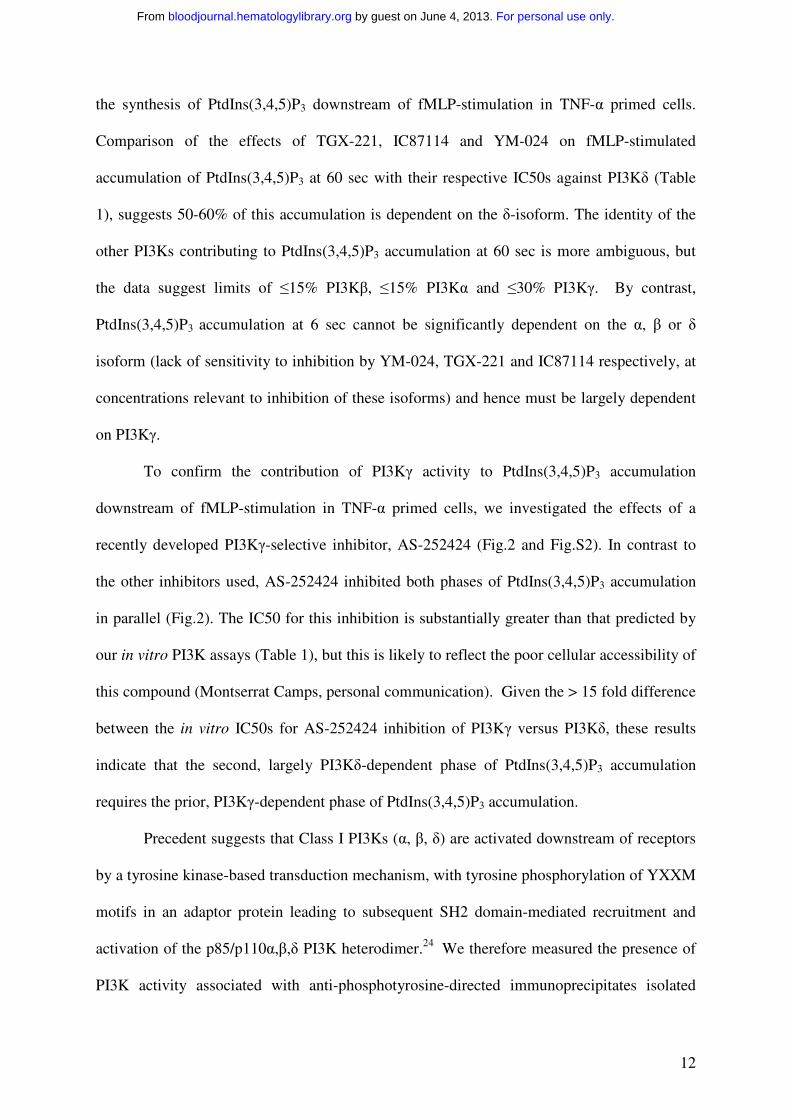
Measurement of PI3K activity in TNF-α primed, fMLP-stimulated human neutrophils
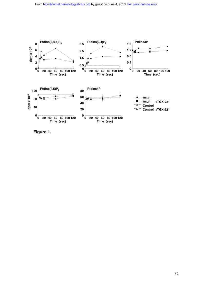
We measured the levels of [32P]-phosphoinositides in [32P]Pi-prelabelled, TNF-α primed
human neutrophils stimulated with 100nM fMLP (Fig.1). The low basal levels of
PtdIns(3,4,5)P3 and PtdIns(3,4)P2 and the dramatic rise in their concentrations on exposure to
fMLP are consistent with previous work describing fMLP stimulation of Class I PI3K
activity, catalysing conversion of PtdIns(4,5)P2 to PtdIns(3,4,5)P3, followed by 5-phosphatase
linked conversion to PtdIns(3,4)P2.32, 34 The significantly higher basal level of [32P]PtdIns3P
and smaller increase on fMLP-stimulation are consistent with previous reports of negligible
effects of fMLP on Class III or Class II catalysed conversion of PtdIns to PtdIns3P.32 The
transient decrease in [32P]PtdIns(4,5)P2 on fMLP-stimulation is consistent with competing
effects of consumption (by PI3K and PLC) and re-synthesis of this lipid (by PI4P5K), as
previously reported.32
The fMLP-stimulated synthesis of PtdIns(3,4,5)P3 in TNF-α primed human
neutrophils is biphasic, with levels peaking approx 10 sec and 60 sec after addition of fMLP
(Fig.1). We have previously shown that the size of this latter peak of PtdIns(3,4,5)P3
accumulation is dependent on prior exposure to TNF-α (5-6 fold reduced in the absence of
TNF-α),22 suggesting significant complexity in the response. We sought to define the roles of
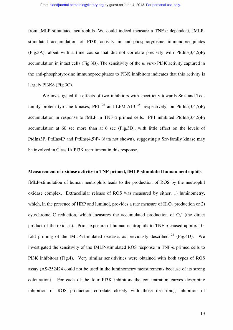
the various Class I PI3K isoforms under these conditions by using several recently developed
isoform-selective, catalytic site inhibitors. The origins and structures of the compounds
chosen for this study are given in the Methods and Fig.S1 and their reported PI3K
selectivities in vitro are given in Table 1. We confirmed the relative selectivity of these
compounds by assessing their IC50s against the in vitro lipid kinase activities of recombinant
PI3Kα, β, δ and γ in identical assays (Table S1); given that each of these compounds act as
competitive ATP-site inhibitors and that the concentration of ATP used in our assays differed
For personal use only. by guest on June 4, 2013. bloodjournal.hematologylibrary.orgFrom
11
from those of others, the data is in good agreement with that presented for these compounds
in the relevant Patent applications..
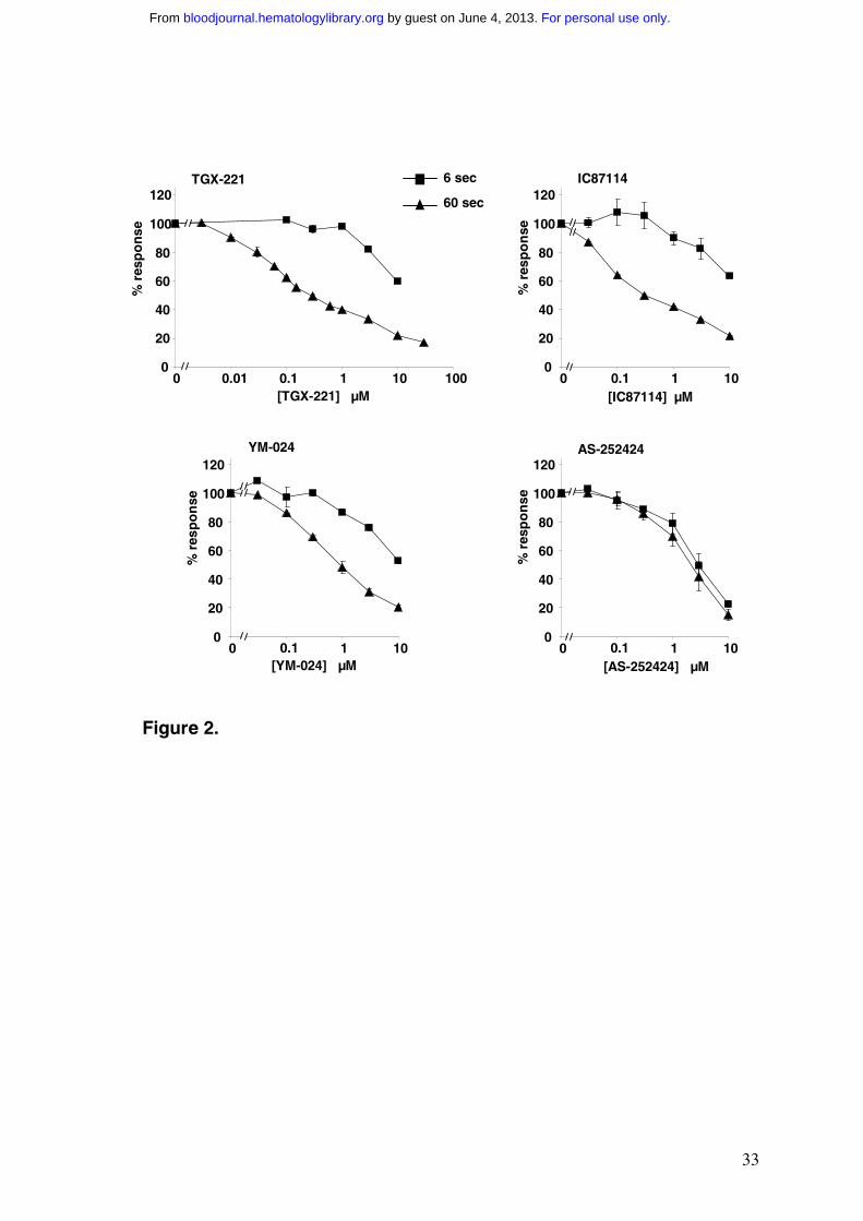
The effect of 0.3µM TGX-221 on the time course of [32P]-phosphoinositide
accumulation in fMLP-stimulated, TNF-α primed human neutrophils is shown in Fig.1. At
this concentration, TGX-221 has a negligible effect on the rising phase of the first peak of
PtdIns(3,4,5)P3 accumulation (6 sec time point, Fig.1) but a substantial effect on the second
peak of PtdIns(3,4,5)P3 accumulation (60 sec time point, Fig.1). Concentration-response
curves for the effects of TGX-221 on PtdIns(3,4,5)P3 accumulation after 6 or 60 sec
stimulation of TNF-α primed cells are shown in Fig.2; effects of TGX-221 on the other lipids
measured are described in supplementary material (Fig.S2). The results clearly indicate that
TGX-221 more potently inhibits the second phase of PtdIns(3,4,5)P3 accumulation than the
first.
Concentration-response curves for the effects of IC87114 and YM-024 on fMLP-
stimulated accumulation of PtdIns(3,4,5)P3 at 6 and 60 sec are also shown in Fig.2 and on
other phosphoinositides in Fig.S2. The relative lack of effect of TGX-221, IC87114 and
YM-024 on the levels of PtdIns(4,5)P2 and PtdIns4P suggest that, at the concentrations tested,
these compounds have few non-specific effects on other neutrophil phosphoinositide kinases.
The effects of TGX-221 on basal PtdIns3P levels suggest this compound may inhibit a Class
III PI3K with an IC50 of approx 1µM (Fig.S2A). The lag in fMLP-stimulated accumulation
of PtdIns(3,4)P2 means that the 6 sec data for this lipid is a composite of roughly equal
proportions of basal and stimulated synthesis (Fig.S2C) and hence difficult to interpret. The
results for PtdIns(3,4,5)P3 accumulation however, allow several clear conclusions to be
drawn. TGX-221, IC87114 and YM-024 each inhibit the second phase of fMLP-stimulated
PtdIns(3,4,5)P3 accumulation more potently than the first and there is multi-component
inhibition of the second phase (Fig.2). Thus more than one isoform of PI3K is involved in
For personal use only. by guest on June 4, 2013. bloodjournal.hematologylibrary.orgFrom
12
the synthesis of PtdIns(3,4,5)P3 downstream of fMLP-stimulation in TNF-α primed cells.
Comparison of the effects of TGX-221, IC87114 and YM-024 on fMLP-stimulated
accumulation of PtdIns(3,4,5)P3 at 60 sec with their respective IC50s against PI3Kδ (Table
1), suggests 50-60% of this accumulation is dependent on the δ-isoform. The identity of the
other PI3Ks contributing to PtdIns(3,4,5)P3 accumulation at 60 sec is more ambiguous, but
the data suggest limits of ≤15% PI3Kβ, ≤15% PI3Kα and ≤30% PI3Kγ. By contrast,
PtdIns(3,4,5)P3 accumulation at 6 sec cannot be significantly dependent on the α, β or δ
isoform (lack of sensitivity to inhibition by YM-024, TGX-221 and IC87114 respectively, at
concentrations relevant to inhibition of these isoforms) and hence must be largely dependent
on PI3Kγ.
To confirm the contribution of PI3Kγ activity to PtdIns(3,4,5)P3 accumulation
downstream of fMLP-stimulation in TNF-α primed cells, we investigated the effects of a
recently developed PI3Kγ-selective inhibitor, AS-252424 (Fig.2 and Fig.S2). In contrast to
the other inhibitors used, AS-252424 inhibited both phases of PtdIns(3,4,5)P3 accumulation
in parallel (Fig.2). The IC50 for this inhibition is substantially greater than that predicted by
our in vitro PI3K assays (Table 1), but this is likely to reflect the poor cellular accessibility of
this compound (Montserrat Camps, personal communication). Given the > 15 fold difference
between the in vitro IC50s for AS-252424 inhibition of PI3Kγ versus PI3Kδ, these results
indicate that the second, largely PI3Kδ-dependent phase of PtdIns(3,4,5)P3 accumulation
requires the prior, PI3Kγ-dependent phase of PtdIns(3,4,5)P3 accumulation.
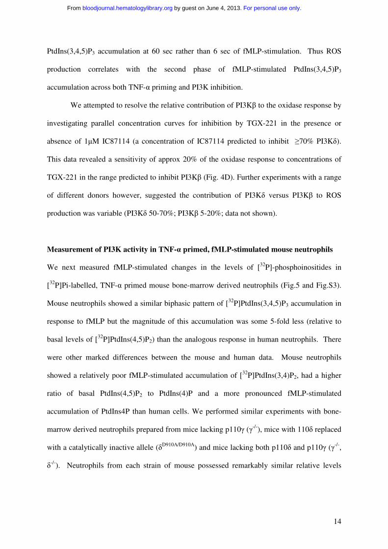
Precedent suggests that Class I PI3Ks (α, β, δ) are activated downstream of receptors
by a tyrosine kinase-based transduction mechanism, with tyrosine phosphorylation of YXXM
motifs in an adaptor protein leading to subsequent SH2 domain-mediated recruitment and
activation of the p85/p110α,β,δ PI3K heterodimer.24 We therefore measured the presence of
PI3K activity associated with anti-phosphotyrosine-directed immunoprecipitates isolated
For personal use only. by guest on June 4, 2013. bloodjournal.hematologylibrary.orgFrom
13
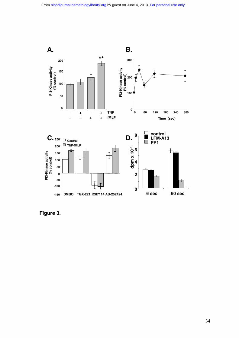
from fMLP-stimulated neutrophils. We could indeed measure a TNF-α dependent, fMLP-
stimulated accumulation of PI3K activity in anti-phosphotyrosine immunoprecipitates
(Fig.3A), albeit with a time course that did not correlate precisely with PtdIns(3,4,5)P3
accumulation in intact cells (Fig.3B). The sensitivity of the in vitro PI3K activity captured in
the anti-phosphotyrosine immunoprecipitates to PI3K inhibitors indicates that this activity is
largely PI3Kδ (Fig.3C).
We investigated the effects of two inhibitors with specificity towards Src- and Tec-
family protein tyrosine kinases, PP1 26 and LFM-A13 35, respectively, on PtdIns(3,4,5)P3
accumulation in response to fMLP in TNF-α primed cells. PP1 inhibited PtdIns(3,4,5)P3
accumulation at 60 sec more than at 6 sec (Fig.3D), with little effect on the levels of
PtdIns3P, PtdIns4P and PtdIns(4,5)P2 (data not shown), suggesting a Src-family kinase may
be involved in Class IA PI3K recruitment in this response.
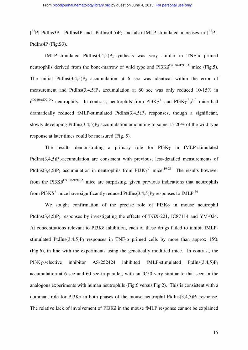
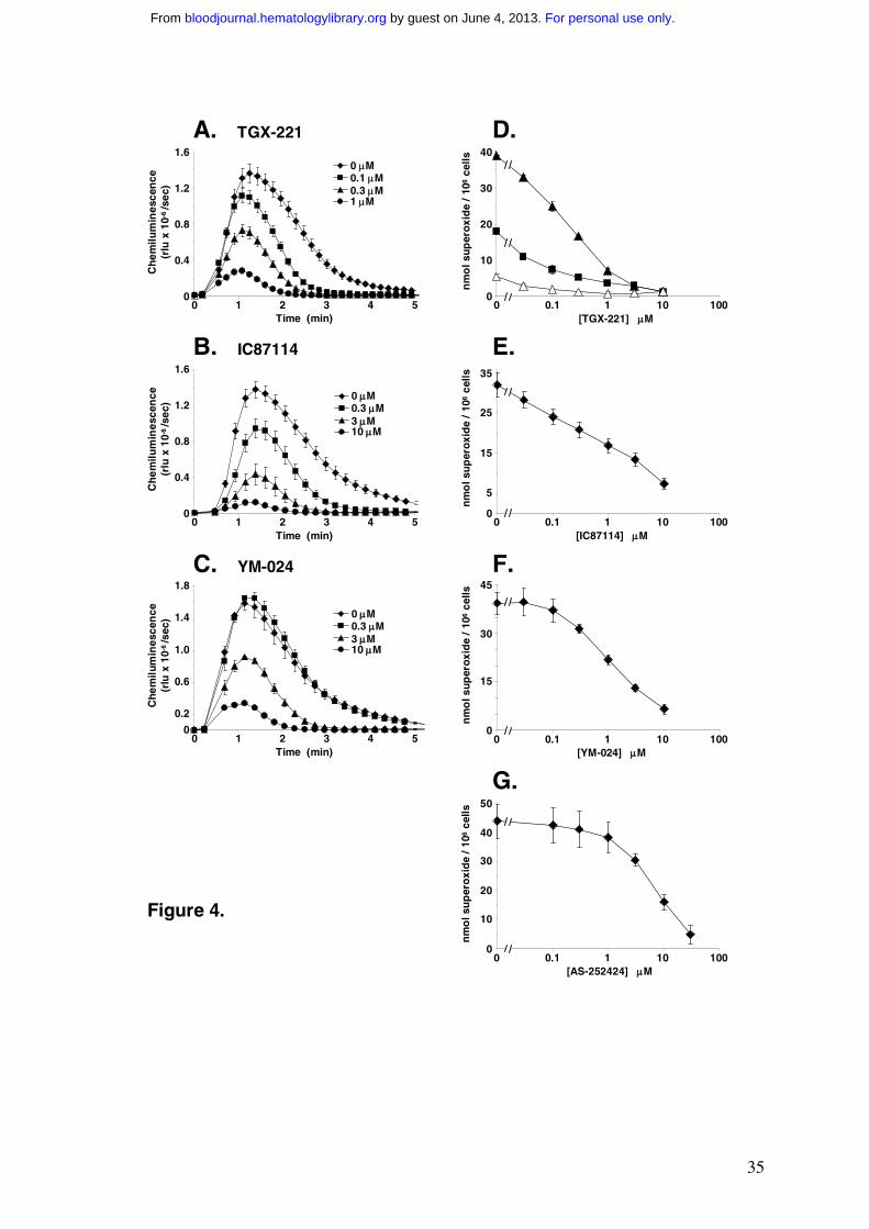
Measurement of oxidase activity in TNF-primed, fMLP-stimulated human neutrophils
fMLP-stimulation of human neutrophils leads to the production of ROS by the neutrophil
oxidase complex. Extracellular release of ROS was measured by either, 1) luminometry,
which, in the presence of HRP and luminol, provides a rate measure of H2O2 production or 2)
cytochrome C reduction, which measures the accumulated production of O2- (the direct
product of the oxidase). Prior exposure of human neutrophils to TNF-α caused approx 10-
fold priming of the fMLP-stimulated oxidase, as previously described 22 (Fig.4D). We
investigated the sensitivity of the fMLP-stimulated ROS response in TNF-α primed cells to
PI3K inhibitors (Fig.4). Very similar sensitivities were obtained with both types of ROS
assay (AS-252424 could not be used in the luminometry measurements because of its strong
colouration). For each of the four PI3K inhibitors the concentration curves describing
inhibition of ROS production correlate closely with those describing inhibition of
For personal use only. by guest on June 4, 2013. bloodjournal.hematologylibrary.orgFrom
14
PtdIns(3,4,5)P3 accumulation at 60 sec rather than 6 sec of fMLP-stimulation. Thus ROS
production correlates with the second phase of fMLP-stimulated PtdIns(3,4,5)P3
accumulation across both TNF-α priming and PI3K inhibition.
We attempted to resolve the relative contribution of PI3Kβ to the oxidase response by
investigating parallel concentration curves for inhibition by TGX-221 in the presence or
absence of 1µM IC87114 (a concentration of IC87114 predicted to inhibit ≥70% PI3Kδ).
This data revealed a sensitivity of approx 20% of the oxidase response to concentrations of
TGX-221 in the range predicted to inhibit PI3Kβ (Fig. 4D). Further experiments with a range
of different donors however, suggested the contribution of PI3Kδ versus PI3Kβ to ROS
production was variable (PI3Kδ 50-70%; PI3Kβ 5-20%; data not shown).
Measurement of PI3K activity in TNF-α primed, fMLP-stimulated mouse neutrophils
We next measured fMLP-stimulated changes in the levels of [32P]-phosphoinositides in
[32P]Pi-labelled, TNF-α primed mouse bone-marrow derived neutrophils (Fig.5 and Fig.S3).
Mouse neutrophils showed a similar biphasic pattern of [32P]PtdIns(3,4,5)P3 accumulation in
response to fMLP but the magnitude of this accumulation was some 5-fold less (relative to
basal levels of [32P]PtdIns(4,5)P2) than the analogous response in human neutrophils. There
were other marked differences between the mouse and human data. Mouse neutrophils
showed a relatively poor fMLP-stimulated accumulation of [32P]PtdIns(3,4)P2, had a higher
ratio of basal PtdIns(4,5)P2 to PtdIns(4)P and a more pronounced fMLP-stimulated
accumulation of PtdIns4P than human cells. We performed similar experiments with bone-
marrow derived neutrophils prepared from mice lacking p110γ (γ-/-), mice with 110δ replaced
with a catalytically inactive allele (δD910A/D910A) and mice lacking both p110δ and p110γ (γ-/-,
δ-/-). Neutrophils from each strain of mouse possessed remarkably similar relative levels
For personal use only. by guest on June 4, 2013. bloodjournal.hematologylibrary.orgFrom
15
[32P]-PtdIns3P, -PtdIns4P and -PtdIns(4,5)P2 and also fMLP-stimulated increases in [32P]-
PtdIns4P (Fig.S3).
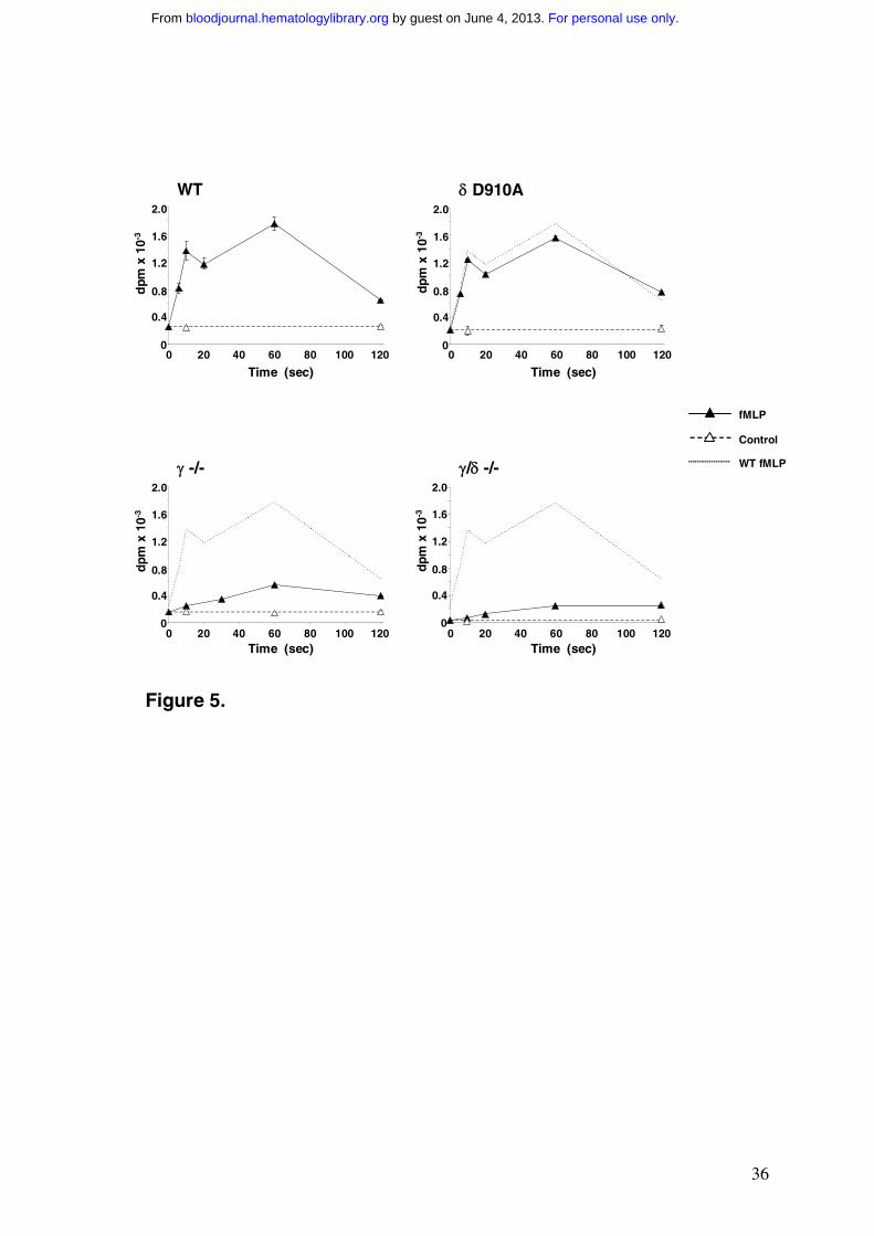
fMLP-stimulated PtdIns(3,4,5)P3-synthesis was very similar in TNF-α primed
neutrophils derived from the bone-marrow of wild type and PI3KδD910A/D910A mice (Fig.5).
The initial PtdIns(3,4,5)P3 accumulation at 6 sec was identical within the error of
measurement and PtdIns(3,4,5)P3 accumulation at 60 sec was only reduced 10-15% in
δD910A/D910A neutrophils. In contrast, neutrophils from PI3Kγ-/- and PI3Kγ-/-,δ-/- mice had
dramatically reduced fMLP-stimulated PtdIns(3,4,5)P3 responses, though a significant,
slowly developing PtdIns(3,4,5)P3 accumulation amounting to some 15-20% of the wild type
response at later times could be measured (Fig. 5).
The results demonstrating a primary role for PI3Kγ in fMLP-stimulated
PtdIns(3,4,5)P3-accumulation are consistent with previous, less-detailed measurements of
PtdIns(3,4,5)P3 accumulation in neutrophils from PI3Kγ-/- mice.19-21 The results however
from the PI3KδD910A/D910A mice are surprising, given previous indications that neutrophils
from PI3Kδ-/- mice have significantly reduced PtdIns(3,4,5)P3-responses to fMLP.36
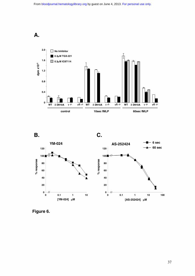
We sought confirmation of the precise role of PI3Kδ in mouse neutrophil
PtdIns(3,4,5)P3 responses by investigating the effects of TGX-221, IC87114 and YM-024.
At concentrations relevant to PI3Kδ inhibition, each of these drugs failed to inhibit fMLP-
stimulated PtdIns(3,4,5)P3 responses in TNF-α primed cells by more than approx 15%
(Fig.6), in line with the experiments using the genetically modified mice. In contrast, the
PI3Kγ-selective inhibitor AS-252424 inhibited fMLP-stimulated PtdIns(3,4,5)P3
accumulation at 6 sec and 60 sec in parallel, with an IC50 very similar to that seen in the
analogous experiments with human neutrophils (Fig.6 versus Fig.2). This is consistent with a
dominant role for PI3Kγ in both phases of the mouse neutrophil PtdIns(3,4,5)P3 response.
The relative lack of involvement of PI3Kδ in the mouse fMLP response cannot be explained
For personal use only. by guest on June 4, 2013. bloodjournal.hematologylibrary.orgFrom
16
on the basis of p110δ expression levels, which are similar in human and mouse neutrophils
(Fig. S4). There are also no reported compensatory changes in the expression of other Class I
PI3K catalytic subunits in either the PI3Kγ-/- or PI3KδD910A/D910A mice.21, 28
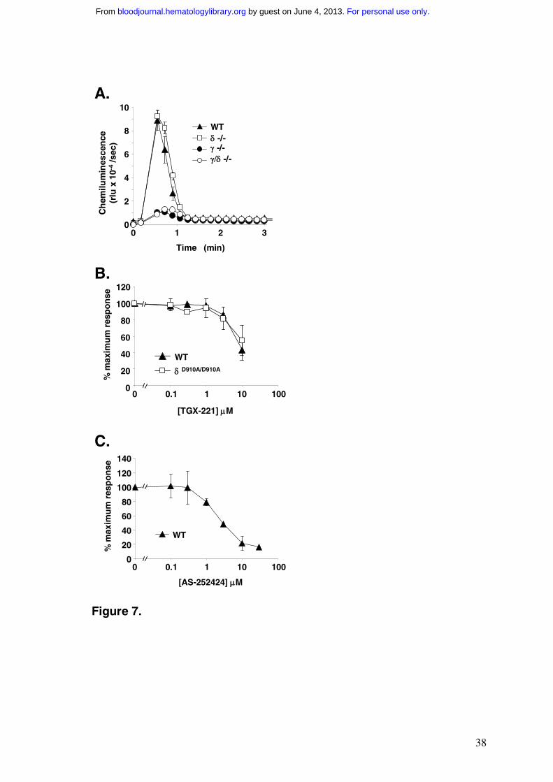
Measurement of oxidase activity in TNF-α primed, fMLP-stimulated mouse neutrophils
We measured the activation of the neutrophil oxidase in mouse bone-marrow derived
neutrophils in experiments similar to those performed on human neutrophils. The scale of
ROS production to fMLP, the PKC agonist PMA and opsonised zymosan were all
substantially lower (approx 10-fold based on neutrophil numbers) in mouse neutrophils than
human neutrophils (data not shown). The degree of TNF-α priming of subsequent responses
to fMLP or C5a were also lower in mouse neutrophils, in the range 3-4 fold versus 8-12 fold
in the human cells. Furthermore, mouse neutrophils were relatively more responsive to
fMLP when first isolated and lost this sensitivity in the absence, but not the presence, of
TNF-α during subsequent incubation at 37ºC for 30min, leading to a substantial effect of
TNF-α when assayed at this point. In contrast, human neutrophils were relatively
unresponsive to fMLP alone when first isolated and gained sensitivity when subsequently
incubated with TNF-α. The time course of fMLP-stimulated ROS production in TNF-α
primed mouse neutrophils was also different to that in human cells (compare Fig.7A to
Fig.4A); in mouse cells ROS production was essentially over by 1 min, whereas maximal
ROS production in the human cells occurred between 1 and 2 min after addition of fMLP
(averaged over ≥ 20 experiments the mean durations of the mouse and human oxidase bursts
were 1.2 min and 3.3 min respectively).
fMLP stimulated very similar oxidase activation in TNF-α primed neutrophils derived
from either wild type or PI3Kδ-/- mice (Fig.7A; similar results were obtained with
PI3KδD910A/D910A mice, data not shown). In contrast, oxidase activation in response to fMLP
For personal use only. by guest on June 4, 2013. bloodjournal.hematologylibrary.orgFrom
17
in TNF-α primed neutrophils from PI3Kγ-/- or PI3Kγ-/-/δ-/- mice was inhibited by over 80%
(Fig.7A). PMA stimulated similar oxidase responses in all four genotypes tested (data not
shown). These results are consistent with previously published defects in oxidase activation
to G-protein coupled agonists in PI3Kγ-/- neutrophils.19-21
We investigated the sensitivity of the primed fMLP-stimulated oxidase response in
mouse neutrophils to PI3K inhibitors. The concentration-response curves for inhibition of
ROS formation by TGX-221 (Fig.7B), IC87114 (Fig.S5) and YM-024 (Fig.S5) were all
shifted to substantially higher concentrations compared to the analogous experiments in
human neutrophils. Furthermore the concentration-response curves for TGX-221 and
IC87114 were very similar in both wild type and PI3KδD910A/D910A neutrophils (Figs. 7B and
S5). These results all point to a relatively small role for PI3Kδ in the mouse oxidase response
to fMLP, consistent with a minor role for this isoform in PtdIns(3,4,5)P3 accumulation at 60
sec. In contrast, the inhibition of fMLP-stimulated ROS production by AS-252424 was very
similar to that observed in human neutrophils (Fig.7C), supporting a key role for PI3Kγ in the
mouse in both PtdIns(3,4,5)P3 accumulation and the activation of the oxidase.
For personal use only. by guest on June 4, 2013. bloodjournal.hematologylibrary.orgFrom
18
DISCUSSION
Stimulation of unprimed human neutrophils with fMLP results in a rapid pulse of
PtdIns(3,4,5)P3 accumulation, peaking at about 6-10 sec, and minimal activation of ROS
production. Pretreatment of human neutrophils with TNF-α results in additional, fMLP-
dependent accumulation of PtdIns(3,4,5)P3 at around 60 sec, and substantial, parallel,
activation of ROS production.22, 23 Specific pharmacological inhibition of this later pulse of
PtdIns(3,4,5)P3 by isoform-selective, PI3K inhibitors correlates with ROS production,
indicating PtdIns(3,4,5)P3 accumulation at later times (and/or PtdIns(3,4)P2 derived from it)
is essential and indeed of high ‘control-strength’ for the pathways by which fMLP regulates
the oxidase. There are now several suggestions for signalling pathways by which
PtdIns(3,4,5)P3/PtdIns(3,4)P2 may regulate oxidase activity, including
PtdIns(3,4,5)P3/PtdIns(3,4)P2-regulated GEFs for rac (P-Rex1; vav),37
PtdIns(3,4,5)P3/PtdIns(3,4)P2-regulated phosphorylation of p47phox via atypical PKCs or PAK
2, 3 and also direct binding of PtdIns(3,4)P2 to p47phox itself.38 There is still no clear
assessment however, of the relative importance of these mechanisms to ROS production in
vivo.
Our data indicate the initial phase of PtdIns(3,4,5)P3 synthesis on addition of fMLP to
human neutrophils is entirely dependent on PI3Kγ. fMLP receptors transduce their signals
through direct activation of Gi heterotrimeric G-proteins, leading to substantial release of Gαi
and Gβγ subunits. Gβγ subunits have been shown to elicit direct and substantial activation of
PI3Kγ in vitro,39 providing a plausible mechanism for rapid, PI3Kγ-dependent synthesis of
PtdIns(3,4,5)P3. The small GTPase Ras also activates PI3Kγ directly through binding to its
p110 catalytic subunit, though the activations measured so far are relatively modest.40 fMLP
For personal use only. by guest on June 4, 2013. bloodjournal.hematologylibrary.orgFrom
19
receptors are known to stimulate GTP-loading of Ras,41 therefore it is also plausible Ras may
play a role in this response.
The second phase of fMLP-stimulated PtdIns(3,4,5)P3 accumulation is augmented by
pre-treatment with TNF-α and is driven largely by Class IA PI3Ks. Our data suggest PI3Kδ
is the predominant PI3K activity in this phase of the response (50-60%) with smaller
contributions from PI3Kβ (15%) and possibly also PI3Kα (≤15%). It is not clear whether it is
simply the timing of this second phase of PtdIns(3,4,5)P3 accumulation that is critical to
oxidase activation or its source. Close comparison of the inhibition-curves for PtdIns(3,4,5)P3
synthesis versus ROS production suggest the oxidase must recognise PtdIns(3,4,5)P3
generated by PI3Kδ and β, but the shallower curve for IC87114 inhibition of ROS production
versus that for TGX-221 (compared with their analogous inhibition curves for PtdIns(3,4,5)P3
synthesis) suggests the oxidase may recognise PtdIns(3,4,5)P3 generated by the γ-isoform less
effectively. However, given the error in the measurements and the relatively small, direct
contribution of the γ-isoform to PtdIns(3,4,5)P3 synthesis at 60 sec, further experiments will
be needed to clarify this. A striking feature of this second phase of Class IA-driven
PtdIns(3,4,5)P3 accumulation is that it is significantly dependent on the activity of PI3Kγ; this
idea that PtdIns(3,4,5)P3 may amplify its own accumulation is also thought to be relevant to
understanding the ‘feed forward’ loops in signalling neutrophil migration.42
The mechanism by which Class IA PI3Ks are activated by fMLP receptors and also
the point of intervention of TNF-α signalling is currently unknown. In principle TNF-α could
be activating the relevant PI3Ks or inhibiting the relevant phosphatases (parallel changes in
PtdIns(3,4)P2 and PtdIns(3,4,5)P3 suggest the phosphatase would have to be a 3-phosphatase
of the PTEN family). Whatever the point of action of TNF-α, precedent would suggest a
tyrosine kinase-based signalling pathway is important at some level for switching on the
Class IA PI3Ks.24 There is significant conflict in the literature concerning the contribution
For personal use only. by guest on June 4, 2013. bloodjournal.hematologylibrary.orgFrom
20
that tyrosine kinase-based signalling pathways make to PtdIns(3,4,5)P3 and O2- production
elicited by fMLP, though our data suggest some of this confusion is likely to arise from the
extent of priming, controlled or otherwise, in the various studies.19-21, 25-27, 35, 43-45 There is
also a substantial body of work describing the activation of Src-family tyrosine kinases
downstream of fMLP-receptors in human neutrophils 46 and several suggestions that they are
important for the activation of PI3K and O2- production.26, 47 Our data offer circumstantial
support to the idea that there is recruitment of PI3Kδ to phosphotyrosine-containing
complexes downstream of fMLP-stimulation, that this is dependent on TNF-α priming and
that src-family kinases may be involved but more work needs to be done to establish the
relevance of these observations to the accumulations of PtdIns(3,4,5)P3 measured.
Our observations that Class IA PI3Ks do not play a major role in PtdIns(3,4,5)P3
production and oxidase activation in response to fMLP in TNF-α primed mouse neutrophils
are a surprise; both because of our results in the analogous experiments in human neutrophils
and also in the light of a recent report suggesting there is a substantial role for PI3Kδ in
fMLP-stimulated PtdIns(3,4,5)P3-synthesis in mouse neutrophils.36 Both studies are internally
consistent with respect to the effect of a PI3Kδ inhibitor (IC87114) and the PI3Kδ-/- mouse
strain used and at present we cannot understand this discrepancy. Our results do suggest that
mouse bone marrow-derived neutrophils have a superficially similar, bi-phasic
PtdIns(3,4,5)P3 response to fMLP when compared to human neutrophils; however, the
magnitude of this response is much lower in the mouse cells. It is surprising that PI3Kγ is the
major isoform responsible for both phases of PtdIns(3,4,5)P3 production in murine cells;
multiple receptors, multiple mechanisms of PI3K regulation (e.g. Gβγ versus ras), potential
regulation of PtdIns(3,4,5)P3 hydrolysis and potential secretion of further active agents, all
provide plausible mechanisms for obtaining temporal differences in PtdIns(3,4,5)P3-
accumulation driven through this single isoform.
For personal use only. by guest on June 4, 2013. bloodjournal.hematologylibrary.orgFrom
21
There are also very significant differences in fMLP-stimulated ROS production in
TNF-α primed, mouse and human neutrophils. In human cells maximal production of ROS
occurs between 1 and 2 min after addition of fMLP and there is substantial accumulation of
Class IA PI3K-derived PtdIns(3,4,5)P3/PtdIns(3,4)P2. In mouse cells there is a much smaller
and shorter burst of ROS production and much lower PtdIns(3,4,5)P3/PtdIns(3,4)P2
accumulation. We do not understand the mechanistic basis for this difference. It may be
related to the different ways in which oxidase activation is organised in mouse and human
cells but it is also plausible that our results are due to differences in the state of differentiation
and/or the cellular environment between the pool of ‘reserve’ neutrophils in the bone marrow
and those in the circulation, although only small differences in ROS production have been
measured between these two different populations of neutrophils in the mouse.48 It may also
be due to a difference in the relative physiological importance of the particular agonists we
have chosen for the mouse and human cells. These are obviously important points to resolve
but, whatever the cause of the differences between our results with the mouse and human
neutrophils, they raise a cautionary note in using the mouse assays as a model for human
neutrophil behaviour, particularly with regard to testing the potential efficacy of PI3K
inhibitors in PtdIns(3,4,5)P3 and ROS production.
Given the established importance of the Class I PI3K signalling pathway in many
aspects of human disease, particularly in cancer and inflammation, the pharmaceutical
industry is showing great interest in developing isoform-selective PI3K inhibitors.49, 50 The
results presented here and elsewhere 27, 51 suggest it may indeed be possible to generate
compounds with the appropriate selectivity and furthermore, that the complexity of PI3K
responses may afford useful operating windows for specificity in the inhibition of a particular
cellular response. The results presented here suggest PI3Kγ is a ‘master’ PI3K downstream of
fMLP receptors in human neutrophils and that inhibition of this isoform will inhibit all
For personal use only. by guest on June 4, 2013. bloodjournal.hematologylibrary.orgFrom
22
PtdIns(3,4,5)P3/PtdIns(3,4)P2-dependent responses to this agonist; however, inhibition of
PI3Kδ may inhibit only a subset of these responses e.g. ROS production. Similar levels of
complexity have been revealed recently in the roles of the PI3Kγ and PI3Kδ isoforms in the
co-operation between IgE and adenosine receptors in the regulation of mast cell secretion.52,
53 It will be interesting to establish whether this level of PI3K complexity exists in many
other contexts of cellular regulation.
ACKNOWLEDGEMENTS
The authors would like to thank Brian Hutchinson for synthesis of Yamanouchi Compound
24, Marion Dorning for measurement of the compound IC50s and Patrick Slocombe, Gillian
Hutchinson, Alaina Smith, Carl Doyle and Kathryn McLaughlin for the production of the
purified Class 1 PI3Ks (all employees of Celltech). We thank Cedric Favré for preparation of
human T cells. TGX-221 was supplied by Kinacia Pty Ltd, a wholly owned subsidiary of
Cerylid Biosciences Ltd (Richmond, Victoria, Australia).
SUPPLEMENTAL MATERIAL IS AVAILABLE ONLINE AT THE TIME OF FINAL
PUBLICATION ONLY.
For personal use only. by guest on June 4, 2013. bloodjournal.hematologylibrary.orgFrom
23
REFERENCES
1. Haslett C, Savill JS, Meagher L. The neutrophil. Current Opinion in Immunology.1989;2:10-18.
2. Babior BM. NADPH oxidase: an update. Blood. 1999;93:1464-1476.3. Cross AR, Segal AW. The NADPH oxidase of professional phagocytes-prototype of
the NOX electron transport chain systems. Biochimica et Biophysica Acta. 2004;1657:1-22.
4. Thrasher AJ, Keep NH, Wientjes F, Segal AW. Chronic granulomatous disease. Biochimica et Biophysica Acta. 1994;1227:1-24.
5. Hampton MB, Kettle AJ, Winterbourn CC. Inside the neutrophil phagosome: oxidants, myeloperoxidase, and bacterial killing. Blood. 1998;92:3007-17.
6. Reeves EP, Lu H, Jacobs HL, et al. Killing activity of neutrophils is mediated through activation of proteases by K+ flux. Nature. 2002;416:291-297.
7. Roos D, Winterbourn CC. Immunology. Lethal weapons. Science. 2002;296:669-671.8. Rajagopalan S, Meng XP, Ramasamy S, Harrison DG, Galis ZS. Reactive oxygen
species produced by macrophage-derived foam cells regulate the activity of vascular matrix metalloproteinases in vitro. Implications for atherosclerotic plaque stability. Journal of Clinical Investigation. 1996;98:2572-2579.
9. Thannickal VJ, Fanburg BL. Reactive oxygen species in cell signaling. American Journal of Physiology-Lung Cellular and Molecular Physiology. 2000;279:L1005-1028.
10. Akgur FM., Brown MF, Zibari GB, et al. Role of superoxide in hemorrhagic shock-induced P-selectin expression. American Journal of Physiology-Heart and Circulatory Physiology. 2000;279:H791-797.
11. Bowler RP, Nicks M, Tran K, et al. Extracellular superoxide dismutase attenuates lipopolysaccharide-induced neutrophilic inflammation. American Journal of Respiratory Cell and Molecular Biology. 2004;31:432-439.
12. Kobayashi SD, Voyich JM, Braughton KR et al. Gene expression profiling provides insight into the pathophysiology of chronic granulomatous disease. Journal of Immunology. 2004;172:636-643.
13. Smedly LA, Tonnesen MG, Sandhans RA, et al. Neutrophil-mediated injury to endothelial cells. Enhancement by endotoxin and essential role of neutrophil elastase. The Journal of Clinical Investigation. 1986;77:1233-43.
14. Worthen GS. Lipid mediators, neutrophils and endothelial injury. The AmericanReview of Respiratory Disease. 1987;136:455-8.
15. Berkow RL, Wang D, Larrick JW, Dodson RW, Howard TH. Enhancement of neutrophil superoxide production by preincubation with recombinant human tumor necrosis factor. Journal of Immunology. 1987;139:3783-3791.
16. Downey GP, Fukushima T, Fialkow L, Waddell TK. Intracellular signaling in neutrophil priming and activation. Seminars in Cell Biology. 1995;6:345-356.
17. Coffer PJ, Koenderman L. Granulocyte signal transduction and priming: cause without effect? Immunology Letters. 1997;57:27-31.
18. Arcaro A, Wymann MP. Wortmannin is a potent phosphatidylinositol 3-kinase inhibitor: the role of phosphatidylinositol 3,4,5-trisphosphate in neutrophil responses. Biochemical Journal. 1993;296:297-301.
19. Sasaki T, Irie-Sasaki J, Jones RG, et al. Function of PI3Kgamma in thymocyte development, T cell activation, and neutrophil migration. Science. 2000;287:1040-1046.
For personal use only. by guest on June 4, 2013. bloodjournal.hematologylibrary.orgFrom
24
20. Li Z, Jiang H, Xie W, Zhang Z, Smrcka AV, Wu D. Roles of PLC-beta2 and -beta3 and PI3Kgamma in chemoattractant-mediated signal transduction. Science.2000;287:1046-1049.
21. Hirsch E, Katanaev VL, Garlanda C, et al. Central role for G protein-coupled phosphoinositide 3-kinase gamma in inflammation. Science. 2000;287:1049-1053.
22. Condliffe AM, Hawkins PT, Stephens LR, Haslett C, Chilvers ER. Priming of human neutrophil superoxide generation by tumour necrosis factor-alpha is signalled by enhanced phosphatidylinositol 3,4,5-trisphosphate but not inositol 1,4,5-trisphosphate accumulation. FEBS Letters. 1998;439:147-151.
23. Cadwallader KA, Condliffe AM, McGregor A, et al. Regulation of phosphatidylinositol 3-kinase activity and phosphatidylinositol 3,4,5-trisphosphate accumulation by neutrophil priming agents. Journal of Immunology. 2002;169:3336-3344.
24. Vanhaesebroeck B, Leevers SJ, Ahmadi K, et al. Synthesis and function of 3-phosphorylated inositol lipids. Annual Review of Biochemistry. 2001;70:535-602.
25. Ptasznik A., Prossnitz ER, Yoshikawa D, Smrcka A, Traynor-Kaplan AE, BokochGM. A tyrosine kinase signaling pathway accounts for the majority of phosphatidylinositol 3,4,5-trisphosphate formation in chemoattractant-stimulated human neutrophils. Journal of Biological Chemistry. 1996;271:25204-25207.
26. Nijhuis E, Lammers JW, Koenderman L, Coffer PJ. Src kinases regulate PKB activation and modulate cytokine and chemoattractant-controlled neutrophil functioning. Journal of Leukocyte Biology. 2002;71:115-24.
27. Sadhu C, Dick K, Tino WT, Staunton DE. Selective role of PI3K delta in neutrophil inflammatory responses. Biochemical and Biophysical Research Communications.2003;308:764-769.
28. Okkenhaug K, Bilancio A, Farjot G, et al. Impaired B and T cell antigen receptor signaling in p110delta PI 3-kinase mutant mice. Science. 2002;297:1031-1034.
29. Clayton E, Bardi G, Bell SE, et al. A crucial role for the p110delta subunit of phosphatidylinositol 3-kinase in B cell development and activation. Journal of Experimental Medicine. 2002;196:753-763.
30. Haslett C, Guthrie LA, Kopaniak MM, Johnston Jr. RB, Henson PM. Modulation of multiple neutrophil functions by preparative methods or trace concentrations of bacterial lipopolysaccharide. American Journal of Pathology. 1985;119:101-110
31. Jackson TR, Stephens LR, Hawkins PT. Receptor specificity of growth factor-stimulated synthesis of 3-phosphorylated inositol lipids in Swiss 3T3 cells. Journal of Biological Chemistry. 1992;267:16627-16636.
32. Stephens LR, Hughes KT, Irvine RF. Pathway of phosphatidylinositol(3,4,5)-trisphosphate synthesis in activated neutrophils. Nature. 1991;351:33-39.
33. Anderson KE, Stephens LR, Hawkins PT. Phosphoinositide 3-kinases. In: Milligan G, ed. Signal Transduction: A Practical Approach. Oxford University Press, Oxford;1999:283-300.
34. Traynor-Kaplan AE, Thompson BL, Harris AL, Taylor P, Omann GM, Sklar LA. Transient increase in phosphatidylinositol 3,4-bisphosphate and phosphatidylinositol trisphosphate during activation of human neutrophils. Journal of Biological Chemistry. 1989;264:15668-15673.
35. Gilbert C, Levasseur S, Desaulniers P, Dusseault AA, et al. Chemotactic factor-induced recruitment and activation of Tec family kinases in human neutrophils. II. Effects of LFM-A13, a specific Btk inhibitor. Journal of Immunology.2003;170:5235-5243.
For personal use only. by guest on June 4, 2013. bloodjournal.hematologylibrary.orgFrom
25
36. Puri KD, Doggett TA, Douangpanya J, et al. Mechanisms and implications of phosphoinositide 3-kinase delta in promoting neutrophil trafficking into inflamed tissue. Blood. 2004;103:3448-3456.
37. Welch HC, Coadwell WJ, Stephens LR, Hawkins PT. Phosphoinositide 3-kinase-dependent activation of Rac. FEBS Letters. 2003;546:93-97.
38. Kanai F, Liu H, Field SJ, et al. The PX domains of p47phox and p40phox bind to lipid products of PI(3)K. Nature Cell Biology. 2001;3:675-678.
39. Stephens LR, Eguinoa A, Erdjument-Bromage H, et al. The G beta gamma sensitivity of a PI3K is dependent upon a tightly associated adaptor, p101. Cell. 1997;89:105-114.
40. Pacold ME, Suire S, Perisic O, et al. Crystal structure and functional analysis of Ras binding to its effector phosphoinositide 3-kinase gamma. Cell. 2000;103:931-943.
41. Worthen GS, Avdi N, Buhl AM, Suzuki N, Johnson GL. FMLP activates Ras and Raf in human neutrophils. Potential role in activation of MAP kinase. Journal of Clinical Investigation. 1994;94:815-823.
42. Weiner OD, Neilsen PO, Prestwich GD, Kirschner MW, Cantley LC, Bourne HR. A PtdInsP(3) - and Rho GTPase-mediated positive feedback loop regulates neutrophil polarity. Nature Cell Biology. 2002;4:509-13.
43. Naccache PH, Levasseur S, Lachance G, Chakravarti S, Bourgoin SG, McColl SR. Stimulation of human neutrophils by chemotactic factors is associated with the activation of phosphatidylinositol 3-kinase gamma. Journal of Biological Chemistry.2000;275:23636-23641.
44. Stephens L, Eguinoa A, Corey S, Jackson T, Hawkins PT. Receptor stimulated accumulation of phosphatidylinositol (3,4,5)-trisphosphate by G-protein mediated pathways in human myeloid derived cells. EMBO Journal. 1993;12:2265-2273.
45. Momose H, Kurosu H, Tsujimoto N, et al. Dual phosphorylation of phosphoinositide 3-kinase adaptor Grb2-associated binder 2 is responsible for superoxide formation synergistically stimulated by Fc gamma and formyl-methionyl-leucyl-phenylalanine receptors in differentiated THP-1 cells. Journal of Immunology. 2003;171:4227-4234.
46. Berton G. Tyrosine kinases in neutrophils. Current Opinion in Hematology.1999;6:51-58.
47. Yan SR, Novak MJ. Src-family kinase-p53/ Lyn p56 plays an important role in TNF-alpha-stimulated production of O2
- by human neutrophils adherent to fibrinogen. Inflammation. 1999;23:167-178.
48. Boxio R, Bossenmeyer-Pourie C, Dournon SNC, Nusse O. Mouse bone marrow contains large numbers of functionally competent neutrophils. Journal of Leukocyte Biology. 2004;75:604-611.
49. Ward SG, Finan P. Isoform-specific phosphoinositide 3-kinase inhibitors as therapeutic targets. Current Opinion in Pharmacology. 2003;3:426-34.
50. Wymann MP, Zvelebil M, Laffargue M. Phosphoinositide 3-kinase signalling - which way to target? Trends in Pharmacological Sciences. 2003;24:366-76.
51. Knight ZA, Chiang CG, Alaimo PJ, et al. Isoform-specific phosphoinositide 3-kinase inhibitors from an arylmorpholine scaffold. Bioorganic & Medicinal Chemistry.2004;12:4749-59.
52. Wymann MP, Bjorklof K, Calvez R, et al. Phosphoinositide 3-kinase gamma: a key modulator in inflammation and allergy. Biochemical Society Transactions. 2003;31:275-80.
53. Ali K, Bilancio A, Thomas M, et al. Essential role for the p110 delta phosphoinositide 3-kinase in the allergic response. Nature. 2004;431:1007-11.
For personal use only. by guest on June 4, 2013. bloodjournal.hematologylibrary.orgFrom
26
TABLES
Table I Inhibition of PI3K activity in vitro
IC50 (µM)
α β γ δ
TGX-221 5 0.007 3.5 0.1
IC87114 >100 75 29 0.5
YM-024 0.30 2.65 9.07 0.33
AS-252424 1.07 >20 0.035 >20
The IC50s for inhibition of the different catalytic subunits of Class I PI3Ks by the
compounds used in this study are listed. The chemical structures of the inhibitors are shown
in Supplementary Material (Fig. S1). The data has been drawn from the appropriate patent
and publication information (see methods); the concentrations of ATP used in the relevant in
vitro PI3K assays were: TGX-221 (100µM), IC87114 (200µM), YM-024 (2µM) and AS-
252424 (each PI3K isoform was assayed at its respective Km for ATP i.e., 89µM, 70µM,
23µM and 59µM for the α,β,γ and δ isoforms respectively). We have also generated
analogous data comparing each of these compounds in the same in vitro assay at 2µM ATP
(see Supplementary Material, Table S1).
For personal use only. by guest on June 4, 2013. bloodjournal.hematologylibrary.orgFrom
27
FIGURE LEGENDS
Figure 1 Time course of changes in phosphoinositide levels during fMLP-stimulation
of TNF-α primed human neutrophils
Human neutrophils were labelled with [32P]Pi in the presence of 200U/ml TNF-α as described
in the Methods. Neutrophils were washed, warmed for 5 min at 37ºC in the presence or
absence of 0.3µM TGX-221 and then challenged with 100nM fMLP or its vehicle for the
times indicated. Incubations were terminated, lipids extracted and [32P] phosphoinositides
quantified as described in the Methods. Data are means ± SD (n = 3-5) collected from
separate preparations of neutrophils (where not shown, errors lie within the symbols).
Figure 2 Effects of PI3K inhibitors on PtdIns(3,4,5)P3 accumulation in fMLP
stimulated, TNF-α primed human neutrophils
Human neutrophils were labelled with [32P]Pi in the presence of 200U/ml TNF-α as described
in the Methods. Neutrophils were washed and warmed for 5 min at 37ºC with the indicated
concentration of inhibitor and then challenged with 100nM fMLP for either 6 sec or 60 sec.
Incubations were terminated and [32P] phosphoinositides quantified as described in the
Methods. Data shown are for the accumulation of PtdIns(3,4,5)P3 expressed as a percentage
of the value with no inhibitor; data are means ± range or SD (n = 2-5) collected from separate
preparations of neutrophils (where not shown, errors lie within the symbols). Analogous dose
curves describing the effects of the inhibitors on the other [32P] phosphoinositides are shown
in Supplementary Material (Fig. S2).
For personal use only. by guest on June 4, 2013. bloodjournal.hematologylibrary.orgFrom
28
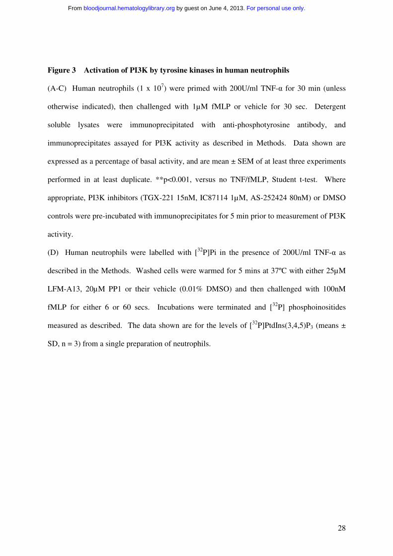
Figure 3 Activation of PI3K by tyrosine kinases in human neutrophils
(A-C) Human neutrophils (1 x 10⁷) were primed with 200U/ml TNF-α for 30 min (unless
otherwise indicated), then challenged with 1µM fMLP or vehicle for 30 sec. Detergent
soluble lysates were immunoprecipitated with anti-phosphotyrosine antibody, and
immunoprecipitates assayed for PI3K activity as described in Methods. Data shown are
expressed as a percentage of basal activity, and are mean ± SEM of at least three experiments
performed in at least duplicate. **p<0.001, versus no TNF/fMLP, Student t-test. Where
appropriate, PI3K inhibitors (TGX-221 15nM, IC87114 1µM, AS-252424 80nM) or DMSO
controls were pre-incubated with immunoprecipitates for 5 min prior to measurement of PI3K
activity.
(D) Human neutrophils were labelled with [32P]Pi in the presence of 200U/ml TNF-α as
described in the Methods. Washed cells were warmed for 5 mins at 37ºC with either 25µM
LFM-A13, 20µM PP1 or their vehicle (0.01% DMSO) and then challenged with 100nM
fMLP for either 6 or 60 secs. Incubations were terminated and [32P] phosphoinositides
measured as described. The data shown are for the levels of [32P]PtdIns(3,4,5)P3 (means ±
SD, n = 3) from a single preparation of neutrophils.
For personal use only. by guest on June 4, 2013. bloodjournal.hematologylibrary.orgFrom
29
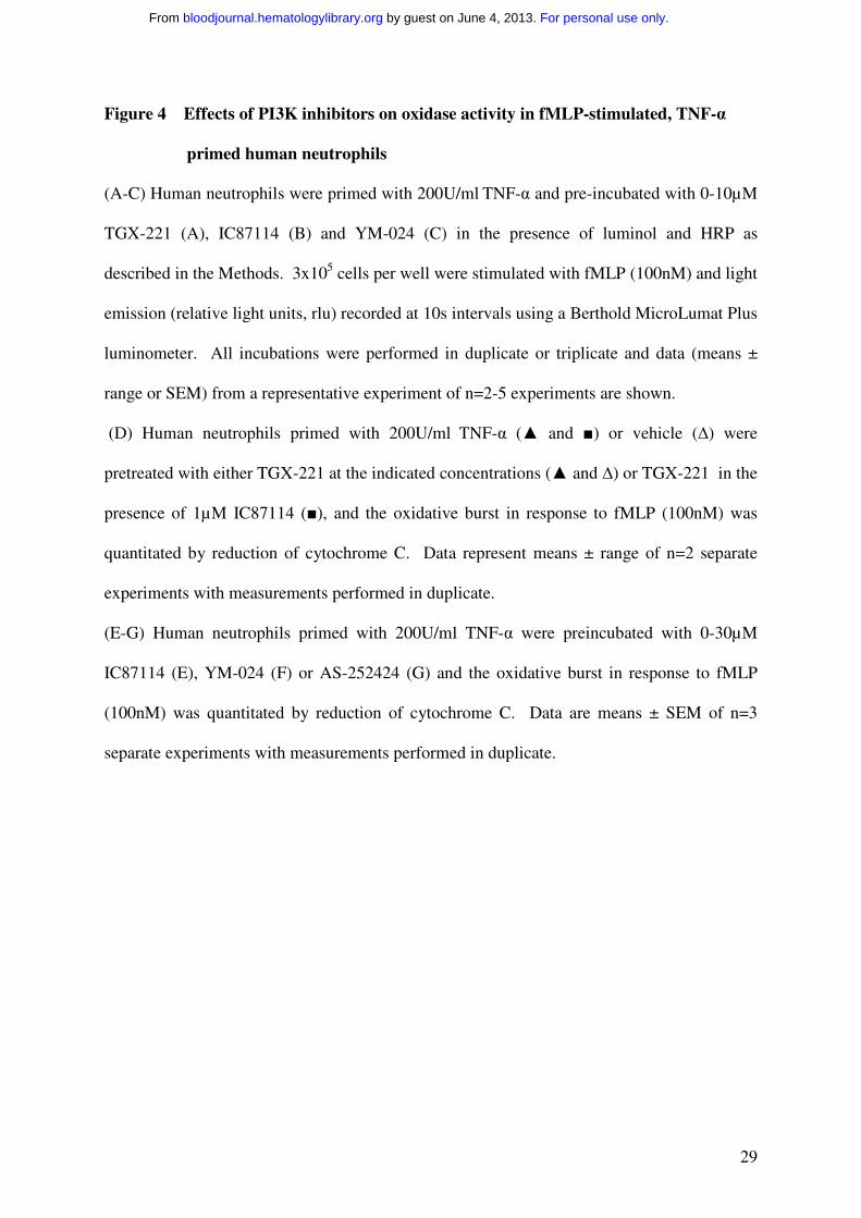
Figure 4 Effects of PI3K inhibitors on oxidase activity in fMLP-stimulated, TNF-α
primed human neutrophils
(A-C) Human neutrophils were primed with 200U/ml TNF-α and pre-incubated with 0-10µM
TGX-221 (A), IC87114 (B) and YM-024 (C) in the presence of luminol and HRP as
described in the Methods. 3x10⁵ cells per well were stimulated with fMLP (100nM) and light
emission (relative light units, rlu) recorded at 10s intervals using a Berthold MicroLumat Plus
luminometer. All incubations were performed in duplicate or triplicate and data (means ±
range or SEM) from a representative experiment of n=2-5 experiments are shown.
(D) Human neutrophils primed with 200U/ml TNF-α (▲ and ■) or vehicle (∆) were
pretreated with either TGX-221 at the indicated concentrations (▲ and ∆) or TGX-221 in the
presence of 1µM IC87114 (■), and the oxidative burst in response to fMLP (100nM) was
quantitated by reduction of cytochrome C. Data represent means ± range of n=2 separate
experiments with measurements performed in duplicate.
(E-G) Human neutrophils primed with 200U/ml TNF-α were preincubated with 0-30µM
IC87114 (E), YM-024 (F) or AS-252424 (G) and the oxidative burst in response to fMLP
(100nM) was quantitated by reduction of cytochrome C. Data are means ± SEM of n=3
separate experiments with measurements performed in duplicate.
For personal use only. by guest on June 4, 2013. bloodjournal.hematologylibrary.orgFrom
30
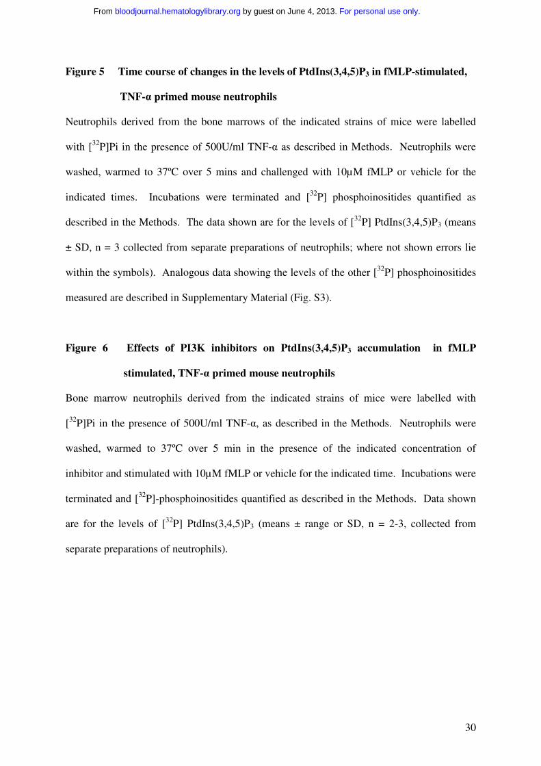
Figure 5 Time course of changes in the levels of PtdIns(3,4,5)P3 in fMLP-stimulated,
TNF-α primed mouse neutrophils
Neutrophils derived from the bone marrows of the indicated strains of mice were labelled
with [32P]Pi in the presence of 500U/ml TNF-α as described in Methods. Neutrophils were
washed, warmed to 37ºC over 5 mins and challenged with 10µM fMLP or vehicle for the
indicated times. Incubations were terminated and [32P] phosphoinositides quantified as
described in the Methods. The data shown are for the levels of [32P] PtdIns(3,4,5)P3 (means
± SD, n = 3 collected from separate preparations of neutrophils; where not shown errors lie
within the symbols). Analogous data showing the levels of the other [32P] phosphoinositides
measured are described in Supplementary Material (Fig. S3).
Figure 6 Effects of PI3K inhibitors on PtdIns(3,4,5)P3 accumulation in fMLP
stimulated, TNF-α primed mouse neutrophils
Bone marrow neutrophils derived from the indicated strains of mice were labelled with
[32P]Pi in the presence of 500U/ml TNF-α, as described in the Methods. Neutrophils were
washed, warmed to 37ºC over 5 min in the presence of the indicated concentration of
inhibitor and stimulated with 10µM fMLP or vehicle for the indicated time. Incubations were
terminated and [32P]-phosphoinositides quantified as described in the Methods. Data shown
are for the levels of [32P] PtdIns(3,4,5)P3 (means ± range or SD, n = 2-3, collected from
separate preparations of neutrophils).
For personal use only. by guest on June 4, 2013. bloodjournal.hematologylibrary.orgFrom
31
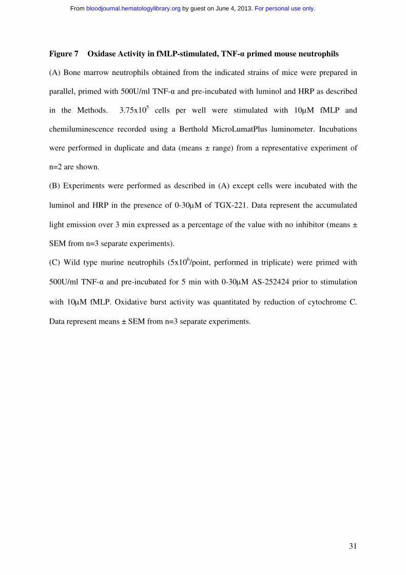
Figure 7 Oxidase Activity in fMLP-stimulated, TNF-α primed mouse neutrophils
(A) Bone marrow neutrophils obtained from the indicated strains of mice were prepared in
parallel, primed with 500U/ml TNF-α and pre-incubated with luminol and HRP as described
in the Methods. 3.75x10⁵ cells per well were stimulated with 10µM fMLP and
chemiluminescence recorded using a Berthold MicroLumatPlus luminometer. Incubations
were performed in duplicate and data (means ± range) from a representative experiment of
n=2 are shown.
(B) Experiments were performed as described in (A) except cells were incubated with the
luminol and HRP in the presence of 0-30µM of TGX-221. Data represent the accumulated
light emission over 3 min expressed as a percentage of the value with no inhibitor (means ±
SEM from n=3 separate experiments).
(C) Wild type murine neutrophils (5x10⁶/point, performed in triplicate) were primed with
500U/ml TNF-α and pre-incubated for 5 min with 0-30µM AS-252424 prior to stimulation
with 10µM fMLP. Oxidative burst activity was quantitated by reduction of cytochrome C.
Data represent means ± SEM from n=3 separate experiments.
For personal use only. by guest on June 4, 2013. bloodjournal.hematologylibrary.orgFrom
32
PtdIns3P
PtdIns4P
PtdIns(3,4)P2
PtdIns(4,5)P2
PtdIns(3,4,5)P3
0 40 60 80 100 12020Time (sec)
4
6
8
0
2dp
mx
10-3
dp
mx
10-3
1.5
2.5
3.5
00.5
0.8
1.2
1.6
0
0.4
80
120
0
4040
60
80
0
20
fMLPfMLP +TGX-221Control Control +TGX-221
Figure 1.
0 40 60 80 100 12020Time (sec)
0 40 60 80 100 12020Time (sec)
0 40 60 80 100 12020Time (sec)
0 40 60 80 100 12020Time (sec)
For personal use only. by guest on June 4, 2013. bloodjournal.hematologylibrary.orgFrom
33
0 0.1 1 10 1000.01
0.1 1 100
0.1 1 100
0.1 1 100
100
80
60
40
20
0
120
100
80
60
40
20
0
120
100
80
60
40
20
0
120
100
80
60
40
20
0
120
[TGX-221] µM [IC87114] µM
[AS-252424] µM[YM-024] µM
% r
esp
on
se
% r
esp
on
se
% r
esp
on
se
% r
esp
on
se
TGX-221 IC87114
AS-252424YM-024
6 sec
60 sec
Figure 2.
For personal use only. by guest on June 4, 2013. bloodjournal.hematologylibrary.orgFrom
34
200
150
100
50
0TNF
Time (sec)
A. B.
C.
+ 0
fMLP+ ++
___
_
300
200
100
0120 180 240 30060
**P
I3-K
inas
e ac
tivi
ty(%
co
ntro
l)
PI3
-Kin
ase
acti
vity
(% c
ont
rol)
D.
6 sec 60 sec
2
4
6
8
0
dp
mx
10-3
LFM-A13PP1
controlControl
TNF-fMLP
250
200
100
-100
-150
150
50
-50
0
PI3
-Kin
ase
acti
vity
(% c
ont
rol)
DMSO TGX-221 IC87114 AS-252424
Figure 3.
For personal use only. by guest on June 4, 2013. bloodjournal.hematologylibrary.orgFrom
35
A. TGX-221 D.
Time (min)0 1 2 3 4 5
0
1.6
1.2
0.8
0.4
Ch
emilu
min
esce
nce
(rlu
x 10
-6 /s
ec)
0 µM0.1 µM0.3 µM1 µM
[TGX-221] µM0 0.1 1 10 100
0
40
30
20
10
nm
ols
uper
oxid
e/
106
cells
B. IC87114 E.
Time (min)0 1 2 3 4 5
0
1.6
1.2
0.8
0.4
Ch
emilu
min
esce
nce
(rlu
x 10
-6 /s
ec)
0 µM0.3 µM3 µM10 µM
[IC87114] µM0 0.1 1 10 100
0
35
25
15
5
nm
ols
uper
oxid
e/
106
cells
C. YM-024 F.
Time (min)0 1 2 3 4 5
0
1.8
1.4
1.0
Che
milu
min
esce
nce
(rlu
x 10
-6 /s
ec)
0.6
0.2
0 µM0.3 µM3 µM10 µM
[YM-024] µM0 0.1 1 10 100
0
45
30
15
nm
ols
uper
oxid
e/
106
cells
G.
[AS-252424] µM0 0.1 1 10 100
nm
ols
up
ero
xid
e/ 1
06ce
lls
0
50
30
10
40
20
Figure 4.
For personal use only. by guest on June 4, 2013. bloodjournal.hematologylibrary.orgFrom
36
γ/δ -/-
δ D910A
γ -/-
WT
0 40 60 80 100 12020 0 40 60 80 100 12020
0 40 60 80 100 12020 0 40 60 80 100 12020
Time (sec)
dpm
x 10
-3
dpm
x 10
-3
dpm
x 10
-3
dpm
x 10
-3
0.8
1.2
2.0
0
0.4
1.6
0.8
1.2
2.0
0
0.4
1.6
0.8
1.2
2.0
0
0.4
1.6
0.8
1.2
2.0
0
0.4
1.6
fMLP
Control
WT fMLP
Time (sec)
Time (sec) Time (sec)
Figure 5.
For personal use only. by guest on June 4, 2013. bloodjournal.hematologylibrary.orgFrom
37
0.1 1 100
100
80
60
40
20
0
120
100
80
60
40
20
0
120
[AS-252424] µM[YM-024] µM
% r
esp
on
se
% r
esp
on
se
AS-252424YM-024 6 sec
60 sec
0.1 1 100 100
2.0
1.6
1.2
0.8
0.4
0
dpm
x10-3
control 10sec fMLP 60sec fMLP
A.
C.B.
WT δ D910A γ -/- γ/δ -/- WT δ D910A γ -/- γ/δ -/- WT δ D910A γ -/- γ/δ -/-
No Inhibitor
0.3µM TGX-221
0.3µM IC87114
Figure 6.
For personal use only. by guest on June 4, 2013. bloodjournal.hematologylibrary.orgFrom
38
A.
B.
wild type
delta kinase dead
120
100
80
60
40
20
0
% m
axim
um
res
po
nse
[TGX-221] µM
C.
wildtype
140
100
80
60
40
20
0
% m
axim
um
res
po
nse
[AS-252424] µM
120
0 0.1 1 10010
10
8
6
4
2
0
Ch
emilu
min
esce
nce
(rlu
x 10
-4/s
ec)
0 1 2 3
Time (min)
WTδ -/-γ -/-γ/δ -/-
WT
δ D910A/D910A
WT
0 0.1 1 10010
Figure 7.
For personal use only. by guest on June 4, 2013. bloodjournal.hematologylibrary.orgFrom