Arabidopsis CBF5 interacts with the H/ACA snoRNP assembly factor NAF1

JOURNAL OF VIROLOGY, Nov. 2005, p. 13735–13746 Vol. 79, No. 210022-538X/05/$08.00�0 doi:10.1128/JVI.79.21.13735–13746.2005Copyright © 2005, American Society for Microbiology. All Rights Reserved.
Human Immunodeficiency Virus Type 1 Vpr Interacts withAntiapoptotic Mitochondrial Protein HAX-1
Venkat S. R. K. Yedavalli,1† Hsiu-Ming Shih,2† Yu-Ping Chiang,3 Chun-Yi Lu,3Luan-Yin Chang,3 Mao-Yuan Chen,4 Che-Yen Chuang,4 Andrew I. Dayton,5
Kuan-Teh Jeang,1* and Li-Min Huang*3
Molecular Virology Section, Laboratory of Molecular Microbiology, National Institutes of Allergy andInfectious Diseases, Maryland 20892-04601; Institute of Biomedical Sciences, Academia Sinica,2 and
Department of Pediatrics3 and Internal Medicine,4 National Taiwan University Hospital, Taipei,Taiwan 100; and Center for Biologics Evaluation and Research, Food and Drug
Administration, Bethesda, Maryland 20892-04605
Received 13 April 2005/Accepted 7 August 2005
Human immunodeficiency virus type 1 viral protein R (Vpr) is required for viral pathogenesis and has beenimplicated in T-cell apoptosis through its activation of caspase 3 and caspase 9 and perturbation of mito-chondrial membrane potential. To understand better Vpr-mitochondria interaction, we report here the iden-tification of antiapoptotic mitochondrial protein HAX-1 as a novel Vpr target. We show that Vpr and HAX-1physically associate with each other. Overexpression of Vpr in cells dislocates HAX-1 from its normal residencein mitochondria and creates mitochondrion instability and cell death. Conversely, overexpression of HAX-1suppressed the proapoptotic activity of Vpr.
Apoptosis or programmed cell death contributes to the elim-ination of damaged, aged, or virus-infected cells (13). Apopto-sis can be initiated by an extrinsic pathway in which deathreceptors expressed at the cell surface trigger receptor-activa-tion of caspases leading to mitochondrial membrane perme-abilization (MMP) (6, 18, 42). Alternatively, a cell-intrinsicapoptotic pathway can act directly on mitochondria, leadingfirst to mitochondrial membrane permeabilization and thenactivation of execution caspases (6, 18, 30, 42). MMP is tightlyregulated by Bcl2 family proteins which contain both pro- andantiapoptotic members (2, 5, 29). Once triggered, MMP marksthe “point of no return” for the apoptotic process (29, 58),whether it be a caspase-dependent or caspase-independentdeath (26, 39, 50). Because apoptosis is used as a means by thehost to defend against invading pathogens, viruses understand-ably have evolved strategies that target the intrinsic and extrin-sic apoptotic pathways. Increasingly, examples illustrate thatmany viruses, including human immunodeficiency virus type 1(HIV-1), hepatitis B virus, Sindbis virus, and baculovirus, en-code proteins that modulate cell death (reviewed in reference4).
Human immunodeficiency virus (HIV) principally infects Thelper (TH) cells and cells of the monocyte-macrophage lin-eage, which express the CD4 cell surface protein. The gradualand selective loss of the CD4 subset of T-lymphocytes is acentral feature of the pathogenesis of HIV which correlateswith the progression from asymptomatic HIV infection to
AIDS. Several mechanisms have been proposed to explain thisdecline, including the rapid turnover and death of infected hostcells, as well as “bystander” cell death via indirect means (17).Moreover, several HIV-1 proteins, including Nef, Vif, Vpr,Vpu, Tat, and Rev, have been implicated in apoptosis induc-tion.
HIV-1 Vpr, a 96-amino-acid, 14-kDa protein, is criticallyinvolved in HIV-1 pathogenesis in vivo (10, 15, 16). Severalfunctions have been attributed to Vpr including (i) interactionwith and translocation of the HIV-1 preintegration complexthrough the nuclear pore, (ii) induction of apoptosis, (iii) in-duction of host cell cycle arrest during G2-to-M transition, and(iv) stimulation of viral gene expression (1, 7, 9, 12, 14, 19–21,25, 32, 40, 41, 43, 46, 48, 53, 55, 60, 61). Studies have docu-mented Vpr-induced apoptosis in human fibroblasts, T-celllines, and primary cells, including lymphocytes and monocytes(1, 23, 38, 44, 47). Indeed, death of uninfected bystander Tcells has also been attributed to secreted Vpr protein. Amongseveral explanations, a leading mechanistic model suggests thatVpr induces cellular apoptosis through dysregulation of MMP.Using isolated mitochondria, others have found that Vpr cantarget the mitochondrial permeability transition pore complexand promote permeabilization of mitochondrial membranes(23). Whether Vpr’s mitochondrion effect can be entirely ex-plained through its binding of inner mitochondria membraneprotein, adenine nucleotide translocator (ANT), remains to beclarified (52).
Apoptosis of infected cells may mute the host’s immuneresponse to the virus (54). In this regard, we wanted to furtherunderstand the details of Vpr’s interaction with mitochondriaand its apoptotic consequences. Here, we report the identifi-cation of HAX-1 (for HS1-associated protein X-1) as a newmitochondrial target for Vpr. HS-1 (for hematopoietic lineagecell-specific protein 1) is a B-cell signaling protein that is asubstrate for intracellular protein tyrosine kinases involved in
* Corresponding author. Mailing address for K.-T. Jeang: Building4, Room 306, 9000 Rockville Pike, NIH, Bethesda, MD 20892-0460.Phone: (301) 496-6680. Fax: (301) 480-3686. E-mail: [email protected] address for L.-M. Huang: National Taiwan University Hospi-tal, Department of Pediatrics, Taipei, Taiwan 10016. Phone: 886-2-2397-0800. Fax: (886) 22-2393-4749. E-mail: [email protected].
† V.S.R.K.Y. and H.-M.S. contributed equally to this study.
13735

the immune response to extracellular stimuli and in cell dif-ferentiation induced by cytokines. HAX-1 was initially re-ported as an HS-1-binding protein; HAX-1 is a 279-amino-acid(35-kDa) protein with homology to Bcl2. HAX-1 has beenshown by others to be an antiapoptotic factor (51). We nowdocument that (i) Vpr binds HAX-1 directly, (ii) overexpres-sion of Vpr causes the egress of HAX-1 from the mitochondriainto the cytoplasm, and (iii) overexpression of HAX-1 countersthe proapoptotic effect of Vpr in cells.
MATERIALS AND METHODS
Plasmids, cell culture, and transfections. HeLa cells were maintained in Dul-becco modified Eagle medium supplemented with 10% fetal bovine serum.Full-length and deletion mutants were generated by PCR amplification andcloning. Plasmids pCDNA-Vpr, pCDNA-FLAG Vpr, and pEYFP-Vpr wereconstructed by PCR amplification of pNL4-3 Vpr and cloning of PCR productsinto pCDNA3.1 and pEYFP vectors, respectively. pCDNA-HAX-1 and pCDNA-HA-HAX-1 were constructed by cloning HAX-1 ORF into pCDNA3.1 vector(Invitrogen). HeLa cells were transfected with Lipofectamine Plus and Lipo-fectamine according to the manufacturer’s instructions (Invitrogen).
Antibodies. Mouse monoclonal anti-HA and anti-FLAG M2 antibodies werecommercially purchased (Sigma-Aldrich); rabbit polyclonal 46-amino-acid anti-body was from the AIDS Reference and Reagent Program. Mouse monoclonalanti-HAX-1 antibody was from BD Pharmingen.
Western blotting, immunoprecipitation, and confocal imaging. Western blot-ting and immunoprecipitation were performed as previously described (28, 56).For confocal microscopy, HeLa cells were cultured on 25-mm coverslips(Thomas Scientific, Swedesboro, NJ) and transfected with plasmid DNA. Oneday later, cells were fixed with 3.7% formaldehyde, permeabilized with phos-phate-buffered saline containing 0.1% Triton X-100, and incubated with primaryantibodies followed with anti-mouse/anti-rabbit antibodies conjugated to AlexaFluor 488/594 (Molecular Probes, Eugene, OR). Nuclei were stained with DAPI(4�,6�-diamidino-2-phenylindole; Molecular Probes). Coverslips were mountedonto glass slides with ProLong Antifade Kit (Invitrogen) and examined with aLeica laser-scanning microscope. For staining of cells with Mitotracker (Invitro-gen), the cultured cells were incubated with Mitotracker dye for 15 min in theincubator before washing and fixation for staining.
Coimmunoprecipitation. MT4/HeLa cells were lysed in radioimmunoprecipi-tation assay (RIPA) buffer (Tris-buffered saline [pH 8.0] containing 1% TritonX-100 or NP-40, 1 mg of bovine serum albumin/ml, and 1 mM EDTA) withprotease inhibitor (phenylmethylsulfonyl fluoride and aprotinin [10 �g/ml]),0.5% sodium deoxycholate, and 0.1% sodium dodecyl sulfate (SDS). Cell lysateswere prepared by sonication and precleared first with anti-mouse/anti-rabbitimmunoglobulin G-agarose before incubation with either anti-HAX or Anti-Vprantibody. Immune complexes were captured by using protein A- or G-agarosebeads, followed by analysis by SDS-polyacrylamide gel electrophoresis (PAGE)and Western blotting.
GST pull-down assay. Expression and purification of glutathione S-transferase(GST) fusion proteins were performed as described previously (3). 35S-labeledHAX-1 proteins were made with TNT reticulocyte lysate system (Promega).35S-labeled proteins were incubated with 2 �g of GST-Vpr fusion or GST proteinin 0.2 ml of binding buffer (10 mM HEPES [pH 7.5], 50 mM NaCl, 0.1% NonidetP-40, 0.5 mM dithiothreitol, 0.5 mM EDTA) for 1 to 2 h, washed four times, andanalyzed by SDS-PAGE, followed by autoradiography. A portion of the reactionmixture was also analyzed by Coomassie blue staining to visualize GST fusionproteins.
Yeast two-hybrid screen and assays. The complete coding sequence of HIV-1pNL4-3 vpr was amplified by PCR and ligated into the BamHI and PstI sites ofthe yeast pBTM116 vector to produce LexA-Vpr as bait for yeast two-hybridscreening. Yeast two-hybrid library screening was performed as described pre-viously (24, 33). Briefly, the yeast reporter strain L40 was transformed withpBTM-Vpr bait plasmid, followed by 250 �g of human bone marrow cDNAlibrary (Clontech). Approximately 107 transformants were screened on mediumcontaining 2 mM 3-amino-(1,2,4) triazole (Sigma) lacking leucine, tryptophan,and histidine. After 3 days of growth, His� colonies were assayed for �-galac-tosidase activity by using colony lift filter assays. Positive cDNA clones wererecovered and tested for interaction specificity by retransformation into yeasteither with Vpr bait or with a nonspecific bait, lamin.
RESULTS
Identification of HAX-1 as a Vpr-interacting protein. Tosearch for Vpr-associated factor(s), we performed a yeast two-hybrid library screen using as bait a fusion protein comprisedof full-length Vpr fused to the LexA protein (LexA-Vpr).HAX-1 was initially identified from an open-ended screeningof the library. The specificity of the interactions between Vprand HAX-1 was next verified by directly transforming yeastcells with isolated plasmids of LexA-Vpr and GalAD-HAX-1(HAX-1 fused with the Gal4 activation domain); positive in-teraction between the two proteins was indicated by blue col-onies on an X-Gal (5-bromo-4-chloro-3-indolyl-�-D-galactopy-ranoside) plate (Fig. 1A). Control transformation of yeast withLexA-lamin plus GalAD-HAX-1 produced no blue colonies,supporting the specificity of Vpr–HAX-1 interaction.
Vpr binds to HAX-1 in vitro and inside cells. To checkfurther the interaction between Vpr and HAX-1, GST pull-down assays were performed with recombinant GST-Vpr fu-sion protein and in vitro-synthesized 35S-labeled HAX-1 pro-tein. Proteins captured by the beads were washed, solubilized,and resolved by SDS-PAGE, followed by autoradiography. Asshown in Fig. 1B, GST-Vpr fusion protein but not GST alonecaptured HAX-1 (Fig. 1B, compare lanes 2 and 3); this resultis fully consistent with a physical protein-protein interactionbetween Vpr and HAX-1.
To examine Vpr-HAX-1 interaction inside cells, we nextattempted coimmunoprecipitate cell-endogenous HAX-1 withVpr expressed from an infecting HIV-1 molecular clone,NL4-3. MT4 cells were infected with HIV-1 molecular cloneNL4-3 and harvested 4 days postinfection. Cells lysed in RIPAbuffer were sonicated and immunoprecipitated with anti-Vprantibody. The immunoprecipitates were resolved by SDS-PAGE and analyzed by Western blotting with anti-HAX-1antibody. As shown in Fig. 1C (lane 4), HAX-1 was found tocoprecipitate with Vpr, a finding consistent with the resultsfrom yeast two-hybrid (Fig. 1A) and GST pull down (Fig. 1B)assays. Taken together, the results suggest that the Vpr inter-acts directly with cell endogenous HAX-1 in HIV-1-infectedcells.
Expression of DN HAX-1 mutant induces apoptosis. Previ-ously, it was reported that HAX-1 contained limited homologywith the BH1 and BH2 domains of Bcl-2 family members (45,51). Elsewhere, in other experimental systems, HAX-1 wasfound to be a mitochondria protein (51), which when overex-pressed blocked Bax-induced apoptosis (45). Formally, it hasnot been fully clarified whether HAX-1 serves a direct role inpreventing cellular apoptosis or only plays an indirect role ininfluencing the expression, stability, and/or function of Bax. Toaddress HAX-1 function in our experimental setting, we con-structed several deletion mutants. We reasoned that some orall of the mutants might be dominant negative (DN) forHAX-1 function. One prediction is that if HAX-1 were to playa constitutive role in suppressing cellular apoptosis, then ex-pression of DN HAX-1 should produce cell death. To checkthis prediction, we introduced wild-type HAX-1, HAX-1(142-279) mutant, or HAX-1(1-141) mutant separately into HeLacells, along with a green fluorescent protein (GFP) plasmidthat marked the transfected cells. After 36 to 48 h, cells werefixed and stained with DAPI. We scored GFP-positive cells for
13736 YEDVALLI ET AL. J. VIROL.

apoptosis based on the visualization of condensed nuclei withnuclear fragmentation. We found that neither full-lengthHAX-1 nor HAX-1(142-279) (Fig. 1, Fig. 2, Fig. 3, Fig. 4, Fig.5 A1 through 6) induced apoptosis. However, overexpressionof HAX-1 (1–141) produced frequent nuclear condensationand fragmentation consistent with apoptosis (Fig. 2A7 to A12).An interpretation of these results is that HAX-1(142-279) is aloss-of-function mutant, whereas HAX-1(1-141) is a DN mu-tant. The finding that DN HAX-1(1-141) induced apoptosis is
consistent with an intrinsically protective role served by cell-endogenous HAX-1 against cell death.
Vpr-induced apoptosis is suppressed by overexpression ofHAX-1. Several studies have shown that expression of Vprcauses apoptotic cell death (22, 23, 38, 44, 47). Because Vprbinds HAX-1 (Fig. 1) and because DN-HAX-1, in itself, pro-moted apoptosis (Fig. 2A), we wondered whether Vpr’s effecton cell death might arise from its physical sequestration oflimiting amounts of intracellular HAX-1. To explore this pos-
FIG. 1. Interaction between Vpr and HAX-1. (A) Vpr and HAX-1 interact in yeast two-hybrid assay. The L40 yeast expressing both LexA BDand GAL4AD hybrid proteins was analyzed on selective plates containing 2 mM concentration of 3-amino-(1,2,4) triazole. Growth in the absenceof His with a positive �-galactosidase signal are indications of the interaction between the hybrid proteins. LexA-lamin fusion protein was usedas a negative control. (B) HAX-1 and Vpr interact in vitro. Bacterially expressed purified GST-Vpr was used for in vitro binding assay with[35S]methionine-labeled in vitro-translated HAX-1 protein. HAX-1 bound GST-Vpr (lane 3) but not GST-alone (lane 2). (C) Interaction betweenVpr and HAX-1 in human cells. HAX-1 interacts with Vpr expressed from HIV-1 in MT4 cells infected with infectious HIV-1 NL4-3. MT4 cellswere harvested 4 days postinfection by centrifugation and were lysed in RIPA buffer. Cell lysates were subjected to sonication and coimmuno-precipitation assays with anti-Vpr or with irrelevant control antibody (isotype control). The immunoprecipitates were resolved by SDS-PAGE,transferred to membrane, and probed with mouse monoclonal anti-HAX-1 antibody. The top panel shows coimmunoprecipitation of HAX-1 withVpr. The middle panel shows Western blotting of endogenous HAX-1. The bottom panel shows expression of Vpr from pNL4-3.
VOL. 79, 2005 HIV-1 Vpr INTERACTS WITH MITOCHONDRIAL PROTEIN HAX-1 13737
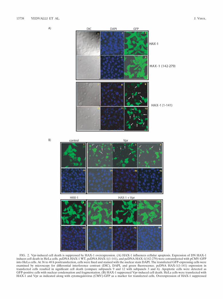
FIG. 2. Vpr-induced cell death is suppressed by HAX-1 overexpression. (A) HAX-1 influences cellular apoptosis. Expression of DN HAX-1induces cell death in HeLa cells. pcDNA HAX-1 WT, pcDNA HAX-1(1-141), and pcDNA HAX-1(142-279) were cotransfected with pCMV-GFPinto HeLa cells. At 36 to 48 h posttransfection, cells were fixed and stained with the nuclear stain DAPI. The transfected GFP-expressing cells wereexamined by microscopy for differential interference contrast (DIC), DAPI, and green fluorescence. pcDNA HAX-1(1-141) expression intransfected cells resulted in significant cell death (compare subpanels 9 and 12 with subpanels 3 and 6). Apoptotic cells were detected asGFP-positive cells with nuclear condensation and fragmentation. (B) HAX-1 suppressed Vpr-induced cell death. HeLa cells were transfected withHAX-1 and Vpr as indicated along with cytomegalovirus (CMV)-GFP as a marker for transfected cells. Overexpression of HAX-1 suppressed
13738 YEDVALLI ET AL. J. VIROL.

sibility, we revisited Vpr’s capacity to induce cell death. In ourcultured cells, we observed that nuclear fragmentation rapidlyensued after Vpr expression (Fig. 2B2-4). Provocatively, whenHAX-1 was coexpressed with Vpr in cells, mortality of recip-
ient cells normally expected from receiving Vpr-alone disap-peared (Fig. 2B, compare panels 6 to 8 to 2 to 4). These resultsare compatible with Vpr’s proapoptotic effect being manifestthrough its sequestration of cell-endogenous HAX-1 and with
Vpr-induced cell death (compare subpanels 2 to 4 with subpanels 6 to 8). (C) Western blot analysis of the expression of Vpr and HAX-1 intransfected HeLa cells. Cell lysates prepared from transfections above (Fig. 2B) were analyzed by blotting for expression of the indicated proteinsfrom transiently transfected plasmids. The top row shows detection of transfected HAX-1 with a light exposure of film. The middle row showsdetection of Flag-tagged transfected Vpr. The bottom row shows �-actin signals as loading controls. (D) Graphic representation of suppressioncell death induced by HIV-1 Vpr. Cell viability of transfected cells was observed 36 to 48 h posttransfection by counting the number of live green(total cells) and apoptotic green (apoptotic cells) fluorescent cells under the microscope. Values are representative of three independent assays.
VOL. 79, 2005 HIV-1 Vpr INTERACTS WITH MITOCHONDRIAL PROTEIN HAX-1 13739
such sequestration being competitively muted by overexpres-sion of exogenous HAX-1. In Fig. 2C, we performed controlWestern blotting to monitor the degree of expression of trans-fected HAX-1 and Vpr. Indeed, based on Western blotting, it
was evident that the level of expression of transfected HAX-1exceeds the amount detected for cell endogenous HAX-1 (Fig.2C, compare lanes 1 and 3 to lanes 2 and 4).
We next quantified the ability of HAX-1 to suppress Vpr-
FIG. 3. Localization of Vpr and HAX-1 in HeLa cells. Transfected cells were visualized 36 to 48 h after transfection. (A) Flag-Vpr andGFP-HAX-1 colocalize. Flag-Vpr is primarily nuclear (subpanel 1); GFP-HAX-1 protein is mainly mitochondrial (subpanel 2). The cotransfectedGFP-HAX-1 and Flag-Vpr colocalize outside of the nucleus and the mitochondria (subpanels 3 and 4). In contrast, Flag-Vpr and GFP-Bcl2 donot colocalize (subpanels 5 to 7). (B) HAX-1 localizes to extra mitochondrial sites in the presence of Vpr. HeLa cells transfected with HAX-1 alone(subpanels 1 to 4) and HAX-1 plus Vpr (subpanels 5 to 8) were stained with Mitotracker before fixation and were probed with anti-HAantibody–anti-mouse Alexa 488. HAX-1 in the absence of Vpr colocalized with Mitotracker-stained mitochondria but in the presence of Vprformed cytoplasmic bodies that are not stained by Mitotracker.
13740 YEDVALLI ET AL. J. VIROL.

induced apoptosis. In Fig. 2D, we counted the number ofGFP-positive cells (total cells) and the number of green cellsthat were apoptotic (apoptotic cells). We found that Vpr-in-duced apoptosis was reduced by threefold when HAX-1 wascoexpressed in trans. Expression of HAX-1-alone, Vpr1-70mutant alone or of Vpr1-70 mutant plus HAX-1 producedapoptosis similar to the controls cells transfected withpCDNA3 vector.
Mutually altered cellular localizations of HAX-1 and Vprupon simultaneous overexpression. Although Vpr is generallylocated at the nucleus and nuclear membrane, some investiga-tors have found its additional presence in mitochondria (22).On the other hand, HAX-1 is largely mitochondrial and isfound rarely in the endoplasmic reticulum (51). To verify thenotion that Vpr might sequester HAX-1 and perturb the lat-ter’s activity, we investigated whether the two proteins showsome level of colocalization inside cells. We transfected HeLacells with either Flag-Vpr (Fig. 3A1) or GFP-HAX-1 (Fig.3A2) or both (Fig. 3A3 and 4). Cells were fixed 24 h later andstained with anti-Flag antibody, as well as visualized for GFP.We found that GFP-HAX-1 (Fig. 3A2) showed a speckledpattern that mirrored Mitotracker staining of mitochondria(Fig. 3B3); on the other hand, Flag-Vpr alone was diffuselynuclear (Fig. 3A1). Intriguingly, coexpression of GFP-HAX-1with Flag-Vpr resulted in a remarkable redistribution of bothmolecules (Fig. 3A3 and 4). Under such conditions, Flag-Vprrelocated from the nucleus into the cytoplasm, whereas GFP-HAX-1 departed the mitochondria to appear with the cyto-plasmic Flag-Vpr. The redistributed Vpr-HAX-1 bodies do notcostain with Golgi, endoplasmic reticulum, or mitochondria(data not shown). Currently, it is not clear whether these bod-ies localize with bona fide cytoplasmic organelles.
As controls, we also visualized mitochondrion-residentGFP-Bcl2 (Fig. 3A5) and GFP-Bcl2 plus Flag-Vpr (Fig. 3A6).Here, simultaneous expression of Bcl2 and Vpr had neither asignificantly perturbing nor a colocalizing effect on each other(compare Fig. 3A3, 4). A minor “blurring” by confocal visual-ization of Bcl2 in the mitochondria may be due to perturbationof mitochondrial membrane potential by Vpr. Overall, ourfindings suggest that Vpr has a specific interaction with HAX-1but not Bcl2. Next, to make sure that our results are notidiosyncratic to GFP-tagged HAX-1, we also examined thebehavior of an hemagglutinin (HA)-tagged HAX-1 (Fig. 3B).In agreement with GFP-HAX-1, HA-HAX-1 was also dislo-cated from the mitochondria by overexpressed Flag-Vpr (com-pare Fig. 3B4). Altogether, our results are compatible with ascenario in which the mitochondrion-intrinsic cell-protectivefunction of HAX-1 is disturbed by Vpr expression.
Physical interaction between Vpr and HAX-1 correlates withapoptosis. To characterize how HAX-1 is targeted by Vpr, wesought to better understand the domain in Vpr responsible forthis function. We tested one point (VprS79A) and two deletion[Vpr(1-70) and Vpr(1-51)] mutants of Vpr (Fig. 4A and B) fortheir association with HAX-1. As shown in Fig. 4A, two distinctintracellular profiles were observed. VprS79A was entirely nu-clear (Fig. 4A1), whereas Vpr(1-70) (Fig. 4A4) and Vpr(1-50)(data not shown) were located in extranuclear speckles. Inter-estingly, when the Vpr mutants were coexpressed with HAX-1,two different patterns were seen. VprS79A exited the nucleusand colocated with HAX-1 (Fig. 4A3). On the other hand,
neither Vpr1-70 (Fig. 4A4) nor Vpr1-51 (data not shown)appeared to colocalize with HAX-1, nor did these two mutantsperturb HAX-1’s normal mitochondrial location (Fig. 4A6).
We also checked the region of interaction between HAX-1and Vpr by coimmunoprecipitations. We transfected HA-tagged HAX-1 with YFP alone, YFP-Vpr, YFP-Vpr(1-70) andYFP-Vpr(1-51) (Fig. 4B) into cells. Cell lysates were immuno-precipitated with anti-HA, and immunoprecipitates wereWestern blotted with anti-YFP. As shown in Fig. 4B, YFP-Vprcoprecipitated with HAX-1, a finding consistent with the re-sults from yeast two-hybrid (Fig. 1A) and GST pull down (Fig.1B) and earlier coimmunoprecipitation (Fig. 1C) assays. Incontrast, Vpr(1-70) and Vpr(1-51) failed to associate withHAX-1 (Fig. 4B, lanes 3 and 4), suggesting that the C terminusof Vpr spanning amino acids 71 to 96 is required for interac-tion with HAX-1.
We also examined our transfected cells for signs of apoptosis(Fig. 4C). We found agreement with previous data on Vpr-induced apoptosis. Hence, Jacotot et al. (23) had reported thatfull-length Vpr and a carboxy-Vpr form (Vpr52-95), but not anamino-Vpr form [Vpr(1-51) and Vpr(1-70)], triggered cellularapoptosis. Our data are consistent with those earlier resultsand further suggest a correlation between Vpr-induced apo-ptosis and Vpr’s ability to associate with HAX-1. Thus, weobserved that Vpr proteins competent or incompetent forHAX-1 binding are, respectively, active or inactive for inducingapoptosis (Fig. 4C).
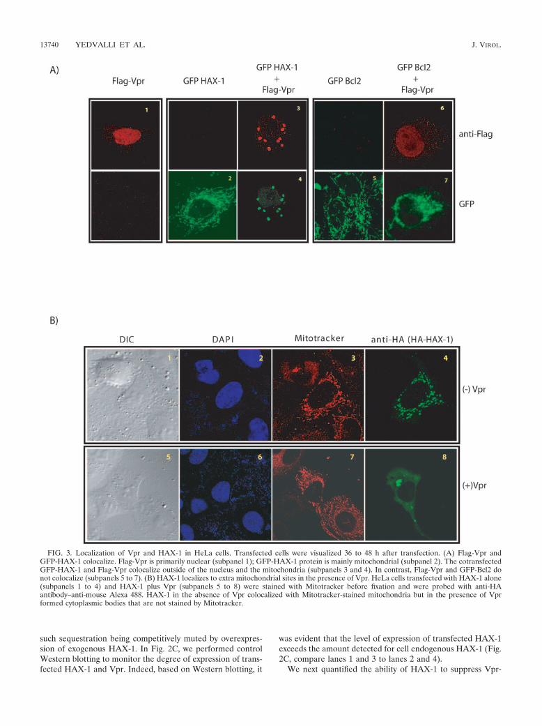
Sequestration of Vpr and suppression of apoptosis maps toamino acids 118 to 141 of HAX-1. Our results to this point areconsistent with a model in which Vpr sequesters and dislo-cates mitochondrion-protective HAX-1, leading to cellularapoptosis. This model predicts that there should be aHAX-1 mutant which, when overexpressed, would bind Vprcompetitively and prevent Vpr’s disturbance of wild-typeHAX-1 function. To check this prediction, we constructedseveral overlapping HAX-1 deletion mutants (Fig. 5B). InFig. 5A, we observed that some HAX-1 deletion mutantsshowed minor differences from wild-type HAX-1 in intra-cellular localization (Fig. 3). Interestingly, all mutants, withthe exception of HAX-1(141-279), were competent for se-questering Vpr (Fig. 5A and B).
When we examined the functional consequences of HAX-1mutant overexpression, a one-to-one correlation emerged be-tween HAX-1 mutants capable of binding Vpr and their abilityto suppress Vpr-induced apoptosis in cells (Fig. 5B). Theseresults are complementary to the above findings (Fig. 4) anddemonstrate that overexpressed HAX-1 mutants can compet-itively capture Vpr and abrogate Vpr’s ability to perturb thelimiting apoptosis-protective function of cell-endogenous mi-tochondria-located HAX-1. We quantified that overexpressionof HAX-1 mutants [HAX-1(81-279), HAX-1(91-279), HAX-1(118-279), and HAX-1(1-260)] suppressed Vpr induced apo-ptosis by �2-3-fold (data not shown). On the other hand,HAX-1(141-279) failed entirely to suppress Vpr-induced apo-ptosis. One interpretation of these results is that HAX-1 isrequired to stabilize mitochondria and that Vpr can physicallybind and dislocate HAX-1 from mitochondria leading to de-stabilized MMP and apoptosis.
VOL. 79, 2005 HIV-1 Vpr INTERACTS WITH MITOCHONDRIAL PROTEIN HAX-1 13741

13742 YEDVALLI ET AL. J. VIROL.

DISCUSSION
Mitochondrial physiology is intimately tied to cellular apo-ptosis (18, 31, 57) One hypothesis for mitochondrial cell deathinvolves a changes in MMP culminating in the release of apo-ptogenic factors such as cytochrome c and apoptosis inhibitingfactor from the mitochondrial intermembrane space (34, 35,49, 59) Whether cells live or die is tied to a balance betweenthe actions of antiapoptotic (e.g., Bcl-2/Bcl-XL) and proapo-ptotic (e.g., Bax, ANT-1, and VDAC) factors. Within this con-text, HAX-1 is an antiapoptotic factor thought most likely tobe located at the outer mitochondrial membrane (8). Overex-pression of HAX-1 has been shown previously to prevent Bax-induced MMP and the subsequent apoptosis of cells (45).
Vpr has been found to perturb MMP (23). Previously, Ja-cotot et al. (23) used a synthetic HIV-1 Vpr peptide to study itsinteraction with isolated mitochondria in vitro. At micromolarconcentrations of Vpr, these authors observed rapid dissipa-tion of MMP; this change in potential correlated with a leakagefrom purified mitochondria of apoptogenic proteins such ascytochrome c. Similarly, in Jurkat T cells, Vpr expression alsoproduced aberrant MMP with accompanying apoptosis (23,37). Currently, Vpr’s apoptogenic MMP effect has been largelyattributed to its interaction with an inner mitochondrial mem-brane protein, ANT-1 (22, 23). Although Vpr-ANT-1 inter-action is consistent with many extant findings, it remains per-plexing how an interaction or disturbance at the innermitochondrial membrane mechanistically provokes perfora-tion of the outer membrane leading to the release of apopto-genic factors normally resident in the intermembranous space.Our new finding that an outer membrane factor, HAX-1, isalso targeted by Vpr clarifies how HIV-1 might fully perforateboth layers of mitochondria membranes leading to apoptosis.Our Vpr-HAX-1 finding also validates a prediction made byJacotot et al. based on their Vpr52-96 results that, separatefrom ANT-1, Vpr must have a second target located in theouter mitochondrial membrane (22). The current finding thatoverexpression of HAX-1 is sufficient to abolish Vpr-inducedapoptosis in cells (Fig. 2) is further consistent with our inter-pretation and suggests that for purposes of apoptosis, stabili-zation of the outer mitochondrial membrane is dominant overdestabilization of the inner membrane (i.e., via ANT-1).
HAX-1 was originally identified by yeast two-hybrid screenas a protein that associates with HS-1 (for hematopoietic lin-eage cell-specific protein 1). HS-1 is a B-cell signaling proteinand is a substrate for intracellular protein tyrosine kinasesinvolved in the immune response to extracellular stimuli and incell differentiation induced by cytokines (51). HAX-1 possessestwo Bcl2 homologous domains BH1 and BH2 which are con-served among Bcl-2 family proteins. Additional amino acidsequences in HAX-1 retain similarities with apoptosis regulat-ing protein Nip3, which has been found to bind antiapoptoticfactor Bcl2 (51). Indeed, recent findings have also shown thatHAX-1 can directly bind to Bcl2, which is also predominantlylocated in the outer mitochondrial membrane (36). It is inter-esting that two other human viruses, Epstein-Barr virus andKaposi’s sarcoma herpesvirus, have also been found to encodeproteins that target HAX-1 (11, 27, 45). Currently, it is notfully understood why disparate viruses would commonly con-verge on this mitochondrial protein. Potentially, if apoptosis of
FIG
.4.
HA
X-1
inte
ract
ion
with
the
Cte
rmin
usof
Vpr
corr
elat
edw
ithsu
ppre
ssio
nof
apop
tosi
s.(A
)C
onfo
cale
xam
inat
ion
ofdi
stri
butio
nof
Vpr
mut
ants
inH
eLa
cells
inth
epr
esen
ceor
abse
nce
ofH
AX
-1.S
tain
ing
patt
erns
ofY
FP-
Vpr
S79A
and
YF
P-V
pr(1
-70)
are
show
n.(B
)M
appi
ngof
the
Vpr
regi
onre
quir
edfo
rH
AX
-1in
tera
ctio
n.H
eLa
cells
tran
sien
tlyex
pres
sing
the
indi
cate
dpr
otei
n(s)
wer
eim
mun
opre
cipi
tate
dw
ithm
ouse
mon
oclo
nala
ntib
ody
toH
AX
-1.T
heim
mun
opre
cipi
tate
sw
ere
subj
ecte
dto
SDS-
PAG
E,t
rans
ferr
edto
mem
bran
e,an
dpr
obed
with
rabb
itpo
lycl
onal
anti-
YF
P.T
heto
ppa
nels
how
sth
atY
FP-
Vpr
WT
coim
mun
opre
cipi
tate
dw
ithH
A-H
AX
-1,w
here
asY
FP
alon
e,Y
FP-
Vpr
(1-7
0),a
ndY
FP-
Vpr
(1-5
1)di
dno
t.T
hem
iddl
epa
nel
show
seq
ual
expr
essi
onle
vels
ofal
lY
FP-
tagg
edpr
otei
ns.
The
bott
ompa
nel
veri
fied
for
equa
lex
pres
sion
oftr
ansf
ecte
dH
A-H
AX
-1.
(C)
Sche
mat
icre
pres
enta
tions
ofV
prm
utan
ts,
thei
rin
tera
ctio
nsw
ithH
AX
-1,a
ndth
eir
apop
tosi
s-in
duci
ngca
paci
ty.
VOL. 79, 2005 HIV-1 Vpr INTERACTS WITH MITOCHONDRIAL PROTEIN HAX-1 13743
FIG. 5. Mapping of amino acids 118 to 141 of HAX-1 as a region of interaction with Vpr. (A) Confocal examination of expression anddistribution of HAX-1 in HeLa cells. Fluorescent images indicate that the various HAX-1 deletion mutants are either capable or not capable ofsequestering Vpr. (B) Diagrammatic summary of HAX-1 deletion mutant correlating the ability to sequester Vpr with ability to halt Vpr-inducedapoptosis. Bcl2 homology domains 1 and 2 (BH1 and -2, green), PEST sequence (PEST, red), and transmembrane domain (TMD, brown) areindicated.
13744 YEDVALLI ET AL. J. VIROL.

infected cells is one way for viruses to evade the elicitation ofa vigorous host immune response, then it could be reasonablewhy Epstein-Barr virus, Kaposi’s sarcoma herpesvirus, andHIV-1 would share this interaction. If this reasoning is correct,then the discovery of small molecules which might interdictVpr-HAX-1 interaction could contribute as an adjunct to thebetter development of an immunity-eliciting AIDS vaccine.
ACKNOWLEDGMENTS
This study was supported in part by grants (NSC93-2314-B-002-104and NSC94-2314-B-002-062) from the National Science Council, Tai-wan, and by intramural research funds from the NIAID, NIH.
REFERENCES
1. Ayyavoo, V., A. Mahboubi, S. Mahalingam, R. Ramalingam, S. Kudchodkar,W. V. Williams, D. R. Green, and D. B. Weiner. 1997. HIV-1 Vpr suppressesimmune activation and apoptosis through regulation of nuclear factor �B.Nat. Med. 3:1117–1123.
2. Belzacq, A. S., H. L. Vieira, F. Verrier, G. Vandecasteele, I. Cohen, M. C.Prevost, E. Larquet, F. Pariselli, P. X. Petit, A. Kahn, R. Rizzuto, C. Bren-ner, and G. Kroemer. 2003. Bcl-2 and Bax modulate adenine nucleotidetranslocase activity. Cancer Res. 63:541–546.
3. Bouhamdan, M., S. Benichou, F. Rey, J. M. Navarro, I. Agostini, B. Spire, J.Camonis, G. Slupphaug, R. Vigne, R. Benarous, and J. Sire. 1996. Humanimmunodeficiency virus type 1 Vpr protein binds to the uracil DNA glyco-sylase DNA repair enzyme. J. Virol. 70:697–704.
4. Boya, P., A. L. Pauleau, D. Poncet, R. A. Gonzalez-Polo, N. Zamzami, and G.Kroemer. 2004. Viral proteins targeting mitochondria: controlling cell death.Biochim. Biophys. Acta 1659:178–189.
5. Brenner, C., H. Cadiou, H. L. Vieira, N. Zamzami, I. Marzo, Z. Xie, B. Leber,D. Andrews, H. Duclohier, J. C. Reed, and G. Kroemer. 2000. Bcl-2 and Baxregulate the channel activity of the mitochondrial adenine nucleotide trans-locator. Oncogene 19:329–336.
6. Brenner, C., and G. Kroemer. 2000. Apoptosis. Mitochondria–the deathsignal integrators. Science 289:1150–1151.
7. Chen, R., E. Le Rouzic, J. A. Kearney, L. M. Mansky, and S. Benichou. 2004.Vpr-mediated incorporation of UNG2 into HIV-1 particles is required tomodulate the virus mutation rate and for replication in macrophages. J. Biol.Chem. 279:28419–28425.
8. Cilenti, L., M. M. Soundarapandian, G. A. Kyriazis, V. Stratico, S. Singh, S.Gupta, J. V. Bonventre, E. S. Alnemri, and A. S. Zervos. 2004. Regulation ofHAX-1 antiapoptotic protein by Omi/HtrA2 protease during cell death.J. Biol. Chem. 279:50295–50301.
9. Cohen, E. A., R. A. Subbramanian, and H. G. Gottlinger. 1996. Role ofauxiliary proteins in retroviral morphogenesis. Curr. Top. Microbiol. Immu-nol. 214:219–235.
10. Connor, R. I., B. K. Chen, S. Choe, and N. R. Landau. 1995. Vpr is requiredfor efficient replication of human immunodeficiency virus type-1 in mono-nuclear phagocytes. Virology 206:935–944.
11. Dufva, M., M. Olsson, and L. Rymo. 2001. Epstein-Barr virus nuclear anti-gen 5 interacts with HAX-1, a possible component of the B-cell receptorsignalling pathway. J. Gen. Virol. 82:1581–1587.
12. Emerman, M. 1996. HIV-1, Vpr and the cell cycle. Curr. Biol. 6:1096–1103.13. Ferri, K. F., and G. Kroemer. 2001. Organelle-specific initiation of cell death
pathways. Nat. Cell Biol. 3:E255–E263.14. Forget, J., X. J. Yao, J. Mercier, and E. A. Cohen. 1998. Human immuno-
deficiency virus type 1 Vpr protein transactivation function: mechanism andidentification of domains involved. J. Mol. Biol. 284:915–923.
15. Gibbs, J. S., A. A. Lackner, S. M. Lang, M. A. Simon, P. K. Sehgal, M. D.Daniel, and R. C. Desrosiers. 1995. Progression to AIDS in the absence ofa gene for vpr or vpx. J. Virol. 69:2378–2383.
16. Goh, W. C., M. E. Rogel, C. M. Kinsey, S. F. Michael, P. N. Fultz, M. A.Nowak, B. H. Hahn, and M. Emerman. 1998. HIV-1 Vpr increases viralexpression by manipulation of the cell cycle: a mechanism for selection ofVpr in vivo. Nat. Med. 4:65–71.
17. Gougeon, M. L., and L. Montagnier. 1999. Programmed cell death as amechanism of CD4 and CD8 T cell deletion in AIDS: molecular control andeffect of highly active anti-retroviral therapy. Ann. N. Y. Acad. Sci. 887:199–212.
18. Green, D. R., and J. C. Reed. 1998. Mitochondria and apoptosis. Science281:1309–1312.
19. He, J., S. Choe, R. Walker, P. Di Marzio, D. O. Morgan, and N. R. Landau.1995. Human immunodeficiency virus type 1 viral protein R (Vpr) arrestscells in the G2 phase of the cell cycle by inhibiting p34cdc2 activity. J. Virol.69:6705–6711.
20. Heinzinger, N. K., M. I. Bukinsky, S. A. Haggerty, A. M. Ragland, V. Kewal-ramani, M. A. Lee, H. E. Gendelman, L. Ratner, M. Stevenson, and M.
Emerman. 1994. The Vpr protein of human immunodeficiency virus type 1influences nuclear localization of viral nucleic acids in nondividing host cells.Proc. Natl. Acad. Sci. USA 91:7311–7315.
21. Hrimech, M., X. J. Yao, F. Bachand, N. Rougeau, and E. A. Cohen. 1999.Human immunodeficiency virus type 1 (HIV-1) Vpr functions as an imme-diate-early protein during HIV-1 infection. J. Virol. 73:4101–4109.
22. Jacotot, E., K. F. Ferri, C. El Hamel, C. Brenner, S. Druillennec, J. Hoebeke,P. Rustin, D. Metivier, C. Lenoir, M. Geuskens, H. L. Vieira, M. Loeffler,A. S. Belzacq, J. P. Briand, N. Zamzami, L. Edelman, Z. H. Xie, J. C. Reed,B. P. Roques, and G. Kroemer. 2001. Control of mitochondrial membranepermeabilization by adenine nucleotide translocator interacting with HIV-1viral protein rR and Bcl-2. J. Exp. Med. 193:509–519.
23. Jacotot, E., L. Ravagnan, M. Loeffler, K. F. Ferri, H. L. Vieira, N. Zamzami,P. Costantini, S. Druillennec, J. Hoebeke, J. P. Briand, T. Irinopoulou, E.Daugas, S. A. Susin, D. Cointe, Z. H. Xie, J. C. Reed, B. P. Roques, and G.Kroemer. 2000. The HIV-1 viral protein R induces apoptosis via a directeffect on the mitochondrial permeability transition pore. J. Exp. Med. 191:33–46.
24. Jin, D. Y., F. Spencer, and K. T. Jeang. 1998. Human T-cell leukemia virustype 1 oncoprotein Tax targets the human mitotic checkpoint proteinMAD1. Cell 93:81–91.
25. Jowett, J. B., V. Planelles, B. Poon, N. P. Shah, M. L. Chen, and I. S. Chen.1995. The human immunodeficiency virus type 1 vpr gene arrests infected Tcells in the G2 � M phase of the cell cycle. J. Virol. 69:6304–6313.
26. Joza, N., S. A. Susin, E. Daugas, W. L. Stanford, S. K. Cho, C. Y. Li, T.Sasaki, A. J. Elia, H. Y. Cheng, L. Ravagnan, K. F. Ferri, N. Zamzami, A.Wakeham, R. Hakem, H. Yoshida, Y. Y. Kong, T. W. Mak, J. C. Zuniga-Pflucker, G. Kroemer, and J. M. Penninger. 2001. Essential role of themitochondrial apoptosis-inducing factor in programmed cell death. Nature410:549–554.
27. Kawaguchi, Y., K. Nakajima, M. Igarashi, T. Morita, M. Tanaka, M. Suzuki,A. Yokoyama, G. Matsuda, K. Kato, M. Kanamori, and K. Hirai. 2000.Interaction of Epstein-Barr virus nuclear antigen leader protein (EBNA-LP)with HS1-associated protein X-1: implication of cytoplasmic function ofEBNA-LP. J. Virol. 74:10104–10111.
28. Kibler, K. V., and K. T. Jeang. 2001. CREB/ATF-dependent repression ofcyclin a by human T-cell leukemia virus type 1 Tax protein. J. Virol. 75:2161–2173.
29. Kroemer, G. 2003. Mitochondrial control of apoptosis: an introduction.Biochem. Biophys. Res. Commun. 304:433–435.
30. Kroemer, G., B. Dallaporta, and M. Resche-Rigon. 1998. The mitochondrialdeath/life regulator in apoptosis and necrosis. Annu. Rev. Physiol. 60:619–642.
31. Kroemer, G., and J. C. Reed. 2000. Mitochondrial control of cell death. Nat.Med. 6:513–519.
32. Le Rouzic, E., and S. Benichou. 2005. The Vpr protein from HIV-1: distinctroles along the viral life cycle. Retrovirology 2:11.
33. Lin, D. Y., and H. M. Shih. 2002. Essential role of the 58-kDa microspheruleprotein in the modulation of Daxx-dependent transcriptional repression asrevealed by nucleolar sequestration. J. Biol. Chem. 277:25446–25456.
34. Liu, X., C. N. Kim, J. Yang, R. Jemmerson, and X. Wang. 1996. Induction ofapoptotic program in cell-free extracts: requirement for dATP and cyto-chrome c. Cell 86:147–157.
35. Mancini, M., D. W. Nicholson, S. Roy, N. A. Thornberry, E. P. Peterson,L. A. Casciola-Rosen, and A. Rosen. 1998. The caspase-3 precursor has acytosolic and mitochondrial distribution: implications for apoptotic signaling.J. Cell Biol. 140:1485–1495.
36. Matsuda, G., K. Nakajima, Y. Kawaguchi, Y. Yamanashi, and K. Hirai.2003. Epstein-Barr virus (EBV) nuclear antigen leader protein (EBNA-LP)forms complexes with a cellular anti-apoptosis protein Bcl-2 or its EBVcounterpart BHRF1 through HS1-associated protein X-1. Microbiol. Immu-nol. 47:91–99.
37. Muthumani, K., A. Y. Choo, D. S. Hwang, M. A. Chattergoon, N. N. Dayes,D. Zhang, M. D. Lee, U. Duvvuri, and D. B. Weiner. 2003. Mechanism ofHIV-1 viral protein R-induced apoptosis. Biochem. Biophys. Res. Commun.304:583–592.
38. Muthumani, K., D. S. Hwang, B. M. Desai, D. Zhang, N. Dayes, D. R. Green,and D. B. Weiner. 2002. HIV-1 Vpr induces apoptosis through caspase 9 inT cells and peripheral blood mononuclear cells. J. Biol. Chem. 277:37820–37831.
39. Penninger, J. M., and G. Kroemer. 2003. Mitochondria, AIF and caspases–rivaling for cell death execution. Nat. Cell Biol. 5:97–99.
40. Popov, S., M. Rexach, G. Zybarth, N. Reiling, M. A. Lee, L. Ratner, C. M.Lane, M. S. Moore, G. Blobel, and M. Bukrinsky. 1998. Viral protein Rregulates nuclear import of the HIV-1 pre-integration complex. EMBO J.17:909–917.
41. Re, F., D. Braaten, E. K. Franke, and J. Luban. 1995. Human immunode-ficiency virus type 1 Vpr arrests the cell cycle in G2 by inhibiting the activa-tion of p34cdc2-cyclin B. J. Virol. 69:6859–6864.
42. Reed, J. C., and G. Kroemer. 2000. Mechanisms of mitochondrial membranepermeabilization. Cell Death Differ. 7:1145.
43. Rogel, M. E., L. I. Wu, and M. Emerman. 1995. The human immunodefi-
VOL. 79, 2005 HIV-1 Vpr INTERACTS WITH MITOCHONDRIAL PROTEIN HAX-1 13745

ciency virus type 1 vpr gene prevents cell proliferation during chronic infec-tion. J. Virol. 69:882–888.
44. Roumier, T., H. L. Vieira, M. Castedo, K. F. Ferri, P. Boya, K. Andreau, S.Druillennec, N. Joza, J. M. Penninger, B. Roques, and G. Kroemer. 2002.The C-terminal moiety of HIV-1 Vpr induces cell death via a caspase-independent mitochondrial pathway. Cell Death Differ. 9:1212–1219.
45. Sharp, T. V., H. W. Wang, A. Koumi, D. Hollyman, Y. Endo, H. Ye, M. Q. Du,and C. Boshoff. 2002. K15 protein of Kaposi’s sarcoma-associated herpesvi-rus is latently expressed and binds to HAX-1, a protein with antiapoptoticfunction. J. Virol. 76:802–816.
46. Stewart, S. A., B. Poon, J. B. Jowett, Y. Xie, and I. S. Chen. 1999. Lentiviraldelivery of HIV-1 Vpr protein induces apoptosis in transformed cells. Proc.Natl. Acad. Sci. USA 96:12039–12043.
47. Stewart, S. A., B. Poon, J. Y. Song, and I. S. Chen. 2000. Human immuno-deficiency virus type 1 vpr induces apoptosis through caspase activation.J. Virol. 74:3105–3111.
48. Subbramanian, R. A., A. Kessous-Elbaz, R. Lodge, J. Forget, X. J. Yao, D.Bergeron, and E. A. Cohen. 1998. Human immunodeficiency virus type 1 Vpris a positive regulator of viral transcription and infectivity in primary humanmacrophages. J. Exp. Med. 187:1103–1111.
49. Susin, S. A., H. K. Lorenzo, N. Zamzami, I. Marzo, B. E. Snow, G. M.Brothers, J. Mangion, E. Jacotot, P. Costantini, M. Loeffler, N. Larochette,D. R. Goodlett, R. Aebersold, D. P. Siderovski, J. M. Penninger, and G.Kroemer. 1999. Molecular characterization of mitochondrial apoptosis-in-ducing factor. Nature 397:441–446.
50. Susin, S. A., N. Zamzami, and G. Kroemer. 1998. Mitochondria as regulatorsof apoptosis: doubt no more. Biochim. Biophys. Acta 1366:151–165.
51. Suzuki, Y., C. Demoliere, D. Kitamura, H. Takeshita, U. Deuschle, and T.Watanabe. 1997. HAX-1, a novel intracellular protein, localized on mito-
chondria, directly associates with HS1, a substrate of Src family tyrosinekinases. J. Immunol. 158:2736–2744.
52. Vieira, H. L., D. Haouzi, C. El Hamel, E. Jacotot, A. S. Belzacq, C. Brenner,and G. Kroemer. 2000. Permeabilization of the mitochondrial inner mem-brane during apoptosis: impact of the adenine nucleotide translocator. CellDeath Differ. 7:1146–1154.
53. Vodicka, M. A., D. M. Koepp, P. A. Silver, and M. Emerman. 1998. HIV-1Vpr interacts with the nuclear transport pathway to promote macrophageinfection. Genes Dev. 12:175–185.
54. Welsh, R. M., K. Bahl, and X. Z. Wang. 2004. Apoptosis and loss of virus-specific CD8� T-cell memory. Curr. Opin. Immunol. 16:271–276.
55. Yao, X. J., A. J. Mouland, R. A. Subbramanian, J. Forget, N. Rougeau, D.Bergeron, and E. A. Cohen. 1998. Vpr stimulates viral expression and in-duces cell killing in human immunodeficiency virus type 1-infected dividingJurkat T cells. J. Virol. 72:4686–4693.
56. Yedavalli, V. S., C. Neuveut, Y. H. Chi, L. Kleiman, and K. T. Jeang. 2004.Requirement of DDX3 DEAD box RNA helicase for HIV-1 Rev-RREexport function. Cell 119:381–392.
57. Zamzami, N., and G. Kroemer. 2001. The mitochondrion in apoptosis: howPandora’s box opens. Nat. Rev. Mol. Cell. Biol. 2:67–71.
58. Zamzami, N., and G. Kroemer. 2003. Apoptosis: mitochondrial membranepermeabilization—the (w)hole story? Curr. Biol. 13:R71–R73.
59. Zamzami, N., S. A. Susin, P. Marchetti, T. Hirsch, I. Gomez-Monterrey, M.Castedo, and G. Kroemer. 1996. Mitochondrial control of nuclear apoptosis.J. Exp. Med. 183:1533–1544.
60. Zhao, R. Y., and R. T. Elder. 2005. Viral infections and cell cycle G2/Mregulation. Cell Res. 15:143–149.
61. Zhou, Y., Y. Lu, and L. Ratner. 1998. Arginine residues in the C terminus ofHIV-1 Vpr are important for nuclear localization and cell cycle arrest.Virology 242:414–424.
13746 YEDVALLI ET AL. J. VIROL.
Copyright © 2022 FDOKUMEN