Neurosteroid Dehydroepiandrosterone Interacts with Nerve Growth Factor (NGF) Receptors, Preventing...
14
Neurosteroid Dehydroepiandrosterone Interacts with Nerve Growth Factor (NGF) Receptors, Preventing Neuronal Apoptosis Iakovos Lazaridis 1. , Ioannis Charalampopoulos 1. , Vassilia-Ismini Alexaki 2 , Nicolaos Avlonitis 3 , Iosif Pediaditakis 1 , Paschalis Efstathopoulos 1 , Theodora Calogeropoulou 3 , Elias Castanas 2 , Achille Gravanis 1 * 1 Department of Pharmacology, School of Medicine, University of Crete, Heraklion, Greece, 2 Experimental Endocrinology, School of Medicine, University of Crete, Heraklion, Greece, 3 Institute of Organic and Pharmaceutical Chemistry, National Hellenic Research Foundation, Athens, Greece Abstract The neurosteroid dehydroepiandrosterone (DHEA), produced by neurons and glia, affects multiple processes in the brain, including neuronal survival and neurogenesis during development and in aging. We provide evidence that DHEA interacts with pro-survival TrkA and pro-death p75 NTR membrane receptors of neurotrophin nerve growth factor (NGF), acting as a neurotrophic factor: (1) the anti-apoptotic effects of DHEA were reversed by siRNA against TrkA or by a specific TrkA inhibitor; (2) [ 3 H]-DHEA binding assays showed that it bound to membranes isolated from HEK293 cells transfected with the cDNAs of TrkA and p75 NTR receptors (K D : 7.461.75 nM and 5.660.55 nM, respectively); (3) immobilized DHEA pulled down recombinant and naturally expressed TrkA and p75 NTR receptors; (4) DHEA induced TrkA phosphorylation and NGF receptor- mediated signaling; Shc, Akt, and ERK1/2 kinases down-stream to TrkA receptors and TRAF6, RIP2, and RhoGDI interactors of p75 NTR receptors; and (5) DHEA rescued from apoptosis TrkA receptor positive sensory neurons of dorsal root ganglia in NGF null embryos and compensated NGF in rescuing from apoptosis NGF receptor positive sympathetic neurons of embryonic superior cervical ganglia. Phylogenetic findings on the evolution of neurotrophins, their receptors, and CYP17, the enzyme responsible for DHEA biosynthesis, combined with our data support the hypothesis that DHEA served as a phylogenetically ancient neurotrophic factor. Citation: Lazaridis I, Charalampopoulos I, Alexaki V-I, Avlonitis N, Pediaditakis I, et al. (2011) Neurosteroid Dehydroepiandrosterone Interacts with Nerve Growth Factor (NGF) Receptors, Preventing Neuronal Apoptosis. PLoS Biol 9(4): e1001051. doi:10.1371/journal.pbio.1001051 Academic Editor: Joseph S. Dillon, The University of Iowa, United States of America Received December 29, 2010; Accepted March 15, 2011; Published April 26, 2011 Copyright: ß 2011 Lazaridis et al. This is an open-access article distributed under the terms of the Creative Commons Attribution License, which permits unrestricted use, distribution, and reproduction in any medium, provided the original author and source are credited. Funding: This work was funded by a grant from Bionature Ltd and EmergoMed Co (http://www.bionature.com.cy, http://www.emergo.net). The funders had no role in study design, data collection and analysis, decision to publish, or preparation of the manuscript. Competing Interests: The authors have declared that no competing interests exist. Abbreviations: BDNF, brain-derived neurotrophic factor; CYP17, cytochrome P450 17-hydroxylase/17,20-lyase; DHEA, dehydroepiandrosterone; DHEA-BSA, DHEA–Bovine Serum Albumin; DHEA-BSA-FITC, DHEA-BSA–Fluorescein Isothiocyanate; DHEA-PEG, DHEA–polyethylene glycol amino resin; DHEAS, dehydroepiandrosterone sulfate; DRG, dorsal root ganglia; GAPDH, glyceraldehyde-3-phosphate dehydrogenase; HEK293, human embryonic kidney cell line 293; MPTP, 1-methyl-4-phenyl-1,2,3,6-tetrahydropyridine; NGF, nerve growth factor; p75 NTR , p75 neurotrophin receptor; PTX, pertussis toxin; RhoGDI, rho GDP dissociation inhibitor; RIP2, receptor-interacting protein 2; SCG, superior cervical ganglia; TRAF6, TNF receptor-associated factor 6; Trk, tropomyosin related kinase * E-mail: [email protected] . These authors contributed equally to this work. Introduction Dehydroepiandrosterone (DHEA) is a steroid, produced in adrenals, in neurons and in glia [1]. The physiological role of brain DHEA appears to be local, i.e. paracrine, while that produced from adrenals, which represents the almost exclusive source of circulating DHEA, is systemic. The precipitous decline of both brain and circulating DHEA with advancing age has been associated with aging-related neurodegenerative diseases [1,2]. It is experimentally supported that DHEA protects neurons against noxious conditions [3–6]. DHEA exerts its multiple pro-survival effects either directly modulating at micromolar concentrations c- aminobutiric acid type A (GABA A ), N-methyl-D-aspartate (NMDA), or sigma1 receptors, or following its conversion to estrogens and androgens. We have recently shown that nanomolar concentrations of DHEA protect sympathoadrenal PC12 cells from apoptosis [7]. PC12 cells do not express functional GABA A or NMDA receptors and cannot metabolize DHEA to estrogens and androgens [8]. The anti-apoptotic effect of DHEA in PC12 cells is mediated by high affinity (K D at nanomolar levels) specific membrane binding sites [9]. Activation of DHEA membrane binding sites results in an acute, transient, and sequential phosphorylation of the pro-survival MEK/ERK kinases, which, in turn, activate transcription factors CREB and NFkB, which afford the transcriptional control of anti-apoptotic Bcl-2 proteins. In parallel, activation of DHEA membrane binding sites induces the phosphorylation of PI3K/Akt kinases, leading to phosphory- lation/deactivation of the pro-apoptotic Bad protein and protec- tion of PC12 cells from apoptosis [10]. In fact, the anti-apoptotic pathways in sympathoadrenal cells initiated by DHEA at the membrane level strikingly resemble those sensitive to neurotrophin nerve growth factor (NGF). NGF promotes survival and rescues from apoptosis neural crest–derived sympathetic neurons (including their related sympathoadrenal cells) and sensory neurons involved in noniception. NGF binds with high affinity (K D : 0.01 nM) to transmembrane tyrosine kinase PLoS Biology | www.plosbiology.org 1 April 2011 | Volume 9 | Issue 4 | e1001051
Transcript of Neurosteroid Dehydroepiandrosterone Interacts with Nerve Growth Factor (NGF) Receptors, Preventing...

Neurosteroid Dehydroepiandrosterone Interacts withNerve Growth Factor (NGF) Receptors, PreventingNeuronal ApoptosisIakovos Lazaridis1., Ioannis Charalampopoulos1., Vassilia-Ismini Alexaki2, Nicolaos Avlonitis3, Iosif
Pediaditakis1, Paschalis Efstathopoulos1, Theodora Calogeropoulou3, Elias Castanas2, Achille Gravanis1*
1 Department of Pharmacology, School of Medicine, University of Crete, Heraklion, Greece, 2 Experimental Endocrinology, School of Medicine, University of Crete,
Heraklion, Greece, 3 Institute of Organic and Pharmaceutical Chemistry, National Hellenic Research Foundation, Athens, Greece
Abstract
The neurosteroid dehydroepiandrosterone (DHEA), produced by neurons and glia, affects multiple processes in the brain,including neuronal survival and neurogenesis during development and in aging. We provide evidence that DHEA interactswith pro-survival TrkA and pro-death p75NTR membrane receptors of neurotrophin nerve growth factor (NGF), acting as aneurotrophic factor: (1) the anti-apoptotic effects of DHEA were reversed by siRNA against TrkA or by a specific TrkAinhibitor; (2) [3H]-DHEA binding assays showed that it bound to membranes isolated from HEK293 cells transfected with thecDNAs of TrkA and p75NTR receptors (KD: 7.461.75 nM and 5.660.55 nM, respectively); (3) immobilized DHEA pulled downrecombinant and naturally expressed TrkA and p75NTR receptors; (4) DHEA induced TrkA phosphorylation and NGF receptor-mediated signaling; Shc, Akt, and ERK1/2 kinases down-stream to TrkA receptors and TRAF6, RIP2, and RhoGDI interactors ofp75NTR receptors; and (5) DHEA rescued from apoptosis TrkA receptor positive sensory neurons of dorsal root ganglia inNGF null embryos and compensated NGF in rescuing from apoptosis NGF receptor positive sympathetic neurons ofembryonic superior cervical ganglia. Phylogenetic findings on the evolution of neurotrophins, their receptors, and CYP17,the enzyme responsible for DHEA biosynthesis, combined with our data support the hypothesis that DHEA served as aphylogenetically ancient neurotrophic factor.
Citation: Lazaridis I, Charalampopoulos I, Alexaki V-I, Avlonitis N, Pediaditakis I, et al. (2011) Neurosteroid Dehydroepiandrosterone Interacts with Nerve GrowthFactor (NGF) Receptors, Preventing Neuronal Apoptosis. PLoS Biol 9(4): e1001051. doi:10.1371/journal.pbio.1001051
Academic Editor: Joseph S. Dillon, The University of Iowa, United States of America
Received December 29, 2010; Accepted March 15, 2011; Published April 26, 2011
Copyright: � 2011 Lazaridis et al. This is an open-access article distributed under the terms of the Creative Commons Attribution License, which permitsunrestricted use, distribution, and reproduction in any medium, provided the original author and source are credited.
Funding: This work was funded by a grant from Bionature Ltd and EmergoMed Co (http://www.bionature.com.cy, http://www.emergo.net). The funders had norole in study design, data collection and analysis, decision to publish, or preparation of the manuscript.
Competing Interests: The authors have declared that no competing interests exist.
Abbreviations: BDNF, brain-derived neurotrophic factor; CYP17, cytochrome P450 17-hydroxylase/17,20-lyase; DHEA, dehydroepiandrosterone; DHEA-BSA,DHEA–Bovine Serum Albumin; DHEA-BSA-FITC, DHEA-BSA–Fluorescein Isothiocyanate; DHEA-PEG, DHEA–polyethylene glycol amino resin; DHEAS,dehydroepiandrosterone sulfate; DRG, dorsal root ganglia; GAPDH, glyceraldehyde-3-phosphate dehydrogenase; HEK293, human embryonic kidney cell line293; MPTP, 1-methyl-4-phenyl-1,2,3,6-tetrahydropyridine; NGF, nerve growth factor; p75NTR, p75 neurotrophin receptor; PTX, pertussis toxin; RhoGDI, rho GDPdissociation inhibitor; RIP2, receptor-interacting protein 2; SCG, superior cervical ganglia; TRAF6, TNF receptor-associated factor 6; Trk, tropomyosin related kinase
* E-mail: [email protected]
. These authors contributed equally to this work.
Introduction
Dehydroepiandrosterone (DHEA) is a steroid, produced in
adrenals, in neurons and in glia [1]. The physiological role of brain
DHEA appears to be local, i.e. paracrine, while that produced
from adrenals, which represents the almost exclusive source of
circulating DHEA, is systemic. The precipitous decline of both
brain and circulating DHEA with advancing age has been
associated with aging-related neurodegenerative diseases [1,2]. It
is experimentally supported that DHEA protects neurons against
noxious conditions [3–6]. DHEA exerts its multiple pro-survival
effects either directly modulating at micromolar concentrations c-
aminobutiric acid type A (GABAA), N-methyl-D-aspartate
(NMDA), or sigma1 receptors, or following its conversion to
estrogens and androgens. We have recently shown that nanomolar
concentrations of DHEA protect sympathoadrenal PC12 cells
from apoptosis [7]. PC12 cells do not express functional GABAA
or NMDA receptors and cannot metabolize DHEA to estrogens
and androgens [8]. The anti-apoptotic effect of DHEA in PC12
cells is mediated by high affinity (KD at nanomolar levels) specific
membrane binding sites [9]. Activation of DHEA membrane
binding sites results in an acute, transient, and sequential
phosphorylation of the pro-survival MEK/ERK kinases, which,
in turn, activate transcription factors CREB and NFkB, which
afford the transcriptional control of anti-apoptotic Bcl-2 proteins.
In parallel, activation of DHEA membrane binding sites induces
the phosphorylation of PI3K/Akt kinases, leading to phosphory-
lation/deactivation of the pro-apoptotic Bad protein and protec-
tion of PC12 cells from apoptosis [10].
In fact, the anti-apoptotic pathways in sympathoadrenal cells
initiated by DHEA at the membrane level strikingly resemble
those sensitive to neurotrophin nerve growth factor (NGF). NGF
promotes survival and rescues from apoptosis neural crest–derived
sympathetic neurons (including their related sympathoadrenal
cells) and sensory neurons involved in noniception. NGF binds
with high affinity (KD: 0.01 nM) to transmembrane tyrosine kinase
PLoS Biology | www.plosbiology.org 1 April 2011 | Volume 9 | Issue 4 | e1001051
TrkA receptor and with lower affinity (KD: 1.0 nM) to p75NTR
receptor, a membrane protein belonging to the TNF receptor
superfamily [11]. In the presence of TrkA receptors, p75NTR
participates in the formation of high affinity binding sites and
enhances NGF responsiveness, leading to cell survival signals. In
the absence of TrkA, p75NTR generates cell death signals. Indeed,
docking of TrkA by NGF initiates receptor dimerization and
phosphorylation of cytoplasmic tyrosine residues 490 and 785 on
the receptor. Phosphotyrosine-490 interacts with Shc and other
adaptor proteins resulting in activation of PI3K/Akt and MEK/
ERK signaling kinase pathways [11]. These signals lead to the
activation of prosurvival transcription factors CREB and NFkB,
the subsequent production of anti-apoptotic Bcl-2 proteins, and
prevention of apoptotic cell death of sympathetic neurons and
sympathoadrenal cells, including PC12 cells [12].
Intrigued by the similarities in the prosurvival membrane
signaling of DHEA and NGF, we set out to examine in the present
study whether the anti-apoptotic effects of DHEA are mediated by
NGF receptors. To address this issue we employed a multifaceted
approach, designing an array of specific experiments: we used
RNA interference (RNAi) to define the involvement of TrkA and
p75NTR receptors in the anti-apoptotic action of DHEA; we
assessed membrane binding of DHEA in HEK293 cells transfect-
ed with the TrkA and p75NTR plasmid cDNAs, using binding
assays, confocal laser microscopy, and flow cytometry; to
investigate the potential direct physical interaction of DHEA with
NGF receptors, we tested the ability of immobilized DHEA to
pull-down recombinant or naturally expressed TrkA and p75NTR
receptors; finally, we examined the ability of DHEA to rescue from
apoptosis NGF receptor sensitive dorsal root ganglia sensory
neurons of NGF null mice and NGF deprived rat superior cervical
ganglia sympathetic neurons in culture [13]. We provide evidence
that DHEA directly binds to NGF receptors to protect neuronal
cells against apoptosis, acting as a neurotrophic factor.
Results
RNA Interference against TrkA Receptors Reverses theAnti-Apoptotic Effect of DHEA
To test the involvement of NGF receptors in the anti-apoptotic
effect of DHEA in serum deprived PC12 cells we have used a
combination of three different sequences of siRNAs for TrkA and
two different shRNAs for p75NTR transcripts [14]. The effective-
ness of si/shRNAs was shown by the remarkable decrease of TrkA
and p75NTR protein levels in PC12 cells, observed by immuno-
blotting analysis, using GAPDH as reference standard (Figure 1B).
Scrambled siRNAs were ineffective in decreasing TrkA and
p75NTR protein levels and did not significantly alter the effect of
DHEA (unpublished data). FACS analysis of apoptotic cells
(stained with Annexin V) has shown that DHEA and membrane
impermeable DHEA-BSA conjugate at 100 nM diminished the
number of apoptotic cells in serum deprived PC12 cell cultures
from 53.5%617.6% increase of apoptosis in serum free condition
(control) to 6%61.4% and 13%65.2%, respectively (n = 8,
p,0.01 versus control) (Figure 1A). Decreased TrkA expression
in serum deprived PC12 cells with siRNAs resulted in the almost
complete reversal of the anti-apoptotic effects of NGF and DHEA
or DHEA-BSA membrane-impermeable conjugate (Figure 1A).
Co-transfection of serum deprived PC12 cells with the si/shRNAs
for TrkA and p75NTR receptors did not modify the effect of the
TrkA deletion alone. Furthermore, transfection of serum deprived
PC12 cells with shRNAs against p75NTR receptor alone did not
significantly alter the anti-apoptotic effects of NGF and DHEA,
suggesting that their anti-apoptotic effects are primarily afforded
by TrkA receptors.
Transfection of serum deprived PC12 cells with the siRNAs
against the TrkA transcript fully annulled the ability of DHEA to
maintain elevated levels of anti-apoptotic Bcl-2 protein (Figure 1B).
Again, transfection with the shRNA against p75NTR receptor
alone did not significantly affect Bcl-2 induction by DHEA, further
supporting the hypothesis that TrkA is the main mediator of the
anti-apoptotic effect of DHEA in this system.
It appears that the ratio of TrkA and p75NTR receptors
determines the effect of DHEA or NGF on cell apoptosis and
survival. Indeed, both NGF and DHEA induced apoptosis of nnr5
cells, a clone of PC12 cell line, known to express only pro-death
p75NTR receptors (Figure 1C), confirming the pro-apoptotic
function of this receptor. Blockade of p75NTR expression by
shRNA almost completely reversed the pro-apoptotic effect of
both agents. The anti-apoptotic effect of NGF and DHEA was
remarkably restored after transfection of nnr5 cells with the TrkA
cDNA, the efficacy of reversal being proportionally dependent on
the amount of transfected TrkA cDNA (Figure 1C).
DHEA was also controlling the response of NGF receptor-
positive cells, by regulating TrkA and p75NTR receptor levels,
mimicking NGF. Serum deprived PC12 cells were exposed to
100 nM of DHEA or 100 ng/ml of NGF for 12, 24, and 48 h;
TrkA and p75NTR protein levels were measured in cell lysates with
immunoblotting, using specific antibodies against TrkA and
p75NTR proteins, and were normalized against GAPDH. Both
NGF and DHEA significantly increased pro-survival TrkA
receptor levels in the time frame studied, i.e. from 12 to 48 h
(n = 5, p,0.01) (Figure S1). Furthermore, DHEA and NGF
significantly decreased p75NTR receptor levels between 24 and
48 h of exposure (n = 5, p,0.01).
We have also tested the anti-apoptotic effects of DHEA in
neural crest deriving superior cervical ganglia (SCG), a classical
NGF/TrkA sensitive mammalian neuronal tissue, containing
primarily one class of neurons, principal sympathetic neurons.
Indeed, NGF and TrkA receptors are absolutely required for SCG
sympathetic neuron survival during late embryogenesis and early
postnatal development [13,15]. TrkC receptors are barely
detectable after E15.5, and no significant TrkB receptors are
present in the SCG at any developmental stage [16]. Dispersed rat
sympathetic SCG neurons at P1 were isolated and cultured for at
least 7 d in the presence of 100 ng/ml NGF before the
experiments are performed, in order to obtain an enriched,
Author Summary
Dehydroepiandrosterone (DHEA) and its sulphate ester arethe most abundant steroid hormones in humans, andDHEA was described as the first neurosteroid produced inthe brain. DHEA is known to participate in multiple eventsin the brain, including neuronal survival and neurogenesis.However, to date no specific cellular receptor has beendescribed for this important neurosteroid. In this study, weprovide evidence that DHEA exerts its neurotrophic effectsby directly interacting with the TrkA and p75NTR mem-brane receptors of nerve growth factor (NGF), andefficiently activates their downstream signaling pathways.This activation prevents the apoptotic loss of NGF receptorpositive sensory and sympathetic neurons. The interactionof DHEA with NGF receptors may also offer a mechanisticexplanation for the multiple actions of DHEA in otherperipheral biological systems expressing NGF receptors,such as the immune, reproductive, and cardiovascularsystems.
DHEA Interacts with NGF Receptors
PLoS Biology | www.plosbiology.org 2 April 2011 | Volume 9 | Issue 4 | e1001051

quasi-homogenous (95%) neuronal cell culture. Enriched SCGs
were then incubated in the presence of 100 ng/ml NGF or in the
same medium as above but lacking NGF and containing a
polyclonal rabbit anti-NGF-neutralizing antiserum in the absence
or the presence of 100 nM DHEA. Withdrawal of NGF strongly
increased the number of apoptotic sympathetic neurons stained
with Annexin V, while DHEA effectively compensated for NGF
by decreasing the levels of apoptotic neurons. This effect was
blocked by a specific TrkA inhibitor, thus suggesting the
involvement of TrkA receptors as the main mediator of the anti-
apoptotic action of DHEA (Figure 2).
[3H]-DHEA Binds to HEK293TrkA and HEK293p75NTR CellMembranes
We have previously shown the presence of specific DHEA
binding sites to membranes isolated from PC12, primary human
sympathoadrenal, and primary rat hippocampal cells, with KD at
the nanomolar level [9]. The presence of DHEA-specific
membrane binding sites on PC12 cells has been confirmed by
flow cytometry and confocal laser microscopy of cells stained with
the membrane impermeable DHEA-BSA-FITC conjugate. In
contrast to estrogens, glucocorticoids and androgens displaced
[3H]DHEA from its membrane binding sites, acting as pure
antagonists by blocking the anti-apoptotic effect of DHEA in
serum deprived PC12 cells [9]. In the present study, we repeated
this series of experiments using membranes isolated from HEK293
cells transfected with the plasmid cDNAs of TrkA or p75NTR
receptors.
HEK293 cells (not expressing TrkA or p75NTR) were transfect-
ed with an empty vector (control) or a specific TrkA or p75NTR
vector; transfection efficiency was assessed by Western blot
(Figure 3A and C,F inserts), confocal laser microscopy, and flow
cytometry (Figure 3B,D). Saturation binding experiments have
shown that [3H]-DHEA bound to membranes isolated from
HEK293 cells, transfected with the cDNAs of TrkA or p75NTR
receptors. Membranes isolated from HEK293 cells transfected
with the empty vector showed no specific binding. The KD values
calculated after Scatchard analysis of saturation curves were, for
incubation of membranes at 25uC for 30 min, 7.461.75 nM and
5.660.55 nM for TrkA or p75NTR, respectively (n = 3)
(Figure 3A,C), and for overnight incubation of membranes at
4uC, 7.863.1 nM and 5.961.7 nM for TrkA or p75NTR,
respectively (n = 3) (Figure S2). DHEA was previously shown to
bind with low affinity (KD: 2 mM) to androgen receptors (AR) [17].
We have thus tested the hypothesis that specific binding of DHEA
to membranes of HEK293 cells transfected with the TrkA and
p75NTR cDNAs might be due to the presence of AR receptors,
induced by the transfection with NGF receptors. However, RT-
PCR analysis showed no detectable levels of androgen receptors
mRNA in RNA preparations isolated from naıve and TrkA or
p75NTR transfected HEK23 cells (Figure S3).
Transfection of PC12 cells, endogenously expressing NGF
receptors, with shRNAs against both TrkA and p75NTR receptors
resulted in a complete loss of [3H]-DHEA specific membrane
binding (Figure 3E,F). To rule out the possibility that the loss of
specific binding might be due to the transfection process, we tested
Figure 1. RNA interference against NGF receptors reverses the anti-apoptotic effect of DHEA. PC12 or PC12nnr5 cells were transfectedwith si/shRNAs of TrkA and/or p75NTR (A and B) and/or expressing vectors of TrkA (c). Twenty-four hours later the medium was replaced either withcomplete medium (serum supplemented) or serum free medium, in the absence or the presence of DHEA, DHEA-BSA (100 nM), or NGF (100 ng/ml).Apoptosis was quantified 24 h later by FACS using Annexin V-FITC and PI. (A) Upper panel: levels of apoptosis expressed as % of difference fromserum supplemented cells [* p,0.01 versus control (serum conditions), n = 8]. Lower panel: representative FACS analysis of Annexin V-FITC and PIstaining. (B) Levels of Bcl-2 protein in serum deprived PC12 cells with or without DHEA treatment. Cellular extracts containing total proteins werecollected and levels of Bcl-2 protein were measured by Western blot, and normalized per GAPDH protein content. Upper panel: mean 6 SE of Bcl-2levels, normalized against GAPDH (* p,0.01 versus control, n = 4), lower panel: representative Western blots of Bcl-2, TrkA, p75NTR, and GAPDHproteins. (C) Upper panel: levels of apoptosis in PC12nnr5 cells expressed as % of difference from serum deprivation condition. (* p,0.01 versuscontrol-naive cells, n = 4). Lower panel: Western blots of TrkA, p75NTR, and GAPDH proteins for each condition.doi:10.1371/journal.pbio.1001051.g001
DHEA Interacts with NGF Receptors
PLoS Biology | www.plosbiology.org 3 April 2011 | Volume 9 | Issue 4 | e1001051

binding of [3H]-DHEA to membranes isolated from PC12 cells
transfected with siRNA against GAPDH. Saturation binding and
Scatchard analysis have shown that [3H]-DHEA bound to
membranes from PC12-siRNA GAPDH cells with a
KD = 1.06860.43 nM (Figure 3E).
The selectivity of DHEA binding to HEK293TrkA and
HEK293p75NTR cell membranes was examined by performing
heterologous [3H]-DHEA displacement experiments using a
number of non-labeled steroids or NGF. Binding of [3H]-DHEA
to membranes isolated from both HEK293TrkA and
HEK293p75NTR cells was effectively displaced by NGF (IC50:
0.860.2 and 1.1960.45 nM, respectively) (Figure S4). NGF was
also effective in displacing [3H]-DHEA binding on membranes
isolated from PC12 cells (IC50: 0.9260.32 nM, unpublished data).
Estradiol failed to displace [3H]-DHEA from its binding to
membranes from HEK293TrkA and HEK293p75NTR cells at
concentrations ranging from 0.1 to 1000 nM. In contrast,
displacement of [3H]-DHEA binding to membranes from both
HEK293TrkA and HEK293p75NTR cells was shown by sulfated
ester of DHEA, DHEAS (IC50: 6.161.1 and 8.161.2 nM,
respectively, n = 3), and testosterone (Testo) (IC50: 5.362.1 and
7.463.2 nM, respectively). Glucocorticoid dexamethasone (DEX)
effectively competed [3H]-DHEA binding to membranes from
HEK293TrkA (IC50: 9.564.6 nM) but was ineffective in displacing
DHEA binding to membranes from HEK293p75NTR cells.
Homologous [125I]-NGF displacement experiments with unla-
beled NGF confirmed the presence of specific NGF binding on
membranes from both HEK293TrkA and HEK293p75NTR cells
with IC50 0.360.09 and 1.760.38 nM, respectively. It is of note
that in contrast to unlabeled NGF, DHEA was unable to displace
binding of [125I]-NGF to membranes isolated from HEK293TrkA
and HEK293p75NTR transfectants (unpublished data).
DHEA-BSA-FITC Conjugate Stains HEK293TrkA andHEK293p75NTR Cell Membranes
Incubation of PC12 cells with the membrane impermeable,
fluorescent DHEA-BSA-fluorescein conjugate results in a specific
spot-like membrane fluorescent staining [9]. In the present study,
we have tested the ability of DHEA-BSA-FITC conjugate to stain
HEK293TrkA and HEK293p75NTR transfectants. Fluorescence
microscopy analysis revealed that DHEA-BSA-FITC clearly
stained the membranes of HEK293TrkA and HEK293p75NTR cells
(Figure 3B,D). No such staining was found in non-transfected
HEK293 cells (unpublished data) or in HEK293 cells transfected
Figure 2. DHEA rescues TrkA positive primary sympathetic neurons from NGF deprivation-induced apoptosis, in a NGF receptordependent manner. Light and Annexin V-FITC-green stained fluorescence microscopy photographs of dispersed primary sympathetic neurons inculture, isolated from rat superior cervical ganglia (SCG) at P1. SCG dispersed neurons were isolated and cultured for at least 7 d in the presence ofthe antimitotic drug cytosine-beta-D-arabinofuranoside (AraC) and of NGF before the experiments are performed, in order to obtain an enriched,quasi-homogenous (95%) neuronal cell culture. Sympathetic neurons were cultured in the presence of 100 ng/ml NGF or in the same mediumwithout NGF and containing a polyclonal rabbit anti-NGF-neutralizing antiserum and/or 100 nM DHEA, in the absence or the presence of TrkA-inhibitor. The results shown are the means from three separate experiments where over 300 neurons were counted in six to seven randomly selectedoptical fields (*p,0.01 versus anti-NGF condition). Inserted photograph depicts tyrosine hydroxylase (TH) staining of sympathetic neurons.doi:10.1371/journal.pbio.1001051.g002
DHEA Interacts with NGF Receptors
PLoS Biology | www.plosbiology.org 4 April 2011 | Volume 9 | Issue 4 | e1001051
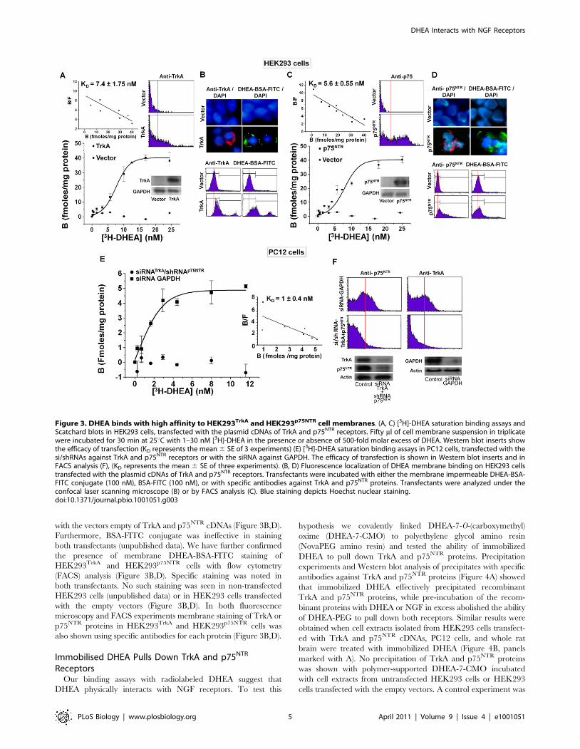
with the vectors empty of TrkA and p75NTR cDNAs (Figure 3B,D).
Furthermore, BSA-FITC conjugate was ineffective in staining
both transfectants (unpublished data). We have further confirmed
the presence of membrane DHEA-BSA-FITC staining of
HEK293TrkA and HEK293p75NTR cells with flow cytometry
(FACS) analysis (Figure 3B,D). Specific staining was noted in
both transfectants. No such staining was seen in non-transfected
HEK293 cells (unpublished data) or in HEK293 cells transfected
with the empty vectors (Figure 3B,D). In both fluorescence
microscopy and FACS experiments membrane staining of TrkA or
p75NTR proteins in HEK293TrkA and HEK293p75NTR cells was
also shown using specific antibodies for each protein (Figure 3B,D).
Immobilised DHEA Pulls Down TrkA and p75NTR
ReceptorsOur binding assays with radiolabeled DHEA suggest that
DHEA physically interacts with NGF receptors. To test this
hypothesis we covalently linked DHEA-7-O-(carboxymethyl)
oxime (DHEA-7-CMO) to polyethylene glycol amino resin
(NovaPEG amino resin) and tested the ability of immobilized
DHEA to pull down TrkA and p75NTR proteins. Precipitation
experiments and Western blot analysis of precipitates with specific
antibodies against TrkA and p75NTR proteins (Figure 4A) showed
that immobilized DHEA effectively precipitated recombinant
TrkA and p75NTR proteins, while pre-incubation of the recom-
binant proteins with DHEA or NGF in excess abolished the ability
of DHEA-PEG to pull down both receptors. Similar results were
obtained when cell extracts isolated from HEK293 cells transfect-
ed with TrkA and p75NTR cDNAs, PC12 cells, and whole rat
brain were treated with immobilized DHEA (Figure 4B, panels
marked with A). No precipitation of TrkA and p75NTR proteins
was shown with polymer-supported DHEA-7-CMO incubated
with cell extracts from untransfected HEK293 cells or HEK293
cells transfected with the empty vectors. A control experiment was
Figure 3. DHEA binds with high affinity to HEK293TrkA and HEK293p75NTR cell membranes. (A, C) [3H]-DHEA saturation binding assays andScatchard blots in HEK293 cells, transfected with the plasmid cDNAs of TrkA and p75NTR receptors. Fifty ml of cell membrane suspension in triplicatewere incubated for 30 min at 25uC with 1–30 nM [3H]-DHEA in the presence or absence of 500-fold molar excess of DHEA. Western blot inserts showthe efficacy of transfection (KD represents the mean 6 SE of 3 experiments) (E) [3H]-DHEA saturation binding assays in PC12 cells, transfected with thesi/shRNAs against TrkA and p75NTR receptors or with the siRNA against GAPDH. The efficacy of transfection is shown in Western blot inserts and inFACS analysis (F), (KD represents the mean 6 SE of three experiments). (B, D) Fluorescence localization of DHEA membrane binding on HEK293 cellstransfected with the plasmid cDNAs of TrkA and p75NTR receptors. Transfectants were incubated with either the membrane impermeable DHEA-BSA-FITC conjugate (100 nM), BSA-FITC (100 nM), or with specific antibodies against TrkA and p75NTR proteins. Transfectants were analyzed under theconfocal laser scanning microscope (B) or by FACS analysis (C). Blue staining depicts Hoechst nuclear staining.doi:10.1371/journal.pbio.1001051.g003
DHEA Interacts with NGF Receptors
PLoS Biology | www.plosbiology.org 5 April 2011 | Volume 9 | Issue 4 | e1001051
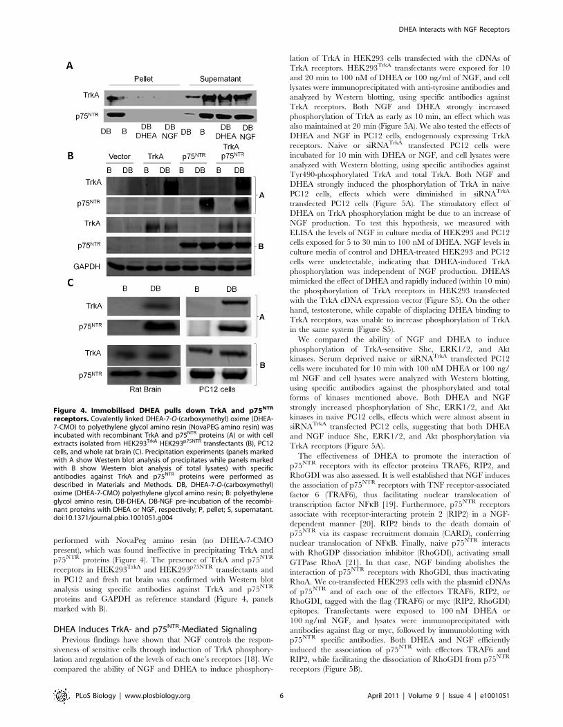
performed with NovaPeg amino resin (no DHEA-7-CMO
present), which was found ineffective in precipitating TrkA and
p75NTR proteins (Figure 4). The presence of TrkA and p75NTR
receptors in HEK293TrkA and HEK293p75NTR transfectants and
in PC12 and fresh rat brain was confirmed with Western blot
analysis using specific antibodies against TrkA and p75NTR
proteins and GAPDH as reference standard (Figure 4, panels
marked with B).
DHEA Induces TrkA- and p75NTR-Mediated SignalingPrevious findings have shown that NGF controls the respon-
siveness of sensitive cells through induction of TrkA phosphory-
lation and regulation of the levels of each one’s receptors [18]. We
compared the ability of NGF and DHEA to induce phosphory-
lation of TrkA in HEK293 cells transfected with the cDNAs of
TrkA receptors. HEK293TrkA transfectants were exposed for 10
and 20 min to 100 nM of DHEA or 100 ng/ml of NGF, and cell
lysates were immunoprecipitated with anti-tyrosine antibodies and
analyzed by Western blotting, using specific antibodies against
TrkA receptors. Both NGF and DHEA strongly increased
phosphorylation of TrkA as early as 10 min, an effect which was
also maintained at 20 min (Figure 5A). We also tested the effects of
DHEA and NGF in PC12 cells, endogenously expressing TrkA
receptors. Naive or siRNATrkA transfected PC12 cells were
incubated for 10 min with DHEA or NGF, and cell lysates were
analyzed with Western blotting, using specific antibodies against
Tyr490-phosphorylated TrkA and total TrkA. Both NGF and
DHEA strongly induced the phosphorylation of TrkA in naive
PC12 cells, effects which were diminished in siRNATrkA
transfected PC12 cells (Figure 5A). The stimulatory effect of
DHEA on TrkA phosphorylation might be due to an increase of
NGF production. To test this hypothesis, we measured with
ELISA the levels of NGF in culture media of HEK293 and PC12
cells exposed for 5 to 30 min to 100 nM of DHEA. NGF levels in
culture media of control and DHEA-treated HEK293 and PC12
cells were undetectable, indicating that DHEA-induced TrkA
phosphorylation was independent of NGF production. DHEAS
mimicked the effect of DHEA and rapidly induced (within 10 min)
the phosphorylation of TrkA receptors in HEK293 transfected
with the TrkA cDNA expression vector (Figure S5). On the other
hand, testosterone, while capable of displacing DHEA binding to
TrkA receptors, was unable to increase phosphorylation of TrkA
in the same system (Figure S5).
We compared the ability of NGF and DHEA to induce
phosphorylation of TrkA-sensitive Shc, ERK1/2, and Akt
kinases. Serum deprived naive or siRNATrkA transfected PC12
cells were incubated for 10 min with 100 nM DHEA or 100 ng/
ml NGF and cell lysates were analyzed with Western blotting,
using specific antibodies against the phosphorylated and total
forms of kinases mentioned above. Both DHEA and NGF
strongly increased phosphorylation of Shc, ERK1/2, and Akt
kinases in naive PC12 cells, effects which were almost absent in
siRNATrkA transfected PC12 cells, suggesting that both DHEA
and NGF induce Shc, ERK1/2, and Akt phosphorylation via
TrkA receptors (Figure 5A).
The effectiveness of DHEA to promote the interaction of
p75NTR receptors with its effector proteins TRAF6, RIP2, and
RhoGDI was also assessed. It is well established that NGF induces
the association of p75NTR receptors with TNF receptor-associated
factor 6 (TRAF6), thus facilitating nuclear translocation of
transcription factor NFkB [19]. Furthermore, p75NTR receptors
associate with receptor-interacting protein 2 (RIP2) in a NGF-
dependent manner [20]. RIP2 binds to the death domain of
p75NTR via its caspase recruitment domain (CARD), conferring
nuclear translocation of NFkB. Finally, naive p75NTR interacts
with RhoGDP dissociation inhibitor (RhoGDI), activating small
GTPase RhoA [21]. In that case, NGF binding abolishes the
interaction of p75NTR receptors with RhoGDI, thus inactivating
RhoA. We co-transfected HEK293 cells with the plasmid cDNAs
of p75NTR and of each one of the effectors TRAF6, RIP2, or
RhoGDI, tagged with the flag (TRAF6) or myc (RIP2, RhoGDI)
epitopes. Transfectants were exposed to 100 nM DHEA or
100 ng/ml NGF, and lysates were immunoprecipitated with
antibodies against flag or myc, followed by immunoblotting with
p75NTR specific antibodies. Both DHEA and NGF efficiently
induced the association of p75NTR with effectors TRAF6 and
RIP2, while facilitating the dissociation of RhoGDI from p75NTR
receptors (Figure 5B).
Figure 4. Immobilised DHEA pulls down TrkA and p75NTR
receptors. Covalently linked DHEA-7-O-(carboxymethyl) oxime (DHEA-7-CMO) to polyethylene glycol amino resin (NovaPEG amino resin) wasincubated with recombinant TrkA and p75NTR proteins (A) or with cellextracts isolated from HEK293TrkA HEK293p75NTR transfectants (B), PC12cells, and whole rat brain (C). Precipitation experiments (panels markedwith A show Western blot analysis of precipitates while panels markedwith B show Western blot analysis of total lysates) with specificantibodies against TrkA and p75NTR proteins were performed asdescribed in Materials and Methods. DB, DHEA-7-O-(carboxymethyl)oxime (DHEA-7-CMO) polyethylene glycol amino resin; B: polyethyleneglycol amino resin, DB-DHEA, DB-NGF pre-incubation of the recombi-nant proteins with DHEA or NGF, respectively; P, pellet; S, supernatant.doi:10.1371/journal.pbio.1001051.g004
DHEA Interacts with NGF Receptors
PLoS Biology | www.plosbiology.org 6 April 2011 | Volume 9 | Issue 4 | e1001051

DHEA Reverses the Apoptotic Loss of TrkA PositiveSensory Neurons in Dorsal Root Ganglia of NGF NullMouse Embryos
NGF null mice have fewer sensory neurons in dorsal root
ganglia (DRG) due to their apoptotic loss [13]. Heterozygous mice
for the NGF deletion were interbred to obtain mice homozygous
for the NGF gene disruption. The mothers were treated daily with
an intraperitoneal injection of DHEA (2 mg) or vehicle (4.5%
ethanol in 0.9% saline). Embryos were collected at E14 day of
pregnancy and sections were stained for Caspase 3 and Fluoro
jade C, markers of apoptotic and degenerative neurons,
respectively. ngf2/2 embryos at E14 showed a dramatic increase
in the number of Fluoro Jade C and Caspase 3 positive neurons in
the DRG compared to the ngf+/2 embryos (Figure 6A,B). DHEA
treatment significantly reduced Fluoro Jade C and Caspase 3
positive neurons in the DRG to levels of ngf+/2 embryos.
Furthermore, TrkA and TUNEL double staining of DRGs has
shown that in ngf+/2 embryos, numbers of TUNEL-positive
apoptotic neurons were minimal, while TrkA positive staining was
present in a large number of neuronal cell bodies of the DRG and
their collaterals were extended within the marginal zone to the
most dorsomedial region of the spinal cord. On the contrary, in
DRG of ngf2/2 embryos levels of TUNEL-positive apoptotic
neurons were dramatically increased, while TrkA neuronal
staining was considerably decreased and DRG collaterals of the
dorsal funiculus were restricted in the dorsal root entry zone
(Figure 6C). DHEA treatment resulted in a significant increase of
TrkA positive staining and the extension of TrkA staining within
the marginal zone to the most dorsomedial region of the spinal
cord similarly to the ngf+/2 embryos (Figure 6D), while staining of
TUNEL-positive apoptotic neurons was decreased to levels shown
in ngf+/2 embryos.
Discussion
DHEA exerts multiple actions in the central and peripheral
nervous system; however, no specific receptor has been reported to
date for this neurosteroid. Most of its actions in the nervous tissue
were shown to be mediated via modulation, at micromolar
concentrations, of membrane neurotransmitter receptors, such as
NMDA, GABAA, and sigma1 receptors. DHEA may also
influence brain function by direct binding, also at micromolar
concentrations, to dendritic brain microtubule-associated protein
MAP2C [22]. In the present study we provide evidence that
DHEA binds to NGF receptors. This is the first report showing a
direct binding of a steroid to neurotrophin receptors. Saturation
experiments and Scatchard analysis of [3H]-DHEA binding to
membranes isolated from HEK293 cells transfected with the
cDNAs of TrkA and p75NTR receptors showed that DHEA binds
to both membranes (7.461.75 nM and 5.660.55 nM for TrkA or
p75NTR, respectively). Non-radioactive NGF effectively displaced
[3H]-DHEA binding to both membrane preparations, with IC50:
0.860.2 and 1.1960.45 nM, respectively. Furthermore, pull-
down experiments using DHEA covalently immobilized on
NovaPEG amino resin suggest that DHEA binds directly to TrkA
and p75NTR proteins. Indeed, polymer-supported DHEA-7-CMO
effectively pulled down recombinant TrkA and p75NTR proteins
Figure 5. DHEA induces TrkA- and p75NTR-mediated signaling. (A) upper panel: HEK293TrkA transfectants were exposed for 10 and 20 min to100 nM of DHEA or 100 ng/ml of NGF, and cell lysates were immunoprecipitated with anti-tyrosine antibodies and analyzed by Western blotting,using specific antibodies against TrkA receptors. lower panel: Serum deprived naive or siRNATrkA transfected PC12 cells were incubated for 10 minwith 100 nM of DHEA or 100 ng/ml of NGF and cell lysates were analyzed with Western blotting, using specific antibodies against thephosphorylated and total forms of TrkA receptor and of Shc, ERK1/2, and Akt kinases. (B) HEK293 cells were co-transfected with the plasmid cDNAs ofp75NTR and of each one of the effectors TRAF6, RIP2, or RhoGDI, tagged with the flag (TRAF6) or myc (RIP2, RhoGDI) epitopes. Transfectants wereexposed for 30 min to 100 nM of DHEA or 100 ng/ml of NGF, and lysates were immunoprecipitated with antibodies against flag or myc, followed byimmunoblotting with p75NTR specific antibodies.doi:10.1371/journal.pbio.1001051.g005
DHEA Interacts with NGF Receptors
PLoS Biology | www.plosbiology.org 7 April 2011 | Volume 9 | Issue 4 | e1001051

and precipitated both proteins from extracts prepared from cells
expressing both receptors (HEK293TrkA, HEK293p75NTR, and
PC12 cells and freshly isolated rat brain). Interestingly, DHEA was
unable to effectively displace binding of [125I]-NGF on mem-
branes isolated from HEK293TrkA and HEK293p75NTR transfec-
tants. It is possible that dissociation of binding of peptidic NGF
from its receptors lasts longer due to the multiple sites of
interaction within the binding cleft of this large peptidic molecule
compared to smaller in volume steroid. Another explanation might
be that NGF and DHEA bind to different domains of NGF
receptors, the NGF domain being non-recognizable by DHEA. It
is of note that antidepressant amitryptiline cannot chase NGF
from TrkA receptors because it binds to a different domain on
TrkA protein compared to NGF. Indeed, other small molecules,
like antidepressant amitriptyline and gamboge’s natural extract
gambogic amide, bind in the extracellular and the cytoplasmic
juxtamembrane domains of TrkA receptor, although with much
lower affinity compared to DHEA (Kd 3 mM and 75 nM,
respectively) [23,24]. The domains of TrkA and p75NTR proteins
involved in DHEA binding were not defined in the present study.
Mutagenesis assays combined with NMR spectroscopy are
planned to map the domains of both receptors related to DHEA
binding.
Our findings suggest that binding of DHEA to NGF receptors is
functional, mediating its anti-apoptotic effects. Indeed, blocking of
TrkA expression by RNAi almost completely reversed the ability
of DHEA to protect PC12 cells from serum deprivation-induced
apoptosis and to maintain elevated levels of the anti-apoptotic Bcl-
2 protein. Additionally, in dispersed primary sympathetic neurons
in culture, DHEA effectively compensated NGF deprivation by
decreasing the levels of apoptotic neurons, an effect which was
reversed by a specific TrkA inhibitor, further supporting the
involvement of TrkA receptors in the anti-apoptotic action of
DHEA. Finally, DHEA effectively rescued from apoptosis TrkA-
Figure 6. DHEA decreases the apoptotic loss of TrkA positive sensory neurons in dorsal root ganglia of NGF null mouse embryos.Heterozygous mice for the NGF deletion were interbred to obtain mice homozygous for the NGF gene disruption. The mothers were treated dailywith an intraperitoneal injection of DHEA (2 mg) or vehicle (4.5% ethanol in 0.9% saline). Embryos were collected at E14 day of pregnancy asdescribed in Material and Methods, and sections were stained for various apoptotic and neuronal markers: (A) Caspase 3, Fluoro Jade C,and TUNELpositive neurons were counted. The results shown are the mean 6 SE from three embryos in each group. In each embryo apoptotic neurons werecounted in at least eight sections from different DRGs (* p,0.01 versus NGF null mice). (B) Staining for Caspase 3 and Fluoro jade C, markers ofapoptotic and degenerative neurons, respectively; (C) double staining for TrkA positive and TUNEL apoptotic neurons (note the co-staining ofapoptotic neurons for both TrkA and TUNNEL in NGF null embryos); and (D) TrkA positive collaterals of DRG sensory neurons. Scale bars at B, C, and D:200 mm.doi:10.1371/journal.pbio.1001051.g006
DHEA Interacts with NGF Receptors
PLoS Biology | www.plosbiology.org 8 April 2011 | Volume 9 | Issue 4 | e1001051

positive dorsal root ganglia sensory neurons of NGF null mouse
embryos.
It appears that the decision between survival and death among
DHEA-responsive cells is determined by the ratio of TrkA and
p75NTR receptors. In fact, DHEA and NGF induced apoptosis of
nnr5 cells, a clone of PC12 cells expressing only pro-death p75NTR
receptors. The pro-death effects of both agents were completely
blocked by p75NTR shRNA and were remarkably restored after
transfection of nnr5 cells with the TrkA cDNA. It is of note that
during brain development the ratio of TrkA to p75NTR varies
tempospatially [25]. Thus, the ability of DHEA to act in a positive
or negative manner on neuronal cell survival may depend upon
the levels of the two receptors during different stages of neuronal
development.
Binding of DHEA on both TrkA and p75NTR receptors was
effectively competed by sulfated DHEA, DHEAS (IC50: 6.161.1
and 8.161.2 nM, respectively), suggesting that DHEAS may also
bind to NGF receptors. Testosterone displaced DHEA binding to
TrkA and p75NTR (IC50: 5.362.1 and 7.463.2 nM, respectively),
while synthetic glucocorticoid dexamethasone displaced DHEA
binding only to pro-survival TrkA receptors (IC50: 9.564.6 nM).
In a previous study we had shown that both steroids effectively
displaced DHEA from its specific membrane binding sites of
sympathoadrenal cells, acting as DHEA antagonists by blocking its
anti-apoptotic effect and the induction of anti-apoptotic Bcl-2
proteins [9]. Our findings suggest that testosterone and glucocor-
ticoids may act as neurotoxic factors by antagonizing endogenous
DHEA and NGF for their binding to NGF receptors, explaining
previously published data. Indeed, testosterone was shown to
increase NMDA and GABAA-mediated neurotoxicity [26,27].
Our findings suggest that testosterone may act as a neurotoxic
factor by also antagonizing the neuroprotective effects of
endogenous DHEA. Furthermore, glucocorticoids show a bimodal
effect on hippocampal neurons causing acutely an increase in
performance of spatial memory tasks, while chronic exposure has
been associated with decreased cognitive performance and
neuronal atrophy [28]. Acute administration of glucocorticoids
results in a glucocorticoid receptor-mediated phosphorylation and
activation of hippocampal TrkB receptors, exerting trophic effects
on dentate gyrus hippocampal neurons [29], via an increase in the
sensitivity of hippocampal cells to neurotrophin BDNF, the
endogenous TrkB ligand known to promote memory and learning
[30]. However, overexposure to glucocorticoids during prolonged
periods of stress is detrimental to central nervous system neurons,
especially in aged animals, affecting mainly the hippocampus. It is
possible that part of neurotoxic effects of glucocorticoids may be
due to their antagonistic effect on the neuroprotective effect of
endogenous DHEA and NGF, via TrkA receptor antagonism. The
decline of brain DHEA and NGF levels during aging and in
Alzheimer’s disease [28] might exacerbate this phenomenon,
rendering neurons more vulnerable to glucocorticoid toxicity.
Indeed, glucocorticoid neurotoxicity becomes more pronounced in
aged subjects since cortisol levels in the cerebrospinal fluid increase
in the course of normal aging, as well as in relatively early stages of
Alzheimer’s disease [28].
A number of neurodegenerative conditions are associated with
lower production or action of both DHEA and NGF [31,32].
Animal studies suggest that NGF may reverse, or slow down the
progression of Alzheimer’s related cholinergic basal forebrain
atrophy [32]. Furthermore, the neurotrophic effects of NGF in
experimental animal models of neurodegenerative conditions, like
MPTP (Parkinson’s disease), experimental allergic encephalomy-
elitis (multiple sclerosis), or ischemic retina degeneration mice [33–
35] support its potential as a promising neuroprotective agent.
However, the use of NGF in the treatment of these conditions is
limited, because of its poor brain blood barrier permeability. It is
of interest that DHEA also exerts neuroprotective properties in
some of these animal models [7,36]. These findings suggest that
synthetic DHEA analogs, deprived of endocrine effects, may
represent a new class of brain blood barrier permeable NGF
receptor agonists with neuroprotective properties. We have
recently reported the synthesis of 17-spiro-analogs of DHEA, with
strong anti-apoptotic and neuroprotective properties, deprived of
endocrine effects [37], which are now being tested for their ability
to bind and activate NGF receptors.
We have previously defined the pro-survival signaling pathways
that are initiated by DHEA at the membrane level [3]. These
pathways include MEK1/2/ERK1/2 and PI3K/Akt pro-survival
kinases. We now provide experimental evidence that DHEA
activates these kinases via TrkA receptors. Down-regulation of
TrkA receptors using siRNAs resulted in an almost complete
reversal of the ability of DHEA to increase the phosphorylation of
kinases Shc, Akt, and ERK1/2. In addition to TrkA receptors,
binding of DHEA to the low affinity NGF receptor was also
functional, affording the activation of p75NTR receptors. Unlike
TrkA receptors, p75NTR lacks any enzymatic activity. Signal
transduction by p75NTR proceeds via ligand-dependent recruit-
ment and release of cytoplasmic effectors to and from the receptor.
Indeed, DHEA like NGF facilitated the recruitment of two major
cytoplasmic interactors of p75NTR, TRAF6 and RIP2 proteins.
Additionally, DHEA-mediated activation of p75NTR led to the
dissociation of bound RhoGDI, a protein belonging to small
GTPases and interacting with RhoA [21]. A schematic represen-
tation of our findings is shown in Figure 7.
Previous findings suggest that DHEA protects PC12 cells
against apoptosis via pertussis toxin (PTX) sensitive, G protein-
associated specific plasma membrane-binding sites [9]. Indeed,
PTX was shown to partially reverse the anti-apoptotic effects of
DHEA and its membrane impermeable DHEA-BSA conjugate, as
well as their effects on prosurvival kinases PI3K/Akt, the
activation of transcription factor NFkappaB, and the phosphor-
ylation and inactivation of apoptotic protein Bad [10]. Interest-
ingly, the prosurvival effects of NGF in sympathetic neurons and
PC12 cells are also partially reversed by PTX [38]. Furthermore,
the NGF-dependent activation of Akt is partially attenuated by
PTX, indicating the participation of G(i/o) proteins. In the same
study, NGF-induced phosphorylation of Bad and transcriptional
activity of NFkappaB were also shown to be sensitive to PTX [38].
It appears that other NGF-driven pathways are sensitive to PTX
too. For instance, in PC12 cells and primary cortical neurons the
NGF-induced phosphorylation of tuberin (a critical translation
regulator holding a central role in NGF-promoted neuronal
survival) is partially blocked by PTX, suggesting the participation
of G(i/o) proteins [39]. Finally, NGF-dependent activation of the
p42/p44 mitogen-activated protein kinase (p42/p44 MAPK)
pathway in PC12 cells was effectively blocked by PTX [40].
However in HEK293 cells transfected with TrkA receptors, PTX
was unable to affect the induction of TrkA phosphorylation by
NGF or DHEA (Figure S5). These findings considered together
suggest that TrkA receptors may use down-stream G protein-
coupled receptor pathways, after binding and activation by NGF
or DHEA, to control neuronal cell survival.
It is worth noticing that the interaction of DHEA with the NGF
system was first suggested 15 years ago by Compagnone et al.,
showing co-localized staining of CYP17, the rate limiting enzyme
of DHEA biosynthesis, and NGF receptors in mouse embryonic
DRGs [41]. About one-fifth of CYP17-immunopositive DRG
neurons in the mouse were found to be also TrkA-immunoposi-
DHEA Interacts with NGF Receptors
PLoS Biology | www.plosbiology.org 9 April 2011 | Volume 9 | Issue 4 | e1001051

tive. Among the TrkA-expressing cells, about one-third also
express CYP17, while p75NTR-expressing neurons represent only
13% of the cells in the DRG. Thus, about one-fifth of CYP17-
immunopositive neurons may be able to respond to both DHEA
and NGF stimulation, an observation compatible with our data,
presented in Figure 6C. A recent report further supports the
interaction of DHEA with NGF receptors. Indeed, DHEA was
shown to act as a keratinocyte-deriving neurotrophic signal,
mimicking NGF in promoting axonal outgrowth of NGF non-
producing but TrkA positive sensory neurons, an effect blocked by
TrkA inhibitor K252a [42].
CYP17 is expressed in invertebrate cephalochordata Amphiox-
us [43]. Amphioxus is also expressing TrkA receptor homologous
AmphiTrk, which effectively transduces signals mediated by NGF
[44]. Phylogenetic analysis of neurotrophins revealed that they
emerged with the appearance of vertebrates (530–550 million
years ago), when complexity of neural tissue increased [45].
Invertebrate cephalochordata like Amphioxus are positioned on
the phylogenetic boundary with vertebrates (600 million years
ago). It is thus tempting to hypothesize that DHEA contributed as
one of the ‘‘prehistoric’’ neurotrophic factors in an ancestral,
simpler structurally invertebrate nervous system [46]; then, when a
strict tempospatial regulation of evolving nervous system of
vertebrates was needed, peptidic neurotrophins emerged to afford
rigorous and cell specific neurodevelopmental processes.
In conclusion, our findings suggest that DHEA and NGF cross-
talk via their binding to NGF receptors to afford brain shaping and
maintenance during development. During aging, the decline of
both factors may leave the brain unprotected against neurotoxic
challenges. This may also be the case in neurodegenerative
conditions associated with lower production or action of both
factors. DHEA analogs may represent lead molecules for designing
non-endocrine, neuroprotective, and neurogenic micromolecular
NGF receptor agonists.
Materials and Methods
si/shRNAs, Plasmids, and AntibodiesPC12 cells were transfected with specific si/shRNAs for
blocking the expression of TrkA and/or p75NTR receptors. More
specifically, three siRNAs and two shRNAs for TrkA and p75NTR,
respectively, were obtained. The sequences for TrkA siRNAs
(Ambion) were: GCCUAACCAUCGUGAAGAG (siRNA ID
191894), GCAUCCAUCAUAAUAGCAA (siRNA ID 191895),
and CCUGACGGAGCUCUAUGUG (siRNA ID 191893).
Sequences for p75NTR (Qiagen) were: GACCUAUCUGAGCU-
GAAA (Cat. No. SI00251090) and GCGUGACUUUCAGG-
GAAA (CatNo SI00251083).
Rat TrkA was expressed from the pHA vector backbone and rat
p75NTR was expressed from the pCDNA3 vector backbone
(InVitrogen) using a full length coding sequence flanked by an
N-terminal hemagglutinin (HA) epitope tag. Plasmids to express
RIP2 [19] and RhoGDI [36] were myc-tagged, while TRAF6 [19]
was FLAG-tagged, as previously described.
The origin of antibodies was as follows: Bcl-2 (Cat. No. C-2, sc-
7382, Santa Cruz Biotechnology Inc.), phospho TrkA (Cat.
No. 9141, Cell Signaling), TrkA (Cat. No. 2505, Cell Signaling,
was used for Western Blotting and Cat. No. 06-574, Upstate, was
used for immunostainings), p75NTR (Cat. No. MAB365R, Milli-
pore), c-myc (Cat. No. 9E10, sc-40, Santa Cruz Biotechnology
Inc.), phospho ERK1/2 (Cat. No. 9106, Cell Signaling), Erk1/2
(Cat. No. 9102, Cell Signaling), phospho-Shc (Tyr239/240)
Antibody (Cat. No. 2434, Cell Signaling), Shc (Cat. No. 2432,
Cell Signaling), phospho-Akt (Ser473) (Cat. No. 9271, Cell
Signaling), Akt (Cat. No. 9272, Cell Signaling), anti-FLAG (M2)
mouse monoclonal (Cat. No. F1804, Sigma), pTyr (Cat. No. sc-
508, Santa Cruz Biotechnology Inc.), active Caspase-3 (Cat.
No. ab13847, Abcam), Tyrosine Hydroxylase (Cat. No. ab6211,
Abcam), anti-rabbit-R-phycoerythrin conjugated (Cat. No. P9537,
Sigma), anti-mouse-fluorescein conjugated (Cat. No. AP124F,
Millipore), anti-rabbit Alexa Fluor 488 (Cat. No. A21206), anti-
rabbit Alexa Fluor 546 (Cat. No. A10040), and GAPDH (Cat.
No. 2118, Cell Signaling).
Cell Cultures and TransfectionPC12 cells were obtained from LGC Promochem (LGC
Standards GmbH, Germany) and nnr5 cells from Dr. C.F. Ibanez
(Karolinska Institute). Both cell types were grown in RPMI 1640
containing 2 mM L-glutamine, 15 mM HEPES, 100 units/ml
penicillin, 0.1 mg/ml streptomycin, and 10% horse serum, 5%
fetal calf serum (both charcoal-stripped for removing endogenous
steroids) at 5% CO2 and 37uC. HEK-293 cells were obtained from
LGC Promochem. Cells were grown in DMEM medium
containing 10% fetal bovine serum (charcoal-stripped for
removing endogenous steroids), 100 units/ml penicillin, and
0.1 mg/ml streptomycin, at 5% CO2 and 3uC. HEK-293 and
PC12 cells were transfected with Lipofectamine 2000 (InVitrogen)
according to manufacturer’s instructions. Transfected cells were
typically used on the 2nd day after transfection.
Figure 7. Hypothetical model of NGF receptor-mediatedsignaling pathways involved in the effects of DHEA onneuronal cell fate. DHEA binds with high affinity to TrkA andp75NTR receptors, initiating the following sequence of events: 1) DHEAinduces TrkA-mediated Tyr490-phosphorylation of Shc, ERK1/2, and Aktkinases, controlling the expression and function of apoptotic Bcl-2proteins; and 2) DHEA promotes the interaction of p75NTR receptorswith effector proteins TRAF6, RIP2, and RhoGDI affecting neuronal cellapoptosis. Our findings suggest that the decision between survival anddeath among DHEA-responsive cells is determined by the balance of itsinteractions with TrkA and p75NTR receptors.doi:10.1371/journal.pbio.1001051.g007
DHEA Interacts with NGF Receptors
PLoS Biology | www.plosbiology.org 10 April 2011 | Volume 9 | Issue 4 | e1001051

Measurement of ApoptosisPC12 cells were cultured in 12-well plates, and 24 h later they
were transfected with the si/shRNAs for TrkA and/or p75NTR.
Twenty-four hours later the medium was aspirated and replaced
either with complete medium (serum supplemented) or serum free
medium in the absence or the presence of DHEA or DHEA-BSA
conjugate at 100 nM. Apoptosis was quantified 24 h later with
annexin V-FITC and PI (BD Pharmingen) according to our
protocol [8].
[3H]-DHEA Binding AssaysMembrane preparation. HEK293 cells transfected with an
empty vector (HEK293-Vector) or overexpressing p75NTR
(HEK293-p75) or TrkA (HEK293-TrkA), and PC12 cells wild
type or shRNA knocked down for p75NTR and TrkA, were
cultured, collected by scraping on ice and washed twice with cold
Phosphate Buffer Saline (PBS), pH 7.4. After centrifugation at
1,200 rpm, cells were homogenized in a 50 mM Tris-HCl buffer,
pH 7.4 (at 4uC), containing freshly added protease inhibitors
(1 mM PMSF and 1 mg/ml aprotinin). Crude membrane fractions
were isolated by differential centrifugation at 2,5006g (10 min at
4uC, to remove unbroken cells and nuclei) and 102,0006g (1 h, at
4uC). Membranes were washed once with ice-cold 50 mM Tris–
HCl buffer, pH 7.4, and re-suspended in the same buffer.
Membranes were then briefly acidified with 50 mM glycine
pH 3 for 3 min on ice to elute membrane adsorbed proteins,
washed once, resuspended in PBS (pH 7.4) with protease
inhibitors, at a concentration of 2 mg/ml, and used immediately
for binding experiments.
Binding conditions. Fifty ml of membrane suspension
(2 mg/ml) in triplicate were incubated with 10 ml of 1–30 nM
[3H]-DHEA (Perkin Elmer, Boston, MA) in the presence or
absence of 500-fold molar excess of DHEA, in PBS, pH 7.4 with
protease inhibitors, in a final volume of 100 ml. Membranes were
incubated for 30 min at 25uC or overnight at 4uC, on a rotating
plate; then they were collected on GF/B filters, prewetted in 0.5%
PEI solution at 4uC. Filters were washed three times with ice-cold
PBS, dried, and counted in scintillation fluid (SigmaFluor, Sigma)
in a scintillation counter (Perkin Elmer, Foster City, CA) with 60%
efficiency for Tritium. For saturation curves, specific binding
(Bound, B) was calculated as the difference of Total Binding 2
Non Specific Binding. KDs were calculated from B/F over B
Scatchard plots. Non-specific binding was ranging from 30% to
50% of total binding shown in the micromolar concentrations of
ligand. Relatively higher levels of non-specific binding have been
typically reported in binding assays for specific membrane binding
of various steroid hormones, due to the highly lipophilic nature of
phospholipid containing cell membranes combined with the strong
lipophilicity of steroids. For displacement experiments, a constant
concentration of [3H]-DHEA (1 nM) was incubated with
increasing concentrations of competitors (10212–1026 M), under
the same conditions as for saturation binding.
Fluorescence MicroscopyHEK293 cells were allowed to grow on gelatin-coated glass
coverslips for 24 h in culture medium, and 24 h later they were
transfected with the cDNAs for TrkA, and p75NTR receptors or
the vector alone. Staining was performed 48 h after transfection.
Culture medium was aspirated and transfectants were washed
twice with PBS buffer. Primary antibodies against TrkA (rabbit,
Upstate, No. 06-574, diluted 1:100) or p75NTR (mouse monoclo-
nal ab, MAB365R, Millipore, dilution 1:500) were added for
30 min at 37uC. Secondary antibodies, anti-rabbit-R-phycoery-
thrin conjugated (Sigma, No. P9537), and anti-mouse-fluorescein
conjugated (No. AP124F, Millipore) were added at 1:100 dilution
and transfectants were incubated for 30 min at 37uC; then they
were washed three times with PBS and counterstained with
Hoechst nuclear stain (Molecular Probes) for 5 min. Transfectants
were also incubated with the DHEA-BSA-FITC or the BSA-FITC
conjugates (1026M) for 15 min at room temperature in the dark;
then they were washed with serum free culture medium and
incubated for another 15 min in serum free culture medium
containing 4% BSA. Coverslips were mounted to slides with 90%
glycerin and were observed with a confocal laser scanning
microscope (Leica TCS-NT, Leica Microsystems GmbH, Heidel-
berg, Germany), mounted with a digital camera.
Flow CytometryHEK293 cells were cultured in 12-well plates, and 24 h later
they were transfected with the cDNAs for TrkA and/or p75NTR
receptors, or the vector alone. Staining was performed 48 h later.
Transfectants (56105 cells) were pelleted and incubated with 20 ml
of the primary antibodies against TrkA or p75NTR receptors for
30 min over ice. Afterwards, transfectants were washed three times
with PBS and 20 ml of the secondary antibodies, and anti-rabbit-
R-phycoerythrin conjugated and anti-mouse-fluorescein conjugat-
ed were added, as described above. For DHEA-BSA-FITC
binding on cells, 20 ml (100 nM) were added on the pelleted cells
for 10 min at RT, and then they were washed with serum free
culture medium and incubated for another 15 min in serum free
culture medium containing 4% BSA. Transfectants were washed
twice with PBS, resuspended in 500 ml of PBS, and were analyzed
in a Beckton-Dickinson FACSArray apparatus and the CELL-
Quest software (Beckton-Dickinson, Franklin Lakes, NJ).
Synthesis of Immobilised DHEA-7-CMONovaPEG amino resin (loading value 0.78 mmol/g) was
purchased from Novabiochem. NMR spectra were recorded on
a Varian 300 spectrometer operating at 300 MHz for 1H and
75.43 MHz for 13C or on a Varian 600 operating at 600 MHz for1H. 1H NMR spectra are reported in units of d relative to the
internal standard of signals of the remaining protons of deuterated
chloroform, at 7.24 ppm. 13C NMR shifts are expressed in units of
d relative to CDCl3 at 77.0 ppm. 13C NMR spectra were proton
noise decoupled. IR spectra was recorded at Bruker Tensor 27.
Absorption maxima are reported in wavenumbers (cm21).
3b-Acetoxy-17,17-ethylenedioxyandrost-5-ene (0.74 g, 1.98
mmol) and N-hydroxy phthalimide (0.71 g, 2.2 mmol) were
dissolved in acetone (39 mL) containing 1 mL of pyridine. The
mixture was stirred vigorously at room temperature and sodium
dichromate dihydrate (0.89 g, 3 mmol) was added. Additional
portions of solid sodium dichromate dihydrate (0.89 g, 3 mmol)
were added after 10 and 20 h stirring at room temperature. After
reaction completion (48 h), the mixture was diluted with
dichloromethane, filtered through a bed of celite, and the filtrate
was washed with water, saturated sodium bicarbonate solution,
and brine. The organic layer was dried over anhydrous sodium
sulfate, the solvent evaporated in vacuo, and the residue purified by
flash column chromatography using hexane/acetone/25%
NH4OH (85:15:0.1 mL) as eluent to afford 3b-acetoxy-17,17-
ethylenedioxyandrost-5-ene-7-one (0.6 g, yield: 78%). 1H NMR
(CDCl3, 300 MHz) d: 0.87 (s, 3H), 1.21 (s, 3H), 1.26–2.00 (m,
14H), 2.05 (s, 3H), 2.20–2.51 (m, 3H), 3.84–3.92 (m, 4H), 4.68–
4.76 (m, 1H), 5.70 (d, J = 1.58 Hz, 1H).
To a solution of 3b-acetoxy-17,17-ethylenedioxyandrost-5-en-7-
one (0.1 g, 0.26 mmol) in pyridine (1.9 mL) was added O-
(carboxymethyl)hydroxylamine hemihydrochloride (0.11 g,
0.52 mmol) and the reaction mixture was stirred overnight under
DHEA Interacts with NGF Receptors
PLoS Biology | www.plosbiology.org 11 April 2011 | Volume 9 | Issue 4 | e1001051

argon. After completion of the reaction, the solvent was
evaporated and the residue was diluted with ethyl acetate. The
organic layer was washed with water and brine, dried over
anhydrous sodium sulfate, and the solvent was evaporated in vacuo
to afford 3b-acetoxy-17,17-ethylenedioxyandrost-5-en-7-one7-(O-
carboxymethy1) oxime as a white foam (0.12 g, yield: 100%). 1H
NMR (CDCl3, 300 MHz) d: 0.88 (s, 3H), 1.13 (s, 3H), 1.16–1.95
(m, 12H), 2.04 (s, 3H), 2.25–2.59 (m, 5H), 3.84–3.95 (m, 4H), 4.59
(d, J = 2.29 Hz, 2H), 4.62–4.73 (m, 1H), 6.51 (d, J = 1.47 Hz, 1H).
To a solution of 3b-acetoxy-17,17-ethylenedioxyandrost-5-en-7-
one-7-(O-carboxymethy1) oxime (0.12 g, 0.26 mmol) in a mixture
of acetone/water (5:1, 6.3 mL) was added p-toluenesulfonic acid
monohydrate (0.019 g, 0.10 mmol), and the reaction mixture was
stirred until the starting material was consumed (48 h). The
solvent was evaporated in vacuo and the residue was diluted with
ethyl acetate. The organic layer was washed with water and brine,
dried over anhydrous sodium sulfate, and the solvent was
evaporated in vacuo to afford 3b-acetoxy-androst-5-en-7,17-dione
7-(O-carboxymethy1) oxime as a white foam (0.11 g, yield: 100%).1H NMR (CDCl3, 600 MHz) d: 0.90 (s, 3H), 1.15 (s, 3H), 1.20–
1.95 (m, 12H), 2.05 (s, 3H), 2.09–2.68 (m, 5H), 4.63 (d,
J = 4.18 Hz, 2H), 4.65–4.71 (m, 1H), 6.56 (d, J = 1.39 Hz, 1H).
To a solution of 3b-acetoxy-androst-5-en-7,17-dione 7-(O-
carboxymethy1) oxime (0.11 g, 0.26 mmol) in methanol (3.9 mL)
was added LiOH (1.5 mL, 1.5 mmol, 1N solution), and the reaction
mixture was stirred until the starting material was consumed (4 h).
The solvent was evaporated in vacuo and the residue was diluted with
water. The solution was acidified with 10% hydrochloric acid and
DHEA-7-CMO precipitated as a white solid, which was isolated by
filtration (0.097 g, yield: 100%). 1H NMR (CDCl3/CD3OD,
600 MHz) d: 0.90 (s, 3H), 1.14 (s, 3H), 1.20–2.75 (m, 17H), 3.49–
3.54 (m, 1H), 4.54 (s, 2H), 6.54 (s, 1H).
3b-Hydroxy-17-oxoandrost-5-en-7-O-(carboxymethyl)oxime
(DHEA-7-CMO) (192 mg, 0.511 mmol) in DMF (5 mL) was treated
with HOBt (69 mg, 0.511 mmol) and DIC (0.08 mL, 0.511 mmol),
and the resulting mixture was stirred at room temperature for
30 min. This solution was added to NovaPEG amino resin (130 mg,
0.102 mmol, 0.78 mmol/gr) (pre-swollen with DMF for 1 h) and the
slurry was shaken at room temperature overnight. The mixture was
filtered, the resin was sequentially washed with dichloromethane (36),
methanol (36), and diethyl ether (36), and was dried in vacuo
overnight. Yield 175 mg (100%), loading value 0.61 mmol/gr. 13C
NMR (gel phase, CDCl3) d: 220.66, 170.15, 157.10, 154.15, 113.11,
72.57, 66.59, 49.92, 47.86, 42.15, 38.46, 37.08, 36.53, 35.49, 31.20,
30.71, 24.96, 20.15, 18.05, 13.95; IR: nmax/cm21 2865 (s), 1735 (m),
1669 (w), 1653 (w), 1637 (w), 1456 (m), 1348 (w), 1289 (w), 1247 (w),
1093 (s), 946 (w).
Co-immunoprecipitation and Pull-Down AssaysHEK293 cells were transfected with the appropriate plasmids
(TrkA, p75NTR, RIP2, TRAF-6, and RhoGDI) by using
Lipofectamine 2000 (Invitrogen). Cells were harvested 48 h after
transfection and suspended in lysis buffer (50 mM Tris-HCl,
0.15 M NaCl, 1% Triton-X100, pH 7.4) supplemented with
protease inhibitors. Lysates were precleared for 1 h with Protein
A-Sepharose beads (Amersham) and immunoprecipitated with the
appropriate antibody (pTyr, Flag, or c-myc) overnight at 4uC.
Protein A Sepharose beads were incubated with the lysates for 4 h
at 4uC with gentle shaking. In the case of immobilized DHEA-7-
CMO, HEK293 or PC12 cells lysates or purified receptors (both
from R&D Systems, Recombinant Mouse NGF R/TNFRSF16/
Fc Chimera, Cat. No.: 1157-NR and Recombinant Rat Trk A/Fc
Chimera, Cat. No.: 1056-TK) were incubated overnight at 4uCwith the NovaPEG amino resin alone or conjugated with DHEA.
Beads were collected by centrifugation, washed four times with
lysis buffer, and resuspended in SDS loading buffer. Proteins were
separated by SDS/PAGE, followed by immunoblotting with
specific antibodies.
Western Blot AnalysisPC12 or HEK293 cells lysates were electrophoresed through a
12% SDS-polyacrylamide gel, and then proteins were transferred
to nitrocellulose membranes, which were processed according to
standard Western blotting procedures, as previously described [8].
To detect protein levels, membranes were incubated with the
appropriate antibodies: Bcl-2 (dilution 1:500), phospho TrkA
(dilution 1:500), total TrkA (dilution1:500), p75NTR (dilution
1:500), phospho Shc (dilution 1:1000), total Shc (dilution
1:1000), phospho Akt (dilution 1:500), total Akt (dilution 1:500),
phospho ERK1/2 (dilution 1:500), and total ERK1/2 (dilution
1:500). Proteins were visualized using the ECL Western blotting
kit (ECL Amersham Biosciences, UK), and blots were exposed to
Kodak X-Omat AR films. A PC-based Image Analysis program
was used to quantify the intensity of each band (Image Analysis,
Inc., Ontario, Canada).
To normalize for protein content the blots were stripped and
stained with GAPDH antibody (dilution 1:1000); the concentra-
tion of each target protein was normalized versus GAPDH. Where
phosphorylation of TrkA or kinases was measured, membranes
were first probed for the phosphorylated form of the protein, then
stripped, and probed for the total protein.
Superior Cervical Ganglia Neuronal CulturesSuperior cervical ganglia (SCG) were removed from newborn
(P0–P1) rat pups and dissociated in 0.25% trypsin (Gibco, 15090)
for 30 min at 37uC. After dissociation SCG neurons were re-
suspended in culture medium (Gibco, Neurobasal Cat. No. 21103)
containing 1% fetal bovine serum (FBS), 100 units/ml penicillin,
0.1 mg/ml streptomycin, 3 mg/ml araC antimitotic, and 100 ng/
ml NGF (Millipore, 01-125). Cells were plated on collagen coated
24-well plates and cultured for 5 d prior to use. For NGF
withdrawal experiments, cells were washed twice with Neurobasal
containing 1% FBS and fresh culture medium lacking NGF and
containing anti-NGF antibody at 1:50 dilution (Millipore,
AB1526). DHEA, TrkA-inhibitor (Calbiochem, 648450) and
anti-p75NTR (mouse, MAB365R Millipore) were used at
100 nM, 100 nM, and 1:50, respectively.
In Vivo Experiments with the NGF Null Micengf +/2 mice [13] were obtained from the Jackson Laboratory
and maintained on C57BL/6 background. All procedures
described below were approved by the Animal Care Committee
of the University of Crete, School of Medicine. Animals were
housed in cages maintained under a constant 12 h light–dark cycle
at 21–23uC, with free access to food and tap water. Genotyping
was performed on tail DNA using the following primers:
NGFKOU2 (59CCG TGA TAT TGC TGA AGA GC39),
NGFU6 (59CAG AAC CGT ACA CAG ATA GC39), and
NGFD1 (59TGT GTC TAT CCG GAT GAA CC39). Genomic
PCR reactions containing the 3 primers were incubated for 32
cycles at 95uC (30 s)/59uC (30 s)/72uC (1 min).
Mice heterozygous for the NGF null mutation were interbred to
obtain mice homozygous for the NGF gene disruption and the first
day of gestation determined by the discovery of a copulation plug.
The mothers were treated daily with a subcutaneous injection of
DHEA (2 mg/day) or vehicle (4.5% ethanol in 0.9% saline)
starting from the third day after gestation. Animals were collected at
E14. At the day of collection the mothers were deeply anesthetized
DHEA Interacts with NGF Receptors
PLoS Biology | www.plosbiology.org 12 April 2011 | Volume 9 | Issue 4 | e1001051

with sodium pentobarbital (Dolethal 0.7 ml/kg i.p.) followed by
transcardial perfusion with saline solution containing heparin for
about 7 min, and with 4% PFA, 15% Picric Acid, 0.05% GA in
phosphate buffer 0.1 M, for another 7 min. After the perfusion the
embryos were collected and maintained in the same fixative
overnight at 4uC. Embryos were then washed in 0.1 M phosphate
buffer and cryoprotected by using 10% sucrose followed by 20%
sucrose overnight at 4uC. Finally, embryos were frozen in OCT in
iso-pentane over liquid nitrogen for 5 min and the frozen tissues
were stored for later use at 280uC. The samples were sectioned
(20 mm) and mounted onto Superfrost plus slides (Menzel-Glaser
J1800AMNZ). Slides were left to air-dry overnight at room
temperature (RT) and were then either used immediately or were
fixed in cold acetone for 1 min and stored at 280uC for later use.
Stored or fresh slides were fixed for 15 min in cold acetone at 4uCand left to dry for 10 min at room temperature. They were then
washed in PB 0.1 M, then in TBS, and incubated for 45 min with
10% horse serum in TBS-T 0.1%. The normal serum was drained
off and the primary antibodies (anti-TrkA diluted 1:400 and active
Caspase-3 diluted 1:50), diluted in TBS-T 0.1% with 1% horse
serum, were added. Sections were incubated for 4 h at RT and
overnight at 4uC; they were then washed in TBS-T 0.1% and the
anti-rabbit secondary antibodies (Alexa Fluor 488 and Alexa Fluor
546, 1:1000 in TBS-T 0.1%) were added for 6 h at RT. Sections
were washed in TBS-T, TBS, and in PB 0.1 M and were
coverslipped with Vectashield (Vector, H-1400) and visualized in
a confocal microscope. TUNEL (Roche, Cat. No. 12156792910)
and Fluoro-Jade C (Millipore, Cat. No. AG325) staining of
apoptotic and degenerating neurons, respectively, was performed
according to the manufacturer’s instructions.
Statistical AnalysisFor the statistical evaluation of our data we have used analysis of
variance, post hoc comparison of means, followed by the Fisher’s
least significance difference test. For data expressed as percent
changes we have used the nonparametric Kruskal-Wallis test for
several independent samples.
Supporting Information
Figure S1 DHEA regulates the levels of TrkA and p75NTR
receptors, mimicking NGF. Serum deprived PC12 cells were
exposed to 100 nM of DHEA or 100 ng/ml of NGF for 12, 14, and
48 h. TrkA and p75NTR protein levels were measured in cell lysates
with immunoblotting, using specific antibodies, and were normal-
ized against GAPDH (* p,0.01 versus control-Serum Free, n = 5).
(TIF)
Figure S2 [3H]-DHEA saturation binding assays and Scatchard
blots in HEK293 cells, transfected with the plasmid cDNAs of TrkA
and p75NTR receptors. Fifty ml of cell membrane suspension in
triplicate were incubated overnight at 4uC with 1–30 nM [3H]-
DHEA in the presence or absence of 500-fold molar excess of DHEA.
(TIF)
Figure S3 RT-PCR of androgen receptors (AR) mRNA in
HEK23 cells, transfected with the plasmid cDNAs of TrkA and
p75NTR receptors. Total RNA was extracted from LNCaP and
naıve HEK293 cells or HEK293 cells transfected with the plasmid
cDNAS of TrkA, p75NTR, or both plasmids, by using the Trizol
Reagent (InVitrogen). One microgram of total RNA was reverse
transcribed by using the Thermo-Script RT-PCR System
(InVitrogen). The cDNA was amplified by PCR. PCR was
performed in a Perkin–Elmer DNA Thermal Cycler with the
following conditions: 60 s at 94uC, 60 s at 60uC, and 3 min at
72uC (for Androgen Receptor, AR) or 30 s at 94uC, 30 s at 58uC,
and 60 s at 72uC (for GAPDH), for 30 cycles to detect the product
at the exponential phase of the amplification. Ten microliters of
the amplified products (1.4 Kb for AR and 483 bp for GAPDH)
were separated on a 1.5% agarose gel and visualized by ethidium
bromide staining. Primers for AR were 59-AGCTACTCCG-
GACCTTACG-39 (sense) and 59-AGGTGCCATGGGAGGGT-
TAG-39 (antisense) and primers for GAPDH were 59-GCCA-
CATCGCTCAGACACCA-39 (sense) and 59-GATGACCCTTT-
TGGCTCCCC-39 (antisense).
(TIF)
Figure S4 Representative curves of [3H]-DHEA displacement
experiments. The selectivity of DHEA binding to HEK293TrkA
and HEK293p75NTR cell membranes was examined by performing
heterologous [3H]-DHEA displacement experiments using a
number of non-labeled steroids or NGF. Membranes (at a final
concentration of 2 mg protein/ml) isolated from HEK293 cells
transfected with the plasmid cDNAs of TrkA and p75NTR
receptors were incubated with 1 nM [3H]-DHEA in the absence
or the presence of various unlabeled steroids or NGF at
concentrations varying from 0.01 to 1,000 nM. Binding of [3H]-
DHEA to membranes isolated from both HEK293TrkA and
HEK293p75NTR cells was effectively displaced by NGF (IC50:
0.860.2 and 1.1960.45 nM, respectively, n = 4). Estradiol failed
to displace [3H]-DHEA from its binding on membranes from
HEK293TrkA and HEK293p75NTR cells at concentrations ranging
from 0.1 to 1,000 nM. In contrast, displacement of [3H]-DHEA
binding to membranes from both HEK293TrkA and
HEK293p75NTR cells was shown by sulfated ester of DHEA,
DHEAS (IC50: 6.161.1 and 8.161.2 nM, respectively, n = 3), and
testosterone (Testo) (IC50: 5.362.1 and 7.463.2 nM, respectively,
n = 4). Glucocorticoid dexamethasone (DEX) effectively competed
[3H]-DHEA binding to membranes from HEK293TrkA (IC50:
9.564.6 nM, n = 4) but was ineffective in displacing DHEA
binding to membranes from HEK293p75NTR cells.
(TIF)
Figure S5 Effects of PTX, DHEAS, and testosterone on TrkA
phosphorylation in HEK293TrkA cells. HEK293TrkA transfectants
cultured in serum free conditions were exposed for 10 min to
100 nM of DHEA or 100 ng/ml of NGF, in the absence or the
presence of 100 ng/ml PTX, or to 100 nM of DHEAS or
testosterone. Cell lysates were then immunoprecipitated overnight
at 4uC with anti-tyrosine antibody and analyzed by Western
blotting, using specific antibodies against TrkA receptors.
(TIF)
Acknowledgments
We thank Professor Carlos F. Ibanez for his generous gift of TrkA, p75NTR,
RIP2 (originally constructed from Dr. Moses Chao), RhoGDI (originally
constructed from Dr. Toshihide Yamashita), and TRAF-6 (originally
constructed from Dr. Bruce Carter) expression plasmids. We also thank
graduate students Vera Vergou, Apostolos Georgiannakis, and Athanasia
Pantzou for their technical assistance. This work was funded by a grant
from Bionature EA Ltd and EmergoMed Ltd and is dedicated to the
memory of Professor Costas Sekeris.
Author Contributions
The author(s) have made the following declarations about their
contributions: Conceived and designed the experiments: IC EC AG.
Performed the experiments: IL IC VIA IP PE NA. Analyzed the data: IL
IC VIA EC AG. Contributed reagents/materials/analysis tools: NA TC.
Wrote the paper: AG.
DHEA Interacts with NGF Receptors
PLoS Biology | www.plosbiology.org 13 April 2011 | Volume 9 | Issue 4 | e1001051

References
1. Baulieu EE, Robel P (1995) Dehydroepiandrosterone (DHEA) and DHEAsulfate as neuroactive neurosteroids. Proc Natl Acad Sci U S A 95: 4089–4091.
2. Compagnone NA, Mellon SH (2000) Neurosteroids: biosynthesis and function ofthese novel neuromodulators. Front Neuroendocrinol 21: 1–56.
3. Charalampopoulos I, Remboutsika E, Margioris AN, Gravanis A (2008)Neurosteroids as modulators of neurogenesis and neuronal survival. Trends
Endocrinol Metab 19: 300–307.
4. Kimonides VG, Khatibi NH, Svendsen CN, Sofroniew MV, Herbert J (1998)Dehydroepiandrosterone (DHEA) and DHEA-sulfate (DHEAS) protect hippo-
campal neurons against excitatory amino acid-induced neurotoxicity. Proc NatlAcad Sci U S A 95: 1852–1857.
5. Bastianetto S, Ramassamy C, Poirier J, Quirion R (1999) Dehydroepiandros-
terone (DHEA) protects hippocampal cells from oxidative stress-induceddamage. Brain Res Mol Brain Res 66: 35–41.
6. Lapchak PA, Chapman DF, Nunez SY, Zivin JA (2000) Dehydroepiandroster-one sulfate is neuroprotective in a reversible spinal cord ischemia model: possible
involvement of GABA(A) receptors. Stroke 31: 1953–1956.
7. Belanger N, Gregoire L, Bedard PJ, Di Paolo T (2006) DHEA improvessymptomatic treatment of moderately and severely impaired MPTP monkeys.
Neurobiol Aging 27: 1684–1693.8. Charalampopoulos I, Tsatsanis C, Dermitzaki E, Alexaki VI, Castanas E, et al.
(2004) Dehydroepiandrosterone and allopregnanolone protect sympathoadrenalmedulla cells against apoptosis via antiapoptotic Bcl-2 proteins. Proc Natl Acad
Sci U SA 101: 8209–8214.
9. Charalampopoulos I, Alexaki VI, Lazaridis I, Dermitzaki E, Avlonitis N, et al.(2006) G protein-associated, specific membrane binding sites mediate the
neuroprotective effect of dehydroepiandrosterone. FASEB J 20: 577–579.10. Charalampopoulos I, Margioris AN, Gravanis A (2008) Neurosteroid dehydro-
epiandrosterone exerts anti-apoptotic effects by membrane-mediated, integrated
genomic and non-genomic pro-survival signaling pathways. J Neurochem 107:1457–1469.
11. Reichardt LF (2006) Neurotrophin-regulated signaling pathways. PhilTrans R Soc B 361: 1545–1564.
12. Riccio A, Ahn S, Davenport M, Blendy J, Ginty D (1999) Mediation by a CREBfamily transcription factor of NGF-dependent survival of sympathetic neurons.
Science 286: 2358–2361.
13. Crowley G, Spencer S, Nishimura M, Chen K, Pitts-Meek S, et al. (1994) Micelacking nerve growth factor display perinatal loss of sensory and sympathetic
neurons yet develop basal fore brain cholinergic neurons. Cell 78: 1001–1011.14. Vilar M, Charalampopoulos I, Kenchappa RS, Simi A, Karaca E, et al. (2009)
Activation of the p75 neurotrophin receptor through conformational rearrange-
ment of disulphide-linked receptor dimers. Neuron 62: 72–83.15. Smeyne RJ, Klein R, Schnapp A, Long LK, Bryant S, et al. (1994) Severe
sensory and sympathetic neuropathies in mice carrying a disrupted Trk/NGFreceptor gene. Nature 368: 246–249.
16. Fagan AM, Zhang H, Landis S, Smeyne RJ, Silos-Santiago I, Barbacid M (1996)TrkA, but not TrkC, receptors are essential for survival of sympathetic neurons
in vivo. J Neurosci 16: 6208–6218.
17. Lu SF, Mo Q, Hu S, Garippa C, Simon NG (2003) Dehydroepiandrosteroneupregulates neural androgen receptor level and transcriptional activity.
J Neurobiol 57: 163–171.18. Zhou J, Vallena JS, Grimes ML, Mobley W (1995) Multiple levels for regulation
of TrkA in PC12 cells by nerve growth factor. J Neurochem 65: 1146–1156.
19. Khursigara G, Ortinick JR, Chao MV (1999) Association of the p75neurotrophin receptor with TRAF6. J Biol Chem 274: 2597–2600.
20. Khursigara G, Bertin J, Yano H, Moffett H, DiStefano PS, et al. (2001) Aprosurvival function for the p75 receptor death domain mediated via the caspase
recruitment domain receptor-interacting protein 2. J Neurosci 21: 5854–5863.21. Yamashita T, Toyama M (2003) The p75 receptor acts as a displacement factor
that releases Rho from Rho-GDI. Nat Neurosci 6: 461–467.
22. Laurine E, Lafitte D, Gregoire C, Seree E, Loret E, et al. (2003) Specific bindingof dehydroepiandrosterone to the N-terminus of the microtubule-associated
protein MAP2. J Biol Chem 278: 29979–29986.23. Jang SW, Okada M, Sayeed I, Xiao G, Stein D, et al. (2007) Gambogic amide a
selective agonist for TrkA receptor that possesses robust neurotrophic activity,
prevent neuronal cell death. Proc Natl Acad Sci U S A 104: 16329–16334.24. Jang SW, Liu X, Chan CB, Weinshenker D, Hall RA, et al. (2009) Amitriptyline
is a TrkA and TrkB receptor agonist that promotes TrkA/TrkB heterodimer-ization and has potent neurotrophic activity. Chem Biol 16: 644–656.
25. Barrett GL, Bartlett PF (1994) The p75 nerve growth factor receptor mediates
survival or death depending on the stage of sensory neuron development. Proc
Natl Acad Sci U S A 91: 6501–6505.
26. Yang SH, Perez E, Cutright J, Liu R, He Z, et al. (2002) Testosterone increases
neurotoxicity of glutamate in vitro and ischemia-reperfusion injury in an animal
model. J Appl Physiol 92: 195–201.
27. Nunez JL, McCarthy MM (2008) Androgens predispose males to GABAA-
mediated excitotoxicity in the developing hippocampus. Exp Neurol 210:
699–708.
28. Lupien SJ, McEwen BS, Gunnar MR, Heim C (2009) Effects of stress
throughout the lifespan on the brain, behaviour and cognition. Nat Rev
Neurosci 10: 434–445.
29. Jeanneteau F, Garabedian M, Chao M (2008) Activation of Trk neurotrophin
receptors by glucocorticoids provides a neuroprotective effect. Proc Nat Acad
Sci U S A 105: 4862–4867.
30. Lee JK, Everitt BJ, Thomas KL (2004) Independent cellular processes for
hippocampal memory consolidation and reconsolidation. Science 304: 839–843.
31. Weill-Engerer S, David JP, Sazdovitch V, Liere P, Eychenne B, et al. (2002)
Neurosteroid quantification in human brain regions: comparison between
Alzheimer’s and nondemented patients. J Clin Endocrinol Metab 87:
5138–5143.
32. Capsoni S, Ugolini G, Comparini A, Ruberti F, Berardi N, et al. (2000)
Alzheimer-like neurodegeneration in aged antinerve growth factor transgenic
mice. Proc Natl Acad Sci U S A 97: 6826–6831.
33. Villoslada P, Hauser SL, Bartke I, Unger J, Heald N, et al. (2000) Human nerve
growth factor protects common marmosets against autoimmune encephalomy-
elitis by switching the balance of T helper cell type 1 and 2 cytokines within the
central nervous system. J Exp Med 191: 1799–1806.
34. Shimoke K, Chiba H (2001) Nerve growth factor prevents 1-methyl-4-phenyl-
1,2,3,6-tetrahydropyridine-induced cell death via the Akt pathway by suppress-
ing caspase-3-like activity using PC12 cells: relevance to therapeutical
application for Parkinson’s disease. J Neurosci Res 63: 402–409.
35. Sivilia S, Giuliani A, Fernandez M, Turba ME, Forni M, et al. (2009)
Intravitreal NGF administration counteracts retina degeneration after perma-
nent carotid artery occlusion in rat. BMC Neurosci 10: 52.
36. Du C, Khalil MW, Sriram S (2001) Administration of dehydroepiandrosterone
suppresses experimental allergic encephalomyelitis in SJL/J mice. J Immunol
167: 7094–7101.
37. Calogeropoulou T, Avlonitis N, Minas V, Pantzou A, Alexi X, et al. (2009)
Novel dehydroepiandrosterone derivatives with anti-apoptotic, neuroprotective
activity. J Med Chem 52: 6569–6587.
38. Wu EH, Wong YH (2005) Pertussis toxin-sensitive Gi/o proteins are involved in
nerve growth factor-induced pro-survival Akt signaling cascade in PC12 cells.
Cell Signal 17: 881–890.
39. Wu EH, Wong YH (2005) Involvement of G i/o proteins in nerve growth factor-
stimulated phosphorylation and degradation of tuberin in PC-12 cells and
cortical neurons. Mol Pharmacol 67: 1195–1205.
40. Rakhit S, Pyne S, Pyne NJ (2001) Nerve growth factor stimulation of p42/p44
mitogen-activated protein kinase in PC12 cells: role of G(i/o), G protein-coupled
receptor kinase 2, beta-arrestin I, and endocytic processing. Mol Pharmacol 60:
63–70.
41. Compagnone N, Bulfone A, Rubenstein J, Mellon S (1995) Steroidogenic
enzyme P450c17 is expressed in the embryonic central nervous system.
Endocrinology 136: 5213–5223.
42. Ulmann L, Rodeau JL, Danoux L, Contet-Audonneau JL, Pauly G, et al. (2009)
Dehydroepiandrosterone and neurotrophins favor axonal growth in a sensory
neuron-keratinocyte coculture model. Neuroscience 159: 514–525.
43. Mizuta T, Kubokawa K (2007) Presence of sex steroids and cytochrome P450
genes in Amphioxus. Endocrinology 148: 3554–3505.
44. Benito-Gutierrez E, Nake C, Llovera M, Comella J, Garcia-Fernadez J (2005)
The single AphiTrk receptor highlights increased complexity of neurotrophin
signaling in vertebrates and suggests an early role in developing sensory
neuroepidermal cells. Development 132: 2191–2202.
45. Hallbook F (1999) Evolution of the vertebrate neurotrophin and Trk receptor
gene families. Curr Opin Neurobiol 9: 616–621.
46. Miller G (2009) On the origin of the nervous system. Science 325: 24–26.
DHEA Interacts with NGF Receptors
PLoS Biology | www.plosbiology.org 14 April 2011 | Volume 9 | Issue 4 | e1001051