
A gel-free proteomic-based method for the characterization of Bordetella pertussis clinical isolates
15
A gel-free proteomic-based method for the characterization of Bordetella pertussis clinical isolates Yulanda M. Williamson a , Hercules Moura a , Kaneatra Simmons b , Jennifer Whitmon b , Nikkol Melnick b , Jon Rees a , Adrian Woolfitt a , David M. Schieltz a , Maria L. Tondella b , Edwin Ades b , Jacquelyn Sampson b , George Carlone b , John R. Barr a, ⁎ a Division of Laboratory Sciences, National Center for Environmental Health, Centers for Disease Control and Prevention, Chamblee, Georgia 30341, USA b Division of Bacterial Diseases, National Center for Immunizations and Respiratory Diseases, Centers for Disease Control and Prevention, Atlanta, Georgia 30333, USA abstract article info Article history: Received 23 December 2011 Received in revised form 4 April 2012 Accepted 10 April 2012 Available online 18 April 2012 Keywords: Pertussis Proteomics Membrane Gel-free LC-MS/MS Antibody-affinity Bordetella pertussis (Bp) is the etiologic agent of pertussis or whooping cough, a highly contagious respiratory disease occurring primarily in infants and young children. Although vaccine preventable, pertussis cases have increased over the years leading researchers to re-evaluate vaccine control strategies. Since bacterial outer membrane proteins, comprising the surfaceome, often play roles in pathogenesis and antibody-mediated im- munity, three recent Bp circulating isolates were examined using proteomics to identify any potential changes in surface protein expression. Fractions enriched for outer membrane proteins were digested with trypsin and the peptides analyzed by nano liquid chromatography-electrospray ionization-mass spectrome- try (nLC-ESI-MS), followed by database analysis to elucidate the surfaceomes of our three Bp isolates. Fur- thermore, a less labor intensive non-gel based antibody affinity capture technology in conjunction with MS was employed to assess each Bp strains' immunogenic outer membrane proteins. This novel technique is gen- erally applicable allowing for the identification of immunogenic surface expressed proteins on pertussis and other pathogenic bacteria. Published by Elsevier B.V. 1. Introduction Bordetella pertussis (Bp) is the etiologic agent of pertussis or whooping cough, a highly contagious respiratory disease occurring primarily in infants and young children (Bordet and Gengou, 1906; Singh and Lingappan, 2006). Current vaccines used in the United States are acellular. They consist of three to five Bp proteins (Locht, 2008; Taylor and Fahm, 1999), including filamentous hemagglutinin adhesin (FHA), pertactin (Prn), pertussis toxin (Ptx), and fimbrae 2 and 3. These latter proteins are purified from Bp strains isolated from the 1940s and 1950s. Many countries throughout the world, however, continue to use whole-cell vaccines. Although pertussis is a vaccine-preventable disease, the World Health Organization (WHO) es- timates that 30–50 million cases per year occur worldwide, with ap- proximately 300,000 deaths (http://www.cdc.gov/). In fact, pertussis cases have increased over the years, leading researchers to reevaluate vaccine control strategies. This resurgence in pertussis cases has oc- curred globally (King et al., 2001; Das, 2002) and has occurred in popu- lations or areas previously immunoprotected by vaccination. Although the cause for the observed increased disease incidence is not fully un- derstood, possible contributing factors are better diagnostics and sur- veillance, waning vaccine-induced immunity, suboptimal vaccine formulation, and variation between circulating isolates and vaccine strains (He and Mertsola, 2008; Matoo and Cherry, 2005; Bart et al., 2010) which are all currently under investigation. With the sequencing of many microbial pathogen genomes com- plete (Parkhill et al., 2003) or underway, many researchers have re- lied on functional genomics to translate the genetic “blueprint” of an organism and to understand biological processes. But with con- stantly advancing methods and technologies, the use of proteomic- based strategies has emerged as an option to study cellular function. In general, proteomics revolutionized in the mid 1970s (O'Farrell, 1975; Wilkins et al., 2007) is the analysis of an organism's proteome or, in essence, its complete array of expressed genes or proteins. Traditional proteomic approaches, such as one- (1D) or two- dimensional (2D) gel electrophoresis (GE), are common technologies to visualize and separate proteins based on molecular weight and/or isolectric point (pI). 1D and 2D-GE, albeit fruitful, can be labor inten- sive and not without technical challenges. For instance, 1D-GE cannot sufficiently resolve very large proteins or complexes that generally are membrane-affiliated and hydrophobic in nature. Also, small pro- teins often expressed in low abundance may escape visual detection dependent upon the rate of gel migration (Kustos et al., 2007). Journal of Microbiological Methods 90 (2012) 119–133 ⁎ Corresponding author at: 4770 Buford Highway, MS-F50, Chamblee, Georgia 30341, USA. Tel.: +1 770 488 7848 (Office); fax: +1 770 488 0509. E-mail address: [email protected] (J.R. Barr). 0167-7012/$ – see front matter. Published by Elsevier B.V. doi:10.1016/j.mimet.2012.04.007 Contents lists available at SciVerse ScienceDirect Journal of Microbiological Methods journal homepage: www.elsevier.com/locate/jmicmeth
Transcript of A gel-free proteomic-based method for the characterization of Bordetella pertussis clinical isolates
Journal of Microbiological Methods 90 (2012) 119–133
Contents lists available at SciVerse ScienceDirect
Journal of Microbiological Methods
j ourna l homepage: www.e lsev ie r .com/ locate / jmicmeth
A gel-free proteomic-based method for the characterization of Bordetella pertussisclinical isolates
Yulanda M. Williamson a, Hercules Moura a, Kaneatra Simmons b, Jennifer Whitmon b, Nikkol Melnick b,Jon Rees a, Adrian Woolfitt a, David M. Schieltz a, Maria L. Tondella b, Edwin Ades b, Jacquelyn Sampson b,George Carlone b, John R. Barr a,⁎a Division of Laboratory Sciences, National Center for Environmental Health, Centers for Disease Control and Prevention, Chamblee, Georgia 30341, USAb Division of Bacterial Diseases, National Center for Immunizations and Respiratory Diseases, Centers for Disease Control and Prevention, Atlanta, Georgia 30333, USA
⁎ Corresponding author at: 4770 Buford Highway30341, USA. Tel.: +1 770 488 7848 (Office); fax: +1 77
E-mail address: [email protected] (J.R. Barr).
0167-7012/$ – see front matter. Published by Elsevier Bdoi:10.1016/j.mimet.2012.04.007
a b s t r a c t
a r t i c l e i n f oArticle history:Received 23 December 2011Received in revised form 4 April 2012Accepted 10 April 2012Available online 18 April 2012
Keywords:PertussisProteomicsMembraneGel-freeLC-MS/MSAntibody-affinity
Bordetella pertussis (Bp) is the etiologic agent of pertussis or whooping cough, a highly contagious respiratorydisease occurring primarily in infants and young children. Although vaccine preventable, pertussis cases haveincreased over the years leading researchers to re-evaluate vaccine control strategies. Since bacterial outermembrane proteins, comprising the surfaceome, often play roles in pathogenesis and antibody-mediated im-munity, three recent Bp circulating isolates were examined using proteomics to identify any potentialchanges in surface protein expression. Fractions enriched for outer membrane proteins were digested withtrypsin and the peptides analyzed by nano liquid chromatography-electrospray ionization-mass spectrome-try (nLC-ESI-MS), followed by database analysis to elucidate the surfaceomes of our three Bp isolates. Fur-thermore, a less labor intensive non-gel based antibody affinity capture technology in conjunction with MSwas employed to assess each Bp strains' immunogenic outer membrane proteins. This novel technique is gen-erally applicable allowing for the identification of immunogenic surface expressed proteins on pertussis andother pathogenic bacteria.
Published by Elsevier B.V.
1. Introduction
Bordetella pertussis (Bp) is the etiologic agent of pertussis orwhooping cough, a highly contagious respiratory disease occurringprimarily in infants and young children (Bordet and Gengou, 1906;Singh and Lingappan, 2006). Current vaccines used in the UnitedStates are acellular. They consist of three to five Bp proteins (Locht,2008; Taylor and Fahm, 1999), including filamentous hemagglutininadhesin (FHA), pertactin (Prn), pertussis toxin (Ptx), and fimbrae 2and 3. These latter proteins are purified from Bp strains isolatedfrom the 1940s and 1950s. Many countries throughout the world,however, continue to use whole-cell vaccines. Although pertussis is avaccine-preventable disease, theWorldHealth Organization (WHO) es-timates that 30–50million cases per year occur worldwide, with ap-proximately 300,000 deaths (http://www.cdc.gov/). In fact, pertussiscases have increased over the years, leading researchers to reevaluatevaccine control strategies. This resurgence in pertussis cases has oc-curred globally (King et al., 2001; Das, 2002) and has occurred in popu-lations or areas previously immunoprotected by vaccination. Although
, MS-F50, Chamblee, Georgia0 488 0509.
.V.
the cause for the observed increased disease incidence is not fully un-derstood, possible contributing factors are better diagnostics and sur-veillance, waning vaccine-induced immunity, suboptimal vaccineformulation, and variation between circulating isolates and vaccinestrains (He and Mertsola, 2008; Matoo and Cherry, 2005; Bart et al.,2010) which are all currently under investigation.
With the sequencing of many microbial pathogen genomes com-plete (Parkhill et al., 2003) or underway, many researchers have re-lied on functional genomics to translate the genetic “blueprint” ofan organism and to understand biological processes. But with con-stantly advancing methods and technologies, the use of proteomic-based strategies has emerged as an option to study cellular function.In general, proteomics revolutionized in the mid 1970s (O'Farrell,1975; Wilkins et al., 2007) is the analysis of an organism's proteomeor, in essence, its complete array of expressed genes or proteins.
Traditional proteomic approaches, such as one- (1D) or two-dimensional (2D) gel electrophoresis (GE), are common technologiesto visualize and separate proteins based on molecular weight and/orisolectric point (pI). 1D and 2D-GE, albeit fruitful, can be labor inten-sive and not without technical challenges. For instance, 1D-GE cannotsufficiently resolve very large proteins or complexes that generallyare membrane-affiliated and hydrophobic in nature. Also, small pro-teins often expressed in low abundance may escape visual detectiondependent upon the rate of gel migration (Kustos et al., 2007).
120 Y.M. Williamson et al. / Journal of Microbiological Methods 90 (2012) 119–133
However, to overcome these limitations subcellular compartments,such as outer membrane proteins (OMPs) or surfaceomes can beisolated by physical or chemical means, and further enriched usingsodium carbonate (Thein et al., 2010) followed by differential centri-fugation. Fractionation reduces sample complexity and promotesfurther examination by GE or mass spectrometry (MS). Nano liquidchromatography-electrospray ionization tandem MS (nLC-ESI MS/MS)is a powerful and sensitive analytical tool used to further elucidateand characterize proteins in complex mixtures (Dworzanski andSnyder, 2005; Han et al., 2008). Proteins can be proteolyticallycleaved by enzymes such as trypsin, generating peptides that arefirst separated by differential retention on the LC column then ion-ized and separated based on their mass-to-charge ratio (m/z). Theproteins from which they originate are identified based on the com-parison between MS/MS fragmentation patterns and protein data-bases (Chen and Prama, 2008).
Over the years, gel-based strategies in parallel with MS have beenused with great success in the characterization of bacterial surfa-ceomes (surface membrane fraction). For example, Somner et al.(2010) performed a comparative surfaceome analysis of pathogenicenterotoxigenic Escherichia coli (ETEC) and commensal strains usinggel and MS-based proteomic approaches. In addition, nLC-ESI MS/MSanalysis was used to profile the surfaceome of four genetically distinctStaphylococcus aureus strains (Dreisbach et al., 2010). Thein et al.(2010) evaluated the efficiency of multiple surfaceome isolationsusing nLC-ESI MS/MS, GE, and immunoblotting methodologies forOMP identification of various gram negative bacteria, including Pseudo-monas aeruginosa. And Jabbour et al. (2010) performed a high-throughput proteomics study using nLC-ESI MS/MS to identify cellularproteins from pathogenic E. coli 0157:H7 and Yersinia pestis. Lastly,Bottero et al. (2007) described a procedure for the enrichmentof Bp outer membrane proteins (OMPs) followed by protein identi-fication using GE-associated mass spectrometric technologies anddatabase search analysis as the basis for novel pertussis vaccinedevelopment.
Bp, a gram-negative organism, contains outer and cytoplasmic (orinner) membranes separated by a periplasmic space. Proteins embed-ded within the membrane and surface-exposed are of biological im-portance. These bacterial proteins act as front-line barriers to thehosts' antibody-mediated cellular environment. They contain possi-ble virulence factors, and they play a role in the attachment to hostcells as well as in the transport of nutrients into the bacteria neededfor growth and survival (Poolman et al., 1990; van den Berg et al.,1999; Kustos et al., 2007; Lee et al., 2008). Thus, further examinationof this subproteome (in particular for clinical pathogens) by GE or ad-vanced technologies such as MS, would be fruitful for the develop-ment of novel diagnostics, strain comparison, or potentially forimproved vaccine development.
In this study, a comparative qualitative proteomic assessment ofthree clinical Bp strains isolated in the United States and the well-typed acellular and whole-cell vaccine strain Tohama I was investi-gated. Since changes in OMP expression might affect several bacterialfunctions such as adherence and pathogenesis with possible implica-tions on host cellular immunity (Kustos et al., 2007; Jabbour et al.,2010), we examined the surfaceome and immunoproteome (i.e., an-tigenic proteins that invoke an immune response). Protein profileswere generated using a multi-combinatorial approach of 1D-GEand/or direct nLC-ESI MS/MS tryptic peptide detection and OMP iden-tification via database search analysis. Additionally, immunoblot-associated MS analysis and a novel approach using antibody affinitymagnetic-bead-capture coupled to MS were used to identify Bp immu-noreactive proteins. This antibody affinity, magnetic-bead-capturetechnique proved to be a labor-saving strategy that assisted in thequick assessment of Bp protein immunoreactivity. This strategy showsgreat promise as an expeditious approach – applicable to a broad spec-trum of organisms – to identify surface-expressed antigens which, once
detected and identified, could be used for strain comparisons and forimproved diagnostics.
2. Materials and methods
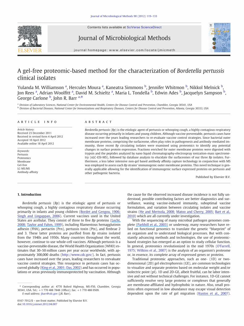
Fig. 1 is a flow diagram schematically summating the core meth-odologies used in this study.
2.1. Reagents
All reagents and media not vendor-specified were prepared at theCenters for Disease Control and Prevention (CDC) Core Facility. In ad-dition, chemicals used in experimentation were obtained from eitherSigma-Aldrich Chemical Company (St. Louis, MO, USA) or Fisher Sci-entific (Pittsburg, PA, USA) as noted.
2.2. Bacterial strains
Four Bp strains, Tohama I (T) and three clinical isolates designatedby CDC as C056 (C), D946 (D) and F656 (F) were used in the proteo-mic comparison (Table 1). T, first isolated in Japan in 1954, is a well-characterized and completely sequenced Bp strain that has been usedas the basis of vaccines in many countries for several years (Advaniet al., 2004). By pulse-field gel electrophoresis (PFGE) analysis, it ischaracterized as type II and possesses the pertactin 1 (prnA1) andpertussis toxin (ptxS1B) genotype typical of prevaccine-era isolates(Advani et al., 2004; van Loo et al., 2002; Litt et al., 2009). Strain Cwas isolated in Minnesota in 1998, has a PFGE CDC type 10 (Hardwicket al., 2002), and a prnA2, ptxS1A genotype common among currentlycirculating isolates (Litt et al., 2009). The D strain, a clinical isolate iden-tified in Georgia in 2002, has a PFGE CDC 21, prnA1 and ptxS1A geno-type. It has shown resistance to the antibiotic erythromycin. Lastly,the F strain (PFGE CDC 206, prnA2, ptxS1A) was isolated from a clinicalcase in 2007 in the Virgin Islands.
2.3. Bacterial cell culture
T, C, D, and F were plated on Bordet-Gengou agar and incubated at35 °C with 5% CO2 for 4 days (Hulbert and Cotter, 2009). The bacteriawere subsequently subcultured into Modified Stainer-Schulte (MSS)media at 35 °C, with aeration at 200 rpm in a Beckman-Coulter shaker(Beckman-Coulter, Brea, CA, USA) until an optical 1.0 density wasreached. The bacterial strains were then pelleted from MSS by centri-fugation at 8000×g for 30 min (min) at 4 °C. The pellets were washedtwo times in distilled water (dH2O) and stored at −70 °C for furtheruse.
2.4. Enriched membrane fraction collection
Enrichedmembrane fractions (EMFs)were collected as previously de-scribed (Molloy, 2008) with the following modifications. Briefly, cell pel-lets of Bp isolates were allowed to thaw gently on ice. The pellets wereFrench-pressed at 16,000 psi in 5 ml of a 50-mMTris–HCl (pH 8.0) buffercontaining a protease inhibitor cocktail added at the manufacturer's rec-ommendation (GE Healthcare, Piscataway, NJ, USA), and 25U of benzo-nase to rupture bacterial cells. The lysates were centrifuged (8000×g,20 min, 4 °C) to remove unbroken cells, and the supernatant containingthe total extracted proteome was retained. 50 ml of ice-cold sodium car-bonate (pH 11.0) was added to 5 ml of each bacterial supernatant. Themixture was stirred gently at 4 °C for 2 h. The sodium carbonateinfused-supernatants were subjected to ultracentrifugation (Beckman-Coulter) (115,000×g, 60 min, 4 °C) to enrich for amembraneprotein frac-tion. The pellets containing the EMFs were washed in a 50 mM Tris–HClbuffer (pH 8.0) and ultracentrifuged twice (115,000×g, 30 min, 4 °C) toremove the enrichment buffer. The final EMFs were solubilized in 1 mlof solubilization buffer containing 7 M urea, 2 M thiourea, 2% CHAPS,
Fig. 1. Flow diagram of core methodologies used in the proteomic study. Red Asterisk (*) indicates the mouse sera was used for the immunoblot analysis and immunoprecipitation.Black asterisk (*) indicates human convalescent sera was used for the immunoprecipitation.
121Y.M. Williamson et al. / Journal of Microbiological Methods 90 (2012) 119–133
10% isopropanol, and protease inhibitor cocktail, with an optional addi-tion of 0.5% bromophenol blue to visually assess the integrity of proteinisolation. Protein concentrations of the samples were determined usinga 2D-Quant Kit (GE Healthcare), and the samples were aliquoted and fro-zen at−20 °C until further use.
2.5. B. pertussis immune sera
Three-week-old female BALB/C mice were initially injected intra-peritoneally (i.p.) with 1×10⁹ colony forming units (cfu) of T, C, D,or F suspended in 10 μl of physiological saline (pH 7.2). Before injec-tion, strains were cobalt-irradiated using 5×10⁶ γ RAD to inhibit bac-terial replication and infectivity, while preserving bacterial surfacestructures. The process was repeated 2 weeks later, every 2 weeksthereafter, with three separate i.p. immunizations of similar dosagefor 6 weeks. At this time, mice were euthanized according to AALACand IACUC standards and the Bp immune sera generated from eachstrain were collected from blood. The collected serum was aliquotedand stored at −70 °C until use. Additionally, a serum pool composedof sera drawn from convalescent pertussis human patients obtainedfrom the CDC Pertussis Laboratory was used in this analysis. Thispool is the Pertussis Laboratory ELISA standard reference sera
Table 1Genomic profiles of Bordetella pertussis (Bp) strains assessed in the study. Abbrevia-tions: T — Tohama I, C — C056, D — D946 and F — F656; USA — United States of Amer-ica; CDC — Centers for Disease Control and Prevention; PFGE — pulse field gelelectrophoresis; Prn — pertactin; Ptx — pertussis toxin.
Strain Isolation location (Year) PFGE Pertactin Pertussis toxin
T Japan (1954) Type II prnA1 ptxS1BC Minnesota, USA (1998) CDC type I0 prnA2 ptxS1AD Georgia, USA (2002) CDC type 21 prnA1 ptxS1AF Virgin Islands, USA (2007) CDC type 206 prnA2 ptxS1A
acquired in accordance with CDC Institutional Review Board stan-dards and regulations.
2.6. 1D sodium dodecyl sulfate-polyacrylamide gel electrophoresis(SDS-PAGE) and immunoblot analysis of B. pertussis EMF
Unless specified, materials, antibodies, and procedures for GE and im-munoblot analysiswere obtained fromBio-Rad (Hercules, CA, USA). 10 μgof total EMF proteins from each strain was suspended in Laemmli samplebuffer and electrophoresed on 12.5% SDS-PAGE gels following standardprotocols (Laemmli, 1970). To visualize separated proteins, gels werestained using the hot coomassie blue staining protocol. EMF proteinswere electroblotted (Towbin et al., 1979) onto polyvinylidene fluoride(PVDF) membranes for 1 h and probed with either a primary Bp strain-specific immune or preimmune (normal)mouse serum. Immunoreactivebands were further probed with a secondary goat-anti-mouse horserad-ish peroxidase-conjugated IgG antibody and subsequently visualizedwith 1,4-benzenediamine dihydrochloride (Sigma-Aldrich).
2.7. PVDF on-membrane protein extraction
Protein extraction directly from blotted-PVDF membranes wasperformed based on Bienvenut et al. (1999), but modified according-ly. PVDF membrane bands containing EMF proteins from T and C as-sociated with immunoreactivity were excised and destained with50% methanol (500 μl) for 2 h at room temperature (RT). Afterdestaining, the supernatant was removed. The membrane pieceswere air dried, followed by the addition of 50 mM ammonium bicar-bonate (NH4(CO3)2) digestion buffer in 30% acetonitrile (ACN) (Fish-er Scientific). The protein-containing membrane pieces were thenincubated overnight (ON) with trypsin (0.1 μg/μl) (Promega Corpora-tion, Carlsbad, CA, USA) at 37 °C. After digestion, the supernatant wascollected and the membranes were treated with 80% ACN to extractthe peptides and sonicated at level nine (Aquasonic™ model 150-D)(VWR Scientific Products, Suwanee, GA) for 15 min. Following son-ication, the extract was pooled with the previous supernatant, dried
122 Y.M. Williamson et al. / Journal of Microbiological Methods 90 (2012) 119–133
via vacuum centrifugation, and resuspended in dH2O (50 μl). Sampleswere prepared for nLC-ESI-MS/MS, in which peptides were sus-pended in equal volumes of 0.1% formic acid.
2.8. EMF protein identification
T, C, D, or F EMFs (10 μg) before direct proteolytic cleavage weretreated with 0.1% rapigest (RG) (Waters Corporation, Milford, MA,USA) in NH4(CO3)2 digestion buffer at 100 °C for 5 min to denatureproteins. Upon cooling at RT, the samples were incubated ON withtrypsin (10 μg) (Promega) at 37 °C. After incubation, the RG was inac-tivated in the presence of 1 M HCl for 30 min at 37 °C, and centrifugedat 12,000×g for 15 min. The supernatant was removed and sus-pended in equal volumes of 0.1% formic acid and analyzed by nLC-ESI MS/MS. The data obtained represent two distinct biological prep-arations, each performed in triplicate.
2.9. Immunoprecipitation studies using antibody affinity magnetic beadcapture technology
Dynal beads (Invitrogen Corporation, Carlsbad, CA, USA) coatedwith protein G for immunoglobulin (IgG) capture and subsequentimmunoprecipitaton (IP) of EMFs were used as per the manufacturer'srecommendation with the following changes. The Dynal beads (200 μlper sample) were initially washed three times via resuspension in800 μl phosphate citrate buffer (PCB), pH 5.0 (Sigma-Aldrich). Next,the beads were resuspended in 800 μl PCB and incubated ON at 37 °Cin the presence of immune sera (100 μg total) from mice immunizedagainst T, C, D or F. We also prepared controls containing normalmouse immune sera (100 μg total) and beads only. After incubation,the beads were magnetically stabilized, the supernatant was removed,and the beads were washed two times with 2 M triethanolamine, pH8.2 (Sigma-Aldrich). This was to remove unbound antibodies and toequilibrate the beads for antibody crosslinking. The immune-serabound beads were next cross-linked with 1 ml 20 mM dimethyl pime-limidate (Sigma-Aldrich) in 2 M triethanolamine for 30 min at RT viainversion. Following crosslinking, the beads were washed two timesin 800 μl phosphate buffer saline (PBS), pH 7.0 with 0.1% Tween 20and further incubated with 800 μl TBE (Sigma-Aldrich) to reduce non-specific (NS) protein binding. The beads were resuspended in 50 μldH2O. Then they were incubated at 37 °C ON in the presence of T, C,D or F EMFs (10 μg) that corresponded with the bead-Ab sourcestrain (e.g., beads bound with T-specific IgG were incubated in thepresence of T-EMF). After magnetic stabilization, the beads werewashed via a mixer three times for 5 min with 100 μl PBS at RT to re-move any unbound or NS-bound EMF proteins. The protein-boundAb-coupled complexes were resuspended in 50 μl NH4(CO3)2 diges-tion buffer treated with 0.1% RG followed by ON trypsin (10 μg) di-gestion at 37 °C.
Upon incubation, the IP complex was magnetically stabilized, andthe supernatant containing EMF tryptic peptides was transferred to afresh tube and dried via vacuum centrifugation to concentrate samples.The RGwas inactivated and the samples prepared for nLC-ESIMS/MS, inwhich peptides were suspended in equal volumes of 0.1% formic acid.The data represent two biological preparations, each performed in du-plicate. Simultaneously, Dynal beads were conjugated with pooledhuman convalescent sera (100 μg total) resulting from Bp infection inaddition to normal human IgG (Interstate Blood Bank, Inc., Memphis,TN, USA). The IgG bound-beads were incubated with T, C, D, or F EMF(10 μg), and the samples were processed as described above.
2.10. Nano liquid chromatography electrospray ionization massspectrometry
Protein identification was achieved by using nanoflow liquid chro-matography (nano-LC), data-dependent tandem mass spectrometry,
and database searching. A pulled-needle, fused silica capillary(365 μm O.D. by 75 μm I.D.) (New Objective, Inc., Woburn, MA) waspacked with 10 cm of 5 μm Symmetry 300 reverse-phase packing ma-terial (Waters Inc., Bedford, MA). Protein digests were loaded ontothe analytical column and separated by gradient elution using anEksigent 2D nanoLC system (Eksigent Technologies, Inc, Dublin, CA).The mobile phase solvents consisted of (solvent A) 0.2% formic acid(Thermo Scientific, Rockford, IL), 0.005% trifluoroacetic acid (Sigma-Aldrich) in water (Burdick and Jackson, Muskegon, MI), and (solventB) 0.2% formic acid, 0.005% trifluoroacetic acid in acetonitrile (Burdickand Jackson). The gradient flow was set at 400 nl/min. The profileconsisted of a hold at 5% B for 5 min followed by a ramp to 30% Bover 100 min, then a ramp up to 90% B in 5 min and a hold at 90%for 2 min before returning to 5% B in 2 min and re-equilibration at5% B for 20 min. After chromatography, peptides were introducedinto an LTQ Orbitrap tandem mass spectrometer (Thermo Scientific,San Jose, CA). A 2.0 kV voltage was applied to the nano-LC column.The mass spectrometer was programmed to perform data-dependentacquisition by scanning the mass range from mass-to-charge (m/z)400 to 1600 at a nominal resolution setting of 60,000 for parent ion ac-quisition in the Orbitrap. Most tryptic peptides fall within the statedm/z range and served as the basis for this selection. For MS/MS anal-ysis the mass spectrometer chose the top 10 most intense ions withtwo or more charges. Singly charged ions were rejected for MS/MSas these ions are likely due to detergents or other sample additives.In particular for a data-dependent acquisition, time is better utilizedacquiring for doubly and triply charged amino acids, which predom-inantly have greater sequence specificity since they are larger pep-tides and thus provide a higher likelihood in which to uniquelyidentify a protein.
All tandem mass spectra were extracted from the raw data fileusing Mascot Distiller (Matrix Science, London, UK; version 2.2.1.0)and searched using Mascot (version 2.2.0). Mascot was set up tosearch using the entire NCBInr database or a modified NCBInr data-base created to search “Bordetella”- or “pertussis”- recognized pro-teins in which trypsin is used as the digestion agent. Mascot wassearched with two missed cleavages, a fragment ion tolerance massof 0.80 Da, and a parent ion tolerance of 200 ppm, while oxidationwas selected as a variable modification. Scaffold (Proteome Software,Portland, OR) was used to validate MS/MS based peptide and proteinidentifications.
Peptide identifications were accepted if they could be establishedat greater than 95.0% probability as specified by the Peptide Prophetalgorithm (Keller et al., 2002). Protein identifications were acceptedif they could be established at greater than 99.0% probability and con-tained at least two identified peptides (Nesvizhskii et al., 2003). Withthese stringent parameters of Peptide Prophet and Protein Prophetwithin the Scaffold software, the probability of a wrong assignmentis below 0.1%. PSORTb subcellular scores were used to predict and lo-calize identified EMF proteins (http://www.psort.org/psortb/) (Yuet al., 2010). Lastly, KEGG identifiers using NCBI Gi accession numberswere employed to assign functions to each of the identified proteinshttp://www.genome.jp/kegg/kegg3.html (Tefon et al., 2011).
3. Results
3.1. 1GE and immunoblot analysis of B. pertussis species EMFs
Carbonate-enriched EMF proteins were initially separated by 1D-SDS-PAGE (1D-GE). This was to observe common and differentialbanding patterns between the T-reference strain and clinical isolatesC, D, and F. Overall, similar protein profiles among the strains wereobserved, with 1D-GE revealing slight differences and no unique pro-tein banding patterns between the strains (Fig. 2). Subsequently, afterobserving no major protein differences, we used classical immuno-blotting approaches to assess the strain's ability to invoke an immune
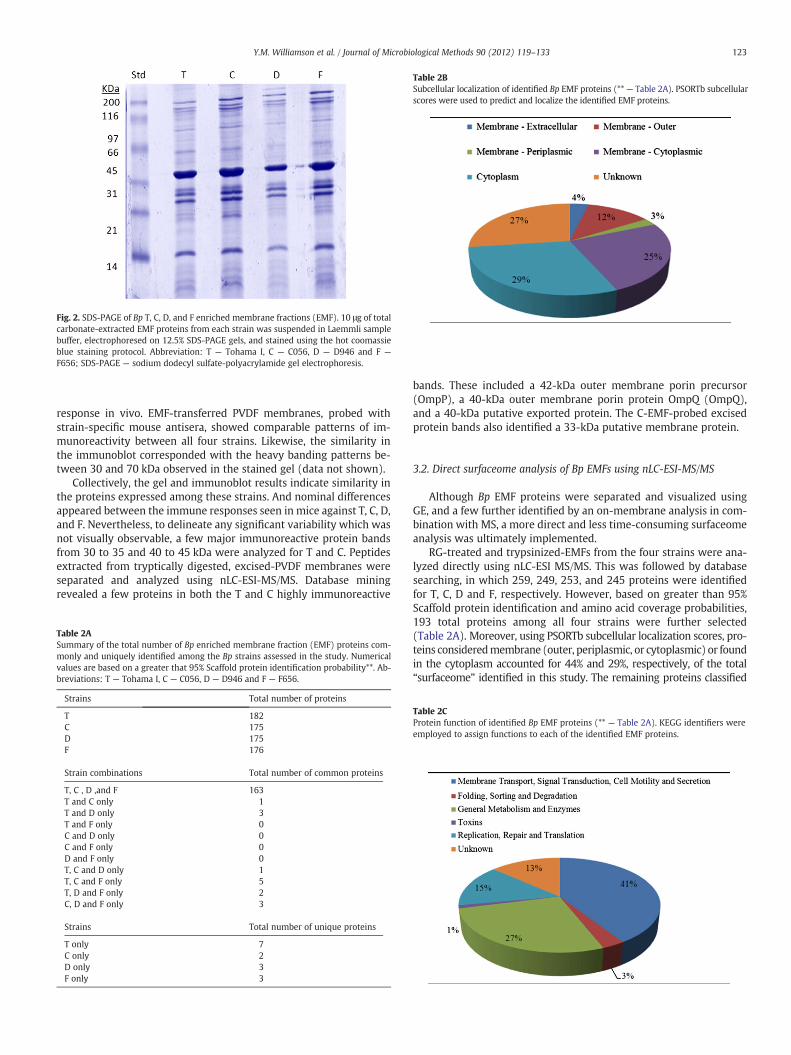
Fig. 2. SDS-PAGE of Bp T, C, D, and F enriched membrane fractions (EMF). 10 μg of totalcarbonate-extracted EMF proteins from each strain was suspended in Laemmli samplebuffer, electrophoresed on 12.5% SDS-PAGE gels, and stained using the hot coomassieblue staining protocol. Abbreviation: T — Tohama I, C — C056, D — D946 and F —
F656; SDS-PAGE — sodium dodecyl sulfate-polyacrylamide gel electrophoresis.
Table 2BSubcellular localization of identified Bp EMF proteins (** — Table 2A). PSORTb subcellularscores were used to predict and localize the identified EMF proteins.
123Y.M. Williamson et al. / Journal of Microbiological Methods 90 (2012) 119–133
response in vivo. EMF-transferred PVDF membranes, probed withstrain-specific mouse antisera, showed comparable patterns of im-munoreactivity between all four strains. Likewise, the similarity inthe immunoblot corresponded with the heavy banding patterns be-tween 30 and 70 kDa observed in the stained gel (data not shown).
Collectively, the gel and immunoblot results indicate similarity inthe proteins expressed among these strains. And nominal differencesappeared between the immune responses seen in mice against T, C, D,and F. Nevertheless, to delineate any significant variability which wasnot visually observable, a few major immunoreactive protein bandsfrom 30 to 35 and 40 to 45 kDa were analyzed for T and C. Peptidesextracted from tryptically digested, excised-PVDF membranes wereseparated and analyzed using nLC-ESI-MS/MS. Database miningrevealed a few proteins in both the T and C highly immunoreactive
Table 2ASummary of the total number of Bp enriched membrane fraction (EMF) proteins com-monly and uniquely identified among the Bp strains assessed in the study. Numericalvalues are based on a greater that 95% Scaffold protein identification probability**. Ab-breviations: T — Tohama I, C — C056, D — D946 and F — F656.
Strains Total number of proteins
T 182C 175D 175F 176
Strain combinations Total number of common proteins
T, C , D ,and F 163T and C only 1T and D only 3T and F only 0C and D only 0C and F only 0D and F only 0T, C and D only 1T, C and F only 5T, D and F only 2C, D and F only 3
Strains Total number of unique proteins
T only 7C only 2D only 3F only 3
bands. These included a 42-kDa outer membrane porin precursor(OmpP), a 40-kDa outer membrane porin protein OmpQ (OmpQ),and a 40-kDa putative exported protein. The C-EMF-probed excisedprotein bands also identified a 33-kDa putative membrane protein.
3.2. Direct surfaceome analysis of Bp EMFs using nLC-ESI-MS/MS
Although Bp EMF proteins were separated and visualized usingGE, and a few further identified by an on-membrane analysis in com-bination with MS, a more direct and less time-consuming surfaceomeanalysis was ultimately implemented.
RG-treated and trypsinized-EMFs from the four strains were ana-lyzed directly using nLC-ESI MS/MS. This was followed by databasesearching, in which 259, 249, 253, and 245 proteins were identifiedfor T, C, D and F, respectively. However, based on greater than 95%Scaffold protein identification and amino acid coverage probabilities,193 total proteins among all four strains were further selected(Table 2A). Moreover, using PSORTb subcellular localization scores, pro-teins consideredmembrane (outer, periplasmic, or cytoplasmic) or foundin the cytoplasm accounted for 44% and 29%, respectively, of the total“surfaceome” identified in this study. The remaining proteins classified
Table 2CProtein function of identified Bp EMF proteins (** — Table 2A). KEGG identifiers wereemployed to assign functions to each of the identified EMF proteins.
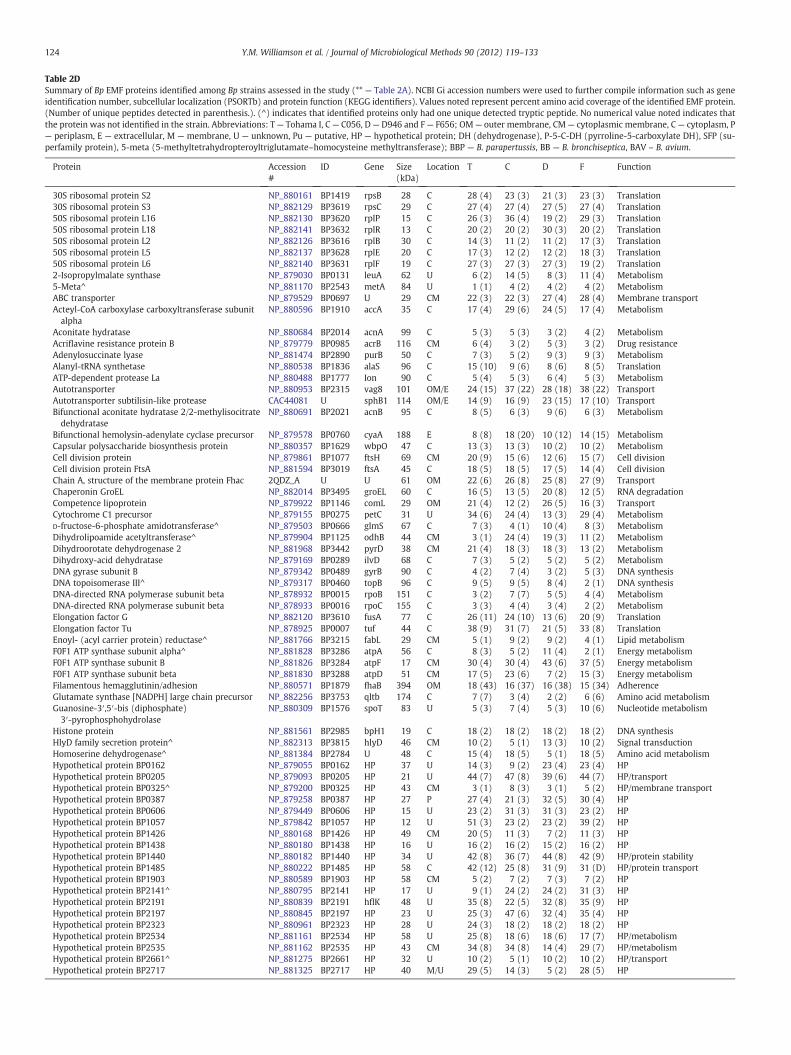
Table 2DSummary of Bp EMF proteins identified among Bp strains assessed in the study (** — Table 2A). NCBI Gi accession numbers were used to further compile information such as geneidentification number, subcellular localization (PSORTb) and protein function (KEGG identifiers). Values noted represent percent amino acid coverage of the identified EMF protein.(Number of unique peptides detected in parenthesis.). (^) indicates that identified proteins only had one unique detected tryptic peptide. No numerical value noted indicates thatthe protein was not identified in the strain. Abbreviations: T— Tohama I, C— C056, D— D946 and F— F656; OM— outer membrane, CM— cytoplasmic membrane, C— cytoplasm, P— periplasm, E — extracellular, M — membrane, U — unknown, Pu — putative, HP — hypothetical protein; DH (dehydrogenase), P-5-C-DH (pyrroline-5-carboxylate DH), SFP (su-perfamily protein), 5-meta (5-methyltetrahydropteroyltriglutamate–homocysteine methyltransferase); BBP — B. parapertussis, BB — B. bronchiseptica, BAV – B. avium.
Protein Accession#
ID Gene Size(kDa)
Location T C D F Function
30S ribosomal protein S2 NP_880161 BP1419 rpsB 28 C 28 (4) 23 (3) 21 (3) 23 (3) Translation30S ribosomal protein S3 NP_882129 BP3619 rpsC 29 C 27 (4) 27 (4) 27 (5) 27 (4) Translation50S ribosomal protein L16 NP_882130 BP3620 rplP 15 C 26 (3) 36 (4) 19 (2) 29 (3) Translation50S ribosomal protein L18 NP_882141 BP3632 rplR 13 C 20 (2) 20 (2) 30 (3) 20 (2) Translation50S ribosomal protein L2 NP_882126 BP3616 rplB 30 C 14 (3) 11 (2) 11 (2) 17 (3) Translation50S ribosomal protein L5 NP_882137 BP3628 rplE 20 C 17 (3) 12 (2) 12 (2) 18 (3) Translation50S ribosomal protein L6 NP_882140 BP3631 rplF 19 C 27 (3) 27 (3) 27 (3) 19 (2) Translation2-Isopropylmalate synthase NP_879030 BP0131 leuA 62 U 6 (2) 14 (5) 8 (3) 11 (4) Metabolism5-Meta^ NP_881170 BP2543 metA 84 U 1 (1) 4 (2) 4 (2) 4 (2) MetabolismABC transporter NP_879529 BP0697 U 29 CM 22 (3) 22 (3) 27 (4) 28 (4) Membrane transportActeyl-CoA carboxylase carboxyltransferase subunitalpha
NP_880596 BP1910 accA 35 C 17 (4) 29 (6) 24 (5) 17 (4) Metabolism
Aconitate hydratase NP_880684 BP2014 acnA 99 C 5 (3) 5 (3) 3 (2) 4 (2) MetabolismAcriflavine resistance protein B NP_879779 BP0985 acrB 116 CM 6 (4) 3 (2) 5 (3) 3 (2) Drug resistanceAdenylosuccinate lyase NP_881474 BP2890 purB 50 C 7 (3) 5 (2) 9 (3) 9 (3) MetabolismAlanyl-tRNA synthetase NP_880538 BP1836 alaS 96 C 15 (10) 9 (6) 8 (6) 8 (5) TranslationATP-dependent protease La NP_880488 BP1777 Ion 90 C 5 (4) 5 (3) 6 (4) 5 (3) MetabolismAutotransporter NP_880953 BP2315 vag8 101 OM/E 24 (15) 37 (22) 28 (18) 38 (22) TransportAutotransporter subtilisin-like protease CAC44081 U sphB1 114 OM/E 14 (9) 16 (9) 23 (15) 17 (10) TransportBifunctional aconitate hydratase 2/2-methylisocitratedehydratase
NP_880691 BP2021 acnB 95 C 8 (5) 6 (3) 9 (6) 6 (3) Metabolism
Bifunctional hemolysin-adenylate cyclase precursor NP_879578 BP0760 cyaA 188 E 8 (8) 18 (20) 10 (12) 14 (15) MetabolismCapsular polysaccharide biosynthesis protein NP_880357 BP1629 wbpO 47 C 13 (3) 13 (3) 10 (2) 10 (2) MetabolismCell division protein NP_879861 BP1077 ftsH 69 CM 20 (9) 15 (6) 12 (6) 15 (7) Cell divisionCell division protein FtsA NP_881594 BP3019 ftsA 45 C 18 (5) 18 (5) 17 (5) 14 (4) Cell divisionChain A, structure of the membrane protein Fhac 2QDZ_A U U 61 OM 22 (6) 26 (8) 25 (8) 27 (9) TransportChaperonin GroEL NP_882014 BP3495 groEL 60 C 16 (5) 13 (5) 20 (8) 12 (5) RNA degradationCompetence lipoprotein NP_879922 BP1146 comL 29 OM 21 (4) 12 (2) 26 (5) 16 (3) TransportCytochrome C1 precursor NP_879155 BP0275 petC 31 U 34 (6) 24 (4) 13 (3) 29 (4) MetabolismD-fructose-6-phosphate amidotransferase^ NP_879503 BP0666 glmS 67 C 7 (3) 4 (1) 10 (4) 8 (3) MetabolismDihydrolipoamide acetyltransferase^ NP_879904 BP1125 odhB 44 CM 3 (1) 24 (4) 19 (3) 11 (2) MetabolismDihydroorotate dehydrogenase 2 NP_881968 BP3442 pyrD 38 CM 21 (4) 18 (3) 18 (3) 13 (2) MetabolismDihydroxy-acid dehydratase NP_879169 BP0289 ilvD 68 C 7 (3) 5 (2) 5 (2) 5 (2) MetabolismDNA gyrase subunit B NP_879342 BP0489 gyrB 90 C 4 (2) 7 (4) 3 (2) 5 (3) DNA synthesisDNA topoisomerase III^ NP_879317 BP0460 topB 96 C 9 (5) 9 (5) 8 (4) 2 (1) DNA synthesisDNA-directed RNA polymerase subunit beta NP_878932 BP0015 rpoB 151 C 3 (2) 7 (7) 5 (5) 4 (4) MetabolismDNA-directed RNA polymerase subunit beta NP_878933 BP0016 rpoC 155 C 3 (3) 4 (4) 3 (4) 2 (2) MetabolismElongation factor G NP_882120 BP3610 fusA 77 C 26 (11) 24 (10) 13 (6) 20 (9) TranslationElongation factor Tu NP_878925 BP0007 tuf 44 C 38 (9) 31 (7) 21 (5) 33 (8) TranslationEnoyl- (acyl carrier protein) reductase^ NP_881766 BP3215 fabL 29 CM 5 (1) 9 (2) 9 (2) 4 (1) Lipid metabolismF0F1 ATP synthase subunit alpha^ NP_881828 BP3286 atpA 56 C 8 (3) 5 (2) 11 (4) 2 (1) Energy metabolismF0F1 ATP synthase subunit B NP_881826 BP3284 atpF 17 CM 30 (4) 30 (4) 43 (6) 37 (5) Energy metabolismF0F1 ATP synthase subunit beta NP_881830 BP3288 atpD 51 CM 17 (5) 23 (6) 7 (2) 15 (3) Energy metabolismFilamentous hemagglutinin/adhesion NP_880571 BP1879 fhaB 394 OM 18 (43) 16 (37) 16 (38) 15 (34) AdherenceGlutamate synthase [NADPH] large chain precursor NP_882256 BP3753 qltb 174 C 7 (7) 3 (4) 2 (2) 6 (6) Amino acid metabolismGuanosine-3′,5′-bis (diphosphate)3′-pyrophosphohydrolase
NP_880309 BP1576 spoT 83 U 5 (3) 7 (4) 5 (3) 10 (6) Nucleotide metabolism
Histone protein NP_881561 BP2985 bpH1 19 C 18 (2) 18 (2) 18 (2) 18 (2) DNA synthesisHlyD family secretion protein^ NP_882313 BP3815 hlyD 46 CM 10 (2) 5 (1) 13 (3) 10 (2) Signal transductionHomoserine dehydrogenase^ NP_881384 BP2784 U 48 C 15 (4) 18 (5) 5 (1) 18 (5) Amino acid metabolismHypothetical protein BP0162 NP_879055 BP0162 HP 37 U 14 (3) 9 (2) 23 (4) 23 (4) HPHypothetical protein BP0205 NP_879093 BP0205 HP 21 U 44 (7) 47 (8) 39 (6) 44 (7) HP/transportHypothetical protein BP0325^ NP_879200 BP0325 HP 43 CM 3 (1) 8 (3) 3 (1) 5 (2) HP/membrane transportHypothetical protein BP0387 NP_879258 BP0387 HP 27 P 27 (4) 21 (3) 32 (5) 30 (4) HPHypothetical protein BP0606 NP_879449 BP0606 HP 15 U 23 (2) 31 (3) 31 (3) 23 (2) HPHypothetical protein BP1057 NP_879842 BP1057 HP 12 U 51 (3) 23 (2) 23 (2) 39 (2) HPHypothetical protein BP1426 NP_880168 BP1426 HP 49 CM 20 (5) 11 (3) 7 (2) 11 (3) HPHypothetical protein BP1438 NP_880180 BP1438 HP 16 U 16 (2) 16 (2) 15 (2) 16 (2) HPHypothetical protein BP1440 NP_880182 BP1440 HP 34 U 42 (8) 36 (7) 44 (8) 42 (9) HP/protein stabilityHypothetical protein BP1485 NP_880222 BP1485 HP 58 C 42 (12) 25 (8) 31 (9) 31 (D) HP/protein transportHypothetical protein BP1903 NP_880589 BP1903 HP 58 CM 5 (2) 7 (2) 7 (3) 7 (2) HPHypothetical protein BP2141^ NP_880795 BP2141 HP 17 U 9 (1) 24 (2) 24 (2) 31 (3) HPHypothetical protein BP2191 NP_880839 BP2191 hflK 48 U 35 (8) 22 (5) 32 (8) 35 (9) HPHypothetical protein BP2197 NP_880845 BP2197 HP 23 U 25 (3) 47 (6) 32 (4) 35 (4) HPHypothetical protein BP2323 NP_880961 BP2323 HP 28 U 24 (3) 18 (2) 18 (2) 18 (2) HPHypothetical protein BP2534 NP_881161 BP2534 HP 58 U 25 (8) 18 (6) 18 (6) 17 (7) HP/metabolismHypothetical protein BP2535 NP_881162 BP2535 HP 43 CM 34 (8) 34 (8) 14 (4) 29 (7) HP/metabolismHypothetical protein BP2661^ NP_881275 BP2661 HP 32 U 10 (2) 5 (1) 10 (2) 10 (2) HP/transportHypothetical protein BP2717 NP_881325 BP2717 HP 40 M/U 29 (5) 14 (3) 5 (2) 28 (5) HP
124 Y.M. Williamson et al. / Journal of Microbiological Methods 90 (2012) 119–133
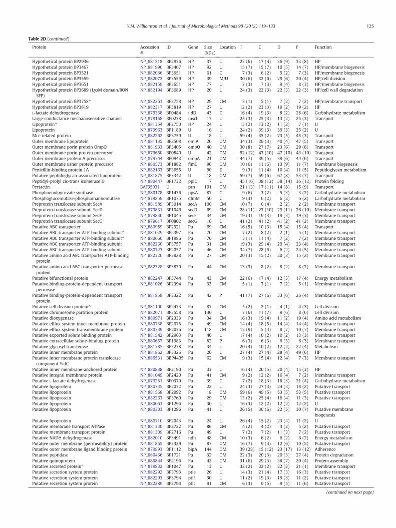
Table 2D (continued)
Protein Accession#
ID Gene Size(kDa)
Location T C D F Function
Hypothetical protein BP2936 NP_881518 BP2936 HP 37 U 23 (6) 17 (4) 36 (9) 33 (8) HPHypothetical protein BP3467 NP_881990 BP3467 HP 92 U 15 (7) 15 (7) 10 (5) 14 (7) HP/membrane biogenesisHypothetical protein BP3521 NP_882036 BP3651 HP 61 C 7 (3) 6 (2) 5 (2) 7 (3) HP/membrane biogenesisHypothetical protein BP3559 NP_882072 BP3559 HP 39 M/U 30 (6) 32 (6) 29 (6) 20 (4) HP/cell divisionHypothetical protein BP3651 NP_882159 BP3651 HP 77 U 7 (3) 7 (3) 9 (4) 8 (3) HP/membrane biogenesisHypothetical protein BP3689 (LysM domain/BONSFP)
NP_882194 BP3689 HP 20 U 24 (3) 22 (3) 22 (3) 22 (3) HP/cell wall degradation
Hypothetical protein BP3758^ NP_882261 BP3758 HP 29 CM 3 (1) 5 (1) 7 (2) 7 (2) HP/membrane transportHypothetical protein BP3819 NP_882317 BP3819 HP 27 U 12 (2) 23 (3) 19 (2) 19 (2) HPL-lactate dehydrogenase NP_879338 BP0484 ildD 43 C 16 (4) 19 (3) 8 (2) 28 (6) Carbohydrate metabolismLarge-conductance mechanosensitive channel NP_879158 BP0278 mscl 17 U 25 (3) 25 (3) 13 (2) 25 (3) TransportLipoprotein^ NP_881354 BP2750 HP 24 U 13 (2) 13 (2) 11 (2) 7 (1) ULipoprotein NP_879963 BP1189 U 16 U 24 (2) 39 (3) 39 (3) 25 (2) UMce related protein NP_882262 BP3759 U 18 U 59 (4) 35 (2) 73 (5) 45 (3) TransportOuter membrane lipoprotein NP_881135 BP2508 omlA 20 OM 34 (3) 29 (3) 40 (4) 47 (5) TransportOuter membrane porin protein OmpQ NP_881933 BP3405 ompQ 40 OM 30 (8) 27 (7) 23 (6) 29 (8) TransportOuter membrane porin protein precursor NP_879650 BP0840 U 42 OM 52 (12) 43 (10) 47 (10) 43 (10) TransportOuter membrane protein A precursor NP_879744 BP0943 ompA 21 OM 44 (7) 39 (5) 39 (6) 44 (6) TransportOuter membrane usher protein precursor NP_880573 BP1882 fimC 96 OM 10 (6) 11 (6) 13 (9) 11 (7) Membrane biogenesisPenicillin-binding protein 1A NP_882163 BP3655 U 90 E 9 (3) 11 (4) 10 (4) 11 (5) Peptidoglycan metabolismPutative peptidoglycan-associated lipoprotein NP_881875 BP3342 U 18 OM 59 (7) 59 (6) 67 (8) 53 (7) TransportPeptidyl-prolyl cis-trans isomerase D NP_880447 BP1732 ppiD 7 U 45 (16) 38 (13) 38 (14) 36 (12) Protein foldingPertactin BAF35031 U prn 101 OM 21 (13) 17 (11) 14 (8) 15 (9) TransportPhosphoenolpyruvate synthase NP_880178 BP1436 ppsA 87 C 9 (6) 3 (2) 5 (3) 3 (2) Carbohydrate metabolismPhosphoglucomutase/phosphomannomutase NP_879859 BP1075 qlmM 50 C 9 (3) 6 (2) 6 (2) 6 (2) Carbohydrate metabolismPreprotein translocase subunit SecA NP_881589 BP3014 secA 100 CM 10 (7) 6 (4) 2 (2) 2 (2) Membrane transportPreprotein translocase subunit SecD NP_879831 BP1046 secD 69 CM 28 (11) 23 (10) 29 (11) 26 (10) Membrane transportPreprotein translocase subunit SecF NP_879830 BP1045 secF 34 CM 19 (3) 19 (3) 19 (3) 19 (3) Membrane transportPreprotein translocase subunit SecG NP_879617 BP0802 secG 16 U 41 (2) 41 (2) 41 (2) 41 (2) Membrane transportPutative ABC transporter NP_880959 BP2321 Pu 69 CM 16 (5) 10 (3) 15 (4) 15 (4) TransportPutative ABC transporter ATP-binding subunit^ NP_881029 BP2397 Pu 70 CM 7 (2) 8 (2) 2 (1) 5 (1) Membrane transportPutative ABC transporter ATP-binding subunit^ NP_880660 BP1986 Pu 70 CM 3 (1) 11 (4) 7 (2) 7 (2) Membrane transportPutative ABC transporter ATP-binding subunit NP_882260 BP3757 Pu 31 CM 19 (3) 29 (4) 29 (4) 23 (4) Membrane transportPutative ABC transporter ATP-binding subunit NP_880723 BP2057 Pu 46 CM 34 (7) 28 (6) 6 (2) 24 (5) Membrane transportPutative amino acid ABC transporter ATP-bindingprotein
NP_882326 BP3828 Pu 27 CM 20 (3) 15 (2) 20 (3) 15 (2) Membrane transport
Putative amino acid ABC transporter permeaseprotein
NP_882328 BP3830 Pu 44 CM 13 (3) 8 (2) 8 (2) 8 (2) Membrane transport
Putative bifunctional protein NP_882247 BP3744 Pu 43 CM 22 (6) 17 (4) 12 (3) 17 (4) Energy metabolismPutative binding-protein-dependent transportpermease
NP_881026 BP2394 Pu 33 CM 5 (1) 3 (1) 7 (2) 5 (1) Membrane transport
Putative binding-protein-dependent transportprotein
NP_881859 BP3322 Pu 42 P 41 (7) 27 (6) 33 (6) 26 (4) Membrane transport
Putative cell division protein^ NP_881100 BP2473 Pu 87 CM 5 (2) 2 (1) 4 (1) 4 (3) Cell divisionPutative chromosome partition protein NP_882071 BP3558 Pu 130 C 7 (6) 11 (7) 9 (6) 8 (6) Cell divisionPutative dioxygenase NP_880971 BP2333 Pu 34 CM 16 (3) 19 (4) 11 (2) 19 (4) Amino acid metabolismPutative efflux system inner membrane protein NP_880738 BP2075 Pu 49 CM 14 (4) 18 (5) 14 (4) 14 (4) Membrane transportPutative efflux system transmembrane protein NP_880739 BP2076 Pu 118 CM 12 (9) 5 (4) 8 (7) 10 (7) Membrane transportPutative exported solute binding protein NP_881542 BP2963 Pu 40 U 17 (4) 10 (2) 10 (2) 13 (3) Membrane transportPutative extracellular solute-binding protein NP_880657 BP1983 Pu 82 P 6 (3) 6 (3) 6 (3) 6 (3) Membrane transportPutative glycosyl transferase NP_881785 BP3238 Pu 34 U 20 (4) 10 (2) 12 (2) 22 (4) MetabolismPutative inner membrane protein NP_881862 BP3326 Pu 26 U 27 (4) 27 (4) 28 (4) 49 (6) HPPutative inner membrane protein translocasecomponent YidC
NP_886531 BBP4405 Pu 62 CM 9 (3) 15 (4) 12 (4) 7 (3) Membrane transport
Putative inner membrane-anchored protein NP_880838 BP2190 Pu 33 U 16 (4) 20 (5) 20 (4) 15 (3) HPPutative integral membrane protein NP_881049 BP2420 Pu 41 CM 9 (2) 12 (2) 16 (4) 7 (2) Membrane transportPutative L-lactate dehydrogenase NP_879251 BP0379 Pu 39 C 7 (2) 18 (3) 18 (3) 21 (4) Carbohydrate metabolismPutative lipoprotein NP_880735 BP2072 Pu 22 U 24 (3) 27 (3) 24 (3) 18 (2) Putative transportPutative lipoprotein NP_881568 BP2992 Pu 18 OM 59 (6) 49 (5) 53 (5) 53 (5) Putative transportPutative lipoprotein NP_882263 BP3760 Pu 29 OM 13 (2) 25 (4) 16 (4) 11 (3) Putative transportPutative lipoprotein NP_880063 BP1296 Pu 30 U 16 (3) 12 (2) 12 (2) 12 (2) UPutative lipoprotein NP_880303 BP1296 Pu 41 U 26 (5) 30 (6) 22 (5) 30 (7) Putative membrane
biogenesisPutative lipoprotein NP_880710 BP2043 Pu 24 U 26 (4) 15 (2) 23 (4) 11 (2) UPutative membrane transport ATPase NP_881330 BP2722 Pu 86 CM 4 (2) 4 (2) 3 (2) 5 (2) Putative transportPutative membrane transport protein NP_881309 BP2716 Pu 49 U 7 (2) 7 (2) 11 (3) 7 (2) Putative transportPutative NADH dehydrogenase NP_882010 BP3491 ndh 48 CM 10 (3) 6 (2) 6 (2) 6 (2) Energy metabolismPutative outer membrane (permeability) protein NP_881865 BP3329 Pu 87 OM 16 (7) 9 (4) 12 (6) 10 (5) Putative transportPutative outer membrane ligand binding protein NP_879893 BP1112 bipA 144 OM 39 (28) 15 (12) 23 (17) 13 (12) AdherencePutative peptidase NP_880436 BP1721 Pu 32 OM 22 (3) 20 (3) 20 (3) 27 (4) Protein degradationPutative quinoprotein NP_880844 BP2196 Pu 42 OM 31 (6) 29 (5) 38 (7) 20 (4) Protein assemblyPutative secreted protein^ NP_879832 BP1047 Pu 13 U 32 (2) 32 (2) 32 (2) 21 (1) Membrane transportPutative secretion system protein NP_882292 BP3793 ptle 26 U 14 (3) 21 (4) 17 (3) 16 (3) Putative transportPutative secretion system protein NP_882293 BP3794 ptlf 30 U 11 (2) 19 (3) 19 (3) 11 (2) Putative transportPutative secretion system protein NP_882289 BP3794 ptlc 91 CM 6 (3) 9 (5) 9 (5) 11 (6) Putative transport
(continued on next page)
125Y.M. Williamson et al. / Journal of Microbiological Methods 90 (2012) 119–133
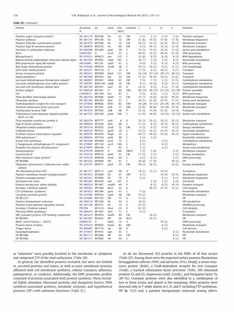
Table 2D (continued)
Protein Accession#
ID Gene Size(kDa)
Location T C D F Function
Putative sugar transport protein^ NP_881176 BP2549 Pu 61 CM 5 (2) 2 (1) 5 (2) 5 (2) Putative transportPutative sulfatase NP_881701 BP3136 Pu 72 CM 21 (8) 10 (5) 17 (8) 17 (8) Membrane biogenesisPutative TolQ-like translocation protein NP_881879 BP3346 Pu 25 CM 22 (3) 22 (3) 23 (4) 19 (3) Membrane transportPutative type III secretion protein NP_880879 BP2235 Pu 66 CM 8 (3) 10 (3) 12 (5) 12 (4) Membrane transportPyrroline-5-carboxylate reductase NP_880048 BP1280 proC 30 C 15 (3) 15 (3) 10 (2) 11 (2) amino acid metabolismRecA AAK85426 U recA 31 C 36 (6) 31 (5) 28 (4) 22 (4) DNA processingRibonuclease E NP_879331 BP0475 rne 115 C 12 (8) 8 (5) 6 (4) 8 (5) RNA degradationRibonucleotide-diphosphate reductase subunit alpha NP_881559 BP2983 nrdA 107 C 10 (7) 5 (3) 3 (2) 8 (5) Nucleotide metabolismRNA polymerase sigma 80 subunit AAC45085 BP1191 rpoD 81 C 9 (4) 5 (2) 6 (3) 6 (2) RNA processingRod shape-determining protein NP_879246 BP0374 mreB 38 C 16 (3) 19 (4) 8 (2) 12 (3) Cell morphologySCO1/SenC family protein^ NP_882237 BP3734 U 22 U 17 (3) 6 (1) 10 (2) 6 (1) TransportSerum resistance protein NP_882013 BP3494 brkA 111 OM 23 (14) 27 (14) 29 (17) 38 (21) TransportSignal peptidase I NP_881060 BP2432 lep 32 CM 11 (2) 19 (4) 14 (3) 12 (2) TransportSuccinate dehydrogenase flavoprotein subunit^ NP_880997 BP2361 sdhA 65 CM 7 (3) 7 (3) 3 (1) 11 (5) Carbohydrate metabolismSuccinate dehydrogenase iron–sulfur protein^ YP_785707 BAV1185 sdhB 26 CM 4 (1) 14 (4) 9 (3) 14 (3) Carbohydrate metabolismSuccinyl-CoA synthetase subunit beta NP_881168 BP2541 sucC 42 U 14 (3) 9 (2) 9 (2) 17 (4) Carbohydrate metabolismSurface antigen NP_880169 BP1427 U 86 OM 29 (15) 29 (15) 25 (13) 35 (18) Protein assemblyTex CAA64672 BP1144 tex 87 C 5 (3) 4 (2) 3 (2) 4 (2) RNA processingThiol:disulfide interchange protein NP_882154 BP3646 DsbA 71 CM 15 (7) 14 (6) 14 (6) 10 (4) Membrane biogenesisThreonine synthase NP_881383 BP2783 thrC 51 C 16 (5) 31 (9) 10 (3) 17 (5) Amino acid metabolismTonB-dependent receptor for iron transport NP_879666 BP0856 bfrD 82 OM 34 (20) 35 (21) 29 (18) 28 (15) Membrane transportTracheal colonization factor precursor NP_879974 BP1201 tcfA 71 OM 12 (6) 20 (8) 19 (10) 10 (6) Membrane transportTranslocation protein TolB NP_881876 BP3343 tolB 48 P 13 (3) 15 (4) 21 (6) 14 (4) TransportTrifunctional transcriptional regulator/prolineDH/P-5-C DH
NP_881353 BP2749 putA 140 C 11 (9) 10 (8) 14 (12) 14 (12) Amino acid metabolism
Twin argininte translocase protein A NP_882278 BP3777 tatA 8 U 35 (2) 35 (2) 35 (2) 35 (2) Membrane transportType II citrate synthase NP_880994 BP2358 qltA 48 C 11 (3) 8 (2) 10 (3) 10 (3) Carbohydrate metabolismUbiquinol oxidase polypeptide I NP_881514 BP2932 cyoB 72 CM 6 (3) 6 (2) 9 (4) 6 (2) Energy metabolismUridylate kinase NP_880163 BP1421 pyrH 26 C 13 (3) 10 (2) 23 (4) 16 (3) Nucleotide metabolismVirulence factors transcription regulator NP_880570 BP1878 bvgA 23 C 43 (7) 59 (9) 52 (8) 34 (5) Signal transductionDNA polymerase I NP_880026 BP1254 polA 99 C 5 (3) 5 (3) Nucleotide metabolismAcetyl-CoA synthetase NP_881040 BP2409 acsA 72 C 6 (3) 4 (2) Carbohydrate metabolism2-Oxoglutarate dehydrogenase E1 component^ NP_879903 BP1124 sucA 106 C 2 (1) 4 (2) MetabolismProbable Orn/Arg/Lys decarboxylase^ NP_879079 BP0190 U 83 C 3 (1) 4 (2) Amino acid metabolismAutotransporter NP_879378 BP0529 U 246 OM/E 7 (2) 5 (6) 2 (2) Membrane transportCytochrome B^ NP_879156 BP0276 petB 51 CM 17 (5) 3 (1) 5 (2) Energy metabolismDNA mismatch repair protein^ NP_879129 BP0244 mutL 69 C 4 (2) 6 (2) 5 (1) DNA processingHP BP3084 NP_881655 BP3084 HP 41 C 18 (4) 17 (4) 10 (2) HPUbiquinol-cytochrome C reductase iron–sulfursubunit
NP_879157 BP0277 petA 23 CM 24 (3) 24 (3) 19 (2) Energy metabolism
30s ribosomal protein S20^ NP_881377 BP2773 rspT 90 U 14 (2) 13 (1) 14 (2) TranslationPutative membrane-bound transglycosylase^ NP_881812 BP3268 Pu 47 OM 4 (1) 14 (4) 16 (4) Membrane biogenesisPutative transglycosylase^ NP_881631 BP3060 Pu 76 P 4 (2) 4 (1) 4 (1) Membrane biogenesisDermonecrotic toxin NP_881965 BP3439 dnt 161 U 5 (4) 2 (2) 4 (4) Cell deathPutative periplasmic solute-binding NP_880224 BP1487 smoM 40 U 8 (2) 8 (2) 16 (4) Putative transportSerotype 3 fimbrial subunit NP_880302 BP1568 fim3 22 E 20 (3) 27 (4) 15 (2) Cell integrityCTP synthetase (synthase) NP_881022 BP2389 pyrG 61 C 5 (2) Nucleotide metabolismCycolysin secretion protein NP_879580 BP0762 cyaD 48 CM 12 (3) Membrane transportHP BP1123 NP_879902 BP1123 HP 75 U 11 (5) HPPutative ketopantoate reductase NP_880110 BP1360 Pu 34 C 10 (2) HP/metabolismPutative transcriptional regulation protein NP_881198 BP2571 Pu 15 U 27 (2) HP/RNA processingSerotype 2 fimbrial subunit P05788 BP1119 fim2 23 E 21 (2) Cell integrityThreonyl-tRNA-synthetase NP_880233 BP1497 thrS 71 C 8 (3) TranslationABC transport protein, ATP-binding component NP_881414 BP2816 metN 40 CM 10 (2) Membrane transportHP BP3441 NP_881967 BP3441 HP 36 M/U 19 (3) HPBpH2 (novel histone — 18323) AAB40156 U bph2 16 U 19 (2) DNA processingPutative heme receptor NP_879314 BP0456 hemC 82 OM 6 (3) Membrane transportTrigger factor NP_880485 BP1774 tig 48 U 8 (2) Cell divisionExopolyphosphatase NP_879857 BP1073 ppx 55 C 4 (2) Nucleotide metabolismHP BP2486 NP_881113 BP2486 HP 45 CM 8 (2) HPHP BP3002 NP_881577 BP3002 HP 69 C 6 (2) HP
126 Y.M. Williamson et al. / Journal of Microbiological Methods 90 (2012) 119–133
as “unknown” were possibly localized to the membrane or cytoplasmand comprised 27% of the total surfaceome (Table 2B).
In general, the identified proteins included, but were not limitedto, secreted proteins and toxins, as well as outer membrane proteinsaffiliated with cell membrane synthesis, cellular transport, adhesion,pathogenesis, or virulence. Additionally, the EMF proteomic profilesconsisted of proteins associated with protein synthesis. These includ-ed highly abundant ribosomal proteins and elongation factors, DNAsynthesis-associated proteins, metabolic enzymes, and hypotheticalproteins (HP) with unknown functions (Table 2C).
In all, we discovered 163 proteins in the EMFs of all four strains(Table 2D). Among themwere the expected surface proteinsfilamentoushemagglutinin adhesin (FHA) and pertactin (Prn), OmpQ, a serum resis-tance protein (BrkA), a TonB-dependent receptor for iron transport(TonB), a tracheal colonization factor precursor (TcfA), 30S ribosomalproteins S2 and S3, chaperonin GroEL (GroEL), and elongation factor Tu(EF-Tu). Common proteins were also identified in a combination oftwo or three strains and absent in the remaining. Other proteins weredetected only in T while absent in C, D, and F, including CTP synthetase,HP Bp 1123 and, a putative ketopantoate reductase among others.
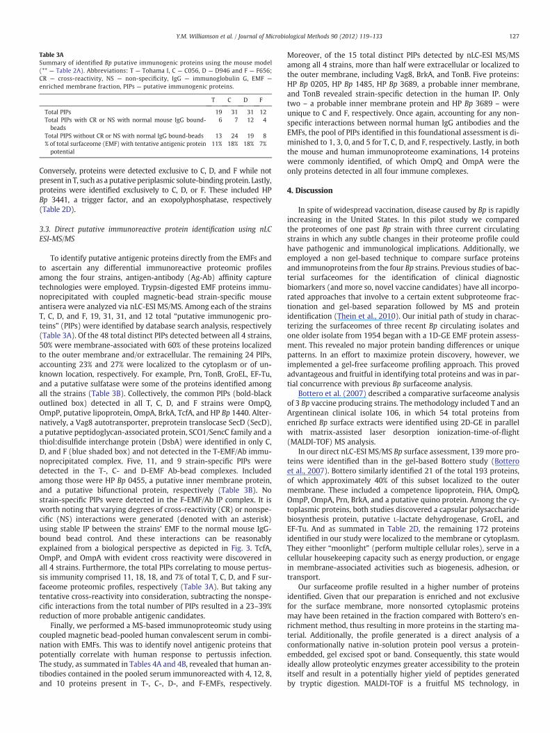
Table 3ASummary of identified Bp putative immunogenic proteins using the mouse model(** — Table 2A). Abbreviations: T — Tohama I, C — C056, D — D946 and F — F656;CR — cross-reactivity, NS — non-specificity, IgG — immunoglobulin G, EMF —
enriched membrane fraction, PIPs — putative immunogenic proteins.
T C D F
Total PIPs 19 31 31 12Total PIPs with CR or NS with normal mouse IgG bound-beads
6 7 12 4
Total PIPS without CR or NS with normal IgG bound-beads 13 24 19 8% of total surfaceome (EMF) with tentative antigenic proteinpotential
11% 18% 18% 7%
127Y.M. Williamson et al. / Journal of Microbiological Methods 90 (2012) 119–133
Conversely, proteins were detected exclusive to C, D, and F while notpresent in T, such as a putative periplasmic solute-binding protein. Lastly,proteins were identified exclusively to C, D, or F. These included HPBp 3441, a trigger factor, and an exopolyphosphatase, respectively(Table 2D).
3.3. Direct putative immunoreactive protein identification using nLCESI-MS/MS
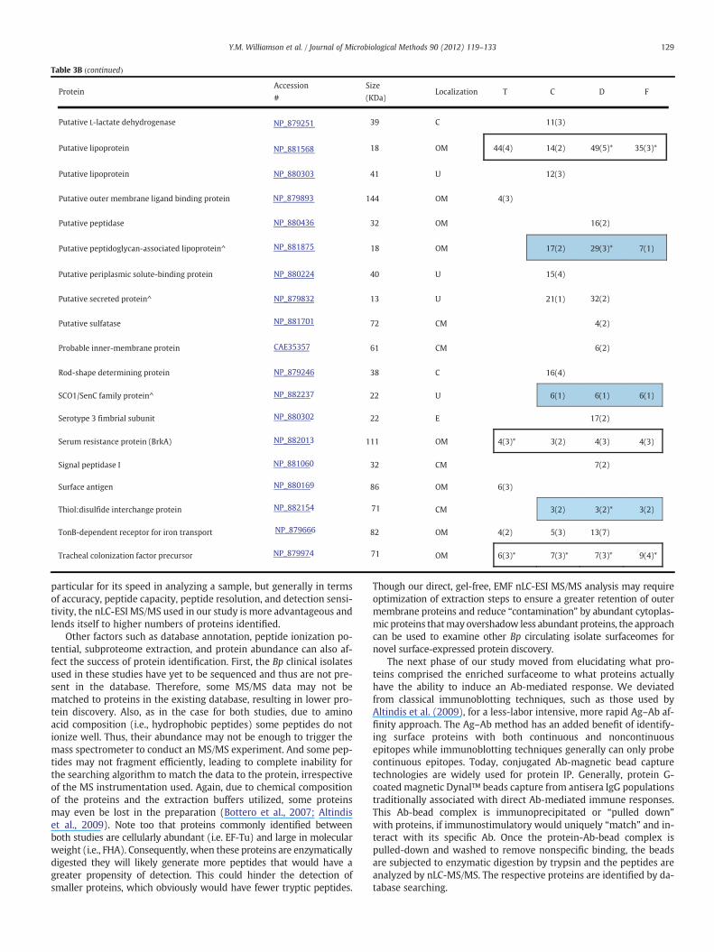
To identify putative antigenic proteins directly from the EMFs andto ascertain any differential immunoreactive proteomic profilesamong the four strains, antigen-antibody (Ag-Ab) affinity capturetechnologies were employed. Trypsin-digested EMF proteins immu-noprecipitated with coupled magnetic-bead strain-specific mouseantisera were analyzed via nLC-ESI MS/MS. Among each of the strainsT, C, D, and F, 19, 31, 31, and 12 total “putative immunogenic pro-teins” (PIPs) were identified by database search analysis, respectively(Table 3A). Of the 48 total distinct PIPs detected between all 4 strains,50% were membrane-associated with 60% of these proteins localizedto the outer membrane and/or extracellular. The remaining 24 PIPs,accounting 23% and 27% were localized to the cytoplasm or of un-known location, respectively. For example, Prn, TonB, GroEL, EF-Tu,and a putative sulfatase were some of the proteins identified amongall the strains (Table 3B). Collectively, the common PIPs (bold-blackoutlined box) detected in all T, C, D, and F strains were OmpQ,OmpP, putative lipoprotein, OmpA, BrkA, TcfA, and HP Bp 1440. Alter-natively, a Vag8 autotransporter, preprotein translocase SecD (SecD),a putative peptidoglycan-associated protein, SCO1/SencC family and athiol:disulfide interchange protein (DsbA) were identified in only C,D, and F (blue shaded box) and not detected in the T-EMF/Ab immu-noprecipitated complex. Five, 11, and 9 strain-specific PIPs weredetected in the T-, C- and D-EMF Ab-bead complexes. Includedamong those were HP Bp 0455, a putative inner membrane protein,and a putative bifunctional protein, respectively (Table 3B). Nostrain-specific PIPs were detected in the F-EMF/Ab IP complex. It isworth noting that varying degrees of cross-reactivity (CR) or nonspe-cific (NS) interactions were generated (denoted with an asterisk)using stable IP between the strains' EMF to the normal mouse IgG-bound bead control. And these interactions can be reasonablyexplained from a biological perspective as depicted in Fig. 3. TcfA,OmpP, and OmpA with evident cross reactivity were discovered inall 4 strains. Furthermore, the total PIPs correlating to mouse pertus-sis immunity comprised 11, 18, 18, and 7% of total T, C, D, and F sur-faceome proteomic profiles, respectively (Table 3A). But taking anytentative cross-reactivity into consideration, subtracting the nonspe-cific interactions from the total number of PIPs resulted in a 23–39%reduction of more probable antigenic candidates.
Finally, we performed a MS-based immunoproteomic study usingcoupled magnetic bead-pooled human convalescent serum in combi-nation with EMFs. This was to identify novel antigenic proteins thatpotentially correlate with human response to pertussis infection.The study, as summated in Tables 4A and 4B, revealed that human an-tibodies contained in the pooled serum immunoreacted with 4, 12, 8,and 10 proteins present in T-, C-, D-, and F-EMFs, respectively.
Moreover, of the 15 total distinct PIPs detected by nLC-ESI MS/MSamong all 4 strains, more than half were extracellular or localized tothe outer membrane, including Vag8, BrkA, and TonB. Five proteins:HP Bp 0205, HP Bp 1485, HP Bp 3689, a probable inner membrane,and TonB revealed strain-specific detection in the human IP. Onlytwo – a probable inner membrane protein and HP Bp 3689 – wereunique to C and F, respectively. Once again, accounting for any non-specific interactions between normal human IgG antibodies and theEMFs, the pool of PIPs identified in this foundational assessment is di-minished to 1, 3, 0, and 5 for T, C, D, and F, respectively. Lastly, in boththe mouse and human immunoproteome examinations, 14 proteinswere commonly identified, of which OmpQ and OmpA were theonly proteins detected in all four immune complexes.
4. Discussion
In spite of widespread vaccination, disease caused by Bp is rapidlyincreasing in the United States. In this pilot study we comparedthe proteomes of one past Bp strain with three current circulatingstrains in which any subtle changes in their proteome profile couldhave pathogenic and immunological implications. Additionally, weemployed a non gel-based technique to compare surface proteinsand immunoproteins from the four Bp strains. Previous studies of bac-terial surfaceomes for the identification of clinical diagnosticbiomarkers (and more so, novel vaccine candidates) have all incorpo-rated approaches that involve to a certain extent subproteome frac-tionation and gel-based separation followed by MS and proteinidentification (Thein et al., 2010). Our initial path of study in charac-terizing the surfaceomes of three recent Bp circulating isolates andone older isolate from 1954 began with a 1D-GE EMF protein assess-ment. This revealed no major protein banding differences or uniquepatterns. In an effort to maximize protein discovery, however, weimplemented a gel-free surfaceome profiling approach. This provedadvantageous and fruitful in identifying total proteins and was in par-tial concurrence with previous Bp surfaceome analysis.
Bottero et al. (2007) described a comparative surfaceome analysisof 3 Bp vaccine producing strains. The methodology included T and anArgentinean clinical isolate 106, in which 54 total proteins fromenriched Bp surface extracts were identified using 2D-GE in parallelwith matrix-assisted laser desorption ionization-time-of-flight(MALDI-TOF) MS analysis.
In our direct nLC-ESI MS/MS Bp surface assessment, 139 more pro-teins were identified than in the gel-based Bottero study (Botteroet al., 2007). Bottero similarly identified 21 of the total 193 proteins,of which approximately 40% of this subset localized to the outermembrane. These included a competence lipoprotein, FHA, OmpQ,OmpP, OmpA, Prn, BrkA, and a putative quino protein. Among the cy-toplasmic proteins, both studies discovered a capsular polysaccharidebiosynthesis protein, putative L-lactate dehydrogenase, GroEL, andEF-Tu. And as summated in Table 2D, the remaining 172 proteinsidentified in our study were localized to the membrane or cytoplasm.They either “moonlight” (perform multiple cellular roles), serve in acellular housekeeping capacity such as energy production, or engagein membrane-associated activities such as biogenesis, adhesion, ortransport.
Our surfaceome profile resulted in a higher number of proteinsidentified. Given that our preparation is enriched and not exclusivefor the surface membrane, more nonsorted cytoplasmic proteinsmay have been retained in the fraction compared with Bottero's en-richment method, thus resulting in more proteins in the starting ma-terial. Additionally, the profile generated is a direct analysis of aconformationally native in-solution protein pool versus a protein-embedded, gel excised spot or band. Consequently, this state wouldideally allow proteolytic enzymes greater accessibility to the proteinitself and result in a potentially higher yield of peptides generatedby tryptic digestion. MALDI-TOF is a fruitful MS technology, in
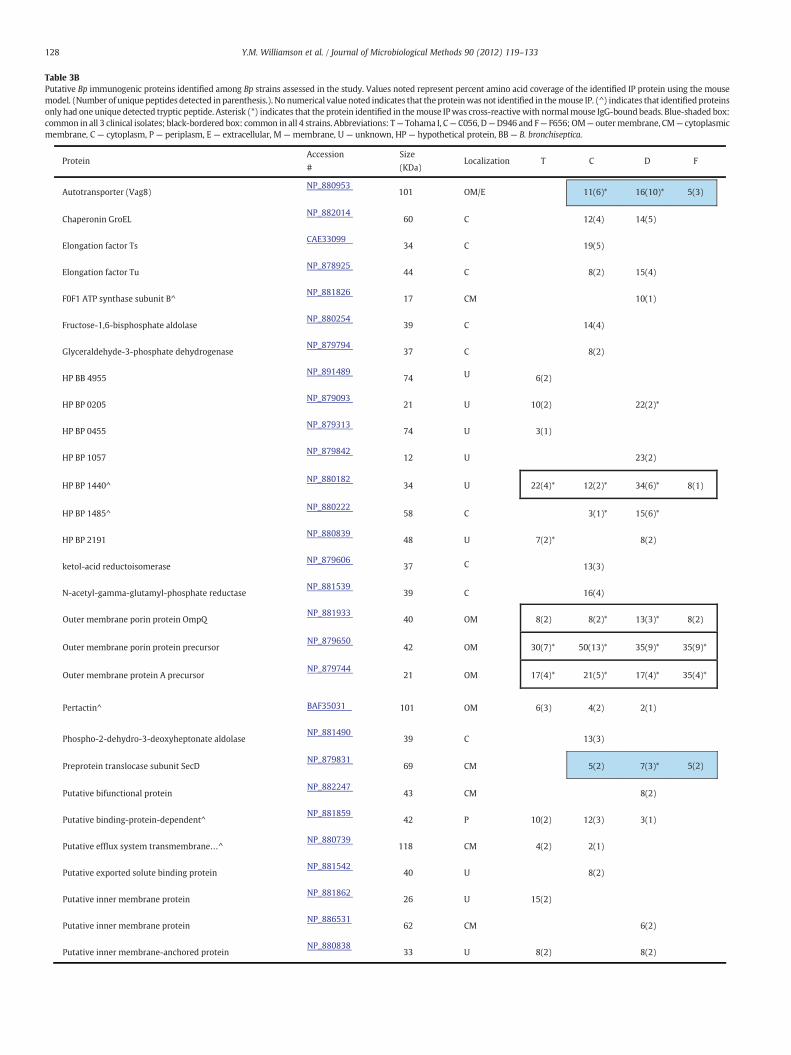
Table 3BPutative Bp immunogenic proteins identified among Bp strains assessed in the study. Values noted represent percent amino acid coverage of the identified IP protein using the mousemodel. (Number of uniquepeptides detected in parenthesis.). No numerical value noted indicates that theproteinwas not identified in themouse IP. (^) indicates that identifiedproteinsonly had one unique detected tryptic peptide. Asterisk (*) indicates that the protein identified in themouse IPwas cross-reactivewith normalmouse IgG-boundbeads. Blue-shaded box:common in all 3 clinical isolates; black-bordered box: common in all 4 strains. Abbreviations: T— Tohama I, C— C056, D—D946 and F— F656; OM— outermembrane, CM— cytoplasmicmembrane, C — cytoplasm, P — periplasm, E — extracellular, M — membrane, U — unknown, HP — hypothetical protein, BB — B. bronchiseptica.
Protein Accession
#
Size
(KDa) Localization
Autotransporter (Vag8) NP_880953
101 11(6)* 16(10)* 5(3)
Chaperonin GroEL NP_882014
60
Elongation factor Ts CAE33099
34
Elongation factor Tu NP_878925
44
F0F1 ATP synthase subunit B^ NP_881826
17
Fructose-1,6-bisphosphate aldolase NP_880254
39
Glyceraldehyde-3-phosphate dehydrogenase NP_879794
37
HP BB 4955 NP_891489
74
HP BP 0205 NP_879093
21
HP BP 0455 NP_879313
74
HP BP 1057 NP_879842
12
HP BP 1440^ NP_880182
34
HP BP 1485^ NP_880222
58
HP BP 2191 NP_880839
48
ketol-acid reductoisomerase NP_879606
37
N-acetyl-gamma-glutamyl-phosphate reductase NP_881539
39
Outer membrane porin protein OmpQ NP_881933
40
Outer membrane porin protein precursor NP_879650
42
Outer membrane protein A precursor NP_879744
21
Pertactin^ BAF35031 101
Phospho-2-dehydro-3-deoxyheptonate aldolase NP_881490
39
Preprotein translocase subunit SecD NP_879831
69 5(2) 7(3)* 5(2)
Putative bifunctional protein NP_882247
43
Putative binding-protein-dependent^ NP_881859
42
Putative efflux system transmembrane…^ NP_880739
118
Putative exported solute binding protein NP_881542
40
Putative inner membrane protein NP_881862
26
Putative inner membrane protein NP_886531
62
Putative inner membrane-anchored protein NP_880838
33
FDCT
14(5)12(4)
19(5)
15(4)8(2)
10(1)
14(4)
8(2)
6(2)
22(2)*10(2)
3(1)
23(2)
8(1)34(6)*12(2)*22(4)*
15(6)*3(1)*
8(2)7(2)*
13(3)
16(4)
8(2)13(3)*8(2)*8(2)
35(9)*35(9)*50(13)*30(7)*
35(4)*17(4)*21(5)*17(4)*
2(1)4(2)6(3)
13(3)
8(2)
3(1)12(3)10(2)
2(1)4(2)
8(2)
15(2)
6(2)
8(2)8(2)
OM/E
C
C
C
CM
C
C
U
U
U
U
U
C
U
C
C
OM
OM
OM
OM
C
CM
CM
P
CM
U
U
CM
U
128 Y.M. Williamson et al. / Journal of Microbiological Methods 90 (2012) 119–133
Putative periplasmic solute-binding protein NP_880224 40
Putative secreted protein^ NP_879832 13
Putative sulfatase NP_881701 72
Probable inner-membrane protein CAE35357 61
Rod-shape determining protein NP_879246 38
15(4)
32(2)21(1)
4(2)
6(2)
16(4)
Protein Accession
#
Size
(KDa) Localization FDCT
SCO1/SenC family protein^ NP_882237 22 6(1) 6(1) 6(1)
Serotype 3 fimbrial subunit NP_880302 22
Serum resistance protein (BrkA) NP_882013 111
Signal peptidase I NP_881060 32
Surface antigen NP_880169 86
Thiol:disulfide interchange protein NP_882154 71 3(2) 3(2)* 3(2)
TonB-dependent receptor for iron transport NP_879666 82
Tracheal colonization factor precursor NP_879974 71
17(2)
4(3)4(3)3(2)4(3)*
7(2)
6(3)
13(7)5(3)4(2)
9(4)*7(3)*7(3)*6(3)*OM
OM
CM
OM
CM
OM
E
U
C
CM
CM
U
U
Putative L-lactate dehydrogenase NP_879251 39
Putative lipoprotein NP_881568 18
Putative lipoprotein NP_880303 41
Putative outer membrane ligand binding protein NP_879893 144
Putative peptidase NP_880436 32
Putative peptidoglycan-associated lipoprotein^ NP_881875 18 17(2) 29(3)* 7(1)
11(3)
35(3)*49(5)*14(2)44(4)
12(3)
4(3)
16(2)
C
OM
U
OM
OM
OM
Table 3B (continued)
129Y.M. Williamson et al. / Journal of Microbiological Methods 90 (2012) 119–133
particular for its speed in analyzing a sample, but generally in termsof accuracy, peptide capacity, peptide resolution, and detection sensi-tivity, the nLC-ESI MS/MS used in our study is more advantageous andlends itself to higher numbers of proteins identified.
Other factors such as database annotation, peptide ionization po-tential, subproteome extraction, and protein abundance can also af-fect the success of protein identification. First, the Bp clinical isolatesused in these studies have yet to be sequenced and thus are not pre-sent in the database. Therefore, some MS/MS data may not bematched to proteins in the existing database, resulting in lower pro-tein discovery. Also, as in the case for both studies, due to aminoacid composition (i.e., hydrophobic peptides) some peptides do notionize well. Thus, their abundance may not be enough to trigger themass spectrometer to conduct an MS/MS experiment. And some pep-tides may not fragment efficiently, leading to complete inability forthe searching algorithm to match the data to the protein, irrespectiveof the MS instrumentation used. Again, due to chemical compositionof the proteins and the extraction buffers utilized, some proteinsmay even be lost in the preparation (Bottero et al., 2007; Altindiset al., 2009). Note too that proteins commonly identified betweenboth studies are cellularly abundant (i.e. EF-Tu) and large in molecularweight (i.e., FHA). Consequently, when these proteins are enzymaticallydigested they will likely generate more peptides that would have agreater propensity of detection. This could hinder the detection ofsmaller proteins, which obviously would have fewer tryptic peptides.
Though our direct, gel-free, EMF nLC-ESI MS/MS analysis may requireoptimization of extraction steps to ensure a greater retention of outermembrane proteins and reduce “contamination” by abundant cytoplas-mic proteins thatmay overshadow less abundant proteins, the approachcan be used to examine other Bp circulating isolate surfaceomes fornovel surface-expressed protein discovery.
The next phase of our study moved from elucidating what pro-teins comprised the enriched surfaceome to what proteins actuallyhave the ability to induce an Ab-mediated response. We deviatedfrom classical immunoblotting techniques, such as those used byAltindis et al. (2009), for a less-labor intensive, more rapid Ag–Ab af-finity approach. The Ag–Ab method has an added benefit of identify-ing surface proteins with both continuous and noncontinuousepitopes while immunoblotting techniques generally can only probecontinuous epitopes. Today, conjugated Ab-magnetic bead capturetechnologies are widely used for protein IP. Generally, protein G-coated magnetic Dynal™ beads capture from antisera IgG populationstraditionally associated with direct Ab-mediated immune responses.This Ab-bead complex is immunoprecipitated or “pulled down”with proteins, if immunostimulatory would uniquely “match” and in-teract with its specific Ab. Once the protein-Ab-bead complex ispulled-down and washed to remove nonspecific binding, the beadsare subjected to enzymatic digestion by trypsin and the peptides areanalyzed by nLC-MS/MS. The respective proteins are identified by da-tabase searching.
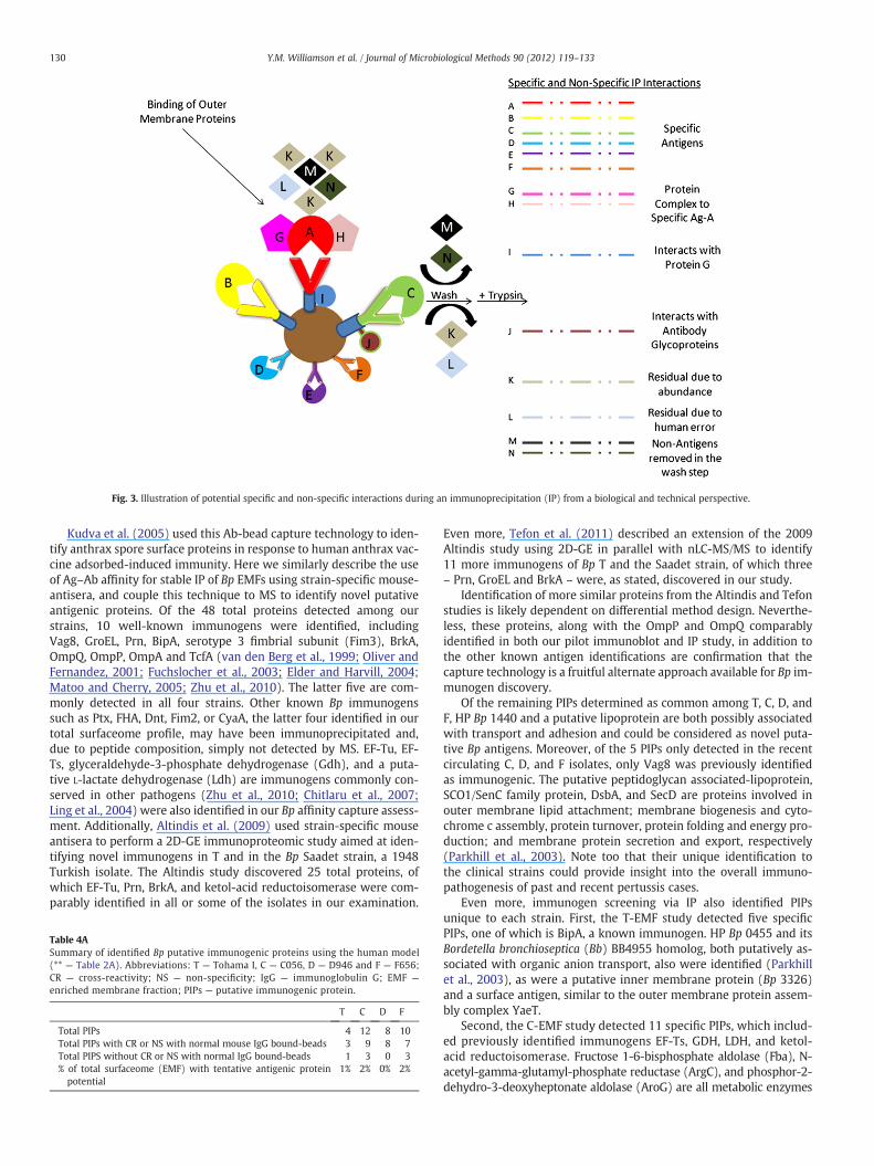
Fig. 3. Illustration of potential specific and non-specific interactions during an immunoprecipitation (IP) from a biological and technical perspective.
130 Y.M. Williamson et al. / Journal of Microbiological Methods 90 (2012) 119–133
Kudva et al. (2005) used this Ab-bead capture technology to iden-tify anthrax spore surface proteins in response to human anthrax vac-cine adsorbed-induced immunity. Here we similarly describe the useof Ag–Ab affinity for stable IP of Bp EMFs using strain-specific mouse-antisera, and couple this technique to MS to identify novel putativeantigenic proteins. Of the 48 total proteins detected among ourstrains, 10 well-known immunogens were identified, includingVag8, GroEL, Prn, BipA, serotype 3 fimbrial subunit (Fim3), BrkA,OmpQ, OmpP, OmpA and TcfA (van den Berg et al., 1999; Oliver andFernandez, 2001; Fuchslocher et al., 2003; Elder and Harvill, 2004;Matoo and Cherry, 2005; Zhu et al., 2010). The latter five are com-monly detected in all four strains. Other known Bp immunogenssuch as Ptx, FHA, Dnt, Fim2, or CyaA, the latter four identified in ourtotal surfaceome profile, may have been immunoprecipitated and,due to peptide composition, simply not detected by MS. EF-Tu, EF-Ts, glyceraldehyde-3-phosphate dehydrogenase (Gdh), and a puta-tive L-lactate dehydrogenase (Ldh) are immunogens commonly con-served in other pathogens (Zhu et al., 2010; Chitlaru et al., 2007;Ling et al., 2004) were also identified in our Bp affinity capture assess-ment. Additionally, Altindis et al. (2009) used strain-specific mouseantisera to perform a 2D-GE immunoproteomic study aimed at iden-tifying novel immunogens in T and in the Bp Saadet strain, a 1948Turkish isolate. The Altindis study discovered 25 total proteins, ofwhich EF-Tu, Prn, BrkA, and ketol-acid reductoisomerase were com-parably identified in all or some of the isolates in our examination.
Table 4ASummary of identified Bp putative immunogenic proteins using the human model(** — Table 2A). Abbreviations: T — Tohama I, C — C056, D — D946 and F — F656;CR — cross-reactivity; NS — non-specificity; IgG — immunoglobulin G; EMF —
enriched membrane fraction; PIPs — putative immunogenic protein.
T C D F
Total PIPs 4 12 8 10Total PIPs with CR or NS with normal mouse IgG bound-beads 3 9 8 7Total PIPS without CR or NS with normal IgG bound-beads 1 3 0 3% of total surfaceome (EMF) with tentative antigenic proteinpotential
1% 2% 0% 2%
Even more, Tefon et al. (2011) described an extension of the 2009Altindis study using 2D-GE in parallel with nLC-MS/MS to identify11 more immunogens of Bp T and the Saadet strain, of which three– Prn, GroEL and BrkA – were, as stated, discovered in our study.
Identification of more similar proteins from the Altindis and Tefonstudies is likely dependent on differential method design. Neverthe-less, these proteins, along with the OmpP and OmpQ comparablyidentified in both our pilot immunoblot and IP study, in addition tothe other known antigen identifications are confirmation that thecapture technology is a fruitful alternate approach available for Bp im-munogen discovery.
Of the remaining PIPs determined as common among T, C, D, andF, HP Bp 1440 and a putative lipoprotein are both possibly associatedwith transport and adhesion and could be considered as novel puta-tive Bp antigens. Moreover, of the 5 PIPs only detected in the recentcirculating C, D, and F isolates, only Vag8 was previously identifiedas immunogenic. The putative peptidoglycan associated-lipoprotein,SCO1/SenC family protein, DsbA, and SecD are proteins involved inouter membrane lipid attachment; membrane biogenesis and cyto-chrome c assembly, protein turnover, protein folding and energy pro-duction; and membrane protein secretion and export, respectively(Parkhill et al., 2003). Note too that their unique identification tothe clinical strains could provide insight into the overall immuno-pathogenesis of past and recent pertussis cases.
Even more, immunogen screening via IP also identified PIPsunique to each strain. First, the T-EMF study detected five specificPIPs, one of which is BipA, a known immunogen. HP Bp 0455 and itsBordetella bronchioseptica (Bb) BB4955 homolog, both putatively as-sociated with organic anion transport, also were identified (Parkhillet al., 2003), as were a putative inner membrane protein (Bp 3326)and a surface antigen, similar to the outer membrane protein assem-bly complex YaeT.
Second, the C-EMF study detected 11 specific PIPs, which includ-ed previously identified immunogens EF-Ts, GDH, LDH, and ketol-acid reductoisomerase. Fructose 1-6-bisphosphate aldolase (Fba), N-acetyl-gamma-glutamyl-phosphate reductase (ArgC), and phosphor-2-dehydro-3-deoxyheptonate aldolase (AroG) are all metabolic enzymes
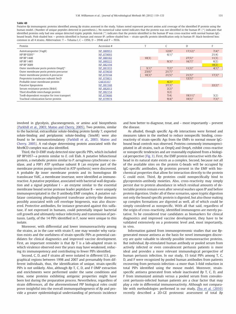
Table 4BPutative Bp immunogenic proteins identified among Bp strains assessed in the study. Values noted represent percent amino acid coverage of the identified IP protein using thehuman model. (Number of unique peptides detected in parenthesis.). No numerical value noted indicates that the protein was not identified in the human IP. (^) indicates thatidentified proteins only had one unique detected tryptic peptide. Asterisk (*) indicates that the protein identified in the human IP was cross-reactive with normal human IgG-bound beads. Pink-shaded box — protein identified in human and mouse IP; yellow-shaded box — strain-specific protein identification only in human IP; black-bordered box:common in all 4 strains. Abbreviation: T — Tohama I, C — C056, D — D946 and F — F656.
Protein Accession # T C D F
Autotransporter (Vag8) NP_880953 12(8)* 17(12)* 7(4)*
HP BP 0205^ NP_879093 6(1)* 21(4)
HP BP 1440 NP_880182 10(3) 27(5)* 11(3)
HP BP 1485 NP_880222 9(3)* 16(7)* 6(2)
HP BP 3689 NP_882194 18(2)
Outer membrane porin protein OmpQ^ NP_881933 3(1)* 7(2)* 11(3)* 4(1)*
Outer membrane porin protein precursor NP_879650 23(6)* 21(7)*
Outer membrane protein A precursor NP_879744 10(3)* 21(5)* 21(5)* 17(3)*
Preprotein translocase subunit SecD NP_879831 12(5)*
Probable inner membrane protein CAE35357 4(2)*
Putative lipoprotein NP_881568 37(3)* 22(2)*
Serum resistance protein (BrkA) NP_882013 2(2)*
Thiol:disulfide interchange protein NP_882154 3(2)
TonB-dependent receptor for iron transport NP_879666 7(4) 19(11)* 3(2)
Tracheal colonization factor protein NP_879974 9(4)* 10(5)* 7(3)*
131Y.M. Williamson et al. / Journal of Microbiological Methods 90 (2012) 119–133
involved in glycolysis, gluconeogenesis, or amino acid biosynthesis(Parkhill et al., 2003; Matoo and Cherry, 2005). Two proteins, similarto the bacterial, extracellular solute-binding protein family 7, exportedsolute-binding and periplasmic solute-binding (SmoM) were alsofound to be immunostimulatory (Parkhill et al., 2003; Matoo andCherry, 2005). A rod-shape determining protein associated with theMreBCD complex was also identified.
Third, the D-EMF study detected nine specific PIPs, which includedHP BP1057—a protein similar to E. coli Elab. A putative bifunctionalprotein, a metabolic protein similar to P. aeruginosa cytochrome c ox-idase, and a F0F1 ATP synthase subunit B (an enzyme part of themembrane proton pump involved in ATP synthesis) were discovered.A probable Bp inner membrane protein and its homologous Bbtranslocase YidC, a membrane insertase, were identified as immuno-reactive. A putative peptidase, associated with bacterial wall degrada-tion and a signal peptidase I – an enzyme similar to the essentialmembrane bound serine protease leader peptidase B –were uniquelyimmunoprecipitated in the D antibody/EMF complex. A putative sul-fatase containing phosphoglycerol transferase activity-like domainspossibly associated with cell envelope biogenesis, was also discov-ered. Protective antibodies, for instance generated against this sulfa-tase, if not expressed in humans, could potentially impair bacterialcell growth and ultimately reduce infectivity and transmission of per-tussis. Lastly, of the 14 PIPs identified in F, none were unique to thisisolate.
Moreover, with differential and lower immunoreactivity amongthe strains, as in the case with strain F, one may wonder why varia-tion exists and the usefulness of strain-specific PIPs as potential can-didates for clinical diagnostics and improved vaccine development.First, an important reminder is that Bp T is a lab-adapted strain inwhich virulence observed over the years may have weakened, reduc-ing its immunopotency and contributing to fewer PIPs identified.
Second, C, D, and F strains all were isolated in different U.S. geo-graphical regions between 1998 and 2007 and presumably from dif-ferent individuals, thus, the identification of unique, strain-specificPIPs is not unlikely. Also, although Bp T, C, D, and F OMP extractionand enrichments were performed under the same conditions andtime, some proteins exhibiting antigenic properties might havebeen lost during the preparation process. Nevertheless, regardless ofstrain differences, all the aforementioned PIP biological roles couldprove insightful into the overall immunopathogenesis of Bp and pro-vide a greater epidemiological understanding of pertussis incidence
and how better to diagnose, treat, and – most importantly – preventthe disease.
As alluded, though specific Ag–Ab interactions were formed andmeasures taken in the method to reduce nonspecific binding, cross-reactivity of strain-specific Ags from the EMFs to normal mouse IgG-bound bead controls was observed. Proteins commonly immunopreci-pitated in all strains, such as OmpQ and OmpA, exhibit cross-reactiveor nonspecific tendencies and are reasonably explained from a biologi-cal perspective (Fig. 3). First, the EMF protein interactive with the Ab-bead in its natural state exists as a complex. Second, because not allof the available sites on the protein G-beads will be occupied byIgG-specific antibodies, Bp proteins present in the EMF with bio-chemical properties that allow for interaction directly to the proteinG could exist. Third, Bp proteins could nonspecifically bind toglycoprotein-antibody moieties. Also, cross-reactivity may simplypersist due to protein abundance in which residual amounts of de-tectable protein remain even after several washes upon IP and beforeprotein digestion. Under all of these scenarios, upon tryptic digestionnot only the bound specific protein is digested, but also thosemakingup complex formations are digested as well, all of which could besimply considered as nonspecific. With all that said, regardless ofthe origin of cross-reactivity, identified immunogens are merely pu-tative. To be considered true candidates as biomarkers for clinicaldiagnostics and improved vaccine development, they have to bevalidated extensively on a proteomic level and, most importantly,in vivo.
Information gained from immunoproteomic studies that use Bp-generated mouse antisera as the basis for novel immunogen discov-ery are quite valuable to identify possible immunoreactive proteins.But individual, Bp-stimulated human antibody or pooled serum fromactively infected or even convalescent pertussis patients is moreideal and provides a more relevant immunological perspective ofhuman pertussis infection. In our study, 15 total PIPs among T, C,D, and F were recognized by pooled human antibodies from patientsrecovering from pertussis infection—a more than 3-fold reduction intotal PIPs identified using the mouse model. Moreover, strain-specific antisera generated from whole inactivated Bp T, C, D, andF from immunized animals versus a pooled serum from convales-cent pertussis-infected human patients are a clear factor that mayplay a role in differential immunoreactivity. Although not compara-ble with methodologies performed in our study, Zhu et al. (2010)recently described a 2D-GE proteomic assessment of total Bp
132 Y.M. Williamson et al. / Journal of Microbiological Methods 90 (2012) 119–133
membrane enriched and extracellular protein preparations. The pur-pose was to investigate the complete set of Bp antigenic proteins usingantisera generated in response to Chinese whole-cell, vaccine-inducedimmunity.
In the Zhu study, twice as many total immunogens were identifiedfor human sera compared with our human sera pool. The disparitywas possibly due to Bp strain differences and lower post-pertussis infec-tion specific circulating Ab titer levels of the pooled human antisera(caused by prior serum dilution) used in our study. Interestingly, irre-spective of the subproteome extraction, protein enrichment, andimmunodetection methods used, OmpQ was the only protein pulleddown using both mouse and human antisera from all four Bp EMFs inour study, as well as in Zhu's immunoproteomic examinations.
In conclusion, we used Ag–Ab affinity capture technologies in con-junction with MS to identify immunoreactive proteins in three recentcirculating Bp strains. The overall goals were to identify biomarkersthat could be evaluated for use in pertussis strain differentiationand in clinical diagnostics, and to detect novel targets for design ofprevention and therapeutic strategies.
As a first step in this qualitative proteomics study, we examinedthe applicability of a gel-free direct enzymatic-treated Bp EMF surfa-ceome analysis. Common and strain-specific surface expressed pro-tein profiles compared to the current vaccine-producing T strainwere generated, suggesting that there may be subtle differences ofsurface protein expression among Bp strains. Further characterizationof these differentially expressed proteins at the molecular level iswarranted; which may reveal minor changes that could have implica-tions on virulence and pathogenesis.
Second, antibodies generated from pertussis interperitoneal mouseinoculation were used to assess protein immunogenicity and revealedthe identification of known and unknown Bp antigens using a stableIP approach. A comprehensive proteomic examination and an in vivoimmunopotency assessment are further needed to validate these PIPsas true biomarker and vaccine candidates. Also, reassessing the immu-noproteome of our unique Bp strains by stable IP using several humansera from active pertussis infected human patients will provide amore consistent depiction of the pertussis immunoproteome. Addition-ally, efforts to enhance novel immunogen discoverymay require immu-nodepletion of known and highly abundant antigenic-specific antibodypopulations, such as OmpQ and BrkA. Highly abundant antigenic pro-teins would in essence generate a larger specific IgG antibody pool.This larger antibody subset would have a greater chance of binding toprotein G-beads, which invariably could overshadow lower expressedimmunostimulatory proteins.
Nonetheless, the gel-free approach described here for immunopro-teome identification remains a fruitful alternative for antigen discovery.And because this is a pilot study using a small number of temporallylimited and genetically diverse strains, future studies will entail usingthe described methods to examine a larger set of epidemiologically sig-nificant strainswith these putative, novel candidates laying the founda-tion for clinical diagnostic design and improved vaccine development,leading to the ultimate prevention of this deadly infection.
4.1. Disclaimer
References in this article to any specific commercial products, process,service, manufacturer, or company do not constitute an endorsement ora recommendation by the U.S. Government or the Centers for DiseaseControl and Prevention. The findings and conclusions in this report arethose of the authors and do not necessarily represent the views of CDC.
References
Advani, A., Donnelly, D., Hallander, H., 2004. Reference system for characterization ofBordetella pertussis pulsed-field gel electrophoresis profiles. J. Clin. Microbiol. 42(7), 2890–2897.
Altindis, E., Tefon, B.E., Özcengiz, Y., Becher, D., Hecker, M., Özcengiz, G., 2009. Immu-noproteomic analysis of Bordetella pertussis and identification of new immunogen-ic proteins. Vaccine 27, 542–548.
Bart, M.J., van Gent, M., van der Heide, H.G., Boekhorst, J., Hermans, P., Parkhill, J., Mooi,F.R., 2010. Comparative genomics of prevaccination and modern Bordetella pertus-sis strains. BMC Genomics 11 (627), 1–10.
Bienvenut, W.V., Sanchez, J.C., Karmime, A., Rouge, V., Rose, K., Binz, P.A., Hochstrasser,D.F., 1999. Toward a clinical molecular scanner for proteome research: parallelprotein chemical processing before and during western blot. Anal. Chem. 17(21), 4800–4807.
Bordet, J., Gengou, O., 1906. Le microbe de la coqueluche. Ann. Inst. Pasteur 20, 48–68.Bottero, D., Gaillard, M.E., Fingermann, M., Weltman, G., Fernandez, J., Sisti, F., Graieb,
A., Roberts, R., Rico, O., Rios, G., Regueira, M., Binsztein, N., Hozbor, D., 2007.Pulsed-field gel electrophoresis, pertactin, pertussis toxin S1 subunit polymor-phisms, and surfaceome analysis of vaccine and clinical Bordetella pertussis strains.Clin. Vaccine Immunol. 14, 1490–1498.
Chen, G., Prama, B.N., 2008. LC-MS for protein characterization: current capabilities andfuture trends. Expert Rev. Proteomics 5, 435–444.
Chitlaru, T., Gat, O., Grosfeld, H., Inbar, I., Gozlan, Y., et al., 2007. Identification of invivo-expressed immunogenic proteins by serological proteome analysis of the Ba-cillus anthracis secretome. Infect. Immun. 75, 2841–2852.
Das, P., 2002. Whooping cough makes global comeback. Lancet Infect. Dis. 2, 322.Dreisbach, A., Hempel, K., Buist, G., Hecker, M., Cecher, D., van Dijl, J.M., 2010. Profiling
the surfaceome of Staphylococcus aureus. Proteomics 10, 1–15.Dworzanski, J.P., Snyder, A.P., 2005. Classification and identification of bacteria using
mass spectrometry-based proteomics. Exp. Rev. Proteomics 2 (6), 863–878.Elder, K.D., Harvill, E.T., 2004. Strain-dependent role of BrkA during Bordetella pertussis
infection of the murine respiratory tract. Infect. Immun. 72, 5919–5924.Fuchslocher, B., Millar, L.L., Cotter, P.A., 2003. Comparison of bipA alleles within and
across Bordetella species. Infect. Immun. 71, 3043–3052.Han, X., Aslanian, A., Yates III, J.R., 2008. Mass spectrometry for proteomics. Curr. Opin.
Chem. Biol. 12, 483–490.Hardwick, T.H., Cassiday, P., Weyant, R.S., Bisgard, K.M., Sanden, G.N., 2002. Changes in
predominance and diversity of genomic subtypes of Bordetella pertussis isolated inthe United States, 1935 to 1999. Emerg. Infect. Dis. 8 (1), 44–49.
He, Q., Mertsola, J., 2008. Factors contributing to pertussis resurgence. Future Micro-biol. 3, 329–339.
http://www.cdc.gov/.Hulbert, R.R., Cotter, P.A., 2009. Laboratory maintenance of Bordetella pertussis. Curr.
Protoc. Microbiol. 15 (4B), 1.1–1.9.Jabbour, R.E., Wade, M.M., Desphande, S.V., Stanford, M.F., Wick, C.H., Zullich, A.W.,
Snyder, A.P., 2010. Identification of Yersinia pestis and Escherichia coli strains bywhole cell and membrane protein extracts with mass spectrometry-based proteo-mics. J. Proteome Res. 9 (7), 3647–3655.
Keller, A., Nesvizhskii, A.I., Kolker, E., Aebersold, R., 2002. Empirical statistical model toestimate the accuracy of peptide identifications made by MS/MS and databasesearch. Anal. Chem. 74, 5383–5392.
King, A.J., Berbers, G., van Oirschot, H.F., Hoogerhout, P., Knipping, K., Mooi, F.R., 2001.Role of the polymorphic region 1 of the Bordetella pertussis protein pertactin in im-munity. Microbiology 147, 2885–2895.
Kudva, I.T., Griffin, R.W., Garren, J.M., Calderwood, S.B., John, M., 2005. Identification ofa protein subset of the anthrax spore immunome in humans immunized with theanthrax vaccine adsorbed preparation. Infect. Immun. 73 (9), 5685–5696.
Kustos, I., Kocsis, B., Kilár, F., 2007. Bacterial outer membrane protein analysis by elec-trophoresis and microchip technology. Expert Rev. Proteomics 4 (1), 91–106.
Laemmli, U.K., 1970. Cleavage of structural proteins during the assembly of the head ofbacteriophage t4. Nature 227, 680–685.
Lee, E.Y., Choi, D.S., Kim, K.P., Gho, Y.S., 2008. Proteomics in gram-negative bacterialouter membrane vesicles. Mass Spec. Rev. 27, 535–555.
Ling, E., Feldman, G., Portnoi, M., Dagan, R., Overweg, K., Mulholland, F., Chalifa-Caspi,V., Wells, J., Mizrachi-Nebenzahl, Y., 2004. Glycolytic enzymes associated withthe cell surface of Streptococcus pneumoniae are antigenic in humans and elicit pro-tective immune responses in the mouse. Clin. Exp. Immunol. 138, 290–298.
Litt, D.J., Neal, S.E., Fry, N.K., 2009. Changes in genetic diversity of the Bordetella pertus-sis population in the United Kingdom between 1920 and 2006 reflect vaccinationcoverage and emergence of a single dominant clonal type. J. Clin. Microbiol. 47,680–688.
Locht, C., 2008. A common vaccination strategy to solve unsolved problems of tubercu-losis and pertussis? Microbes Infect. 10, 1051–1056.
Matoo, S., Cherry, J.D., 2005. Molecular pathogenesis, epidemiology, and clinical mani-festations of respiratory infections due to Bordetella pertussis and other Bordetellasubspecies. Clin. Microbiol. Rev. 18 (2), 326–382.
Molloy, M.D., 2008. Isolation of bacterial cell membrane proteins using carbonate ex-traction. Methods Mol. Biol. 424, 397–401.
Nesvizhskii, A.I., Keller, A., Kolker, E., Aebersold, R., 2003. A statistical model for identi-fying proteins by tandem mass spectrometry. Anal. Chem. 75, 4646–4658.
O'Farrell, P.H., 1975. High resolution two-dimensional electrophoresis of protein. J.Biol. Chem. 250, 4007–4021.
Oliver, D.C., Fernandez, R.C., 2001. Antibodies to BrkA augment killing of Bordetella per-tussis. Vaccine 20, 235–241.
Parkhill, J., Sebaihia, M., Preston, A., Murphy, L.D., Thomson, N., Harris, D.E., et al., 2003.Comparative analysis of the genome sequences of Bordetella pertussis, Bordetellaparapertussis and Bordetella bronchioseptica. Nat. Genet. 35, 32–40.
Poolman, J., Hamstra, H.J., Barlow, A., Kuipers, B., Loggen, H., Nagel, J., 1990. Outermembrane vesicles of Bordetella pertussis are protective antigens in the mouse in-tracerebral challenge model. FDA 202–206.

133Y.M. Williamson et al. / Journal of Microbiological Methods 90 (2012) 119–133
Singh, M., Lingappan, K., 2006. Whooping cough—the current scene. Chest 130, 1547–1553.Somner, U., Peterson, J., Pfeiffer, M., Schrotz-King, P., Morsczeck, C., 2010. Comparison
of surface proteomes of enterotoxigenic (ETEC) and commensal Escherichia colistrains. J Microbiol Methods 83 (2010), 13–19.
Taylor, B., Fahm, R., 1999. Pasteur Merieux Connaught five-component acellular per-tussis vaccine. Biologicals 27, 103–104.
Tefon, B., MaaB, S., Ozcengiz, E., Becher, D., Hecker, M., Ozcengiz, G., 2011. A compre-hensive analysis of Bordetella pertussis surface proteome and identification ofnew immunogenic proteins. Vaccine 29 (19), 3583–3595.
Thein, M., Sauer, G., Paramasivam, N., Grin, I., Linke, D., 2010. Efficient subfractionation ofgram-negative bacteria for proteomics studies. J. Proteome Res. 9 (12), 6135–6147.
Towbin, H., Staehelin, T., Gordon, J., 1979. Electrophoretic transfer of proteins frompolyacrylamide gets to nitrocellulose sheets: procedure and some applications.Proc. Natl. Acad. Sci. U. S. A. 76, 4350–4354.
van den Berg, B.M., Beekhuizen, H., Mooi, F.R., van Furth, R., 1999. Role of antibodies againstBordetella pertussis virulence factors in adherence of Bordetella pertussis and Bordetellaparapertussis to human bronchial epithelial cells. Infect. Immun. 67, 1050–1055.
van Loo, I.H., Heuevelman, K.J., King, A.J., Mooi, F.R., 2002. Multilocus sequence typingof Bordetella pertussis based on surface protein genes. J. Clin. Micro. 40, 1994–2001.
Wilkins, M.R., Appel, R.D., Williams, K.L., Hochstrasser, D.F. (Eds.), 2007. Proteome Re-search: Concepts, Technology and Application, 2nd Ed. Springer, Berlin. [pp.].
Yu, N.Y., Wagner, J.R., Laird, M.R., Mellj, G., Rey, S., Lo, R., Dao, P., Sahinalp, S.C., Ester, M.,Foster, L.J., Brinkman, F.S., 2010. Improved protein subcellular localization predic-tion with refined localization subcategories and predictive capabilities for all pro-karyotes. Bioinformatics 26 (13), 1608–1615.
Zhu, Y.Z., Cai, C.S., Zhang,W., Guo, H.X., Zhang, J.P., Ji, Y.Y., Ma, G.Y.,Wu, J.L., Li, Q.T., Lu, C.P.,Guo, X.K., 2010. Immunoproteomic analysis of human serological antibody responsesto vaccination and whole-cell pertussis vaccine (WCV). PLoS One 5 (11), 1–11.