from accreting and transitional millisecond pulsars to rotation ...
ORIGINAL ARTICLE
Distinct proteomic profiles of amphetamine
self-administration transitional states
WM Freeman1
K Brebner2
SG Amara3
MS Reed4
J Pohl4
AG Phillips2
1Department of Pharmacology, Penn StateCollege of Medicine, Hershey, PA, USA;2Department of Psychiatry, University of BritishColumbia, Vancouver, BC, Canada; 3Departmentof Neurobiology, University of Pittsburgh Schoolof Medicine, Pittsburgh, PA, USA; 4Microchemical& Proteomics Facility, Emory University School ofMedicine, Atlanta GA, USA
Correspondence:WM Freeman, Department ofPharmacology, Penn State College ofMedicine, 500 University Drive, H078, POBox 850, Hershey, PA 17033, USA.Tel: 717 531 0003 ext.280248Fax: 717 531 5013E-mail: [email protected]
Received: 17 December 2004Revised: 23 February 2005Accepted: 25 February 2005Published online 26 April 2005
ABSTRACTIn the rat, continuous access to d-amphetamine (d-AMPH) leads to lengthybouts of self-administration, voluntary abstinence, and relapse to self-administration. Previous studies have revealed that the progression frompsychostimulant self-administration to abstinence to relapse is mediated inpart by the ventral hippocampus. Stimulation of the ventral subiculum(vSub) during voluntary abstinence from d-AMPH self-administrationreinstates self-administration and increases nucleus accumbens (NAc)dopamine efflux. Quantitative proteomic examination of the hippocampusfrom rats naı̈ve to amphetamine, during a self-administration session ‘Binge’,during voluntarily abstinence ‘Abstinent’, and after reinstatement of self-administration ‘Relapse’, revealed a differential proteomic state duringabstinence. Actin- and cytoskeletal-related proteins were over-representedin the changes occurring during abstinence and suggest a decrease in actinfilament polymerization. These changes may underlie alterations in neuronaltone during abstinence that could affect both neurotransmission andbehavior. These data provide the first classification of addiction-relatedbehaviors based on clustering of quantitative proteomic measurements.The Pharmacogenomics Journal (2005) 5, 203–214. doi:10.1038/sj.tpj.6500309Published online 26 April 2005
Keywords: drug abuse; gene expression; bioinformatics; 2-DIGE; ontology; massspectrometry
INTRODUCTIONThe psychostimulant and reinforcing properties of d-amphetamine (d-AMPH)appear to be mediated in part by dopamine (DA), specifically DA efflux in theventral striatum/nucleus accumbens (NAc).1 Rats readily engage in intravenousself-administration (IVSA) of d-AMPH and given continuous access, will engagein a cyclic IVSA pattern of binge, abstinence, and relapse,2 reflecting the typicaluse pattern of human drug abusers.3 Understanding the neurobiologicalsubstrates mediating the transition from one behavioral phase to the next iscritical to the development of more effective treatment strategies. The onset ofabstinence can be characterized neurochemically by diminished NAc DA efflux,whereas increased NAc DA efflux precedes reinstatement.2,4 If d-AMPH isadministered noncontingently during abstinence, no resulting increase in NAcDA efflux is observed.4 This point emphasizes the importance of using IVSA inthe study of drug reinforcement since noncontingent drug administration doesnot replicate the neurochemistry of self-administered drugs.2,5
The NAc receives glutamatergic inputs from the ventral subiculum (vSub) ofthe hippocampal formation6 and this circuit has been implicated in the
The Pharmacogenomics Journal (2005) 5, 203–214& 2005 Nature Publishing Group All rights reserved 1470-269X/05 $30.00
www.nature.com/tpj

reinstatement of d-AMPH IVSA. Stimulation of the vSubincreases NAc DA levels and reinstates d-AMPH seekingbehavior in a glutamate-dependent manner.7–9 Theta burststimulation of the vSub reinstates cocaine IVSA in aglutamate-dependent manner,10 while inactivation of thevSub blocks cocaine IVSA reinstatement elicited by both adrug-prime and a conditioned stimulus.11 These reportsclearly suggest that hippocampal and glutamatergic me-chanisms may be involved in modulating reinstatement/relapse of psychostimulant drug self-administration.
The application of functional genomic approaches tosubstance abuse has provided considerable information ondrug-induced alterations in gene expression within variousbrain regions.12,13 Advances in proteomic technology nowmake it possible to detect and quantify alterations inabundance and modification of thousands of proteinssimultaneously.14 Given that drug addiction is a dynamicbehavioral and biochemical process, the ability to simulta-neously assess the expression and modification of multipleproteins would lead to a more complete understanding ofaddiction pathophysiology. Combining proteomics technol-ogy with animal models of d-AMPH IVSA binge/abstinencecycling will facilitate the discovery of mechanisms thatmediate long-term behavioral changes associated with theaddictive process.
Using two-dimensional difference gel electrophoresis (2-DIGE), which separates proteins based on isoelectric pointand molecular weight and uses fluorescent dyes forquantitation,15,16 we quantitatively compared hippocampalprotein expression and modification in four temporalgroups: d-AMPH-naı̈ve controls (Naı̈ve), 3-h into IVSAsession (Binge), after a d-AMPH binge followed by 3-h ofvoluntary abstinence from IVSA (Abstinent), and afterbinging and abstinence followed by 3-h of IVSA reinstate-ment (Relapse). The goal of this study was to examine abrain region containing the cell bodies of glutamatergicafferents known to alter d-AMPH drug self-administration.Proteins were identified by matrix-assisted laser desorptionionization time-of-flight–time-of-flight tandem mass spec-trometry (MALDI-ToF/ToF), and patterns of protein expres-sion during the different behaviors were compared.
MATERIALS AND METHODSAnimal MethodsSubjects
Male Sprague–Dawley rats (University of British Columbiabreeding facility, Vancouver, Canada), weighing approxi-mately 300 g, were individually housed in plastic cages in acolony room with an ambient temperature of 221C, andadapted to a reverse light : dark cycle (lights off at 0400hours). Food (Purina Rat Chow) and water were available adlibitum during all phases of the experiments, except as notedbelow. All experiments were conducted in accordance withthe standards of the Canadian Council on Animal Care andwere approved by the Committee on Animal Care, Uni-versity of British Columbia.
Surgery and training
After a 7-day environmental acclimation period, rats werefood deprived for 16 h before being placed in operantchambers (Med Associates, Inc., VT, USA). Responding onthe active lever was engendered and maintained under afixed ratio-1 (FR1) schedule of reinforcement with 45 mgNoyes food pellets. Food training was conducted daily in 2-hsessions for a period of 7 days. The operant chambers usedfor food training were identical to the chambers used fordrug self-administration; however, they were only equippedwith one lever and there was no house-light to signal thestart or end of the session. Upon completion of training, ratswere returned to free-feeding conditions. Following foodtraining, rats were surgically implanted with a chronicallyindwelling jugular cannula, as previously described.17
Briefly, rats were anaesthetized with ketamine (100 mg/kg,i.p.) and xylazine (8 mg/kg, i.p.) prior to surgery. A silasticcannula was implanted into the right jugular vein and thefree end of the cannula was passed subcutaneously to thehead of the rat and was connected to a plastic screw-onconnector (Plastics-One, Inc.). The assembly was mountedon an exposed area of the skull using four stainless steeljewelers’ screws and dental acrylic. All cannulae wereflushed regularly with heparinized saline (10 IU) to helpmaintain patency. d-AMPH IVSA training sessions began 7days after surgery.
Amphetamine self-administration
Rats were trained to self-administer d-AMPH (0.1 mg/kg perinjection) under an FR1 schedule of reinforcement (max-imum 12 injections per day over a 3-day period, withsessions lasting 5 h). Rats were placed in an operant chamber(Med Associates, Inc., St Albans, VT, USA) equipped with ahouse-light, one active, and one nonactive lever. Cue-lightswere located above each lever and drug infusions wereaccompanied by flashing of the cue-light located above theactive lever. The start of the drug session was signaled byillumination of the house-light for 30 s prior to the deliveryof a priming injection of d-AMPH. The house-light remainedilluminated for the remainder of the session to signal theavailability of the drug. After the priming injection, eachresponse on the active lever resulted in the delivery of salinesolution containing d-AMPH over a 5-s period. Infusionswere followed by a 20-s timeout period during which thecue-light was turned off and presses on the levers had noprogrammed consequence. Responses on both the activeand inactive levers were recorded throughout the session.Rats were removed from the test chambers 30 min after theend of each session. Rats that completed the maximumnumber of injections available during all 3 daily trainingsessions (a total of 13 infusions including the priming doseper session per day) were then placed in the operantchambers for a 24-h session on day 4. Following 24 h inhome cages (day 5), rats were randomly assigned to theBinge, Abstinent, or Relapse group, and on day 6 werereturned to the operant chambers. Rats in the Binge groupself-administered d-AMPH for 3 h before being killed via an
Proteomic profiles of amphetamine self-administrationWM Freeman et al
204
The Pharmacogenomics Journal
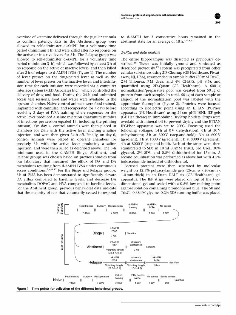
overdose of ketamine delivered through the jugular cannulato confirm patency. Rats in the Abstinent group wereallowed to self-administer d-AMPH for a voluntary timeperiod (minimum 3 h) and were killed after no responses onthe active or inactive levers for 3 h. The Relapse group wasallowed to self-administer d-AMPH for a voluntary timeperiod (minimum 3 -h), which was followed by at least 3 h ofno response on the active or inactive levers, and then killedafter 3 h of relapse to d-AMPH IVSA (Figure 1). The numberof lever presses on the drug-paired lever as well as thenumber of lever presses on the inactive lever, and interinfu-sion time for each infusion were recorded via a computerinterface system (MED Associates Inc.), which controlled thedelivery of drug and food. During the 24-h and unlimitedaccess test sessions, food and water were available in theoperant chamber. Naı̈ve control animals were food trained,implanted with cannulae, and recuperated for 7 days beforereceiving 3 days of IVSA training where responses on theactive lever produced a saline injection (maximum numberof injections per session equaled 13, including the priminginfusion). On day 4, control animals were then placed inchambers for 24 h with the active lever eliciting a salineinjection, and were then given 24-h off. Finally, on day 6,control animals were placed in operant chambers forprecisely 3 h with the active lever producing a salineinjection, and were then killed as described above. The 3-hminimum used in the d-AMPH Binge, Abstinent, andRelapse groups was chosen based on previous studies fromour laboratory that measured the efflux of DA and DAmetabolites resulting from d-AMPH IVSA under continuousaccess conditions.2,4,8,17 For the Binge and Relapse groups,3 h of IVSA has been demonstrated to significantly elevateDA efflux compared to baseline levels, and decrease DAmetabolites DOPAC and HVA compared to baseline levels.For the Abstinent group, previous behavioral data indicatethat the majority of rats that voluntarily ceased to respond
to d-AMPH for 3 consecutive hours remained in theabstinent state for an average of 18 h.2,4,8,17
2-DIGE and data analysis
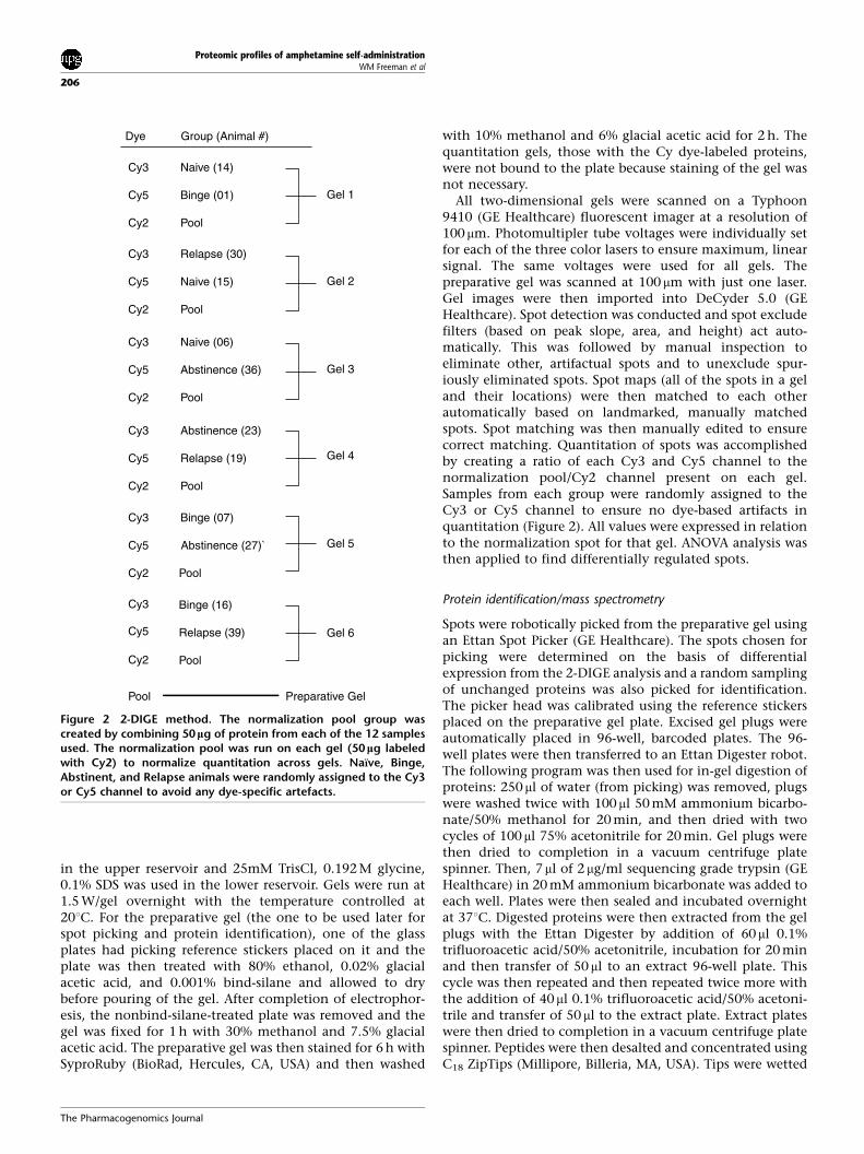
The entire hippocampus was dissected as previously de-scribed.18 Tissue was initially ground and sonicated asdescribed previously.19 Protein was precipitated from othercellular substances using 2D-Cleanup (GE Healthcare, Piscat-away, NJ, USA), resuspended in sample buffer (30 mM TrisCl,2 M Thiourea, 7 M Urea, and 4% CHAPS, pH 8.5), andquantified using 2D-Quant (GE Healthcare). A 600 mgnormalization/preparative pool was created from 50mg ofprotein from each sample. In total, 50 mg of each sample oraliquot of the normalization pool was labeled with theappropriate fluorophor (Figure 2). Proteins were focusedaccording to isoelectric point using an ETTAN IPGPhorapparatus (GE Healthcare) using 24 cm pH3-10NL IEF gels(GE Healthcare) in Immobiline DryStrip holders. Strips wereoverlaid with mineral oil to prevent drying and the ETTANIPGPhor apparatus was set to 201C. Focusing used thefollowing voltages: 14 h at 0 V (rehydration); 6 h at 30 V(rehydration); 3 h at 300 V (step-and-hold); 3 h at 600 V(gradient); 3 h at 1000 V (gradient); 3 h at 8000 V (gradient);4 h at 8000 V (step-and-hold). Each of the strips were thenequilibrated to SDS in 10 ml 50 mM TrisCl, 6 M Urea, 30%glycerol, 2% SDS, and 0.5% dithiothreitol for 15 min. Asecond equilibration was performed as above but with 4.5%iodoacetomide instead of dithiothreitol.
Focused proteins were then separated by molecularweight on 12.5% polyacrylamide gels (26 cm-w�20 cm-h�1.0 mm-thick) in an Ettan DALT six (GE Healthcare) gelapparatus. The IEF strips were placed on top of the two-dimensional gel and sealed with a 0.5% low melting pointagarose solution containing bromophenol blue. The 50 mMTrisCl, 0.384 M glycine, 0.2% SDS running buffer was placed
Food training Surgery Recuperationd-AMPHtraining
d-AMPHIVSA
No access
7 days 1 day7 days 3 days 1 day
7 days 1 day7 days 3 days 1 day
d-AMPHIVSA
3 hrs
d-AMPHIVSA
Voluntary length(32.8-h±6.2)
Voluntaryabstinence
3 hrs
d-AMPHIVSA
Voluntary length(29.8-h±3.4)
Voluntaryabstinence
Voluntary length(15-h±4.8)
d-AMPHIVSA
3 hrs
Binge
Abstinent
Relapse
Food training Surgery RecuperationSalinetraining
24hr accesssaline
No access Saline access
3hrsNaive
Sacrifice
Sacrifice
Sacrifice
Sacrifice
Trai
ning
Ses
sion
sTe
st S
essi
on
Figure 1 Time points for collection of the different behavioral groups.
Proteomic profiles of amphetamine self-administrationWM Freeman et al
205
www.nature.com/tpj
in the upper reservoir and 25mM TrisCl, 0.192 M glycine,0.1% SDS was used in the lower reservoir. Gels were run at1.5 W/gel overnight with the temperature controlled at201C. For the preparative gel (the one to be used later forspot picking and protein identification), one of the glassplates had picking reference stickers placed on it and theplate was then treated with 80% ethanol, 0.02% glacialacetic acid, and 0.001% bind-silane and allowed to drybefore pouring of the gel. After completion of electrophor-esis, the nonbind-silane-treated plate was removed and thegel was fixed for 1 h with 30% methanol and 7.5% glacialacetic acid. The preparative gel was then stained for 6 h withSyproRuby (BioRad, Hercules, CA, USA) and then washed
with 10% methanol and 6% glacial acetic acid for 2 h. Thequantitation gels, those with the Cy dye-labeled proteins,were not bound to the plate because staining of the gel wasnot necessary.
All two-dimensional gels were scanned on a Typhoon9410 (GE Healthcare) fluorescent imager at a resolution of100 mm. Photomultipler tube voltages were individually setfor each of the three color lasers to ensure maximum, linearsignal. The same voltages were used for all gels. Thepreparative gel was scanned at 100 mm with just one laser.Gel images were then imported into DeCyder 5.0 (GEHealthcare). Spot detection was conducted and spot excludefilters (based on peak slope, area, and height) act auto-matically. This was followed by manual inspection toeliminate other, artifactual spots and to unexclude spur-iously eliminated spots. Spot maps (all of the spots in a geland their locations) were then matched to each otherautomatically based on landmarked, manually matchedspots. Spot matching was then manually edited to ensurecorrect matching. Quantitation of spots was accomplishedby creating a ratio of each Cy3 and Cy5 channel to thenormalization pool/Cy2 channel present on each gel.Samples from each group were randomly assigned to theCy3 or Cy5 channel to ensure no dye-based artifacts inquantitation (Figure 2). All values were expressed in relationto the normalization spot for that gel. ANOVA analysis wasthen applied to find differentially regulated spots.
Protein identification/mass spectrometry
Spots were robotically picked from the preparative gel usingan Ettan Spot Picker (GE Healthcare). The spots chosen forpicking were determined on the basis of differentialexpression from the 2-DIGE analysis and a random samplingof unchanged proteins was also picked for identification.The picker head was calibrated using the reference stickersplaced on the preparative gel plate. Excised gel plugs wereautomatically placed in 96-well, barcoded plates. The 96-well plates were then transferred to an Ettan Digester robot.The following program was then used for in-gel digestion ofproteins: 250 ml of water (from picking) was removed, plugswere washed twice with 100 ml 50 mM ammonium bicarbo-nate/50% methanol for 20 min, and then dried with twocycles of 100 ml 75% acetonitrile for 20 min. Gel plugs werethen dried to completion in a vacuum centrifuge platespinner. Then, 7 ml of 2 mg/ml sequencing grade trypsin (GEHealthcare) in 20 mM ammonium bicarbonate was added toeach well. Plates were then sealed and incubated overnightat 371C. Digested proteins were then extracted from the gelplugs with the Ettan Digester by addition of 60ml 0.1%trifluoroacetic acid/50% acetonitrile, incubation for 20 minand then transfer of 50 ml to an extract 96-well plate. Thiscycle was then repeated and then repeated twice more withthe addition of 40ml 0.1% trifluoroacetic acid/50% acetoni-trile and transfer of 50 ml to the extract plate. Extract plateswere then dried to completion in a vacuum centrifuge platespinner. Peptides were then desalted and concentrated usingC18 ZipTips (Millipore, Billeria, MA, USA). Tips were wetted
Cy3 Naive (14)
Cy5 Binge (01)
Cy2 Pool
Gel 1
Cy3 Abstinence (23)
Cy5 Relapse (19)
Cy2 Pool
Cy3 Relapse (30)
Cy5 Naive (15)
Cy2 Pool
Gel 2
Gel 3
Gel 4
Gel 5
Gel 6
Cy3 Binge (07)
Cy5 Abstinence (27)`
Cy2
Cy3
Cy5
Cy2
Pool
Cy3 Naive (06)
Cy5 Abstinence (36)
Cy2 Pool
Binge (16)
Relapse (39)
Pool
Pool Preparative Gel
Dye Group (Animal #)
Figure 2 2-DIGE method. The normalization pool group wascreated by combining 50 lg of protein from each of the 12 samplesused. The normalization pool was run on each gel (50 lg labeledwith Cy2) to normalize quantitation across gels. Naı̈ve, Binge,Abstinent, and Relapse animals were randomly assigned to the Cy3or Cy5 channel to avoid any dye-specific artefacts.
Proteomic profiles of amphetamine self-administrationWM Freeman et al
206
The Pharmacogenomics Journal

with 10ml 50% acetonitrile/50% water and equilibrated with10ml 0.1% trifluoroacetic acid in H2O pHo4. Samples werethen drawn into the ZipTip column and washed twice with0.1% trifluoroacetic acid in H2O. Peptides were then elutedfrom the column in 5 ml 0.1% trifluoroacetic acid/50%acetonitrile.
Peptides were then analyzed by MALDI-ToF/ToF massspectrometry using a 4700 Proteomics Analyzer (AppliedBiosystems, Foster City, CA, USA).20 In total, 0.9 ml of eachZipTip-cleaned peptide sample was spotted onto a 192position MALDI-ToF/ToF target plate (Applied Biosystems);0.8ml of 2.5 mg/ml ACH-cinnamic acid in 60 : 40 acetoni-trile : water (both HPLC grade) was then spotted onto eachposition of the plate. For each sample, an initial massspectrum was collected. Based on that mass spectrum, up to15 precursors were selected for tandem mass spectrometry(MS/MS) analysis. Using GPS Explorer 2.0 software (AppliedBiosystems), the MS and MS/MS data were submitted to aMASCOT search engine for identification. The NCBI non-redundant database and the Mammalia taxonomy were usedfor the searches. A protein was considered identified if theMASCOT confidence interval was 485th percentile.
Clustering and ontology
Normalized signal intensities were imported into VectorX-pression 3.0 (Informax Inc., Frederick, MD, USA). Proteinswith significant changes (ANOVA, n¼3/group, Po0.05) andno missing values were then ranked according to the Fisherdistance difference between Naı̈ve and Abstinent groups.These groups were chosen due to the preliminary observa-tion that the Abstinent group contained the greatestnumber of changes. Using an iterative Principal ComponentAnalysis (PCA) process and starting with the largest Fisherdistance difference, the largest group of proteins was found,which contained the most variance in the first two principalcomponents. PCA analysis was performed to identify thesubset of changes that contained the largest set of informa-tion and that allowed the greatest discrimination betweenbehavioral groups. Once this group was found, this subset ofproteins was used to generate PCA and K-means clusteringgraphs. The K-means clustering used a nonhierarchical K-means divisive scheme based on Euclidean distance. Thismethod starts by splitting all objects into two clusters andrepeats for each daughter cluster until each cluster containsonly one object and is used to find samples/subjects thathave similar expression profiles in terms of the magnitude oftheir expression levels. Protein accession numbers for all theproteins identified in the study were imported into theCelera Panther Database.21,22 In Celera classifications, eachprotein was assigned a molecular function. Over/under-representation of particular classifications was accomplishedwith the list compare function.
Statistics
Sample size for proteomics experiments was determined byan ANOVA power calculation (minimum difference 0.5;standard deviation of residuals 0.1, groups 4, power 0.8, and
alpha 0.05). A post hoc Tukey multicomparison test (Po0.05)was used to determine pairwise differences betweengroups.
RESULTSd-AMPH IVSAThe patterns of d-AMPH IVSA observed during continuousaccess were similar to previous studies that have used thismodel.2,4,17 During the first 24 h of unlimited access period(Training Sessions, day 4), there were no significantdifferences in the total d-AMPH intake between Binge,Abstinent, and Relapse groups, with all three groupsaveraging approximately 12.5 mg/kg (data not shown).During both the first and second unlimited access session(Testing Session, day 6), all animals directed their respondsesalmost entirely towards the active lever. There were sporadicsigns of increased responding evident on the inactive leverin some rats immediately prior to voluntary abstinence,which could be attributed to a generalized increase in motoractivity.
During the Testing Session, the Binge group self-adminis-tered d-AMPH for 3 h before being killed. Examination ofthe pattern of responses on the active lever during theTesting Session (Figure 3) indicated that on average the ratsin the Abstinent group self-administered d-AMPH for32.876.2 h before abruptly abstaining. All Abstinent ani-mals were then killed after 3 h of no response on the activelever. For the Relapse group, the initial d-AMPH IVSA was29.873.4 h followed by an average of 15.374.8 h ofabstinence. The Relapse group was only allowed to resumed-AMPH IVSA for 3 h before being killed. The total averageintake of animals in the Abstinent group and the Relapsegroup did not differ (27.573.3 vs 27.873.5 mg/kg, respec-tively), despite the fact that animals in the Relapse grouphad three additional hours of IVSA exposure. This is in partdue to the fact that only a small dose of d-AMPH wasavailable in each infusion and intake during the relapseperiod was limited to 3 h, a short time period compared tothe average 31.5 h of the initial binge.
2-DIGE and MALDI-ToF/ToF AnalysisLabeled proteins were separated and produced typical spotpatterns for a two-dimensional gel (Figure 4). The sixquantitation gels and the preparative gel produced highlysimilar spot patterns (data not shown). Spots were detectedfrom gel images by DeCyder 5.0 and matched automaticallyacross all seven gels. In total, 1294 spots were detected. Aftermanual editing of the spot matches, 917 (70%) werematched across at least three gels (enough to providequantitation for all groups). This step was required toeliminate artifactual spots, such as dust particles and glasssmudges, that were not deleted automatically by theprogram. Signal intensities were then normalized to theCy2 channel in each gel and the resultant normalized signalwas used to test for potential changes (ANOVA Po0.05,n¼3/group) of which 73 were found.
All potential changes and a sampling of unchangedproteins (214 protein spots in total) were subjected to
Proteomic profiles of amphetamine self-administrationWM Freeman et al
207
www.nature.com/tpj

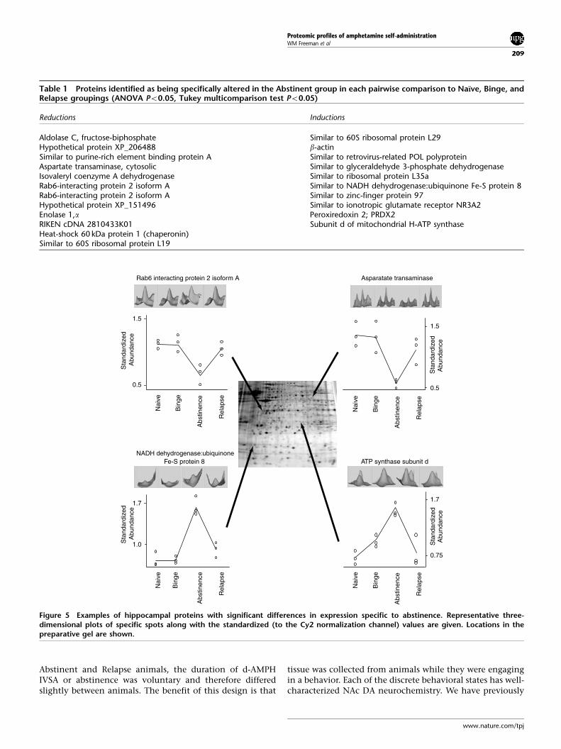
digestion and MALDI-ToF/ToF. A total of 108 proteins wereidentified with a high degree of confidence (see SupportingResults, Supplementary Tables I–III); 49 of the identifiedproteins were potentially altered (ANOVA, Po0.05) at sometime point of d-AMPH IVSA compared to naı̈ve animals (seeSupporting Results, Supplementary Tables I and II). Theseproteins have a variety of biochemical functions and a rangeof temporal expression profiles across the separate phases ofIVSA. Some proteins were seen to increase or decreases inabundance solely at one time point, while others weredifferent at two or more time points. Of particular interestwas the finding that the majority of changes occurred in theAbstinent group. Post hoc testing revealed that 12 proteinswere reduced and 10 proteins induced in Abstinent animalsin each pairwise comparison to Naı̈ve, Binge, and Relapsegroups (Table 1 and see Figure 5 for examples).
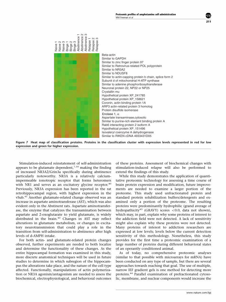
Clustering and ontological analysisGiven the number of changes found and that many changeswere specific to the Abstinent group, clustering analysis wasused to find those changes that were most indicative of thebehavioral state.23,24 An iterative PCA process was used tocluster individual animals and therefore find those changesthat were most indicative of the behavioral state. A group of22 proteins/isoforms were found in which the first twocomponents accounted for 93.8% of the variance in thestudy and which completely segregated the Abstinentanimals from animals in all other groups (Figure 6a). Anonhierarchical K-means divisive clustering of the subjectsbased on this classification group of proteins also segmentedthe Abstinent animals from all other subjects (Figure 6b).Relative expression levels of the classification group alsoclearly show the uniqueness of the Abstinent animals fromall others (Figure 7). The proteins used for classificationdisplayed both increases and decreases in expression, and/ormodification (eg phosphorylation) as, in some cases, multi-ple spots were identified as the same protein but only certainspecies were altered in quantity.
To ascertain processes and potential biological outcomesof the changes observed,25 all the proteins identified wereimported into the Celera Panther database.21,22 Differentgroupings of proteins (eg all proteins identified, unchangedproteins, and proteins from the classification cluster) werecompared for under- or over-representation of particularmolecular functions (Figure 8). The classification groupingwas found to have an under-representation of ribosomalproteins and an overabundance of actin- and cytoskeletal-related proteins.
DISCUSSIONThe present study is the first report of proteomic profilesduring transitional states of psychostimulant self-adminis-tration. We identified a number of proteins and proteinmodifications differentially regulated in the hippocampus atdifferent points of a d-AMPH IVSA cycle of binge–absti-nence–relapse. Hippocampal tissue was collected fromanimals at time points that were based on their operantbehavioral state (Naı̈ve, Binge, Abstinent, Relapse). For the
0
100
150
200
250
300
Training Unlimited access (24 hr)
RelapseUnlimited access
20
24 hr off
Training Sessions Test Session
Self-administration Trials
NaiveBinge
AbstinentRelapse
Num
ber
of le
ver
pres
ses
Figure 3 Total number of lever presses (7SEM) on the active leverduring different cycles of intravenous self-administration of d-amphetamine (d-AMPH). Rats were randomly assigned to theBinge, Abstinent, or Relapse group following 3 days of trainingunder an FR1 schedule of reinforcement. All rats were givenunlimited access to 0.1 mg/kg per injection in a 24-h self-adminis-tration session. There were no significant differences betweenlever-pressing behavior in any of the groups. After a 24-h restperiod, rats were given a second unlimited access session. Rats inthe Binge group were permitted to self-administer d-AMPH for 3 hbefore being killed. Rats in the Abstinent group were permitted toself-administer until they entered voluntary abstinence, and werekilled at the end of a 3-h period during which there was no responseon either the active or inactive lever. Animals in the Relapse groupwere permitted to reinitiate self-administration after an abstinenceperiod that lasted at least 3 h, and were killed 3 h after theyresumed responding. Control animals self-administered saline, andwere killed 3 h after the start of the second unlimited access session.
103pHMW
Dec
reas
ing
Nonlinear
Figure 4 Example of 2-DIGE gel from the study.
Proteomic profiles of amphetamine self-administrationWM Freeman et al
208
The Pharmacogenomics Journal
Abstinent and Relapse animals, the duration of d-AMPHIVSA or abstinence was voluntary and therefore differedslightly between animals. The benefit of this design is that
tissue was collected from animals while they were engagingin a behavior. Each of the discrete behavioral states has well-characterized NAc DA neurochemistry. We have previously
0.5
1.5
Nai
ve
Bin
ge
Abs
tinen
ce
Rel
apse
Nai
ve
Bin
ge
Abs
tinen
ce
Rel
apse
Nai
ve
Bin
ge
Abs
tinen
ce
Rel
apse
Nai
ve
Bin
ge
Abs
tinen
ce
Rel
apse
Sta
ndar
dize
d A
bund
ance
Rab6 interacting protein 2 isoform A
ATP synthase subunit d
Sta
ndar
dize
dA
bund
ance
0.75
1.7
Asparatate transaminase
NADH dehydrogenase:ubiquinone Fe-S protein 8
Sta
ndar
dize
d A
bund
ance
1.7
1.0
0.5
1.5
Sta
ndar
dize
dA
bund
ance
Figure 5 Examples of hippocampal proteins with significant differences in expression specific to abstinence. Representative three-dimensional plots of specific spots along with the standardized (to the Cy2 normalization channel) values are given. Locations in thepreparative gel are shown.
Table 1 Proteins identified as being specifically altered in the Abstinent group in each pairwise comparison to Naı̈ve, Binge, andRelapse groupings (ANOVA Po0.05, Tukey multicomparison test Po0.05)
Reductions Inductions
Aldolase C, fructose-biphosphate Similar to 60S ribosomal protein L29Hypothetical protein XP_206488 b-actinSimilar to purine-rich element binding protein A Similar to retrovirus-related POL polyproteinAspartate transaminase, cytosolic Similar to glyceraldehyde 3-phosphate dehydrogenaseIsovaleryl coenzyme A dehydrogenase Similar to ribosomal protein L35aRab6-interacting protein 2 isoform A Similar to NADH dehydrogenase:ubiquinone Fe-S protein 8Rab6-interacting protein 2 isoform A Similar to zinc-finger protein 97Hypothetical protein XP_151496 Similar to ionotropic glutamate receptor NR3A2Enolase 1,a Peroxiredoxin 2; PRDX2RIKEN cDNA 2810433K01 Subunit d of mitochondrial H-ATP synthaseHeat-shock 60 kDa protein 1 (chaperonin)Similar to 60S ribosomal protein L19
Proteomic profiles of amphetamine self-administrationWM Freeman et al
209
www.nature.com/tpj

demonstrated2,4 that NAc DA efflux increases during a d-AMPH binge, reaches an apex and then begins to decrease,even as self-administration continues. Despite continuedaccess to d-AMPH, the animals will voluntarily cease IVSAand enter an abstinent phase during which NAc DA levelsfall to a nadir. As the abstinence phase continues, NAc DAlevels will begin to recover towards baseline levels, and uponvoluntary resumption of d-AMPH IVSA NAc DA levels onceagain increase in response to d-AMPH exposure. Previousdata from this laboratory have also indicated that themesolimbic DA system is pharmacologically unresponsive tothe actions of d-AMPH immediately prior to, and duringabstinence, and that recovery of function in this system isaccompanied by reinstatement of d-AMPH IVSA.2,4 Stimula-tion of the vSub during abstinence induces NAc DA effluxand reinstatement to d-AMPH IVSA, and to a greater degree
than d-AMPH priming.26,9,8 Similar findings of changeshave been reported with cocaine self-administration andinactivation or activation of the vSub.27,10 Given that thevSub sends direct and indirect glutamatergic afferents to theNAc and stimulation of these afferents induces d-AMPHIVSA relapse, we chose to investigate whether there werechanges in hippocampal protein expression and modifica-tion across each phase of the binge–abstinent–relapse cycleof d-AMPH IVSA.
Differential expression of proteins was found at eachphase of the d-AMPH IVSA cycle. In particular, the Abstinentgroup exhibited a proteomic profile unlike those of theother behavioral states. It is possible that protein changes inthe Abstinent group are related to disruption in circadianrhythms associated with extended hours of drug self-administration; however, the animals in the Abstinentgroup were killed at various times of the day and night,depending on when they entered the abstinence phase.Animals in the Relapse group were also killed at varioustimes of day or night depending on the length of their IVSAbinges and their abstinent period and would therefore belikely to exhibit similar disruptions in circadian rhythms.Also of note is that protein levels were similar in Naı̈ve,Binge, and Relapse groups, and that many of the changes(increases or decreases) observed were specific to theAbstinent group. The proteins identified in this study havea wide variety of functions (Figure 8). Clustering of theprotein changes revealed a subset of proteins that segregatedthe Abstinent animals from all others. This subset ofproteins contained an overabundance of actin-relatedproteins and an under-representation of ribosomal-relatedproteins.
The paucity of ribomsomal proteins in the classificationcluster suggests that translational changes are not indicativeof the Abstinent subjects. While there were ribosomalproteins identified as being changed in some groups, thesedifferences were not part of the classification cluster. Thiswould suggest that protein translation mechanisms are in anormative state during abstinence.
It is of note that in the classification cluster there areseveral proteins involved in the regulation of actin filamentpolymerization. CapZb decreases actin polymerization byblocking actin barbed ends,28,29 and is increased specificallyduring abstinence. ARP3 and Coronin 1A, both of whichincrease actin filament assembly,30,31 are decreased specifi-cally during abstinence. Taken together, this would suggest adecrease in actin polymerization during abstinence whichcould alter potassium,32 sodium,33 calcium, AMPA,34 andNMDA receptor channel function.35 Owing to its regulationof receptor functionality, actin polymerization regulationcan affect the potentiation or depression of a neuron.Inhibition of actin polymerization impairs LTP but notLTD.36,37 Furthermore, electrical stimulation can changeactin polymerization dynamics.36,38,39 These findings com-bined with those of this study suggest a changed neuronaltone in the hippocampus where potentiation may bediminished and depression enhanced, and that mightcontribute to behaviors associated with drug intake.
Abstinence 3
Abstinence 2
Abstinence 1
Relapse 1
Naive 3
Binge 3
Binge 2
Relapse 2
Naive 1
Relapse 3
Naive 2
Binge 1
1
0
− 4 − 3 − 2 − 1 0 1
1st Component
2nd Com
ponent
a
b
Figure 6 Classification of behaviors by proteomic profile. (a) PCAof changes in which the first two principal components account forthe greatest percentage of the variability. X-axis values representPCA value for the first component and y-axis is the secondcomponent (&, Naı̈ve; =, Binge; þ , Abstinent; o, Relapse). (b)The group of hippocampal proteins found by the PCA clusteringwas also used for K-means clustering of the animals. Thisnonhierarchical K-means divisive scheme reveals a cluster solelycomposed of Abstinent animals.
Proteomic profiles of amphetamine self-administrationWM Freeman et al
210
The Pharmacogenomics Journal
Stimulation-induced reinstatement of self-administrationappears to be glutamate dependent,7,10 making the findingof increased NR3A2/Grin3a specifically during abstinenceparticularly noteworthy. NR3A is a relatively calcium-impermeable ionotropic receptor that forms heteromerswith NR1 and serves as an excitatory glycine receptor.40
Previously, NR3A expression has been reported in the ratretrohippocampal region, with highest expression in thevSub.41 Another glutamate-related change observed was anincrease in aspartate aminotransferase (AST), which was alsoevident only in the Abstinent rats. Aspartate aminotransfer-ase, the enzyme that catalyzes the transamination betweenaspartate and 2-oxoglutarate to yield glutamate, is widelydistributed in the brain.42 Changes in AST may reflectalterations in glutamate metabolism, or changes in excita-tory neurotransmission that could play a role in thetransition from self-administration to abstinence after highlevels of d-AMPH intake.
For both actin- and glutamate-related protein changesobserved, further experiments are needed to both localizeand determine the functionality of these changes. As theentire hippocampal formation was examined in this study,more discrete anatomical techniques will be used in futurestudies to determine in which subregion of the hippocam-pus the alterations take place, and the nature of the cell typeaffected. Functionally, manipulations of actin polymeriza-tion or NR3A agonism/antagonism are needed to assess thebiochemical, electrophysiological, and behavioral outcomes
of these proteins. Assessment of biochemical changes withstimulation-induced relapse will also be performed toextend the findings of this study.
While this study demonstrates the application of quanti-tative proteomic technology for assessing a time course ofbrain protein expression and modification, future improve-ments are needed to examine a larger portion of theproteome. This study used unfractionated protein andstandard protein solubilization buffers/detergents and ex-amined only a portion of the proteome. The resultingproteins were predominantly hydrophilic (grand average ofhydropathicity43 (GRAVY) scores o0.0, data not shown),which may, in part, explain why some proteins of interest tothe addiction field were not detected. A lack of sensitivitymight also explain why these proteins were not detected.Many proteins of interest to addiction researchers areexpressed at low levels; levels below the current detectionsensitivity of this methodology. Nonetheless, this studyprovides for the first time a proteomic examination of alarge number of proteins during different behavioral statesof an operantly conditioned animal.
As of today, no comprehensive proteomic analyses(similar to that possible with microarrays for mRNA) havebeen conducted on any type of sample, but there are severalapproaches towards reaching that goal. The use of multiple,narrow IEF gradient gels is one method for detecting moreproteins.44 Parallel examination of prefractionated cytoso-lic, membrane, and nuclear components would increase the
Nai
ve 1
Nai
ve 2
Nai
ve 3
Bin
ge 1
Bin
ge 2
Bin
ge 3
Abs
tinen
ce 1
Abs
tinen
ce 2
Abs
tinen
ce 3
Rel
apse
1R
elap
se 2
Rel
apse
3
Beta-actinSimilar to GAPDHSimilar to zinc finger protein 97Similar to Retrovirus-related POL polyproteinSimilar to NR3A2Similar to NDUSF8Similar to actin-capping protein b chain, splice form 2Subunit d of mitochondrial H-ATP synthaseSimilar to adenine phosphoribosyltransferaseNeuronal protein 22, NP22 or NP25Crystallin muHypothetical protein XP_241785Hypothetical protein XP_158621Coronin, actin binding protein 1AARP3 actin-related protein 3 homologProtein disulfide isomerase
Aspartate transaminase,cytosolicSimilar to purine-rich element binding protein ARab6 interacting protein 2 isoform AHypothetical protein XP_151496Isovaleryl coenzyme A dehydrogenaseSimilar to RIKEN cDNA 4933431D05
Enolase 1, α
Figure 7 Heat map of classification proteins. Proteins in the classification cluster with expression levels represented in red for lowexpression and green for higher expression.
Proteomic profiles of amphetamine self-administrationWM Freeman et al
211
www.nature.com/tpj

coverage of the proteome. Analysis of membrane proteinscarries additional technical hurdles.45 There remains somecontroversy as to capabilities of electrophoresis-based se-paration of proteins combined with MS identification asopposed to HPLC-based separations of peptides combinedwith MS identification in analyzing hydrophobic/mem-brane proteins. A recent study46 demonstrated that electro-phoresis and HPLC separations are comparable in theirability to detect hydrophobic/membrane proteins. The mostimportant hurdle with respect to increasing the number ofproteins analyzed is sensitivity. The current study used alimited amount (50 mg) of protein per sample in the
quantitative gels. Even more sensitive dyes47 or a methodof protein amplification are needed to examine specific cellpopulations within the hippocampus.
This study was conducted to examine the proteomicchanges in the hippocampus during the d-AMPH binge–abstinence–relapse cycle of behavior and demonstrates theapplicability of quantitative proteomic technologies tobehavioral pharmacology research. Future studies will assessthe proteomic response of the NAc and other brain regionsduring these behaviors. It is anticipated that the proteomicchanges in the NAc will be different from those observed inthis study, reflecting the contrast between the types and
Cytoskeletal Protein
Nucleic Acid Binding
Oxidoreductase
UnclassifiedRibosomal Protein
Actin Family
Dehydrogenase
Regulatory Molecule
Hydrolase
Kinase
Actin-Binding Isomerase
All Proteins
Cytoskeletal Protein
IsomeraseActin-Binding
Hydrolase
Regulatory Molecule
Dehydrogenase
Actin Family
Ribosomal ProteinUnclassified
Oxidoreductase
Nucleic Acid Binding All Changes
Unclassified
Oxidoreductase
Nucleic Acid Binding
Cytoskeletal Protein
Isomerase
Actin-Binding
Hydrolase
Regulatory Molecule
Dehydrogenase
Actin Family
Classification cluster
Figure 8 Ontological analysis of hippocampal protein groupings from proteins in the study. All proteins identified were groupedaccording to Celera Molecular Function classification grouping (top), those proteins differentially expressed at a minimum of one timepoint (middle), and those found in the classification cluster (bottom). An overabundance of actin-related proteins and an under-representation of ribosomal proteins were observed in the classification cluster.
Proteomic profiles of amphetamine self-administrationWM Freeman et al
212
The Pharmacogenomics Journal

functions of neurons found in the NAc and those in thehippocampus. These future studies will also need to utilizethe strategies outlined above to gain a more comprehensivepicture of the proteome. Also, the results of this study will beexamined further by localization of the changes to specificcell types in the hippocampus and testing of the behavioraleffects of actin depolymerizing agents.
This work represents the first classification of behaviorsbased on clustering of quantitative proteomics measure-ments. These findings also present the first indication ofnumerous protein expression and modification changes inthe hippocampus of rats engaged in d-AMPH IVSA, volun-tary abstinence, and reinstatement of self-administration.
ACKNOWLEDGEMENTSThis work was supported by NIH Grants DA015945 (to WMF),DA013830-03S1 (to SGA), and CIHR Grant FRN:36372 (to AGP).NIH Grant DA013772 to Scott E Hemby (Wake Forest UniversitySchool of Medicine) provided generous financial support andscientific infrastructure.
DUALITY OF INTERESTNone declared.
Supplementary InformationSupplementary Information accompanies the paper on theThe Pharmacogenomics Journal website (http://www.nature.com/tpj).
ABBREVIATIONS
d-AMPH d-amphetamineDA dopamineNAc nucleus accumbensIVSA intravenous self-administrationvSub ventral subiculum2-DIGE two-dimensional difference gel electrophoresisMALDI-ToF/ToF matrix-assisted laser desorption ionization time-of-
flight-time-of-flight tandem mass spectrometryPCA Principal Component Analysis
REFERENCES1 Roberts DC, Corcoran ME, Fibiger HC. On the role of ascending
catecholaminergic systems in intravenous self-administration of cocaine.Pharmacol Biochem Behav 1977; 6: 615–620.
2 Di Ciano P, Blaha CD, Phillips AG. Changes in dopamine oxidationcurrents in the nucleus accumbens during unlimited-access self-administration of d-amphetamine by rats. Behav Pharmacol 1996; 7:714–729.
3 Gawin FH, Kleber HD. Abstinence symptomatology and psychiatricdiagnosis in cocaine abusers. Clinical observations. Arch Gen Psychiatry1986; 43: 107–113.
4 Di Ciano P, Blaha CD, Phillips AG. Inhibition of dopamine efflux in therat nucleus accumbens during abstinence after free access to d-amphetamine. Behav Brain Res 2002; 128: 1–12.
5 Hemby SE, Co C, Koves TR, Smith JE, Dworkin SI. Differences inextracellular dopamine concentrations in the nucleus accumbensduring response-dependent and response-independent cocaine admin-istration in the rat. Psychopharmacology (Berl) 1997; 133: 7–16.
6 Groenewegen HJ, Vermeulen-Van der Zee E, te Kortschot A, Witter MP.Organization of the projections from the subiculum to the ventral
striatum in the rat. A study using anterograde transport of Phaseolusvulgaris leucoagglutinin. Neuroscience 1987; 23: 103–120.
7 Legault M, Rompre PP, Wise RA. Chemical stimulation of the ventralhippocampus elevates nucleus accumbens dopamine by activatingdopaminergic neurons of the ventral tegmental area. J Neurosci 2000;20: 1635–1642.
8 Taepavarapruk P, Phillips AG. Neurochemical correlates of relapse to d-amphetamine self-administration by rats induced by stimulation of theventral subiculum. Psychopharmacology (Berl) 2003; 168: 99–108.
9 Taepavarapruk P, Floresco SB, Phillips AG. Hyperlocomotion andincreased dopamine efflux in the rat nucleus accumbens evoked byelectrical stimulation of the ventral subiculum: role of ionotropicglutamate and dopamine D1 receptors. Psychopharmacology (Berl)2000; 151: 242–251.
10 Vorel SR, Liu X, Hayes RJ, Spector JA, Gardner EL. Relapse to cocaine-seeking after hippocampal theta burst stimulation. Science 2001; 292:1175–1178.
11 Sun W, Rebec GV. Lidocaine inactivation of ventral subiculumattenuates cocaine-seeking behavior in rats. J Neurosci 2003; 23:10258–10264.
12 Nestler EJ. Psychogenomics: opportunities for understanding addiction.J Neurosci 2001; 21: 8324–8327.
13 Freeman WM, Dougherty KE, Vacca SE, Vrana KE. An interactivedatabase of cocaine-responsive gene expression. Sci World J 2002; 2:701–706.
14 Freeman WM, Hemby SE. Proteomics for protein expression profiling inneuroscience. Neurochem Res 2004; 29: 1065–1081.
15 Unlu M, Morgan ME, Minden JS. Difference gel electrophoresis: a singlegel method for detecting changes in protein extracts. Electrophoresis1997; 18: 2071–2077.
16 Alban A, David SO, Bjorkesten L, Andersson C, Sloge E, Lewis S et al. Anovel experimental design for comparative two-dimensional gelanalysis: two-dimensional difference gel electrophoresis incorporatinga pooled internal standard. Proteomics 2003; 3: 36–44.
17 Di Ciano P, Coury A, Depoortere RY, Egilmez Y, Lane JD, Emmett-Oglesby MW et al. Comparison of changes in extracellular dopamineconcentrations in the nucleus accumbens during intravenous self-administration of cocaine or d-amphetamine. Behav Pharmacol 1995; 6:311–322.
18 Freeman WM, Brebner K, Lynch WJ, Robertson DJ, Roberts DC, VranaKE. Cocaine-responsive gene expression changes in rat hippocampus.Neuroscience 2001; 108: 371–380.
19 Freeman WM, Nader MA, Nader SH, Robertson DJ, Gioia L, Mitchell SMet al. Chronic cocaine-mediated changes in non-human primatenucleus accumbens gene expression. J Neurochem 2001; 77:542–549.
20 Medzihradszky KF, Campbell JM, Baldwin MA, Falick AM, Juhasz P,Vestal ML et al. The characteristics of peptide collision-induceddissociation using a high-performance MALDI-TOF/TOF tandem massspectrometer. Anal Chem 2000; 72: 552–558.
21 Thomas PD, Campbell MJ, Kejariwal A, Mi H, Karlak B, Daverman R et al.PANTHER: a library of protein families and subfamilies indexed byfunction. Genome Res 2003; 13: 2129–2141.
22 Thomas PD, Kejariwal A, Campbell MJ, Mi H, Diemer K, Guo N et al.PANTHER: a browsable database of gene products organized bybiological function, using curated protein family and subfamilyclassification. Nucleic Acids Res 2003; 31: 334–341.
23 Raychaudhuri S, Stuart JM, Altman RB. Principal components analysis tosummarize microarray experiments: application to sporulation timeseries. Pac Symp Biocomput 2000; 5: 455–466.
24 Misra J, Schmitt W, Hwang D, Hsiao LL, Gullans S, Stephanopoulos G etal. Interactive exploration of microarray gene expression patterns in areduced dimensional space. Genome Res 2002; 12: 1112–1120.
25 Cho RJ, Campbell MJ. Transcription, genomes, function. Trends Genet2000; 16: 409–415.
26 Blaha CD, Yang CR, Floresco SB, Barr AM, Phillips AG. Stimulation of theventral subiculum of the hippocampus evokes glutamate receptor-mediated changes in dopamine efflux in the rat nucleus accumbens. EurJ Neurosci 1997; 9: 902–911.
27 Black YD, Green-Jordan K, Eichenbaum HB, Kantak KM. Hippocampalmemory system function and the regulation of cocaine self-administra-tion behavior in rats. Behav Brain Res 2004; 151: 225–238.
Proteomic profiles of amphetamine self-administrationWM Freeman et al
213
www.nature.com/tpj
28 Yamashita A, Maeda K, Maeda Y. Crystal structure of CapZ: structuralbasis for actin filament barbed end capping. EMBO J 2003; 22:1529–1538.
29 Pantaloni D, Le Clainche C, Carlier MF. Mechanism of actin-basedmotility. Science 2001; 292: 1502–1506.
30 Welch MD, Iwamatsu A, Mitchison TJ. Actin polymerization is inducedby Arp2/3 protein complex at the surface of Listeria monocytogenes.Nature 1997; 385: 265–269.
31 Goode BL, Wong JJ, Butty AC, Peter M, McCormack AL, Yates JR et al.Coronin promotes the rapid assembly and cross-linking of actinfilaments and may link the actin and microtubule cytoskeletons inyeast. J Cell Biol 1999; 144: 83–98.
32 Huang H, Rao Y, Sun P, Gong LW. Involvement of actin cytoskeleton inmodulation of Ca(2+)-activated K(+) channels from rat hippocampalCA1 pyramidal neurons. Neurosci Lett 2002; 332: 141–145.
33 Berdiev BK, Prat AG, Cantiello HF, Ausiello DA, Fuller CM, Jovov B et al.Regulation of epithelial sodium channels by short actin filaments. J BiolChem 1996; 271: 17704–17710.
34 Zhou Q, Xiao M, Nicoll RA. Contribution of cytoskeleton to theinternalization of AMPA receptors. Proc Natl Acad Sci USA 2001; 98:1261–1266.
35 Furukawa K, Fu W, Li Y, Witke W, Kwiatkowski DJ, Mattson MP. Theactin-severing protein gelsolin modulates calcium channel and NMDAreceptor activities and vulnerability to excitotoxicity in hippocampalneurons. J Neurosci 1997; 17: 8178–8186.
36 Fukazawa Y, Saitoh Y, Ozawa F, Ohta Y, Mizuno K, Inokuchi K.Hippocampal LTP is accompanied by enhanced F-actin content withinthe dendritic spine that is essential for late LTP maintenance in vivo.Neuron 2003; 38: 447–460.
37 Chen Y, Bourne J, Pieribone VA, Fitzsimonds RM. The role of actin in theregulation of dendritic spine morphology and bidirectional synapticplasticity. NeuroReport 2004; 15: 829–832.
38 Ackermann M, Matus A. Activity-induced targeting of profilin andstabilization of dendritic spine morphology. Nat Neurosci 2003; 6:1194–1200.
39 Star EN, Kwiatkowski DJ, Murthy VN. Rapid turnover of actin indendritic spines and its regulation by activity. Nat Neurosci 2002; 5:239–246.
40 Chatterton JE, Awobuluyi M, Premkumar LS, Takahashi H, Talantova M,Shin Y et al. Excitatory glycine receptors containing the NR3 family ofNMDA receptor subunits. Nature 2002; 415: 793–798.
41 Wong HK, Liu XB, Matos MF, Chan SF, Perez-Otano I, Boysen Met al. Temporal and regional expression of NMDA receptorsubunit NR3A in the mammalian brain. J Comp Neurol 2002; 450:303–317.
42 Najlerahim A, Harrison PJ, Barton AJ, Heffernan J, Pearson RC.Distribution of messenger RNAs encoding the enzymes glutaminase,aspartate aminotransferase and glutamic acid decarboxylase in ratbrain. Brain Res Mol Brain Res 1990; 7: 317–333.
43 Kyte J, Doolittle RF. A simple method for displaying the hydropathiccharacter of a protein. J Mol Biol 1982; 157: 105–132.
44 Wildgruber R, Harder A, Obermaier C, Boguth G, Weiss W, Fey SJ et al.Towards higher resolution: two-dimensional electrophoresis of Sacchar-omyces cerevisiae proteins using overlapping narrow immobilized pHgradients. Electrophoresis 2000; 21: 2610–2616.
45 Santoni V, Molloy M, Rabilloud T. Membrane proteins and proteomics:un amour impossible? Electrophoresis 2000; 21: 1054–1070.
46 Schmidt F, Donahoe S, Hagens K, Mattow J, Schaible UE, Kaufmann SHet al. Complementary analysis of the Mycobacterium tuberculosisproteome by two-dimensional electrophoresis and isotope-codedaffinity tag technology. Mol Cell Proteomics 2004; 3: 24–42.
47 Shaw J, Rowlinson R, Nickson J, Stone T, Sweet A, Williams K et al.Evaluation of saturation labeling two-dimensional difference gelelectrophoresis fluorescent dyes. Proteomics 2003; 3: 1181–1195.
Proteomic profiles of amphetamine self-administrationWM Freeman et al
214
The Pharmacogenomics Journal
Copyright © 2022 FDOKUMEN