Proteomic profiling of various human dental stem cells
24
WJSC https://www.wjgnet.com 1214 October 26, 2020 Volume 12 Issue 10 World Journal of Stem Cells W J S C Submit a Manuscript: https://www.f6publishing.com World J Stem Cells 2020 October 26; 12(10): 1214-1236 DOI: 10.4252/wjsc.v12.i10.1214 ISSN 1948-0210 (online) SYSTEMATIC REVIEWS Proteomic profiling of various human dental stem cells - a systematic review Jagadish Hosmani, Khalil Assiri, Hussain Mohammed Almubarak, Master Luqman Mannakandath, Ahmed Al- Hakami, Shankargouda Patil, Deepa Babji, Sachin Sarode, Anantharam Devaraj, Harish C Chandramoorthy ORCID number: Jagadish Hosmani 0000-0003-3579-0906; Khalil Assiri 0000-0002-0570-3376; Hussain Mohammed Almubarak 0000-0001- 9027-4133; Master Luqman Mannakandath 0000-0002-9932-8254; Ahmed Al-Hakami 0000-0003938- 5182; Shankargouda Patil 0000-0001- 7246-5497; Deepa Babji 0000-0002- 1493-335X; Sachin Sarode 0000- 0003-1856-0957; Anantharam Devaraj 0000-0002-8685-6728; Harish C Chandramoorthy 0000-0002-8780- 8971. Author contributions: Hosmani J and Chandramoorthy HC made substantial contributions to the conception and design of the study; Babji D and Sarode S performed the literature search, and acquisition, analysis and interpretation of the data; Hosmani J, Patil S, Al-Hakami and Chandramoorthy HC drafted the article and performed critical revisions related to important intellectual content of the manuscript; Devraj A, Assiri K, Almubarak HA and Mannakandath ML designed the figures and contributed specific sections of content; All authors read and approved the final version of the manuscript. Supported by Deanship of Scientific Research, King Khalid University through Large Research Jagadish Hosmani, Diagnostic Dental Sciences, College of Dentistry, King Khalid University, Abha 61471, Asir, Saudi Arabia Khalil Assiri, Hussain Mohammed Almubarak, Master Luqman Mannakandath, Diagnostic Dental Sciences, King Khalid University, Abha 61471, Asir, Saudi Arabia Ahmed Al-Hakami, Anantharam Devaraj, Harish C Chandramoorthy, Center for Stem Cell Research and Department of Microbiology and Clinical Parasitology, King Khalid University, Abha 61421, Asir, Saudi Arabia Shankargouda Patil, Maxillofacial Surgery and Diagnostic Sciences, Division of oral Pathology, Jazan 45142, Jazan, Saudi Arabia Deepa Babji, Department of Oral Pathology and Microbiology, Maratha Mandal's NG Halgekar Institute of Dental Sciences and Research Centre, Belgaun 590 010, Karnataka, India Sachin Sarode, Department of Oral Pathology, Y Patil Dental College and Hospital, Pune 411018, Maharashtra, India Corresponding author: Harish C Chandramoorthy, PhD, Associate Professor, Center for Stem Cell Research and Department of Microbiology and Clinical Parasitology, King Khalid University, College of Medicine, Griger, Abha 61421, Asir, Saudi Arabia. [email protected] Abstract BACKGROUND The proteomic signature or profile best describes the functional component of a cell during its routine metabolic and survival activities. Additional complexity in differentiation and maturation is observed in stem/progenitor cells. The role of functional proteins at the cellular level has long been attributed to anatomical niches, and stem cells do not deflect from this attribution. Human dental stem cells (hDSCs), on the whole, are a combination of mesenchymal and epithelial coordinates observed throughout craniofacial bones to pulp. AIM To specify the proteomic profile and compare each type of hDSC with other mesenchymal stem cells (MSCs) of various niches. Furthermore, we analyzed the characteristics of the microenvironment and preconditioning changes associated
-
Upload
khangminh22 -
Category
Documents
-
view
0 -
download
0
Transcript of Proteomic profiling of various human dental stem cells

WJSC https://www.wjgnet.com 1214 October 26, 2020 Volume 12 Issue 10
World Journal of
Stem CellsW J S CSubmit a Manuscript: https://www.f6publishing.com World J Stem Cells 2020 October 26; 12(10): 1214-1236
DOI: 10.4252/wjsc.v12.i10.1214 ISSN 1948-0210 (online)
SYSTEMATIC REVIEWS
Proteomic profiling of various human dental stem cells - a systematic review
Jagadish Hosmani, Khalil Assiri, Hussain Mohammed Almubarak, Master Luqman Mannakandath, Ahmed Al-Hakami, Shankargouda Patil, Deepa Babji, Sachin Sarode, Anantharam Devaraj, Harish C Chandramoorthy
ORCID number: Jagadish Hosmani 0000-0003-3579-0906; Khalil Assiri 0000-0002-0570-3376; Hussain Mohammed Almubarak 0000-0001-9027-4133; Master Luqman Mannakandath 0000-0002-9932-8254; Ahmed Al-Hakami 0000-0003938-5182; Shankargouda Patil 0000-0001-7246-5497; Deepa Babji 0000-0002-1493-335X; Sachin Sarode 0000-0003-1856-0957; Anantharam Devaraj 0000-0002-8685-6728; Harish C Chandramoorthy 0000-0002-8780-8971.
Author contributions: Hosmani J and Chandramoorthy HC made substantial contributions to the conception and design of the study; Babji D and Sarode S performed the literature search, and acquisition, analysis and interpretation of the data; Hosmani J, Patil S, Al-Hakami and Chandramoorthy HC drafted the article and performed critical revisions related to important intellectual content of the manuscript; Devraj A, Assiri K, Almubarak HA and Mannakandath ML designed the figures and contributed specific sections of content; All authors read and approved the final version of the manuscript.
Supported by Deanship of Scientific Research, King Khalid University through Large Research
Jagadish Hosmani, Diagnostic Dental Sciences, College of Dentistry, King Khalid University, Abha 61471, Asir, Saudi Arabia
Khalil Assiri, Hussain Mohammed Almubarak, Master Luqman Mannakandath, Diagnostic Dental Sciences, King Khalid University, Abha 61471, Asir, Saudi Arabia
Ahmed Al-Hakami, Anantharam Devaraj, Harish C Chandramoorthy, Center for Stem Cell Research and Department of Microbiology and Clinical Parasitology, King Khalid University, Abha 61421, Asir, Saudi Arabia
Shankargouda Patil, Maxillofacial Surgery and Diagnostic Sciences, Division of oral Pathology, Jazan 45142, Jazan, Saudi Arabia
Deepa Babji, Department of Oral Pathology and Microbiology, Maratha Mandal's NG Halgekar Institute of Dental Sciences and Research Centre, Belgaun 590 010, Karnataka, India
Sachin Sarode, Department of Oral Pathology, Y Patil Dental College and Hospital, Pune 411018, Maharashtra, India
Corresponding author: Harish C Chandramoorthy, PhD, Associate Professor, Center for Stem Cell Research and Department of Microbiology and Clinical Parasitology, King Khalid University, College of Medicine, Griger, Abha 61421, Asir, Saudi Arabia. [email protected]
AbstractBACKGROUND The proteomic signature or profile best describes the functional component of a cell during its routine metabolic and survival activities. Additional complexity in differentiation and maturation is observed in stem/progenitor cells. The role of functional proteins at the cellular level has long been attributed to anatomical niches, and stem cells do not deflect from this attribution. Human dental stem cells (hDSCs), on the whole, are a combination of mesenchymal and epithelial coordinates observed throughout craniofacial bones to pulp.
AIM To specify the proteomic profile and compare each type of hDSC with other mesenchymal stem cells (MSCs) of various niches. Furthermore, we analyzed the characteristics of the microenvironment and preconditioning changes associated
Hosmani J et al. Proteomics of human dental stem cells
WJSC https://www.wjgnet.com 1215 October 26, 2020 Volume 12 Issue 10
Group Project, No. G.R.P 2/27/40.
Conflict-of-interest statement: The authors declare no conflicts of interest.
PRISMA 2009 Checklist statement: The authors have read the PRISMA 2009 Checklist, and the manuscript was prepared and revised according to the PRISMA 2009 Checklist.
Open-Access: This article is an open-access article that was selected by an in-house editor and fully peer-reviewed by external reviewers. It is distributed in accordance with the Creative Commons Attribution NonCommercial (CC BY-NC 4.0) license, which permits others to distribute, remix, adapt, build upon this work non-commercially, and license their derivative works on different terms, provided the original work is properly cited and the use is non-commercial. See: http://creativecommons.org/licenses/by-nc/4.0/
Manuscript source: Unsolicited manuscript
Received: March 18, 2020 Peer-review started: March 18, 2020 First decision: July 5, 2020 Revised: August 6, 2020 Accepted: September 1, 2020 Article in press: September 1, 2020 Published online: October 26, 2020
P-Reviewer: Li YH S-Editor: Ma YJ L-Editor: Filipodia P-Editor: Wang LL
with the proteomic profile of hDSCs and their influence on committed lineage differentiation.
METHODS Literature searches were performed in PubMed, EMBASE, Scopus, and Web of Science databases, from January 1990 to December 2018. An extra inquiry of the grey literature was completed on Google Scholar, ProQuest, and OpenGrey. Relevant MeSH terms (PubMed) and keywords related to dental stem cells were used independently and in combination.
RESULTS The initial search resulted in 134 articles. Of the 134 full-texts assessed, 96 articles were excluded and 38 articles that met the eligibility criteria were reviewed. The overall assessment of hDSCs and other MSCs suggests that differences in the proteomic profile can be due to stem cellular complexity acquired from varied tissue sources during embryonic development. However, our comparison of the proteomic profile suffered inconsistencies due to the heterogeneity of various hDSCs. We believe that the existence of a heterogeneous population of stem cells at a given niche determines the modalities of regeneration or tissue repair. Added prominences to the differences present between various hDSCs have been reasoned out.
CONCLUSION Systematic review on proteomic studies of various hDSCs are promising as an eye-opener for revisiting the proteomic profile and in-depth analysis to elucidate more refined mechanisms of hDSC functionalities.
Key Words: Apical papilla stem cells; Dental follicle stem cells; Dental pulp stem cells; Periodontal ligament stem cells; Proteomics
©The Author(s) 2020. Published by Baishideng Publishing Group Inc. All rights reserved.
Core Tip: Neural crest-derived dental stem cells (DSCs) are ubiquitously present around the tooth anlage, are spatiotemporally related to each other, and possess the potential for self-renewal and ability to differentiate into different cell types and lineages under suitable microenvironments. Unearthing the spectrum, cascade and arrays of proteins present in these stem cells at different stages of tooth development and all the post-translational modifications of these proteins is possible by proteomics. Here, we present a systematic review on proteomic studies of various DSCs. Emphases on the differences present between various DSCs have been reasoned out.
Citation: Hosmani J, Assiri K, Almubarak HM, Mannakandath ML, Al-Hakami A, Patil S, Babji D, Sarode S, Devaraj A, Chandramoorthy HC. Proteomic profiling of various human dental stem cells - a systematic review. World J Stem Cells 2020; 12(10): 1214-1236URL: https://www.wjgnet.com/1948-0210/full/v12/i10/1214.htmDOI: https://dx.doi.org/10.4252/wjsc.v12.i10.1214
INTRODUCTIONHuman teeth are unique by being composite in nature and are created through the support and equal cooperation of neural crest-determined mesenchymal stem cells (MSCs) and oral ectoderm-derived stem cells during initial development. They are distinct in their composition by the presence of the outer mineralized enamel layer, the hardest structure in the body, closely and intimately apposed with the adjacent mineralized dentin layer, the soft tissue component, the dental pulp and radicular structures that are anchored to the alveolar socket via the periodontium[1,2].
Isolation of pluripotent MSCs from several oral tissues has been successful[3]. Stem cells of dental origin (DSCs) have the attributes of auto-renewal and multilineage differentiation, similar to any other MSCs in the body. Owing to their derivation from

Hosmani J et al. Proteomics of human dental stem cells
WJSC https://www.wjgnet.com 1216 October 26, 2020 Volume 12 Issue 10
the neural crest, they have a different origin from bone-marrow-derived MSCs, which are derived from mesoderm[4]. DSCs have been successfully harvested and found to differentiate into osteoblast-like cells that form bone in vitro. Several studies have revealed the potential of DSCs to form dentin-like tissue. These DSCs were differentiated into several cell types, including neurons, adipocytes, and chondrocytes[5,6].
DSCs, owing to many advantages, such as noninvasive cell culturing, high clinical potential and ease in access, remain favorable among all the MSCs studied. The repair and regeneration capabilities of DSCs, similar to those of MSCs from different organs, were observed in several in vitro studies. To date, five different human DSCs have been depicted: Dental pulp stem cells (DPSCs), stem cells from human exfoliated deciduous teeth (SHEDs), periodontal ligament stem cells (PDLSCs), stem cells from apical papilla (APSCs), and dental follicle stem cells (DFSCs). Thus, the heterogeneity of DSCs remains one of the key hindrances to determining ideal ways to approach cell-based tissue design. The advancement of genome-wide research systems empowers the depiction and observation of the gene expression patterns of various cells. Utilizing this information, we can more readily comprehend the components overseeing the presentation of every cell's attributes[7].
Proteomics provides an amazing technique to describe the whole protein profile of stem cell phenotypes with different specializations. This innovation is useful for understanding the components that control their self-restoration, differentiation potential and capacity to recover the unique microenvironments from which they are determined[8].
Different investigations have investigated the protein expression profiles in MSCs derived from DPSCs, PDLSCs, DFSCs and APSCs to generate a database of proteins regularly or differentially expressed among various DSCs[9].
The aim of this systematic review is to quantify the existing literature on proteomic profiling of DSCs.
MATERIALS AND METHODSProtocol and registrationThe international prospective register of systematic reviews (PROSPERO) database was searched for any enrolled protocol on comparative subjects. Similarly, the current systematic review was enrolled as a protocol with PROSPERO (ID: CRD42019120267). The review considered part of the Preferred Reporting Items for Systematic Reviews and Meta-Analyses proclamation.
Eligibility criteriaInclusion criteria: The PICO framework was used to develop a literature search strategy: (1) P: Population, human DMSCs; (2) I: Intervention, proteomic analysis; (3) C: Comparison, human MSCs such as bone marrow stem cells, adipocyte stem cells, peripheral blood stem cells and comparison of various DSCs with each other; and (4) O: Outcomes, assessing similarities of and differences in proteomic profiles between different human DSCs.
Exclusion criteria: The following exclusion criteria were applied: (1) Studies that did not assess the proteomics of human DSCs; and (2) Case reports, reviews, experimental studies on animals, short communications and personal opinions, letters to the editor, and conference abstracts.
Focused question: Is the comprehensive proteomic analysis of the regulatory network during the differentiation of human DSCs unique compared to the other MSCs of the body?
Search strategyDefinite mechanized literature searches were performed in PubMed, EMBASE, Scopus, and Web of Science from January 1990 to December 2018. An extra inquiry of the grey literature was completed on Google Scholar, ProQuest, and OpenGrey. Reference arrangements of every single included article were physically searched to distinguish any potential applicable articles. End-Note programming (EndNote X7®; Thomson Reuters, Philadelphia, PA, United States) was utilized to organize the references and expel any duplicate articles.
The following MeSH terms (PubMed) and keywords were used independently and in combination: DSCs; dental MSCs; DPSCs; PDLSCs; stem cells of human exfoliated

Hosmani J et al. Proteomics of human dental stem cells
WJSC https://www.wjgnet.com 1217 October 26, 2020 Volume 12 Issue 10
deciduous teeth; dental follicle stem cells; apical papilla stem cells; DSC proliferation; dental mesenchymal stem cell proliferation; proteomic analysis; proteomic profiling; and secretome.
Study determination and information extractionThe chosen procedure for the investigation was completed in two phases. In the primary stage, the titles and abstracts of every single recognized article were screened by two independent analysts (Hosmani J and Chandramoorthy HC) utilizing an institutionalized guide. This was trailed by recovery of full texts of studies that met the qualification criteria and inspected autonomously by two similar reviewers utilizing an institutionalized and pilot-tried structure. Any contradictions regarding study determination were commonly examined, and an agreement was made before consideration of the investigation. The two reviewers (Hosmani J and Chandramoorthy HC) independently collected the data on study characteristics (author, year of study and country), type of DSCs, study design, and technique used for proteomic analysis.
RESULTSSearch strategy and outcomeThe initial search resulted in 134 articles. Out of 134 full-texts assessed, 96 articles were excluded. Thus, 38 articles that met the eligibility criteria were included (Figure 1).
Description of the studiesThe articles assessed for this systematic review could be categorized as follows: (1) Proteomic analysis of individual human DSCs (Table 1); (2) Comparative proteomic analysis among various human DSCs (Table 2); (3) Comparative proteomic analysis of human DSCs with other MSCs of the body (Table 3); (4) Proteomic studies on the influence of the microenvironment or preconditioning factors on human DSCs (Table 4). Phenotypic cell surface markers in DSCs (Table 5); and (5) Among the articles reviewed in the present study, 24 studies were carried out on DPSCs, 11 studies on PDLSCs, 7 studies on DFSCs, 5 studies on APSCs and 4 studies on SHEDs either individually or in combination with other DSCs or MSCs.
DISCUSSIONProteomic studies on DPSCsA study was carried out by Niehage et al[10] on the cell surface proteomes of human DPSCs derived from a single donor. Proteomic characterization of DPSCs after culture expansion in low (2%) or high (10%) serum-containing media was assessed. A label-free quantitative method to assess cell surface proteomes was utilized and based on a novel mathematical model developed by the authors; in total, 101 CD surface proteins and 268 non-CD cell surface proteins were identified. Variation in the serum conditions (high or low) yielded 14 surface proteins that varied consistently in abundance. Approximately 400 cell surface proteins were identified in the study, which the authors claim as the most comprehensive analysis. Along with novel MSC markers in their previous study, the authors identified new protein markers belonging to the tumor necrosis factor (TNF) receptor superfamily, integrins, and interleukin receptors. CD87, integrin alpha-10, CBPM and SLIK2 were identified as novel markers differentially expressed in cells cultured in BE media[10].
Wei et al[11] studied the differential protein expression pattern secreted by DPSCs during odontoblast-like differentiation. Twenty-three proteins were produced during odontogenic differentiation. Significant reorganization of the cytoskeleton via upregulation of vimentin and cardiac muscle alpha actin was noted. Downregulation of nuclear proteins (hnRNPc) was noted. Annexin VI was increased as DPSCs differentiated into odontoblast-like cells. Downregulation of collagen VI during the mineralization of DPSCs was noted. Coregulation of collagen VI and matrixion-2 peroxiredoxins (Prdx 4) was upregulated, indicating that odontoblast-like differentiation occurs in DPSCs.
A study on the ability of DPSCS from carious teeth to differentiate into neuronal lineages, specifically DAergic-like cells was carried out by Gnanasegaran et al[12]. These authors found that DPSCs from carious teeth displayed decreased neurite formation,

Hosmani J et al. Proteomics of human dental stem cells
WJSC https://www.wjgnet.com 1218 October 26, 2020 Volume 12 Issue 10
Table 1 Proteomic analysis of individual human dental stem cells
Ref.Human dental stem cells analyzed
Method used Main findings Inference
Morsczeck et al[30]
Dental follicle precursor cells
2-DE combined with LC-MS/MS Bioinformatic analyses
Differentially regulated proteins: 115 upregulated proteins: Glutamine synthetase, lysosomal proteinase cathepsin B Proteins, plastin 3 t-isoform, beta-actin, superoxide dismutases, and transgelin Highly downregulated proteins: Cofilin-1, pro-alpha 1 collagen, destrin, prolyl 4-hydrolase and dihydrolipoamide dehydrogenase Upregulated proteins Downregulated proteins
Actin-bundling and defense against Oxidative cellular stress Collagen biosynthesis Catabolism, cell motility and biological quality Cell cycle progression and protein metabolism
Pivoriunas et al[15]
Human dental pulp derived SHEDs
2-DE and MALDI-TOF-MS
The protein spot designs imagined on 2-DE gels were entirely replicable, and roughly 150–300 protein spots were distinguished on each gel The cells communicated trademark antigens of MSC-like cells, including CD73, CD90, CD105, CD146, and did not express hematopoietic markers CD14, CD34, and CD45, as surveyed with FACS examination
Identification of profoundly communicated proteins in SHEDs uncovered proteomic profiles fundamentally the same as that of MSC-like cells got from different tissues
Niehage et al[10] hPDLSCs LC-MS/MS 400 cell surface proteins were recognized Prominently, a few proteins affiliated with the tumor necrosis factor receptor super family (CD40, CD120a, CD261, CD262, CD264, and CD266), distinctive integrins (alpha-4, alpha-6, and alpha-10), or interleukin receptors (CD121a, CD130, CD213a1, CD217, and CDw210b) were distinguished
Huge, changes in the proteomes of DPSCs cultured in either low (BE medium) or high (S medium) serum content milieu
Xiong et al[25] hPDLSCs 2-DE-MS An aggregate of 80 very much settled proteins spots with an atomic weight scope of 10-110 kDa were recognized. Following spot extraction and investigation by MS, a sum of 32 protein spots were recognized as membrane related proteins PDLSC were likewise found to express two novel cell surface proteins, Annexin A2 and sphingosine kinase 1
These proteomic discoveries give the stage to additionally characterize the cell surface protein articulation profile of PDLSC so as to additionally describe this cell populace and support development of novel isolation and purification strategies
Wei et al[11] hPDLSCs 2-DE coupled with MALDI-TOF MS
23 protein spots identified to the early odontogenic differentiation were distinguished; These proteins included cytoskeleton proteins, nuclear proteins, cell membrane-bound molecules, proteins involved in matrix synthesis, and metabolic enzymes; The expression of four identified proteins, which were heteronuclear ribonuclear proteins C, annexin VI, collagen type VI, and matrilin-2, was confirmed by Western blot and real-time real time polymerase chain reaction
Articulation changes of the recognized proteins uncover the inclusion of involvement of various regulation mechanisms in odontoblast-like differentiation including cell cycle, protein synthesis and degradation, Ca2+ homeostasis, signal transduction, translation, and cellular energy regulation, which are all fascinating focuses for further examinations
Wu et al[26] Human periodontal ligament cells
2-DE combined with LC-MS/MS and peptide mass fingerprinting
61 proteins in periodontal ligament cells experiencing differentiation appeared no less than a 1.5-crease change in intensity, of which 29 differentially communicated proteins were effectively distinguished by MALDI-TOF MS The outflows of a portion of the recognized proteins were moreover confirmed stern blotting and reverse transcription–polymerase chain reaction analysis
These proteins for the most part included cytoskeleton proteins and cytoskeleton-related proteins, nuclear proteins and cell membrane bound particles, and might be related with the remarkable capacity of periodontal ligament cells in keeping up periodontal tissue homeostasis, especially in the midst of the mineralization of periodontal tendon
Gnanasegaran et al[12]
DPSCs from carious teeth
PCR Western blot analysis Impulse detection via microelectrode array
The capability of DPSCs-CT to differentiate into DAergic-like cells was not proportionate to that of DPSCs; This was also reflected in both gene and protein generation whereby key neuronal markers, for example, nestin, NURR1 and beta-III-tubulin were expressed essentially lower when contrasted with separated DPSCs; Also, articulations of transcriptomes identified with neurogenesis uncovered down control of over half of the qualities when contrasted with separated DPSC
DPSCs-CT were able to differentiate into DAergic-like cells but not as efficiently as DPSCs
Dou et al[31] DFCs iTRAQ labeling combined with MS
A total of 2092 transmitted proteins were distinguished in adjusted media of iDFCs. Diverged from primary DFCs, 253 unmistakably conveyed proteins were found in iDFCs secretome (142 up-managed and 111 down-controlled); Bioinformatic examination revealed that the vast majority of emitted proteins were locked in with cell process, metabolic procedure, biologic control, cell part association or biogenesis, immune framework process, formative procedure, reaction to upgrade and flagging
Proteomic profile of cell secretome wasn’t largely affected after immortalization converted by this piggyback immortalization system; The secretome of iDFCs may be a good candidate of primary DFCs for regenerative medicine
Periodontal ligament
900 spots were distinguished and recognized 117 protein spots originating in 74 different genes; In addition to scaffold cytoskeletal proteins, proteins implicated with cellular motility and membrane
Reichenberg et al[27]
2-DE MALDI-TOF Most of these identified proteins are closely related to the extensive PDL fibroblasts’ functions and homeostasis

Hosmani J et al. Proteomics of human dental stem cells
WJSC https://www.wjgnet.com 1219 October 26, 2020 Volume 12 Issue 10
fibroblast trafficking, chaperone, stress and folding proteins, metabolic enzymes, proteins associated with detoxification and membrane activity, biodegradative metabolism, translation and transduction, extracellular proteins, and cell cycle regulation proteins were recognized
Hao et al[28] Periodontal ligament stem cells
Microarray Expression of 116 miRNAs was found to be altered after osteo-induction, with 30 upregulated and 86 downregulated; Thirty-one of these miRNAs (26.7%) had osteogenesis-related target genes
Noteworthy modifications in miRNA articulation profiles were seen amid osteogenic separation of hPDLSCs; These outcomes infer that miRNAs may effectively affect this procedure by focusing on osteogenesis-related genes
Wilson et al[13] DPSCs High-resolution array comparative genomic hybridization
Spontaneously immortalized and hTERT immortalized DPSCs do not demonstrate tumorgenic potential
Cultured DPSC lines that can be differentiated into neurons may be safe for future in vivo therapy for neurobiological diseases
Choi et al[14] DPSCs Microarray Strong expression of BBX during the odontoblast differentiation of DPSCs; The overexpression of BBX cDNA in DPSCs/progenitors induced substantial mineralization and expression of the odontoblast marker genes, such as ALP, OPN, BSP, DMP1, and DSPP
The results show that BBX plays a key role in the regulation of odontoblast differentiation of hPDLSCs/progenitors.
2-DE: Two-dimensional gel electrophoresis; SHEDs: Stem cells from human exfoliated deciduous teeth; LC: Liquid chromatography; MS: Mass spectrometry; MALDI-TOF: Matrix assisted laser desorption/ionization time-of-flight; MSC: Mesenchymal stem cells; FACS: Fluorescent-activated cell sorting; DPSCs: Dental pulp stem cells; PDLSCs: Periodontal ligament stem cells; hPDLSCs: Human PDLSCs; PCR: Polymerase chain reaction; CT: Computed tomography; iTRAQ: Isobaric tag for relative and absolute quantitation; DFCs: Dental follicle cells; iDFCs: Immortalized DFCs; PDL: Periodontal ligament; hTERT: Human telomerase reverse transcriptase.
reduced functionality and downregulated transcription factor genes when compared to DPSCs derived from non-carious teeth. The authors considered that inflammation could be the primary reason for the impairment of functionality of DPSCs from carious teeth. Proinflammatory mediators such as cytokines, interleukins and TNF-α are amplified in the microenvironment of pulpal tissue.
The oncogenic potential of human DPSCs when exposed to prolonged culture conditions was studied by Wilson et al[13]. These authors assessed the characteristics of DPSCs that were spontaneously immortalized in culture. Anchorage-dependent growth, genomic instability and the ability to differentiate into neurons were assessed, and the authors were successful in differentiating into DPSCs into neurons in culture.
A study conducted by Choi et al[14] (2014) identified bobby sox homolog (BBX) as a new transcriptional regulator for the differentiation of DPSCs into odontoblasts. Microarray and validation by RT-PCR were utilized, which yielded the expression of several transcription factors during odontoblast differentiation of DPSCs. The one factor that stood out was BBX. BBX localization to the nucleus appears to be increased during odontoblastic differentiation of DPSCs. It was noted that the knockdown of BBX suppressed the odontoblast differentiation of DPSCs.
The proteomic profile of DPSCs from SHED was studied by Pivoriuūnas et al[15]. It was found that SHEDs may be closely related, if not identical, to MSC-like cells, as they shared a similar proteomic profile. It was found in the study that SHEDs were pre-committed towards differentiation into osteogenic lineages. The authors failed to induce the in vitro differentiation of SHEDs and clonal cells into in vitro adipogenic differentiation. They found a high level of comparability between proteomic marks of essential SHEDs and clonal cell strains.

Hosmani J et al. Proteomics of human dental stem cells
WJSC https://www.wjgnet.com 1220 October 26, 2020 Volume 12 Issue 10
Table 2 Comparative proteomic analysis amongst various human dental stem cells
Ref. Human dental stem cells compared Method used Main findings
Taraslia et al[16]
SHEDs and PDLSCs Nano-LC tandem-MS SHEDs prevalently communicated atoms that are engaged with sorting out the cytoskeletal network, cell migration and adhesion, while PDLSCs are profoundly energy delivering cells, endlessly communicating proteins that are involved in different parts of cell metabolism and multiplication
Eleuterio et al[17]
Periodontal ligament and dental pulp
2-DE MALDI-TOF/TOF DPSCs vs PDLSCs express differentially regulated proteins that are potentially related to growth, regulation and genesis of neuronal cells, suggesting that SCs derived from oral tissue source populations may possess the potential ability of neuronal differentiation which is very consistent with their neural crest origin
Patil et al[18]
Dental follicle, dental pulp and dental papilla
2DE coupled with MALDI-TOF MS
19 proteins either found commonly or differentially expressed among the three types of dental MSCs which were largely similar cellular properties and multilineage potential
Ma et al[19] DPSCs and CDPSCs 2DE electrophoresis (2-D DIGE) in combination with (MALDI-TOF MS)
18 protein spots differentially expressed between DPSCs and CDPSCs; These differently expressed proteins are mostly involved in the regulation of cell proliferation, differentiation, cell cytoskeleton and motility. CDPSCs had a higher expression of antioxidative proteins that might protect CDPSCs from oxidative stress
Akpinar et al[20]
DPSCs from natal, exfoliated deciduous, and an impacted third molar tooth
2DE approach coupled with MALDI-TOF/TOF
61 proteins were predominantly expressed by all three stem cell types. Classification of the identified proteins based on biological function revealed that structurally important proteins and proteins that are involved in protein folding machinery are predominantly expressed by all three stem cell lines
Wang et al[21]
DPSCs and periodontal ligament stem cells
iTRAQ technique A total of 159 differentially expressed proteins in PDLSCs and DPSCs. GO classification terms that distinguish osteo-induced PDLSCs from DPSCs were identified. two thirds of the enriched GO terms belonged to metabolic processes and response to stimulus, suggesting that PDLSCs and DPSCs may undergo distinct metabolic changes during the differentiation process and that the differentiation induction environment could act as a stress condition. Mineralization and migration capacities of PDLSCs were greater than those of DPSCs
Guo et al[32]
Dental follicle and dental papilla cells
2DE approach coupled with MALDI-TOF/TOF
12 proteins were significantly differential, and phosphoserine aminotransferase 1, isoform 2 of hypoxia-inducible factor 1-alpha and Isoform 1 of annexin A2, were the most significantly differential proteins. These proteins are related to regulation of bone balance, angiogenesis and cell survival in an anoxic environment. Both DFCs and DPCs express odontogenic, neurogenic and peridontogenic markers
Tian et al[33]
DFCs and periodontal ligament cells
2DE approach coupled with MALDI-TOF/TOF
32 differentially expressed proteins in DFCs and PDLCs. PDLCs could contribute to regenerate dentin-like tissues in the inductive microenvironment of treated dentin matrix. DFCs presented more remarkable dentinogenic capability than PDLCs
Joo et al[22] Apical papilla and dental pulp
A cytokine membrane array and enzyme-linked immunosorbent assay
Odontoblast differentiation-related cytokines were more strongly expressed in DPSCs-CM, while cell-proliferation–related cytokines were more strongly expressed in DACCs-CM; DPSCs may exert a stronger paracrine effect than DACCs on regeneration of the dentin–pulp complex, in terms of odontoblast differentiation
Wang et al[23]
DPSCs and SHED Flow cytometry analysis of cell surface antigens RT-qPCR Western blot analysis
Notable alterations were exhibited in SHED and DPSCs during the process of extensive expansion in vitro and the results may provide guidance for the selection of safe and effective expanded SHED and DPSCs for regenerative medicine and therapy
Li et al[29] DFCs and periodontal ligament cells
iTRAQ A total of 2138 proteins were identified and 39 of these proteins were consistently differentially expressed between DFCs and PDLCs. Gene ontology analyses revealed that the protein subsets expressed higher in PDLCs were related to actin binding, cytoskeletal protein binding, and structural constituent of muscle. PDLCs display enhanced actin cytoskeletal dynamics relative to DFCs while DFCs may exhibit a more robust antioxidant defense ability relative to PDLCs
SHEDs: Stem cells from human exfoliated deciduous teeth; PDLSCs: Periodontal ligament stem cells; LC: Liquid chromatography; MS: Mass spectrometry; 2-DE: Two-dimensional gel electrophoresis; MALDI-TOF: Matrix assisted laser desorption/ionization time-of-flight; SCs: Schwann cells; MSCs: Mesenchymal stem cells; DPSCs: Dental pulp stem cells; CDPSCs: Carious DPSCs; iTRAQ: Isobaric tag for relative and absolute quantitation; DFCs: Dental follicle cells; DPCs: Dental pulp cells; CM: Conditioned medium; SHEDs: Human exfoliated deciduous teeth; RT-qPCR: Real time quantitative polymerase chain reaction; iTRAQ: Isobaric tag for relative and absolute quantitation.
Hosmani J et al. Proteomics of human dental stem cells
WJSC https://www.wjgnet.com 1221 October 26, 2020 Volume 12 Issue 10
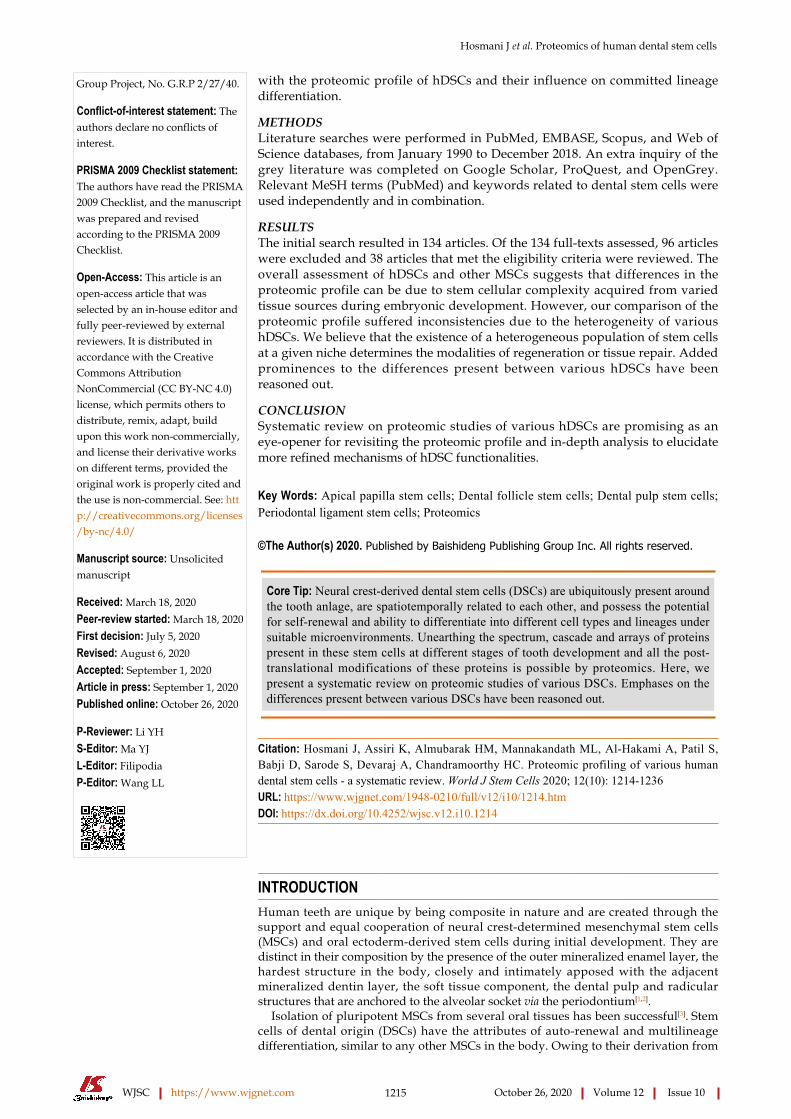
Table 3 Comparative proteomic analysis of human dental stem cells with other mesenchymal stem cells of the body
ComparisonRef. Dental stem
cellsMesenchymal stem cells
Main findings
Kumar et al[24]
DPSCs BMSCs DPSCs and its secretome show an inherent tendency for higher osteogenic differentiation and lower adipogenic differentiation, these may be potential candidates for effective future therapy in osteoporosis where disturbance of osteocyte/adipocyte homeostasis is reported
Eleuterio et al[17]
DPSCs and periodontal ligament stem cells
BMSCs Stem cells from oral tissue represent an easily accessible and autologous niche of stem cells that could give a worthwhile source to regenerative medication including many tissue frameworks and nerve repair
Yu et al[34] Dental apical papilla
BMSCs As for BMSCs, SCAPs indicated extended outflow of proteins that are locked in with metabolic methods and translation and lower measurements of those related with normal bond, developmental strategies, and safe limit; Similarly, SCAPs released on a very basic level greater proportions of chemokines and neurotrophins than BMSCs, however BMSCs discharged more ECM proteins and proangiogenic factors
DPSCs: Dental pulp stem cells; BMSCs: Bone marrow mesenchymal stem cells; SCAPs: Mesenchymal stem cells from dental apical papilla; ECM: Extracellular matrix.
Molecular characterization of DPSCs from SHED was compared with PDLSCs by distinguishing the variety of proteins being communicated in these cells. SHEDS were larger size PDLSCs. The mesenchymal properties of both SHEDs and PDLSCs were similar in terms of expression of the triple panel of protein-surface molecules, CD73, CD90 and CD105, and were negative for hematopoietic cell markers. Differentiation into osteo-, adipo-, and chondro-specific lineages was positive in SHEDs and PDLSCs following induction. High-scale proteomic mapping by engaging in nano-liquid chromatography - mass spectrometry (LC-MS)/MS technology led to the identification of 517 proteins in SHED, whereas in PDLSCs, it was 1721. SHED-explicit proteins appear to be embroiled in cell cohesiveness, regulation, motility, migration, and actin cytoskeleton organization. Wound healing assays performed for SHEDs and PDLSCs revealed that the propensity for higher injury recuperating movement was higher in SHEDs. SHEDs had an increased number of proteins that controlled cellular plasticity and adaptations to extrinsic and environmental stimuli[16].
Eleuterio et al[17] conducted a comparative proteome analysis between DPSCs, PDLSCs, and BMSCs from different donors. They found that the proliferation rate and viability of DPSCs compared to those of BMSCs were significant with the MTT assay at passage 2 after 3 d of culture. Even considering the capacity to differentiate into osteogenic tissue, DPSC was better when compared to BMSC. Approximately 2078 ± 357 proteome spots were detected in DPSCs. The level of coordinating between gels of a similar populace was 86 for DPSCs. Putative ATP-dependent CLP protease proteolytic subunit (CLPP), NAD(P)H dehydrogenase quinine 1 (NQ01), succinyl-CoA:3-ketoacid coenzyme A transferase 1 (SCOT 1) and an additional new isoform of tubulin (TBBS) were identified in DPSCs. Compared to that in BMSCs, N(G), N(G)-dimethylarginine dimethylaminohydrolase 1 (DDAH1) and citrate synthase (CISY) in DPSCs were upregulated. The study revealed that stem cells from dental tissue have different lineage potential than do BMSCs.
Patil et al[18] analyzed the morphology, expansion rate, articulation of MSC-explicit markers, and in vitro differentiation capacity into osteoblasts, adipocytes, chondrocytes and useful hepatocyte-like cells of DPSCs and contrasted them with those of DFSCs and APSCs. The protein spot match of DFSCs vs DPSCs was 61.6% ± 30.5%. Changes among the common protein expression levels between DFSCs and DPSCs were 16.8% ± 3.08% upregulating and 3.5% ± 4.03% downregulating. The potency of DPSC stemness was similar to that of DFSCs and APSCs in terms of the expression of the transcription factors Oct-3/4, Nanog and SOX-2 at both the mRNA and protein levels. It was also noted that the chondrogenic differentiation potential was more intense in DPSCs than in APSCs and DFSCs. It was found that DPSCs, along with DFSCs and APSCs, have the potential for hepatogenic differentiation under suitable culture conditions.
Ma et al[19] carried out a study to recognize the differentially communicated proteins among DPSCs and stem cells from profound carious DPSCs (CDPSCs) and investigated the potential molecular markers contributing to the recovery of dental tissues. The morphology determined via light and electron microscopy of DPSCs and
Hosmani J et al. Proteomics of human dental stem cells
WJSC https://www.wjgnet.com 1222 October 26, 2020 Volume 12 Issue 10
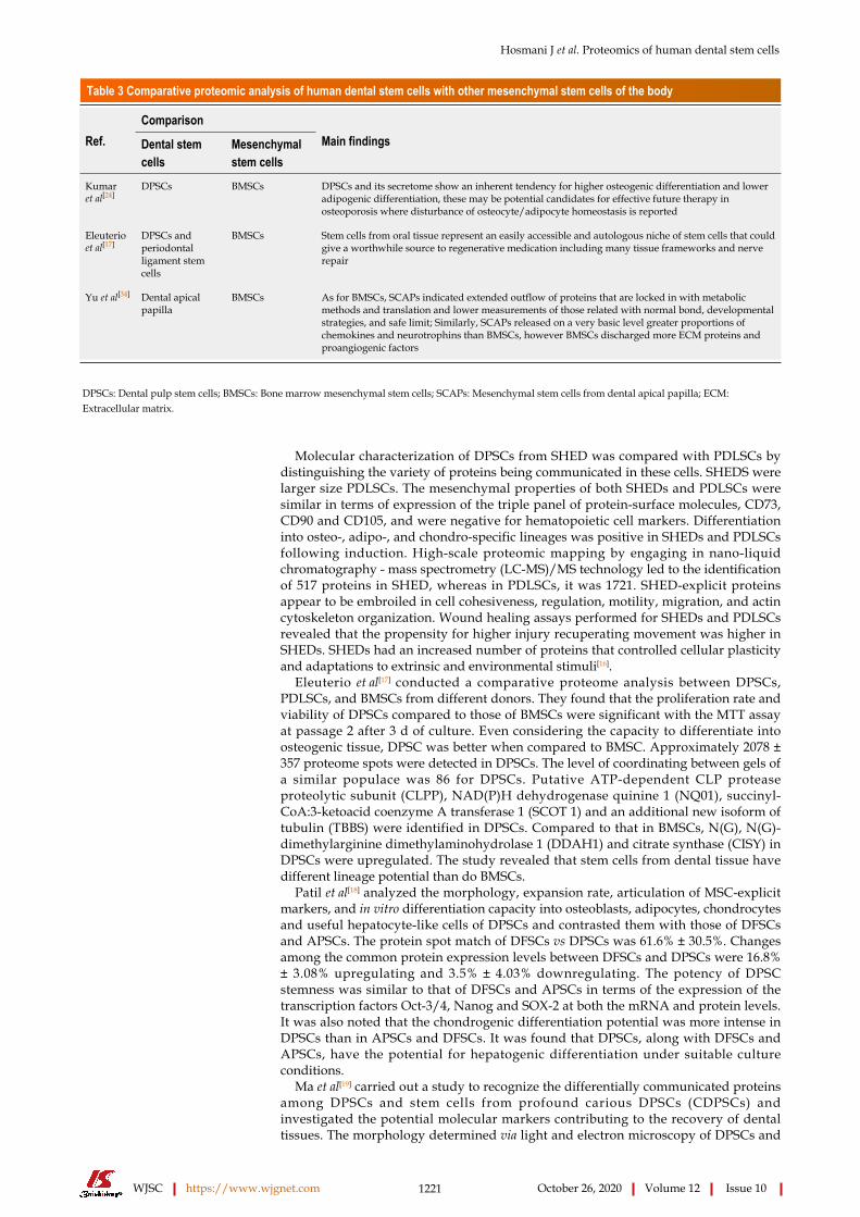
Table 4 Proteomic studies on influence or microenvironment or preconditioning factors on human dental stem cells
Ref. Dental stem cells Influence of niche or microenvironment or preconditioning factors Main findings
Dou et al[37] DPSCs Hypoxia Hypoxic culture of DPSCs under 3D framework incompletely influences the proteome profile of cells. Albeit present moment hypoxic culture (1% O2 for 24 h) changed articulation of a few proteins, the in an unexpected way communicated protein represent 2.7% (57 of 2115); By and large the impact of hypoxia on protein articulation in DPSCs was not substantial
Lee et al[38] DPSCs Preameloblasts conditioned medium Preameloblasts molded medium initiates the odontogenic differentiation of DPSCs and advances dentin development in vivo and in vitro Of the distinguished proteins, Cpne7 is another applicant that is engaged with odontoblast differentiation
Kim et al[39] DPSCs Human LOXL2 LOXL2 negatively affects the differentiation of hDPSCs and blocking LOXL2 can elevate the hDPSC differentiation to odontoblasts
Wang et al[21] DPSCs Periodontal ligament stem cells
Osteogenic induction medium Fewer than 5% of the differentially imparted proteins make up the close proteomic profile between osteo-induced PDLSCs and DPSCs This examination portrays the differences between osteo-induced PDLSCs and DPSCs in vitro The mineralization and migration cutoff points of PDLSCs were more noticeable than those of DPSCs, in which heat shock protein beta-1, Protein S100-A10 and S100-A11 may have an impact
Jung et al[40] Gingival fibroblasts Cyclosporin-A Prx 1 may play a relevant role in CsA-induced proliferation of gingival fibroblasts
Bakopoulou et al[41]
Apical papilla mesenchymal stem cells
Stress microenvironments: Serum deprivation, glucose deprivation, and oxygen deprivation/hypoxia conditions, individually or in combination
Endothelial transdifferentiation potential as well as angiogenic paracrine activity of apical papilla mesenchymal stem cells was significantly upgraded when uncovered for a brief timeframe to combined stress microenvironments
Lin et al[42] hPDLSCs 2,3,5,4’-tetrahydroxystilbene-2-O-β-D-glucosid 2,3,5,4’-tetrahydroxystilbene-2-O-β-D-glucosid enhanced the renewal ability and proliferative potential of hDPSCs via the AMPK/ERK/SIRT1 axis
Rani et al[43] hPDLSCs PVF of the horseshoe crab embryo PVF could improve cell cycle regulatory gene expression in DPSCs as shown by the higher articulation of the considerable number of qualities considered in this investigation at various cell passages in the treated gathering contrasted with the untreated group
Laothumthut et al[44]
hPDLSCs WSM extracted from the nacreous layer of the bivalve pinctada maxima
The human dental pulp cells cultured in nacreous WSM showed higher relative cell suitability than those in DMEM with comparative morphological appearance; Huge changes were found in the overall plenitude of 44 proteins in cells after introduction to WSM for about 14 d; They assume a job in cell adhesion, cell proliferation, metabolic process, signal transduction, stress reaction, transcription, translation, and transport
Bok et al[45] Dental follicle derived stem cells
HUVECs Expanded angiogenic movement in DFSC/HUVEC co-cultures may invigorate osteoblast development of DFSCs. Along these lines, the emission of angiogenic factors from HUVECs may assume a job in the osteogenic differentiation of DFSCs
Tsikandelova et al[46]
Human dental pulpal cells
FGF 8 protein FGF8 treatment could advance endogenous recuperating of the dental mash by means of enlistment of dental pulp progenitors just as by advancing their angiogenic and odontogenic differentiation
Qin et al[47] Human dental pulpal cells
Metformin Metformin can incite DPC differentiation and mineralization in an AMPK-subordinate way and that this all around endured antidiabetic drug has potential in regenerative endodontics just as in other regenerative applications
Wang et al[48] hPDLSCs DFO Early introduction to DFO advanced the mineralization of DPSCs and expanded autophagic action Autophagy restraint stifled DFO-prompted DPSC movement and odontoblast differentiation
Li et al[49] hPDLSCs SCs and its secreted vesicles SC emission demonstrated an overwhelmingly regulatory on the development of hDPCs
DPSCs: Dental pulp stem cells; LOXL2: Lysyl oxidase like 2; hDPSC: Human dental pulp stem cells; PDLSCs: Periodontal ligament stem cells; hPDLSCs: Human PDLSCs; PVF: Perivitelline fluid; WSM: Water-soluble matrix; HUVECs: Human umbilical vein endothelial cells; DFSC: Dental follicle stem cells; DPCs: Dental pulp cells; DFO: Deferoxamine; SC: Schwann cell; hDPCs: Human dental pulp cells.
Hosmani J et al. Proteomics of human dental stem cells
WJSC https://www.wjgnet.com 1223 October 26, 2020 Volume 12 Issue 10
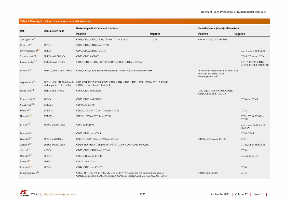
Table 5 Phenotypic cell surface markers in dental stem cells
Mesenchymal stromal cell markers Hematopoietic (stem) cell markersRef. Dental stem cells
Positive Negative Positive Negative
Niehage et al[10] CD29, CD44, CD73, CD90, CD105, CD146, CD166 CD271 CD 34, CD 45, CD133,CD117
Choi et al[14] DPSCs CD29, CD44, CD105 and CD90
Pivoriuunas et al[15] SHEDs CD73, CD90, CD105, CD146 CD14, CD34, and CD45
Taraslia et al[16] SHEDs and PDLSCs CD73, CD90 & CD105 CD45, CD34 and CD31
Eleuterio et al[17] PDLSCs and DPSCs CD13+, CD29+, CD44+,CD105+, CD73+, CD90+, CD146+, CD166+ CD117, CD133, CD144, CD271, CD14, CD34, CD45
Patil et al[18] DPSCs, APSCs and DFSCs CD44, CD73, CD90 & vimentin markers specifically associated with MSCs. A few cells expressed CD34 and CD45 markers associated with hematopoietic cells
Akpinar et al[20] DPSCs of SHED, Natal teeth and impacted third molar
CD3, CD4, CD13, CD14, CD29, CD34, CD44, CD45, CD73, CD90, CD106, CD117, CD146, CD166, HLA-DR, and HLA-ABC
Wang et al[21] SHEDs and DPSCs CD73, CD90 and CD105 Low expression of CD34, CD11b, CD19, CD45 and HLA-DR
Kumar et al[24] DPSCs CD73, CD90 and CD105 CD34 and CD45
Xiong et al[25] PDLSCs CD 73 and CD 90
Wu et al[26] PDLSCs STRO-1, CD146, CD29, CD44 and CD106 CD34
Hao et al[28] PDLSCs STRO-1, CD146, CD29 and CD90 CD31, CD34, CD45 and CD106
Li et al[29] DFSCs and PDLSCs CD73 and CD 90 CD31, CD34 and CD45, HLA-DR
Dou et al[31] CD73, CD90, and CD146 CD34, CD45
Guo et al[32] DPSCs and DFSCs STRO-1, CD29, CD44, CD90 and CD146 DPSCs: CD146 and CD106 CD31
Tian et al[33] DFSCs and PDLSCs CD146 and STRO-1: Higher in DFSCs, CD105, CD90, CD44 and CD29 CD 31, CD34 and CD45
Yu et al[34] APSCs CD73, CD90, CD105 and CD146 CD34
Dou et al[37] DPSCs CD73, CD90, and CD146 CD34 and CD45
Lee et al[38] DPSCs STRO-1 and CD44
Kim et al[39] DPSCs CD44, CD73, and CD105 CD34
CD90/Thy-1, CD73, CD146/MUC18, CD81/TAPA and the cell adhesion molecules CD49f/a6-integrin, CD29/b1-integrin, CD51/av-integrin, and CD166/ALCAM. Lower
Bakopoulou et al[41] CD105 and CD106 CD45

Hosmani J et al. Proteomics of human dental stem cells
WJSC https://www.wjgnet.com 1224 October 26, 2020 Volume 12 Issue 10
expression: STRO-1 (22.6%–2.2%), CD24 (25.8%–1.9%), and CD34 (3.2%–0.5%)
Bok et al[45] DFSCs CD44, CD73, CD90 and CD105 CD34 and CD45
Tsikandelova et al[46] DPSCs CD49f, CD117, CD146, CD271, CD105 and Stro-1
Qin et al[47] CD73, CD90 and CD105 CD34 and CD45
DPSCs: Dental pulp stem cells; SHEDs: Stem cells from human exfoliated deciduous teeth; PDLSCs: Periodontal ligament stem cells; APSCs: Stem cells from apical papilla; DFSCs: Dental follicle stem cells; MSCs: Mesenchymal stem cells.
CDPSCs was similar and had a spindle-fibroblast-like appearance with long cytoplasmic extensions and filipodia. When researchers analyzed the proliferation rates of DPSCs and CDPSCs at passage 3 after 3, 5, and 7 d, it was found that CDPSCs had a higher proliferative potential. Proteomic analysis at a narrow pH (4-7) revealed 18 protein spots that were differentially expressed in CDPSCs. The proteins were mainly related to cell proliferation, differentiation, cell cytoskeleton, motility, and antioxidative functions. They concluded that these proteins might be responsible for the biological differences between CDPSCs and DPSCs. They likewise discovered that CDPSCs have a more elevated antioxidative properties that protect them from oxidative stress.
The characteristics of DPSCs from natal, SHED and impacted third molars were analyzed by Akpinar et al[20] to determine the level of heterogeneity. Each stem cell line indicated the presence of cells with large, flattened or fibroblast-like shapes, and no noteworthy changes in size and shape were observed. However, evaluation of proliferation rates by measuring cell viability at various time points indicated that natal DPSCs had a higher proliferation rate than SHEDs and DPSCs of impacted third molars. The highest telomerase activity was observed with natal DPSCs. It was noted that the natal DPSC cell proteome was more similar to the SHED proteome than the DPSC proteome. Most of the proteins elucidated have a major role in cellular architecture. A total of 61 proteins were predominantly expressed by all three stem cell types.
Differentially expressed proteins in DPSCs were compared with those in PDLSCs under in vitro osteogenic induction using the iTRAQ proteomic approach and bioinformatic analysis by Wang et al[21]. A common set of over 3000 proteins was detected in DPSCs and PDLSCs. Merely 5% of them were differentially expressed, which reflected the extreme particular differentiation potential and different capacities under the individual microenvironments of these two cells. DPSCs were found to be weaker on osteogenic mineralization compared to PDLSCs. Prostacyclin synthase and arginosuccinate lyase were the two of the most significantly expressed proteins between PDLSCs and DPSCs and are important for many metabolic and biosynthetic processes.
Joo et al[22] carried out a study on the expression levels of cytokines from dental pulp (DPSCs) and developing apical complexes of immature teeth (DACCs). A human

Hosmani J et al. Proteomics of human dental stem cells
WJSC https://www.wjgnet.com 1225 October 26, 2020 Volume 12 Issue 10
Figure 1 Flow diagram of literature search and selection criteria.
cytokine array was utilized to assess and match 174 cytokines in DACC-CM and DPSC-CM. They found that there were 25 strongly expressed cytokines, among which 22 and 3 were strongly expressed in DPSC-CM and DACC-CM, respectively. NT-3 and BMP-4, which are related to odontoblastic differentiation, were significantly expressed at higher levels in DPSC-CM. TGF-β, NT-4 and EGFR were exclusively expressed in DPSC-CM. It was inferred that dental pulp cells express more cytokines specific to mineralization than DACCs.
Wang et al[23] examined the impacts of long-term in vitro development on the fundamental properties and gene expression of SHEDs and DPSCs. It was found that long-term culturing of SHEDs and DPSCs led to morphological alterations with irregular and elongated shapes. SHEDs displayed a fundamentally higher proliferation rate in correlation with DPSCs in vitro at an early passage, yet development energy was altogether diminished at passage 20. Immune phenotyping indicated that SHEDs and DPSCs were able to maintain their specific properties of stemness. The percentage of CD-73-positive DPSCs was lower at passage 20 than that in SHEDs at passage 20. The differentiation propensity of SHEDs and DPSCs was found to be markedly reduced with long-term cultivation, which was confirmed by RT-qPCR analysis. Even the migratory abilities of SHEDs and DPSCs were found to be markedly reduced. The number of apoptotic cells in both DPSCs and SHEDs increased during long-term cultivation. Analysis of the molecular markers of senescence showed that DPSCs expressed a higher predisposition of cell death during long-term cultures.

Hosmani J et al. Proteomics of human dental stem cells
WJSC https://www.wjgnet.com 1226 October 26, 2020 Volume 12 Issue 10
SHEDs and DPSCs may undertake unique pathways during cellular senescence, and p19Ink4a appears to play a significant role in DPSC senescence.
The DPSCs were pitted against BMSCs for their ability to differentiate into osteoblasts and adipocytes by Kumar et al[24] who further reported the contribution of secretome proteins in regulating their differentiation. DPSCs displayed higher osteogenic potential than BMSCs. DPSCs demonstrated a higher accumulation of calcium. Quantitation of oil red O uptake by isopropanol extraction showed significantly higher adipogenic potential of BMSCs compared to DPSCs. Proteome analysis of the secretome showed 7 osteogenic lineage-related proteins compared to 5 proteins in BMSCs. Adipogenic lineage-related proteins in the secretome of BMSCs were 4 and 5 in DPSCs. It was concluded that owing to their ability to increase osteogenic and decrease adipogenic potential of DPSCs, the utility of DPSC secretomes in osteoporosis could be a promising “cell-free” therapeutic modality for the future.
PDLSCsXiong et al[25] carried out a study to determine the various proteins associated with cellular functions and properties of PDLSC. PDLSCs had smaller sizes when compared to SHED. A total of 3235 proteins were expressed in PDLSCs, and comparative evaluation revealed 1516 proteins expressed in both PDLSCs and SHEDs, of which 1721 were presented exclusively in PDLSC. The majority of these exclusively identified proteins appeared to be mainly classified in the cellular metabolism network. A total of 545 proteins were found to be involved in the cellular growth and proliferation of 207 proteins strongly involved in DNA replication, recombination and repair. On a whole, PDLSCs revealed a notable mitogenic advantage, as indicated by growth curves and protein molecules identified in the study. The Rho-GDI signaling pathway revealed proteins that were directly involved in the elevated proliferation potency of PDLSC.
Analysis of differentially expressed protein profiles of PDLSCs undergoing mineralization was investigated by Wu et al[26] (2009). Positivity for markers related to bone formation was observed in PDLSCs when cultured in osteogenic media. In total, 29 differentially expressed proteins were identified in PDLSCs during osteogenic differentiation. Proteins involved in cellular functions, such as cytoskeletal-related, nuclear regulation, cell membrane binding, matrix synthesis, metabolic enzymes and signal transduction, were identified. Reorganization of the cytoskeleton during osteoblastic/cementoblastic differentiation of PDLSCs was noted by downregulation of vimentin, caldesmon, tropomyosin and clathrin heavy-chain polypeptide.
Proteomic analysis of PDLSCs was carried out by Reichenberg et al[27]. In this, 117 proteins that corresponded to 74 different gene products were identified. At least 30 spots were identified as proteins associated with the cytoskeleton. The metabolic needs and capabilities of the PDLSCs were demonstrated by the identification of 20 spots as metabolic enzymes. The subcellular localization of proteins was found to be 50.2% in the cytoplasm, 14.9% in the endoplasmic reticulum and 16.3% in mitochondria. Membrane-associated proteins constituted 4%, and cytoplasmic vesicles constituted 1.3%. It was discovered that the primary contrasts among dermal and PDL fibroblasts were in the classification of cytoskeleton-related proteins. It was accepted that this distinction mirrors the broad and exceptional biotopological capacity of PDL in cell- extracellular matrix (ECM) communications, maintaining legitimate collagen homeostasis and cellular reaction to the outer mechanical masticatory loads being applied inside PDL, which requires high articulation of cytoskeleton segments.
A study by Hao et al[28] was carried out to delineate the exact miRNA profile during osteogenic differentiation of PDLSCs. The gene targets of differentially expressed miRNAs were also predicted utilizing bioinformatic tools. The findings of this study indicate that miRNAs play significant roles in the osteogenic differentiation of PDLSCs. Microarray-based analysis was used to investigate osteogenesis-related miRNAs, 116 of which were found to be differentially expressed, and 34 had putative target genes that were intimately associated with osteogenesis. qRT-PCR analysis revealed the upregulation of m-R-654-3p, miR-4288 and miR-34c-5p and downregulation of miR-218-5p, miR-663a and miR-874-3p. The downregulation of miR-218 suggested its suppressive effect on the angiogenesis of PDLSCs. It was also found that the downregulated miR218-5p has three osteogenesis-related targets, RUNX2, COL1A1, and SATB2. The downregulated miRNAs may elicit a negative effect on osteogenic differentiation via direct posttranscriptional inhibition of RUNX2.
Identification of PDLSC-specific biomarkers was performed by Taraslia et al[16] by distinguishing the atlas of proteins being communicated in these cells. It was found that PDLSCs were smaller than SHEDs. PDLSCs were positive for MSC markers and negative for hematopoietic markers. The expression of lineage specific was observed in
Hosmani J et al. Proteomics of human dental stem cells
WJSC https://www.wjgnet.com 1227 October 26, 2020 Volume 12 Issue 10
PDLSCs, and these cells were found to be capable of differentiating into osteo-, adipo- and chondro-specific lineages. In-depth and high-scale proteomic mapping uncovered 1721 proteins solely in PDLSCs and basically grouped in various aspects of cell metabolism and proliferation which clarified the nearness of solid mitochondria contrasted with SHEDs. PDLSC-explicit proteins were additionally bunched in RNA combination and homeostasis. The proteins identified in PDLSC also indicated high proliferative activities of cells.
The PDLSC proteome was compared with those of DPSCs and BMSCs. All of the analyzed stem cells had a similar morphology with a fibroblast-like shape. The expression of stem surface molecules and pluripotency surface markers was positive for all the stem cells analyzed. The proliferation rate was significant in PDLSCs compared to BMSCs. Evaluation of osteogenic differentiation via the expression of osteogenic marker intensity revealed that bone formation was higher in PDLSCs than in BMSCs. Comparative proteome analysis in a broad pH range (3-10) allowed the detection of 2131 ± 290 spots in PDLSCs. The percentage of matching between gels of the same population was approximately 86% for PDLSCs, and the percentage matching between different populations was approximately 80% for DPSCs and PDLSCs, 78% for DPSCs and BMSCs, and 77% for PDLSCs and BMSCs. Four proteins were exclusively expressed in PDLSCs and BMSCs, namely, CLPP, NQ01, SCOT1 and TBBS. DDAH1 and CISY were found to be upregulated in PDLSCs and DPSCs compared to BMSC. Annexin 10 was found to be exclusively present in PDLSCs. COMT was exclusively present in BMSCs and was found to be missing in PDLSCs and DPSCs. In the pH 4-7 range, 1777 ± 152 protein spots were resolved, and in the pH 6-9 range, 1101 ± 108 protein spots for PDLSC were resolved. In this pH range, PDLSC exclusively expressed NECP2, AK1C1 and SEP11[17].
The highly sensitive and easily performed iTRAQ proteome analysis of PDLSCs and DPSCs was completed by Wang et al[21], who also determined the correlation between these two cell types. Regular arrangement of more than 3000 proteins was distinguished in both PDLSCs and DPSCs, and fewer than 5% of them were differentially communicated. It was contemplated that comparative starting points and close limitation lead to solid articulation of such a wide scope of proteomes, and differentially communicated proteins mirrored a definitive particular separation potential and various capacities under one-of-a-kind microenvironments. In the present investigation, PDLSCs had a more grounded mineralization limit than DPSCs, which is a solid marker of osteogenic separation. Prostacyclin synthase and arginosuccinate lyase are two of the largest differentially communicated proteins among PDLSCs and DPSCs. Proteins with collagen, heat shock proteins and S100 families were observed to be downregulated in PDLSCs
Li et al[29] analyzed the immunophenotypic highlights and cell cycle status of two cell lines. The outcome of the study proposed that DFSCs and PDLSCs showed comparative highlights identified with immunophenotype and cell cycle. Molecular differences between DFSCs and PDLSCs were analyzed by iTRAQ. This study provided new insights into the possible mechanism responsible for the different biological features, wherein PDLSCs displayed enhanced actin cytoskeletal dynamics relative to DFSCs, while DFSCs exhibited more robust antioxidant defense ability in comparison with PDLSCs.
Proteomic studies on DFSCsThe proteomic profile of DFSCs before and after osteogenic differentiation and the identification of differentially expressed proteins involved in important cellular processes were studied by Morsczeck et al[30]. The present discoveries include understanding the systems associated with DFSC differentiation. The outcomes provided proof that differentiation influences cell shape and motility and catalysts engaged with the control of oxidative pressure and collagen biosynthesis. Compounds such as superoxide dismutase or glutamine synthetases were most likely associated with the procedure of cell separation. These distinguished proteins are a piece of machinery in charge of the arrangement of membrane-like cell structures containing mineralized clusters. Biological processes associated with osteogenic differentiation of osteoblasts were not recognized in upregulated proteins of DFSCs. The authors believed that modification of the activity of special enzymes, cell motility or ROS may also affect the osteogenic differentiation process of DFSCs.
A comparison of the protein expression profiles of DFSCs with DPSCs and APSCs from a single donor tooth was carried out by Patil et al[18]. With respect to the characterization of DFSCs, the population doubling time was significantly shorter, suggesting a higher proliferative rate when compared to that of DPSCs and APSCs. The DFSCs were found to be proportionally higher in the S phase of the cell cycle

Hosmani J et al. Proteomics of human dental stem cells
WJSC https://www.wjgnet.com 1228 October 26, 2020 Volume 12 Issue 10
compared to APSCs and DPSCs. Approximately 462.6 ± 32.03 well-stained protein spots were detected in DFSCs, which were significantly higher than those of DPSCs and APSCs. The osteogenic differentiation potential of DFSCs and DPSCs was higher than that of APSCs. The adipogenesis potential was found to be higher for DFSCs, which was attributed to the supplementation of IBMX in adipogenic induction medium in this study. It was found that proteins of DFSCs closely resembled proteins secreted by APSCs compared with DPSCs.
An iTRAQ proteomic analysis of immortalized DFSCs was carried out by Dou et al[31]. The iDFSCs did not have fibroblast-like morphology, and the nuclear cytoplasmic ratio was altered. It was noted that iDFSCs had enhanced telomerase activity compared with that of DFSCs. The iDFSC secretome harbored a large number of proteins primarily involved in metabolic, biologic, immunologic and defensive regulation. Among the 2092 detected secretory proteins, only 12.1% were significantly affected by immortalization. Series of bioactive factors, such as different types of collagen, nestin, MMPs, TIMPs, HSPs, PDGF, IGF 1-2, VEGF, and TGF β 1,2, which could be related to ECM remodeling and tissue regeneration and repair, could be identified. Compared with those in DFCs, the level of TGF-β1 was upregulated and the level of IL-6 was downregulated in iDFSCs. The study results revealed that immortalization partly changed the proteomic profile of the DFSC proteome.
The proteome of DFSCs was compared with that of DPSCs by Guo et al[32]. DFSCs were found to be rich in rough endoplasmic reticulum and lysosomes in TEM, but ultrastructural comparison with DPSCs gave a contrasting finding wherein rough endoplasmic reticulum and mitochondria were more abundant. DFSCs showed a higher proliferation rate. A total of 30 spots were identified as being differentially expressed. Among these, 16 proteins were expressed at higher levels in DFSCs. DFSCs were found to be more pluripotent than DPSCs by virtue of having less heterochromatin in the nucleus. The upregulated proteins in DFSCs had a greater capacity to manage bone development and resorption and to advance cell survival in a hypoxic domain.
A similar kind of comparative study on DFSCs and PDLSCs analyzed the biological characteristics and investigated their odontogenic differentiation under the inductive microenvironment of treated dentin matrix. The DFSCs were smaller than PDLSCs. Homogenous electron-dense granules were present in DFSCs but not in PDLSCs. Heterochromatin were observed in DFSCs, but cytoplasmic organelles were abundant in PDLSCs. A total of 35 spots were identified as differentially expressed; among these, 12 proteins were highly expressed in DFSCs. The proteins identified were primarily located in the cytoplasm. DFSCs presented stronger clone-forming abilities compared to PDLSCs. DFSCs were efficient in undergoing differentiation. The study revealed that stemness was higher in DFSCs than in PDLSCs. It was reasoned that DFSCs are derived from tissue at an earlier stage of tooth development and could be of great value in regenerative medicine. Li et al[29] noted in their study that DFSCs exhibited more robust antioxidant defense ability compared to PDLSCs.
In this relative investigation, Tian et al[33] endeavored to reveal insight into the particular organic properties of DFSCs and PDLSCs and to assess their odontogenic differentiation under the equivalent inductive condition of TDM scaffolds. Because of their origin from the developing tissue, DFSCs had a higher proliferative rate and were morphologically smaller and fusiform. DFSCs had less heterochromatin in the nucleus compared to PDLSCs. The organelles present in the DFSCs were all demonstrative of cell homeostasis, such as rebuilding amid general improvement, and assume a basic job in foundational stem cell renewal, multiplication and maturing. DFSCs represented a stronger clone-framing capacity than PDLSCs. This examination found that the dentinogenesis-related qualities communicated distinctively in DFSCs and PDLSCs with or without the microenvironment of TDM.
Proteomic studies on APSCsThe typical features and common and specific cellular proteomic profile of APSCs were compared with those of DPSCs and DFSCs. Evaluation of population doubling time showed that that of APSCs was 39.1 ± 2.7 h. The proportion of cells in the S phase of the cell cycle of APSCs was 21.9% ± 4.1%. The population of APSCs in the G0/G1 phase was 61.8% ± 3.6%. The proportion of APSCs in the G2/M phase was 16.3% ± 3.2%. In APSCs, approximately 3366 ± 10.78 well-stained protein spots were detected. The protein spot matching between DFSCs and APSCs was 69.3% ± 1.52%. Changes among the common protein expression levels between DFSCs and APSCs were 24.6% ± 5.17%. There were 96.7±11.04 specific protein spots of APSCs. In vitro differentiation assays suggested that APSCs are distinct from other DSCs. They were most closely matched to DFSCs[18].

Hosmani J et al. Proteomics of human dental stem cells
WJSC https://www.wjgnet.com 1229 October 26, 2020 Volume 12 Issue 10
Guo et al[32] explored the differences in the odontogenic differentiation ability of APSCs and DPSCs and compared their proteomes. Ultrastructural analysis demonstrated that there were rough endoplasmic reticula and mitochondria in the cytoplasm of APSCs. In terms of proliferation rate, APSCs had a lower rate compared to DFSCs. A total of 30 spots were identified as being differentially expressed between APSCs and DFSCs. Among these, 14 proteins were expressed at higher levels in APSCs. The pluripotency of APSCs was lower, and even the telomerase activity was lower in APSCs. Vascular differentiation was found to be better in APSCs, suggesting that they are more likely to differentiate in vascular endothelial cells. A stronger osteogenic differentiation potential was observed in DFSCs than in APSCs. The upregulated proteins indicated that bone formation and resorption were better in DFSCs than in APSCs. It was noted that APSCs differentiated into odontoblasts quickly when they came in contact with dentin tissue. The treated dentin matrix was shown to be a better microenvironment for inducing the differentiation of APSCs. Higher expression of DSPP protein was noted in APSCs, suggesting its greater role in dentinogenesis. APSCs were found to be at an earlier stage of neural differentiation compared to DFSCs.
Developing apical complex (DACC) along with dental pulp were checked for the expression levels of cytokines by Joo et al[22] (2018). Proliferation-related and anti-inflammatory cytokines were strongly expressed in DACCs. IFG-1, a cytokine expressed in DACC, was responsible for increased cell proliferation, alkaline phosphatase activity and DNA synthesis. Lipopolysaccharide activity inhibition and osteoprotegerin expression of the apical complex reduce osteoclastic activity, thereby creating a microenvironment that facilitates osteoblastic activity. The anti-inflammatory cytokine IL-10, which is the only cytokine present in DACC, facilitates tissue development and osteogenesis in the apical complex.
The functional role of APSC secretomes, the molecular basis of their actions and comparison with the proteomic profile of BMSCs were investigated by Yu et al[34]. The proliferation of APSCs was enhanced compared to that of BMSCs. A total of 2046 proteins were detected from APSCs. Among these proteins, 25.3% matched the extracellular region, and 9.5% matched the ECM. The ECM proteins discharged by APSCs are associated with cell cohesiveness, multiplication, collagen association and proteases, immune framework, odontogenesis, angiogenesis, neuron advancements, and signaling molecules.
Role of microenvironments and preconditioning factors on the proteomic profile of DSCsStem cells more often than not pass through two distinctive natural conditions, from segregation to engraftment. It is unclear how microenvironmental effects on stem cells result in their reactions to bodily insults and how useful stem cells can be cultured in vivo. These questions have contributed to the theory that, given the different tissue-specific stem cell specialties and distinctive microenvironments, initiating a stem cell specialty will result in stem cell self-renewal and controlled differentiation to drive the fast and effective differentiation of other stem cells so they can join the battle[35].
Stem cell microenvironments are becoming progressively acknowledged due to their restorative impacts in controlling stem cell behavior and homeostasis. In their specialty, stem cells are maintained or can experience expansion and differentiation in reaction to injury, disease or maturation to renew misplaced cells or tissues. In diverse situations, stem cells have diverse requirements to work appropriately. In this manner, plastic stem cells and progenitor cells have been examined in an assortment of situations and have been created through physical, chemical, genetic, and pharmacological controls. In reasonable microenvironments, plastic stem cells by and large have progressed cell survival, increased neuronal separation, and enhanced paracrine effects, resulting in expanded trophic support, improved homing to the injury location, and intensive suppression inflammatory factors in immune responses to promote functional recovery. The microenvironment serves to drive the cells into a state of availability in different environments[36].
In the present review, fourteen articles were analyzed for the influence of a niche, a microenvironment or preconditioning factors on the proteomic profile of DSCs.
Dou et al[37] deliberately examined the impact of momentary hypoxia on the gene expression and proteomic profile of human DPSCs under 3D culture conditions. Most of the upregulated proteins engaged in cell processes, angiogenesis, protein binding and transport, direction of reaction to stimuli, metabolic processes, and immune responses. The investigation indicated that hypoxia affected the proteomic profile of DSCs. This present investigation discovered that hypoxia enhanced the expression of

Hosmani J et al. Proteomics of human dental stem cells
WJSC https://www.wjgnet.com 1230 October 26, 2020 Volume 12 Issue 10
several pro-angiogenic genes at the mRNA level. HIF-1É is a key factor regulating angiogenesis actuated by hypoxia. Additionally, expanded TGF-β1 expression was found after low-oxygen incitement. TGF-β1 is considered an indispensable cytokine that directs a broad range of physiological and pathologic processes, including tissue wound healing, inflammation, cell proliferation, differentiation, migration, and ECM synthesis.
Lee et al[38] led an investigation in which they investigated traces of BMP in preameloblast-conditioned medium (PA-CM) influencing hDPSC differentiation. Consequently, they hindered BMP movement in PA-CM with the extracellular BMP antagonist noggin and evaluated DSPP promoter activity. Progressions in DSPP promoter activity after noggin treatment in PA-CM were not noticed, suggesting that PA-CM influences DSPP promoter activity by components that are superfluous to BMP-2. BSP levels were observed to be higher than DSP in the mineralized tissue created in the rhBMP-2-treated group, while mineralized tissue shaped in the PA-CM-treated group showed the opposite trend. Generally, this information proposes that PA-CM promotes the induction of more odontogenic qualities of hDPSCs than rhBMP-2.
Kim et al[39] demonstrated that lysyl oxidase-like 2 (LOXL2) expression is decreased during odontogenic differentiation of DPSCs. Exogenous application of human recombinant LOXL2 protein resulted in a decrease in odontogenic differentiation. The levels of LOXL2 decrease as the cells lose their proliferative capacity and begin differentiation.
Wang et al[21] investigated the differentially communicated proteins in DPSCs and PDLSCs, alongside the underlying mechanism, under in vitro osteogenic induction using an iTRAQ proteomic approach and bioinformatics examination. In vitro osteogenic induction somewhat recreates the microenvironment principal for osteogenesis/odontogenesis of mesenchymal cells. Under such conditions, regardless of the way that DPSCs and PDLSCs can outline osteoid tissue, which is the nature of osteogenesis/odontogenesis, DPSCs differentiate into odontoblast-like cells, while certain PDLSCs differentiate into cementoblast-like cells, adipocytes, and collagen-framing cells. A total of 159 differentially communicated proteins in DPSCs and PDLSCs were recognized. A few individuals from the collagen, heat shock protein and protein S100 families may recognize osteo-induced PDLSCs from DPSCs. Of the enriched terms, 75% belonged to catalytic activity, protein binding and negative regulation of protein metabolic processes, indicating different biosynthetic and regulatory processes of PDLSCs and DPSCs towards differentiation. It was proposed that PDLSCs and DPSCs may experience unmistakable metabolic changes amid the differentiation procedure and that the differentiation enlistment condition could as act as a stress condition. KEGG investigation uncovered a few pathways recognizing osteo-induced PDLSCs from DPSCs.
In a study led by Jung et al[40] a proteomic strategy was utilized to distinguish conceivable changes at the protein level after cyclosporin A (CsA) treatment in human gingival fibroblasts (HGFs). CsA extended the cell appropriateness of HGFs in a proportionate and time-subordinate manner. Seventeen proteins were overexpressed in the CsA-treated HGFs; however, three proteins were observed to be communicated in a manner not matching that in the untreated cells exactly. The recognized proteins were fundamentally related to cell expansion, digestion, and oxidation. The overexpression of peroxiredoxin 1 (Prx 1) confirmed by Western blotting and the diminishing of cytosolic responsive oxygen species (ROS) levels in the CsA-treated HGF indicated that Prx 1 may accept a pressing occupation in the HGF development impelled by CsA. Upregulation of galectin 3 in CsA-treated HGF demonstrated that it is related to CsA-initiated multiplication.
Bakopoulou et al[41] considered three distinct angles characterizing the angiogenic capability of APSCs under different stress conditions related to in vivo conditions in damaged tissues. Specifically, the study demonstrated that the attributes of APSCs under a stressful microenvironment, such as serum, glucose and oxygen deprivation, were increased as much as possible towards endothelial transdifferentiation potential, similar to angiogenic paracrine movement. The authors reported that endothelial transdifferentiation of APSC was associated with noticeable upregulation of Ang-2 and Tie-1 and parallel downregulation of Ang-1 and Tie-2 articulation. An unconventional component in their investigation was that transient presentation (72 h) of APSC to stressful microenvironments was similarly or progressively strong in evoking a proangiogenesis procedure, in contrast to introduction with the angiogenic vehicle for 28 d.
A bioactive compound of Fallopia multiflora, 2,3,5,4’-tetrahydroxystilbene-2-O-β-D-glucoside (THSG), is known for its antioxidative properties and free radical
Hosmani J et al. Proteomics of human dental stem cells
WJSC https://www.wjgnet.com 1231 October 26, 2020 Volume 12 Issue 10
elimination activities, and has been a source of several studies related to reducing the effect of aging, protecting the cardiovascular system and reducing inflammation. It has also been reported that it can induce the proliferation and differentiation of stem cells. Lin et al[42] studied the mechanisms of THSG-enhanced proliferative potentials in hDPSCs. hDPSC proliferation was significantly increased when the concentration of THSG was greater than 0.1 μmol/L, with no cytotoxic effects towards hDPSCs. Higher levels of cell renewal were also noted. Expression of genes such as SIRT1, which is associated with cell proliferation, was noted in hDPSCs after treatment with THSG. THSG significantly induced pluripotent stem cell genes, including NANOG, POU5F1, and SOX2. This meant that THSG might change DPSCs from multipotent stem cells to pluripotent-like stem cells.
Due to its possession of cell proliferation abilities, perivitelline fluid (PVF) of the horseshoe crab embryo has been found to be beneficial in cardiac disorder and cancer therapies. However, its utility in investigating the expression of cell cycle regulatory genes from human DPSCs was never studied, which prompted Rania et al[43] to carry out such research. CDKN2A, PTEN, TP53 and MDM2, the genes with a pivotal role in regulating the cell cycle of DPSCs, were analyzed when DPSCs were exposed to PVF. There was no change in the expression levels of CDKN2A and MDM2 between treated and untreated DPSCs. Enhanced expression of CDKN2A revealed that the presentation to PVF expanded cell expansion and counteracted cell senescence. DPSCs treated with PVF displayed elevated expression of MDM2 at passage 4, drastically depleted expression at passage 5 and slightly increased expression at passage 6. TP53 expression in the DPSC-treated group was higher than that in the untreated group. On the other hand, the expression of PTEN in the DPSC-treated group started to increase from passages 5 to 6. In any case, in general, the PTEN articulation was higher than the untreated gathering in every one of the sections considered. The results demonstrated that PVF could overhaul cell cycle administrative quality articulation in DPSCs, as indicated by the higher verbalization of the impressive number of characteristics considered in this examination at different cell entries in the treated DPSCs being differentiated from the untreated DPSCs.
An examination to assess the impact of the nacreous water-dissolvable framework (WSM) on multiplication and differential protein expression in STRO-1+ human dental pulp cells in vitro was carried out by Laothumthut et al[44]. Nacre, the glossy inward layer of a molluscan shell, is a characteristic material for self-association in biomineralization. Its natural segment has been generally grouped into WSM and water-insoluble network. It was discovered that the STRO-1-positive dental pulp cells refined with DMEM medium enhanced with nacreous WSM demonstrated higher cell suitability than those with DMEM medium. An aggregate of 44 differentially communicated proteins with obvious molecular weight underneath 30 kDa delivered by human pulp cells refined with either DMEM or WSM were distinguished. They were copiously upregulated in cells after introduction to nacreous WSM for approximately 14 d. It was recommended that these proteins function in cell binding, multiplication, metabolism, signal transduction, stress reaction, transcription, translation, and transport and was critical for STRO-1-positive magnetic-isolated human dental pulp cells to react to the outside WSM within 2 wk. The results of the study suggested that WSM seems to be a useful biomaterial in the process of tissue repair after pulp injury.
The impact of human umbilical vein endothelial cells (HUVECs) on osteogenic differentiation of DFSCs in vitro by coculturing DFSCs and HUVECs was studied by Bok et al[45]. Outcomes of the study demonstrated that expanded angiogenic movement in DFSC/HUVEC cocultures may animate osteoblast development of DFSCs. Along these lines, the emission of angiogenic factors from HUVECs may assume a job in the osteogenic separation of DFSCs. Cell viability, in view of metabolic action and DNA content, was most astounding for co-cultures with a DFSC/HUVEC proportion of 50:50 in a 1:1 blend of mesenchymal stem cell development medium and endothelial cell development medium. Osteoblastic and angiogenic phenotypes were upgraded in cocultures with a DFSC/HUVEC proportion of 50:50 compared with DFSC monocultures. Expanded expression of angiogenic phenotypes and vascular endothelial development factor (VEGF) levels were observed after some time in both 50:50 DFSC/HUVEC cocultures and DFSC monocultures during the culture period.
Tsikandelova et al[46] studied the effect of fibroblastic growth factor-8 (FGF-8) on adult heterogeneous DPSCs by assessing their genetic, proteomic, proliferative and mineralization profiles. Enhanced proliferation of DPSCs was noted after short-term exposure to FGF-8. Several proteins implicated in the regulation of cellular proliferation processes, including LMNA, XRCC5 and gelsolin, were noted. 2D and mass spectrometry show that a greater portion of the proteins distinguished through

Hosmani J et al. Proteomics of human dental stem cells
WJSC https://www.wjgnet.com 1232 October 26, 2020 Volume 12 Issue 10
the proteomics approach are intensely ensnared in the control of the actin cytoskeleton and bond elements, which is predictable given that FGF8 has the capacity to advance cell transitory conduct in dental pulp cells. Continuous FGF8 treatment of dental pulp in this study also strongly upregulated genes associated with pulp inflammation, such as leukotriene A4 hydroxylase (LTA4H) and platelet endothelial cell adhesion molecule (PECAM1). In this manner, due to the general requirements for wound recuperation of angiogenesis, improved relocation, a more prominent proliferative limit, endothelial separation and ECM redesigning observed in this study could demonstrate that FGF8 treatment can inspire an angiogenic reaction in a heterogeneous culture of dental pulp.
The effects of metformin on the proliferation and odontoblastic differentiation of dental pulp cells and the role of AMPK signaling in metformin-mediated odontoblastic differentiation were investigated by Qin et al[47]. Metformin is broadly utilized by a large number of diabetic patients as a first-line treatment for type 2 diabetes mellitus. As an insulin-sensitizing drug, it is safe, nontoxic, and overall endured. The impact of metformin on cell multiplication has been accounted for in different types of cells. The study demonstrated that diverse dosages of metformin have no large impact on dental pulp cell expansion. Moreover, metformin was found to not modify the viability of dental pulp cells, suggesting that metformin is safe and nontoxic. Strangely, this investigation demonstrated that the addition of the antiviral agent adenine 9-b-D-arabinofuranoside, a specific AMPK inhibitor, constricted metformin-actuated reactions. These outcomes proposed that metformin actuated the differentiation and mineralization of osteoblast-like cells through the initiation of AMPK signaling. This study showed that metformin can enhance the odontoblastic differentiation of dental pulp cells through activation of the AMPK signaling pathway.
The molecular mechanism underlying the effect of deferoxamine (DFO) on the repair ability of DPSCs was studied by Wang et al[48]. The effect of DFO on autophagy and the role of autophagy in regulating the migration and odontoblast differentiation of DPSCs were elaborated. Enhanced differentiation of DPSCs and mineralization was noted after exposure to DFO. Autophagy in DPSCs was observed after DFO activation. LC3B is a common marker for autophagosome formation, and immunostaining with LC3B was performed in the present study. DFO treatment obviously increased the protein expression of LC3B-II and Beclin 1 and gradually decreased the protein level of p62. To additionally show that the iron chelator (DFO) could prompt autophagy in DPSCs, the autophagic level was identified furthermore of FC (Fe iron). It was shown that DFO could instigate autophagy by sequestering intracellular iron. Strikingly, excess iron could similarly enhanced the outflow of LC3B, showing that autophagy is closely related to iron metabolism.
Li et al[49] investigated the administrative potential outcomes of Schwann cells (SCs) on human dental pulp cells and found that SC-conditioned culture medium (SCmd) limited human dental pulp cells’ expansion and multipotency. These authors further recognized extracellular vesicles (EVs) from SCmd as the essential factors modifying human dental pulp cell differentiation. Finally, the proteomes of EVs and SCs were dissected by mass spectrometry, and 937 ordinary proteins (appeared in the two EVs and SCs) were recognized among EVs and their maternal SCs, similar to 211 specific proteins from EVs and 1371 from SCs. Articulation of the interpretation factor Oct4 was upregulated after treatment of both SC emission and EVs, similar to Sox2 and Nanog. Bounteous enhancement of Oct4 in EVs, which may be responsible for the upregulation of stem cell-related genes in human dental pulp cells, was observed. SC proteome and SC-EV proteome examination indicated that distinctive proteins related to cell survival were perceived. Among the moderately enhanced proteins in SC-EVs, three TGFβ flagging particles (TGFβ1, TGFβ2 and TGFβ3) were exceptionally communicated, supporting our conclusion that TGFβ ligands from EV actuated development-related motioning in human dental pulp cells and balanced cell expansion and differentiation.
CONCLUSIONFindings from our review on the existing literature on proteomic landscaping of DSCs concluded that these cells were unique and different from the other MSCs present in the body. The uniqueness of DSCs could be attributed to their origin and development through the interplay of epithelial-mesenchymal interactions. The presence and localization of the various DSCs (DPSCs, PDLSCs, DFSCs, APSCs and SHEDS) also had an impact on their final differentiation, which was reflected in the proteomic
Hosmani J et al. Proteomics of human dental stem cells
WJSC https://www.wjgnet.com 1233 October 26, 2020 Volume 12 Issue 10
analysis. The microenvironment, niche and preconditioning factors had a substantial impact on the final outcome of the differentiation of DSCs. These factors either enhanced or suppressed the desired final differentiation outcome. Factors such as the source of DSCs (single/multiple donors), the culturing techniques, and the systemic condition of the donor have a bearing on the proteomes. The majority of the proteins secreted by the various DSCs were mainly involved in the regulation of differentiation. The field of proteomics has recently greatly developed, considering the distinguishing proof and quantitation of proteins, even in small samples. Since there is a developing enthusiasm for the remedial impacts of autocrine/paracrine factors from MSCs, future investigations should assess all parts of the secretome and its related extracellular vesicles with regard to DSCs.
ARTICLE HIGHLIGHTSResearch backgroundFive different types of dental stem cells (DSCs) which localize at specific areas of developing tooth anlage are dental pulp stem cells (DPSCs), stem cells from human exfoliated deciduous teeth (SHEDs), periodontal ligament stem cells (PDLSCs), stem cells from apical papilla (APSCs), and dental follicle stem cells (DFSCs). Each population of these stem cells are unique in their own way and have the ability of self-renewal and multipotency. These stem cells are fundamental tenets of regenerative dentistry and can be source for cell therapies and tooth and periodontium engineering. Mechanics of stemness, regulation of cell renewal, proliferation potential and differentiation to specific cell lineages can be explored by the powerful bioinformatics tool known as proteomics.
Research motivationA rapid stride in the field of proteomics provides us with tangible and exponential information for a global understanding of the biological features of stem cells. Critical events in the life cycle of different DSCs like, protein distribution, post-translational modifications, and the protein interactome patterns are deciphered by proteomics and provide the useful links to understand the behavioral pattern of stem cells which can be cloned and replicated in laboratories.
Research objectivesSeveral investigations have investigated the protein expression profiles in DPSCs, PDLSCs, DFSCs and APSCs to generate a database of proteins regularly or differentially expressed among various DSCs. The objectives of this systematic review were to quantify the existing literature on proteomic profiling of DSCs and put forth the comparative proteomic profiles of DSCs, they types of methodologies two-dimensional gel electrophoresis, isobaric tags for relative and absolute quantitation (iTRAQ) and stable isotope labelling by amino acids in cell culture (known as SILAC)), influence or microenvironment or preconditioning factors on DSCs and expression of different phenotypic cell surface markers on various DSCs.
Research methodsThe review was accounted for as per the Preferred Reporting Items for Systematic Reviews and Meta-Analyses proclamation. The PICO framework was used to develop literature search strategy. Definite mechanized literature searches were performed in PubMed, EMBASE, Scopus, and Web of Science from January 1990 up to and including December 2018. An extra inquiry of the grey literature was completed on Google Scholar, ProQuest, and OpenGrey. Reference arrangements of every single included article were physically perused to distinguish any potential applicable articles. MeSH terms (PubMed) and keywords were used independently and in combination: DSCs; dental MSCs; dental pulp stem cell; periodontal ligament stem cell; stem cells of human exfoliated deciduous teeth; dental follicle stem cell; apical papilla stem cell; DSCs proliferation; dental mesenchymal stem cell proliferation; proteomic analysis; proteomic profiling; secretome.
Research resultsThe initial search resulted in 134 articles. Out of 134 full-texts assessed, 96 articles were excluded. Thus, 38 articles that met the eligibility criteria were included. Among the articles reviewed in the present study, 24 studies were carried out on DPSCs, 11
Hosmani J et al. Proteomics of human dental stem cells
WJSC https://www.wjgnet.com 1234 October 26, 2020 Volume 12 Issue 10
studies on PDLSCs, 7 studies on DFSCs, 5 studies on APSCs and 4 studies on SHEDs either individually or in combination with other DSCs or MSCs.
Research conclusionsIn the field of regenerative medicine, DSCs, because of their multipotency, have been the subject of great interest for several researchers. Proteomic analysis of these unique cells has contributed to the identification of important pathways utilized by the various DSCs to differentiate into distinct tissues as well as important proteins responsible for their special function in vivo and in vitro. However, comparison of proteomic studies in DSCs still suffers from the heterogeneity of various DSCs. In addition, as proteomics technology advances, several studies can be revisited in order to increase the depth of analysis and, thereby, elucidate more refined mechanisms involved in DSCs functionalities.
Research perspectivesQuantification and validation of all the differentially expressed proteins of DSCs can be analyzed by bioinformatics. The various facets of the proteome and its associated entities can be evaluated. Identification of specific cell surface markers of each type of DSC can build new vistas in the field of regenerative dentistry.
REFERENCESLa Noce M, Mele L, Tirino V, Paino F, De Rosa A, Naddeo P, Papagerakis P, Papaccio G, Desiderio V. Neural crest stem cell population in craniomaxillofacial development and tissue repair. Eur Cell Mater 2014; 28: 348-357 [PMID: 25350250 DOI: 10.22203/ecm.v028a24]
1
Smith MM, Fraser GJ, Mitsiadis TA. Dental lamina as source of odontogenic stem cells: evolutionary origins and developmental control of tooth generation in gnathostomes. J Exp Zool B Mol Dev Evol 2009; 312B: 260-280 [PMID: 19156674 DOI: 10.1002/jez.b.21272]
2
Egusa H, Sonoyama W, Nishimura M, Atsuta I, Akiyama K. Stem cells in dentistry--part I: stem cell sources. J Prosthodont Res 2012; 56: 151-165 [PMID: 22796367 DOI: 10.1016/j.jpor.2012.06.001]
3
Komada Y, Yamane T, Kadota D, Isono K, Takakura N, Hayashi S, Yamazaki H. Origins and properties of dental, thymic, and bone marrow mesenchymal cells and their stem cells. PLoS One 2012; 7: e46436 [PMID: 23185234 DOI: 10.1371/journal.pone.0046436]
4
Kawai G, Ohno T, Kawaguchi T, Nagano A, Saito M, Takigami I, Matsuhashi A, Yamada K, Hosono K, Tezuka K, Kunisada T, Hara A, Shimizu K. Human Dental Pulp Facilitates Bone Regeneration in a Rat Bone Defect Model. Bone Tissue Regener 2013; 4: 1-10 [DOI: 10.4137/BTRI.S10687]
5
d'Aquino R, Graziano A, Sampaolesi M, Laino G, Pirozzi G, De Rosa A, Papaccio G. Human postnatal dental pulp cells co-differentiate into osteoblasts and endotheliocytes: a pivotal synergy leading to adult bone tissue formation. Cell Death Differ 2007; 14: 1162-1171 [PMID: 17347663 DOI: 10.1038/sj.cdd.4402121]
6
Kim SH, Kim YS, Lee SY, Kim KH, Lee YM, Kim WK, Lee YK. Gene expression profile in mesenchymal stem cells derived from dental tissues and bone marrow. J Periodontal Implant Sci 2011; 41: 192-200 [PMID: 21954424 DOI: 10.5051/jpis.2011.41.4.192]
7
van Hoof D, Krijgsveld J, Mummery C. Proteomic analysis of stem cell differentiation and early development. Cold Spring Harb Perspect Biol 2012; 4 [PMID: 22317846 DOI: 10.1101/cshperspect.a008177]
8
Mrozik KM, Zilm PS, Bagley CJ, Hack S, Hoffmann P, Gronthos S, Bartold PM. Proteomic characterization of mesenchymal stem cell-like populations derived from ovine periodontal ligament, dental pulp, and bone marrow: analysis of differentially expressed proteins. Stem Cells Dev 2010; 19: 1485-1499 [PMID: 20050811 DOI: 10.1089/scd.2009.0446]
9
Niehage C, Karbanová J, Steenblock C, Corbeil D, Hoflack B. Cell Surface Proteome of Dental Pulp Stem Cells Identified by Label-Free Mass Spectrometry. PLoS One 2016; 11: e0159824 [PMID: 27490675 DOI: 10.1371/journal.pone.0159824]
10
Wei X, Wu L, Ling J, Liu L, Liu S, Liu W, Li M, Xiao Y. Differentially expressed protein profile of human dental pulp cells in the early process of odontoblast-like differentiation in vitro. J Endod 2008; 34: 1077-1084 [PMID: 18718369 DOI: 10.1016/j.joen.2008.06.014]
11
Gnanasegaran N, Govindasamy V, Abu Kasim NH. Differentiation of stem cells derived from carious teeth into dopaminergic-like cells. Int Endod J 2016; 49: 937-949 [PMID: 26354006 DOI: 10.1111/iej.12545]
12
Wilson R, Urraca N, Skobowiat C, Hope KA, Miravalle L, Chamberlin R, Donaldson M, Seagroves TN, Reiter LT. Assessment of the Tumorigenic Potential of Spontaneously Immortalized and hTERT-Immortalized Cultured Dental Pulp Stem Cells. Stem Cells Transl Med 2015; 4: 905-912 [PMID: 26032749 DOI: 10.5966/sctm.2014-0196]
13
Choi YA, Seol MY, Shin HI, Park EK. Bobby Sox homology regulates odontoblast differentiation of human dental pulp stem cells/progenitors. Cell Commun Signal 2014; 12: 35 [PMID: 24885382 DOI: 10.1186/1478-811X-12-35]
14
Pivoriuūnas A, Surovas A, Borutinskaite V, Matuzeviccius D, Treigyte G, Savickiene J, Tunaitis V, Aldonyte R, JarmalavicciuÅ«te A, Suriakaite K, Liutkeviccius E, Venalis A, Navakauskas D, Navakauskiene R, Magnusson KE. Proteomic analysis of stromal cells derived from the dental pulp of human exfoliated deciduous teeth. Stem Cells Dev 2010; 19: 1081-1093 [PMID: 19824824 DOI: 10.1089/scd.2009.0315]
15
Taraslia V, Lymperi S, Pantazopoulou V, Anagnostopoulos AK, Papassideri IS, Basdra EK, Bei M, 16
Hosmani J et al. Proteomics of human dental stem cells
WJSC https://www.wjgnet.com 1235 October 26, 2020 Volume 12 Issue 10
Kontakiotis EG, Tsangaris GT, Stravopodis DJ, Anastasiadou E. A High-Resolution Proteomic Landscaping of Primary Human Dental Stem Cells: Identification of SHED- and PDLSC-Specific Biomarkers. Int J Mol Sci 2018; 19 [PMID: 29304003 DOI: 10.3390/ijms19010158]Eleuterio E, Trubiani O, Sulpizio M, Di Giuseppe F, Pierdomenico L, Marchisio M, Giancola R, Giammaria G, Miscia S, Caputi S, Di Ilio C, Angelucci S. Proteome of human stem cells from periodontal ligament and dental pulp. PLoS One 2013; 8: e71101 [PMID: 23940696 DOI: 10.1371/journal.pone.0071101]
17
Patil R, Kumar BM, Lee WJ, Jeon RH, Jang SJ, Lee YM, Park BW, Byun JH, Ahn CS, Kim JW, Rho GJ. Multilineage potential and proteomic profiling of human dental stem cells derived from a single donor. Exp Cell Res 2014; 320: 92-107 [PMID: 24162002 DOI: 10.1016/j.yexcr.2013.10.005]
18
Ma D, Cui L, Gao J, Yan W, Liu Y, Xu S, Wu B. Proteomic analysis of mesenchymal stem cells from normal and deep carious dental pulp. PLoS One 2014; 9: e97026 [PMID: 24809979 DOI: 10.1371/journal.pone.0097026]
19
Akpinar G, Kasap M, Aksoy A, Duruksu G, Gacar G, Karaoz E. Phenotypic and proteomic characteristics of human dental pulp derived mesenchymal stem cells from a natal, an exfoliated deciduous, and an impacted third molar tooth. Stem Cells Int 2014; 2014: 457059 [PMID: 25379041 DOI: 10.1155/2014/457059]
20
Wang H, Ma D, Zhang X, Xu S, Ning T, Wu B. Comparative proteomic profiling of human dental pulp stem cells and periodontal ligament stem cells under in vitro osteogenic induction. Arch Oral Biol 2018; 89: 9-19 [PMID: 29407636 DOI: 10.1016/j.archoralbio.2018.01.015]
21
Joo KH, Song JS, Kim S, Lee HS, Jeon M, Kim SO, Lee JH. Cytokine Expression of Stem Cells Originating from the Apical Complex and Coronal Pulp of Immature Teeth. J Endod 2018; 44: 87-92.e1 [PMID: 29079048 DOI: 10.1016/j.joen.2017.08.018]
22
Wang H, Zhong Q, Yang T, Qi Y, Fu M, Yang X, Qiao L, Ling Q, Liu S, Zhao Y. Comparative characterization of SHED and DPSCs during extended cultivation in vitro. Mol Med Rep 2018; 17: 6551-6559 [PMID: 29532869 DOI: 10.3892/mmr.2018.8725]
23
Kumar A, Kumar V, Rattan V, Jha V, Pal A, Bhattacharyya S. Molecular spectrum of secretome regulates the relative hepatogenic potential of mesenchymal stem cells from bone marrow and dental tissue. Sci Rep 2017; 7: 15015 [PMID: 29118330 DOI: 10.1038/s41598-017-14358-0]
24
Xiong J, Menicanin D, Zilm PS, Marino V, Bartold PM, Gronthos S. Investigation of the Cell Surface Proteome of Human Periodontal Ligament Stem Cells. Stem Cells Int 2016; 2016: 1947157 [PMID: 27579043 DOI: 10.1155/2016/1947157]
25
Wu L, Wei X, Ling J, Liu L, Liu S, Li M, Xiao Y. Early osteogenic differential protein profile detected by proteomic analysis in human periodontal ligament cells. J Periodontal Res 2009; 44: 645-656 [PMID: 19453858 DOI: 10.1111/j.1600-0765.2008.01174.x]
26
Reichenberg E, Redlich M, Cancemi P, Zaks B, Pitaru S, Fontana S, Pucci-Minafra I, Palmon A. Proteomic analysis of protein components in periodontal ligament fibroblasts. J Periodontol 2005; 76: 1645-1653 [PMID: 16253085 DOI: 10.1902/jop.2005.76.10.1645]
27
Hao Y, Ge Y, Li J, Hu Y, Wu B, Fang F. Identification of MicroRNAs by Microarray Analysis and Prediction of Target Genes Involved in Osteogenic Differentiation of Human Periodontal Ligament Stem Cells. J Periodontol 2017; 88: 1105-1113 [PMID: 28598283 DOI: 10.1902/jop.2017.170079]
28
Li J, Li H, Tian Y, Yang Y, Chen G, Guo W, Tian W. Cytoskeletal binding proteins distinguish cultured dental follicle cells and periodontal ligament cells. Exp Cell Res 2016; 345: 6-16 [PMID: 26708290 DOI: 10.1016/j.yexcr.2015.12.011]
29
Morsczeck C, Petersen J, Völlner F, Driemel O, Reichert T, Beck HC. Proteomic analysis of osteogenic differentiation of dental follicle precursor cells. Electrophoresis 2009; 30: 1175-1184 [PMID: 19288589 DOI: 10.1002/elps.200800796]
30
Dou L, Wu Y, Yan Q, Wang J, Zhang Y, Ji P. Secretome profiles of immortalized dental follicle cells using iTRAQ-based proteomic analysis. Sci Rep 2017; 7: 7300 [PMID: 28779163 DOI: 10.1038/s41598-017-07467-3]
31
Guo L, Li J, Qiao X, Yu M, Tang W, Wang H, Guo W, Tian W. Comparison of odontogenic differentiation of human dental follicle cells and human dental papilla cells. PLoS One 2013; 8: e62332 [PMID: 23620822 DOI: 10.1371/journal.pone.0062332]
32
Tian Y, Bai D, Guo W, Li J, Zeng J, Yang L, Jiang Z, Feng L, Yu M, Tian W. Comparison of human dental follicle cells and human periodontal ligament cells for dentin tissue regeneration. Regen Med 2015; 10: 461-479 [PMID: 26022765 DOI: 10.2217/rme.15.21]
33
Yu S, Zhao Y, Ma Y, Ge L. Profiling the Secretome of Human Stem Cells from Dental Apical Papilla. Stem Cells Dev 2016; 25: 499-508 [PMID: 26742889 DOI: 10.1089/scd.2015.0298]
34
Pennings S, Liu KJ, Qian H. The Stem Cell Niche: Interactions between Stem Cells and Their Environment. Stem Cells Int 2018; 2018: 4879379 [PMID: 30405721 DOI: 10.1155/2018/4879379]
35
Votteler M, Kluger PJ, Walles H, Schenke-Layland K. Stem cell microenvironments--unveiling the secret of how stem cell fate is defined. Macromol Biosci 2010; 10: 1302-1315 [PMID: 20715131 DOI: 10.1002/mabi.201000102]
36
Dou L, Yan Q, Liang P, Zhou P, Zhang Y, Ji P. iTRAQ-Based Proteomic Analysis Exploring the Influence of Hypoxia on the Proteome of Dental Pulp Stem Cells under 3D Culture. Proteomics 2018; 18 [PMID: 29327447 DOI: 10.1002/pmic.201700215]
37
Lee JH, Lee DS, Choung HW, Shon WJ, Seo BM, Lee EH, Cho JY, Park JC. Odontogenic differentiation of human dental pulp stem cells induced by preameloblast-derived factors. Biomaterials 2011; 32: 9696-9706 [PMID: 21925730 DOI: 10.1016/j.biomaterials.2011.09.007]
38
Kim JH, Lee EH, Park HJ, Park EK, Kwon TG, Shin HI, Cho JY. The role of lysyl oxidase-like 2 in the odontogenic differentiation of human dental pulp stem cells. Mol Cells 2013; 35: 543-549 [PMID: 23677379 DOI: 10.1007/s10059-013-0080-3]
39
Jung JY, Kang GC, Jeong YJ, Kim SH, Kwak YG, Kim WJ. Proteomic analysis in cyclosporin A-induced overgrowth of human gingival fibroblasts. Biol Pharm Bull 2009; 32: 1480-1485 [PMID: 19652395 DOI: 10.1248/bpb.32.1480]
40
Hosmani J et al. Proteomics of human dental stem cells
WJSC https://www.wjgnet.com 1236 October 26, 2020 Volume 12 Issue 10
Bakopoulou A, Kritis A, Andreadis D, Papachristou E, Leyhausen G, Koidis P, Geurtsen W, Tsiftsoglou A. Angiogenic Potential and Secretome of Human Apical Papilla Mesenchymal Stem Cells in Various Stress Microenvironments. Stem Cells Dev 2015; 24: 2496-2512 [PMID: 26203919 DOI: 10.1089/scd.2015.0197]
41
Lin CY, Chin YT, Kuo PJ, Lee HW, Huang HM, Lin HY, Weng IT, Hsiung CN, Chan YH, Lee SY. 2,3,5,4'-Tetrahydroxystilbene-2-O-β-glucoside potentiates self-renewal of human dental pulp stem cells via the AMPK/ERK/SIRT1 axis. Int Endod J 2018; 51: 1159-1170 [PMID: 29635697 DOI: 10.1111/iej.12935]
42
Rania AQ, Kannana TP, Azmia NI, Binti N, Ibrahima NB, Omar NS, Azlinaa A, Mokhtard KI. Effect of perivitelline fluid from horseshoe crab on the expression of cell cycle regulatory genes in human dental pulp stem cells. Arch Orofac Sci 2016; 11: 7-14
43
Laothumthut T, Jantarat J, Paemanee A, Roytrakul S, Chunhabundit P. Shotgun proteomics analysis of proliferating STRO-1-positive human dental pulp cell after exposure to nacreous water-soluble matrix. Clin Oral Investig 2015; 19: 261-270 [PMID: 24923583 DOI: 10.1007/s00784-014-1256-8]
44
Bok JS, Byun SH, Park BW, Kang YH, Lee SL, Rho GJ, Hwang SC, Woo DK, Lee HJ, Byun JH. The Role of Human Umbilical Vein Endothelial Cells in Osteogenic Differentiation of Dental Follicle-Derived Stem Cells in In Vitro Co-cultures. Int J Med Sci 2018; 15: 1160-1170 [PMID: 30123053 DOI: 10.7150/ijms.27318]
45
Tsikandelova R, Mladenov P, Planchon S, Kalenderova S, Praskova M, Mihaylova Z, Stanimirov P, Mitev V, Renaut J, Ishkitiev N. Proteome response of dental pulp cells to exogenous FGF8. J Proteomics 2018; 183: 14-24 [PMID: 29758290 DOI: 10.1016/j.jprot.2018.05.004]
46
Qin W, Gao X, Ma T, Weir MD, Zou J, Song B, Lin Z, Schneider A, Xu HHK. Metformin Enhances the Differentiation of Dental Pulp Cells into Odontoblasts by Activating AMPK Signaling. J Endod 2018; 44: 576-584 [PMID: 29306537 DOI: 10.1016/j.joen.2017.11.017]
47
Wang X, Wu TT, Jiang L, Rong D, Zhu YQ. Deferoxamine-Induced Migration and Odontoblast Differentiation via ROS-Dependent Autophagy in Dental Pulp Stem Cells. Cell Physiol Biochem 2017; 43: 2535-2547 [PMID: 29131087 DOI: 10.1159/000484506]
48
Li Z, Liang Y, Pan K, Li H, Yu M, Guo W, Chen G, Tian W. Schwann cells secrete extracellular vesicles to promote and maintain the proliferation and multipotency of hDPCs. Cell Prolif 2017; 50 [PMID: 28714175 DOI: 10.1111/cpr.12353]
49

Published by Baishideng Publishing Group Inc
7041 Koll Center Parkway, Suite 160, Pleasanton, CA 94566, USA
Telephone: +1-925-3991568
E-mail: [email protected]
Help Desk: https://www.f6publishing.com/helpdesk
https://www.wjgnet.com
© 2020 Baishideng Publishing Group Inc. All rights reserved.