A chemical proteomic strategy for studying pyridoxal ...
203
TECHNISCHE UNIVERSITÄT MÜNCHEN Department Chemie Lehrstuhl für Organische Chemie II A chemical proteomic strategy for studying pyridoxal phosphate-dependent enzymes Annabelle Dorothée Hoegl Vollständiger Abdruck der von der Fakultät für Chemie der Technischen Universität München zur Erlangung des akademischen Grades eines Doktors der Naturwissenschaften (Dr. rer. nat.) genehmigten Dissertation. Vorsitzender: Prof. Dr. Aymelt Itzen Prüfer der Dissertation 1. Prof. Dr. Stephan A. Sieber 2. Prof. Dr. Kathrin Lang Diese Dissertation wurde am 11.01.2018 bei der Technischen Universität München eingereicht und durch die Fakultät für Chemie am 07.03.2018 angenommen.
-
Upload
khangminh22 -
Category
Documents
-
view
0 -
download
0
Transcript of A chemical proteomic strategy for studying pyridoxal ...

TECHNISCHE UNIVERSITÄT MÜNCHEN Department Chemie
Lehrstuhl für Organische Chemie II
A chemical proteomic strategy for studying pyridoxal phosphate-dependent enzymes
Annabelle Dorothée Hoegl
Vollständiger Abdruck der von der Fakultät für Chemie der Technischen Universität München zur
Erlangung des akademischen Grades eines
Doktors der Naturwissenschaften (Dr. rer. nat.)
genehmigten Dissertation.
Vorsitzender: Prof. Dr. Aymelt Itzen
Prüfer der Dissertation 1. Prof. Dr. Stephan A. Sieber
2. Prof. Dr. Kathrin Lang
Diese Dissertation wurde am 11.01.2018 bei der Technischen Universität München eingereicht
und durch die Fakultät für Chemie am 07.03.2018 angenommen.


TECHNISCHE UNIVERSITÄT MÜNCHEN Department Chemie
Lehrstuhl für Organische Chemie II
Eine proteomische Strategie zur Untersuchung Pyridoxalphosphat-abhängiger Enzyme
Annabelle Dorothée Hoegl
Vollständiger Abdruck der von der Fakultät für Chemie der Technischen Universität München zur
Erlangung des akademischen Grades eines
Doktors der Naturwissenschaften (Dr. rer. nat.)
genehmigten Dissertation.
Vorsitzender: Prof. Dr. Aymelt Itzen
Prüfer der Dissertation 1. Prof. Dr. Stephan A. Sieber
2. Prof. Dr. Kathrin Lang
Diese Dissertation wurde am 11.01.2018 bei der Technischen Universität München eingereicht
und durch die Fakultät für Chemie am 07.03.2018 angenommen.


i
Abstract
Pyridoxal phosphate (PLP) is an enzyme cofactor required for the chemical transformation of
biological amines in various facets of cellular metabolism. PLP-dependent enzymes (PLP-DEs) are
ubiquitous throughout evolution and catalytically diverse, which complicates their classification
using current methods. We present a novel chemical proteomic platform for the global
identification and characterization of PLP-DEs in cells. This platform uses functionalized pyridoxal
cofactor mimics designed to be integrated into cellular PL uptake mechanisms and metabolic
processing to access and report on the full complement of PLP-DEs in Staphylococcus aureus.
Reductive amination of the probes to PLP-DE binding sites mediates an irreversible link, thus
permitting conjugation to reporter tags for enrichment and quantitative proteomic analysis.
A small library of chemical probes for binding to cellular PLP-DEs was synthesized, which
comprised minimally-modified pyridoxal derivatives bearing either an alkyne or azide tag. In so
doing, an effective route for alkylating the PL-scaffold at the 2’-position was developed.
Subsequent biological evaluation of the probes using recombinant proteins and cell-based
experiments established their suitability as chemical reporters of PLP binding. Here, kinetic and
structural studies revealed effective probe phosphorylation by S. aureus pyridoxal kinase as well
as the ability of the activated synthetic cofactors to function catalytically within the model PLP-DE
alanine racemase. Furthermore, targeted metabolomics and cellular labeling experiments in
S. aureus confirmed probe uptake, phosphorylation at physiological rates and probe-dependent
protein labeling by fluorescence SDS-PAGE. Proteomic analysis of labeled proteins upon
enrichment and MS-measurement revealed known as well as novel PLP-DE family members. Here,
a two-tiered filtering strategy based on LFQ-intensity profiles was developed to add confidence to
our data and to discern from non-specific binding. Our PL-probe toolbox was able to collectively
access 73% of the S. aureus PLPome (33 proteins) and consequently identified two novel PLP-DE
family members. Cofactor binding was confirmed for select known and uncharacterized PLP-DEs,
and preliminary insights into the latter proteins could be gained. In addition, various applications
for the method were explored, such as the assessment of in situ inhibitor specificity of the
antibiotic D-cycloserine across the set of PLP-DEs and comparison of enzyme active site
architectures using elaborated probe structures. Finally, labeling in other organisms such as
human cells and the Gram-negative bacterium Pseudomonas aeruginosa was explored in order to
expand the application of the method.
This thesis describes the development of a method for profiling an enzyme family of primary
biological significance using an interdisciplinary approach, which can be used for genome
annotation, comparative profiling and drug development.

ii

iii
Zusammenfassung
Pyridoxalphosphat (PLP) wird als Cofaktor für diverse enzymatische Reaktionen von biologischen
Aminen benötigt und ist essenziell für den zellulären Metabolismus. PLP-abhängige Enzyme (PLP-
DEs) sind daher weit verbreitet und weisen eine enorme katalytische Vielfalt auf, was die
Klassifizierung dieser Proteinfamilie verkompliziert. In dieser Arbeit wurde eine chemisch
proteomische Methode zur globalen Identifizierung und Charakterisierung zellulärer PLP-DEs
entwickelt. Dafür wurden Pyridoxal-Derivate als chemische Sonden genutzt. Diese Cofaktor-
Analoga sollen von Zellen aufgenommen werden und in PLP-abhängige, biologische Netzwerke
eingebaut werden. Durch reduktive Aminierung können die Protein-gebundenen Sonden
irreversibel an PLP-DEs geknüpft werden, was die darauffolgende Kopplung an Affinitätsmarker
ermöglicht. Die so isolierten Proteine können mithilfe quantitativer Massenspektrometrie
identifiziert werden.
PL-Cofaktor-Derivate mit minimalen Alkin- oder Azid-Funktionen wurden hier als Sonden zur
Untersuching der PLP-DEs aus Staphylococcus aureus synthetisiert. Hierzu wurde eine effiziente
Syntheseroute entwickelt um an der 2’-Position verschiedene Alkyl-Einheiten einzuführen. Die
Cofaktor-Analoga wurden durch in-vitro-Assays mit rekombinanten Proteinen und zellbasierten
Experimenten validiert. Beispielsweise konnte mit der Aufnahme von Enzymkinetiken gezeigt
werden, dass die Sonden durch die S. aureus Pyridoxalkinase phosphoryliert und damit aktiviert
werden. Zusätzlich konnte mit der Aktivität des PLP-DE Alaninracemase erfolgreiches PLP-Mimikry
nachgewiesen werden. Metabolomische Studien mit S. aureus bestätigten die Aufnahme und
Phosphorylierung der Sonden auch unter physiologischen Bedingungen, und Fluoreszenz-SDS-
PAGE wies auf die Bindung zellulärer Proteine hin. Diese Proteine konnten in nachfolgenden
Proteomik-Experimenten angereichert und mittels Massenspektrometrie vermessen werden. Die
Ergebnisse offenbarten bekannte wie auch unbekannte PLP-DEs. Eine zweistufige
Datenfilteranalyse, die auf den quantitativen Intensitätsprofilen der Proteine fußte, diskriminierte
mögliche PLP-DEs von Hintergrundproteinen. Mithilfe aller Sonden konnten 73% des
beschriebenen S. aureus PLPoms (33 Proteine) gefunden werden. Darüberhinaus wurden zwei
neue PLP-DEs identifiziert. Angeschlossene Validierungsstudien bewiesen tatsächlich eine in-vitro-
PLP-Bindung. Neben dieser Entdeckung weiterer Mitglieder der PLP-DE-Proteinfamilie, wurden
zusätzliche Anwendungen der Methode erforscht. Es konnte somit die in-situ-Spezifität des
Antibiotikums D-Cycloserin unter PLP-DEs bewertet werden und die aktiven Zentren
verschiedener PLP-DEs mithilfe weiterer PL-Derivate verglichen werden. Des Weiteren wurden
erste Experimente in anderen Organismen durchgeführt, wie z.B. mit Humanzellen oder mit dem
gramnegativen Bakterium Pseudomonas aeruginosa.

iv
Die vorliegende Arbeit umfasst die Entwicklung einer Methode zur Untersuchung einer
Enzymfamilie mit hoher biologischer Relevanz. Sie trägt zur Genom-Annotierung bei und kann bei
der Enzymcharakterisierung oder auch in der Medikamentenentwicklung Anwendung finden.

v
Acknowledgements
I extend my gratitude to Prof. Dr. Stephan Sieber for his excellent supervision throughout my
doctoral thesis and for the opportunities he has provided me with in accepting me into his lab. It
was a pleasure to work on such a challenging and multidisciplinary project, and my development
as a researcher has greatly benefited from it. I appreciate his guidance and trust in managing the
project, the kindness he has shown me on several occasions and his ability to secure excellent
funding and resources to support the lab.
I thank the members of the thesis evaluation committee, Prof. Dr. Kathrin Lang and Prof. Dr.
Aymelt Itzen for their time and effort in assessing this work.
This work was largely funded by the European Research Council (ERC) and the European Union’s
Horizon 2020 research and innovation programme (grant agreement No 725085, CHEMMINE, ERC
consolidator grant to Prof. Dr. Stephan Sieber). Furthermore, I gratefully acknowledge the
Deutscher Akademischer Austauschdienst (DAAD) for financial support in the form of a doctoral
scholarship.
I would like to thank my collaborators Sabine Schneider and Christopher Scheidler for their
contributions to the project including their X-ray crystallographic expertise and countless
crystallization trials.
Many thanks to all current and former colleagues in the Sieber group for support, a productive
work environment, scientific exchange and friendly relations. Thanks to Dr. Matthew B. Nodwell
for inspiring the PLP project and for his continued support and mentorship beyond his time in the
lab. Thanks to Dr. Nina C. Bach for her mentorship inside and out of the lab, her enthusiasm for
the project and her patience in teaching me proteomics. Thanks to our postdocs Dr. Megan
Wright, Dr. Pavel Kielkowski and Dr. Weining Zhao for all the helpful scientific discussions, support
and friendships. Thanks to Philipp Kleiner and Elena Kunold for the friendly work environment and
daily support in Lab C over the years. Thanks to all the contributors of the PLP project, whose
motivation and enthusiasm made it a great pleasure to work together: Volker C. Kirsch for his
dedication as a metabolomics specialist, Matthias Stahl for scientific discussions and
bioinformatics expertise, and Martin Pfanzelt for his synthetic nimbleness, willingness to help and
great teamwork from his early internships onwards. Thanks to other current members of the PLP-
team, Anja Fux and Ines Hübner, for their involvement in related projects, helpful discussions and
exchange of ideas. To many of the people mentioned above, I also extend my gratitude for proof-
reading this thesis. I thank Mona Wolff and Katja Bäuml for the tremendous technical support and
assistance in all aspects of the lab.

vi
Thank you also to previous friends, colleagues and supervisors for guidance and helping to shape
my scientific career.
Most importantly, I thank my family – including my parents Johannes and Christine, as well as my
sister Katharina and her husband Paul – for their unconditional support and encouragement.
Thanks also to the Williams family for their loving support. My final and greatest thanks goes to
Benjamin M. Williams for his love and support along my side every step of the way.

vii
Contributions
Chapter 2 The project was designed by A. Hoegl, Dr. M. Nodwell, and Prof. Dr. S. A. Sieber. Dr. M. Nodwell established the synthesis of PL1 and PL2 probes. A. Hoegl reproduced the synthesis of PL1 and PL2, and developed the syntheses of PL3 and PL4 probes.
Chapter 3 A. Hoegl and Dr. M. Nodwell purified recombinant proteins and Alr holoenzymes, and performed Alr kinetics assays as well as in vitro PLK probe phosphorylation experiments. M. Pfanzelt performed PLK kinetics assays as well as gel-based labeling experiments and UV-Vis measurements of Alr holoenzymes as a research intern under the supervision of A. Hoegl. Dr. S. Schneider performed crystallization and determined X-ray structures of Alr. A. Hoegl carried out intact-protein MS measurements, cellular labeling experiments, S. aureus growth curves and metabolomic experiments. V.C. Kirsch prepared and analyzed targeted metabolomic samples and recorded HRMS measurements of phosphorylated probes.
Chapter 4 The proteomic strategy was designed and developed by A. Hoegl, Dr. N.C. Bach and Prof. Dr. S.A. Sieber. A. Hoegl performed and analyzed proteomic experiments.
Chapter 5 A. Hoegl cloned and purified recombinant proteins, performed intact-protein MS measurements, UV-Vis measurements, analytical size-exclusion chromatography, enzyme assays, binding-site identification, as well as proteomic and metabolomic experiments. M. Pfanzelt performed select UV-Vis measurements and gel-based labeling experiments as a research intern under the supervision of A. Hoegl. Dr. N.C. Bach and M. Stahl analyzed PLP binding-site data and proteomic data. V.C. Kirsch performed and analyzed metabolomics experiments. C. Scheidler and Dr. S. Schneider performed crystallization and determined the X-ray structure of the uncharacterized protein Q2FF14.
Chapter 6 Project applications were designed by A. Hoegl and Prof. Dr. S. A. Sieber. A. Hoegl performed all proteomic experiments and data analysis. M. Pfanzelt synthesized PL5 and PL6 probes under the supervision of A. Hoegl.

viii
ix
Publications
Journal Publication
A. Hoegl, M.B. Nodwell, V.C. Kirsch, N.C. Bach, M. Pfanzelt, M. Stahl, S. Schneider, S.A.
Sieber. Mining the cellular inventory of pyridoxal phosphate-dependent enzymes with
functionalized cofactor mimics. (Manuscript in revision process)
Conferences
A. Hoegl, M.B. Nodwell, N.C. Bach, V.C. Kirsch, M. Stahl, M. Pfanzelt, S. Schneider, S.A.
Sieber. A chemical proteomic strategy for the study of pyridoxal phosphate-dependent
enzymes. Quantitative proteomics: Strategies and tools to probe biology. European
Molecular Biology Laboratory (EMBL) course. Heidelberg, Germany, June 19-23, 2017.
(Poster presentation)
A. Hoegl, M.B. Nodwell, N.C. Bach, V.C. Kirsch, M. Stahl, M. Pfanzelt, S. Schneider, S.A.
Sieber. Using chemical proteomics to study pyridoxal phosphate-dependent enzymes.
100th Canadian Chemistry Conference and Exhibition. Toronto, Canada, May 28 - June 1,
2017. (Conference talk)
A. Hoegl, M.B. Nodwell, S. Schneider, S.A. Sieber. A chemical proteomic strategy for the
study of pyridoxal phosphate-dependent enzymes. Bioorganic Chemistry Gordon
Research Conference: Chemical Approaches for Unraveling Biology. Andover, NH, USA,
June 5-10, 2016. (Poster presentation)

x

xi
Table of Contents
Abstract .............................................................................................................................................. i
Zusammenfassung............................................................................................................................ iii
Acknowledgements ........................................................................................................................... v
Contributions................................................................................................................................... vii
Publications ...................................................................................................................................... ix
Table of Contents ............................................................................................................................. xi
List of Figures .................................................................................................................................. xv
List of Schemes .............................................................................................................................. xvii
List of Tables ................................................................................................................................... xix
1. Introduction................................................................................................................................... 1
1.1 PLP chemistry ........................................................................................................................ 1
1.2 PLP-dependent enzymes ....................................................................................................... 3
1.3 PLP-based drug development ............................................................................................... 5
1.4 The PLP-DE family .................................................................................................................. 7
1.5 Chemical proteomic profiling strategies ............................................................................... 8
1.6 Objectives ............................................................................................................................10
2. Design & Synthesis ......................................................................................................................13
2.1 Introduction .........................................................................................................................13
2.2 Experimental Design ............................................................................................................15
2.3 Synthesis ..............................................................................................................................16
2.3.1 Synthesis of PL1 and PL2 ............................................................................................16
2.3.2 Synthesis of PL3 ..........................................................................................................17
2.3.3 Synthesis of PL4 ..........................................................................................................17
3. Biological Evaluation ...................................................................................................................19
3.1 Introduction .........................................................................................................................19
3.2 Probe phosphorylation by S. aureus pyridoxal kinase ........................................................20
3.3 Cofactor viability of probes with alanine racemase ............................................................22
3.4 Cellular growth, metabolism and labeling by PL-probes ....................................................25
3.5 Conclusion ...........................................................................................................................27
xii
4. Proteomic Analysis ...................................................................................................................... 29
4.1 Introduction ........................................................................................................................ 29
4.2 En route to an optimized proteomic workflow................................................................... 30
4.3 Data processing and filtering strategy ................................................................................ 33
4.4 MS-based analysis of PL1 vs. PL2 proteomic labeling......................................................... 39
4.5 PL3 labeling using the Staudinger-Bertozzi ligation............................................................ 41
4.6 Conclusion ........................................................................................................................... 45
5. Validation & Enzyme Characterization ....................................................................................... 47
5.1 Introduction ........................................................................................................................ 47
5.2 Known PLP-DEs ................................................................................................................... 49
5.3 Uncharacterized PLP-DEs .................................................................................................... 51
5.3.2 Q2FF14 ....................................................................................................................... 52
5.3.2 A0A0H2XGP0 .............................................................................................................. 55
5.3.3 A0A0H2XHH8 ............................................................................................................. 59
5.4 Alternative function proteins .............................................................................................. 60
5.5 Binding site identification ................................................................................................... 61
5.6 Conclusion ........................................................................................................................... 63
6. Applications ................................................................................................................................ 65
6.1 Comparison of PLP-DE active site constraints .................................................................... 65
6.2 In situ inhibitor specificity of the antibiotic D-cycloserine ................................................. 68
6.3 PLPome profiling in Gram-negative bacteria ...................................................................... 72
6.4 Profiling the Human PLPome .............................................................................................. 77
7. Conclusion ................................................................................................................................... 81
8. Experimental procedures............................................................................................................ 85
8.1 Chemical Synthesis.............................................................................................................. 85
Synthesis of PL1 and PL2......................................................................................................... 86
Synthesis of PL3 ...................................................................................................................... 91
Synthesis of PL4 ...................................................................................................................... 95
Synthesis of PL5 ...................................................................................................................... 96
Synthesis of PL6 ...................................................................................................................... 98
8.2 Biological Methods ........................................................................................................... 100
8.3 Crystallography ................................................................................................................. 107
8.4 Proteomics ........................................................................................................................ 109
8.5 Metabolomics ................................................................................................................... 115

xiii
8.6 Binding-site Identification .................................................................................................116
References .....................................................................................................................................119
Appendix .......................................................................................................................................129
Abbreviations ............................................................................................................................129
Supplementary gel images ........................................................................................................131
MS/MS spectra for PLP binding site identification ...................................................................133
NMR Spectra .............................................................................................................................137
Curriculum Vitae............................................................................................................................179

xiv

xv
List of Figures
1.1 Pyridoxal phosphate and related vitamin B6 derivatives. .......................................................... 1
1.2 Internal and external aldimine formation. ................................................................................. 2
1.3 Controlling PLP reaction specificity. ........................................................................................... 3
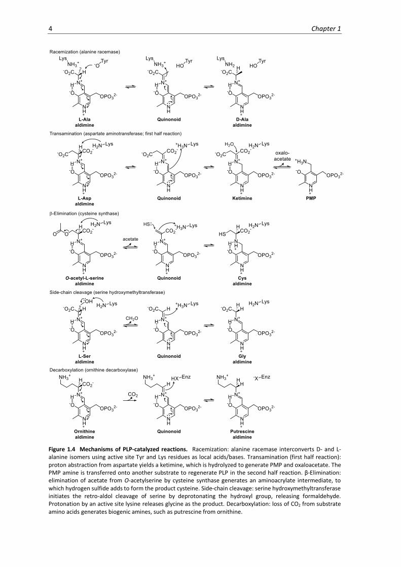
1.4 Mechanisms of PLP-catalyzed reactions. ................................................................................... 4
1.5 Inhibitors of PLP-DEs. ................................................................................................................. 6
1.6 Activity-based protein profiling workflow. ................................................................................ 9
1.7 Overview of chemical proteomic strategy for profiling the family of PLP-DEs........................11
2.1 Chemical structures of PLP derivatives. ...................................................................................14
2.2 Proteomic strategy for profiling PLP-DEs. ................................................................................15
3.1 PLP metabolism. .......................................................................................................................20
3.2 Probe phosphorylation by S. aureus PLK. ................................................................................21
3.3 Reconstitution and activity of Alr with cofactor probes. .........................................................23
3.4 Alr crystallography....................................................................................................................24
3.5 Bacterial growth using synthetic cofactor mimics. ..................................................................25
3.6 Cellular probe labeling. ............................................................................................................26
3.7 Optimization of PL4-labeling. ...................................................................................................27
4.1 Proteomics-based optimization of PL4-labeling in S. aureus. ..................................................31
4.2 PL4 labeling 100 µM, 10 µM.....................................................................................................32
4.3 Filter 1 and confidence plot. ....................................................................................................34
4.4 Filter 2: Concentration-dependent chemical proteomics of PL4 in S. aureus USA300 TnPdxS.
.........................................................................................................................................................37
4.5 Filter 2 Continued: Concentration-dependent chemical proteomics of PL4 in S. aureus
USA300 TnPdxS. ..............................................................................................................................38
4.6 Chemical proteomics with PL1 and PL2 in S. aureus USA300 TnPdxS. ....................................39
4.7 Chemical proteomics with S. aureus USA300 TnPdxS grown in CDM containing 25 µM PL1. 40
4.8 Chemical ligation tools for ABPP with azide-based probes. ....................................................42
4.9 Chemical proteomics with PL3 in S. aureus USA300 TnPdxS using the Staudinger-Bertozzi
ligation. ............................................................................................................................................43
4.10 Chemical proteomics of S. aureus USA300 TnPdxS grown with PL3 supplementation. ........44
4.11 Combined total of S. aureus USA300 TnPdxS PLP-DEs detected with PL1, PL3 and PL4. ......46
5.1 Validation of known PLP-DEs OKR-DC and CS by UV-Vis, MS and gel-based analysis. ............50
5.2 Validation of PLP-dependent aminotransferases by UV-Vis, MS and gel-based analysis. ......51
5.3 Validation of PLP binding for uncharacterized proteins identified using PL4..........................52

xvi
5.4 Sequence alignment of Q2FF14 with homologous sequences from Gram-positive bacteria. 53
5.5 X-ray crystallography and structural analysis of Q2FF14. ....................................................... 55
5.6 Overview of GP0 and metabolomics analysis. ......................................................................... 57
5.7 Overview of HH8. ..................................................................................................................... 60
5.8 Analysis of PLP binding for alternative function proteins. ...................................................... 61
5.9 Binding site identification. ....................................................................................................... 63
6.1 Phosphorylation of PL5 and PL6 by S. aureus PLK. .................................................................. 66
6.2 Chemical proteomics of PL4, PL5 and PL6 in S. aureus USA300 TnPdxS. ................................ 67
6.3 Mechanism of D-cycloserine inhibition of Alr. ........................................................................ 69
6.4 Competitive proteomic profiling reveals PLP-DE off-targets of D-cycloserine. ...................... 71
6.5 Timeline of antibiotic discoveries. ........................................................................................... 72
6.6 Cell walls of Gram-positive and Gram-negative bacteria. ....................................................... 73
6.7 Chemical proteomics with PL4 in P. aeruginosa TnPdxJ. ......................................................... 74
6.8 Chemical proteomics with PL4P in P. aeruginosa lysate. ........................................................ 76
6.9 Chemical proteomics with PL4 in human cells (HeLa). ............................................................ 79
8.1 Validation of S. aureus TnQ2FF14 and TnGP0 transposon insertion.................................... 106
8.2 Validation of P. aeruginosa TnPdxJ transposon insertion. .................................................... 106

xvii
List of Schemes
2.1 Synthesis of PL1 and PL2. .........................................................................................................16
2.2 Synthesis of PL3. .......................................................................................................................17
2.3 Synthesis of PL4. .......................................................................................................................17
6.1 Synthesis of PL5 and PL6. ..........................................................................................................66

xviii
xix
List of Tables
4.1 PLP-DEs and select proteins of interest identified in volcano plot 4.3b. .................................35
4.1 continued ..................................................................................................................................36
4.2 Proteins significantly enriched using PL1 and PL2 in S. aureus USA300 TnPdxS. ....................39
4.3 Proteins significantly enriched from S. aureus USA300 TnPdxS grown in CDM containing 25
µM PL1.............................................................................................................................................41
4.4 Proteins significantly enriched by chemical proteomics using PL3 (100, 10 µM)....................43
4.5 Proteins significantly enriched from bacterial cultures grown using PL3 supplementation (25
µM). .................................................................................................................................................45
5.1 Whole proteome comparison of S. aureus TnGP0 vs. wild-type. ............................................58
5.2 Whole metabolome comparison of S. aureus TnGP0 vs wild-type. .........................................58
6.1 Comparison of PLP-DEs significantly enriched using PL4, PL5 and PL6 in S. aureus USA300
TnPdxS. ............................................................................................................................................68
6.2 Proteins significantly depleted by DCS treatment in S. aureus USA300 TnPdxS. ....................71
6.3 Proteins significantly enriched using 100 µM PL4 in P. aeruginosa TnPdxJ cells. ...................75
6.4 Proteins significantly enriched using 10 µM PL4P in P. aeruginosa lysate. .............................76
6.5 Proteins significantly enriched using 100 µM PL4 in HeLa cells...............................................79
8.1 HPLC Gradients. .........................................................................................................................86
8.2 PCR primers and overexpression conditions for recombinant proteins. ................................100
8.3 Substrates used for N-acetyltransferase activity assay. .........................................................104
8.4 Crystallographic data collection and refinement statistics. ....................................................108

xx

1. Introduction
Pyridoxal phosphate (PLP, Figure 1.1), a bioactive component of vitamin B6, is a versatile and
ubiquitous enzyme cofactor required by all organisms for the metabolism of amino acids and
amine-containing compounds. PLP-dependent enzymes (PLP-DEs) use the PLP cofactor to catalyze
a range of chemical reactions important for essential cellular processes such as glucose, lipid and
amino acid metabolism, as well as heme, nucleotide and neurotransmitter production.(rev.1-3)
Accounting for 238 distinct catalytic activities, PLP-dependent chemistry, including racemization,
transamination, decarboxylation, and carbon-carbon bond cleavage and formation, is estimated
to represent 4% of all known catalytic activities classified to date.4 The study of PLP-DEs and their
catalytic mechanisms has provided important insights into the evolutionary diversity, therapeutic
relevance and encompassing roles of this enzyme family.
Figure 1.1 Pyridoxal phosphate and related vitamin B6 derivatives.
1.1 PLP chemistry
PLP catalysis hinges on basic chemical principles which enable a modular, effective and diversified
biocatalytic system. All PLP-dependent reactions share common mechanistic features which are
elaborated towards different chemical outcomes through the enzyme active site environment. In
the resting state, PLP-DEs bind the cofactor at an active site lysine residue via a reversible imine
bond, termed the internal aldimine (Figure 1.2). The electron-withdrawing effect of the
heteroaromatic pyridine ring, which is often protonated, polarizes the molecule and activates it
towards imine formation. The internal aldimine is stabilized by resonance effects and hydrogen
bonding with the nearby C3 phenoxide. Transaldimation with an incoming substrate amine
proceeds through the formation of a geminal diamine intermediate to generate the substrate-

2 Chapter 1
bound external aldimine. The latter is the catalytically competent complex which facilitates and
directs chemical reactions. PLP functions as an electrophilic catalyst by stabilizing negative charge
formation at Cα of the substrate, thus making adjacent sigma bonds labile towards heterolytic
cleavage. The resulting carbanion is delocalized across an extended π-system in the form of a
coplanar quinonoid intermediate, which is resonance stabilized by pyridine and Schiff base
protonation.
Figure 1.2 Internal and external aldimine formation. Active site lysine residues bind PLP via a reversible imine bond, which is displaced by the incoming substrate to yield the external aldimine via gem-diamine formation.
The external aldimine represents a key point of diversification for PLP chemistry, in which the
formation of one of three different quinonoid intermediates dictates the downstream reaction
path (Figure 1.3).5,6 In the majority of cases, deprotonation at Cα (Cα-H bond cleavage) yields a
carbanion that can subsequently undergo a variety of reactions. Reprotonation of Cα at the
opposite face results in substrate racemization or epimerization, while reprotonation at C4’ of the
cofactor generates a ketimine intermediate which, upon hydrolysis, generates pyridoxamine
phosphate (PMP) for transamination reactions (Figure 1.4). Alternatively, the carbanion can act as
a nucleophile to undergo aldol and Claisen condensations, or can displace a suitable leaving group
at Cβ to generate an aminoacrylate intermediate, as demonstrated in cysteine and tryptophan
biosynthesis. Decarboxylation (Cα-CO2 bond cleavage), on the other hand, is generally followed by
reprotonation of the quinonoid intermediate to produce biogenic amines required for hormone,
alkaloid, nucleic acid and protein biosynthesis. Finally, side-chain cleavage (Cα-Cβ bond cleavage)
leads to retro-aldol-type condensations, which are used by serine hydroxymethyltransferase or
threonine aldolase enzymes. Certain enzymes have also been shown to catalyze combinations of
these reactions, as exemplified by dialkylglycine decarboxylase (decarboxylative transamination).7
Enzymes control PLP reaction specificity through their active site environment. According to
Dunathan,8 stereoelectronic effects within the external aldimine direct which of the three σ-bonds
of the substrate is initially cleaved (Figure 1.3). The most labile bond is that oriented parallel to
neighbouring p-orbitals of the π-system due to optimal charge delocalization and stabilization.
Thus, enzymes affect the reaction outcome by directing the substrate binding orientation through
their active sites. In addition, residues within the enzyme pocket provide the necessary
functionality for catalysis and can tune the electrophilic strength of the system by stabilizing

Introduction 3
specific protonation states of the external aldimine.6 Both the number of protons as well as their
placement can affect the stability and reactivity of the carbanion.9 The evolution of distinct active
site features has influenced the specificity of PLP-DEs for catalyzing certain reaction types at the
expense of others. Therefore, active site amino acid substitutions are known to affect both the
reaction rate as well as the reaction path.10,11 While PLP is able to catalyze reactions independently
of PLP-DEs,12,13 particularly when assisted by metal ion chelation, enzymes represent an optimal
and precise way to achieve a diversified catalytic output.
Figure 1.3 Controlling PLP reaction specificity. In the external aldimine, heterolytic cleavage of a σ-bond adjacent to Cα of the substrate results in the formation of a resonance-stabilized quinonoid intermediate. Enzymes control specificity through stereoelectronic effects (Dunathan’s hypothesis) and their active site environments.
1.2 PLP-dependent enzymes
Enzymes that use PLP for catalysis originated early in evolution and were already specialized
towards basic metabolic pathways in the universal ancestor (~ 1500 million years ago).14,15 PLP-
DEs can be traced to five independent ancestral protein lineages, whose common mechanistic
features likely arose by chemical necessity and are reflective of convergent evolution. These
subfamilies can be classified based on structural fold type and are named after their most
representative protein member.16-18 Fold type I (aspartate aminotransferase) represents the
largest, most diverse PLP-DE subgroup, which is subdivided into six classes including three
aminotransferase types.(rev.2,3) Fold type II (tryptophan synthase) enzymes, which often catalyze β-
elimination reactions, share overarching similarities with the fold type I group, but differ in their
active site placement and the presence of an additional regulatory domain.
4 Chapter 1
Figure 1.4 Mechanisms of PLP-catalyzed reactions. Racemization: alanine racemase interconverts D- and L-alanine isomers using active site Tyr and Lys residues as local acids/bases. Transamination (first half reaction): proton abstraction from aspartate yields a ketimine, which is hydrolyzed to generate PMP and oxaloacetate. The PMP amine is transferred onto another substrate to regenerate PLP in the second half reaction. β-Elimination: elimination of acetate from O-acetylserine by cysteine synthase generates an aminoacrylate intermediate, to which hydrogen sulfide adds to form the product cysteine. Side-chain cleavage: serine hydroxymethyltransferase initiates the retro-aldol cleavage of serine by deprotonating the hydroxyl group, releasing formaldehyde. Protonation by an active site lysine releases glycine as the product. Decarboxylation: loss of CO2 from substrate amino acids generates biogenic amines, such as putrescine from ornithine.

Introduction 5
Fold type IV (D-amino acid aminotransferase) enzymes resemble both fold types I and II, but often
display a near mirror image of their active sites. By contrast, fold type III (alanine racemase) PLP-
DEs are characterized by distinct α/β-barrel cores and are found among bacterial racemases and
eukaryotic decarboxylases. Fold type V enzymes (glycogen phosphorylase) exhibit a unique
catalytic mechanism using the PLP phosphate group for acid/base chemistry. The structural
classification of PLP-DEs facilitates their identification, but is not prognostic of protein function
since each fold type has multiple catalytic activities. Early PLP-DEs catalyzed several reactions to
cover a range of functions and have since evolved towards a narrower substrate scope and
reaction specificity.14 Several enzymes are still promiscuous in their ability to catalyze more than
one reaction type at a physiologically-relevant rate. This phenomenon manifests itself especially
throughout in vitro experiments with purified proteins and has found significant use in
biotechnological applications for the synthesis of unnatural products and metabolites.1
The field of PLP-DE enzymology is dynamic and sustained by continuous discoveries of novel
enzymes, structures, chemical mechanisms and biological roles. For example, a recently
discovered indolmycin biosynthetic enzyme was found to be capable of coupling O2 and PLP
reactivity in order to efficiently oxidize an unactivated carbon-carbon bond.19 Other studies have
identified new PLP-DE fold types among aminomutases, which do not closely resemble any of the
currently known PLP-DE structures.20,21 Moreover, regulatory roles of PLP have been elucidated,
including enzyme inhibition (PPAT decarboxylase, mevalonate kinase glyceraldehyde-3-phosphate
dehydrogenase, pyridoxal kinase),22-26 transcriptional regulation (MocR/GabR family)27-29 and
chaperone-like capacities.30 These discoveries not only expand our knowledge of the versatility of
PLP as a cofactor but also enrich our toolbox for biocatalytic, biotechnological, biomedical and
biochemical applications.
1.3 PLP-based drug development
Due to their widespread involvement in basic metabolic processes, PLP-DEs have been implicated
in human disease and are recognized as important drug targets.(rev.2,31,32) PLP-DEs are well-suited
to mechanism-based inhibition, whereby reactive substrate derivatives bind and rearrange to
modify the enzyme covalently or to generate tight-binding PLP-substrate complexes in the active
sites. Examples include β-chloroalanine, which eliminates to an electrophilic aminoacrylate
intermediate, vinylglycine, which forms a conjugated ketimine (Michael acceptor), and propargyl-
glycine, which rearranges to a reactive allene intermediate (Figure 1.5). Lack of specificity limits
the therapeutic utility of these compounds and is also a general challenge in the development of
PLP-DE inhibitors, which are often associated with numerous side effects.

6 Chapter 1
Figure 1.5 Inhibitors of PLP-DEs. (a) Mechanism-based inhibitors of PLP-DEs (top) and select clinical drugs targeting PLP-DEs for the treatment of neurological disorders and infectious diseases (bottom). (b) Mechanism of inhibition of vinylglycine and propargylglycine.
Inhibitors of human PLP-DEs frequently target their roles in neurological disorders, cancer,
inflammation and metabolic dysfunctions. Enzymes for which clinical drugs have been developed
include serine hydroxymethyltransferase (Mimosine; malaria, tumours), GABA aminotransferase
(Vigabatrin; epilepsy), DOPA decarboxylase (Carbidopa; Parkinson’s), and branched-chain amino
acid aminotransferase (Gabapentin; GABA/glutamate equilibrium pathologies).31 Many of these
drugs function by resolving imbalances in important CNS neurotransmitter levels. Differences in
the mechanisms and types of PLP-DEs in humans versus bacteria or protozoa can also be exploited
to treat infectious diseases. Clinically druggable targets include alanine racemase (D-cycloserine;
antibacterial) and ornithine decarboxylase (Eflornithine; African sleeping sickness). Other
pathways emerging as potential antibacterial targets include peptidoglycan synthesis (N-
acetylornithine aminotransferase, diaminopimelate decarboxylase), trans-sulfuration
(cystathionine γ-synthase, cystathionine β-lyase, methionine γ-lyase, cystalysin, cysteine
synthase), as well as amino acid (threonine synthase, tryptophan synthase) and cofactor (heme,
biotin) biosynthesis. Mining the cell for further essential metabolic pathways can aid the discovery
of new drug targets.

Introduction 7
1.4 The PLP-DE family
Gaining a global overview of PLP-DEs has been the subject of several bioinformatics-driven studies
aimed at both their identification and functional annotation. Despite notoriously low sequence
similarity among PLP-DEs as a result of evolutionary disparity, limited structural diversity has
facilitated their classification into five distinct fold types. Here, genomics-based methods capable
of extracting structural information from gene sequences have been useful in predicting PLP-DE
fold type, identifying new PLP-DEs and drawing preliminary functional conclusions.4,14 An
advantage of these algorithms is their ability to detect distant homology between proteins despite
low degrees of sequence identity. This has helped to characterize the molecular evolution of the
PLP-DE family and has enabled comparison of their number and variety in different organisms. It
was thereby determined that bacteria and archaea contain approximately 20-40 genes for PLP-
DEs, representing 1.5% of most prokaryotic genomes.4 Although higher organisms have a few
more PLP-DE genes, their genomic proportion decreases with greater genomic size and
complexity, presumably because of their basic metabolic involvement. Results from these
analyses have been published in the form of an online B6-database which integrates bioinformatics
with available experimental information (http://bioinformatics.unipr.it.B6db).33 However,
genomic analyses of this type are restricted to known sequence-structure relationships and are
imprecise at functionally annotating PLP-DEs. Other bioinformatics approaches for classifying PLP-
DE function have therefore compared the structures of active sites, which show high conservation
of catalytic residues essential to enzymatic function.34 These structure-based analyses were able
to detect defined active site architectures among the different PLP-DE fold types and were further
able to discriminate between unique variations in active site geometry that clustered to specific
PLP-DE functions. These studies, and extensions thereof, have helped to define functional
evolutionary connections and to decipher the roles of new PLP-DEs.
Nonetheless, our current understanding of the PLPome is incomplete and many PLP-DEs remain
unidentified whilst even more lack functional characterization.4 Although genomic and structure-
based bioinformatic analyses have established an overview of the PLP-DE family, these strategies
rely on the availability of prior knowledge. In a more direct approach, Western-blotting using an
anti-pyridoxine antibody was used to visualize the PLPome, but was unable to identify or quantify
distinct PLP-DEs.35 MS-based approaches attempting to establish a method for the direct
detection of PLP-modified residues in proteomes are limited by low intensity and poor stability of
the PLP modification during measurement,36 highlighting the need for a strategy to enrich PLP-
bound proteins or peptides. Profiling the family of PLP-DEs would not only help to address
important questions in the field concerning evolutionary relationships and novel PLP-dependent
functions, but would also enable a range of comparative PLPome analyses.
8 Chapter 1
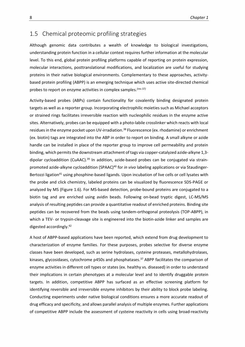
1.5 Chemical proteomic profiling strategies
Although genomic data contributes a wealth of knowledge to biological investigations,
understanding protein function in a cellular context requires further information at the molecular
level. To this end, global protein profiling platforms capable of reporting on protein expression,
molecular interactions, posttranslational modifications, and localization are useful for studying
proteins in their native biological environments. Complementary to these approaches, activity-
based protein profiling (ABPP) is an emerging technique which uses active site-directed chemical
probes to report on enzyme activities in complex samples.(rev.37)
Activity-based probes (ABPs) contain functionality for covalently binding designated protein
targets as well as a reporter group. Incorporating electrophilic moieties such as Michael acceptors
or strained rings facilitates irreversible reaction with nucleophilic residues in the enzyme active
sites. Alternatively, probes can be equipped with a photo-labile crosslinker which reacts with local
residues in the enzyme pocket upon UV-irradiation.38 Fluorescence (ex. rhodamine) or enrichment
(ex. biotin) tags are integrated into the ABP in order to report on binding. A small alkyne or azide
handle can be installed in place of the reporter group to improve cell permeability and protein
binding, which permits the downstream attachment of tags via copper-catalyzed azide-alkyne 1,3-
dipolar cycloaddition (CuAAC).39 In addition, azide-based probes can be conjugated via strain-
promoted azide-alkyne cycloaddition (SPAAC)40 for in vivo labeling applications or via Staudinger-
Bertozzi ligation41 using phosphine-based ligands. Upon incubation of live cells or cell lysates with
the probe and click chemistry, labeled proteins can be visualized by fluorescence SDS-PAGE or
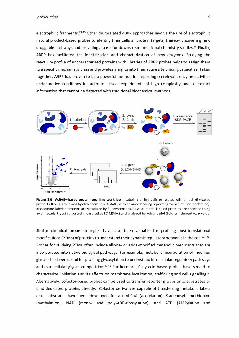
analyzed by MS (Figure 1.6). For MS-based detection, probe-bound proteins are conjugated to a
biotin tag and are enriched using avidin beads. Following on-bead tryptic digest, LC-MS/MS
analysis of resulting peptides can provide a quantitative readout of enriched proteins. Binding site
peptides can be recovered from the beads using tandem-orthogonal proteolysis (TOP-ABPP), in
which a TEV- or trypsin-cleavage site is engineered into the biotin-azide linker and samples are
digested accordingly.42
A host of ABPP-based applications have been reported, which extend from drug development to
characterization of enzyme families. For these purposes, probes selective for diverse enzyme
classes have been developed, such as serine hydrolases, cysteine proteases, metallohydrolases,
kinases, glycosidases, cytochrome p450s and phosphatases.37 ABPP facilitates the comparison of
enzyme activities in different cell types or states (ex. healthy vs. diseased) in order to understand
their implications in certain phenotypes at a molecular level and to identify druggable protein
targets. In addition, competitive ABPP has surfaced as an effective screening platform for
identifying reversible and irreversible enzyme inhibitors by their ability to block probe labeling.
Conducting experiments under native biological conditions ensures a more accurate readout of
drug efficacy and specificity, and allows parallel analysis of multiple enzymes. Further applications
of competitive ABPP include the assessment of cysteine reactivity in cells using broad-reactivity
Introduction 9
electrophilic fragments.43-45 Other drug-related ABPP approaches involve the use of electrophilic
natural product-based probes to identify their cellular protein targets, thereby uncovering new
druggable pathways and providing a basis for downstream medicinal chemistry studies.46 Finally,
ABPP has facilitated the identification and characterization of new enzymes. Studying the
reactivity profile of uncharacterized proteins with libraries of ABPP probes helps to assign them
to a specific mechanistic class and provides insights into their active site binding capacities. Taken
together, ABPP has proven to be a powerful method for reporting on relevant enzyme activities
under native conditions in order to dissect experiments of high complexity and to extract
information that cannot be detected with traditional biochemical methods.
Figure 1.6 Activity-based protein profiling workflow. Labeling of live cells or lysates with an activity-based probe. Cell lysis is followed by click chemistry (CuAAC) with an azide-bearing reporter group (biotin or rhodamine). Rhodamine-labeled proteins are visualized by fluorescence SDS-PAGE. Biotin-labeled proteins are enriched using avidin beads, trypsin digested, measured by LC-MS/MS and analyzed by volcano plot (fold-enrichment vs. p-value)
Similar chemical probe strategies have also been valuable for profiling post-translational
modifications (PTMs) of proteins to understand their dynamic regulatory networks in the cell.(rev.47)
Probes for studying PTMs often include alkyne- or azide-modified metabolic precursors that are
incorporated into native biological pathways. For example, metabolic incorporation of modified
glycans has been useful for profiling glycosylation to understand intracellular regulatory pathways
and extracellular glycan composition.48,49 Furthermore, fatty acid-based probes have served to
characterize lipidation and its effects on membrane localization, trafficking and cell signalling.50
Alternatively, cofactor-based probes can be used to transfer reporter groups onto substrates or
bind dedicated proteins directly. Cofactor derivatives capable of transferring metabolic labels
onto substrates have been developed for acetyl-CoA (acetylation), S-adenosyl-L-methionine
(methylation), NAD (mono- and poly-ADP-ribosylation), and ATP (AMPylation and

10 Chapter 1
phosphorylation). Their use has helped to identify the substrates of these modifications and has
provided insights into their biological roles. Cofactor probes designed to directly report on protein
binding have required the incorporation of a photo-crosslinker to mediate irreversible binding.
Thereby, probes for vitamin B12 have unraveled new regulatory roles of the cofactor in folate,
ubiquinone and methionine metabolism, and probes for other B-vitamins (thiamine, riboflavin and
biotin) identified several vitamin transporters.51,52 Taken together, chemical probes are valuable
tools for exploring biological systems which can serve a vast array of discovery-based applications.
1.6 Objectives
Enzymes utilizing PLP for catalysis are ubiquitous throughout evolution and functionally diverse.
The importance of this cofactor in sustaining essential cellular pathways can hardly be
understated, though our current understanding of this enzyme family is incomplete. To date,
there is no comprehensive method for profiling the cellular PLPome and it is thus estimated that
many PLP-DEs remain unidentified while even more lack complete functional classification.
Profiling the family of PLP-DEs could help to uncover novel biological roles of PLP as well as
potential druggable pathways, and would enable diverse comparative applications.
This thesis describes the development and evaluation of a chemical proteomic platform for the
global identification and characterization of PLP-DEs in cells. We envisaged the use of
functionalized cofactor mimics that are able to integrate into cellular PL uptake mechanisms and
metabolic processing to access and report on the full complement of PLP-DEs (Figure 1.7). A small
library of PL-derivatives containing an alkyne or azide tag as chemical probes was therefore
designed. Here, the 4’-aldehyde acts as an effective handle for mediating an irreversible link to
PLP-DEs upon sodium borohydride reduction of the internal aldimine. A stable covalent bond is a
prerequisite for downstream proteomic manipulation in order to attach reporter or affinity tags
by CuAAC. This permits the enrichment of probe-bound proteins for subsequent identification and
quantification by MS-analysis. We aimed to establish a robust proteomic method supported by in-
depth biochemical evaluation to ensure specific and authentic probe binding to PLP-DEs, which
has the potential for application in diverse experiments. The clinically-relevant Gram-positive
bacterial pathogen Staphylococcus aureus was selected in order to develop our experimental
workflow. Our goals were to use our designed method to identify novel PLP-DE functions or
cellular cofactor roles and to investigate its applicability to therapeutically-relevant scenarios,
such as off-target screening of PLP-DE drugs and global profiling of enzyme active sites. The
method could ideally be further expanded to other organisms and provides an effective tool for
probing PLP-related biological questions.

Introduction 11
Figure 1.7 Overview of chemical proteomic strategy for profiling the family of PLP-DEs. PL-based probes were designed to be taken up by bacterial cells and integrated into cellular metabolism. Irreversible modification and subsequent MS-based analysis of probe-bound proteins serves to report on PLP-DEs.

12 Chapter 1

2. Design & Synthesis
2.1 Introduction
Reporting on natural PLP binding events requires a functional, minimally-modified synthetic
cofactor probe capable of accessing the complement of cellular PLP-DEs and labeling them
covalently. Previous investigations into the effects of modifying the PLP scaffold on cofactor
viability have helped to define the core functionality required for PLP catalysis and guided our
probe design. It was found that PLP-DEs tolerate modification of the cofactor to varying extents.
In some cases, the PLP derivatives bound to the enzymes and supported catalysis, though often
at reduced rates. In other cases, altering the PLP structure was detrimental to catalysis either
because essential cofactor functionality was removed or an ensuing change in binding geometry
was not favourable for catalysis. Interestingly, several cofactor derivatives that were non-
activating still maintained the capacity to bind PLP-DE active sites and were even competitive for
PLP binding. As a result, a number of PLP-derivatives have been synthesized and evaluated for
antivitamin and inhibitory properties.53-55
The functional groups essential for PLP catalysis include the 3-phenol, 4’-aldehyde and 5’-
phosphate. The 4’-aldehyde mediates cofactor binding to PLP-DEs and substrates as previously
described and the Schiff base aldimines are stabilized through hydrogen bonding and resonance
with the 3-phenolate, which is deprotonated under physiological conditions. Analogues modifying
the phenol, such as 3-methoxy-PLP or 3-deoxy-PLP (Figure 2.1), are generally unable to activate
apo-PLP-DEs for catalysis but maintain some binding ability.56,57 The 5’-phosphate of PLP serves as
a stable anchoring point to PLP-DEs, mediating up to 9 hydrogen-bonding interactions with a
defined phosphate-binding motif conserved among PLP-DE active sites.58 Modifications at the 5’-
position that disrupt phosphorylation (5’-deoxy-PL) or the electrostatic capacity of the phosphate
group (methyl-phosphonate, carboxymethyl etc.) significantly decrease binding, while close
mimics (5’-deoxy-methylenephosphonate, ethylene phosphate or 5’-methyl-phosphate) bind
relatively well and support catalysis to varying degrees.56,57,59 Pyridine protonation has widely
been assumed to be necessary for PLP catalysis by acting as an electron sink. However, recent
studies have shown that N-modified PLP derivatives (N-oxide-PLP, de-aza-PLP) support binding

14 Chapter 2
and certain PLP-DE functions.56,60 For example, a de-aza-PLP analogue yielded functionally
competent alanine racemase and cysteine synthase, but not aspartate aminotransferase.60 In
combination with computational studies of enzyme reactions and activation barriers, these
studies indicate that not all PLP-DEs require the electrophilicity of the protonated pyridine for
carbanion stabilization. Instead, the electrophilic propensity of PLP is tuned towards specific
reactions by residues lining the enzyme active site. Finally, it has been found that removal of the
2-methyl group (nor-PLP) or limited expansion thereof (ethyl, propyl) was tolerated by several
enzymes for both catalysis and binding, though sometimes with reduced efficacy.57,61-63 Similarly,
methylation at the 6 position (6-methyl PLP) is tolerated.57,61 The 2-methyl group does not have a
distinct catalytic function and is believed to fulfill a spatial role in directing the conformation of
cofactor binding. Its modification with bulky substituents can therefore affect the cofactor binding
geometry within the enzyme pocket to the point of disrupting catalysis.
In summary, PLP-DEs show varying tolerances towards modification of the PLP cofactor. The
2-position is amenable to alteration without significant or broad repercussions on both binding
and catalysis. We therefore reasoned that this site is suitable for the introduction of a small alkyne
or azide handle in order to generate a PLP-probe that mimics the natural cofactor and permits
integration into PLP-dependent biological pathways.
Figure 2.1 Chemical structures of PLP derivatives. Various PLP derivatives have been synthesized and evaluated for their ability to bind to PLP-DEs and facilitate catalysis.

Design & Synthesis 15
2.2 Experimental Design
Following the example of activity-based protein profiling,37 we designed PL-probes containing a
small alkyne tag either directly on the pyridine ring (PL1, PL2) or with an ethylene spacer (PL4), as
well as a 2’-azide analogue (PL3) to account for chemical preferences within protein binding sites
(Figure 2.2a). These probes are intended to make use of cellular PL-uptake mechanisms and
metabolism to yield phosphorylated PLP derivatives capable of binding PLP-DEs (Figure 2.2b). Our
strategy harnesses the intrinsic reactivity of the internal aldimine to anchor the probe to the
enzyme irreversibly upon sodium borohydride (NaBH4)-mediated reduction,36 circumventing the
need for additional reactive groups. A stable covalent bond is a prerequisite for downstream
proteome manipulation, in which denaturing conditions are necessary for protein identification.
Subsequent bioorthogonal ligation of the alkyne tag to biotin-azide or vice versa allows gel- and
MS-based detection of the PLPome. This methodology was developed with the goal of generating
effective chemical tools for the discovery, characterization and exploration of PLP-binding events
in the complex biological context of the living cell.
Figure 2.2 Proteomic strategy for profiling PLP-DEs. (a) Chemical structures of PL-based probes as tools for reporting on PLP-binding. (b) PLPome detection strategy: PL-probes are taken up by live S. aureus cells, phosphorylated and incorporated into PLP-DEs. Upon cell lysis, NaBH4 reduction of the imine bond and click chemistry with fluorescent or enrichment tags permit proteomic identification of labeled PLP-DEs.

16 Chapter 2
2.3 Synthesis
2.3.1 Synthesis of PL1 and PL2
A semisynthetic strategy starting from pyridoxine was developed for PL1 and PL2, which was
inspired by previous chemistry by Korytnyk et al.54 (Scheme 2.1). While a 2’-alkynylated derivative
of pyridoxine similar to PL1 has previously been reported, its cofactor activity was not assessed.54
For the synthesis, pyridoxine hydrochloride was first reacted with three equivalents of TBSCl to
yield protected intermediate 1. N-oxidation using mCPBA was followed by a Boekelheide
rearrangement64 using trifluoroacetic anhydride and quenching with MeOH. This served to install
the 2’-alcohol of intermediate 2 and resulted in loss of the phenolic TBS group. The primary alcohol
was then oxidized to the corresponding aldehyde (3) using MnO2 and the phenol was reprotected
using MOMCl. Reaction of 4 with the Ohira-Bestmann reagent65 (dimethyl-1-diazo-2-
oxopropylphosphonate) in the presence of K2CO3 gave terminal alkyne 5. Upon removal of silane
protecting groups, oxidation of 6 with MnO2 in methanol gave a 2.5:1 mixture of isomers, 7a and
7b, which were separated by preparative HPLC and assigned by 2D NMR spectroscopy. A structural
isomer of PL1 with inverse functionality at the 4’ and 5’ positions that is electronically incapable
of PLP catalysis (PL2) was obtained as a synthetic by-product and was later used as a control to
test for non-specific reactivity of the PL-scaffold. Hydrolysis under acidic conditions yielded the
final PL1 and PL2 probes. Based on the NMR spectra, the probes were primarily present in the
favoured hemiacetal form.53
Scheme 2.1 Synthesis of PL1 and PL2. Pyridoxine hydrochloride was TBS-protected and oxidized at the 2-methyl position. An Ohira-Bestmann reaction was used to install the alkyne handle of probes PL1 and PL2. Upon deprotection and MnO2 oxidation to the aldehyde, isomeric probe precursors were separated by HPLC.
Design & Synthesis 17
2.3.2 Synthesis of PL3
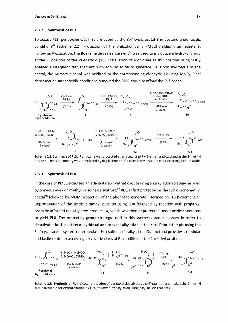
To access PL3, pyridoxine was first protected as the 3,4’-cyclic acetal 8 in acetone under acidic
conditions66 (Scheme 2.2). Protection of the 5’alcohol using PMBCl yielded intermediate 9.
Following N-oxidation, the Boekelheide rearrangement64 was used to introduce a hydroxyl group
at the 2’ position of the PL-scaffold (10). Installation of a chloride at this position using SOCl2
enabled subsequent displacement with sodium azide to generate 11. Upon hydrolysis of the
acetal, the primary alcohol was oxidized to the corresponding aldehyde 12 using MnO2. Final
deprotection under acidic conditions removed the PMB group to afford the PL3 probe.
Scheme 2.2 Synthesis of PL3. Pyridoxine was protected as an acetal and PMB-ether, and oxidized at the 2-methyl position. The azide moiety was introduced by displacement of a transiently installed chloride using sodium azide.
2.3.3 Synthesis of PL4
In the case of PL4, we devised an efficient new synthetic route using an alkylation strategy inspired
by previous work on methyl-pyridine derivatives.67 PL was first protected as the cyclic monomethyl
acetal68 followed by MOM-protection of the phenol to generate intermediate 13 (Scheme 2.3).
Deprotonation of the acidic 2-methyl position using LDA followed by reaction with propargyl
bromide afforded the alkylated product 14, which was then deprotected under acidic conditions
to yield PL4. The protecting group strategy used in this synthesis was necessary in order to
deactivate the 4’ position of pyridoxal and prevent alkylation at this site. Prior attempts using the
3,4’-cyclic acetal system (intermediate 9) resulted in 4’-alkylation. Our method provides a modular
and facile route for accessing alkyl derivatives of PL modified at the 2-methyl position.
Scheme 2.3 Synthesis of PL4. Acetal protection of pyridoxal deactivates the 4’-position and makes the 2-methyl group available for deprotonation by LDA, followed by alkylation using alkyl halide reagents.

18 Chapter 2

3. Biological Evaluation
3.1 Introduction
Vitamin B6 is a collective term used to describe pyridoxal (PL), pyridoxine (PN) and pyridoxamine
(PM), as well as their 5’-phosphorylated forms (PLP, PNP and PMP). A series of biosynthetic,
catabolic and salvage enzymes generate and interconvert the different forms of vitamin B6 to suit
the needs of the cell (Figure 3.1).69 While microorganisms and plants are capable of de novo PLP
biosynthesis, animals rely on vitamin B6 uptake from the diet. PL, PN and PM are believed to be
absorbed through facilitated diffusion or active transport, though the exact mechanism of cellular
uptake remains unclear.70,71 A salvage pathway, consisting of pyridoxal kinases (PLKs, PdxK),
pyridoxine-5’-phosphate oxidase (PNPOx, PdxH) and pyridoxal phosphatases, is used by all cells to
recycle PLP liberated from enzymatic turnover or from nutritional uptake.72 PLKs use ATP to
phosphorylate the 5’ alcohol of each of the three vitamers (PL, PN and PM), while PNPOx oxidizes
PNP and PMP to form PLP. The salvage enzymes are believed to maintain homeostasis by
regulating cellular PLP levels through feedback inhibition as well as the direct distribution of PLP
to corresponding apoenzymes, though this is not fully understood.22,23,72 The PLP aldehyde is much
more reactive than its unphosphorylated counterpart, PL, which exists primarily in the hydrated
acetal form. The concentration of PLP is therefore maintained at low levels to prevent undesired
binding to cellular nucleophiles.23,73 Studies have shown that imbalances in PLP metabolism and
homeostasis can cause severe neurological pathologies.74
Two distinct PLP biosynthetic pathways have recently been characterized.69,72 The DXP-dependent
pathway is found among members of the γ-subdivision of proteobacteria, which include many
Gram-negative bacteria (ex. Escherichia coli, Pseudomonas aeruginosa and Salmonella enterica).
In this pathway, pyridoxine synthase (PdxJ) condenses de-oxyxylulose 5-phosphate (DXP) with
3-hydroxy-1-aminoacetone phosphate to generate PNP, which is then converted to PLP by PNPOx
(PdxH). Although it was discovered first, the DXP-pathway is not widely distributed and rather
complex. The R5P pathway is more common and employs a PLP synthase complex, consisting of
Pdx1 (PdxS) and Pdx2 (PdxT) subunits, to condense D-ribose 5-phosphate (R5P), glutamine and
glyceraldehyde 3-phosphate, generating PLP directly. PLP catabolism proceeds through oxidation

20 Chapter 3
of PL, mainly to 4-pyridoxic acid, and excretion in the urine by humans and animals.
Microorganisms can further degrade the cofactor to usable metabolites such as succinic
semialdehyde or 2-(hydroxymethyl)-4-oxobutanoate, acetate, ammonia and carbon dioxide.
Figure 3.1 PLP metabolism. Microorganisms synthesize PLP via DXP- or R5P-dependent pathway. A vitamin B6 salvage pathway found in all organisms interconverts the different forms of the cofactor as needed by the cell. Catabolism occurs through oxidation of PL to pyridoxic acid, followed by excretion (humans, animals) or further degradation (microorganisms).
3.2 Probe phosphorylation by S. aureus pyridoxal kinase
PLP is the main bioactive component of vitamin B6 and it is generated through the phosphorylation
of PL by PLKs. The 5’-phosphate group is important for binding PLP-DE active sites, where it helps
to anchor and correctly position the cofactor through hydrogen-bonding interactions.58 S. aureus

Biological Evaluation 21
PLK (PdxK) is capable of phosphorylating all forms of vitamin B6 (PL, PN, PM) and was recently
discovered to have roles in both PLP salvage as well as thiamine biosynthesis.75 Structural studies
revealed S. aureus PLK to adopt a classic ribokinase fold and lent insights into its unique
mechanism of action. A flexible loop of PLK engages the pyridoxal aldehyde as a hemithioacetal at
a highly conserved cysteine residue and folds down in order to sequester the phosphorylation
reaction within the catalytic pocket of the enzyme.75
We first studied the ability of S. aureus PLK to accept our synthetic probes as substrates in order
to yield activated cofactor mimics that can bind to PLP-DEs. The kinetics of probe phosphorylation
by PLK were therefore determined using a previously described assay that couples the ATP-
dependent activity of PLK to the consumption of NADH (340 nm) via a pyruvate kinase/lactate
dehydrogenase system.75 Both PL3 and PL4 were efficiently converted to phosphorylated products
at levels comparable to PL (Figure 3.2). Formation of PL3P and PL4P could be confirmed by MS
upon overnight incubation of the probes with PLK in the presence of ATP to generate the
phosphorylated products. Although significant overlap in the absorption spectra of NADH
(340 nm) and PL1 (325 nm, extended conjugation) precluded kinetic analysis, MS-based detection
of PL1P served to confirm phosphorylation by PLK. In contrast, phosphorylation could not be
detected for PL2, the negative control probe.
Figure 3.2 Probe phosphorylation by S. aureus PLK. PLK uses ATP to phosphorylate the 5’-OH of PL. Kinetics of probe phosphorylation compared to PL formation are shown (n = 4, error bars: mean ± standard deviation) as well as HRMS confirmation of phosphorylated products. Kinetic analysis of PL1 (325 nm) was not possible due to significant spectral overlap with NADH (340 nm).

22 Chapter 3
3.3 Cofactor viability of probes with alanine racemase
Next, we examined the compatibility of the phosphorylated probes with alanine racemase (Alr) as
a model PLP-DE since it is well-studied and universally present in bacteria. Alr catalyzes the
interconversion of L- and D-alanine, an essential component for bacterial peptidoglycan synthesis,
and is therefore considered an important antibiotic target.76 Recombinant, strep-tagged S. aureus
Alr (Uniprot ID: P63479) was first purified as the PLP-holoenzyme using affinity chromatography
and was subsequently transformed to its apo-form (Figure 3.3a). Methods for resolving the PLP
cofactor from PLP-DEs are enzyme-dependent, but have included nucleophilic displacement by
small molecule amines or extensive dialysis.77 In some cases, harsher treatments such as partial
unfolding or precipitation of the enzyme are required in order to remove the cofactor
completely.59,78 On-column nucleophilic displacement of bound PLP using hydroxylamine was
effective at generating the alanine racemase apoenzyme (Apo-Alr), and its formation could be
monitored spectrally by the loss of absorbance of the internal aldimine (410-420 nm).60 Upon
reconstitution of Apo-Alr with the phosphorylated probes, UV-Vis peaks indicative of internal
aldimine formation were observed (Figure 3.3c). Probe binding and the degree of cofactor loading
within the different holoenzymes were further confirmed by MS (Figure 3.3d). Therefore, the
holoenzymes were reduced using NaBH4 and measured by intact-protein MS, revealing protein
adduct formation (80-100%) corresponding to the respective masses of the phosphorylated
probes. Having confirmed reductive amination of the internal aldimine to stably fix the probes to
Alr, we subsequently investigated whether Alr could be chemically labeled with a fluorescent
reporter tag using copper-catalyzed azide-alkyne 1,3-dipolar cycloaddition (CuAAC, click
chemistry) with rhodamine-azide.79-81 Visualization by fluorescence SDS-PAGE revealed specific
labeling of Alr by PL1P and PL4P (Figure 3.3e), while unphosphorylated probes and the control
probe PL2 helped gauge nonspecific binding of the reactive aldehyde. For PL3P, CuAAC with
rhodamine alkyne proved more specific over strain-promoted click chemistry (SPAAC) with DBCO-
rhodamine for labeling Alr, presumably due to nucleophilic attack of other enzyme residues on
the strained alkyne system.82
Subsequently, we investigated the capacity of the phosphorylated probes to support enzyme
function. The ability of the individual probe-bound Alr holoenzymes to catalyze alanine
racemization was therefore characterized kinetically. The synthetic cofactors yielded catalytically
competent Alr-holoenzymes with nearly identical efficiency compared to the PLP-holoenzyme
(Figure 3.3b). PL1P even showed higher turnover compared to PLP, which may result from
increased stabilization due to its extended conjugation or perhaps favoured binding within the Alr
active site. Such hyper-activity has previously been described for other cofactor derivative-enzyme
systems.63 The remarkable structural tolerance within the Alr active site was consequently
reaffirmed by X-ray crystallography.

Biological Evaluation 23
Figure 3.3 Reconstitution and activity of Alr with cofactor probes. (a) Reconstitution of PLP or phosphorylated probes with Alr. (b) Kinetics of alanine racemization (D- to L-Ala direction) by S. aureus Alr holoenzymes (0.01 µM) containing PLP or cofactor analogues (n = 3, error bars: mean ± standard deviation). (c) UV-Vis absorption spectra of different S. aureus Alr holoenzymes (100 µM). Data was normalized to absorbance of the internal aldimine (420-430 nm). (d) Percent of S. aureus holoenzymes bound by PLP or cofactor analogues upon NaBH4 reduction and measurement by intact protein MS (n = 3, error bars: mean ± standard deviation). (e) Fluorescence SDS-PAGE of S. aureus Alr (10 µM) labeling using PLP-probes (0.5, 1, 5 equivalents) or unphosphorylated PL probes (5 equivalents) upon click chemistry to rhodamine-azide or alkyne. DBCO = dibenzyl cyclooctyne. See appendix for full gels and Coomassie staining.
In order to understand the binding mode of the PLP-derivatives within the Alr active site, we
determined the X-ray crystal structures of Alr bound to PL1P and PL4P, representing the smallest
and largest probes respectively. Several crystal structures of Alr have been resolved, which
revealed a conserved overall fold (type III) consisting of an N-terminal α/β barrel and a
predominantly β-stranded C-terminus. Alr functions as a homodimer, forming two active sites that
are lined with positive charge at the dimer interface, and PLP binds to a highly conserved lysine
residue (Lys39) near the mouth of the barrel. Both PL1P and PL4P bound the structurally
equivalent position of the natural PLP cofactor (comparison with PDB: 4A3Q,83 Figure 3.4), and

24 Chapter 3
clear electron density was indicative of internal aldimine formation with Lys39. The phosphate
groups are coordinated by a conserved hydrogen-bonding network present in all available
structures of Alr and its homologues.83 Though the active site geometry generally matched the
PLP-holoenzyme, a slight realignment of the Lys131 and Arg138 sidechains was necessary to fit
the alkyne handle of the PLP-analogues. Our studies confirm the adequate space within the Alr
aldimine binding pocket for accommodating structural changes imparted to the synthetic cofactor
without influencing catalytic activity, and led us to conclude that the probes represent good
cofactor mimics for binding and reporting on PLP-DEs.
Figure 3.4 Alr crystallography. (a) Crystal structure of S. aureus Alr bound to PL1P (left) and PL4P (center). Simulated annealing omit electron density map contoured at 2.5 σ. Superimposed active sites (right) of PLP-bound Alr (purple, PDB 4A3Q),83 Alr-PL1P (grey) and Alr-PL4P (blue). Residues lining the active site as well as the PL1P (grey) and PL4P (gold) cofactors are shown in stick models; hydrogen bonds are drawn as dashed lines. (b) Surface representation of the S. aureus Alr dimer (blue, grey) in complex with PL4P (gold, stick representation); the two active sites are indicated by red arrows. (c) Two-dimensional LIGPLOT1-representation of the interactions between S. aureus Alr and PL4P. Figures courtesy of Dr. S. Schneider.

Biological Evaluation 25
3.4 Cellular growth, metabolism and labeling by PL-probes
The ability of our PL probes to function globally in native biological systems was assessed using
bacterial growth experiments. To promote the cellular uptake and use of our probes as PL
surrogates, we used a transposon mutant strain of S. aureus that is incapable of de novo PLP
biosynthesis but is able to salvage exogenous PL and convert it to PLP. Since S. aureus employs the
R5P-biosynthetic pathway, which requires the functioning of a PLP-synthase complex consisting
of PdxS and PdxT subunits to generate PLP, we reasoned that disruption of the pdxS gene would
provide the desired phenotype. The S. aureus USA300 TnPdxS transposon mutant (Nebraska
transposon mutant library)84 was therefore selected. Bacteria were incubated in defined, PL-free
media supplemented with various concentrations of the probe and growth was measured after
24 h. We found that < 1 µM PL was required to support full bacterial growth, while absence of PL
or incubation with PL2 prohibited growth (OD600 < 0.1), thereby confirming the transposon
phenotype. Both PL1 and PL3 were able to support bacterial growth to the same maximal level as
PL, though required much higher concentrations (approximately 25 µM) (Figure 3.5). However,
S. aureus cells were unable to grow using PL4 as a cofactor substitute under the conditions tested.
These results suggest that small modifications of the cofactor are tolerated for a range of cellular
functions, while the increased steric bulk of the PL4 alkyne linker may preclude its use for certain
essential biological pathways.
Figure 3.5 Bacterial growth using synthetic cofactor mimics. Growth curves of S. aureus USA300 TnPdxS (Nebraska transposon library)84 in defined, PL-free media supplemented with PL (left) or PL probes (right). Bacterial growth was measured (OD600) after 24 h and data was obtained from three biological replicates (error bars: mean ± standard deviation).

26 Chapter 3
Our studies on the growth of S. aureus TnPdxS with our probes as cofactor surrogates indicated
that forcing conditions would be required to compete with endogenous PLP levels for PLP-DE
binding. However, as the probes were unable to support sufficient bacterial growth on
synthetically practical amounts for labeling experiments, we adopted an alternative strategy in
which bacteria were first grown to stationary phase in PL-rich media and subsequently transferred
to chemically-defined media containing 100 µM of the respective probes in place of PL. Upon
completion of a two-hour labeling period with the probes, the cells were lysed and treated with
NaBH4 in order to fix the probes to the enzymes by reductive amination. Conjugation to rhodamine
azide by CuAAC and fluorescence SDS-PAGE analysis revealed various extents of protein labeling
by the different probes (Figure 3.6a). While PL4 labeled a large number of proteins very strongly,
the PL1 probe showed only weak labeling above PL2 or DMSO treatment (controls). We further
investigated the reason for the poor labeling efficiency of PL1 and found labeling of purified Alr-
PL1P to decrease significantly when spiked into cell lysate, media or reaction mixtures containing
nucleophilic amino acids such as cysteine (Figure 3.6b). Labeling could be rescued to a certain
degree by quenching reactive nucleophiles using iodoacetamide alkylation82 prior to click
chemistry. Furthermore, adduct formation could be observed by MS when incubating PL1 with N-
acetyl-cysteine in the presence of copper (data not shown). We therefore reasoned that copper
may coordinate to the PL1 pyridine and activate the 2’-alkyne, which is in conjugation with the
aromatic system, towards conjugate addition by cellular nucleophiles. Future proteomic
experiments were therefore conducted using an additional iodoacetamide treatment for PL1-
labeled samples. Similarly, PL3-based labeling proved weak over background. The reasons for this
are unclear, but may stem from lower specificity and efficiency of inverse click reactions.80
Figure 3.6 Cellular probe labeling. (a) Fluorescence SDS-PAGE showing probe labeling (100 µM for 2 h at 37°C) in S. aureus USA300 TnPdxS. See appendix for Coomassie stained gels. (b) Optimizations of PL1-labeling. Fluorescence SDS-PAGE showing labeling of Alr-PL1P in the presence of cell media (CDM, top), PBS containing 1 mM cysteine (middle), or S. aureus lysate (bottom). PL1P-bound Alr holoenzyme (5 µM) was reduced with NaBH4 (10 mM) and treated with iodoacetamide (IAA) prior to CuAAC with rhodamine azide. The proposed mechanism of inactivation of PL1P by conjugate addition of cellular nucleophiles in the presence of copper is illustrated.

Biological Evaluation 27
Figure 3.7 Optimization of PL4-labeling. (a) Time-dependent (left) and concentration-dependent labeling (right) of PL4 in S. aureus USA300 TnPdxS by fluorescence SDS-PAGE. See appendix for Coomassie stained gels. (b) In situ metabolism of PL4 to PL4P (n = 3, error bars: mean ± standard deviation) in S. aureus USA300 TnPdxS (100 µM for 2 h at 37°C).
Further optimizations with regards to labeling time and probe concentration were directed at PL4
due to its robust labeling profile. We found that incubation time does not significantly affect
labeling efficiency and that PL4-based labeling is concentration-dependent (Figure 3.7a). Using a
targeted metabolomics approach, we also verified in situ probe phosphorylation under these
experimental conditions. It was found that approximately 80% of the probe was converted to its
phosphorylated form within 2 h of incubation with the bacteria, and formation of PL4P increased
with higher probe concentrations (Figure 3.7b). A concentration of 100 µM of PL4 was eventually
selected as a good balance between labeling strength and non-specific background binding, which
was later reaffirmed through proteomics experiments.
3.5 Conclusion
In summary, the biological activity and suitability of three PL-derivatives as chemical probes for
reporting on PLP-binding were evaluated. Each probe is efficiently phosphorylated to its
biologically active form by the S. aureus PLK at levels comparable to PL, and binding was assessed

28 Chapter 3
by reconstituting the model PLP-DE Alr with the activated probes. The catalytic activity of artificial
Alr holoenzymes was not affected by the chemical modification of the cofactor, which can be
attributed to structural accommodation within the Alr binding site. Labeling experiments with Alr-
PL3P revealed significant background labeling linked to the reactivity of alkyne- and DBCO-
rhodamine reagents, while cellular PL1-labeling suffered from the conjugate addition of biological
nucleophiles. Although these probes showed the best biological uptake and incorporation, as
evidenced by their ability to support bacterial growth as cofactor surrogates, chemical factors limit
their use as effective and specific probes under the present experimental conditions. By
comparison, the extended bulk of PL4 prevented it from substituting for all essential cellular PLP-
functions, but strong gel-based labeling led to its selection for further optimizations and cell-based
metabolic experiments. We were encouraged by previous observations that PLP-derivatives are
sometimes unable to support enzymatic catalysis though are nonetheless able to bind PLP-DEs.
With a robust PLP-labeling strategy in hand, we proceeded with MS-based proteomic investigation
in order to identify and quantify cellular proteins accessed by the PL-probes in S. aureus.

4. Proteomic Analysis
4.1 Introduction
Mass spectrometry represents a powerful analytical platform for proteomic investigations. The
development of advanced MS-based methods that are capable of identifying and quantifying
proteins have made possible the detailed and global comparison of biological systems.
Furthermore, in combination with chemical tools to selectively enrich protein subtypes, MS-
analysis can be used to study distinct cellular events of interest. Recent technological advances in
peptide separation techniques, mass spectrometer instruments and data analysis have been
critical in establishing MS-methods for routine use in diverse applications and proteomic analyses.
Quantitative proteomic analysis has traditionally relied on the use of dyes, fluorophores or
radioactive tags in combination with high-resolution protein separation techniques, such as 2D
gel electrophoresis.85 However, these methods are limited by their inability to simultaneously
identify proteins, a shortcoming which has been addressed by modern LC-MS/MS techniques. A
challenge for the absolute quantification of proteins by MS is the range in physicochemical
properties of proteolytic peptides (size, charge, hydrophobicity). As such, strategies for the
relative quantification of peptides between different samples using stable isotope tags have been
developed.86-88 These labels are introduced either at the level of the protein or peptide by
metabolic, enzymatic or chemical means, and resulting peptides behave identically to their non-
labeled counterparts throughout LC but can be distinguished by MS.89 For metabolic
incorporation, labels are introduced to the living cell by substituting heavy- or light-isotope amino
acids into the growth medium (ex. SILAC; stable isotope labeling by amino acids in cell culture).90
The early-stage introduction of the labels allows samples to be pooled and processed together,
minimizing handling or instrumental errors to produce high-accuracy results. Alternatively, labels
can be introduced chemically at the peptide level. For example, in a method termed dimethyl
labeling, deuterated and 13C-labeled formaldehyde and sodium cyanoborohydride (NaCNBH3) are
used to methylate lysine residues by reductive amination.91 While dimethyl labeling is affordable
and effective, only three experimental conditions can be compared due to the limited number of
reagent combinations that produce a detectable mass difference (≥ 4 Da) and minor shifts during

30 Chapter 4
chromatography can complicate data analysis. These challenges have been addressed by isobaric
mass tagging techniques, such as iTRAQ (isotope tags for relative and absolute quantification)92
and tandem mass tags (TMT).93 Here, labeled peptides co-migrate exactly during LC and are only
distinguished and quantified upon MS2 fragmentation, while multiplexing of up to 10 samples is
possible. Finally, label-free quantification (LFQ) strategies have recently gained popularity due to
their ability to compare unlimited samples at a low cost. Since comparative samples are processed
independently, errors in sample handling and MS measurement are reflected in the obtained data
and advanced data analysis and statistical evaluation methods are required to extract reliable
quantitative data from LFQ-based MS measurements.94 In conclusion, the field of MS-based
proteomic quantification is rapidly developing and, with it, technological advances in mass
spectrometry instruments as well as methods for bioinformatic data analysis are keeping pace.
Previous investigations into the cellular PLPome have labeled and visualized PLP-binding proteins
by Western blot using an α-pyridoxine antibody.35 However, these reports were unable to identify
and quantify distinct PLP-DEs, which could be addressed through MS-based analysis. Direct
analysis of PLP-modified peptides from cell mixtures is complicated by their suppressed ionization
efficiency and poor detection, reminiscent of phosphopeptides.36,95 Neutral-loss scanning for
phosphate and PLP moieties during mass fingerprinting has been effective at localizing the PLP-
binding site of purified protein.36 An additional enrichment strategy using immobilized metal ion
affinity chromatography (IMAC) coupled to PLP phosphatase treatment identified only few sites
in E. coli lysate.95 We present an MS-based proteomic approach using chemical probes for the
enrichment and identification of PLP-DEs. We apply supplementary data analysis strategies in
order to add confidence to LFQ data and to improve the accuracy of the method for our purposes.
4.2 En route to an optimized proteomic workflow
Having established a viable labeling strategy supported by in-depth biochemical characterization
(Chapter 3), we proceeded with the development of an MS-based proteomic workflow to evaluate
PL-probe binding in S. aureus USA300 TnPdxS cells. Experiments were initially optimized using the
PL4 probe due to its promising gel-based labeling pattern, and subsequently applied to PL1, PL2
and PL3. A stationary-phase bacterial culture was therefore treated with PL4 in defined media,
devoid of PL, over the course of a two-hour labeling period. Upon cell lysis and reductive fixation
of the probe to PLP-DEs, a protein precipitation step followed by washing of the protein fraction
was necessary to remove excess NaBH4. The labeled proteins were then solubilized (0.4% SDS in
PBS) and conjugated by CuAAC to a trifunctional rhodamine-biotin-azide linker96 for enrichment
using avidin-beads. The introduction of a fluorescent tag onto labeled proteins was critical during
experimental optimizations in order to qualitatively assess protein enrichment by gel. Samples
were subsequently trypsin digested and the resulting peptides were analyzed by LC-MS/MS.
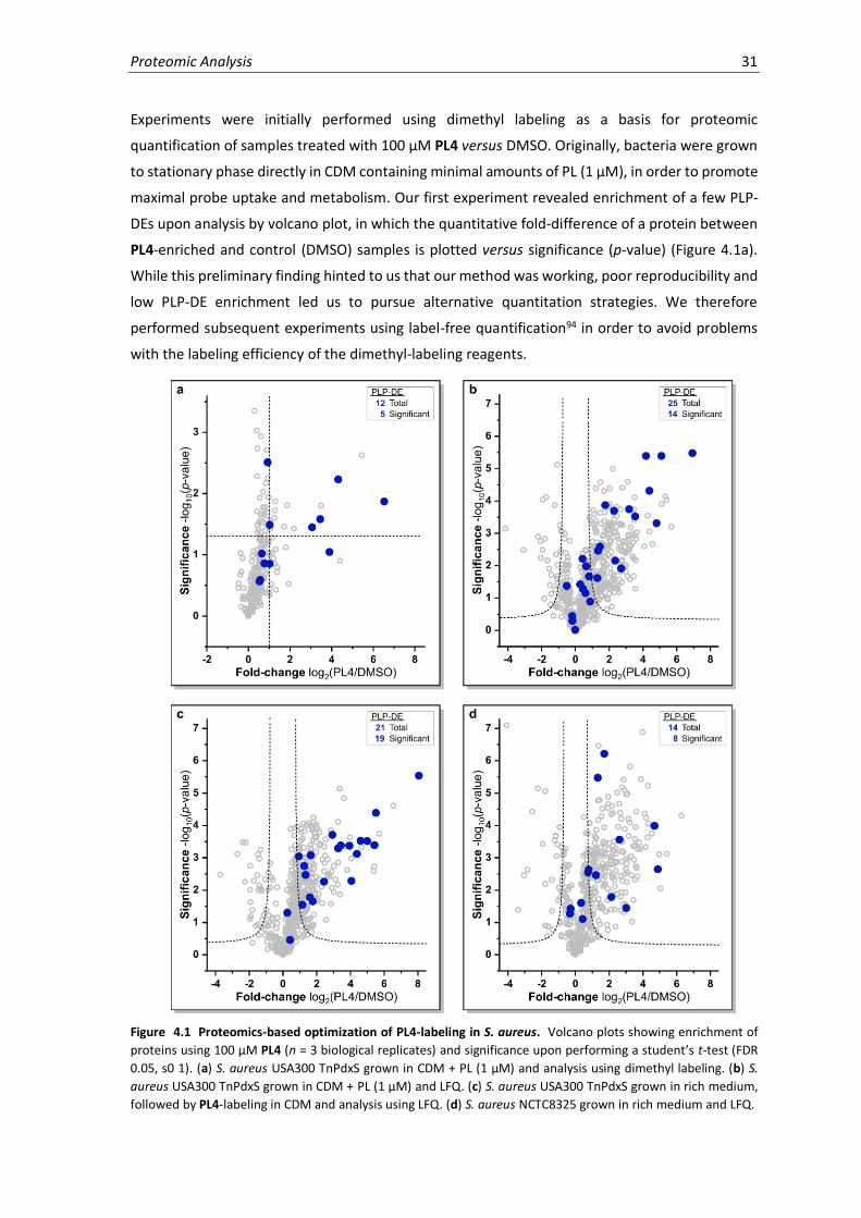
Proteomic Analysis 31
Experiments were initially performed using dimethyl labeling as a basis for proteomic
quantification of samples treated with 100 µM PL4 versus DMSO. Originally, bacteria were grown
to stationary phase directly in CDM containing minimal amounts of PL (1 µM), in order to promote
maximal probe uptake and metabolism. Our first experiment revealed enrichment of a few PLP-
DEs upon analysis by volcano plot, in which the quantitative fold-difference of a protein between
PL4-enriched and control (DMSO) samples is plotted versus significance (p-value) (Figure 4.1a).
While this preliminary finding hinted to us that our method was working, poor reproducibility and
low PLP-DE enrichment led us to pursue alternative quantitation strategies. We therefore
performed subsequent experiments using label-free quantification94 in order to avoid problems
with the labeling efficiency of the dimethyl-labeling reagents.
Figure 4.1 Proteomics-based optimization of PL4-labeling in S. aureus. Volcano plots showing enrichment of
proteins using 100 µM PL4 (n = 3 biological replicates) and significance upon performing a student’s t-test (FDR
0.05, s0 1). (a) S. aureus USA300 TnPdxS grown in CDM + PL (1 µM) and analysis using dimethyl labeling. (b) S.
aureus USA300 TnPdxS grown in CDM + PL (1 µM) and LFQ. (c) S. aureus USA300 TnPdxS grown in rich medium,
followed by PL4-labeling in CDM and analysis using LFQ. (d) S. aureus NCTC8325 grown in rich medium and LFQ.
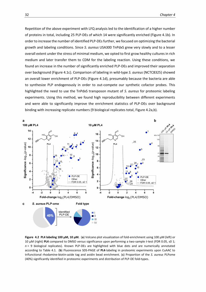
32 Chapter 4
Repetition of the above experiment with LFQ analysis led to the identification of a higher number
of proteins in total, including 25 PLP-DEs of which 14 were significantly enriched (Figure 4.1b). In
order to increase the number of identified PLP-DEs further, we focused on optimizing the bacterial
growth and labeling conditions. Since S. aureus USA300 TnPdxS grew very slowly and to a lesser
overall extent under the stress of minimal medium, we opted to first grow healthy cultures in rich
medium and later transfer them to CDM for the labeling reaction. Using these conditions, we
found an increase in the number of significantly enriched PLP-DEs and improved their separation
over background (Figure 4.1c). Comparison of labeling in wild-type S. aureus (NCTC8325) showed
an overall lower enrichment of PLP-DEs (Figure 4.1d), presumably because the bacteria are able
to synthesize PLP endogenously in order to out-compete our synthetic cofactor probes. This
highlighted the need to use the TnPdxS transposon mutant of S. aureus for proteomic labeling
experiments. Using this method, we found high reproducibility between different experiments
and were able to significantly improve the enrichment statistics of PLP-DEs over background
binding with increasing replicate numbers (9 biological replicates total, Figure 4.2a,b).
Figure 4.2 PL4 labeling 100 µM, 10 µM. (a) Volcano plot visualization of fold-enrichment using 100 µM (left) or
10 µM (right) PL4 compared to DMSO versus significance upon performing a two-sample t-test (FDR 0.05, s0 1;
n = 9 biological replicates). Known PLP-DEs are highlighted with blue dots and are numerically annotated
according to Table 4.1. (b) Fluorescence SDS-PAGE of PL4-labeling in proteomic experiments upon CuAAC to
trifunctional rhodamine-biotin-azide tag and avidin bead enrichment. (c) Proportion of the S. aureus PLPome
(40%) significantly identified in proteomic experiments and distribution of PLP-DE fold-types.
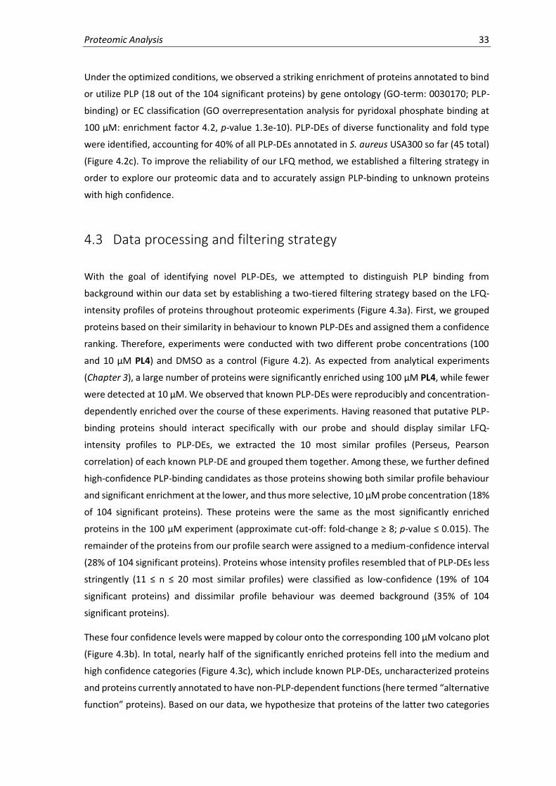
Proteomic Analysis 33
Under the optimized conditions, we observed a striking enrichment of proteins annotated to bind
or utilize PLP (18 out of the 104 significant proteins) by gene ontology (GO-term: 0030170; PLP-
binding) or EC classification (GO overrepresentation analysis for pyridoxal phosphate binding at
100 µM: enrichment factor 4.2, p-value 1.3e-10). PLP-DEs of diverse functionality and fold type
were identified, accounting for 40% of all PLP-DEs annotated in S. aureus USA300 so far (45 total)
(Figure 4.2c). To improve the reliability of our LFQ method, we established a filtering strategy in
order to explore our proteomic data and to accurately assign PLP-binding to unknown proteins
with high confidence.
4.3 Data processing and filtering strategy
With the goal of identifying novel PLP-DEs, we attempted to distinguish PLP binding from
background within our data set by establishing a two-tiered filtering strategy based on the LFQ-
intensity profiles of proteins throughout proteomic experiments (Figure 4.3a). First, we grouped
proteins based on their similarity in behaviour to known PLP-DEs and assigned them a confidence
ranking. Therefore, experiments were conducted with two different probe concentrations (100
and 10 µM PL4) and DMSO as a control (Figure 4.2). As expected from analytical experiments
(Chapter 3), a large number of proteins were significantly enriched using 100 µM PL4, while fewer
were detected at 10 µM. We observed that known PLP-DEs were reproducibly and concentration-
dependently enriched over the course of these experiments. Having reasoned that putative PLP-
binding proteins should interact specifically with our probe and should display similar LFQ-
intensity profiles to PLP-DEs, we extracted the 10 most similar profiles (Perseus, Pearson
correlation) of each known PLP-DE and grouped them together. Among these, we further defined
high-confidence PLP-binding candidates as those proteins showing both similar profile behaviour
and significant enrichment at the lower, and thus more selective, 10 µM probe concentration (18%
of 104 significant proteins). These proteins were the same as the most significantly enriched
proteins in the 100 µM experiment (approximate cut-off: fold-change ≥ 8; p-value ≤ 0.015). The
remainder of the proteins from our profile search were assigned to a medium-confidence interval
(28% of 104 significant proteins). Proteins whose intensity profiles resembled that of PLP-DEs less
stringently (11 ≤ n ≤ 20 most similar profiles) were classified as low-confidence (19% of 104
significant proteins) and dissimilar profile behaviour was deemed background (35% of 104
significant proteins).
These four confidence levels were mapped by colour onto the corresponding 100 µM volcano plot
(Figure 4.3b). In total, nearly half of the significantly enriched proteins fell into the medium and
high confidence categories (Figure 4.3c), which include known PLP-DEs, uncharacterized proteins
and proteins currently annotated to have non-PLP-dependent functions (here termed “alternative
function” proteins). Based on our data, we hypothesize that proteins of the latter two categories
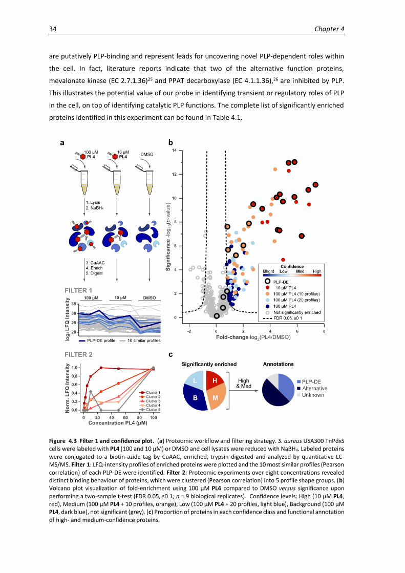
34 Chapter 4
are putatively PLP-binding and represent leads for uncovering novel PLP-dependent roles within
the cell. In fact, literature reports indicate that two of the alternative function proteins,
mevalonate kinase (EC 2.7.1.36)25 and PPAT decarboxylase (EC 4.1.1.36),26 are inhibited by PLP.
This illustrates the potential value of our probe in identifying transient or regulatory roles of PLP
in the cell, on top of identifying catalytic PLP functions. The complete list of significantly enriched
proteins identified in this experiment can be found in Table 4.1.
Figure 4.3 Filter 1 and confidence plot. (a) Proteomic workflow and filtering strategy. S. aureus USA300 TnPdxS cells were labeled with PL4 (100 and 10 µM) or DMSO and cell lysates were reduced with NaBH4. Labeled proteins were conjugated to a biotin-azide tag by CuAAC, enriched, trypsin digested and analyzed by quantitative LC-MS/MS. Filter 1: LFQ-intensity profiles of enriched proteins were plotted and the 10 most similar profiles (Pearson correlation) of each PLP-DE were identified. Filter 2: Proteomic experiments over eight concentrations revealed distinct binding behaviour of proteins, which were clustered (Pearson correlation) into 5 profile shape groups. (b) Volcano plot visualization of fold-enrichment using 100 µM PL4 compared to DMSO versus significance upon performing a two-sample t-test (FDR 0.05, s0 1; n = 9 biological replicates). Confidence levels: High (10 µM PL4, red), Medium (100 µM PL4 + 10 profiles, orange), Low (100 µM PL4 + 20 profiles, light blue), Background (100 µM PL4, dark blue), not significant (grey). (c) Proportion of proteins in each confidence class and functional annotation of high- and medium-confidence proteins.

Proteomic Analysis 35
Table 4.1 PLP-DEs and select proteins of interest identified in volcano plot 4.3b. See Table 4.1 continued (p. 36) for remaining significant proteins. (Red = High confidence, Orange = Medium confidence, Grey = Non-significant).
Fold-
change Significance Name Uniprot ID Cluster
PL
P-D
Es
1. 7.36 11.12 Cystathionine gamma-synthase (EC 4.4.1.8) A0A0H2XFF8 1
2. 6.82 10.69 Diaminopimelate decarboxylase (EC 4.1.1.20) A0A0H2XFD9 2
3. 6.34 6.83 Cysteine desulfurase (EC 2.8.1.7) A0A0H2XHJ5 2
4. 5.81 13.02 Cysteine synthase (EC 2.5.1.47) A0A0H2XFQ3 1
5. 5.79 10.10 Aminotransferase, class I (EC 2.6.1.-) A0A0H2XFA8 2
6. 5.37 12.96 Threonine synthase (EC 4.2.3.1) A0A0H2XH24 3
7. 4.94 9.71 Orn/Lys/Arg decarboxylase (EC 4.1.1.18) A0A0H2XII6 1
8. 4.71 10.32 Aminotransferase, class V A0A0H2XKI8 2
9. 4.69 7.29 L-threonine dehydratase catabolic TdcB (EC 4.3.1.19) Q2FH01 2
10. 4.56 7.34 Uncharacterized protein A0A0H2XHH8 1
11. 4.48 10.06 Branched-chain-amino-acid aminotransferase (EC 2.6.1.42) A0A0H2XIS2 2
12. 3.41 9.11 D-alanine aminotransferase (EC 2.6.1.21) A0A0H2XHU6 1
13. 2.75 9.49 Alanine racemase (EC 5.1.1.1) Q2FF55 1
14. 2.43 7.88 Aminotransferase, class I (EC 2.6.1.-) A0A0H2XFY9 3
15. 2.01 6.04 Probable glycine dehydrogenase subunit 2 (EC 1.4.4.2) Q2FGI7 3
16. 1.46 5.21 Serine hydroxymethyltransferase (EC 2.1.2.1) Q2FF15 3
17. 0.89 5.68 Ornithine aminotransferase (EC 2.6.1.13) A0A0H2XIK4 2
18. 0.83 7.61 Putative pyridoxal phosphate-dependent acyltransferase A0A0H2XJW8 2
19. 0.81 2.20 Probable glycine dehydrogenase subunit 1 (EC 1.4.4.2) Q2FGI6
20. 0.53 1.72 Aminotransferase A0A0H2XJK0
21. 0.51 0.82 Cysteine synthase/cystathionine beta-synthase (EC 2.5.1.47) A0A0H2XG73
22. 0.04 0.13 Glutamate-1-semialdehyde 2,1-aminomutase 1 (EC 5.4.3.8) Q2FG69
Oth
er
23. 6.88 9.47 Pseudouridine-5'-phosphate glycosidase (EC 4.2.1.70) A0A0H2XJC6 2
24. 5.92 9.69 Mevalonate kinase (EC 2.7.1.36) A0A0H2XHE9 3
25. 5.38 12.29 Putative heme-dependent peroxidase (EC 1.11.1.-) Q2FJ56 2
26. 4.96 4.83 Ferrochelatase (EC 4.99.1.1) A0A0H2XGS5 2
27. 3.81 8.02 Glutamyl-aminopeptidase (EC 3.4.11.7) A0A0H2XJ82 2
28. 3.42 7.96 UPF0340 protein SAUSA300_2068 Q2FF14 1
29. 4.37 12.41 Uncharacterized protein A0A0H2XGP0 2
30. 4.33 6.39 Phosphopantothenoylcysteine decarboxylase (EC 4.1.1.36) A0A0H2XFW7 3
31. 1.55 4.33 D-alanine-D-alanine ligase (EC 6.3.2.4) Q2FF43 3
Having classified enriched proteins based on their similarity to PLP-DE profile behaviour, we next
focused on the properties of probe binding for individual proteins as a secondary filter. Proteomic
experiments were therefore conducted over a larger range of probe concentrations, and the
profile plots of all proteins significantly enriched in the above confidence plot (Filter 1, Tabel 4.1)
were examined. Upon plotting the normalized LFQ-intensities over the different probe
concentrations, defined binding curves could be revealed for proteins interacting specifically with
the probe. The curve shapes were clustered into five groups of similar behaviour (Perseus, Pearson
correlation, Figure 4.4a,b) as illustrated by representative examples in Figure 4.3a (bottom). For
some protein populations, binding was saturated at low probe concentrations (cluster 1), while
others never reached saturation under the conditions tested but still bound the probe in a
concentration-dependent manner (clusters 2-3). Proteins that interacted with the probe non-
specifically showed sporadic, unpredictable LFQ intensities over the range of probe
concentrations (clusters 4-5). Most PLP-DEs could be assigned to clusters 1 or 2, which depict the
most pronounced binding curve behaviour and also include the majority of high-confidence
binding proteins determined by Filter 1 (Figure 4.5a-d). While these data cannot be directly
correlated to protein affinity for PL4P because the cellular protein concentrations are unknown,
we suspect that these trends reflect binding affinity (ie. clusters 1 and 2 = higher affinity).

36 Chapter 4
Table 4.1 continued Additional proteins significantly enriched using PL4 (100 µM). (Orange = Medium confidence, Light Blue = Low confidence, Dark Blue = Background, Grey = Non-significant).
Fold-
change Significance Name Uniprot ID Cluster
33. 4.18 9.93 Dihydroorotate dehydrogenase (quinone) (EC 1.3.5.2) A0A0H2XFW6 3 34. 4.10 9.47 Signal peptidase I (EC 3.4.21.89) A0A0H2XEA7 2 35. 4.03 8.63 Glutamyl-aminopeptidase A0A0H2XFP6 3 36. 2.75 4.03 Methicillin resistance protein FemB A0A0H2XHG6 2 37. 2.75 8.40 GTPase Obg (EC 3.6.5.-) Q2FG83 2
38. 2.51 9.25 Phosphoglucosamine mutase (EC 5.4.2.10) Q2FEX1 2 39. 2.38 7.61 Oxidoreductase, aldo/keto reductase family A0A0H2XG74 2 40. 2.30 2.99 Uncharacterized protein A0A0H2XJQ7 5 41. 2.27 5.40 3-phosphoshikimate 1-carboxyvinyltransferase (EC 2.5.1.19) (EPSPS) Q2FGX6 2 42. 2.06 7.38 Isocitrate dehydrogenase [NADP] (EC 1.1.1.42) A0A0H2XHS2 3
43. 1.98 5.72 Elongation factor P (EF-P) Q2FGJ3 3 44. 1.97 3.65 Putative NAD(P)H nitroreductase Q2FDY2 2 45. 1.73 6.48 Uncharacterized protein A0A0H2XGS0 3 46. 1.70 6.55 Uncharacterized protein A0A0H2XJ48 4 47. 1.42 2.10 Deoxyribose-phosphate aldolase (DERA) (EC 4.1.2.4) A0A0H2XFP3 1
48. 1.35 3.06 Lipoate--protein ligase (EC 2.7.7.63) A0A0H2XFJ1 3 49. 1.34 4.49 Bifunctional protein FolD (EC 1.5.1.5); (EC 3.5.4.9) Q2FI15 2 50. 1.26 3.82 Uncharacterized protein A0A0H2XEY2 2 51. 1.13 6.34 Uracil phosphoribosyltransferase (EC 2.4.2.9) (UMP pyrophosphorylase) Q2FF16 4 52. 0.94 6.90 Transcription termination/antitermination protein NusA A0A0H2XEX6 1
53. 0.82 3.33 Single-stranded DNA-binding protein (SSB) A0A0H2XJY5 3
54. 2.72 6.25 GTP pyrophosphokinase (EC 2.7.6.5) A0A0H2XGF7 4 55. 2.68 4.14 2-dehydropantoate 2-reductase (EC 1.1.1.169) A0A0H2XJU5 3 56. 2.11 3.08 Uncharacterized protein A0A0H2XI47 4
57. 2.04 2.33 Uroporphyrinogen decarboxylase (UPD) (EC 4.1.1.37) Q2FFR2 4
58. 2.01 4.05 Protoporphyrinogen oxidase (EC 1.3.3.4) A0A0H2XFK8 3 59. 1.92 5.98 Glutamate dehydrogenase A0A0H2XHC2 2 60. 1.50 6.41 Transcription termination/antitermination protein NusG A0A0H2XEI4 3 61. 1.49 7.10 Protein GrpE (HSP-70 cofactor) Q2FGE2 2 62. 1.49 2.65 Chorismate synthase (CS) (EC 4.2.3.5) Q2FGX4 2 63. 1.45 1.71 Glutathione peroxidase A0A0H2XEC3 3
64. 1.43 3.25 50S ribosomal protein L5 Q2FEQ1 5 65. 1.39 4.50 Transcriptional regulator SarA (Staphylococcal accessory regulator A) Q2FJ20 4 66. 1.39 3.04 Uncharacterized protein A0A0H2XI18 4 67. 1.31 4.75 Orotidine 5'-phosphate decarboxylase (EC 4.1.1.23) (OMPDCase) Q2FHN4 2 68. 1.25 4.97 50S ribosomal protein L27 Q2FG82 5
69. 1.17 3.71 Putative universal stress protein Q2FG28 2 70. 1.12 3.01 Putative dipeptidase (EC 3.4.13.-) Q2FFY7 3 71. 1.04 2.48 ATP-dependent protease ATPase subunit HslU (Unfoldase HslU) Q2FHI4 5 72. 1.02 2.32 6,7-dimethyl-8-ribityllumazine synthase (Lumazine synthase) (EC 2.5.1.78) Q2FFX3 2 73. 0.97 3.73 Chorismate mutase/phospho-2-dehydro-3-deoxyheptonate aldolase (EC
5.4.99.5)
A0A0H2XI85 2
74. 2.23 2.85 Inositol monophosphatase family protein (EC 3.1.3.25) A0A0H2XHI3 3 75. 2.11 5.44 Serine acetyltransferase (EC 2.3.1.30) A0A0H2XK24 3 76. 1.89 4.15 Phosphate acetyltransferase (EC 2.3.1.8) A0A0H2XHL4 2 77. 2.23 2.85 Inositol monophosphatase family protein (EC 3.1.3.25) A0A0H2XHI3 3
78. 2.11 5.44 Serine acetyltransferase (EC 2.3.1.30) A0A0H2XK24 3 79. 1.89 4.15 Phosphate acetyltransferase (EC 2.3.1.8) A0A0H2XHL4 2 80. 1.66 2.40 UDP-N-acetylenolpyruvoylglucosamine reductase (EC 1.3.1.98) (UDP-N-
acetylmuramate dehydrogenase) Q2FIQ3
4
81. 1.53 1.90 Ribulokinase (EC 2.7.1.16) Q2FJ88 5
82. 1.47 2.11 30S ribosomal protein S20 Q2FGD8 5 83. 1.42 1.78 Pyridine nucleotide-disulfide oxidoreductase (EC 1.8.1.9) A0A0H2XJR2 5 84. 1.39 3.69 50S ribosomal protein L25 (General stress protein CTC) Q2FJE0 4 85. 1.37 1.62 Orotate phosphoribosyltransferase (OPRTase) (EC 2.4.2.10) A0A0H2XH75 5 86. 1.36 3.99 SUF system FeS assembly protein, NifU family A0A0H2XJC0 2
87. 1.30 1.56 Uncharacterized protein A0A0H2XJG9 5 88. 1.24 1.11 UPF0457 protein SAUSA300_2132 Q2FEV9 3 89. 1.21 2.84 Tryptophan--tRNA ligase (EC 6.1.1.2) (Tryptophanyl-tRNA synthetase) A0A0H2XGX6 3 90. 1.17 2.51 Uncharacterized N-acetyltransferase SAUSA300_1070 (EC 2.3.1.-) A0A0H2XHJ3 3 91. 1.14 1.60 S1 RNA binding domain protein A0A0H2XFU0 5
92. 1.13 5.96 Protein RecA (Recombinase A) A0A0H2XFW9 4 93. 1.13 2.16 Uncharacterized protein A0A0H2XH52 3 94. 1.12 2.47 50S ribosomal protein L6 Q2FEQ4 3 95. 1.10 1.90 Enoyl-[acyl-carrier-protein] reductase [NADPH] (ENR) (EC 1.3.1.39) A0A0H2XIJ1 5 96. 1.09 3.11 Uncharacterized protein A0A0H2XFW2 3
97. 1.09 1.15 50S ribosomal protein L9 Q2FKP1 5 98. 1.05 2.44 Staphylococcal accessory regulator A0A0H2XHA2 5 99. 1.03 2.16 Bifunctional protein PyrR [Includes: Pyrimidine operon regulatory
protein;Uracil phosphoribosyltransferase (UPRTase) (EC 2.4.2.9)] Q2FHP0
1
100. 1.00 4.74 UTP-glucose-1-phosphate uridylyltransferase (EC 2.7.7.9) Q2FE05 3
101. 0.99 4.67 50S ribosomal protein L17 Q2FER6 3 102. 0.98 1.43 Uncharacterized protein A0A0H2XH92 5 103. 0.97 1.76 50S ribosomal protein L24 Q2FEQ0 5 104. 0.94 1.58 Polyribonucleotide nucleotidyltransferase (EC 2.7.7.8) A0A0H2XEG5 5 105. 0.94 2.89 Ribosome-binding factor A Q2FHG8 4
106. 0.92 1.97 Uncharacterized protein A0A0H2XH40 2 107. 0.92 2.27 Alkaline shock protein 23 Q2FEV0 4 108. 0.88 3.10 Phosphoglycerate kinase (EC 2.7.2.3) Q2FIM0 4

Proteomic Analysis 37
Figure 4.4 Filter 2: Concentration-dependent chemical proteomics of PL4 in S. aureus USA300 TnPdxS. (a) Heatmap representation of the mean LFQ intensities of significant proteins (from Figure 4.3b) over several PL4 concentrations (100, 50, 25, 10, 5, 2.5, 1, and 0 µM) performed as a separate experiment (n = 3 biological replicates). Data were normalized to DMSO (= 0, light red) and absolute max binding (= 1, red) within individual protein datasets; proteins negatively enriched compared to DMSO (< 0) are shown in white. Proteins are labeled by their Uniprot ID and PLP-DEs are bolded in red. (b) Clustering of significantly enriched proteins based on LFQ-intensity profiles (hierarchical clustering, Pearson correlation, 5 groups).

38 Chapter 4
Figure 4.5 Filter 2 Continued: Concentration-dependent chemical proteomics of PL4 in S. aureus USA300 TnPdxS. (a) Percent of proteins from each confidence class (Filter 1) per cluster (Filter 2). The number of proteins per cluster are written above each respective bar. (b) Fluorescence SDS-PAGE showing concentration-dependent PL4 labeling during proteomic experiments upon enrichment. (c) LFQ intensity profiles of all PLP-DEs identified. (d) LFQ intensity profiles of uncharacterized proteins (left) and alternative enzymes (right) selected for validation.
The application of our two-fold filtering strategy helped to narrow down the selection of
uncharacterized (Q2FF14, A0A0H2XGP0, A0A0H2XHH8) as well as alternative function proteins
(HemQ: Q2FJ56, PsuG: A0A0H2XJC6, HemH: A0A0H2XGS5) as candidates for further
characterization (see Chapter 5). First, LFQ-intensity profile searches were used to correlate the
data of known PLP-DEs to unknown proteins and thereby define PLP-DE confidence levels.
Secondly, assessing the defined binding curve shapes of proteins over several probe
concentrations highlighted PLP-binding over random probe binding. Proteins selected for further
investigation were all identified with high or medium confidence (Table 4.1) and showed strong
concentration-dependent enrichment (Figure 4.5d). Our study highlights the effectiveness with
which concentration-dependent profile analysis can add certainty to proteomic investigations and
can help to distinguish weak binding from background in complex experiments.

Proteomic Analysis 39
4.4 MS-based analysis of PL1 vs. PL2 proteomic labeling
We next investigated chemical proteomics using PL1 and PL2 in S. aureus TnPdxS. Only a few
protein hits could be identified using PL1 (Figure 4.6a,b), which included 5 PLP-DEs as well as the
uncharacterized protein Q2FF14 and the putative heme-dependent peroxidase (Q2FJ56),
previously identified with PL4 (high-confidence). Generally, there was little overlap between the
proteins identified using the inactive control probe PL2 (Figure 4.6a) in comparison to PL1 and
PL4, supporting the labeling specificity of the probes. Nonetheless, cysteine synthase
(A0A0H2XFQ3) and the putative PLP-dependent acyltransferase (A0A0H2XJW8) were significantly
enriched with PL2, suggesting that these proteins bind non-specifically or are able to tolerate the
structural and chemical alterations of PL2. While PL1 represents a specific chemical probe for
identifying PLP-DEs in cells, its chemical lability appears to prevent it from labeling as efficiently
as PL4 under comparable experimental conditions.
Figure 4.6 Chemical proteomics with PL1 and PL2 in S. aureus USA300 TnPdxS. (a) Volcano plots showing enrichment of proteins using 100 µM PL1 (left) or PL2 (right) (n = 3 biological replicates). Known PLP-DEs are highlighted with blue dots and are numerically annotated according to Table 4.2. (b) Fluorescence SDS-PAGE showing PL1 and PL2 labeling during proteomic experiments upon avidin bead enrichment.
Table 4.2 Proteins significantly enriched using PL1 and PL2 in S. aureus USA300 TnPdxS. Corresponding volcano
plots in Figure 4.6a.
Fold-
change Significance Name Uniprot ID
PL
1
1. 4.69 4.24 Putative pyridoxal phosphate-dependent acyltransferase A0A0H2XJW8
2. 4.13 4.19 Cysteine synthase (EC 2.5.1.47) A0A0H2XFQ3
3. 2.39 1.37 Cysteine desulfurase (EC 2.8.1.7) A0A0H2XHJ5
4. 2.18 1.56 Uncharacterized protein A0A0H2XHH8
5. 1.44 2.05 Aminotransferase A0A0H2XJK0
6. 1.76 2.81 UPF0340 protein SAUSA300_2068 Q2FF14
7. 1.37 1.91 Putative heme-dependent peroxidase (EC 1.11.1.-) Q2FJ56
PL
2
8. 2.46 4.19 Putative pyridoxal phosphate-dependent acyltransferase A0A0H2XJW8
9. 0.78 2.40 Cysteine synthase (EC 2.5.1.47) A0A0H2XFQ3
10. 2.27 1.60 Protein-tyrosine-phosphatase PtpA (EC 3.1.3.48) Q2FFL4
11. 1.56 1.04 Putative thioredoxin A0A0H2XHS5
12. 1.42 1.33 Lipoate--protein ligase (EC 6.3.1.20) A0A0H2XFJ1
13. 1.36 1.88 ATP-dependent protease ATPase subunit HslU (Unfoldase HslU) Q2FHI4
14. 0.98 1.74 Transferrin receptor A0A0H2XKM7

40 Chapter 4
In order to improve PL1 labeling, we attempted to increase its metabolic incorporation into
biological processes by growing bacteria directly in CDM supplemented with PL1. Previous
experiments showed that PL1 acts as a global PL-surrogate and is capable of supporting the growth
of S. aureus USA300 TnPdxS in CDM in the absence of PL (see Figure 3.5). Therefore, the probe
appears to function in a vast amount of cellular PLP-dependent processes. For proteomic
experiments, bacterial cultures were inoculated into CDM containing 25 µM PL1, which was the
concentration required to support maximum bacterial growth with respect to PL-substituted
media. Cultures grown in CDM containing 0.5 µM PL served as a control in proteomic experiments.
Due to the minimal growth conditions, long incubation times (30 h) and aeration (baffled flasks)
were required for the culture to reach a maximum OD600 (~ 1.3-1.5). At this point, bacteria were
harvested, lysed and processed as described previously, including an additional IAA alkylation step
prior to click chemistry to improve PL1-conjugation. Upon enrichment and LC-MS/MS analysis, the
fold change in proteins from bacteria grown in the presence of PL1 versus PL were plotted (Figure
4.7a,b). We found significant enrichment of PLP-DEs – 36% of the annotated S. aureus PLPome –
with an enrichment factor of 13.8, p = 1.9e-17. Considerably less background was observed using
this method by comparison to our 2 h labeling protocol with PL4. Furthermore, we were able to
access 7 additional PLP-DEs not previously identified using the PL4 probe, while the remaining 9
PLP-DEs matched prior experiments (see comparison in Figure 4.11).
Figure 4.7 Chemical proteomics with S. aureus USA300 TnPdxS grown in CDM containing 25 µM PL1. (a)
Volcano plot showing enrichment of proteins from cultures grown in CDM containing PL1 (25 µM) versus PL
(0.5 µM) (n = 3 biological replicates). Known PLP-DEs are highlighted with blue dots and are numerically annotated
according to Table 4.3. (b) Proportion of the S. aureus PLPome (36%) significantly identified in proteomic
experiments and distribution of PLP-DE fold-types.

Proteomic Analysis 41
Table 4.3 Proteins significantly enriched from S. aureus USA300 TnPdxS grown in CDM containing 25 µM PL1. PLP-DEs (blue) and other significant proteins (grey); corresponding volcano plot in Figure 4.7a.
In summary, using conditions in which bacteria are allowed to grow directly in the presence of PL1
significantly improved labeling, probably by increasing the metabolic incorporation of PL1 as a
cofactor surrogate. This enabled the identification of additional PLP-DEs that could not be
identified in PL4 experiments. Interestingly, we also significantly identify the uncharacterized
protein Q2FF14 and the alternative function protein Q2FJ56, which were both highly enriched and
presumed to have PLP-dependent roles based on proteomic experiments with the PL4 probe.
Therefore, our findings with PL1 support the hypothesis that these proteins may interact with PLP.
4.5 PL3 labeling using the Staudinger ligation
Bioorthogonal chemical ligations are useful for studying biological processes and engineering
novel biochemical interactions. Prerequisites for bioorthogonal reactions include the ability to
selectively form covalent adducts within a complex biological matrix, as well as fast reaction rates
and high yields under biological conditions. Azides represent good biorthogonal functionalities
which can easily be introduced into diverse biological molecules by metabolic incorporation of
azide-bearing substrates. Bioorthogonal reactions applicable for conjugating azide-bearing
activity-based probes include both copper-catalyzed39 and copper-free40 Huisgen [2+3]
cycloadditions,79,81,97 as well as the Staudinger ligation.41 Comparison of CuAAC directionality has
Fold-change
Significance Name Uniprot ID
1. 7.78 3.23 Aminotransferase, class V A0A0H2XKI8 2. 6.65 6.85 Putative pyridoxal phosphate-dependent acyltransferase A0A0H2XJW8 3. 5.15 5.45 Alanine racemase (EC 5.1.1.1) Q2FF55 4. 5.13 2.44 Tryptophan synthase beta chain (EC 4.2.1.20) Q2FH64
5. 4.85 4.69 L-threonine dehydratase biosynthetic IlvA (EC 4.3.1.19) (Threonine deaminase)
Q2FF63
6. 4.83 3.87 Uncharacterized protein A0A0H2XHH8 7. 3.57 5.50 Cysteine desulfurase (EC 2.8.1.7) A0A0H2XHJ5 8. 3.38 5.32 Cysteine synthase (EC 2.5.1.47) A0A0H2XFQ3
9. 2.83 3.58 Aminotransferase, class V A0A0H2XGZ7 10. 2.01 4.09 Trans-sulfuration enzyme family protein A0A0H2XG37 11. 1.88 5.12 Serine hydroxymethyltransferase (SHMT) (Serine methylase) (EC 2.1.2.1) Q2FF15 12. 1.82 3.96 Alanine racemase (EC 5.1.1.1) A0A0H2XI95 13. 1.52 3.77 Cys/Met metabolism PLP-dependent enzyme (EC 4.4.1.8) A0A0H2XFH9
14. 1.51 5.48 Threonine synthase (EC 4.2.3.1) A0A0H2XH24 15. 1.44 2.13 Transcriptional regulator, gntR family protein A0A0H2XF80 16. 0.82 3.59 Aminotransferase, class I (EC 2.6.1.-) A0A0H2XFY9 17. 4.27 3.47 UPF0340 protein SAUSA300_2068 Q2FF14 18. 2.58 2.62 Bacterial luciferase family protein A0A0H2XF54
19. 2.46 1.89 NADPH-dependent oxidoreductase (EC 1.6.-.-) Q2FJN3 20. 2.37 1.48 UPF0348 protein SAUSA300_1025 Q2FHV5 21. 2.05 3.99 Transcriptional repressor, ArsR family A0A0H2XG84 22. 1.75 1.67 Glyoxalase family protein (EC 1.13.11.39) A0A0H2XI94 23. 1.54 1.54 Peptide methionine sulfoxide reductase MsrA (EC 1.8.4.11) A0A0H2XEQ6
24. 1.36 4.03 Bifunctional purine biosynthesis protein PurH [Includes: Phosphoribosyl-aminoimidazolecarboxamide formyltransferase (EC 2.1.2.3); IMP cyclohydrolase (EC 3.5.4.10)
Q2FI05
25. 1.26 3.22 Organic hydroperoxide resistance protein-like Q2FIJ2 26. 1.18 2.74 Lantibiotic epidermin immunity protein F A0A0H2XGC6 27. 1.12 3.00 Phosphoribosylaminoimidazole-succinocarboxamide synthase (EC 6.3.2.6) Q2FI12
28. 1.11 2.09 Uncharacterized oxidoreductase SAUSA300_2422 (EC 1.-.-.-) Q2FE21 29. 1.03 3.34 Putative heme-dependent peroxidase SAUSA300_0569 (EC 1.11.1.-)
(UPF0447 protein SAUSA300_0569) Q2FJ56
30. 0.93 3.86 UPF0173 metal-dependent hydrolase SAUSA300_1653 Q2FG31
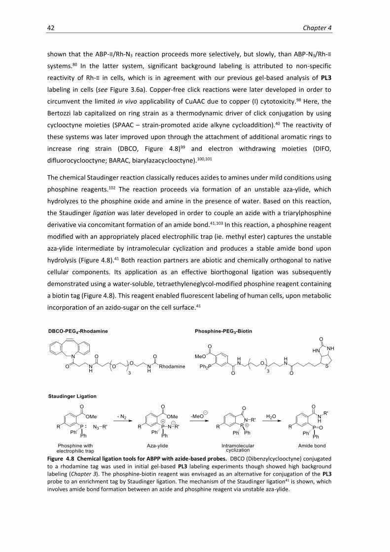
42 Chapter 4
shown that the ABP-≡/Rh-N3 reaction proceeds more selectively, but slowly, than ABP-N3/Rh-≡
systems.80 In the latter system, significant background labeling is attributed to non-specific
reactivity of Rh-≡ in cells, which is in agreement with our previous gel-based analysis of PL3
labeling in cells (see Figure 3.6a). Copper-free click reactions were later developed in order to
circumvent the limited in vivo applicability of CuAAC due to copper (I) cytotoxicity.98 Here, the
Bertozzi lab capitalized on ring strain as a thermodynamic driver of click conjugation by using
cyclooctyne moieties (SPAAC – strain-promoted azide alkyne cycloaddition).40 The reactivity of
these systems was later improved upon through the attachment of additional aromatic rings to
increase ring strain (DBCO, Figure 4.8)99 and electron withdrawing moieties (DIFO,
difluorocyclooctyne; BARAC, biarylazacyclooctyne).100,101
The chemical Staudinger reaction classically reduces azides to amines under mild conditions using
phosphine reagents.102 The reaction proceeds via formation of an unstable aza-ylide, which
hydrolyzes to the phosphine oxide and amine in the presence of water. Based on this reaction,
the Staudinger ligation was later developed in order to couple an azide with a triarylphosphine
derivative via concomitant formation of an amide bond.41,103 In this reaction, a phosphine reagent
modified with an appropriately placed electrophilic trap (ie. methyl ester) captures the unstable
aza-ylide intermediate by intramolecular cyclization and produces a stable amide bond upon
hydrolysis (Figure 4.8).41 Both reaction partners are abiotic and chemically orthogonal to native
cellular components. Its application as an effective biorthogonal ligation was subsequently
demonstrated using a water-soluble, tetraethyleneglycol-modified phosphine reagent containing
a biotin tag (Figure 4.8). This reagent enabled fluorescent labeling of human cells, upon metabolic
incorporation of an azido-sugar on the cell surface.41
Figure 4.8 Chemical ligation tools for ABPP with azide-based probes. DBCO (Dibenzylcyclooctyne) conjugated to a rhodamine tag was used in initial gel-based PL3 labeling experiments though showed high background labeling (Chapter 3). The phosphine-biotin reagent was envisaged as an alternative for conjugation of the PL3 probe to an enrichment tag by Staudinger ligation. The mechanism of the Staudinger ligation41 is shown, which involves amide bond formation between an azide and phosphine reagent via unstable aza-ylide.

Proteomic Analysis 43
Our earlier biochemical investigations proved PL3 to have suitable probe qualities, though
significant background due to nonspecific reactivity of the alkyne- and DBCO-rhodamine reagents
observed by SDS-PAGE (Figure 3.6) precluded its application for our method. Similar problems in
the literature have been addressed through the use of the Staudinger ligation, which showed
improved specificity.48 We therefore pursued PL3-based chemical proteomics using the
commercially available Staudinger biotin-phosphine reagent (Figure 4.8). PL3-labeled samples
(100 and 10 µM, 2 h, 37°C) were reacted with this reagent (24 h incubation) and enriched using
avidin beads. MS-analysis revealed enrichment of 10 PLP-DEs with nearly no background labeling
at 100 µM PL3, and accordingly fewer proteins at the lower probe concentration of 10 µM (Figure
4.9). Significant proteins corresponded well to previous PL4-based experiments, and also included
several of the non-PLP-DEs selected for further characterization.
Figure 4.9 Chemical proteomics with PL3 in S. aureus USA300 TnPdxS using the Staudinger-Bertozzi ligation. (a) Volcano plots showing enrichment of proteins with 100 µM (left) or 10 µM (right) PL3. (n = 6 biological replicates). Known PLP-DEs are highlighted with blue dots and are numerically annotated according to Table 4.4.
Table 4.4 Proteins significantly enriched by chemical proteomics using PL3 (100, 10 µM).
No. Fold-
change Significance Name Uniprot ID
1. 4.56 7.58 Cysteine synthase (EC 2.5.1.47) A0A0H2XFQ3
2. 4.28 7.12 Cysteine desulfurase (EC 2.8.1.7) A0A0H2XHJ5 3. 4.17 5.00 Aminotransferase, class V A0A0H2XKI8 4. 3.98 4.29 Orn/Lys/Arg decarboxylase (EC 4.1.1.18) A0A0H2XII6 5. 1.89 3.47 Threonine synthase (EC 4.2.3.1) A0A0H2XH24 6. 1.58 6.27 Putative pyridoxal phosphate-dependent acyltransferase A0A0H2XJW8
7. 1.44 4.84 Branched-chain-amino-acid aminotransferase (EC 2.6.1.42) A0A0H2XIS2 8. 1.43 2.55 Cysteine synthase/cystathionine beta-synthase (EC 2.5.1.47) A0A0H2XG73 9. 1.29 5.16 Alanine racemase (EC 5.1.1.1) Q2FF55
10. 1.01 4.43 L-threonine dehydratase catabolic TdcB (EC 4.3.1.19) (Threonine deaminase) Q2FH01 11. 4.55 8.09 Pseudouridine-5'-phosphate glycosidase (PsiMP glycosidase) (EC 4.2.1.70) A0A0H2XJC6
12. 3.38 3.99 Signal peptidase I (EC 3.4.21.89) A0A0H2XEA7 13. 2.12 2.61 UPF0340 protein SAUSA300_2068 Q2FF14 14. 0.79 6.54 Putative heme-dependent peroxidase SAUSA300_0569 (EC 1.11.1.-) Q2FJ56
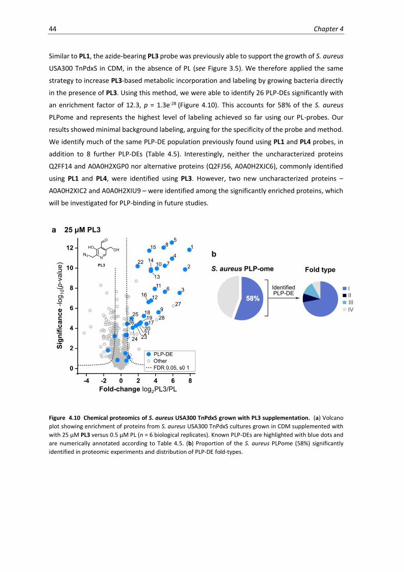
44 Chapter 4
Similar to PL1, the azide-bearing PL3 probe was previously able to support the growth of S. aureus
USA300 TnPdxS in CDM, in the absence of PL (see Figure 3.5). We therefore applied the same
strategy to increase PL3-based metabolic incorporation and labeling by growing bacteria directly
in the presence of PL3. Using this method, we were able to identify 26 PLP-DEs significantly with
an enrichment factor of 12.3, p = 1.3e-28 (Figure 4.10). This accounts for 58% of the S. aureus
PLPome and represents the highest level of labeling achieved so far using our PL-probes. Our
results showed minimal background labeling, arguing for the specificity of the probe and method.
We identify much of the same PLP-DE population previously found using PL1 and PL4 probes, in
addition to 8 further PLP-DEs (Table 4.5). Interestingly, neither the uncharacterized proteins
Q2FF14 and A0A0H2XGP0 nor alternative proteins (Q2FJ56, A0A0H2XJC6), commonly identified
using PL1 and PL4, were identified using PL3. However, two new uncharacterized proteins –
A0A0H2XIC2 and A0A0H2XIU9 – were identified among the significantly enriched proteins, which
will be investigated for PLP-binding in future studies.
Figure 4.10 Chemical proteomics of S. aureus USA300 TnPdxS grown with PL3 supplementation. (a) Volcano
plot showing enrichment of proteins from S. aureus USA300 TnPdxS cultures grown in CDM supplemented with
with 25 µM PL3 versus 0.5 µM PL (n = 6 biological replicates). Known PLP-DEs are highlighted with blue dots and
are numerically annotated according to Table 4.5. (b) Proportion of the S. aureus PLPome (58%) significantly
identified in proteomic experiments and distribution of PLP-DE fold-types.

Proteomic Analysis 45
Table 4.5 Proteins significantly enriched from bacterial cultures grown using PL3 supplementation (25 µM).
No. Fold-
change Significance Name Uniprot ID
1. 7.90 11.82 Glutamate-1-semialdehyde 2,1-aminomutase 2 (GSA 2) (EC 5.4.3.8) Q2FFN1 2. 7.41 9.86 Aminotransferase A0A0H2XKB4 3. 6.78 7.53 Aminotransferase, class V A0A0H2XKI8 4. 5.91 10.94 D-alanine aminotransferase (EC 2.6.1.21) A0A0H2XHU6 5. 5.88 12.54 Putative pyridoxal phosphate-dependent acyltransferase A0A0H2XJW8
6. 5.08 7.64 Trans-sulfuration enzyme family protein A0A0H2XG37 7. 5.07 10.23 Cysteine desulfurase (EC 2.8.1.7) A0A0H2XHJ5 8. 4.94 12.04 Ornithine aminotransferase (OAT) (EC 2.6.1.13) A0A0H2XIK4 9. 4.34 5.60 Cystathionine gamma-synthase (EC 4.4.1.8) A0A0H2XFF8
10. 4.15 9.95 Probable glycine dehydrogenase (decarboxylating) subunit 2 (EC 1.4.4.2)
(Glycine dehydrogenase (aminomethyl-transferring) subunit 2)
Q2FGI7
11. 3.91 7.93 Serine hydroxymethyltransferase (SHMT) (Serine methylase) (EC 2.1.2.1) Q2FF15 12. 3.49 6.77 Cysteine synthase (EC 2.5.1.47) A0A0H2XFQ3 13. 3.47 9.72 Cys/Met metabolism PLP-dependent enzyme (EC 4.4.1.8) A0A0H2XFH9 14. 3.46 9.87 Branched-chain-amino-acid aminotransferase (EC 2.6.1.42) A0A0H2XIS2
15. 3.27 11.73 Glutamate-1-semialdehyde 2,1-aminomutase 1 (GSA 1) (EC 5.4.3.8) Q2FG69 16. 3.19 6.61 Cysteine synthase/cystathionine beta-synthase (EC 2.5.1.47) A0A0H2XG73 17. 2.96 4.45 Ornithine aminotransferase (OAT) (EC 2.6.1.13) A0A0H2XHG0 18. 2.65 5.23 Aminotransferase, class V A0A0H2XGZ7 19. 2.28 4.59 Orn/Lys/Arg decarboxylase (EC 4.1.1.18) A0A0H2XII6
20. 2.16 4.45 Aminotransferase, class V (EC 1.12.-.-) A0A0H2XJX6 21. 2.08 4.42 Diaminopimelate decarboxylase (DAP decarboxylase) (EC 4.1.1.20) A0A0H2XFD9 22. 1.93 10.19 Alanine racemase (EC 5.1.1.1) Q2FF55 23. 1.75 4.26 Uncharacterized protein A0A0H2XHH8 24. 1.37 4.06 Histidinol-phosphate aminotransferase (EC 2.6.1.9) (Imidazole acetol-
phosphate transaminase)
Q2FIR7
25. 1.24 4.99 Aminotransferase A0A0H2XJK0 26. 0.71 4.44 Aminotransferase, class I (EC 2.6.1.-) A0A0H2XFA8 27. 6.05 6.22 Uncharacterized protein A0A0H2XIC2 28. 4.13 4.85 Uncharacterized protein A0A0H2XIU9 29. 2.09 1.93 PTS system, glucose-specific IIA component (EC 2.7.1.69) A0A0H2XJ98 30. 1.84 3.43 Iron-sulfur cluster repair protein ScdA Q2FK11
31. 1.76 8.70 Bifunctional purine biosynthesis protein PurH [Includes: Phosphoribosylaminoimidazolecarboxamide formyltransferase (EC 2.1.2.3);IMP cyclohydrolase (EC 3.5.4.10) (IMP synthase) (Inosinicase)]
Q2FI05
4.6 Conclusion
In conclusion, our chemical proteomic strategy was effective at labeling and enriching PLP-DEs
from S. aureus USA300 TnPdxS. The use of three PL-derivatives with different chemical and
biological properties as probes for PLP-binding enabled labeling of a combined total of 73% of the
S. aureus PLPome (Figure 4.11). From these results, protein candidates were selected for further
validation as putative PLP-DEs. Several of these candidates were identified throughout multiple
proteomic experiments and across different probe-labeled samples, which adds additional
confidence to their interaction with PLP. Nonetheless, we find marked differences in our labeling
results, the types of PLP-DEs identified and the methodological requirements. PL4 shows effective
enrichment of PLP-DEs during a 2-hour in situ labeling period, which can be coupled to a two-
tiered confidence filtering strategy to extract reliable results. While PL1 was less effective using
this protocol due to chemical lability, its ability to support bacterial growth enabled an alternative
labeling strategy which increased its metabolic incorporation and led to the identification of
additional PLP-DEs not labeled by PL4. The azide-containing PL3 probe requires the employment
of the Staudinger ligation to achieve labeling specificity suitable for proteomic experiments.

46 Chapter 4
Labeling is highly specific during the 2-hour labeling protocol, but fewer proteins are identified
than with PL4. However, when bacteria are grown directly in the presence of PL3 as a cofactor
substitute, the most complete and specific PLPome labeling profile thus far could be accessed.
This enabled the identification of two additional uncharacterized proteins, which were not
previously identified, as candidates for further characterization. The validation of PLP-binding to
known PLP-DEs, uncharacterized and alternative proteins is described in Chapter 5.
Figure 4.11 Combined total of S. aureus USA300 TnPdxS PLP-DEs detected with PL1, PL3 and PL4. A total of 33 proteins, ie. 73 % of the S. aureus PLPome could be identified with our probe-labeling strategy. A Venn diagram illustrates the differences in proteins significantly enriched (Uniprot identifiers) using the different probes. The comparison comprises data from the following volcano plots: PL1 (Figure 4.7a, CDM supplementation with PL1), PL3 (Figure 4.10a, CDM supplementation with PL3), and PL4 (Figure 4.3b).

5. Validation & Enzyme Characterization
5.1 Introduction
Recent estimates indicate that about 30-40% of genes in each genome remain unannotated or
only putatively assigned a function.104 The evolutionary conservation of many uncharacterized
proteins (70%) implies that they are functionally relevant or metabolically important.105,106
Chemical profiling strategies are able to put these proteins into a functional context by their
reactivity or affinity, and thereby provide starting points for further investigation of protein
function. Current strategies for the full characterization of proteins require the integration of
multiple approaches, including bioinformatics, structural, biochemical and cell-based methods.
Bioinformatic strategies for the functional analysis of proteins rely on sequence- or structure-
homology searches within corresponding databases. While sequence-based searches have
evolved advanced algorithms for detecting distant structural similarities and phylogenetic
relationships among proteins, they are not accurate at discriminating protein function. For PLP-
DEs, sequence-based analyses has led to their classification into different fold types, but could not
assign specific catalytic function due to overlap and multiplicity among the different subgroups.18
In an alternative approach, the use of reference search models tailored to specific catalytic
activities rather than structural fold provided a more detailed genomic overview of PLP-DE
activities across different organisms.4 Search methods that rely on structural data are more
accurate at assigning protein function. Strategies that have been applied to PLP-DEs include
multiple structure alignments (3DM database analysis and overlays) as well as comparisons of
active site architecture.34,107,108 A combination of these methods was applied by Fleischmann et al.
in order to establish a robust workflow for characterizing PLP-DEs using three uncharacterized
proteins from the human microbiome.107 Initial secondary structure matching (SSM) identified
similarity to known aminotransferases and a Pfam search109 (protein family database search) was
able to assign the corresponding aminotransferase subtypes. Upon structural superimposition
with a subset of representative family members, the best matches were selected for structure-
based multiple sequence alignment in order to reveal conservation of distinct residues. It was
found that active site motifs characteristic of aminotransferases were strictly conserved. PLP-DE

48 Chapter 5
active site structures are often similar due to common mechanistic features associated with the
cofactor. However, unique arrangements of residues essential for catalysis remain highly
conserved throughout evolution and can be used to infer enzyme function. Active site
fingerprinting of slow-evolving features using the program CPASS (comparison of active site
structures) has been used to cluster PLP-DE function based on conserved residues proximal to the
PLP ligand (6 Å).34 Structural data and in silico analysis thus represent a powerful combination for
obtaining an initial hypothesis to pursue in subsequent biological investigations.
Substantial literature precedence (since the 1940’s) for the family of PLP-DEs facilitates their study
by providing important information on their diverse catalytic mechanisms, unique characteristics
and kinetic activities. A common method for ascertaining cofactor binding and deriving qualitative
insights into protein function is UV-Vis spectroscopy, which permits the observation of
characteristic catalytic intermediates such as the internal aldimine (330-360 nm; 420-430 nm),
quinonoid (500 nm), aminoacrylate (470 nm) or PMP (330 nm).60,110 This strategy was applied by
Fleischmann et al. to assess substrate preferences of their aminotransferases, revealing
considerable substrate promiscuity (≥ 8 amino acids each).107 The natural enzyme substrates were
narrowed down using kinetics assays to screen possible amine acceptors (ex. ketoglutarate,
oxaloacetate, pyruvate), as well as slow-cooling molecular dynamics simulation of the modeled
external aldimines. A variety of coupled assays have been reported for studying PLP-DE activity,
and stopped-flow kinetics are frequently used to directly monitor quinonoid formation or other
characteristic intermediates. Additionally, PLP-DE substrates can be determined by screening
libraries of compounds and analyzing reaction products by LC-MS or HPLC. Nonetheless, standard
in vitro experiments do not account for complex biological settings, such as protein interactions,
cryptic natural substrates or protein regulation. In particular, studying the roles of PLP-DEs in vitro
is complicated by substrate promiscuity and functional redundancy.
In vivo or ex vivo studies are useful for understanding protein function directly in a native biological
environment. Here, the application of proteomic and transcriptomic methods can help to dissect
the roles of proteins within biological pathways or networks, as well as their regulation,
interaction partners and cellular localization. Moreover, coupling these methods to metabolomic
analysis can provide powerful datasets for gaining direct insights into the state of a cell.111 For
example, these strategies can be applied to study the function of a protein by analyzing its cellular
implications upon over- or in-activation (ie. overexpression or genetic disruption). This often
triggers the activation of compensatory mechanisms, and the resulting proteomic or metabolomic
changes can thereby provide clues as to the cellular function of a protein. Alternatively,
metabolomics can be used to measure the activity of a protein under near-native conditions using
a strategy termed activity-based metabolomic profiling (ABMP).112,113 The purified protein is
therefore incubated with extracted metabolome or cell lysate, and differences in metabolite levels
are measured by MS as a readout of enzyme activity. Using native metabolomes ensures the

Validation & Enzyme Characterization 49
screening of physiologically-relevant metabolites from the parent organism and provides access
to non-commercial substrates. There are several challenges associated with metabolomics, in
which compound identification and annotation rely on matching to corresponding database
entries (MS, MS/MS). Technical difficulties include the inability to discern isobaric compounds or
to account for in-source fragmentation, and selecting the correct measurement technique for
metabolites of interest. Furthermore, high variability between samples due to cellular dynamics,
compound stability, and metabolite extraction complicate analysis.
Using a suite of bioinformatic, structural, biochemical and cell-based methods, we attempted to
confirm the PLP-dependent function of select proteins identified from chemical proteomic
experiments (Chapter 4). Techniques for validating PLP-binding were first established using known
PLP-DEs and then applied to study uncharacterized and alternative-function proteins. We further
studied the uncharacterized proteins using several of the strategies outlined above in order to
gain functional insights. The identification of novel PLP-dependent roles in the cell represents a
major application of our PLPome profiling method.
5.2 Known PLP-DEs
To validate the binding of PLP to proteins identified in our proteomic experiments, we first
established a PLP-detection strategy with known PLP-DEs using UV-Vis spectroscopy, gel-based
labeling, and intact-protein MS. Therefore, five significantly enriched (high and medium
confidence) PLP-DEs of binding clusters 1 and 2 were selected from PL4-based proteomic
experiments. Different types of PLP-DEs were chosen, including two class I aminotransferases (AT1
and AT2; A0A0H2XFY9 and A0A0H2XFA8), D-alanine aminotransferase (DAT; A0A0H2XHU6),
ornithine/lysine/arginine decarboxylase (OKR-DC; A0A0H2XII6) and cysteine synthase (CS;
A0A0H2XFQ3), to study a range of PLP-dependent processes. These proteins were recombinantly
expressed and purified on an analytical scale by streptavidin affinity chromatography. UV-Vis
spectra indicated the presence of the PLP cofactor bound as an internal aldimine (360 or 420 nm,
Figure 5.1a) for each protein (Figure 5.1b, 5.2b). We further used UV-Vis in order to observe
characteristic spectral properties of PLP-dependent catalytic intermediates.60,110 For example,
upon incubation of CS with the substrate O-acetylserine (OAS), formation of the aminoacrylate
intermediate (470 nm)60 was observed (Figure 5.1a,b). It also enabled the qualitative assessment
of AT1, AT2 and DAT substrate preferences in vitro upon incubating each aminotransferase with
various amino acids in the presence of PLP and monitoring PMP formation (330 nm, Figure 5.2a,b).
We observed that DAT converts many D-amino acids (7 out of 11 tested), while AT1 and AT2
converted only L-amino acids (3 and 6 out of 17 tested, respectively; Figure 5.2c).

50 Chapter 5
Figure 5.1 Validation of known PLP-DEs OKR-DC and CS by UV-Vis, MS and gel-based analysis. (a) Characteristic spectral properties of internal aldimine and aminoacrylate intermediates.60,110 (b) UV-Vis spectra of OKR-DC (top) and CS ± OAS (10 equivalents) to observe aminoacrylate formation (470 nm, bottom). Curves were normalized to absorbance at 300 nm. (c) Proportion of PLP-DE bound upon incubation with PLP or cofactor analogues (4 equivalents), NaBH4 reduction and measurement by intact protein MS (n = 3, error bars: mean ± standard deviation). Up to 6% nonspecific addition was observed for CS (PL, PL4). (d) Fluorescence SDS-PAGE showing in vitro labeling of OKR-DC and CS with PL4P (0.5, 1, 5 equivalents) or PL4 (5 equivalents) as a negative control. See appendix for full gels and Coomassie staining.
Our findings are in line with previous reports of the in vitro promiscuity of PLP-DEs, and are a
starting point for more detailed kinetic investigations of substrate scope. Spectroscopic
measurements therefore represent a reliable and sensitive method to confirm the activity of an
enzyme and to gain preliminary functional insights, which could be coupled to stopped-flow
kinetic experiments for more detailed analysis.
We next corroborated PLP and PL4P binding to the selected PLP-DEs using intact-protein mass
spectrometry. Incubation of the proteins with excess cofactor followed by NaBH4 reduction
resulted in quantifiable, single-fold adduct formation (Figure 5.1c, 5.2d). Control experiments with
non-phosphorylated cofactors (PL and PL4) did not show adduct formation, thereby proving the
specificity of the interaction. For most PLP-DEs, cofactor binding could not be detected without
prior incubation with excess PLP. MS-based analysis of binding thus appears to be less suitable for
monitoring probe binding than UV-Vis, which detected bound cofactor in all PLP-DEs, perhaps due
to poor efficiency of the NaBH4 reduction step. CS and AT1 were approximately 40% bound by PLP
and PL4P appeared to bind the remaining fraction of apoenzyme without significantly displacing
the cofactor from the PLP-holoenzymes. Probe binding and specificity was further reflected in gel-
based labeling experiments, in which only the active probe labeled proteins significantly (Figure
5.1d, 5.2e). We therefore conclude that the methods presented here are simple and effective at
detecting authentic PLP binding, and can be used across a panel of PLP-DEs.

Validation & Enzyme Characterization 51
Figure 5.2 Validation of PLP-dependent aminotransferases by UV-Vis, MS and gel-based analysis. (a) Characteristic spectral properties of PLP/PMP intermediates during transamination. (b) UV-Vis spectra of DAT, AT1 (A0A0H2XFY9) and AT2 (A0A0H2XFA8) ± substrate amino acids (10 equivalents) to observe PMP formation (330 nm). Curves were normalized to absorbance at 300 nm. (c) Heatmap representation of in vitro substrate preferences of aminotransferases observed by spectral shifts. DAT was tested with D-amino acids; AT1 and AT2 were tested with L-amino acids. (d) Proportion of PLP-DE bound upon incubation with PLP or cofactor analogues (4 equivalents), NaBH4 reduction and measurement by intact protein MS (n = 3, error bars: mean ± standard deviation). (e) Sample fluorescence SDS-PAGE showing in vitro labeling of DAT with PL4P (0.5, 1, 5 equivalents) or PL4 (5 equivalents) as a negative control. See appendix for full gel and Coomassie staining.
5.3 Uncharacterized PLP-DEs
Having validated cofactor binding for several known PLP-DEs, we used the above techniques to
assess the PLP-binding of uncharacterized proteins identified from PL4-based proteomics. These
included Q2FF14 (Uniprot ID; high-confidence; cluster 1), GP0 (Uniprot: A0A0H2XGP0; medium-
confidence; cluster 2), and HH8 (Uniprot: A0A0H2XHH8; high-confidence; cluster 1). While the
latter protein is annotated to be functionally uncharacterized, it is predicted to bind PLP by
homology.108,114,115 Each of the three proteins appears to bind PLP and PL4P based on initial UV-
Vis, MS and gel-based labeling experiments (Figure 5.3). UV-Vis spectra were recorded from large-
scale protein batches, in which E. coli expression lysate was treated with PLP (100 µM, 20 min,
4°C) prior to purification to saturate PLP binding and improve the spectroscopic signal strength.
We next proceeded with biochemical characterization according to the strategies outlined in the
introduction, in order to gain insights into potential roles of these proteins.

52 Chapter 5
Figure 5.3 Validation of PLP binding for uncharacterized proteins identified using PL4. (a) UV-Vis spectra of uncharacterized proteins saturated with PLP (100 µM) in E. coli expression lysate; curves were normalized to absorbance at 300 nm. (b) Proportion of protein bound upon incubation with PLP or cofactor analogues (Q2FF14, GP0: 4 equivalents, HH8: 1 equivalent), NaBH4 reduction and measurement by intact protein MS (n = 3, error bars: mean ± standard deviation). (c) Fluorescence SDS-PAGE showing labeling of uncharacterized proteins with PL4P (0.5, 1, 5 equivalents) or PL4 (5 equivalents) as a control. See appendix for full gels and Coomassie staining.
5.3.2 Q2FF14
Q2FF14 is currently annotated as a member of the YwlG superfamily (uncharacterized protein
family UPF0340; domain of unknown function DUF436), comprising 68 highly conserved proteins
found in Gram-positive bacteria. The protein is 174 residues in length and has a molecular weight
of 18.9 kDa. A sequence alignment of Q2FF14 with homologues from other species shows distinct
regions of conservation (Figure 5.4). The corresponding gene (SAUSA300_2068) is located
upstream from the PLP-DE serine hydroxymethyltransferase, in an operon together with UDP-
GlcNAc 2-epimerase and uracil phosphoribosyltransferase (Figure 5.5a), which are important for
one-carbon, aminosugar and pyrimidine metabolism, respectively.
Q2FF14 shows a strong absorbance at 330-340 nm, indicative of either the enolimine form of the
internal aldimine or a PL-derivative saturated at the C4A carbon, in addition to a modest keto-
enamine peak at 420 nm (Figure 5.3a). The peaks are very weak when isolating the protein directly
from E. coli expression lysate, but prior incubation of the lysate with PLP generates a protein that
is visibly yellow and has a correspondingly strong signal. However, the apparent holoenzyme is
unstable and readily precipitates at 4°C within 1 h. Our MS-based analysis suggests that Q2FF14
binds PLP and PL4P weakly, but specifically over non-phosphorylated counterparts (Figure 5.3b).
This weak binding is also reflected in gel-based labeling, in which a 5-fold excess of probe yields
only minimal fluorescent signal (Figure 5.3c). These results match our observation that Q2FF14
may be unstable in its PLP-bound form, or has low affinity towards PLP, though requires further
confirmation.

Validation & Enzyme Characterization 53
Figure 5.4 Sequence alignment of Q2FF14 with homologous sequences from Gram-positive bacteria.
Sequences were selected by Blast search of the Q2FF14 protein sequence, excluding S. aureus. Sequences were
aligned using ClustalX116 and processed using ESPript.117 Figure courtesy of Dr. S. Schneider.
54 Chapter 5
We determined the X-ray crystal structure of Q2FF14 to 1.75 Å resolution and did not detect
bound PLP or other ligands (Figure 5.5b,c,d). The protein fold consists of a central 8-stranded β-
sheet decorated with α-helices and a long C-terminal extension (Figure 5.5b). Q2FF14 forms a
hexameric structure that is made up of two stacked trimeric rings (Figure 5.5d), which matched
solution-based SEC analysis (Figure 5.5e). Structural alignment search (DALI)118 reveals high
similarity to the conserved hypothetical protein TT1679 from Thermus thermophilus HB8 (PDB:
1v8d),119 with which it shares 40% sequence identity,119 as well as distant similarity (11% sequence
identity) to glutamate dehydrogenases (e.g. PDB: 1v9l).120
We next attempted to elucidate the function of Q2FF14 using substrate screens and cell-based
experiments. Incubation of apo-Q2FF14 with a mixture of D/L-amino acids in the presence of PLP
and other cofactors, however, did not result in any characteristic spectral peaks nor changes by
MS (data not shown). We conclude that Q2FF14 does not transform amino acid substrates or that
the protein is inactive under the conditions tested. An activity-based metabolomic profiling
approach using S. aureus metabolome may therefore be more suitable for identifying a possible
substrate under more biologically-relevant conditions.
Cell-based experiments were performed to understand the functional implications of Q2FF14 in a
native environment. A strain of S. aureus USA300 in which the gene encoding Q2FF14 was
disrupted by transposon insertion was obtained from the Nebraska transposon mutant library.84
The availability of this strain and its growth viability indicate that Q2FF14 is not essential. A whole-
proteome analysis served to compare stationary-phase cultures of TnQ2FF14 and wild-type
S. aureus USA300 (Figure 5.5f). We find that all of the downregulated proteins are within the same
operon, initiated by Q2FF14. It is therefore possible that Q2FF14 regulates the expression of other
genes within its operon; however, this observation may also result from non-specific mRNA
processing effects due to transposon insertion (3.2 kb). Only one uncharacterized protein
(probable β-lactamase, A0A0H2XJY4) was significantly upregulated. Similarly, we did not observe
any significant dysregulations of metabolites in corresponding metabolomic profiling experiments
(data not shown). This suggests that Q2FF14 influences cellular pathways that cannot be
significantly detected under the experimental conditions.
To summarize, the conserved uncharacterized protein Q2FF14 appears to bind PLP or a derivative
thereof, but subsequently shows in vitro instability. We are unable to draw functional conclusions
based on the obtained structural data, substrate analysis or cell-based studies. Some of these
experiments may require optimizations of conditions, such as cellular growth, in order to clearly
distinguish an effect of Q2FF14 activity.

Validation & Enzyme Characterization 55
Figure 5.5 X-ray crystallography and structural analysis of Q2FF14. (a) Q2FF14 (174 residues) contains a domain of unknown function (DUF436) and initiates a 4-gene operon. (b) Secondary structure and protein fold of a Q2FF14 monomer (ribbon representation). (c) Electrostatic surface representation of a Q2FF14 monomer. (d) Assembly of Q2FF14 into a hexamer composed of two stacked trimers. Crystallography figures courtesy of Dr. S. Schneider. (e) Size exclusion chromatography of Q2FF14 shows hexamer formation. (f) Volcano plot showing fold-difference between S. aureus USA300 Q2FF14 transposon mutant versus wild-type upon performing a two-sample t-test (FDR 0.5, s0 0.5; n = 3 replicates).
5.3.2 A0A0H2XGP0
GP0 is an uncharacterized protein composed of 295 residues (34.8 kDa), which contains a
C-terminal domain (150-295) predicted to have N-acyltransferase function (InterPro, Figure 5.6a).
Specifically, this domain belongs to the family of Gcn5-related N-acetyltransferases (GNAT,
Interpro IPR000182) which use acetyl-CoA to transfer an acetyl group onto primary amines of
small molecules and proteins. GNAT proteins have diverse cellular functions in both prokaryotes

56 Chapter 5
and eukaryotes, and acetylate a variety of substrates such as aminoglycoside antibiotics
(resistance mechanism), glucosamine-6-phosphate (bacterial cell wall biosynthesis), histones
(transcriptional activation), serotonin and spermidine. They are characterized by a structurally
conserved fold comprising an arrangement of 6-7 β-strands and 4 α-helices (β1-α1-α2-β2-β3-β4-α3-
β5-α 4-β6-β7). The gene encoding GP0 (SAUSA300_2209) is conserved among staphylococci and is
within its own operon, frequently located in proximity to the sugar transporter glcU
(SAUSA300_2210). S. aureus contains 35 putative GNAT enzymes, many of which are functionally
and structurally uncharacterized.121
UV-Vis, MS and gel-based analyses revealed a definitive interaction of GP0 with the PLP cofactor
(Figure 5.3). In solution, the protein exists primarily as a monomer with small amounts of
homodimerization, a common feature of PLP-DEs (Figure 5.6c).2 Preliminary substrate studies
using a pool of D/L-amino acids did not show any conversion by UV-Vis or MS (data not shown).
The previous annotation of GP0 as an acetyltransferase led us to conduct a broad substrate screen
for acetyltransferase activity with amino acids, antibiotics, and diverse other metabolites (for
exact substrates see Table 8.3). The assay was based on a literature protocol, using Ellman’s
reagent (5,5′-dithiobis(2-nitrobenzoic acid), DTNB) as a quantitative, spectrophotometric reporter
of the production of free CoA thiol.122 Using this plate-assay with 48 possible substrates available
in our lab (see Table 8.3), we did not detect any GP0-based acetylation activity above controls.
The application of an activity-based metabolomic profiling approach using extracted S. aureus
metabolome would enable screening of a broader substrate scope and may therefore lend further
insights into GP0 function.
We next assessed the cellular effect of GP0 disruption by comparing a TnGP0 transposon mutant
of S. aureus USA300 to wild-type on both a proteomic and metabolomic level. We obtained the
TnGP0 mutant strain from the Nebraska transposon mutant library,84 which grew similarly to the
wild-type strain. In proteomic experiments, a significant downregulation of proteins involved in
amino acid import and biosynthesis was observed (Figure 5.6b, Table 5.1). Particularly, pathways
of Ser/Thr, Met/Cys and Val/Ile/Leu were affected, as observed by a decrease in the methionine
importer (Q2FJI0) and aspartokinase (A0A0H2XKG7). Furthermore, several lipoproteins and ABC
transporters were also downregulated. Only the uncharacterized protein A0A0H2XHF8 was
significantly upregulated in the absence of GP0. Considerable alterations in amino acid
biosynthesis could also be detected by metabolomic profiling, in which levels of amino acids,
acetylated amino acids and short peptides appear to be altered (Figure 5.6d, Table 5.2). Putative
pathways that were significantly affected include lysine, proline and tryptophan biosynthesis, as
well as UDP-N-acetyl-D-glucosamine biosynthesis (biochemical precursor for cell wall
components) and cell wall recycling (anhydromuropeptides) (Figure 5.6e)
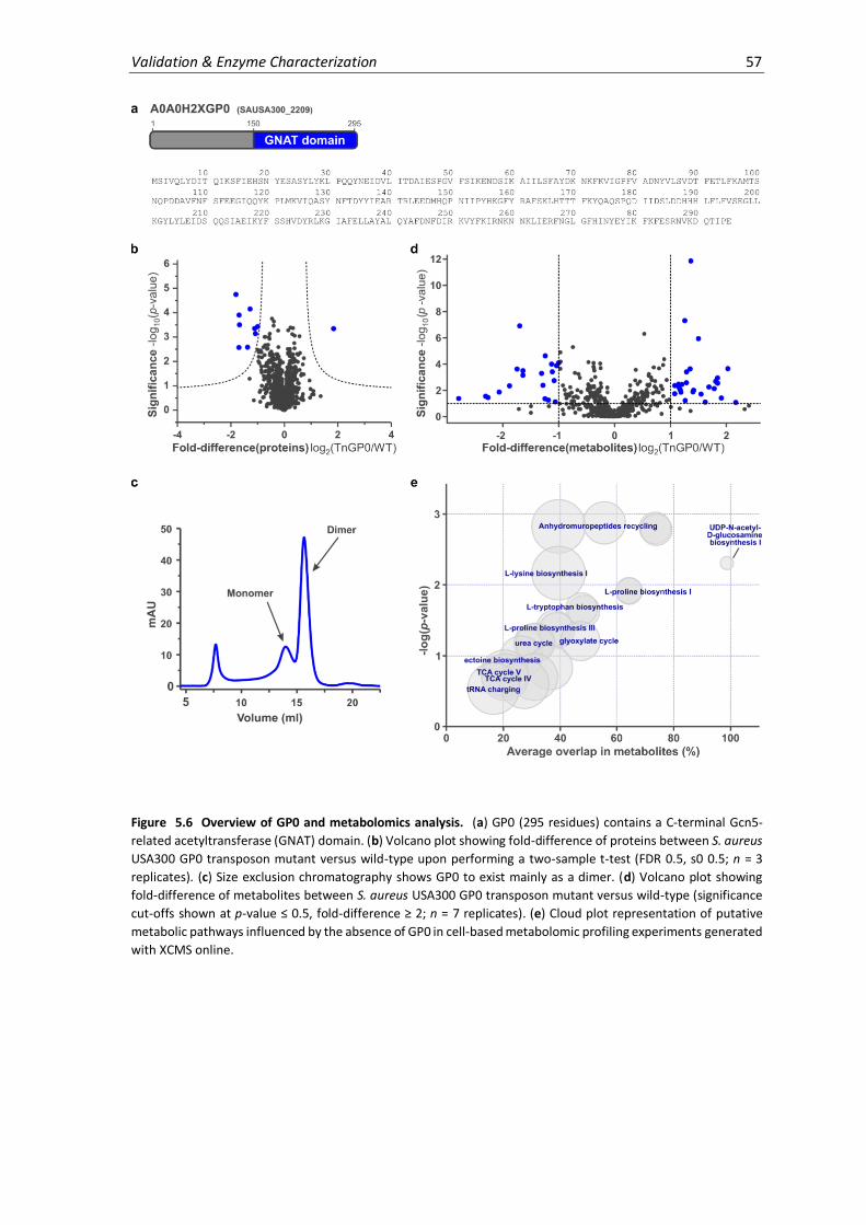
Validation & Enzyme Characterization 57
Figure 5.6 Overview of GP0 and metabolomics analysis. (a) GP0 (295 residues) contains a C-terminal Gcn5-
related acetyltransferase (GNAT) domain. (b) Volcano plot showing fold-difference of proteins between S. aureus
USA300 GP0 transposon mutant versus wild-type upon performing a two-sample t-test (FDR 0.5, s0 0.5; n = 3
replicates). (c) Size exclusion chromatography shows GP0 to exist mainly as a dimer. (d) Volcano plot showing
fold-difference of metabolites between S. aureus USA300 GP0 transposon mutant versus wild-type (significance
cut-offs shown at p-value ≤ 0.5, fold-difference ≥ 2; n = 7 replicates). (e) Cloud plot representation of putative
metabolic pathways influenced by the absence of GP0 in cell-based metabolomic profiling experiments generated
with XCMS online.

58 Chapter 5
Table 5.1 Whole proteome comparison of S. aureus TnGP0 vs. wild-type. Corresponding volcano plot in Figure 5.6b.
Uniprot ID Name Gene Gene Ontology
Up-regulated proteins
A0A0H2XHF8 Uncharacterized protein SAUSA300_1489
Down-regulated proteins
Q2FF67 2-isopropylmalate synthase (EC 2.3.3.13) (Alpha-isopropylmalate synthase)
SAUSA300_2010 (leuA)
leucine biosynthesis [GO:0009098]
A0A0H2XKG7 Aspartokinase (EC 2.7.2.4) SAUSA300_1225 lysine biosynthesis via DAP [GO:0009089]; threonine biosynthesis process [GO:0009088]
A0A0H2XHB0 Uncharacterized protein SAUSA300_0177
A0A0H2XI71 Peptide ABC transporter, ATP-binding protein SAUSA300_0200 peptide transport [GO:0015833]
A0A0H2XJL5 Uncharacterized protein SAUSA300_0173
A0A0H2XFT9 Lipoprotein SAUSA300_0437
Q2FJI0 Methionine import ATP-binding protein MetN 1
(EC 3.6.3.-)
SAUSA300_0435
(metN1)
methionine transport [GO:0015821]
A0A0H2XJJ1 Putative lipoprotein SAUSA300_0175 A0A0H2XIF6 Putative lipoprotein SAUSA300_0203
Table 5.2 Whole metabolome comparison of S. aureus TnGP0 vs wild-type. Select up/downregulated features from volcano plot Figure 5.6d identified using the Metlin database.123
Feature Fold-
difference Significance
Predicted Metabolite #1
Predicted Metabolite #2
Predicted Metabolite #3
Up-regulated metabolites
374.2245 2.17 1.08 - - -
491.3030 1.91 1.42 CysLeuLeuLys GlyAsnLysArg
118.0853 1.84 2.94 L-Valine
231.1330 1.50 5.94 Hydroxyprolylvaline Valylhydroxyproline
189.1224 1.40 1.89 N-ε-acetyllysine ValAla
175.1069 1.36 11.85 N-acetylornithine ValGly N-acetylproline
90.0544 1.26 7.31 L-Alanine
Down-regulated metabolites
442.2354 -2.79 1.38 AspProLeuPro
441.2327 -2.31 1.55 IleLeuSerSer
441.2319 -2.27 1.46 AspLeuProPro
104.1062 -1.19 1.26 - - 203.1018 -1.00 4.09 SerPro Alanylhydroxyproline N-acetyl-β-glucosamine
In summary, we can confirm GP0 to bind PLP and could detect alterations in PLP-dependent
processes, such as amino acid biosynthesis, on a cellular level in its absence. However, it is difficult
to draw specific function from cell-based proteomic and metabolomic analyses, in which multiple
mechanisms may compensate for the loss of a protein and the interrelatedness of biological
networks makes interpretation difficult. Though GP0 is predicted to have an acetyltransferase
domain similar to GNAT proteins, we do not detect acetyltransferase activity across a range of
possible substrates. Limitations in structural data acquisition currently prevent us from confirming
this structure-based prediction and drawing conclusions as to the N-terminal half of the protein,
which often serves substrate recognition in GNAT proteins. To date, the only reported PLP-
dependent acetyltransferase activity is that of glycine C-acetyltransferase, also known as 2-amino-
3-ketobutyrate CoA ligase (A0A0H2XJW8).

Validation & Enzyme Characterization 59
5.3.3 A0A0H2XHH8
HH8 (A0A0H2XHH8) is annotated as an uncharacterized protein (UPF001) which is predicted to be
part of a functionally unknown subgroup of the alanine racemase family (Pfam: PF01168). The
universal distribution and evolutionary conservation of proteins belonging to this subgroup argue
for its importance in cellular functions. Crystal structures of homologues from Saccharomyces
cerevisiae (YBL036C, PDB: 1CT5/1B54)114 and Escherichia coli (YggS, PDB: 1W8G)124 have revealed
similarity to bacterial alanine racemase and eukaryotic ornithine decarboxylase, which were
previously not detected by sequence-based searches. However, unlike Alr, HH8 homologues are
single-domain monomeric structures which lack the Alr active site base Tyr265.114 Furthermore,
they are unable to racemize amino acids114,115 or showed only weak activity upon overnight
incubation,108 leading to the hypothesis that they are non-specific racemases with a yet unknown
substrate. However, other functional investigations have implicated this protein class in PLP
homeostasis.115,125 Here, it was shown that the ΔYggS E. coli strain accumulated PNP and that high
levels of PN (but not PM, PL or PLP) were toxic to this strain.125 Comparative genomics in E. coli
further linked this protein subfamily to PLP salvage, cell division and cell wall biosynthesis.125 In a
previous genomic clustering experiment, the human homologue PROSC (proline synthetase co-
transcribed protein) was linked to proline synthesis, though this likely relates to another function.
In humans, deficiency of PROSC causes vitamin B6-dependent epilepsy.126 This strengthens its
functional link to PLP homeostasis, and its dysregulation may thus influence secondary processes
such as amino acid metabolism.
In our studies, we confirm the S. aureus USA300 enzyme HH8 to bind PLP based on UV-Vis and MS
experiments (Figure 5.3). Interestingly, we found HH8 to be nearly completely saturated with PLP
upon purification from the E. coli expression strain, even without any prior addition of PLP to the
lysate. This was not the case for any other PLP-DE studied here, where maximally half of the
protein population showed PLP-binding (see Figures 5.1c, 5.2d). Furthermore, PLP adduct
formation could be detected even in the absence of NaBH4 reduction (data not shown). These
observations argue for a strong interaction of HH8 with PLP and a high cellular binding affinity. In
agreement with previous studies, SEC analysis indicates that HH8 is present exclusively in its
monomeric form (Figure 5.7b), and we do no detect Alr activity at a relevant timescale using our
coupled assay (data not shown). Studies have suggested that HH8 homologues are essential for
some organisms (ex. P. aeruginosa and S. aureus) but are dispensable for others.125 The lack of a
corresponding HH8 transposon mutant in the S. aureus USA300 Nebraska transposon mutant
library supports this conclusion. In S. aureus, we find the gene encoding HH8 (SAUSA300_1082) to
be located in an operon with three other hypothetical proteins (Figure 5.7a) and therefore cannot
draw any conclusions from its genomic environment. In summary, our results match previous
literature reports of this PLP-dependent uncharacterized protein family, whose function remains
elusive though likely includes a role in PLP homeostasis.

60 Chapter 5
Figure 5.7 Overview of HH8. (a) HH8 contains a domain similar to Alr and is part of a 4-gene operon with
hypothetical proteins of unknown function. (b) Size exclusion chromatography of HH8 shows it is present
exclusively as a monomer. (c) Protein sequence of HH8 (222 residues).
5.4 Alternative function proteins
Finally, we proceeded with the analysis of three alternative function proteins, which represent
proteins identified using our probe strategy that are annotated to have non-PLP-related functions.
These included coprohaem decarboxylase (HemQ; Q2FJ56), pseudouridine-5’-phosphate
glycosidase (PsuG; A0A0H2XJC6) and ferrochelatase (HemH; A0A0H2XGS5). HemQ was initially
annotated as a putative haem peroxidase127 and was recently classified as coprohaem
decarboxylase.128 This enzyme forms a non-classical haem biosynthetic pathway found in Gram
positive bacteria together with HemY and HemH,128,129 the latter of which has been suggested to
interact with PLP for ferrous iron chelation.130 PsuG, on the other hand, is part of a large family of
pseudouridine-5’-phosphate glycosidases, which are uniquely present in bacteria and catalyze the
metabolism of the nonclassical RNA nucleotide pseudouridine (5-ribosyluracil).131,132 While the
role of pseudouridine is unclear, it appears to be important in the rigidification of higher-order
RNA structures. Not all proteins annotated as PsuG have the same function, as exemplified by a
subclass involved in the biosynthesis of blue pigments.132 The selected proteins were ranked as
high-confidence binders based on their significant and reproducible appearance throughout
multiple proteomic replicates and experimental setups. We therefore hypothesized that they may
have a yet unknown interaction with PLP or have additional unannotated roles.
The alternative proteins were purified on a small scale and their binding to PLP was assessed using
UV-Vis, MS and fluorescence SDS-PAGE methods. Based on MS- and gel-based labeling
experiments, HemQ appears to bind the PL scaffold non-specifically since both phosphorylated

Validation & Enzyme Characterization 61
(PL4P) and non-phosphorylated (PL4) probes bound the protein to equal extents (Figure 5.8c).
Contrastingly, HemH and PsuG bound the phosphorylated cofactors PLP and PL4P weakly, but
specifically over inactive forms by MS-analysis, and showed enhanced binding to the probe over
the natural cofactor. These trends were further reflected in gel-based experiments; however, PLP
binding was not observed spectrally for any of the purified proteins. Lack of UV-Vis absorption
suggests that the proteins do not carry PLP in their pockets as an internal aldimine, and it is
therefore possible that an interaction with PLP may instead be transient. Due to the unclear
binding trends observed in our experiments, we conclude that the alternative enzymes studied
here either bind our probe non-specifically, are inactive using the current purification protocol, or
require certain conditions not present in our in vitro system to incite PLP-binding.
Figure 5.8 Analysis of PLP binding for alternative function proteins. (a) Fluorescence SDS-PAGE showing labeling of select alternative function proteins with PL4P (0.5, 1, 5 equivalents) or PL4 (5 equivalents) as a negative control. (b) UV-Vis spectra of alternative function proteins. Curves were normalized to absorbance at 300 nm. (c) Percent of alternative function proteins bound upon incubation with PL, PLP, PL4, PL4P (4 equivalents), NaBH4 reduction and measurement by intact protein MS (n = 3, error bars: mean ± standard deviation). See appendix for full gels and Coomassie staining.
5.5 Binding site identification
MS-based methods have successfully been used to map a variety of posttranslational
modifications to distinct protein residues. PLP-binding sites have classically been determined
using laborious methods such as point mutational studies coupled with UV-Vis spectroscopy and
enzyme activity assays,133,134 or crystallography. Recent reports have attempted to establish MS-
methodology for directly assigning PLP-binding sites to recombinant proteins36 as well as protein
mixtures from cell lysates.95 These studies have underscored the challenges as well as the unique
characteristics associated with PLP-bound peptides that are fixed by reductive amination
throughout MS-measurement. For example, PLP-peptides exhibit distinct spectral features in CID
that correspond to the neutral loss of phosphate [H3PO4] (-98 Da) and the PLP moiety [C8H10NO5P]
(-231 Da) from the precursor peptide ion. These diagnostic peaks can be used during peptide mass
62 Chapter 5
fingerprinting in order to identify PLP-sites by neutral loss scanning. However, a corresponding
lack in backbone fragmentation often hinders subsequent sequence interpretation, an effect also
observed for phosphopeptides. Thus, the application of alternative fragmentation techniques
such as ETD (electron transfer dissociation), ECD (electron capture dissociation) or neutral loss-
triggered MS3 acquisition may be required, which have proven effective for the analysis of other
labile modifications. Furthermore, PLP-peptides exhibit suppressed precursor ionization efficiency
over non-modified peptides, making their identification in complex mixtures difficult. Therefore,
enrichment techniques such as IMAC (immobilized metal-affinity chromatography) or
fractionation by liquid chromatography can be used to simplify sample peptide mixtures for MS-
measurement. In a recent report, enrichment of PLP-bound peptides using Ti4+-IMAC was followed
by treatment with an alkaline phosphatase (ALP) to selectively remove the PL-phosphate moiety
for improved ionization and to distinguish from co-enriched phosphopeptides. This led to the
identification of one PLP binding site from E. coli lysate (K270, tryptophanase). In summary,
measuring PLP-binding sites on proteins directly using MS is accompanied by several technical
complications which are exacerbated in complex mixtures such as cell lysate.
In light of these challenges, we attempted to use MS-based methods in order to localize PLP
binding to specific lysine residues of purified proteins. Both purified PLP-DEs and uncharacterized
proteins were thus reduced with NaBH4 in the presence of excess PLP. The proteins were then
digested using chymotrypsin or GluC as alternatives to trypsin, which cannot cleave at PLP-
modified sites.135,136 During MS measurement of the resulting peptide mixture, we found that
conducting both the full scan as well as MS/MS fragmentation by HCD in the orbitrap was optimal.
A representative example of an MS/MS spectrum depicting the known PLP binding site of Alr (K39)
is shown in Figure 5.9a; all remaining spectra are shown in the appendix. Using our method, we
did not detect the neutral loss of phosphate or the PLP moiety. Despite also observing low
detection of PLP-modified peptides, we were able to identify binding sites with high scores and
posterior error probability (PEP) (Figure 5.9b). Our data showed excellent sequence coverage and
the experimentally-determined sites were largely in agreement with literature reports for the
known PLP-DEs (Interpro).137 The observed inconsistency of the predicted versus the identified
site for D-amino acid aminotransferase (A0A0H2XHU6) was analyzed several times under various
digestion and measurement conditions, and the predicted site K146 was repeatedly found to be
unmodified. Based on our data, we propose K172 and K58 to be possible binding sites of the
uncharacterized PLP-binding proteins A0A0H2XGP0 and Q2FF14, respectively.
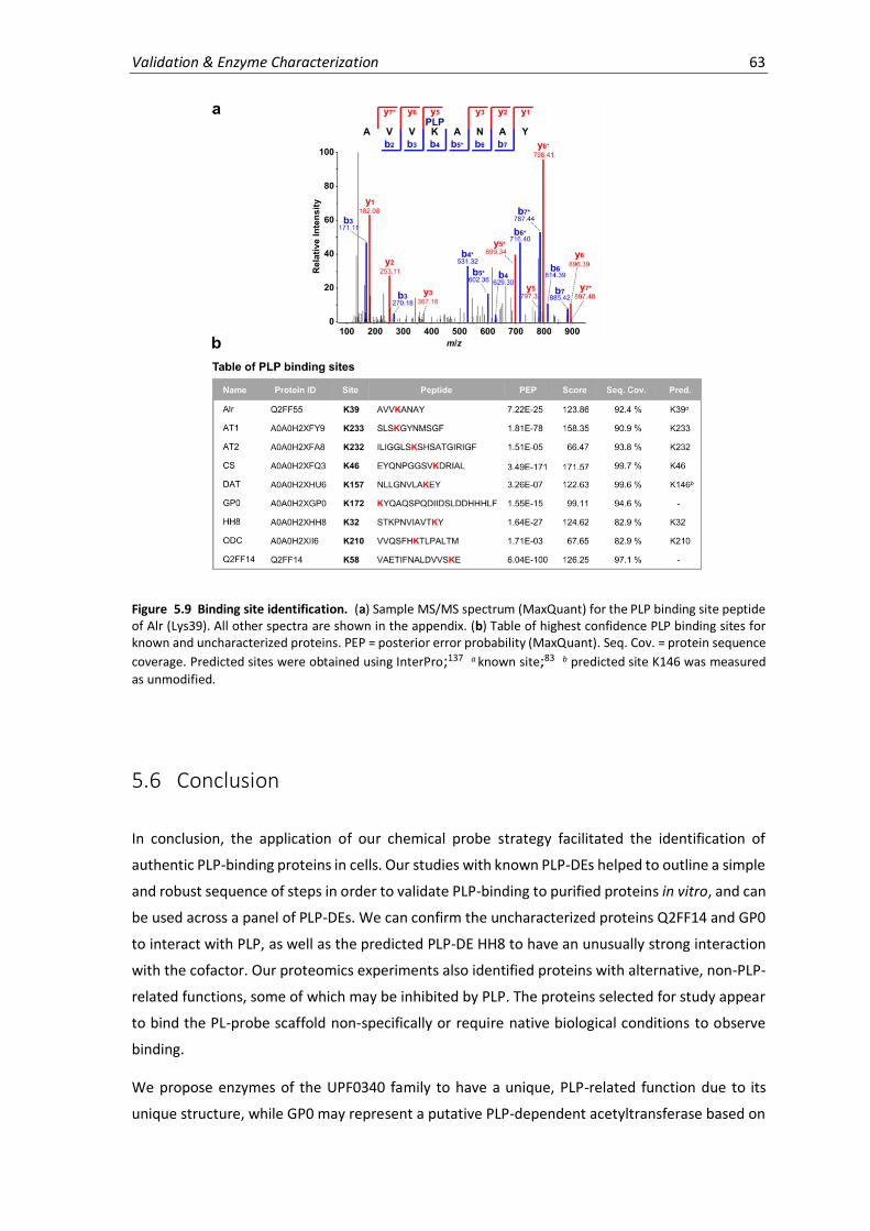
Validation & Enzyme Characterization 63
Figure 5.9 Binding site identification. (a) Sample MS/MS spectrum (MaxQuant) for the PLP binding site peptide of Alr (Lys39). All other spectra are shown in the appendix. (b) Table of highest confidence PLP binding sites for known and uncharacterized proteins. PEP = posterior error probability (MaxQuant). Seq. Cov. = protein sequence
coverage. Predicted sites were obtained using InterPro;137 a known site;83 b predicted site K146 was measured
as unmodified.
5.6 Conclusion
In conclusion, the application of our chemical probe strategy facilitated the identification of
authentic PLP-binding proteins in cells. Our studies with known PLP-DEs helped to outline a simple
and robust sequence of steps in order to validate PLP-binding to purified proteins in vitro, and can
be used across a panel of PLP-DEs. We can confirm the uncharacterized proteins Q2FF14 and GP0
to interact with PLP, as well as the predicted PLP-DE HH8 to have an unusually strong interaction
with the cofactor. Our proteomics experiments also identified proteins with alternative, non-PLP-
related functions, some of which may be inhibited by PLP. The proteins selected for study appear
to bind the PL-probe scaffold non-specifically or require native biological conditions to observe
binding.
We propose enzymes of the UPF0340 family to have a unique, PLP-related function due to its
unique structure, while GP0 may represent a putative PLP-dependent acetyltransferase based on

64 Chapter 5
its sequence similarity to Gcn5-related acetyltransferases. Our functional investigations are only
preliminary and require further experimental optimization to draw useful conclusions. Currently,
we do no detect any cellular repercussions of the Q2FF14 transposon mutation, suggesting its
function cannot be detected under the current experimental setup. The crystal structure of
Q2FF14 does not show significant similarity to other known structures and does not contain bound
PLP, which appears to bind Q2FF14 but renders it unstable. GP0 contains a domain that is highly
similar to Gcn5-related acetyltransferases, but it cannot be ascribed a corresponding function
based on a broad-substrate screen for GNAT activity. It is possible that the enzyme acetylates
other substrates not tested in our experiments or transfers alternative acyl groups. GP0 gene
disruption moderately affects amino acid biosynthetic pathways, though a precise function cannot
yet be pinpointed.
The detailed functional annotation of proteins requires significant experimental work and we have
applied several state-of-the-art techniques to understand the function of two uncharacterized
proteins. Additional experiments worthy of pursuit include activity-based metabolomics as a
native, broad substrate screen of enzyme activity. Furthermore, future experiments will also
investigate the uncharacterized proteins A0A0H2XIC2 and A0A0H2XIU9, identified in PL3-based
proteomic experiments as putative PLP-DEs. In conclusion, we have applied a method for
extrapolating knowledge from the proteome in order to annotate proteins as part of the PLP-DE
family without prior knowledge and provide a starting point for further investigations of UPF0340
proteins and PLP-dependent acetyltransferases.
6. Applications
With a functional proteomic strategy for studying PLP-DEs in hand, we next investigated diverse
applications of the method beyond the identification of novel PLP-DEs. These include drug
development-based applications, such as enzyme active site mapping and inhibitor off-target
screening, as well as expansion of the method for use in other organisms.
6.1 Comparison of PLP-DE active site constraints
The family of PLP-DEs has evolved different active site features which help to direct the specificity
of chemical reactions. Here, the architecture of the binding pocket influences the orientation of
substrate binding and inititally directs reactions through stereoelectronic effects. Furthermore,
active site residues control PLP-chemistry by providing the necessary functionality for reaction
and tuning the electrophilicity of the system. As a result of the active site variability, PLP-DEs are
able to accommodate modification of the PLP scaffold to varying extents. Previous studies into
the effects of structurally modifying PLP on cofactor viability have helped to define the core
functionality required for catalysis (see Chapter 2 Introduction). These include the 3-phenol, 4’-
aldehyde and 5’-phosphate, which are essential for binding to active sites and catalysis, through
hydrogen bonding and resonance.
Activity-based probe libraries have been useful for studying enzyme active sites and their
structural binding preferences in native biological environments. In order to profile structural
constraints within PLP-binding sites on a global level, we generated two additional PL probes
bearing increased steric bulk at the 2-position, including an extended alkyl chain (PL5) and an
aromatic group (PL6) (Scheme 6.1). The probes were synthesized according to the alkylation
strategy developed for PL4. Therefore, acetal-protected PL-intermediate 13 was reacted with
either TMS-protected 6-bromohexyne or 4-ethynyl-benzyl iodide, to generate intermediates 15
and 17, respectively. TMS-deprotection of the alkyne using potassium carbonate was followed by
acidic deprotection of the acetal and MOM groups of both intermediates in order to produce the
final probes PL5 and PL6.
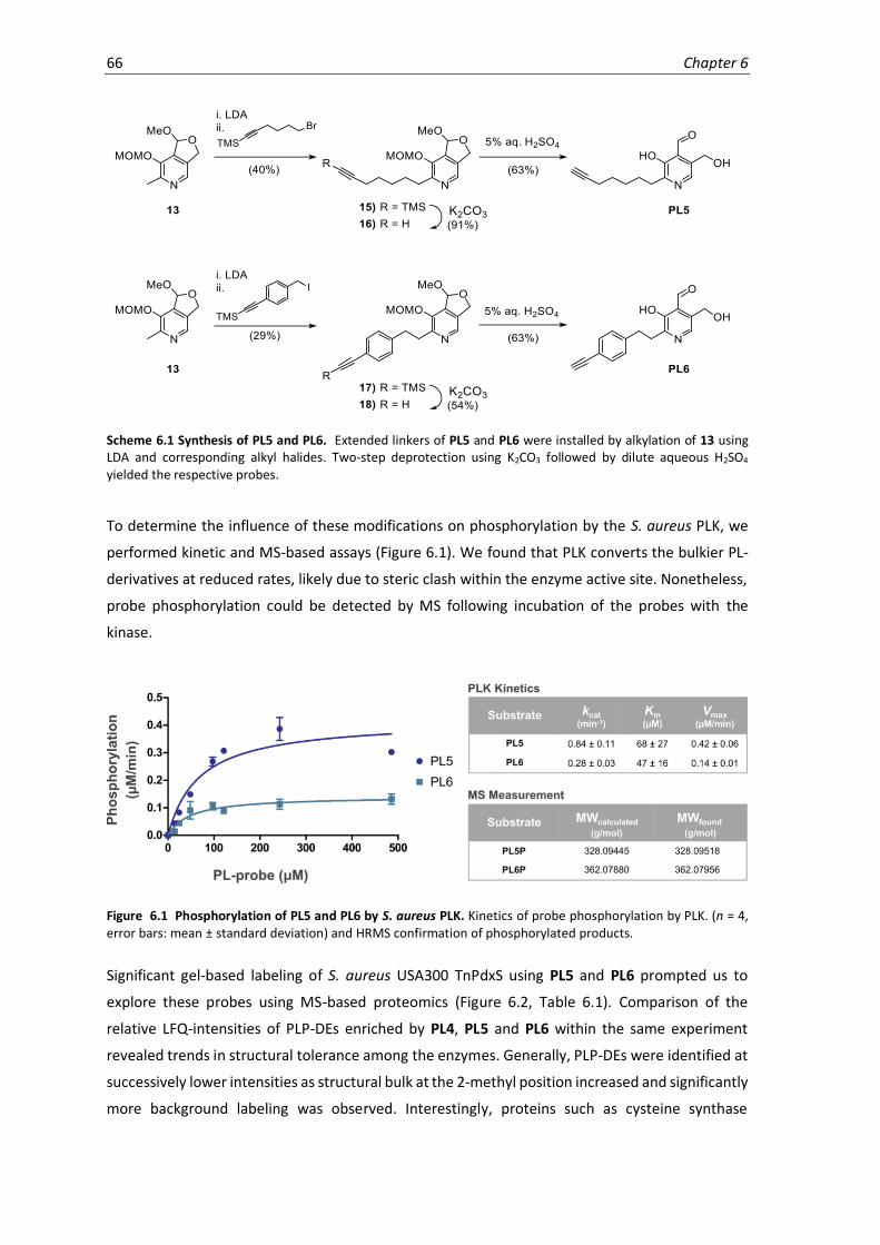
66 Chapter 6
Scheme 6.1 Synthesis of PL5 and PL6. Extended linkers of PL5 and PL6 were installed by alkylation of 13 using LDA and corresponding alkyl halides. Two-step deprotection using K2CO3 followed by dilute aqueous H2SO4 yielded the respective probes.
To determine the influence of these modifications on phosphorylation by the S. aureus PLK, we
performed kinetic and MS-based assays (Figure 6.1). We found that PLK converts the bulkier PL-
derivatives at reduced rates, likely due to steric clash within the enzyme active site. Nonetheless,
probe phosphorylation could be detected by MS following incubation of the probes with the
kinase.
Figure 6.1 Phosphorylation of PL5 and PL6 by S. aureus PLK. Kinetics of probe phosphorylation by PLK. (n = 4, error bars: mean ± standard deviation) and HRMS confirmation of phosphorylated products.
Significant gel-based labeling of S. aureus USA300 TnPdxS using PL5 and PL6 prompted us to
explore these probes using MS-based proteomics (Figure 6.2, Table 6.1). Comparison of the
relative LFQ-intensities of PLP-DEs enriched by PL4, PL5 and PL6 within the same experiment
revealed trends in structural tolerance among the enzymes. Generally, PLP-DEs were identified at
successively lower intensities as structural bulk at the 2-methyl position increased and significantly
more background labeling was observed. Interestingly, proteins such as cysteine synthase

Applications 67
(A0A0H2XFQ3), the class-I aminotransferase (A0A0H2XFY9),cystathionine gamma-synthase
(A0A0H2XFF8), and uncharacterized putative PLP-homeostasis protein (A0A0H2XHH8) all
tolerated the extended chain of PL5 well and could be identified with intensities in the range of
PL4-based labeling. Similarly, alanine racemase (Q2FF55), which was proven to accommodate
structural alterations of the cofactor, was also significantly enriched using PL5. Other proteins,
including threonine synthase (A0A0H2XH24), threonine deaminase (Q2FH01) and the
uncharacterized protein A0A0H2XGP0 preferred the aromatic bulk of PL6. A few proteins showed
higher intensities with PL5 (aminotransferases A0A0H2XFA8 and A0A0H2XIS2) or PL6 (putative
PLP-dependent acyltransferase A0A0H2XJW8 and putative aluminum resistance protein
A0A0H2XHV8) than with PL4. However, nearly half of the proteins were only strongly enriched
using PL4, including the uncharacterized protein Q2FF14. This data therefore reaffirms that
limited modification of the PLP-scaffold permits access to the largest number and variety of PLP-
DEs and underscores the evolution of this protein family towards binding a defined PLP structure.
Figure 6.2 Chemical proteomics of PL4, PL5 and PL6 in S. aureus USA300 TnPdxS. (a) Volcano plots showing enrichment of proteins with 100 µM PL4, PL5 and PL6 (n = 6 biological replicates). Known PLP-DEs are highlighted with blue dots and are listed in Table 6.1. (b) Fluorescence SDS-PAGE showing labeling of PL4, PL5 and PL6 (100 µM each) in S. aureus USA300 TnPdxS upon CuAAC to a trifunctional rhodamine-biotin-azide tag and avidin bead enrichment. (c) Heatmap representation of proteomic data showing normalized LFQ intensities of PLP-DEs and uncharacterized proteins identified.

68 Chapter 6
Table 6.1 Comparison of PLP-DEs significantly enriched using PL4, PL5 and PL6 in S. aureus USA300 TnPdxS. Corresponding volcano plots Figure 6.2a. PLP-DEs significantly enriched with respective probes are marked (+).
PL4
(sig.) PL5
(sig.) PL6
(sig.) Name Uniprot ID
1. + + Alanine racemase (EC 5.1.1.1) Q2FF55 2. + + Aminotransferase, class I (EC 2.6.1.-) A0A0H2XFY9
3. + + + Aminotransferase, class I (EC 2.6.1.-) A0A0H2XFA8 4. + Aminotransferase, class V A0A0H2XKI8 5. + + + Branched-chain-amino-acid aminotransferase (EC 2.6.1.42) A0A0H2XIS2 6. + + Cystathionine gamma-synthase (EC 4.4.1.8) A0A0H2XFF8 7. + + Cysteine desulfurase (EC 2.8.1.7) A0A0H2XHJ5
8. + + + Cysteine synthase (EC 2.5.1.47) A0A0H2XFQ3 9. + Cysteine synthase/cystathionine beta-synthase (EC 2.5.1.47) A0A0H2XG73
10. + D-alanine aminotransferase (EC 2.6.1.21) A0A0H2XHU6 11. + + + Diaminopimelate decarboxylase (DAP decarboxylase) (DAPDC) (EC 4.1.1.20) A0A0H2XFD9 12. + + + L-threonine dehydratase catabolic TdcB (EC 4.3.1.19) (Threonine deaminase) Q2FH01
13. + + + Orn/Lys/Arg decarboxylase (EC 4.1.1.18) A0A0H2XII6 14. + + Threonine synthase (EC 4.2.3.1) A0A0H2XH24 15. + + + Uncharacterized protein A0A0H2XGP0 16. + + + Uncharacterized protein A0A0H2XHH8 17. + UPF0340 protein SAUSA300_2068 Q2FF14
18. Probable glycine dehydrogenase (decarboxylating) subunit 2 (EC 1.4.4.2) Q2FGI7 19. + Putative aluminium resistance protein A0A0H2XHV8 20. + Putative pyridoxal phosphate-dependent acyltransferase A0A0H2XJW8 21. Aminotransferase A0A0H2XJK0 22. Glutamate-1-semialdehyde 2,1-aminomutase 2 (GSA 2) (EC 5.4.3.8) Q2FFN1
23. Ornithine aminotransferase (OAT) (EC 2.6.1.13) A0A0H2XIK4 24. Probable glycine dehydrogenase (decarboxylating) subunit 1 (EC 1.4.4.2) Q2FGI6 25. Serine hydroxymethyltransferase (SHMT) (Serine methylase) (EC 2.1.2.1) Q2FF15
This study acts as a proof-of-concept, illustrating how PLP-derivatives can be applied to actively
probe space within PLP binding sites on a proteome-wide level. Analyses of this type can also help
to define structure-activity and structure-selectivity relationships across a panel of PLP-DEs and
can provide useful information for their specific targeting by PLP-DE inhibitors.
6.2 In situ inhibitor specificity of the antibiotic D-cycloserine
The natural product D-cycloserine, produced by the bacterial strains Streptomyces garyphalus and
lavendulae, is known to inhibit the PLP-DE alanine racemase (Alr).76,138 Alr generates the essential
peptidoglycan precursor D-alanine by interconverting the two amino acid isoforms. The
peptidoglycan is a bacterial cell wall component, composed of a meshwork of long carbohydrate
chains (alternating N-acetylglucosamine and N-acetylmuramic acid) crosslinked by short peptide
bridges (D-ala, D-glu, L-lys), which provides cellular rigidity and support.139 Since Alr is universal to
bacteria but absent from most eukaryotes, it is considered a promising therapeutic target.
Although several bacteria (ex. E. coli, P. aeruginosa, S. aureus) contain two alanine racemase
isozymes – one that is constitutively expressed (Alr) and one that is inducible by the presence of
L-alanine (DadX)140,141 – Alr knockout appears to be lethal to bacteria in the absence of an external
source of D-alanine.142 Several mechanism-based inhibitors of Alr have been reported, including
the reactive alanine analogues β-fluoroalanine, β-chloroalanine, β-trifluoroalanine and alanine
phosphonate. However, lack of target specificity prevents their therapeutic use as antibacterials.
DCS is currently the only clinically marketed drug targeting Alr,31 though it is reserved as a second-
line defense against resistant tuberculosis due to severe neurological side-effects.143-145

Applications 69
Crystal structures of Alr from various species have revealed a highly conserved Alr active site
containing a wide entryway.146 The structure of the Geobacillus stearothermophilus Alr in complex
with DCS helped to unravel its mechanism of inhibition, whereby DCS first binds to the PLP
cofactor through transamination to generate the external aldimine (Figure 6.3a).147 Following a
1,3-prototropic shift, the resulting ketimine intermediate tautomerizes to form a stable isoxazole
ring which is covalently attached to the PLP cofactor (Figure 6.3b). The aromatic PLP-derivative
remains tightly bound within enzyme active sites, though extensive dialysis against PLP at neutral
pH can eventually re-activate the enzyme.148,149
DCS acts synergistically with other peptidoglycan inhibitors known to target Alr (ex. β-
chloroalanine)150 and is known to also inhibit other key enzymes in peptidoglycan biosynthesis.
These include D-ala-D-ala ligase (Ddl),151 which catalyzes the formation of D-alanine dipeptides,
and D-amino acid aminotransferase (DAT), a PLP-DE which converts D-alanine into D-
glutamate.148,149 DCS inhibits DAT in the same manner as for Alr, through the formation of a stable
covalent PLP-adduct which binds the enzyme tightly.148 Other PLP-DEs reported to be inhibited by
DCS include dialkylglycine decarboxylase,152 a unique enzyme which performs decarboxylative
transamination of its substrate, as well as serine palmitoyltransferase,153 in which DCS undergoes
decarboxylative ring-opening to modify and thereby inhibit the enzyme irreversibly.
Figure 6.3 Mechanism of D-cycloserine inhibition of Alr. (a) Mechanism of DCS inhibition by formation of a stable aromatic PLP adduct within enzyme pockets. (b) Crystal structure of Geobacillus stearothermophilus Alr bound to the DCS-PLP inhibitor complex (PDB: 1EPV).147 (c) Kinetics of alanine racemization (D- to L-Ala direction) by S. aureus Alr (0.01 µM) in the presence of DCS (n = 3, error: mean ± standard deviation).

70 Chapter 6
Since DCS is promiscuous in its inhibition of PLP-DEs, we attempted to evaluate the complete set
of DCS off-targets directly in S. aureus cells using our chemical proteomic method. We first
confirmed DCS inhibition in our hands to match literature reports,154,155 observing concentration-
dependent inhibition of DCS on Alr activity in vitro (Figure 6.3c) and a minimum inhibitory
concentration (MIC) of 0.5 mM in S. aureus. To screen for other PLP-DEs inhibited by DCS, we
designed a competitive proteomic experiment in which S. aureus USA300 TnPdxS was incubated
with DCS (10 or 1 mM) for 30 min prior to conducting our 2h labeling protocol with PL4. Analysis
of non-treated versus DCS-treated samples by volcano plot revealed a handful of enzymes
significantly influenced by DCS in a concentration-dependent manner (Figure 6.4, Table 6.2).
Among these was the known target DAT, as well as two previously undescribed PLP-DE targets:
ornithine/lysine/arginine decarboxylase (OKR-DC) and diaminopimelate decarboxylase (DAP-DC).
OKR-DC catalyzes the decarboxylation of amino acid substrates to generate putrescine, agmatine
and cadaverine. These amine building blocks are used for polyamine (spermine and spermidine)
biosynthesis, which are important for cell growth and bacterial virulence.156 DAP-DC
decarboxylates meso-diaminopimelate to the amino acid lysine, which is important for protein
and peptidoglycan biosynthesis. Lysine biosynthesis is recognized as an antibacterial target since
it is essential but absent in mammals.157
Interestingly, Alr labeling was not significantly outcompeted by DCS under these conditions. While
this is in disagreement with several previous in vitro studies,138,155 the known in vitro substrate
promiscuity of PLP-DEs may account for the observed reaction with DCS.158,159 In support of this
conclusion, recent metabolomics studies in mycobacteria have reported that Alr is only weakly
inhibited by DCS in a cellular context, while Ddl is the primary lethal target in cells.111,160 We were
surprised to significantly identify Ddl in our experiment, which, despite being a known target of
DCS, is not known to interact with PLP. Ddl catalyzes the formation of D-ala:D-ala dipeptides in
two steps, in which D-ala first binds to a high-affinity site (N-terminal) and is phosphorylated at its
carboxylate group using ATP.161 Then, a second D-ala residue binds at the lower-affinity site (C-
terminal) and undergoes nucleophilic attack on the phosphorylated D-ala to complete the
coupling reaction. DCS inhibits Ddl competitively and reversibly, and is believed to bind to the
high-affinity D-ala site.162,163 Based on earlier proteomic data (see Figure 4.3b, Table 4.1), we find
Ddl to be significantly enriched (medium-confidence), implying that it interacts with our PL4P
probe and may thus bind PLP by extension. Although a direct role of cofactor binding to Ddl
remains speculative, our data suggests that PLP and DCS compete for binding to Ddl. Interestingly,
Ddl has been reported to be under PLP-based transcriptional regulation by the MocR/GabR-type
protein DdlR in Bacillus brevis.29 Although the exact mechanism of this regulator are not fully
understood, the protein binds PLP as an internal aldimine and is able to form an external aldimine
with the D-ala-D-ala dipeptide.
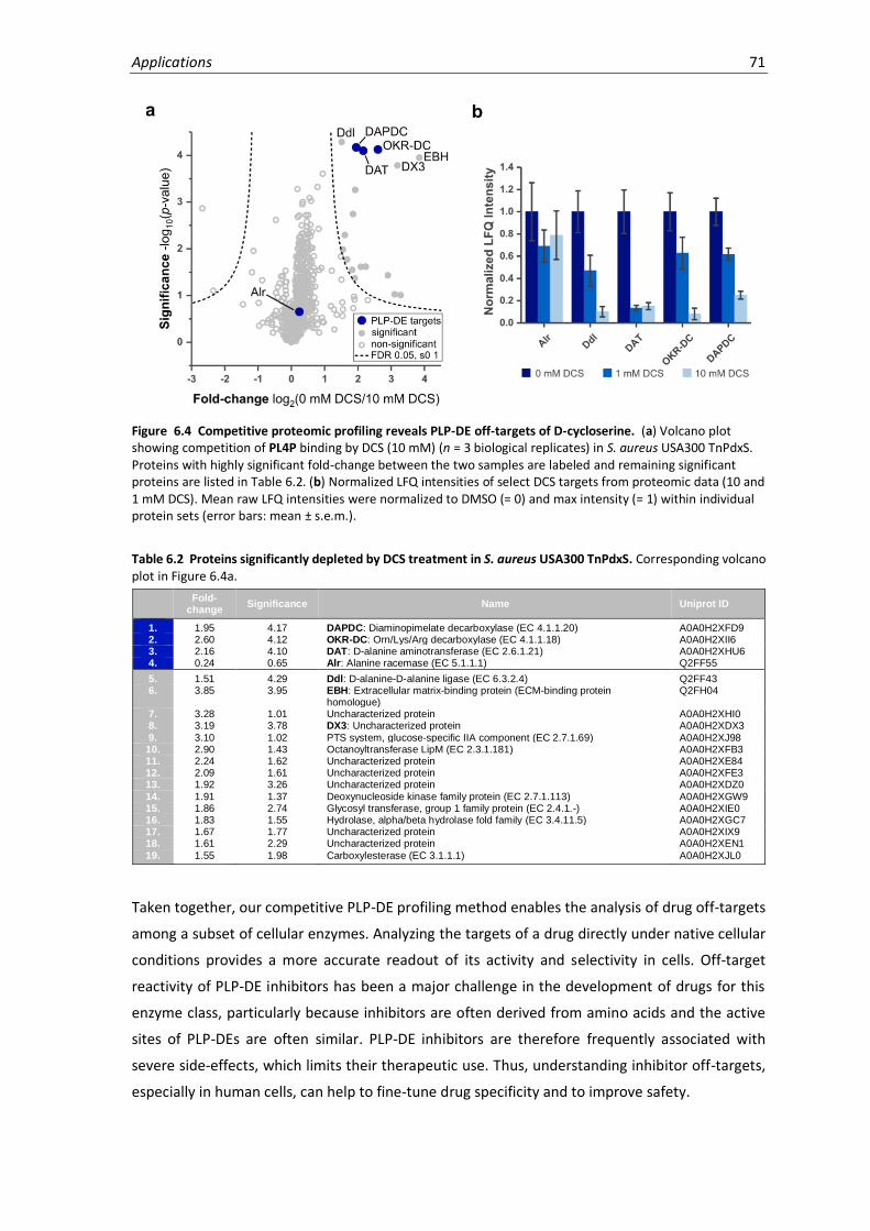
Applications 71
Figure 6.4 Competitive proteomic profiling reveals PLP-DE off-targets of D-cycloserine. (a) Volcano plot showing competition of PL4P binding by DCS (10 mM) (n = 3 biological replicates) in S. aureus USA300 TnPdxS. Proteins with highly significant fold-change between the two samples are labeled and remaining significant proteins are listed in Table 6.2. (b) Normalized LFQ intensities of select DCS targets from proteomic data (10 and 1 mM DCS). Mean raw LFQ intensities were normalized to DMSO (= 0) and max intensity (= 1) within individual protein sets (error bars: mean ± s.e.m.).
Table 6.2 Proteins significantly depleted by DCS treatment in S. aureus USA300 TnPdxS. Corresponding volcano plot in Figure 6.4a.
Fold-
change Significance Name Uniprot ID
1. 1.95 4.17 DAPDC: Diaminopimelate decarboxylase (EC 4.1.1.20) A0A0H2XFD9 2. 2.60 4.12 OKR-DC: Orn/Lys/Arg decarboxylase (EC 4.1.1.18) A0A0H2XII6 3. 2.16 4.10 DAT: D-alanine aminotransferase (EC 2.6.1.21) A0A0H2XHU6 4. 0.24 0.65 Alr: Alanine racemase (EC 5.1.1.1) Q2FF55
5. 1.51 4.29 Ddl: D-alanine-D-alanine ligase (EC 6.3.2.4) Q2FF43 6. 3.85 3.95 EBH: Extracellular matrix-binding protein (ECM-binding protein
homologue) Q2FH04
7. 3.28 1.01 Uncharacterized protein A0A0H2XHI0 8. 3.19 3.78 DX3: Uncharacterized protein A0A0H2XDX3
9. 3.10 1.02 PTS system, glucose-specific IIA component (EC 2.7.1.69) A0A0H2XJ98 10. 2.90 1.43 Octanoyltransferase LipM (EC 2.3.1.181) A0A0H2XFB3 11. 2.24 1.62 Uncharacterized protein A0A0H2XE84 12. 2.09 1.61 Uncharacterized protein A0A0H2XFE3 13. 1.92 3.26 Uncharacterized protein A0A0H2XDZ0
14. 1.91 1.37 Deoxynucleoside kinase family protein (EC 2.7.1.113) A0A0H2XGW9 15. 1.86 2.74 Glycosyl transferase, group 1 family protein (EC 2.4.1.-) A0A0H2XIE0 16. 1.83 1.55 Hydrolase, alpha/beta hydrolase fold family (EC 3.4.11.5) A0A0H2XGC7 17. 1.67 1.77 Uncharacterized protein A0A0H2XIX9 18. 1.61 2.29 Uncharacterized protein A0A0H2XEN1
19. 1.55 1.98 Carboxylesterase (EC 3.1.1.1) A0A0H2XJL0
Taken together, our competitive PLP-DE profiling method enables the analysis of drug off-targets
among a subset of cellular enzymes. Analyzing the targets of a drug directly under native cellular
conditions provides a more accurate readout of its activity and selectivity in cells. Off-target
reactivity of PLP-DE inhibitors has been a major challenge in the development of drugs for this
enzyme class, particularly because inhibitors are often derived from amino acids and the active
sites of PLP-DEs are often similar. PLP-DE inhibitors are therefore frequently associated with
severe side-effects, which limits their therapeutic use. Thus, understanding inhibitor off-targets,
especially in human cells, can help to fine-tune drug specificity and to improve safety.

72 Chapter 6
6.3 PLPome profiling in Gram-negative bacteria
Discovering new therapeutic strategies against pathogenic bacteria is an urgent and unmet need
in the face of antibiotic resistance. Gram-negative bacteria are particularly challenging to target
due to their insensitivity to many commonly used antibiotics. Moreover, a recently reported
subset of Gram-negative “ESKAPE” pathogens (Enterococcus faecium, Staphylococcus aureus,
Klebsiella pneumoniae, Acinetobacter baumanii, Pseudomonas aeruginosa, and Entero-
bacter species) cause the majority of nosocomial infections worldwide and are rapidly developing
multi-drug resistance.164 Drug-resistant bacteria are a major public health concern which is forcing
clinicians to fall back on less safe drug options (ex. colistin) due to lack of available alternatives.
Most antibiotics discovered during the golden era (1940-1960) stem from a limited set of
molecular scaffolds whose useable lifespan has been maximized through synthetic derivatization
(Figure 6.5). However, only few novel developments have arisen in recent years (since 2000).
Strategies currently being pursued to aid antibiotic drug discovery and development include
target-based discovery (high throughput screening, rational drug design), searching for novel
molecular scaffolds by exploring untapped natural product resources and repurposing synthetic
libraries for antibiotics.165,166 However, these platforms are hindered by multiple challenges
associated with antibiotic development, such as cell permeability and unpredictable drug
properties, high toxicity, and low return on cost.165,167
Figure 6.5 Timeline of antibiotic discoveries. Most modern antibiotics originated during the “Golden Era” of antibiotic discovery (1940-1960). This was proceeded by the rise of antibiotic medicinal chemistry (1970-1980), ie. synthetic derivatization to maximize the lifespan of antibiotics against the threat of bacterial resistance. Few drug candidates have been found since, resulting in a discovery void. Figure adapted from G.D. Wright.168
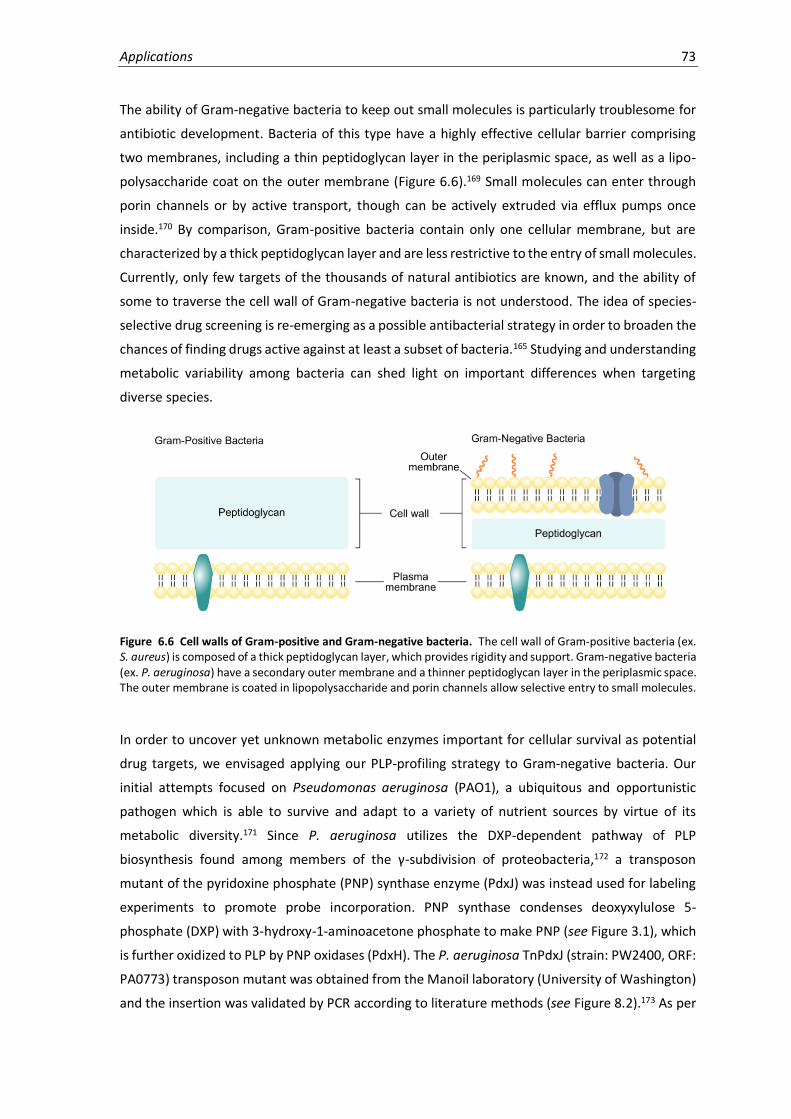
Applications 73
The ability of Gram-negative bacteria to keep out small molecules is particularly troublesome for
antibiotic development. Bacteria of this type have a highly effective cellular barrier comprising
two membranes, including a thin peptidoglycan layer in the periplasmic space, as well as a lipo-
polysaccharide coat on the outer membrane (Figure 6.6).169 Small molecules can enter through
porin channels or by active transport, though can be actively extruded via efflux pumps once
inside.170 By comparison, Gram-positive bacteria contain only one cellular membrane, but are
characterized by a thick peptidoglycan layer and are less restrictive to the entry of small molecules.
Currently, only few targets of the thousands of natural antibiotics are known, and the ability of
some to traverse the cell wall of Gram-negative bacteria is not understood. The idea of species-
selective drug screening is re-emerging as a possible antibacterial strategy in order to broaden the
chances of finding drugs active against at least a subset of bacteria.165 Studying and understanding
metabolic variability among bacteria can shed light on important differences when targeting
diverse species.
Figure 6.6 Cell walls of Gram-positive and Gram-negative bacteria. The cell wall of Gram-positive bacteria (ex. S. aureus) is composed of a thick peptidoglycan layer, which provides rigidity and support. Gram-negative bacteria (ex. P. aeruginosa) have a secondary outer membrane and a thinner peptidoglycan layer in the periplasmic space. The outer membrane is coated in lipopolysaccharide and porin channels allow selective entry to small molecules.
In order to uncover yet unknown metabolic enzymes important for cellular survival as potential
drug targets, we envisaged applying our PLP-profiling strategy to Gram-negative bacteria. Our
initial attempts focused on Pseudomonas aeruginosa (PAO1), a ubiquitous and opportunistic
pathogen which is able to survive and adapt to a variety of nutrient sources by virtue of its
metabolic diversity.171 Since P. aeruginosa utilizes the DXP-dependent pathway of PLP
biosynthesis found among members of the γ-subdivision of proteobacteria,172 a transposon
mutant of the pyridoxine phosphate (PNP) synthase enzyme (PdxJ) was instead used for labeling
experiments to promote probe incorporation. PNP synthase condenses deoxyxylulose 5-
phosphate (DXP) with 3-hydroxy-1-aminoacetone phosphate to make PNP (see Figure 3.1), which
is further oxidized to PLP by PNP oxidases (PdxH). The P. aeruginosa TnPdxJ (strain: PW2400, ORF:
PA0773) transposon mutant was obtained from the Manoil laboratory (University of Washington)
and the insertion was validated by PCR according to literature methods (see Figure 8.2).173 As per
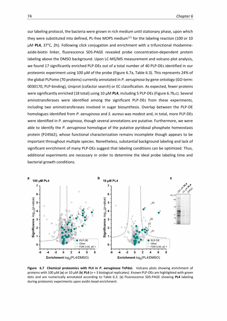
74 Chapter 6
our labeling protocol, the bacteria were grown in rich medium until stationary phase, upon which
they were substituted into defined, PL-free MOPS medium171 for the labeling reaction (100 or 10
µM PL4, 37°C, 2h). Following click conjugation and enrichment with a trifunctional rhodamine-
azide-biotin linker, fluorescence SDS-PAGE revealed probe concentration-dependent protein
labeling above the DMSO background. Upon LC-MS/MS measurement and volcano plot analysis,
we found 17 significantly enriched PLP-DEs out of a total number of 40 PLP-DEs identified in our
proteomic experiment using 100 µM of the probe (Figure 6.7a, Table 6.3). This represents 24% of
the global PLPome (70 proteins) currently annotated in P. aeruginosa by gene ontology (GO-term:
0030170, PLP-binding), Uniprot (cofactor search) or EC classification. As expected, fewer proteins
were significantly enriched (18 total) using 10 µM PL4, including 5 PLP-DEs (Figure 6.7b,c). Several
aminotransferases were identified among the significant PLP-DEs from these experiments,
including two aminotransferases involved in sugar biosynthesis. Overlap between the PLP-DE
homologues identified from P. aeruginosa and S. aureus was modest and, in total, more PLP-DEs
were identified in P. aeruginosa, though several annotations are putative. Furthermore, we were
able to identify the P. aeruginosa homologue of the putative pyridoxal phosphate homeostasis
protein (P24562), whose functional characterization remains incomplete though appears to be
important throughout multiple species. Nonetheless, substantial background labeling and lack of
significant enrichment of many PLP-DEs suggest that labeling conditions can be optimized. Thus,
additional experiments are necessary in order to determine the ideal probe labeling time and
bacterial growth conditions.
Figure 6.7 Chemical proteomics with PL4 in P. aeruginosa TnPdxJ. Volcano plots showing enrichment of proteins with 100 µM (a) or 10 µM (b) PL4 (n = 3 biological replicates). Known PLP-DEs are highlighted with green dots and are numerically annotated according to Table 6.3. (c) Fluorescence SDS-PAGE showing PL4 labeling during proteomic experiments upon avidin bead enrichment.

Applications 75
Table 6.3 Proteins significantly enriched using 100 µM PL4 in P. aeruginosa TnPdxJ cells. Corresponding volcano plot in Figure 6.7a. Green = PLP-DEs; Grey = other significant proteins (cut-offs: enrichment > 3, significance > 3).
No. Fold-
change Significance Name Uniprot ID
1. 7.25 3.86 Probable aminotransferase Q9HV83 2. 5.51 2.91 UDP-4-amino-4-deoxy-L-arabinose aminotransferase Q9HY65 3. 5.51 3.25 Tryptophan synthase beta chain (EC 4.2.1.20) P07345 4. 4.99 4.07 Biosynthetic arginine decarboxylase (ADC) (EC 4.1.1.19) Q9HUX1 5.
4.98 3.74
L-seryl-tRNA(Sec) selenium transferase (EC 2.9.1.1) (Selenocysteine
synthase) Q9HV01 6. 4.92 3.49 Pyridoxal phosphate homeostasis protein (PLP homeostasis protein) P24562 7. 4.83 2.26 Probable aminotransferase Q9HXY0 8. 3.27 3.13 Branched-chain-amino-acid aminotransferase (BCAT) (EC 2.6.1.42) O86428 9. 2.80 1.87 Aminotransferase (EC 2.6.1.-) Q9HV76
10. 2.13 2.33 UDP-2-acetamido-2-deoxy-3-oxo-D-glucuronate aminotransferase (UDP-3-oxo-D-GlcNAcA aminotransferase) (EC 2.6.1.98)
Q9HZ76
11. 1.99 1.76 Probable cystathionine gamma-lyase Q9I6A0 12. 1.99 1.58 O-succinylhomoserine sulfhydrylase (OSH sulfhydrylase) (EC 2.5.1.-) P55218 13. 1.69 3.25 Aromatic-amino-acid aminotransferase (EC 2.6.1.57) P43336
14. 1.40 1.11 Alanine racemase, catabolic (EC 5.1.1.1) Q9HTQ2 15. 1.35 2.64 Uncharacterized protein Q9HZI4 16. 1.34 1.77 Lysine-specific pyridoxal 5'-phosphate-dependent carboxylase, LdcA Q9I2S7 17. 1.02 2.43 Ornithine decarboxylase Q9HVQ3
18. 7.42 3.41 Deoxyuridine 5'-triphosphate nucleotidohydrolase (dUTPase) (EC 3.6.1.23) Q9HTN3
19. 7.35 3.57 Uncharacterized protein Q9HX12 20. 6.60 4.42 Probable oxidoreductase Q9I2R2 21. 6.32 3.29 Transcriptional regulatory protein AlgP (Alginate regulatory protein AlgR3) P15276 22. 6.15 4.27 Nitrogen regulatory protein P-II 2 Q9HTR6 23. 5.74 5.03 UDP-N-acetylmuramoyl-tripeptide--D-alanyl-D-alanine ligase (EC 6.3.2.10) Q9HVZ7
24. 5.29 3.64 Aerotaxis transducer Aer2 Q9I6V6 25. 5.17 3.36 Transcriptional repressor NrdR Q9HWX1 26. 5.04 3.25 Uncharacterized protein Q9I2V2 27. 4.83 4.06 Probable short-chain dehydrogenase Q9I2R7 28. 4.54 3.90 Probable pyrophosphohydrolase Q9HW06 29. 4.49 4.22 Acylphosphatase (EC 3.6.1.7) (Acylphosphate phosphohydrolase) Q9I504
30. 4.43 4.55 Periplasmic serine endoprotease DegP-like (EC 3.4.21.107) G3XD20 31. 4.19 4.33 Uncharacterized protein Q9HVV5 32. 4.11 3.01 Uncharacterized protein Q9HTY8 33. 3.97 6.31 Glutathione peroxidase Q9I5A2 34. 3.72 3.95 Ribonucleoside-diphosphate reductase (EC 1.17.4.1) Q9I4I1
35. 3.43 3.47 Uncharacterized protein Q9I6Z1 36. 3.17 3.17 Probable FAD-dependent monooxygenase Q9HWJ1
To test whether probe uptake by P. aeruginosa was hampered by its effective cellular barrier,
labeling experiments were also conducted in bacterial lysate using the phosphorylated probe. P.
aeruginosa lysate was thus incubated with 10 or 1 µM of PL4P (45 min, 4°C), upon which the
samples were reduced with NaBH4, conjugated to the trifunctional linker, enriched and analyzed
by LC-MS/MS (Figure 6.8a-c, Table 6.4). In this experiment, 19 PLP-DEs out of 49 total were
significantly identified, representing 27% of the annotated P. aeruginosa PLPome. As a whole,
similar PLP-DEs were identified in comparison to labeling in live cells, with some observable shifts
in significance. Notably, the PLP homeostasis protein distinguishes itself from the background in
terms of significance, implying considerable binding to the probe over other proteins. This is in
agreement with its presumed function of regulating cellular PLP levels by scavenging the reactive
aldehyde or passing the cofactor to other proteins. While optimizations are required to improve
labeling over background (labeling time, number of replicates) or filtering as detailed in Chapter 4
may be necessary, this work presents a proof-of-concept indicating that PL4P-labeling can be
performed in bacterial lysates. This strategy thus represents a promising alternative in case a PLP-
biosynthesis mutant strain is not available, or if PL-probe uptake limits the use of the labeling
method in live cells. Establishing the method for use in Gram-negative bacteria enables
comparative analysis of different strains, and may serve to identify essential metabolic enzymes
common to certain bacterial subsets as putative drug targets.

76 Chapter 6
Figure 6.8 Chemical proteomics with PL4P in P. aeruginosa lysate. Volcano plots showing enrichment of proteins with (a) 10 µM or (b) 1 µM PL4P (n = 3 biological replicates). Known PLP-DEs are highlighted with green dots and are numerically annotated according to Table 6.4. (c) Fluorescence SDS-PAGE showing PL4P labeling in lysate during proteomic experiments upon avidin bead enrichment.
Table 6.4 Proteins significantly enriched using 10 µM PL4P in P. aeruginosa lysate. Corresponding volcano plot in Figure 6.8a. Green = PLP-DEs; Grey = other significant proteins (cut-offs: enrichment > 3.5, significance > 3.5).
No. Fold-
change Significance Name
Uniprot ID
1. 7.80 3.87 Pyridoxal phosphate homeostasis protein (PLP homeostasis protein) P24562 2. 3.71 4.41 Probable aminotransferase Q9HXY0 3. 3.42 5.01 Tryptophan synthase beta chain (EC 4.2.1.20) P07345 4. 3.02 4.51 Probable transcriptional regulator Q9HZV0 5. 2.73 4.81 Probable aminotransferase Q9HV83 6. 2.71 4.41 L-seryl-tRNA(Sec) selenium transferase (EC 2.9.1.1) (Selenocysteine synthase) Q9HV01
7. 2.58 1.28 Probable aminotransferase Q9I371 8. 2.29 4.20 UDP-2-acetamido-2-deoxy-3-oxo-D-glucuronate aminotransferase (UDP-3-oxo-
D-GlcNAcA aminotransferase) (EC 2.6.1.98) Q9HZ76
9. 1.99 4.37 Serine hydroxymethyltransferase 3 (SHMT 3) (EC 2.1.2.1) Q9HVI7 10. 1.90 2.53 Serine hydroxymethyltransferase 2 (SHMT 2) (EC 2.1.2.1) Q9I138
11. 1.65 3.83 Biosynthetic arginine decarboxylase (ADC) (EC 4.1.1.19) Q9HUX1 12. 1.62 4.64 Ornithine decarboxylase Q9HVQ3 13. 1.26 3.35 Uncharacterized protein Q9HZI4 14. 1.20 2.98 UDP-4-amino-4-deoxy-L-arabinose--oxoglutarate aminotransferase (EC
2.6.1.87) Q9HY65
15. 1.17 3.45 Probable transcriptional regulator Q9I283 16. 1.13 3.66 Probable aminotransferase Q9I015
17. 1.04 3.57 Aspartate aminotransferase (AspAT) (EC 2.6.1.1) P72173 18. 0.88 3.58 Probable cystathionine gamma-lyase Q9I6A0 19. 0.76 3.51 Aminotransferase (EC 2.6.1.-) Q9HV76
20. 5.66 3.98 Potential phenazine-modifying enzyme Q9I781 21. 4.37 4.20 Uncharacterized protein Q9I585
22. 4.36 4.59 Signal recognition particle receptor FtsY (SRP receptor) Q9I6C1 23. 4.33 4.16 Probable short-chain dehydrogenase Q9I3P2 24. 4.28 5.49 Acetolactate synthase isozyme III small subunit Q9HVA1 25. 3.97 4.59 Uncharacterized protein Q9HX12 26. 3.82 4.63 Uncharacterized protein Q9I2B4
27. 3.82 4.14 Nonspecific ribonucleoside hydrolase Q9I6Y9 28. 3.79 3.54 Phosphopantetheine adenylyltransferase (EC 2.7.7.3) (Dephospho-CoA
pyrophosphorylase) (Pantetheine-phosphate adenylyltransferase) (PPAT) Q9I6D1
29. 3.71 3.67 NAD-dependent protein deacylase 1 (EC 3.5.1.-) (Regulatory protein SIR2 homologue 1)
Q9I4L0
30. 3.71 5.14 Uncharacterized protein Q9HVT3 31. 3.67 3.93 3-oxoacyl-[acyl-carrier-protein] reductase FabG (EC 1.1.1.100) (3-ketoacyl-acyl
carrier protein reductase) O54438
32. 3.66 3.69 Nucleoid-associated protein PA1533 Q9I3I0

Applications 77
6.4 Profiling the Human PLPome
Humans rely on the dietary uptake of vitamin B6 (PN, PM and PL), for which major sources include
whole grain and nuts, vegetables, bananas, milk and certain types of meat. While non-
phosphorylated B6 vitamers are directly absorbed in the intestine by passive diffusion, the
phosphorylated forms first require removal of the phosphate group by an intestinal alkaline
phosphatase in order to pass cell membranes.174 On entering cells, the human pyridoxal kinase
(PDXK) phosphorylates the PL-derivatives to generate the bioactive PLP cofactor. PLP-DEs use the
cofactor throughout essential metabolic processes, such as amino acid and aminosugar
metabolism, as well as cofactor, nucleotide and lipid biosynthesis. Additionally, PLP-DEs are
important for the production of biogenic amines in humans, such as the neurotransmitters of the
central nervous system (CNS). These include histamine (histidine decarboxylase), serotonin
(aromatic L-amino acid decarboxylase), GABA (glutamate decarboxylase) and dopamine (L-3,4-
dihydroxyphenylalanine (DOPA) decarboxylase). Vitamin B6 deficiency, due to diet, malabsorption
(celiac disease), increased excretion (renal dialysis) or enzymatic mutations (ex. antiquitin), can
cause neurological symptoms such as irritability and seizures.175 On the other hand, vitamin B6
abuse, i.e. excessive uptake of PN (9.6 g/day, 3 years), has resulted in modest to severe
neurological and dermatological problems which were reversible upon discontinuation.176,177
Alterations in vitamin B6 metabolism are associated with multiple human diseases and PLP-DE
inhibitors are clinically prescribed for treating CNS neutrotransmitter imbalances as well as
cancer.31 For example, the PLP-DE GABA aminotransferase controls the levels of γ-aminobutyric
acid (GABA), the major inhibitory neurotransmitter of the CNS, by catalyzing its transamination to
succinic semialdehyde. Since low levels of GABA can cause convulsions and seizures,178 which are
symptoms of epilepsy, Parkinson’s, Huntington’s and Alzheimer’s diseases, the GABA
aminotransferase inhibitor vigabatrin is commonly prescribed.179,180 However, vigabatrin is
associated with severe irreversible side effects, such as loss of peripheral vision, the molecular
mechanisms of which are unclear.181 Similarly, the PLP-DE DOPA decarboxylase controls
production of the two major neurotransmitters dopamine and serotonin through the
decarboxylation of DOPA and L-5-HTP substrates, respectively. In the treatment of Parkinson’s
disease, involving the progressive loss of dopamine-producing cells in the midbrain, exogenous
DOPA is co-administered with the DOPA decarboxylase inhibitor carbidopa.31 This drug helps to
increase DOPA levels at the site by preventing premature metabolism in the body prior to reaching
the CNS, where it cannot pass the blood-brain barrier. Unfortunately, as with many hydrazine-
based PLP-DE inhibitors, carbidopa also suffers from poor selectivity. Finally, though the role of
vitamin B6 in cancer is complex and unclear, PLP-dependent pathways appear to be affected by
the metabolic rewiring of rapidly proliferating cells.182 Cancer patients often have decreased levels
of circulating B6 vitamers,183 due to an increased demand for PLP-dependent metabolism for

78 Chapter 6
sustaining the anabolic needs of highly proliferating cells and for their adaption to adverse
conditions during tumour progression.184 For example, serine hydroxymethyltransferase has a
central role in one-carbon metabolism and is thus required for increased DNA synthesis in rapidly
proliferating cells.185,186 Drugs that inhibit SHMT, such as mimosine, are therefore clinically
relevant for the treatment of tumours.187 Contrastingly, in some distinct cases, increased levels of
circulating vitamin B6 also correlated with reduced incidence of certain neoplasms.188,189 Here, high
intratumoural expression of PDXK was prognostic of improved disease outcome, perhaps due to
increased activity of the antitumour immune response.190
Genomic comparison of eukaryotic and prokaryotic PLPomes has revealed that the proportion of
PLP-DE-encoding genes decreases with increasing size and complexity of the genome.4 These
observations are characteristic of enzymes involved in basic metabolic pathways rather than
specialized regulatory functions. Furthermore, this analysis also showed that a substantial fraction
of identified PLP-DE genes (nearly 20%) remain functionally unclassified or only tentatively
classified. Uncovering and functionally characterizing new roles for PLP in the cell using PLP-
profiling tools can help to uncover novel metabolic pathways and identify new therapeutic targets.
Furthermore, chemical probes can be used to analyze PLP-DE off-targets of known inhibitors to
improve therapeutic safety, and to compare and understand PLP-dependent metabolic rewiring
in cancer cells for identification of biomarkers or prognostic factors.
In order to access a range of useful experimental applications using our PLPome profiling method,
we attempted to establish labeling in human cells. We reasoned that human cells would lend
themselves well to probe uptake since they depend on vitamin B6 absorption from the
environment and do not possess PLP biosynthetic machinery. HeLa cells were therefore cultured
in standard medium and subsequently labeled using 100 µM PL4 in PBS. Initial gel-based analysis
revealed significant fluorescence labeling and encouraged us to examine PL4 labeling on a
proteomics level. Samples of PL4-labeled HeLa cells were thus lysed and reduced using NaBH4,
followed by click conjugation to a trifunctional rhodamine-azide-biotin linker and enrichment
using avidin beads. LC-MS/MS measurement of samples, data analysis and volcano plot
representation of the fold enrichment versus significance revealed no distinct enrichment of PLP-
DEs (Figure 6.9, Table 6.5). Only five PLP-DEs were identified throughout the experiment, including
three that were significantly enriched, but dispersed within a cloud of background. Among the
significantly identified PLP-DEs are ornithine aminotransferase, pyridoxal kinase and aspartate
aminotransferase.
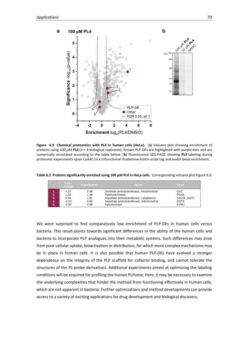
Applications 79
Figure 6.9 Chemical proteomics with PL4 in human cells (HeLa). (a) Volcano plot showing enrichment of proteins using 100 µM PL4 (n = 3 biological replicates). Known PLP-DEs are highlighted with purple dots and are numerically annotated according to the table below. (b) Fluorescence SDS-PAGE showing PL4 labeling during proteomic experiments upon CuAAC to a trifunctional rhodamine-biotin-azide tag and avidin bead enrichment.
Table 6.5 Proteins significantly enriched using 100 µM PL4 in HeLa cells. Corresponding volcano plot Figure 6.9.
Fold-
change Significance Name Gene
1. 4.22 2.98 Ornithine aminotransferase, mitochondrial OAT 2. 1.03 1.48 Pyridoxal kinase PDXK
3. 0.93 1.81 Aspartate aminotransferase, cytoplasmic GIG18, GOT1 4. -0.33 0.86 Aspartate aminotransferase, mitochondrial GOT2 5. -0.34 0.48 Kynureninase KYNU
We were surprised to find comparatively low enrichment of PLP-DEs in human cells versus
bacteria. This result points towards significant differences in the ability of the human cells and
bacteria to incorporate PLP analogues into their metabolic systems. Such differences may arise
from poor cellular uptake, bioactivation or distribution, for which more complex mechanisms may
be in place in human cells. It is also possible that human PLP-DEs have evolved a stronger
dependence on the integrity of the PLP scaffold for cofactor binding, and cannot tolerate the
structures of the PL-probe derivatives. Additional experiments aimed at optimizing the labeling
conditions will be required for profiling the human PLPome. Here, it may be necessary to examine
the underlying complexities that hinder the method from functioning effectively in human cells,
which are not apparent in bacteria. Further optimizations and method developments can provide
access to a variety of exciting applications for drug development and biological discovery.

80 Chapter 6

7. Conclusion
This thesis describes the development of a chemical proteomic strategy for the study of PLP-DEs
in live cells for diverse applications. We present a small library of probes as novel tools for profiling
the family of PLP-DEs, which are easily accessible, compatible with biological systems and
adaptable to suit diverse chemical biology needs.
The synthesis of a small PL-probe library involved core protecting group systems of the PL-scaffold
and outlines strategies for the facile derivatization of the 2’ position using diverse alkyl halides or
nucleophilic groups. Importantly, the probes could be activated to their phosphorylated forms by
S. aureus pyridoxal kinase and are also accommodated by diverse PLP-DEs. As exemplified by Alr,
the additional bulk of the probes did not hinder catalytic activity, which could later be rationalized
through crystallographic evidence. Our results indicate that multiple enzymes show a similar
phenomenon, since two cofactor probes (PL1 and PL3) were able to globally substitute for
essential PLP-processes in order to support bacterial growth. Considering the evolution of PLP-
DEs towards binding the specific cofactor structure, the ability of tool compounds to integrate into
such diverse catalytic facets is astounding. Although the larger ethylene linker of PL4 was not
tolerated as a full cellular substitute of cofactor activity, it nonetheless appears to bind a multitude
of proteins. Using additional probes, some structural preferences could be revealed among
PLP-DEs, implying that different probe structures may be able to selectively target parts of the
PLPome. A challenge here is maintaining the ability to undergo phosphorylation by PLK, which
decreases as bulk of the substituents increases.
Our proteomic strategy capitalizes on the unique mode of cofactor binding to PLP-DEs in the form
of an internal aldimine, which can be fixed by reductive amination. This circumvents the need for
cumbersome and inherently non-specific photocrosslinkers or electrophilic traps, thus allowing
for a more diversified and selective set of probes for reporting on natural PLP-binding events.
Several binding controls (PL2 and unphosphorylated probes) in analytical and proteomic
experiments further highlight the specificity and authenticity of probe binding in the complex
environment of the living cell. Labeling in bacteria directly cultured on the probes (PL1 and PL3)
as a PL supplement provided excellent selectivity and PLPome coverage. Alternatively, PL4
identified many proteins upon a short, in situ labeling treatment (2h, 37°C), and results could be
82 Chapter 7
strengthened using extended MS-based analysis. A two-step filtering strategy based on LFQ-
intensity profile searches across multiple probe concentrations was used to cluster confidence
and probe binding behaviour. We thus demonstrate the effectiveness with which supplementary
analyses can help to distinguish specific and low-affinity binding from background and present
general strategies for extracting additional information from proteomic datasets in order to gain
deeper experimental insights.
Using our probes, we cumulatively labeled 73% of the current S. aureus PLPome and identified
several more putative PLP binding events. This enabled the annotation of uncharacterized
proteins Q2FF14 and GP0 as novel PLP-DEs, as well as IU9 and IC2 as putative PLP-DEs which
require additional validation. A multifaceted exploration of the uncharacterized proteins Q2FF14
and GP0 incorporated in silico, in vitro and in vivo approaches. These studies provided preliminary
functional insights into the PLP-related roles of these proteins, though necessitate further
investigation. We anticipate that our tool compounds provide a resource for unraveling new PLP
roles or yet undiscovered interactions in a cellular context. Additional applications of the method
address challenges of drug development for PLP-DEs, several of which are recognized as drug
targets, but are difficult to target selectively. A competitive proteomic screening platform was
used to assess off-target reactivity of the clinical PLP-DE inhibitor DCS, thereby identifying two
decarboxylases as potential targets while suggesting that the widely accepted target Alr is not
significantly inhibited in vivo in S. aureus. Learning about the off-target mechanisms of PLP-DE
inhibitors can help to pinpoint ways of improving current therapies and can be applied to other
known drugs that are currently not approved due to safety concerns. Furthermore, in situ SAR
profiling using a panel of structurally diverse probes can serve as a starting point for understanding
active site geometries of different PLP-DEs and their selectivity.
A current limitation of our PLP-DE detection strategy is the requirement to promote probe uptake
using a bacterial transposon mutant devoid of de novo PLP biosynthesis. However, this is allayed
by the availability of many bacterial transposon mutant libraries, and the future prospect of
labeling in bacterial lysates. In human cells, the lack of PLP biosynthetic machinery and
dependence on PL salvage from the environment make it a suitable system for our labeling
method, but further investigations are required to understand the underlying mechanisms
currently preventing the method from functioning effectively. In addition, structural modifications
imparted to the cofactor limit our access to enzymes with restricted binding sites, as evidenced
by the inability of PL4 to promote cellular growth. This challenge has been addressed through the
use of a diversified set of probes, which has broadened proteome coverage, but nonetheless does
not yet permit complete access to the PLPome.
Profiling the family of PLP-DEs provides an important foundation for the study of diverse topics in
the field, including evolutionary relationships of PLP-DEs, cellular PLP homeostasis mechanisms,
alternative roles of PLP and comparison of different organisms, cell types or cell states. Thus,

Conclusion 83
future investigations will aim to expand the method for application in different organisms. The
ability to simultaneously monitor multiple PLP-DEs in their natural context and to identify novel
family members without prior information has the potential to fill major gaps in knowledge.
Nonetheless, full functional characterization of PLP-DEs upon their identification is difficult and
requires combined biochemical, structural and bioinformatics approaches. Although significant
literature precedence and flowcharts107 for the functional characterization of PLP-DEs are useful
for providing general insights, extensive experimental work is required to account for the diverse
catalytic activities. Global cellular profiling techniques, such as proteomics and metabolomics, can
further be used to understand cellular implications of PLP-DE transposon mutants in order to
unravel their functions and activity-based metabolomics profiling can also help to identify unusual
enzyme substrates.
By recent estimates, 30-40% of genes in all genomes remain unannotated or only putatively
assigned a function and, of these, a significant fraction is predicted to encode functionally relevant
proteins due to their phylogenetic conservation.104-106 Genomics-based bioinformatic analysis can
help to putatively annotate uncharacterized proteins, but can sometimes fail to identify protein
similarity or results in misannotation due to low sequence identity. Current methods for directly
detecting PLP binding in proteomes are either not quantitative or associated with many technical
challenges, highlighting the need for supplementary chemical probe-based enrichment strategies
for identifying PLP-binding in complex mixtures. Our methodology provides the first detailed
proteomic perspective on the family of PLP-DEs and is concurrent with other recent endeavours
in profiling post-translational modifications and cofactor-dependent families.47,51,52,191-193
Effectively mining proteomes for important metabolic enzymes increases our knowledge of
diverse protein families, thereby providing a basis for downstream drug development
applications. These studies therefore contribute efficient measures for addressing the wealth of
uncharacterized proteins through the exploration of complex biological systems.

84 Chapter 7
8. Experimental procedures
8.1 Chemical Synthesis
General Methods. Reagents were purchased from Sigma-Aldrich, Thermo Fisher Scientific, Merck,
TCI Europe, VWR International, and Alfa Aesar, and were used without further purification. All
reactions involving air or water sensitive chemicals were carried out in oven-dried flasks under
argon atmosphere. Dry solvents were purchased from Sigma-Aldrich and stored over molecular
sieves under argon atmosphere. Flash chromatography and TLC (F254) analysis were performed
using 60 Å silica gel from Merck (Darmstadt, Germany). TLC plates were visualized by staining with
ceric ammonium molybdate (CAM) and phosphomolybdic acid (PMA) stains, or UV-absorption.
HRMS spectra were acquired by ESI using a LTQ-FT Ultra mass spectrometer (Thermo Scientific).
NMR spectra were recorded on Bruker AV-250, AV-360, AV-500, AVHD-300, AVHD-400, and
AVHD-500 instruments, or an AVHD-500 spectrometer coupled to a cryo platform. The chemical
shifts (δ) are reported in parts per million (ppm) and spectra are referenced to residual proton
and carbon signals of the deuterated solvent. The following abbreviations are used to describe
NMR coupling patterns: s – singlet; d – doublet; t – triplet; q – quartet; p – pentet; m – multiplet.
The coupling constants, J, are reported in Hertz (Hz).
HPLC Purification. Compounds were purified by preparative, reversed-phase HPLC using a Waters
2545 quaternary gradient module equipped with a Waters 2998 photodiode array detector and
fraction collector on a YMC Triart C18 column (250 × 10 mm, 5 µm). Gradients are listed in Table
8.1, using ddH2O and HPLC-grade acetonitrile (no TFA) as the mobile phase.
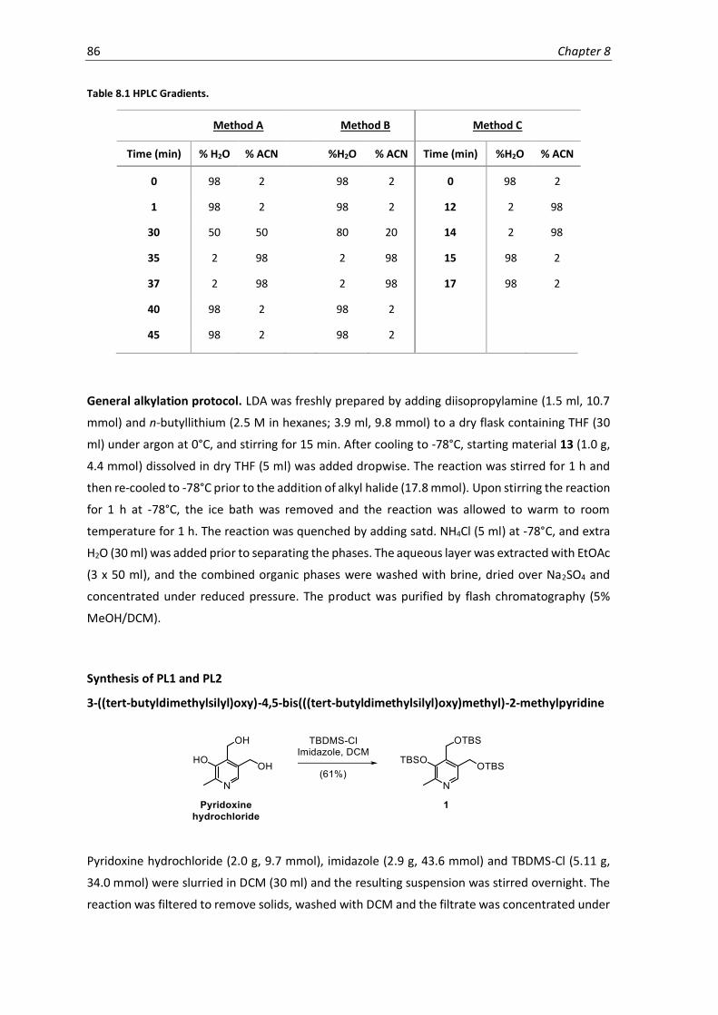
86 Chapter 8
Table 8.1 HPLC Gradients.
Method A Method B Method C
Time (min) % H2O % ACN %H2O % ACN Time (min) %H2O % ACN
0 98 2 98 2 0 98 2
1 98 2 98 2 12 2 98
30 50 50 80 20 14 2 98
35 2 98 2 98 15 98 2
37 2 98 2 98 17 98 2
40 98 2 98 2
45 98 2 98 2
General alkylation protocol. LDA was freshly prepared by adding diisopropylamine (1.5 ml, 10.7
mmol) and n-butyllithium (2.5 M in hexanes; 3.9 ml, 9.8 mmol) to a dry flask containing THF (30
ml) under argon at 0°C, and stirring for 15 min. After cooling to -78°C, starting material 13 (1.0 g,
4.4 mmol) dissolved in dry THF (5 ml) was added dropwise. The reaction was stirred for 1 h and
then re-cooled to -78°C prior to the addition of alkyl halide (17.8 mmol). Upon stirring the reaction
for 1 h at -78°C, the ice bath was removed and the reaction was allowed to warm to room
temperature for 1 h. The reaction was quenched by adding satd. NH4Cl (5 ml) at -78°C, and extra
H2O (30 ml) was added prior to separating the phases. The aqueous layer was extracted with EtOAc
(3 x 50 ml), and the combined organic phases were washed with brine, dried over Na2SO4 and
concentrated under reduced pressure. The product was purified by flash chromatography (5%
MeOH/DCM).
Synthesis of PL1 and PL2
3-((tert-butyldimethylsilyl)oxy)-4,5-bis(((tert-butyldimethylsilyl)oxy)methyl)-2-methylpyridine
Pyridoxine hydrochloride (2.0 g, 9.7 mmol), imidazole (2.9 g, 43.6 mmol) and TBDMS-Cl (5.11 g,
34.0 mmol) were slurried in DCM (30 ml) and the resulting suspension was stirred overnight. The
reaction was filtered to remove solids, washed with DCM and the filtrate was concentrated under
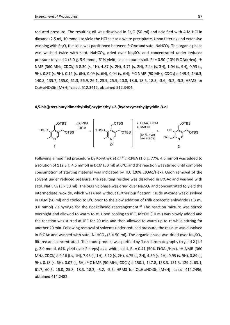
Experimental Procedures 87
reduced pressure. The resulting oil was dissolved in Et2O (50 ml) and acidified with 4 M HCl in
dioxane (2.5 ml, 10 mmol) to yield the HCl salt as a white precipitate. Upon filtering and extensive
washing with Et2O, the solid was partitioned between EtOAc and satd. NaHCO3. The organic phase
was washed twice with satd. NaHCO3, dried over Na2SO4 and concentrated under reduced
pressure to yield 1 (3.0 g, 5.9 mmol, 61% yield) as a colourless oil. Rf = 0.50 (10% EtOAc/Hex). 1H
NMR (360 MHz, CDCl3) δ 8.30 (s, 1H), 4.87 (s, 2H), 4.71 (s, 2H), 2.44 (s, 3H), 1.04 (s, 9H), 0.93 (s,
9H), 0.87 (s, 9H), 0.12 (s, 6H), 0.09 (s, 6H), 0.04 (s, 6H); 13C NMR (90 MHz, CDCl3) δ 149.4, 146.3,
140.8, 135.7, 135.0, 61.3, 56.9, 26.1, 25.9, 25.9, 20.8, 18.6, 18.5, 18.3, -3.6, -5.2, -5.3; HRMS for
C26H53NO3Si3 [M+H]+ calcd. 512.3412, obtained 512.3404.
4,5-bis(((tert-butyldimethylsilyl)oxy)methyl)-2-(hydroxymethyl)pyridin-3-ol
Following a modified procedure by Korytnyk et al,54 mCPBA (1.0 g, 77%, 4.5 mmol) was added to
a solution of 1 (2.3 g, 4.5 mmol) in DCM (50 ml) at 0°C, and the reaction was stirred until complete
consumption of starting material was indicated by TLC (20% EtOAc/Hex). Upon removal of the
solvent under reduced pressure, the resulting residue was dissolved in EtOAc and washed with
satd. NaHCO3 (3 × 50 ml). The organic phase was dried over Na2SO4 and concentrated to yield the
intermediate N-oxide, which was used without further purification. Crude N-oxide was dissolved
in DCM (50 ml) and cooled to 0°C prior to the slow addition of trifluoroacetic anhydride (1.3 ml,
9.0 mmol) via syringe for the Boekelheide rearrangement.64 The reaction mixture was stirred
overnight and allowed to warm to rt. Upon cooling to 0°C, MeOH (10 ml) was slowly added and
the reaction was stirred at 0°C for 20 min and then allowed to warm up to rt while stirring for
another 20 min. Following removal of solvents under reduced pressure, the residue was dissolved
in EtOAc and washed with satd. NaHCO3 (3 × 50 ml). The organic phase was dried over Na2SO4,
filtered and concentrated. The crude product was purified by flash chromatography to yield 2 (1.2
g, 2.9 mmol, 64% yield over 2 steps) as a white solid. Rf = 0.41 (50% EtOAc/Hex). 1H NMR (360
MHz, CDCl3) δ 9.16 (bs, 1H), 7.93 (s, 1H), 5.12 (s, 2H), 4.75 (s, 2H), 4.59 (s, 2H), 0.95 (s, 9H), 0.89 (s,
9H), 0.18 (s, 6H), 0.07 (s, 6H); 13C NMR (90 MHz, CDCl3) δ 150.1, 147.8, 138.3, 131.3, 129.2, 63.1,
61.7, 60.5, 26.0, 25.8, 18.3, 18.3, -5.2, -5.5; HRMS for C20H39NO4Si2 [M+H]+ calcd. 414.2496,
obtained 414.2482.

88 Chapter 8
4,5-bis(((tert-butyldimethylsilyl)oxy)methyl)-3-hydroxypicolinaldehyde
To a solution of alcohol 2 (1.2 g, 2.9 mmol) in DCM (10 ml) was added MnO2 (0.7 g, 8.7 mmol), and
the resulting slurry was stirred vigorously for approximately 3 h until complete consumption of
starting material was indicated by TLC (20% EtOAc/Hex). If the reaction stalled, more MnO2 was
added. Once the oxidation was complete, the reaction mixture was filtered through celite and
concentrated under reduced pressure to yield aldehyde 3 (1.0 g, 2.4 mmol, 83%) as a colourless
oil which did not require further purification. Rf = 0.72 (20% EtOAc/Hex). 1H NMR (360 MHz, CDCl3)
δ 11.03 (s, 1H), 10.04 (s, 1H), 8.59 (s, 1H), 5.00 (s, 2H), 4.81 (s, 2H), 0.96 (s, 9H), 0.89 (s, 9H), 0.13
(s, 6H), 0.09 (s, 6H); 13C NMR (90 MHz, CDCl3) δ 198.7, 155.6, 143.9, 141.6, 135.5, 133.6, 61.0, 54.6,
26.0, 26.0, 18.5, 18.4, -5.3, -5.3; HRMS for C20H37NO4Si2 [M+H]+ calcd. 412.2339, obtained
412.2326.
4,5-bis(((tert-butyldimethylsilyl)oxy)methyl)-3-(methoxymethoxy)picolinaldehyde
DIPEA (145 µl, 0.83 mmol) and MOMCl (86 µl, 1.13 mmol) were sequentially added to a solution
of 3 (310 mg, 0.75 mmol) in DCM (5 ml) and the reaction mixture was stirred at room temperature
for 2 h. Upon removal of the solvents under reduced pressure, the crude product was purified by
flash chromatography to yield 4 (209 mg, 0.46 mmol, 76% yield) as a colourless oil which
crystallizes upon freezing. Rf = 0.30 (20% EtOAc/Hex). 1H NMR (360 MHz, CDCl3) δ 10.17 (s, 1H),
8.82 (s, 1H), 5.10 (s, 2H), 4.99 (s, 2H), 4.84 (s, 2H), 3.57 (s, 3H), 0.95 (s, 9H), 0.89 (s, 9H), 0.12 (s,
6H), 0.10 (s, 6H); 13C NMR (90 MHz, CDCl3) δ 191.7, 152.4, 144.8, 143.9, 142.6, 140.3, 102.4, 61.0,
58.1, 56.1, 26.0, 25.9, 18.5, 18.3, -5.3, -5.4; HRMS for C22H41NO5Si2 [M+H]+ calcd. 456.2602,
obtained 456.2601.

Experimental Procedures 89
4,5-bis(((tert-butyldimethylsilyl)oxy)methyl)-2-ethynyl-3-(methoxymethoxy)pyridine
To a solution of 4 (209 mg, 0.46 mmol) in MeOH (5 ml) were added the Ohira-Bestmann reagent65
(dimethyl-1-diazo-2-oxopropylphosphonate) (131 mg, 0.68 mmol) and solid K2CO3 (124 mg, 0.90
mmol), and the reaction was rapidly stirred for 16 h. EtOAc (20 ml) was then added to the reaction
mixture and the organic phase was washed with H2O (2 × 10 ml), dried over Na2SO4 and
concentrated under reduced pressure. The crude product was purified by flash chromatography
to yield 5 (120 mg, 0.27 mmol, 58% yield) as a yellow solid. Rf = 0.47 (20% EtOAc/Hex). 1H NMR
(360 MHz, CDCl3) δ 8.55 (s, 1H), 5.20 (s, 2H), 4.92 (s, 2H), 4.79 (s, 2H), 3.58 (s, 3H), 3.31 (s, 1H),
0.93 (s, 9H), 0.88 (s, 9H), 0.11 (s, 6H), 0.09 (s, 6H); 13C NMR (90 MHz, CDCl3) δ 153.1, 144.7, 138.8,
137.9, 135.3, 100.7, 80.8, 60.9, 58.1, 56.3, 26.0, 26.0, 25.9, 18.4, 18.3, -5.3, -5.4; HRMS for
C23H41NO4Si2 [M+H]+ calcd. 452.2652, obtained 452.2635.
(6-ethynyl-5-(methoxymethoxy)pyridine-3,4-diyl)dimethanol
TBAF (514 µl, 1 M in THF, 514 mmol) was added to a solution of 5 (116 mg, 0.257 mmol) in THF (5
ml). The reaction was stirred for 10 min at rt and following removal of solvents under reduced
pressure, the crude product was purified by flash chromatography to yield 6 (55 mg, 0.246 mmol,
96% yield) as a tan solid. Rf = 0.48 (10% MeOH/DCM). 1H NMR (360 MHz, MeOD) δ 8.38 (s, 1H),
5.28 (s, 2H), 4.85 (s, 2H), 4.80 (s, 2H), 4.01 (s, 1H), 3.63 (s, 3H); 13C NMR (90 MHz, MeOD) δ 155.5,
145.8, 142.8, 139.4, 137.0, 101.7, 84.1, 80.6, 60.4, 58.3, 55.7; HRMS for C11H13NO4 [M+H]+ calcd.
224.0923, obtained 224.0916.

90 Chapter 8
2-ethynyl-5-(hydroxymethyl)-3-(methoxymethoxy)isonicotinaldehyde and 6-ethynyl-4-
(hydroxylmethyl)-5-(methoxymethoxy)nicotinaldehyde
To a solution of 6 (30 mg, 0.13 mmol) in MeOH (3 ml) was added MnO2 (34 mg, 0.39 mg), and the
resulting slurry was stirred vigorously for 1 h. The reaction mixture was subsequently filtered
through a 0.4 µM syringe filter and concentrated under reduced pressure. The product was
dissolved in H2O and the two isomers, 7a and 7b, were separated by HPLC purification using
method A (7a tR = 13.4 min, 7b tR = 14.0 min). Rf = 0.53 (10% MeOH/DCM). The product distribution
was 2.5:1 and the respective final yields were 7a (15 mg, 0.067 mmol, 52%) and 7b (6 mg, 21%).
7a 1H NMR (500 MHz, MeOD) δ 8.22 (s, 1H), 6.56 (s, 1H), 5.59 (d, J = 6.4 Hz, 1H), 5.28 (d, J = 6.5
Hz, 1H), 5.23 (dd, J = 13.8 Hz, J = 1.2 Hz, 1H), 5.05 (d, J = 13.8 Hz, 1H), 3.92 (s, 1H), 3.55 (s, 3H); 13C
NMR (125 MHz, MeOD) δ 151.8, 140.0, 139.4, 138.3, 135.2, 101.1, 98.1, 83.6, 80.2, 70.6, 57.5;
HRMS for C11H11NO4 [M+H]+ calcd. 222.0766, obtained 222.0759. 7b 1H NMR (500 MHz, MeOD) δ
8.27 (s, 1H), 6.48 (d, J = 2.1 Hz, 1H), 5.33 (dd, J = 14.2, 2.2 Hz, 1H), 5.23 (s, 2H), 5.16 (d, J = 14.2 Hz,
1H), 3.96 (s, 1H), 3.53 (s, 3H); 13C NMR (125 MHz, MeOD) δ 151.4, 140.9, 140.0, 139.8, 135.7,
101.1, 98.1, 84.1, 80.0, 71.0, 57.4; HRMS for C11H11NO4 [M+H]+ calcd. 222.0766, obtained
222.0759.
PL1
7a (20.7 mg, 0.094 mmol) was dissolved in 0.5 N HCl and stirred at rt for 15 min. The solution was
diluted by half with H2O and purified by HPLC using method B (tR = 8.6 min). Upon lyophilization,
the PL1 hydrochloride salt (17.5 mg, 0.082 mmol, 88% yield) was obtained as a light yellow, fluffy
solid. 1H NMR (500 MHz, DMSO-d6) δ 8.05 (s, 1H), 6.43 (s, 1H), 5.08 (d, J = 13.7 Hz, 1H), 4.91 (d, J
= 13.7 Hz, 1H), 4.29 (s, 1H); 13C NMR (125 MHz, DMSO-d6) δ 150.6, 137.8, 134.5, 134.5, 129.5, 98.5,
83.7, 80.4, 69.2; HRMS for C9H7NO3 [M+H]+ calcd. 178.0504, obtained 178.0498.

Experimental Procedures 91
PL2
7b (12.3 mg, 0.056 mmol) was dissolved in 0.5 N HCl and stirred at rt for 15 min. The solution was
diluted by half with H2O and purified by HPLC using method B (tR = 9.1 min). Upon lyophilization,
the PL2 hydrochloride salt (10.9 mg, 0.051 mmol, 92% yield) was obtained as a light yellow, fluffy
solid. 1H NMR (500 MHz, MeOD) δ 8.09 (s, 1H), 6.49 (d, J = 2.3 Hz, 1H), 5.18 (dd, J = 14.3, 2.3 Hz,
1H), 5.01 (d, J = 14.3 Hz, 1H), 3.94 (s, 1H); 13C NMR (126 MHz, MeOD) δ 152.1, 139.5, 137.8, 136.4,
130.7, 101.8, 84.7, 79.3, 70.4; HRMS for C9H7NO3 [M+H]+ calcd. 178.0504, obtained 178.0498.
Synthesis of PL3
(2,2,8-trimethyl-4H-[1,3]dioxino[4,5-c]pyridin-5-yl)methanol
Acetal protection of pyridoxine was performed using a previously described protocol by Yang et
al.66 To a suspension of pyridoxine hydrochloride (5.0 g, 24.3 mmol) in dry acetone (100 ml) were
added 2,2-dimethoxypropane (75 ml) and p-toluenesulfonic acid (18.5 g, 97.2 mmol). The mixture
was stirred overnight upon which the dark-brown solution was neutralized with satd. NaHCO3 until
light yellow. The aqueous layer was extracted with EtOAc (3 × 100 ml) and the combined organic
layers were washed with brine and concentrated under reduced pressure until crystalline. The
crude product was recrystallized from EtOH/Et2O (40:60) to yield pure 8 (2.8 g, 13.4 mmol) as a
white crystalline solid. Rf = 0.29 (10% MeOH/DCM). 1H NMR (400 MHz, CDCl3) δ 8.00 (s, 1H), 4.95
(s, 2H), 4.62 (s, 2H), 2.45 (s, 3H), 1.57 (s, 6H). 13C NMR (91 MHz, CDCl3) δ 147.6, 146.2, 138.6, 129.8,
126.1, 99.9, 60.0, 58.7, 24.9, 18.2. HRMS for C11H15NO3 [M+H]+ calcd. 210.1130, obtained
210.1123.

92 Chapter 8
5-(((4-methoxybenzyl)oxy)methyl)-2,2,8-trimethyl-4H-[1,3]dioxino[4,5-c]pyridine
To a dry flask under argon was added NaH (60% dispersion in mineral oil; 1.9 g, 47.8 mmol)
followed by dry DMF (20 ml). The suspension was heated to 70°C in an oil bath and 8 (2.0 g, 9.6
mmol) dissolved in DMF was slowly added. Once addition was complete, the reaction was left at
70°C for 5 min and then allowed to cool to room temperature. The reaction was cooled to 0°C for
the slow addition of PMBCl (1.6 ml, 11.5 mmol) and was allowed to warm to room temperature
while stirring overnight. Upon cooling on ice, the reaction was slowly quenched with H2O (50 ml),
extracted with EtOAc (3 × 50 ml), dried over Na2SO4 and concentrated under reduced pressure.
The crude product was purified by flash chromatography to yield 9 (2.2 g, 6.7 mmol, 70%) as a
clear oil. Rf = 0.43 (10% MeOH/DCM). 1H NMR (300 MHz, CDCl3) δ 7.94 (s, 1H), 7.24 (d, J = 8.6 Hz,
2H), 6.88 (d, J = 8.7 Hz, 2H), 4.85 (s, 2H), 4.42 (s, 2H), 4.39 (s, 2H), 3.80 (s, 3H), 2.40 (s, 3H), 1.54 (s,
6H). 13C NMR (75 MHz, CDCl3) δ 159.4, 148.2, 146.1, 139.8, 129.6, 126.4, 126.1, 113.9, 99.7, 71.8,
67.1, 58.8, 55.3, 24.8, 18.6. HRMS for C19H23NO4 [M+H]+ calcd. 330.1705, obtained 330.1699.
(5-(((4-methoxybenzyl)oxy)methyl)-2,2-dimethyl-4H-[1,3]dioxino[4,5-c]pyridin-8-yl)methanol
Following a modified procedure by Korytnyk et al,54 mCPBA (0.8 g, 77%, 3.7 mmol) was added to
a solution of 9 (1.0 g, 3.0 mmol) in DCM (50 ml) and the reaction was stirred overnight at room
temperature. Upon removal of the solvent under reduced pressure, the resulting residue was
dissolved in EtOAc and washed with satd. NaHCO3 (3 × 30 ml). The organic phase was dried over
Na2SO4 and concentrated to yield intermediate N-oxide, which was used directly in the next step.
Crude N-oxide was dissolved in DCM (50 ml) and cooled to 0°C prior to the slow addition of
trifluoroacetic anhydride (0.86 ml, 6.0 mmol) via syringe for the Boekelheide rearrangement.64
The ice bath was then removed and the reaction was stirred for 4 h. Upon re-cooling to 0°C, MeOH
(10 ml) was slowly added and the reaction was stirred at 0°C for 20 min and then allowed to warm
up to rt while stirring for another 20 min. Following removal of solvents under reduced pressure,
the residue was dissolved in EtOAc and washed with satd. NaHCO3 (3 × 50 ml). The organic phase
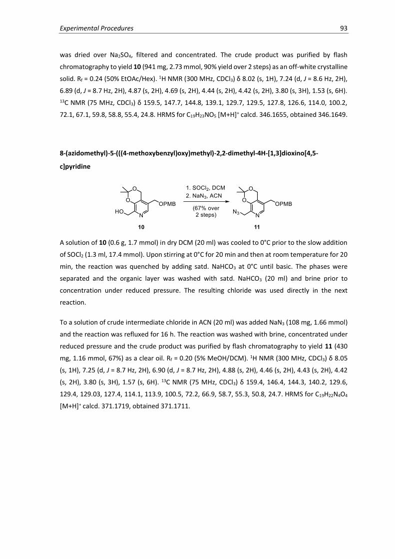
Experimental Procedures 93
was dried over Na2SO4, filtered and concentrated. The crude product was purified by flash
chromatography to yield 10 (941 mg, 2.73 mmol, 90% yield over 2 steps) as an off-white crystalline
solid. Rf = 0.24 (50% EtOAc/Hex). 1H NMR (300 MHz, CDCl3) δ 8.02 (s, 1H), 7.24 (d, J = 8.6 Hz, 2H),
6.89 (d, J = 8.7 Hz, 2H), 4.87 (s, 2H), 4.69 (s, 2H), 4.44 (s, 2H), 4.42 (s, 2H), 3.80 (s, 3H), 1.53 (s, 6H).
13C NMR (75 MHz, CDCl3) δ 159.5, 147.7, 144.8, 139.1, 129.7, 129.5, 127.8, 126.6, 114.0, 100.2,
72.1, 67.1, 59.8, 58.8, 55.4, 24.8. HRMS for C19H23NO5 [M+H]+ calcd. 346.1655, obtained 346.1649.
8-(azidomethyl)-5-(((4-methoxybenzyl)oxy)methyl)-2,2-dimethyl-4H-[1,3]dioxino[4,5-
c]pyridine
A solution of 10 (0.6 g, 1.7 mmol) in dry DCM (20 ml) was cooled to 0°C prior to the slow addition
of SOCl2 (1.3 ml, 17.4 mmol). Upon stirring at 0°C for 20 min and then at room temperature for 20
min, the reaction was quenched by adding satd. NaHCO3 at 0°C until basic. The phases were
separated and the organic layer was washed with satd. NaHCO3 (20 ml) and brine prior to
concentration under reduced pressure. The resulting chloride was used directly in the next
reaction.
To a solution of crude intermediate chloride in ACN (20 ml) was added NaN3 (108 mg, 1.66 mmol)
and the reaction was refluxed for 16 h. The reaction was washed with brine, concentrated under
reduced pressure and the crude product was purified by flash chromatography to yield 11 (430
mg, 1.16 mmol, 67%) as a clear oil. Rf = 0.20 (5% MeOH/DCM). 1H NMR (300 MHz, CDCl3) δ 8.05
(s, 1H), 7.25 (d, J = 8.7 Hz, 2H), 6.90 (d, J = 8.7 Hz, 2H), 4.88 (s, 2H), 4.46 (s, 2H), 4.43 (s, 2H), 4.42
(s, 2H), 3.80 (s, 3H), 1.57 (s, 6H). 13C NMR (75 MHz, CDCl3) δ 159.4, 146.4, 144.3, 140.2, 129.6,
129.4, 129.03, 127.4, 114.1, 113.9, 100.5, 72.2, 66.9, 58.7, 55.3, 50.8, 24.7. HRMS for C19H22N4O4
[M+H]+ calcd. 371.1719, obtained 371.1711.
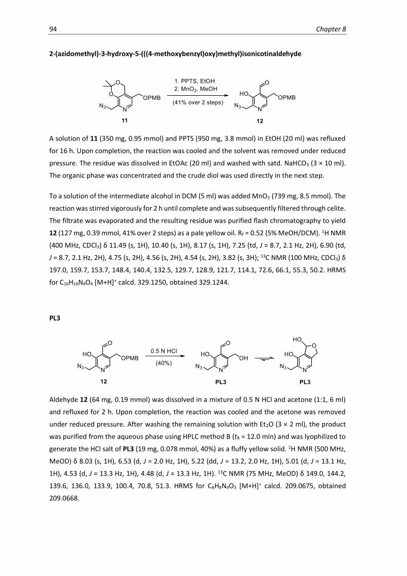
94 Chapter 8
2-(azidomethyl)-3-hydroxy-5-(((4-methoxybenzyl)oxy)methyl)isonicotinaldehyde
A solution of 11 (350 mg, 0.95 mmol) and PPTS (950 mg, 3.8 mmol) in EtOH (20 ml) was refluxed
for 16 h. Upon completion, the reaction was cooled and the solvent was removed under reduced
pressure. The residue was dissolved in EtOAc (20 ml) and washed with satd. NaHCO3 (3 × 10 ml).
The organic phase was concentrated and the crude diol was used directly in the next step.
To a solution of the intermediate alcohol in DCM (5 ml) was added MnO2 (739 mg, 8.5 mmol). The
reaction was stirred vigorously for 2 h until complete and was subsequently filtered through celite.
The filtrate was evaporated and the resulting residue was purified flash chromatography to yield
12 (127 mg, 0.39 mmol, 41% over 2 steps) as a pale yellow oil. Rf = 0.52 (5% MeOH/DCM). 1H NMR
(400 MHz, CDCl3) δ 11.49 (s, 1H), 10.40 (s, 1H), 8.17 (s, 1H), 7.25 (td, J = 8.7, 2.1 Hz, 2H), 6.90 (td,
J = 8.7, 2.1 Hz, 2H), 4.75 (s, 2H), 4.56 (s, 2H), 4.54 (s, 2H), 3.82 (s, 3H); 13C NMR (100 MHz, CDCl3) δ
197.0, 159.7, 153.7, 148.4, 140.4, 132.5, 129.7, 128.9, 121.7, 114.1, 72.6, 66.1, 55.3, 50.2. HRMS
for C16H16N4O4 [M+H]+ calcd. 329.1250, obtained 329.1244.
PL3
Aldehyde 12 (64 mg, 0.19 mmol) was dissolved in a mixture of 0.5 N HCl and acetone (1:1, 6 ml)
and refluxed for 2 h. Upon completion, the reaction was cooled and the acetone was removed
under reduced pressure. After washing the remaining solution with Et2O (3 × 2 ml), the product
was purified from the aqueous phase using HPLC method B (tR = 12.0 min) and was lyophilized to
generate the HCl salt of PL3 (19 mg, 0.078 mmol, 40%) as a fluffy yellow solid. 1H NMR (500 MHz,
MeOD) δ 8.03 (s, 1H), 6.53 (d, J = 2.0 Hz, 1H), 5.22 (dd, J = 13.2, 2.0 Hz, 1H), 5.01 (d, J = 13.1 Hz,
1H), 4.53 (d, J = 13.3 Hz, 1H), 4.48 (d, J = 13.3 Hz, 1H). 13C NMR (75 MHz, MeOD) δ 149.0, 144.2,
139.6, 136.0, 133.9, 100.4, 70.8, 51.3. HRMS for C8H8N4O3 [M+H]+ calcd. 209.0675, obtained
209.0668.
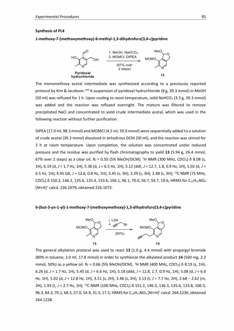
Experimental Procedures 95
Synthesis of PL4
1-methoxy-7-(methoxymethoxy)-6-methyl-1,3-dihydrofuro[3,4-c]pyridine
The monomethoxy acetal intermediate was synthesized according to a previously reported
protocol by Kim & Jacobson.194 A suspension of pyridoxal hydrochloride (8 g, 39.3 mmol) in MeOH
(50 ml) was refluxed for 1 h. Upon cooling to room temperature, solid NaHCO3 (3.3 g, 39.3 mmol)
was added and the reaction was refluxed overnight. The mixture was filtered to remove
precipitated NaCl and concentrated to yield crude intermediate acetal, which was used in the
following reaction without further purification.
DIPEA (17.0 ml, 98.3 mmol) and MOMCl (4.5 ml, 59.0 mmol) were sequentially added to a solution
of crude acetal (39.3 mmol) dissolved in anhydrous DCM (50 ml), and the reaction was stirred for
2 h at room temperature. Upon completion, the solution was concentrated under reduced
pressure and the residue was purified by flash chromatography to yield 13 (5.94 g, 26.4 mmol,
67% over 2 steps) as a clear oil. Rf = 0.50 (5% MeOH/DCM). 1H NMR (300 MHz, CDCl3) δ 8.08 (s,
1H), 6.19 (d, J = 1.7 Hz, 1H), 5.36 (d, J = 6.5 Hz, 1H), 5.12 (ddt, J = 12.7, 1.8, 0.9 Hz, 1H), 5.02 (d, J =
6.5 Hz, 1H), 4.95 (dt, J = 12.6, 0.8 Hz, 1H), 3.45 (s, 3H), 3.39 (s, 3H), 2.48 (s, 3H); 13C NMR (75 MHz,
CDCl3) δ 150.2, 146.2, 135.6, 135.4, 133.6, 106.1, 96.1, 70.0, 56.7, 54.7, 19.6; HRMS for C11H15NO4
[M+H]+ calcd. 226.1079, obtained 226.1072.
6-(but-3-yn-1-yl)-1-methoxy-7-(methoxymethoxy)-1,3-dihydrofuro[3,4-c]pyridine
The general alkylation protocol was used to react 13 (1.0 g, 4.4 mmol) with propargyl bromide
(80% in toluene; 2.0 ml, 17.8 mmol) in order to synthesize the alkylated product 14 (560 mg, 2.2
mmol, 50%) as a yellow oil. Rf = 0.66 (5% MeOH/DCM). 1H NMR (400 MHz, CDCl3) δ 8.19 (s, 1H),
6.26 (d, J = 1.7 Hz, 1H), 5.45 (d, J = 6.6 Hz, 1H), 5.18 (ddd, J = 12.8, 1.7, 0.9 Hz, 1H), 5.08 (d, J = 6.6
Hz, 1H), 5.02 (d, J = 12.8 Hz, 1H), 3.51 (s, 2H), 3.46 (s, 2H), 3.13 (t, J = 7.7 Hz, 2H), 2.68 – 2.62 (m,
2H), 1.93 (t, J = 2.7 Hz, 1H); 13C NMR (100 MHz, CDCl3) δ 151.2, 146.3, 136.3, 135.6, 133.8, 106.3,
96.3, 84.3, 70.1, 68.5, 57.0, 54.9, 31.5, 17.5; HRMS for C14H17NO4 [M+H]+ calcd. 264.1236, obtained
264.1228.

96 Chapter 8
PL4
Compound 14 (40 mg, 0.15 mmol) was dissolved in 5% H2SO4 (2 ml) in acetone/H2O (1:1) and
heated at 85°C for 1 h. The reaction was allowed to cool and the acetone was removed under
reduced pressure. The product was purified by HPLC using method B (tR = 9.8 min) and lyophilized
to generate PL4 as the H2SO4 salt (35 mg, 0.11 mmol, 76%) as a fluffy white solid. 1H NMR (400
MHz, DMSO-d6) δ 9.72 (s, 1H, OH), 7.98 (s, 1H), 6.67 (d, J = 7.7 Hz, 1H, OH), 6.42 (dd, J = 7.5, 1.8
Hz, 1H), 5.05 (ddd, J = 12.9, 1.9, 0.9 Hz, 1H), 4.86 (d, J = 13.1 Hz, 1H), 2.96 – 2.91 (m, 2H), 2.71 (t, J
= 2.6 Hz, 1H), 2.57 – 2.50 (m, 1H); 13C NMR (100 MHz, DMSO-d6) δ 146.6, 145.7, 135.5, 133.8,
132.5, 98.4, 84.5, 71.0, 68.8, 30.5, 16.5; HRMS for C11H11NO3 [M+H]+ calcd. 206.0817, obtained
206.0810.
Synthesis of PL5
1-Methoxy-7-(methoxymethoxy)-6-(7-(trimethylsilyl)hept-6-in-1-yl)-1,3-dihydrofuro[3,4-
c]pyridine
The general alkylation protocol was used to react 13 (200 mg, 0.89 mmol) with TMS-protected 7-
bromohex-1-yne (620 mg, 2.67 mmol) in order to synthesize the alkylated product 15 (132 mg,
0.35 mmol, 40%) as a yellow oil. Rf = 0.67 (50% EtOAc/hexanes). 1H NMR (400 MHz, CDCl3) δ 8.17
(s, 1H), 6.25 (s, 1H), 5.42 (d, J = 6.5 Hz, 1H), 5.17 (d, J = 12.7 Hz, 1H), 5.06 (d, J = 6.5 Hz, 1H), 5.01
(d, J = 12.8 Hz, 1H), 3.51 (s, 3H), 3.45 (s, 3H), 2.89 (t, J = 7.8 Hz, 2H), 2.21 (t, J = 7.0 Hz, 2H), 1.71 (p,
J = 7.6 Hz, 2H), 1.60-1.52 (m, 2H), 1.50-1.44 (m, 2H), 0.13 (s, 9H). 13C NMR (100 MHz, CDCl3) δ
153.8, 146.2, 135.8, 135.4, 134.0, 107.7, 106.3, 96.4, 84.4, 70.1, 57.0, 54.9, 32.5, 29.0, 28.7, 28.4,
19.9, 0.3. HRMS for C20H31NO4Si [M+H]+ calcd. 378.2101, obtained 378.2094.

Experimental Procedures 97
6-(hept-6-yn-1-yl)-1-methoxy-7-(methoxymethoxy)-1,3-dihydrofuro[3,4-c]-pyridin
To a solution of 15 (241 mg, 0.63 mmol) dissolved in dry MeOH (10 ml) was added K2CO3 (441 mg,
3.2 mmol) and the reaction was stirred overnight at room temperature. Upon completion, Et2O
and H2O were added (each 20 ml) and the aqueous layer was extracted with Et2O (3 × 20 ml). The
organic phase was concentrated under reduced pressure and the resulting residue was purified
by flash chromatography to yield 16 (176 mg, 0.58 mmol, 91%) as a yellow oil. Rf = 0.62 (50%
EtOAc/hexane). 1H NMR (400 MHz, CDCl3) δ 8.16 (s, 1H), 6.24 (s, 1H), 5.41 (d, J = 6.5 Hz, 1H), 5.16
(d, J = 13.0 Hz, 1H), 5.05 (d, J = 6.5 Hz, 1H), 5.00 (d, J = 13.0 Hz, 1H), 3.50 (s, 3H), 3.45 (s, 3H), 2.87
(t, J = 7.8 Hz, 2H), 2.18 (td, J = 7.0, 2.6 Hz, 2H), 1.91 (t, J = 2.7 Hz, 1H), 1.77-1.65 (m, 2H), 1.63-1.52
(m, 2H), 1.52-1.44 (m, 2H). 13C NMR (100 MHz, CDCl3) δ 153.8, 146.1, 135.7, 135.7, 133.8, 106.3,
96.4, 84.8, 70.1, 68.3, 56.9, 54.9, 32.6, 28.9, 28.5, 28.4, 18.4. HRMS for C17H23NO4 [M+H]+ calcd.
306.1705, obtained 306.1701.
PL5
Compound 16 (100 mg, 0.372 mmol) was dissolved in 5% H2SO4 (2 ml) in acetone/H2O (1:1) and
heated at 85°C for 1 h. The reaction was allowed to cool and the acetone was removed under
reduced pressure. The product was purified by HPLC using method A (tR = 7.5 min) and lyophilized
to generate PL5 as the H2SO4 salt (80 mg, 0.232 mmol, 63%) as a fluffy white solid. 1H NMR (400
MHz, DMSO-d6) δ 9.49 (s, 1H, OH), 7.94 (s, 1H), 6.63 (d, J = 7.1 Hz, 1H, OH), 6.41 (d, J = 7.1 Hz, 1H),
5.04 (d, J = 12.8 Hz, 1H), 4.85 (d, J = 12.8 Hz, 1H), 2.77 – 2.65 (m, 3H), 2.14 (td, J = 6.9, 2.6 Hz, 2H),
1.62 (p, J = 7.6 Hz, 2H), 1.55-1.43 (m, 2H), 1.43-1.34 (m, 2H). 13C NMR (100 MHz, DMSO-d6) δ 148.9,
145.6, 134.9, 133.7, 132.4, 98.5, 84.6, 71.1, 31.2, 28.1, 27.9, 27.5, 17.6. HRMS for C14H18NO3
[M+H]+ calcd. 248.1287, obtained 248.1281.
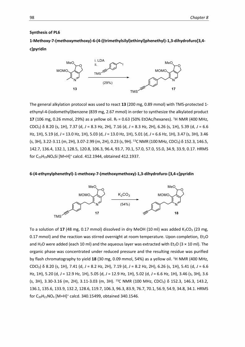
98 Chapter 8
Synthesis of PL6
1-Methoxy-7-(methoxymethoxy)-6-(4-((trimethylsilyl)ethinyl)phenethyl)-1,3-dihydrofuro[3,4-
c]pyridin
The general alkylation protocol was used to react 13 (200 mg, 0.89 mmol) with TMS-protected 1-
ethynyl-4-(iodomethyl)benzene (839 mg, 2.67 mmol) in order to synthesize the alkylated product
17 (106 mg, 0.26 mmol, 29%) as a yellow oil. Rf = 0.63 (50% EtOAc/hexanes). 1H NMR (400 MHz,
CDCl3) δ 8.20 (s, 1H), 7.37 (d, J = 8.3 Hz, 2H), 7.16 (d, J = 8.3 Hz, 2H), 6.26 (s, 1H), 5.39 (d, J = 6.6
Hz, 1H), 5.19 (d, J = 13.0 Hz, 1H), 5.03 (d, J = 13.0 Hz, 1H), 5.01 (d, J = 6.6 Hz, 1H), 3.47 (s, 3H), 3.46
(s, 3H), 3.22-3.11 (m, 2H), 3.07-2.99 (m, 2H), 0.23 (s, 9H). 13C NMR (100 MHz, CDCl3) δ 152.3, 146.5,
142.7, 136.4, 132.1, 128.5, 120.8, 106.3, 96.4, 93.7, 70.1, 57.0, 57.0, 55.0, 34.9, 33.9, 0.17. HRMS
for C23H29NO4Si [M+H]+ calcd. 412.1944, obtained 412.1937.
6-(4-ethynylphenethyl)-1-methoxy-7-(methoxymethoxy)-1,3-dihydrofuro-[3,4-c]pyridin
To a solution of 17 (48 mg, 0.17 mmol) dissolved in dry MeOH (10 ml) was added K2CO3 (23 mg,
0.17 mmol) and the reaction was stirred overnight at room temperature. Upon completion, Et2O
and H2O were added (each 10 ml) and the aqueous layer was extracted with Et2O (3 × 10 ml). The
organic phase was concentrated under reduced pressure and the resulting residue was purified
by flash chromatography to yield 18 (30 mg, 0.09 mmol, 54%) as a yellow oil. 1H NMR (400 MHz,
CDCl3) δ 8.20 (s, 1H), 7.41 (d, J = 8.2 Hz, 2H), 7.19 (d, J = 8.2 Hz, 2H), 6.26 (s, 1H), 5.41 (d, J = 6.6
Hz, 1H), 5.20 (d, J = 12.9 Hz, 1H), 5.05 (d, J = 12.9 Hz, 1H), 5.02 (d, J = 6.6 Hz, 1H), 3.46 (s, 3H), 3.6
(s, 3H), 3.30-3.16 (m, 2H), 3.11-3.03 (m, 3H). 13C NMR (100 MHz, CDCl3) δ 152.3, 146.3, 143.2,
136.1, 135.6, 133.9, 132.2, 128.6, 119.7, 106.3, 96.3, 83.9, 76.7, 70.1, 56.9, 54.9, 34.8, 34.1. HRMS
for C20H21NO4 [M+H]+ calcd. 340.15499, obtained 340.1546.

Experimental Procedures 99
PL6
Compound 18 (30 mg, 0.088 mmol) was dissolved in 5% H2SO4 (2 ml) in acetone/H2O (1:1) and
heated at 85°C for 1 h. The reaction was allowed to cool and was concentrated under reduced
pressure to remove acetone. The product was purified by HPLC using method C (tR = 8.5 min) and
lyophilized to generate PL6 as the H2SO4 salt (21 mg, 0.0554 mmol, 63%) as a fluffy white solid. 1H
NMR (400 MHz, DMSO-d6) δ 9.65 (s, 1H, OH), 7.98 (s, 1H), 7.38 (d, J = 8.2 Hz, 2H), 7.26 (d, J = 8.2
Hz, 2H), 6.66 (d, J = 7.6 Hz, 1H, OH), 6.42 (d, J = 7.6 Hz, 1H), 5.05 (d, J = 12.9 Hz, 1H), 4.87 (d, J =
12.9 Hz, 1H), 4.10 (s, 1H), 3.08 – 2.91 (m, 4H). 13C NMR (100 MHz, DMSO-d6) δ 147.6, 145.7, 143.2,
135.2, 133.8, 132.5, 131.6, 128.6, 119.1, 98.5, 83.6, 68.8, 33.5, 32.7. HRMS for C17H15NO3 [M+H]+
calcd. 282.1130, obtained 282.1123.

100 Chapter 8
8.2 Biological Methods
Bacterial growth media. LB-medium. Peptone (10 g), NaCl (5 g) and yeast extract (5 g) were
combined and dissolved in ddH2O to a final volume of 1 L, and adjusted to pH 7.5. B-medium.
Peptone (10 g), NaCl (5 g), yeast extract (5 g) and K2HPO4 (1 g) were combined and dissolved in
ddH2O to a final volume of 1 L, and adjusted to pH 7.5.
Cloning and overexpression of recombinant proteins. N-terminally STREP-II tagged S. aureus
(Mu50 for Alr and PLK; USA300 for all others) proteins were cloned and expressed using the
primers and conditions listed in Table 8.2. Cloning was performed using the Invitrogen Gateway
cloning system with pDONR201Kan as the donor vector and pET55destAmp as the destination vector.
Proteins were expressed in E. coli BL21(DE3) grown in LB-media containing 0.1 mg/ml Ampicillin
at 37°C to an OD600 of 0.5-0.6 through the addition of 1 mM isopropyl-1-thio-β-galactopyranoside
(IPTG). Bacteria were harvested and washed with PBS prior to cell lysis and protein purification.
Table 8.2 PCR primers and overexpression conditions for recombinant proteins.
No Protein Name Uniprot ID, Gene name
Primers Over-
expression MW found
(Da)
1. (Alr) Alanine racemase 1 P63479, Alr1
(fwd) ggggacaagtttgtacaaaaaagcaggcttt tcggataaatattatagat (rev) ggggaccactttgtacaagaaagctgggtgtt attttaacaattcatttg
30°C, o/n m-Met = 45,099.1
2. (AT1) Aminotransferase, Class I
A0A0H2XFY9, SAUSA300_2497
(fwd) ggggacaagtttgtacaaaaaagcaggcttt
atctctaacaaattagcaaat (rev) ggggaccactttgtacaagaaagctgggtgtt attcatataaatatgctagtt
25°C, 6 h m-Met = 45,024.0
3. (AT2) Aminotransferase,
Class I
A0A0H2XFA8,
SAUSA300_0952
(fwd) ggggacaagtttgtacaaaaaagcaggcttt aaactttctttaaattctaatt
(rev) ggggaccactttgtacaagaaagctgggtgtt atttagtatttaaatatttag
25°C, o/n m-Met =
45,435.4
4. (DAT) D-alanine aminotransferase
A0A0H2XHU6, dat
(fwd) ggggacaagtttgtacaaaaaagcaggcttt gaaaaaatttttttaaatggtgag (rev) ggggaccactttgtacaagaaagctgggtgtt
aaatactgtgtgactctata
25°C, o/n m-Met = 34,184.5
5. (CS) Cysteine synthase A0A0H2XFQ3, cysK
(fwd )ggggacaagtttgtacaaaaaagcaggcttt gcacaaaaaccagtagataatatta (rev) ggggaccactttgtacaagaaagctgggtgtt agtcatcgaatgaatataaaggt
37°C, 4 h m-Met = 35,265.5
6. (ODC) Ornithine/Lysine/ Arginine decarboxylase
A0A0H2XII6, SAUSA300_0458
(fwd) ggggacaagtttgtacaaaaaagcaggcttt aagcaacctattttaaataaattag (rev) ggggaccactttgtacaagaaagctgggtgtt attcatcctcaactaaaatttta
25°C, o/n m-Met = 53,372.2
7. (HemQ) coprohaem decarboxylase
Q2FJ56, SAUSA300_0569
(fwd) ggggacaagtttgtacaaaaaagcaggcttt
agtcaagcagccgaaacatta (rev) ggggaccactttgtacaagaaagctgggtgtt aagaaatcgcaaagaattgatcg
25°C, o/n m-Met = 31,679.7
8. (PsuG) pseudouridine-5’-phosphate glycosidase
A0A0H2XJC6, SAUSA300_0312
(fwd) ggggacaagtttgtacaaaaaagcaggcttt gcaaatttacaaaagtatattga (rev) ggggaccactttgtacaagaaagctgggtgct
acaataatttattaacagcg
25°C, o/n m-Met = 35,154.5
9. (HemH) Ferrochelatase A0A0H2XGS5, SAUSA300_1782
(fwd) ggggacaagtttgtacaaaaaagcaggcttt actaaaaaaatgggattattagtta (rev) ggggaccactttgtacaagaaagctgggtgtt aaaatatagacttgatttcatca
25°C, o/n m-Met = 37,346.6
10. (Q2FF14) UPF0340 uncharacterized protein
Q2FF14, SAUSA300_2068
(fwd) ggggacaagtttgtacaaaaaagcaggcttt aaagatttgacaatgttattag (rev) ggggaccactttgtacaagaaagctgggtgtt attggtatttagcacgtt
25°C, o/n m-Met = 21,184.9
11. (GP0) Uncharacterized protein
A0A0H2XGP0, SAUSA300_2209
(fwd) ggggacaagtttgtacaaaaaagcaggcttt
agcatagttcagttatatga (rev) ggggaccactttgtacaagaaagctgggtgtt attcagggattgtttgat
20°C, o/n m-Met = 37,052.9
12. (HH8) Uncharacterized
protein
A0A0H2XHH8,
SAUSA300_1082
(fwd )ggggacaagtttgtacaaaaaagcaggcttt aaagataatttacaacaaatct
(rev) ggggaccactttgtacaagaaagctgggtgtc actcctcttctcc
25°C, o/n m-Met =
27,636.3
Experimental Procedures 101
Cloning and overexpression of recombinant PLK. The N-terminally STREP-II tagged S. aureus
Mu50 pyridoxal kinase (PLK) was prepared as previously described.75
Analytical-scale protein purification (AT1, AT2, DAT, CS, OKR-DC, HemQ, PsuG, HemH, Q2FF14,
GP0, HH8). Bacterial cell pellets from overexpression cultures (50 ml) were resuspended in 1 ml
phosphate buffer (50 mM NaH2PO4, 300 mM NaCl, pH 8.0). Cells were lysed by sonication and the
lysate was clarified by centrifugation (21,000 x g, 30 min, 4°C). Strep-Tactin® spin columns (Iba
Lifesciences) were equilibrated with phosphate buffer, according to the user manual (2 × 500 µl,
700 x g, 30 sec, 4°C) and lysate was loaded onto the columns (700 x g, 30 sec, 4°C). Columns were
washed with phosphate buffer (4 x 100 µl, 16,200 x g, 30 sec, 4°C) prior to eluting the protein in
buffer containing D-biotin (5 mM) (2 × 100 µl, 700 x g, 30 sec, 4°C). Protein concentrations were
measured on a Tecan infinite M200Pro plate reader by absorption at 280 nm (Nanoquant plate).
A yield of approximately 3 mg/ml (≈ 100 µM) was obtained for each protein and the MW of each
protein was confirmed by intact-protein MS. Proteins were found to be sufficiently pure by SDS-
PAGE and were stored at -80°C.
Large-scale protein purification (Alr, Apo-Alr). The bacterial cell pellet from the overexpression
culture (2 L) was resuspended in phosphate buffer (50 mM NaH2PO4, 300 mM NaCl, pH 8.0). Cells
were lysed by sonication and the lysate was clarified by centrifugation (36,000 x g, 30 min, 4°C).
The supernatant was loaded onto a pre-equilibrated 5 ml StrepTrap column (GE Healthcare) using
an Äkta purification system (GE Healthcare) and washed with phosphate buffer (50 ml, flow rate
= 4 ml/min). Alr was eluted in phosphate buffer containing desthiobiotin (2.5 mM) as the PLP-
holoenzyme. In order to generate the apoenzyme (Apo-Alr), column-bound proteins were
additionally flushed with phosphate buffer containing 25 mM hydroxylamine (50 ml) prior to
washing and elution. Protein-containing fractions were concentrated, desalted into phosphate
buffer using a 5 ml HiTrap Desalting column (GE Healthcare) and protein aliquots were flash frozen
in liquid nitrogen for storage at -80°C. Alr was found to be sufficiently pure by SDS-PAGE and its
MW was confirmed by intact-protein MS. Protein concentrations were measured on a Tecan
infinite M200Pro plate reader by absorption at 280 nm (Nanoquant plate).
Large-scale protein purification (Q2FF14, GP0 and HH8). Holoenzymes were overexpressed and
purified as described above using the following buffers for both purification and desalting: Q2FF14
(50 mM Tris, 250 mM NaCl, pH 8), GP0 (100 mM Tris, 750 mM NaCH, pH 8) and HH8 (50 mM Tris,
750 mM NaCl, pH 8). Expression culture lysate was incubated with 100 µM PLP for 15 min on ice
prior to purification in order to saturate proteins with the cofactor.
PLK kinetics. PLK activity was measured using the previously reported coupled assay.75

102 Chapter 8
In vitro phosphorylation of PL-probes using PLK. A solution containing 2 mM PL-probe, 25 µM
PLK and 10 mM ATP was prepared in 1 ml kinase buffer (50 mM Tris, 50 mM KCl, 10 mM MgCl2,
pH 7.9) and incubated overnight at 4°C with gentle shaking. The solution was filtered through a
10 kDa MWCO centrifugal filter (Sartorius Stedim Biotech) to remove PLK, and phosphorylated
probes were used in subsequent experiments without further purification, assuming full
conversion (800 µM final concentration). Phosphorylation of PL probes could be confirmed by LC-
MS/MS analysis using a H-ESI-II probe and LC conditions as described in the metabolomics section
with slight modifications. A positive multiple reaction monitoring (MRM) was used as the
detection mode and daughter ions of the respective HRMS probe ions were recorded in a range
of 50-2000 m/z. Quantification of PL4P product samples showed the presence of > 99.5% PL4P.
Reconstitution of Apo-Alr with phosphorylated probes. Apo-Alr (50 µM in phosphate buffer) was
incubated with phosphorylated probes (5 equivalents) overnight at 4°C with gentle shaking.
Residual probe was removed by desalting into phosphate buffer using a 5 ml HiTrap Desalting
column (GE Healthcare) on an Äkta purification system.
UV-vis measurements. UV-vis spectra of protein samples (100 µM) were recorded in phosphate
buffer (50 mM NaH2PO4, 300 mM NaCl, pH 8.0) at 25°C on a Tecan infinite M200Pro plate reader
(300-600 nm, 5 nm increments). For substrate studies, proteins were incubated with 10
equivalents of substrate for 15 min prior to measurement.
Alr kinetics. Racemization of D-Ala to L-Ala was measured using a coupled assay that monitors
NADH formation (abs =340 nm) by L-alanine dehydrogenase.195 A premix containing 2 units/ml L-
alanine dehydrogenase (Roche), 10 mM NAD+ and 10 nM Alr was prepared in CHES buffer
(100 mM CHES, 100 mM KCl, pH 8.8). 100 µl of the premix was pipetted into flat-bottom 96-well
plates (NunclonTM Delta Surface, Thermo Scientific) containing 5 µl D-Ala (1, 2, 10, 20, 50, 100, 200
and 300 mM). The reactions were incubated at 37°C and NADH formation was measured at
340 nm in 1 min kinetic intervals using a Tecan infinite M200Pro plate reader. Enzymatic activity
was assayed in three replicates. Initial rates of NADH formation (µM/min) were determined for
the different substrate concentrations and the data (mean ± standard deviation) was fitted to the
Michaelis-Menten equation (non-linear regression) using GraphPad Prism-v5.0 software.
NaBH4-reduction of proteins for intact-protein MS. 10 µM protein samples (25 µl) were incubated
with 4 equivalents of cofactor derivative (1 µl of 500 µM stock PL, PLP, PL4 or PL4P) in PBS for 30
min and subsequently treated with 10 mM NaBH4 (2 µl of 250 mM stock prepared fresh in 0.1 M
NaOH) at room temperature for 30 min. For Alr holoenzymes substituted with phosphorylated
cofactor probes, samples were directly reduced without prior incubation. The NaBH4 was
quenched by acidification to pH 5-6 with HCl (5-10 µl of 0.5% FA) and neutralized to pH 7 with
NaOH (5-10 µl of 0.1 M NaOH). Samples were diluted to 50 µl with PBS (5 µM final enzyme
concentration) for measurement by intact-protein MS.

Experimental Procedures 103
Intact-protein mass spectrometry. Samples were desalted and measured using a MassPREP On-
Line Desalting Cartridge (Waters) on an Ultimate 3000 HPLC system (Dionex) coupled to a Finnigan
LTQ-FT Ultra mass spectrometer (Thermo Scientific) with electrospray ionization (spray voltage
4.0 kV, tube lens 110 V, capillary voltage 48 V, sheath gas 60 arb, aux gas 10 arb, sweep gas 0.2
arb). Xcalibur Xtract Software (Thermo Scientific) was used for data analysis and deconvolution.
In vitro labeling of recombinant proteins. 10 µM protein samples (50 µl in PBS) were labeled with
PL(P)-probes (2 µl of 25× stock) at room temperature for 30 min. Upon reduction with 10 mM
NaBH4 (2 µl of 250 mM prepared fresh in 0.1 M NaOH) at room temperature for 30 min, proteins
were precipitated by adding ice-cold acetone (4× volume) and incubating at -20°C for at least 1 h.
Precipitated proteins were pelleted by centrifugation (18,000 x g, 15 min, 4°C) and washed with
ice-cold MeOH (2 × 0.2 ml), using sonication to resuspend the pellets between washes. Pelletized
proteins were resuspended in 50 µl PBS containing 0.4% (w/v) SDS and CuAAC to rhodamine-azide
was performed by adding 0.1 mM Rh-N3 (1 µl of 5 mM stock in DMSO), 0.5 mM BTTAA ligand196
(2.5 µl of 10 mM stock in ddH2O), 1 mM CuSO4 (1 µl of 50 mM stock in ddH2O), 2 mM NaAsc (1 µl
of 100 mM prepared fresh in ddH2O) and incubating at room temperature for 1 h in the dark. For
PL3(P)-labeled proteins, CuAAC was performed using 0.1 mM Rh-alkyne (1 µl of 5 mM stock in
DMSO) instead of Rh-N3. Strain-promoted click chemistry was performed using 0.02 mM
rhodamine-DBCO reagent (1 µl of 1 mM stock in DMSO). Samples were quenched with 2× gel
loading buffer and analyzed by SDS-PAGE (12.5% polyacrylamide gels) with fluorescence scanning.
Analytical size-exclusion chromatography (SEC). Sample protein (100 µl, 50 µM) was injected
onto a Superdex 200 10/300 gel filtration column (GE Healthcare) connected to an Äkta
purification system 10 (GE Healthcare), which was pre-equilibrated with respective protein buffers
used for purification. UV absorption was recorded at 280 nm. The oligomerization state was
calculated from a calibration curve determined for the column using a protein standard mix (Gel
Filtration Calibration Kit, GE Healthcare).
Enzyme substrate screens by MS. 10 µM enzyme was incubated with a mixture of amino acids
(0.5 mM of each proteinogenic L-amino acid except Tyrosine, including Ornithine) and cofactor
mix (100 µM of each NAD, NADH, NADP, NADPH, FAD, FMN, ATP, SAM, MgCl2, MnCl2, AcCoA, PLP)
in ddH2O for 30 min at 37°C. Reactions were stopped by precipitating protein using 400 µl ice-cold
MeOH and incubating at -20°C for 1 h. Samples were subsequently centrifuged (18,000 x g, 15
min, 4°C) and the supernatant was evaporated in a speedvac centrifuge. The resulting residue was
dissolved 150 µl H2O/ACN (50:50, incl. 1% FA) and centrifuged (18,000 x g, 15 min, 4°C) to remove
undissolved particles prior to measurement on an Ultimate 3000 RSLC system (Thermo Scientific)
coupled to an LTQ Orbitrap XL mass spectrometer (Thermo Scientific). Chromatographic
separation was performed using a SeQuant® ZIC®-pHILIC (250 x 2.1 mm, 5 µm, 100 Å) (Merck
Millipore).

104 Chapter 8
Assay for N-Acetyltransferase activity. A previously described broad-substrate screening assay by
Kuhn et al.122 was used to assess N-acetyltransferase activity of GP0. Therefore, 50 µl of a mixture
containing 2.5 mM substrate (Table 8.3), 0.5 mM AcCoA and 1 µM enzyme in 50 mM Tris-HCl, pH 8
were added to each well of a 96-well plate (NunclonTM Delta Surface, Thermo Scientific) and
incubated at 33°C for 15 min. Next, 50 µl of 6M guanidine-HCl buffer were added, followed by
100 µl of a solution containing Ellman’s reagent (0.4 mM 5,5'-dithio-bis-(2-nitrobenzoic acid) in
100 mM Tris-HCl pH 8). The plate was incubated at room temperature for 10 min and absorption
at a wavelength of 415 nm was measured using a Tecan infinite M200Pro plate reader. Samples
were measured in duplicate.
Table 8.3 Substrates used for N-acetyltransferase activity assay.
No. Substrate No. Substrate
1. Spermine 24. Erythromycin
2. Spermidine 25. Kanamycin
3. L-Lys 26. Streptomycin
4. L-Gln 27. Tobramycin
5. L-Asn 28. Ampicillin
6. L-Arg 29. Puromycin
7. L-Asp 30. Chloramphenicol
8. L-Glu 31. Bacitracin
9. Gly 32. Colistin
10. L-Ala 33. Adenine
11. L-Ser 34. Guanosine
12. L-Thr 35. Cytidine
13. L-Met 36. NAD
14. L-Trp 37. AMP
15. L-Phe 38. Thiamine pyrophosphate
16. L-Tyr 39. Urea
17. L-Val 40. Guanidine
18. L-Ile 41. Pyridoxamine
19. L-Leu 42. Thiamine
20. L-homoserine 43. Folic acid
21. L-Citrulline 44. 4-aminobenzoic acid
22. L-Orn 45. D-glucosamine
23. Gentamicin 46. Tyramine
S. aureus USA300 TnPdxS growth curves. The S. aureus USA300 transposon mutant TnPdxS (gene:
SAUSA300_0504; pyridoxal biosynthesis lyase PdxS) was obtained from the Nebraska Transposon
Mutant Library within the Network on Antimicrobial Resistance in Staphylococcus aureus
(NARSA).197 Bacteria were kept under antibiotic pressure (erythromycin) for all stages of the
experiment to select for the transposon phenotype. Chemically-defined media (CDM) free of PL
was prepared according to Liebeke et al.198 and sterilized by filtration through a 0.2 µM PES filter
(VWR). Overnight cultures of S. aureus USA300 TnPdxS were grown in B-medium containing 5
µg/ml erythromycin (Erm). Cultures were harvested, washed twice with 5 ml CDM-Erm (5 µg/ml)

Experimental Procedures 105
and resuspended in CDM-Erm. 150 µl of bacterial culture (initial OD600 = 0.08) was added to flat-
bottomed 96-well plates (Nunclon Delta surface, Thermo Scientific) containing 1.5 µl PL or PL-
probe (100× stock in DMSO), and the plates were incubated at 37°C for 24 h with shaking (220
rpm). Endpoint measurements of bacterial growth were recorded at OD600 on a Tecan Infinite Pro
platereader. Concentration of probe was plotted versus percent of maximum growth with respect
to bacteria grown on PL-substituted media. Measurements were conducted in triplicate and were
obtained from three individual biological experiments. Wells containing media substituted with
DMSO or the PL2 control probe were used as negative controls and served to validate the
transposon mutant phenotype as no bacterial growth could be detected (OD600 < 0.1).
Labeling of recombinant holoenzymes (Alr-PL1P and Alr-PL4P) spiked into cell lysate. Cell lysate
was prepared from stationary phase S. aureus USA300 TnPdxS (OD600 = 2.2-2.5) grown in
B-medium containing 5 µg/ml Erm. Bacteria were harvested (6,000 rpm, 5 min, 4°C), washed twice
with cold PBS and resuspended in PBS to an OD600 = 40. A 1 ml portion of this suspension was
transferred to a 2 ml glass-bead lysis tube (Precellys® Lysing Kit, soil-grinding SK38) and bacteria
were lysed using a Precellys® 24 Homogenizer (6 × 15 sec shaking at 5400 rpm, with cooling breaks
on ice between cycles). The lysate was clarified by centrifugation (18,000 x g, 30 min, 4°C), and to
45 µl of the clarified lysate were added 5 µM holoenzyme (5 µl of 50 µM stock in PBS). Samples
were subsequently subjected to NaBH4 reduction, protein precipitation, CuAAC and SDS-PAGE
analysis as described above for labeling of recombinant proteins. Proteins were treated with IAA
(2 µl of 25× stock, 30 min, rt) prior to CuAAC as described below.
Analytical Labeling in S. aureus. S. aureus USA300 TnPdxS was grown to early stationary phase (5
h, OD600 = 2.2 - 2.5) in B-medium containing 5 µg/ml Erm. Bacteria were harvested (6,000 rpm, 5
min, 4°C), washed twice with CDM-Erm (5 µg/ml) and resuspended to an OD600 = 40. Per sample,
0.2 ml of this suspension was incubated with 2 µl probe or DMSO (100× stock in DMSO) for 2 h at
37°C, with shaking. Bacteria were subsequently harvested, washed with PBS (2 × 0.5 ml, 4°C),
resuspended in 0.2 ml PBS and transferred to 0.5 ml glass-bead lysis tubes (0.2 g of 0.5 mm glass
beads self-packed into 0.5 ml bulk tubes with caps, MoBio Laboratories). Cells were lysed
mechanically using a Precellys® 24 Homogenizer (6 × 15 sec shaking at 5400 rpm) with cooling
breaks on ice between cycles. The lysates were clarified by centrifugation (18,000 x g, 30 min, 4°C)
and were reduced with 20 mM NaBH4, (8 µl of 500 mM stock prepared fresh in 0.1 M NaOH) at
room temperature for 30 min. Proteins were subsequently precipitated by adding ice-cold
acetone (4× volume) and storage at -20°C overnight. Precipitated proteins were pelleted by
centrifugation (18,000 x g, 15 min, 4°C) and washed with ice-cold MeOH (2 × 0.5 ml), using
sonication to resuspend the pellets between washes. Proteins were solubilized in 50 µl PBS
containing 0.4% (w/v) SDS and subjected to CuAAC by adding 0.1 mM Rh-N3 (1 µl of 5 mM stock
in DMSO), 0.5 mM BTTAA196 ligand (2.5 µl of 10 mM stock in ddH2O), 1 mM CuSO4 (1 µl of 50 mM
stock in ddH2O), 2 mM NaAsc (1 µl of 100 mM prepared fresh in ddH2O) and incubation at room
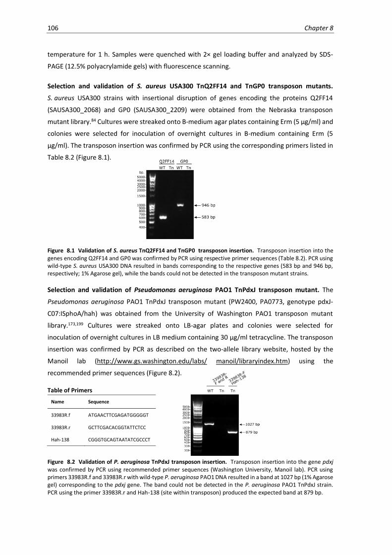
106 Chapter 8
temperature for 1 h. Samples were quenched with 2× gel loading buffer and analyzed by SDS-
PAGE (12.5% polyacrylamide gels) with fluorescence scanning.
Selection and validation of S. aureus USA300 TnQ2FF14 and TnGP0 transposon mutants.
S. aureus USA300 strains with insertional disruption of genes encoding the proteins Q2FF14
(SAUSA300_2068) and GP0 (SAUSA300_2209) were obtained from the Nebraska transposon
mutant library.84 Cultures were streaked onto B-medium agar plates containing Erm (5 µg/ml) and
colonies were selected for inoculation of overnight cultures in B-medium containing Erm (5
µg/ml). The transposon insertion was confirmed by PCR using the corresponding primers listed in
Table 8.2 (Figure 8.1).
Figure 8.1 Validation of S. aureus TnQ2FF14 and TnGP0 transposon insertion. Transposon insertion into the genes encoding Q2FF14 and GP0 was confirmed by PCR using respective primer sequences (Table 8.2). PCR using wild-type S. aureus USA300 DNA resulted in bands corresponding to the respective genes (583 bp and 946 bp, respectively; 1% Agarose gel), while the bands could not be detected in the transposon mutant strains.
Selection and validation of Pseudomonas aeruginosa PAO1 TnPdxJ transposon mutant. The
Pseudomonas aeruginosa PAO1 TnPdxJ transposon mutant (PW2400, PA0773, genotype pdxJ-
C07:ISphoA/hah) was obtained from the University of Washington PAO1 transposon mutant
library.173,199 Cultures were streaked onto LB-agar plates and colonies were selected for
inoculation of overnight cultures in LB medium containing 30 µg/ml tetracycline. The transposon
insertion was confirmed by PCR as described on the two-allele library website, hosted by the
Manoil lab (http://www.gs.washington.edu/labs/ manoil/libraryindex.htm) using the
recommended primer sequences (Figure 8.2).
Table of Primers
Name Sequence
33983R.f ATGAACTTCGAGATGGGGGT
33983R.r GCTTCGACACGGTATTCTCC
Hah-138 CGGGTGCAGTAATATCGCCCT
Figure 8.2 Validation of P. aeruginosa TnPdxJ transposon insertion. Transposon insertion into the gene pdxj was confirmed by PCR using recommended primer sequences (Washington University, Manoil lab). PCR using primers 33983R.f and 33983R.r with wild-type P. aeruginosa PAO1 DNA resulted in a band at 1027 bp (1% Agarose gel) corresponding to the pdxj gene. The band could not be detected in the P. aeruginosa PAO1 TnPdxJ strain. PCR using the primer 33983R.r and Hah-138 (site within transposon) produced the expected band at 879 bp.

Experimental Procedures 107
8.3 Crystallography
Crystallization and structure determination for Alr. High-throughput crystallization screening was
carried out (Phoenix, Art Robbins) and all crystals were obtained by the sitting-drop vapour
diffusion method at 4 °C. Apo-Alr was crystallized with 0.1 M citric acid pH 4, 1.6 M ammonium
sulphate as the precipitant. Reconstituted Alr-PL1P and Alr-PL4P holoenzymes were crystallized
in 0.1 M Hepes pH 7 and 2 M sodium chloride and 0.1 M sodium acetate pH 5 containing 10% 2-
methyl-1,3,-propanediol (MPD), respectively. Prior to cryo-cooling and storage in liquid nitrogen,
either lithium sulphate (Apo-Alr, Alr-PL1P complex) or MPD (Alr-PL4P complex) was added to the
crystallization drop for cryo-protection. Crystals were screened and diffraction data was collected
at the beamlines of the European Synchrotron Radiation Facility (ESRF, ID23-2 and ID29) and the
Swiss Light Source (SLS, PX I). The data were processed with XDS200 and the crystals belonged to
the space group P212121. The resolution cut-offs were chosen according to the correlation
coefficient of random half-data sets (1/2 CC) at about 50%.201-203 The structures were solved using
the coordinates of the previously reported structure (PDB code 4A3Q)83 by molecular replacement
in PHASER.204 This was followed by simulated annealing in PHENIX,205,206 prior to iterative cycles of
manual model building in COOT207 and restraint and TLS refinement in REFMAC5.208 Structure
optimization was carried out using the PDBredo server.209 For data processing and structure
refinement statistics see Table 8.4. All structural figures were prepared with PyMol (Delano
Scientific).
Crystallization and structure determination for Q2FF14. Crystals of S. aureus Q2FF14 were
obtained in 0.1 M bicine, pH 9.0, 1.6 M ammonium sulphate. Prior to cryo-cooling and storage in
liquid nitrogen, 2 M lithium sulphate was added to the crystallisation drop. Data collection was
carried out at the ESRF beamline MASSIF-1 automatically210,211 and the data were processed with
XDS to 1.75 Å (resolution cut-off CC1/2 ~ 50%, symmetry P21). The structure was solved by
molecular replacement with PHASER using the coordinates of the homologous, hypothetical
protein TT1679 from Thermus thermophilus (PDB code 1V8D, 40% sequence identity). Model
building, refinement and validation were carried out as for Alr (see above). For data processing
and structure refinement statistics see Supporting Table 8.4.
All structural figures were prepared with PyMol (Delano Scientific) or UCSF Chimera. Sequence
alignments were done with ClustalX.116

108 Chapter 8
Table 8.4 Crystallographic data collection and refinement statistics. Statistics for the highest-resolution shell
are shown in parantheses.
Apo PL1P-complex PL4P-complex Q2FF14
Wavelength (Å) 0.976 0.873 0.979 0.966
Resolution range 45.2-2.15 (2.2-2.15) 49.5-2.45 (2.54-2.45) 49.4-1.9 (1.96-1.9) 45.3-1.75 (1.81-1.75)
Space group P 21 21 21 P 21 21 21 P 21 21 21 P 21
Unit cell 64.7 114.5 126.1 85.9 106.9 131.0 85.5 106.8 130.8 54.7 136.9 79.2
= 98.6
Total reflections 267,266 (22,547) 224,459 (21,447) 637,180 (55,865) 356,535 (35,255)
Unique reflections 51,458 (4,390) 44,955 (4,399) 95,389 (8,791) 114,397 (11,371)
Multiplicity 5.2 (5.1) 5.0 (4.9) 6.7 (6.4) 3.1 (3.1)
Completeness (%) 1.00 (0.99) 1.00 (0.99) 0.99 (0.92) 0.99 (0.99)
Mean I/sigma(I) 6.0 (0.4) 6.6 (1.4) 10.7 (1.2) 10.4 (0.90)
Wilson B-factor 34.4 32.3 28.7 30.3
R-merge 0.166 (1.73) 0.213 (1.02) 0.13 (1.48) 0.056 (1.19)
CC1/2 0.99 (0.85) 0.983 (0.5) 1.0 (0.45) 0.998 (0.534)
Reflections used in refinement 50,710 (4,797) 44,949 (4,399) 95,378 (8,791) 114,296 (11,325)
Reflections used for R-free 2,534 (237) 2,247 (219) 4,786 (462) 5,713 (563)
R-work 0.2041 (0.337) 0.195 (0.297) 0.199 (0.346) 0.194 (0.398)
R-free 0.234 (0.37) 0.215 (0.33) 0.21 (0.359) 0.220 (0.396)
Number of non-hydrogen atoms 6,475 6,341 6,712 8,510
macromolecules 6,040 6,102 6,104 8,006
ligands 138 68 125 85
Protein residues 763 771 764 1,058
RMS(bonds) 0.011 0.009 0.012 0.017
RMS(angles) 1.42 1.4 1.5 1.9
Ramachandran favoured (%) 97 97 97 96
Ramachandran allowed (%) 2.9 3 2.7 3
Ramachandran outliers (%) 0.3 0 0 0.57
Rotamer outliers (%) 1.7 1.2 0.9 1.6
Clashscore 1.5 2.3 1.1 3.2
Average B-factor 34.0 35.5 22.0 42.16
macromolecules 33.7 35.5 20.2 42.12
ligands 56.0 50.9 46.5 56.3
solvent 29.0 27.7 39.1 40.2
Number of TLS groups 14 2 14 42

Experimental Procedures 109
8.4 Proteomics
S. aureus growth and labeling for chemical proteomics (standard protocol). Overnight cultures
of S. aureus USA300 TnPdxS grown in B-medium containing 5 µg/ml Erm were diluted 1:100 into
fresh media (~ 100 ml for 3 samples) and grown for 5 h until early stationary phase (OD600 = 2.2 -
2.5). Bacteria were harvested (6,000 rpm, 5 min, 4°C), washed twice with 10 ml CDM-Erm
(5 µg/ml) and resuspended to an OD600 = 40. For each sample, 1 ml of bacterial suspension (OD600
= 40) was incubated with 10 µl probe (100× stock in DMSO) or DMSO in culture tubes for 2 h at
37°C, with shaking. For samples treated with DCS, bacteria were incubated with 50 µl DCS (20×
stock prepared fresh in 0.1 M sodium phosphate buffer, pH 8) for 30 min at 37°C, with shaking,
prior to probe labeling. Bacteria were subsequently harvested, washed with PBS (2 × 1 ml, 4°C)
and resuspended in 1 ml PBS for cell lysis.
S. aureus growth and labeling for chemical proteomics (PL1/PL3 growth supplementation).
Overnight cultures of S. aureus USA300 TnPdxS grown in B-medium (20 ml) containing 5 µg/ml
Erm were harvested (6,000 rpm, 5 min, 4°C), washed twice with 5 ml CDM-Erm (5 µg/ml) and
resuspended in 5 ml. This suspension was used to inoculate bacterial cultures to an OD600 = 0.1 in
100 ml CDM-Erm supplemented with either 25 µM probe (25 µl of 100 mM stock in DMSO) or
0.5 µM PL (25 µl of 2 mM stock in DMSO) in baffled flasks. The cultures were grown for 30 h at
37°C with shaking, at which point they were harvested (6,000 rpm, 5 min, 4°C), washed twice with
1 ml PBS, resuspended and adjusted to 1 ml of OD600 = 60 prior to cell lysis.
P. aeruginosa growth and labeling for chemical proteomics (standard protocol). Overnight
cultures of P. aeruginosa PAO1 TnPdxJ (obtained as described in biological methods) grown in LB-
medium were diluted 1:100 into fresh media and grown for 5 h until early stationary phase.
Bacteria were harvested (6,000 rpm, 5 min, 4°C), washed twice with 10 ml MOPS-succinate
medium (50 mM MOPS pH7.2, 20 mM sodium succinate, 20 mM NH4Cl, 1 mM MgSO4, 10 mM KCl,
3.5 µM FeSO4) and resuspended to an OD600 = 40. For each sample, 1 ml of bacterial suspension
(OD600 = 40) was incubated with 10 µl probe (100× stock in DMSO) or DMSO in culture tubes for
2 h at 37°C with shaking. Bacteria were subsequently harvested, washed with PBS (2 × 1 ml, 4°C)
and resuspended in 1 ml PBS for cell lysis.
Bacterial cell lysis and NaBH4 reduction. Bacterial suspensions prepared above were transferred
to 2 ml glass-bead lysis tubes (Precellys® Lysing Kit, soil-grinding SK38). Cells were lysed
mechanically using a Precellys® 24 Homogenizer (6 × 15 sec shaking at 5400 rpm) with 2-min
cooling breaks on ice between cycles. Upon centrifugation (18,000 x g, 30 min, 4°C), the clarified
lysates were reduced with 20 mM NaBH4, (500 mM stock prepared fresh in 0.1 M NaOH) at room
temperature for 30 min. Proteins were subsequently precipitated by adding ice-cold acetone (4×
volume) and stored at -20°C overnight. Precipitated proteins were pelleted by centrifugation

110 Chapter 8
(18,000 x g, 15 min, 4°C) and washed with ice-cold MeOH (2 × 1 ml), using sonication to resuspend
the pellets between washes. Proteins were solubilized in 0.5 ml PBS containing 0.4% (w/v) SDS
and protein concentrations were adjusted to 2 mg/ml (= 1 mg total per sample). Samples from
chemical supplementation experiments were instead solubilized in 1 ml PBS containing 0.4% SDS
(w/v) and adjusted to 2 mg/ml (= 2 mg total per sample).
P. aeruginosa labeling in cell lysate. Overnight cultures of P. aeruginosa PAO1 grown in LB-
medium were diluted 1:100 into fresh media and grown for 5 h until early stationary phase.
Bacteria were harvested (6,000 rpm, 5 min, 4°C), washed twice with 10 ml PBS buffer and
resuspended to an OD600 = 40 in PBS containing protease inhibitors (Complete Mini Protease
Inhibitor Cocktail, 1 tablet per 10 ml PBS, Roche). Bacteria were lysed by sonication and
centrifuged (18,000 x g, 30 min, 4°C). For each labeling reaction, 750 µl clarified lysate was
incubated with 5 µl PL4P (300× stock in kinase buffer: 50 mM Tris, 50 mM KCl, 10 mM MgCl2, pH
7.9) or kinase buffer as a control, for 45 min on ice. Samples were reduced with 20 mM NaBH4,
(500 mM stock prepared fresh in 0.1 M NaOH) at room temperature for 30 min and proteins were
subsequently precipitated by adding ice-cold acetone (4× volume) and storing at -20°C overnight.
Precipitated proteins were pelleted by centrifugation (18,000 x g, 15 min, 4°C) and washed with
ice-cold MeOH (2 × 1 ml), using sonication to resuspend the pellets between washes. Proteins
were solubilized in 0.5 ml PBS containing 0.4% (w/v) SDS and protein concentrations were
adjusted to 2 mg/ml (= 1 mg total per sample) prior to click chemistry and enrichment.
HeLa cell growth and labeling for chemical proteomics. HeLa cells were grown to 80-90%
confluency in a 15 cm dish in Dulbecco’s Modified Eagle Medium (DMEM, Gibco) supplemented
with 5 mM glutamine and 10% FCS. Cells were incubated with 100 or 10 µM PL4 probe (100 mM
stock) or DMSO for 1 h at 37°C, upon which they were detached with a cell scraper, centrifuged
(600 x g, 5 min, 4°C) and washed with 1 ml PBS. Cell pellets were resuspended in 1 ml lysis buffer
(PBS + 1% v/v NP-40 and 1% w/v sodium deoxycholate) and incubated on ice for 20 min. The lysate
was clarified by centrifugation (18,000 x g, 30 min, 4°C) and samples were then reduced with 20
mM NaBH4 (500 mM stock prepared fresh in 0.1 M NaOH) at room temperature for 30 min.
Proteins were subsequently precipitated by adding ice-cold acetone (4× volume) and stored at -
20°C overnight. Precipitated proteins were pelleted by centrifugation (18,000 x g, 15 min, 4°C) and
washed with ice-cold MeOH (2 × 1 ml), using sonication to resuspend the pellets between washes.
Proteins were solubilized in 0.5 ml PBS containing 0.4% (w/v) SDS and protein concentrations were
adjusted to 2 mg/ml (= 1 mg total per sample) prior to click chemistry and enrichment.
Click chemistry. Samples were subjected to CuAAC by adding 0.1 mM tri-functional rhodamine-
biotin-azide96 linker (10 mM stock in DMSO), 0.5 mM BTTAA ligand196 (10 mM stock in ddH2O), 1
mM CuSO4 (50 mM stock in ddH2O) and 2 mM NaAsc (100 mM prepared fresh in ddH2O) to each
sample and incubating for 1 h at room temperature in the dark. PL1 and PL2-labeled samples were
Experimental Procedures 111
incubated with 20 mM IAA (500 mM prepared fresh in ddH2O) for 30 min at rt prior to click
chemistry. PL3-labeled samples were incubated with 0.2 mM EZ-LinkTM Phosphine-PEG3-Biotin
(Sigma Aldrich) (10 mM stock in DMSO) for 4 h at 37°C, followed by 20 h at 25°C. Upon acetone
precipitation and washing (as described above), the protein pellets were re-solubilized in 0.5 ml
PBS containing 0.4% SDS (w/v) and centrifuged (18,000 x g, 5 min, rt) prior to avidin bead
enrichment. Samples from chemical supplementation experiments were instead re-solubilized in
1 ml PBS containing 0.4% (w/v) SDS.
Enrichment and sample preparation for MS. Solubilized, labeled protein (0.5-1 ml, 2 mg/ml in
0.4% (w/v) SDS in PBS) was transferred to Lo-bind Eppendorf tubes containing 50 µl of avidin-
agarose bead slurry (Sigma Aldrich) pre-equilibrated with 0.4% (w/v) SDS in PBS (3 × 1 ml, 400 x g,
5 min, rt), and samples were rotated for 1 h at room temperature. The beads were then washed
with 0.4% (w/v) SDS in PBS (3 × 1 ml), 6 M urea (2 × 1 ml) and finally PBS (3 × 1 ml). During the last
washing step, 10% of the sample was removed for gel-based analysis of enrichment. This fraction
was heated (10 min, 95°C) in 25 µl gel loading buffer, centrifuged (13,000 rpm, 3 min) and analyzed
by fluorescence SDS-PAGE (NuPAGE 4-12% Bis-Tris gel, Thermo Fisher Scientific). The remaining
beads were resuspended in 200 µl X-buffer (7 M urea, 2 M thiourea in 20mM HEPES buffer, pH
7.5). Upon reduction with 5 mM TCEP (2 µl of 500 mM stock in ddH2O) for 1 h at 37°C, proteins
were alkylated using 10 mM IAA (4 µl of 500 mM stock in ddH2O) for 30 min at 25°C and samples
were quenched with 10 mM DTT (4 µl of 500 mM stock in ddH2O) for 30 min at 25°C. Enzymatic
digestion using LysC (1 µl of 0.5 µg/µl, Wako, MS-grade) was first carried out for 2 h at 25°C, upon
which samples were diluted with triethylammonium bicarbonate (TEAB) buffer (600 µl of 50 mM
stock in ddH2O) and digested with trypsin (1.5 µl of 0.5 µg/µl in 50 mM acetic acid, Promega,
sequencing grade) for an additional 16 h at 37°C. Samples were acidified to 1% (v/v) FA and
desalted using SepPak® C18 cartridges (50 mg, Waters) with a vacuum manifold.212 The cartridges
were first washed with ACN (2 × 1 ml) and equilibrated with 0.1% (v/v) TFA (3 × 1 ml) prior to
sample loading. After washing with 0.1% (v/v) TFA (3 × 1 ml) and 0.5% (v/v) FA (1 × 0.5 ml),
peptides were eluted in 80% (v/v) ACN containing 0.5% FA (3 × 0.25 ml) and freeze-dried using a
speedvac centrifuge. Samples were prepared for MS-analysis by dissolving in 25 µl of 1% (v/v) FA
and filtering through 0.22 µm PVDF filters (Millipore). All samples were prepared in three
biological replicates from individual cultures and were processed in parallel.
Dimethyl labeling. Labeled proteins were enriched, reduced, alkylated and digested as described
above. Samples were acidified to 1% (v/v) FA and peptides were dimethyl labeled on column using
SepPak® C18 cartridges (50 mg, Waters) with a vacuum manifold.212 The cartridges were first
washed with ACN (2 × 1 ml) and equilibrated with 0.5% (v/v) FA (3 × 1 ml) prior to loading the
samples. After washing with 0.5% (v/v) FA (4 × 1 ml), each sample was flushed with respective
labeling reagents (5 × 1 ml Light: 30 mM NaBH3CN, 0.2% formaldehyde; Medium: 30 mM
NaBH3CN, 0.2% formaldehyde-d2; Heavy: 30 mM NaBD3CN, 0.2% 13C-formaldehyde-d2, each in 45

112 Chapter 8
mM sodium phosphate buffer pH 7.5). The columns were subsequently washed with 0.5% (v/v)
FA (2 × 1 ml) and eluted in 80% (v/v) ACN containing 0.5% FA (3 × 0.25 ml). Labels were mixed and
freeze-dried using a speedvac centrifuge. Samples were prepared for MS-analysis by dissolving in
20 µl of 1% (v/v) FA and filtering through 0.22 µm PVDF filters (Millipore). All samples were
prepared in three biological replicates from individual cultures and were processed in parallel.
Whole-proteome samples. Overnight cultures of wild-type S. aureus USA300 and TnQ2FF14 or
TnGP0 (obtained as described in Biological Methods) were grown in B-medium containing 5 µg/ml
Erm and diluted 1:100 into fresh media for the day culture. Bacteria were grown for 5 h until early
stationary phase (OD600 = 5), at which point 2 ml of the culture was harvested (6,000 rpm, 5 min,
4°C). The cell pellet was washed with PBS (2 × 1 ml, 4°C) and resuspended in 0.5 ml lysis buffer (50
mM Tris, 75 mM NaCl pH 8, containing 8 M urea, 1 mM EDTA and Complete Mini Protease Inhibitor
Cocktail, Roche (1 tablet per 10 ml)). Samples were lysed in 2 ml glass-bead lysis tubes (Precellys®
Lysing Kit, soil-grinding SK38) using a Precellys® 24 Homogenizer (6 × 15 sec shaking at 5400 rpm)
with 2-min cooling breaks on ice between cycles. Cell lysates were clarified by centrifugation
(18,000 x g, 30 min, 4°C) and protein concentration was adjusted to 0.2 ml of 0.25 mg/ml (= 0.25
mg). Samples were reduced with 10 mM DTT (1 M stock in ddH2O) for 1 h at 37°C, alkylated with
55 mM IAA (500 mM stock in ddH2O) for 30 min at 25°C and quenched with 40 mM DTT (1 M stock
in ddH2O) for 30 min at 25°C. Proteins were first digested using LysC (2 µl of 0.5 µg/µl, Wako, MS-
grade) for 2 h at 25°C, upon which samples were diluted with triethylammonium bicarbonate
(TEAB) buffer (600 µl of 50 mM stock in ddH2O) and digested with trypsin (4 µl of 0.5 µg/µl in 50
mM acetic acid, Promega, sequencing grade) for an additional 16 h at 37°C. Samples were acidified
to 1% (v/v) FA and desalted using SepPak® C18 cartridges (50 mg, Waters) with a vacuum
manifold.212 The cartridges were first washed with ACN (2 × 1 ml) and equilibrated with 0.1% (v/v)
TFA (3 × 1 ml) prior to loading the samples. After washing with 0.1% (v/v) TFA (3 × 1 ml) and 0.5%
(v/v) FA (1 × 0.5 ml), peptides were eluted in 80% (v/v) ACN containing 0.5% FA (3 × 0.25 ml) and
freeze-dried using a speedvac centrifuge. Samples were prepared for MS-analysis by dissolving in
100 µl of 1% (v/v) FA and filtering through 0.22 µm PVDF filters (Millipore). All samples were
prepared in three biological replicates from individual cultures and were processed in parallel.
MS measurement and analysis. LC-MS/MS analysis was performed with an Ultimate3000 Nano-
HPLC system (Thermo Scientific) coupled to an Orbitrap Fusion instrument (Thermo Scientific).
Samples were loaded on a 2 cm PepMap RSLC C18 trap column (particles 2 µm, 100 Å, inner
diameter 75 µm, Thermo Scientific) with 0.1% TFA and separated on a 50 cm PepMap RSLC C18
column (particles 2 µm, 100 Å, inner diameter 75 µm, Thermo Scientific) heated at 50 °C. The
gradient was run from 5-32% ACN containing 0.1% formic acid during a 152 min method (7 min
5%, 105 min to 22%, 10 min to 32%, 10 min to 90%, 10 min wash at 90%, 10 min equilibration at
5%) at a flow rate of 300 nl/min. Survey scans (m/z 300-1500) were acquired in the orbitrap with
a resolution of 120,000 at m/z 200 and the maximum injection time set to 50 ms (target value

Experimental Procedures 113
2e5). The most intense ions of charge states 2-7 were selected for fragmentation with high-energy
collisional dissociation at a normalized collision energy of 30%. The instrument was operated in
top speed mode and spectra were acquired in the ion trap with the maximum injection time set
to 50 ms (target value 1e4). The option to inject ions for all available parallelizable time was
enabled. Dynamic exclusion of sequenced peptides was set to 60 s. Data were acquired using
Xcalibur software version 3.0sp2 (Thermo Scientific).
MS raw files were analyzed with MaxQuant213 software (version 1.5.3.8). MS/MS-based peptide
identification was carried out using the Andromeda214 search engine with the S. aureus USA300
UniProtKB database (May 2016, 2607 accessions). For other experiments, the S. aureus NCTC8325
UniProtKB database (October 2017, 2889 accessions), the P. aeruginosa PAO1 UniProtKB database
(September 2017, 5563 accessions) or human UniprotKB database (May 2016, 70625 accessions)
were used. The following parameter settings were used: peptide and protein FDR 1%, enzyme
specificity trypsin, minimal number of amino acids required for peptide identification 7, variable
modification methionine oxidation, fixed modification carbamidomethylation. At least two unique
peptides were required for the identification of proteins. All other parameters were used
according to the default settings. For label-free protein quantification, the MaxLFQ94 algorithm
was used as part of the MaxQuant environment. The ‘match between runs’ option was enabled
to maximize the number of quantification events across all replicates. Statistical analysis was
performed in Perseus215 (version 1.5.5.3). Proteins identified only by site modification, reverse hits
or contaminants were removed. Data were filtered to retain only those proteins with > 2 MS/MS
counts and quantified in at least 2 out of 3 replicates (alternatively: 5/6 replicates or 8/9
replicates). Missing values were then imputed on the basis of a normal distribution (width=0.3,
down-shift=1.8). Volcano plots were generated by performing a two-sample t-test with
permutation-based statistics (FDR 0.05, s0 = 1, or other as specified). The complete lists of PLP-
DEs (PLPomes) for the selected species were identified by gene ontology (GO-term: 0030170;
pyridoxal-phosphate binding), EC classification (enzyme.expasy.org; cofactor search: pyridoxal 5’-
phosphate), and Uniprot (www.uniprot.org, cofactor search: pyridoxal 5’-phosphate).
Overrepresentation analysis is based on gene ontology annotations and was performed with the
Bingo App in the Cytoscape environment.216 Statistically significant proteins from the volcano plot
were compared to all proteins present in the plot for the annotation pyridoxal phosphate binding
(GO:0030170) in the category of molecular process. According to literature research (see searches
described above), we additionally annotated the following proteins as pyridoxal phosphate
binding and added them to the GO list: A0A0H2XFQ3, A0A0H2XG73, A0A0H2XGD2, A0A0H2XGZ7,
A0A0H2XHH8, A0A0H2XII6, A0A0H2XIJ0, A0A0H2XIS2, A0A0H2XJX6, A0A0H2XKI8, Q2FF55,
Q2FGI6, Q2FGI7, Q2FH63, Q2FH64, Q2FHT1 and Q2FKE3. Analysis was based on a
hypergeometrical test with the multiple testing correction according to Benjamini-Hochberg and
a significance level of 0.05.

114 Chapter 8
Confidence plot analysis. Only proteins significantly enriched (104 proteins, volcano plot Figure
4.3b, Table 4.1) were selected for analysis. Profile plots of log2 LFQ intensities of individual proteins
across proteomic samples were generated using Perseus. Each of the 18 PLP-DEs was individually
selected as a reference profile and the 10 or 20 most similar profiles were identified by Pearson
correlation. Corresponding proteins were grouped into confidence classes as illustrated in
Figure 4.3.
Heatmap and clustering analysis for concentration-dependent probe enrichment. The mean raw
LFQ intensities from triplicate measurements were normalized to DMSO (= 0) and highest LFQ
intensity (= 1) for individual proteins across eight probe concentrations. Samples negatively
enriched compared to DMSO were set equal to 0 for analysis. Profile plots of the normalized mean
LFQ intensities were generated across different probe concentrations using Perseus and
hierarchical clustering (Pearson, max number of clusters = 5) was performed to group similarly
behaving proteins. The heatmap was prepared using OriginPro (2017) graphing and analysis
software.
Heatmap analysis of PL4, PL5 and PL6. The mean raw LFQ intensities from triplicate
measurements were normalized to DMSO (= 0) and highest LFQ intensity (= 1) for individual PLP-
DE within PL4-, PL5- and PL6-enriched samples. The heatmap was prepared using OriginPro (2017)
graphing and analysis software.
Experimental Procedures 115
8.5 Metabolomics
Targeted PL4P metabolomics. Bacteria were grown and labeled as described for the proteomic
workflow (see Proteomics section). For each sample, 2.5 ml of bacterial suspension (OD600 = 40)
was incubated with 25 µl probe (1000× stock in DMSO) in culture tubes for 2 h at 37°C with
shaking. Samples were then shock-cooled to 4°C in a dry ice/ethanol bath while agitating to avoid
local freezing. Bacteria were harvested (6000 rpm, 5 min, 4°C), washed with 0.6% (w/v) NaCl
solution (2 × 5 ml) and resuspended in 5 ml extraction buffer (100% MeOH containing 0.5 µM PL
as an internal standard). Cells were lysed by sonication (3 x 20 sec, with cooling breaks on ice) and
samples were stored overnight at -80°C. Cell debris and precipitated protein were pelleted by
centrifugation (18,000 x g, 30 min, 4°C) and the clarified extract was evaporated under a stream
of nitrogen. Metabolome samples were dissolved in 150 µl H2O/ACN (50:50, incl. 1% FA),
centrifuged (18,000 x g, 15 min, 4°C) and the supernatant was stored at -80 °C until LC-MS analysis.
Amounts of PL4 and PL4P were quantified using calibration curves. Samples were measured on an
Ultimate 3000 RSLC system (Thermo Scientific) coupled to an LTQ Orbitrap XL mass spectrometer
(Thermo Scientific). Chromatographic separation was performed using a SeQuant® ZIC®-pHILIC
(250 x 2.1 mm, 5 µm, 100 Å) (Merck Millipore). Data was processed using Xcalibur 2.2 SP1.48 Quan
Browser (Thermo Scientific).
Whole-metabolome profiling of S. aureus USA300 TnQ2FF14 and TnGP0 vs WT. Overnight
cultures ofwild-type S. aureus USA300 and TnQ2FF14 or TnGP0 (obtained as described in
Biological Methods) were grown in B-medium containing 5 µg/ml Erm and diluted 1:100 into fresh
B-medium for the day culture. Bacteria were grown for 5 h until early stationary phase (OD600 =
5), at which point 15 ml of the culture was harvested (6,000 rpm, 5 min, 4°C) and the bacteria
were washed with 0.9% NaCl (2 × 5 ml, 4°C). Cells were lysed by sonication (3 x 20 sec, with cooling
breaks on ice) and samples were stored overnight at -80°C. Cell debris and precipitated protein
were pelleted by centrifugation (18,000 x g, 30 min, 4°C) and the clarified extract was evaporated
in a speedvac centrifuge. Metabolome samples were dissolved in 150 µl H2O/ACN (50:50, incl. 1%
FA), centrifuged (18,000 x g, 15 min, 4°C) and the supernatant was stored at -80 °C until LC-MS
analysis. Samples were measured on an Ultimate 3000 RSLC system (Thermo Scientific) coupled
to an LTQ Orbitrap XL mass spectrometer (Thermo Scientific). Chromatographic separation was
performed using a SeQuant® ZIC®-pHILIC (250 x 2.1 mm, 5 µm, 100 Å) (Merck Millipore). Data was
processed using Xcalibur 2.2 SP1.48 Quan Browser (Thermo Scientific) and analyzed using
XCMS.217
116 Chapter 8
8.6 Binding-site Identification
Sample preparation. Recombinant protein samples (2 µg, 50 µl) were incubated with 5
equivalents of PLP for 30 min at rt, and were subsequently reduced with 10 mM NaBH4 (2 µl of
250 mM stock prepared fresh in 0.1 M NaOH) for 30 min at rt. Proteins were precipitated by adding
ice-cold acetone (4× volume) and incubating at -20°C for at least 1 h. Precipitated proteins were
pelleted by centrifugation (18,000 x g, 15 min, 4°C) and washed with ice-cold MeOH (2 × 0.2 ml),
using sonication to resuspend the pellets between washes. Samples were resuspended in 100 µl
X-buffer (7 M urea, 2 M thiourea in 20mM HEPES buffer, pH 7.5) and reduced with 5 mM TCEP (1
µl of 500 mM stock in ddH2O) for 1 h at 37°C. Proteins were alkylated using 10 mM IAA (2 µl of
500 mM stock in ddH2O) for 30 min at 25°C, followed by quenching with 10 mM DTT (2 µl of 500
mM stock in ddH2O) for 30 min at 25°C. Samples were diluted with 600 µl chymotrypsin buffer
(100 mM Tris-HCl, 10 mM CaCl2, pH 8) and digested by incubating with chymotrypsin (1 µl of 0.5
µg/µl in 1 mM HCl, Promega, sequencing grade) for 16 h at 25°C. Double-digested samples
(Q2FF14) were first incubated with trypsin (1 µl of 0.5 µg/µl in 50 mM acetic acid, Promega,
sequencing grade) for 30 min at 37°C and were then digested with chymotrypsin as described
above. Samples digested with GluC (Q2FF14) were incubated with the enzyme (5 µl of 0.1 µg/µl in
ddH2O, Roche, sequencing grade) for 16 h at 25°C. Reactions were quenched by acidification to
1% (v/v) FA and peptides were desalted using SepPak® C18 cartridges (50 mg, Waters) with a
vacuum manifold.212 The cartridges were washed with ACN (2 × 1 ml) and equilibrated with 0.1%
(v/v) TFA (3 × 1 ml) prior to loading the samples. After washing with 0.1% (v/v) TFA (3 × 1 ml) and
0.5% (v/v) FA (1 × 0.5 ml), peptides were eluted in 80% (v/v) ACN containing 0.5% FA (3 × 0.25 ml)
and freeze-dried using a speedvac centrifuge. Peptides were prepared for MS-analysis by
dissolving in 25 µl of 1% (v/v) FA and filtering through 0.22 µm PVDF filters (Millipore).
MS measurement and analysis. LC-MS/MS analysis was performed with an Ultimate3000 Nano-
HPLC system (Thermo Scientific) coupled to an Orbitrap Fusion instrument (Thermo Scientific) as
described in the proteomics section, with some modifications. For PLP binding site identification,
chymotrypsin digested samples were measured with a shorter gradient during a 62 min method.
Charge states 1-7 were considered for measurement and depend scans were only performed on
single charge state per precursor and measured in the orbitrap (AGC target 5e4, injection time 50
ms). MS raw files were analyzed with MaxQuant213 software (version 1.5.3.8) as described in the
proteomics section with some modifications. MS/MS-based peptide identification was carried out
using the Andromeda214 search engine. For PLP binding site identification, the enzyme specificity
was set to chymotrypsin (with additional cleavage after leucine and methionine) for all enzymes
except for Q2FF14 which was double-digested with trypsin and chymotrypsin or digested with
GluC. As a variable modification, the PLP moiety (+ 231.02966) at lysine residues was added with
neutral losses of H3PO4 and the PLP moiety implemented. As we expect one PLP modification site

Experimental Procedures 117
per protein, we selected the site with the highest confidence based on 100% localization
probability, best PEP (posterior error probability), score, score difference, delta score and manual
evaluation of MS/MS spectra from MaxQuant (see Appendix).


References
1 Di Salvo, M. L., Budisa, N. & Contestabile, R. in Beilstein Bozen Symposium on Molecular Engineering and Control 27-66 (Beilstein-Institut, Prien (Chiemsee), Germany, 2013).
2 Eliot, A. C. & Kirsch, J. F. Pyridoxal phosphate enzymes: mechanistic, structural, and evolutionary considerations. Annu. Rev. Biochem. 73, 383-415 (2004).
3 Schneider, G., Kack, H. & Lindqvist, Y. The manifold of vitamin B6 dependent enzymes. Structure 8, R1-6 (2000).
4 Percudani, R. & Peracchi, A. A genomic overview of pyridoxal-phosphate-dependent enzymes. EMBO Rep. 4, 850-854 (2003).
5 Toney, M. D. Reaction specificity in pyridoxal phosphate enzymes. Arch. Biochem. Biophys. 433, 279-287 (2005).
6 Toney, M. D. Controlling reaction specificity in pyridoxal phosphate enzymes. Biochim. Biophys. Acta 1814, 1407-1418 (2011).
7 Keller, J. W. et al. Pseudomonas cepacia 2,2-dialkylglycine decarboxylase. Sequence and expression in Escherichia coli of structural and repressor genes. J. Biol. Chem. 265, 5531-5539 (1990).
8 Dunathan, H. C. Conformation and reaction specificity in pyridoxal phosphate enzymes. Proc. Natl. Acad. Sci. U.S.A. 55, 712-716 (1966).
9 Chan-Huot, M., Sharif, S., Tolstoy, P. M., Toney, M. D. & Limbach, H. H. NMR studies of the stability, protonation States, and tautomerism of (13)C- AND (15)N-labeled aldimines of the coenzyme pyridoxal 5'-phosphate in water. Biochemistry 49, 10818-10830 (2010).
10 Graber, R. et al. Conversion of aspartate aminotransferase into an L-aspartate beta-decarboxylase by a triple active-site mutation. J. Biol. Chem. 274, 31203-31208 (1999).
11 Seebeck, F. P. & Hilvert, D. Conversion of a PLP-dependent racemase into an aldolase by a single active site mutation. J. Am. Chem. Soc. 125, 10158-10159 (2003).
12 Metzler, D. E. & Snell, E. E. Deamination of serine. I. Catalytic deamination of serine and cysteine by pyridoxal and metal salts. J. Biol. Chem. 198, 353-361 (1952).
13 Olivard, J., Metzler, D. E. & Snell, E. E. Catalytic racemization of amino acids by pyridoxal and metal salts. J. Biol. Chem. 199, 669-674 (1952).
14 Christen, P. & Mehta, P. K. From cofactor to enzymes. The molecular evolution of pyridoxal-5'-phosphate-dependent enzymes. Chem. Rec. 1, 436-447 (2001).
15 Dunathan, H. C. & Voet, J. G. Stereochemical evidence for the evolution of pyridoxal-phosphate enzymes of various function from a common ancestor. Proc. Natl. Acad. Sci. U.S.A. 71, 3888-3891 (1974).
16 Grishin, N. V., Phillips, M. A. & Goldsmith, E. J. Modeling of the spatial structure of eukaryotic ornithine decarboxylases. Protein Sci. 4, 1291-1304 (1995).
17 Jansonius, J. N. Structure, evolution and action of vitamin B6-dependent enzymes. Curr. Opin. Struct. Biol. 8, 759-769 (1998).
18 Mehta, P. K. & Christen, P. The molecular evolution of pyridoxal-5'-phosphate-dependent enzymes. Adv. Enzymol. Relat. Areas Mol. Biol. 74, 129-184 (2000).
19 Du, Y. L. et al. A pyridoxal phosphate-dependent enzyme that oxidizes an unactivated carbon-carbon bond. Nat. Chem. Biol. 12, 194-199 (2016).
20 Berkovitch, F. et al. A locking mechanism preventing radical damage in the absence of substrate, as revealed by the x-ray structure of lysine 5,6-aminomutase. Proc. Natl. Acad. Sci. U.S.A. 101, 15870-15875 (2004).
21 Lepore, B. W., Ruzicka, F. J., Frey, P. A. & Ringe, D. The x-ray crystal structure of lysine-2,3-aminomutase from Clostridium subterminale. Proc. Natl. Acad. Sci. U.S.A. 102, 13819-13824 (2005).
22 di Salvo, M. L. et al. On the mechanism of Escherichia coli pyridoxal kinase inhibition by pyridoxal and pyridoxal 5'-phosphate. Biochim. Biophys. Acta. 1854, 1160-1166 (2015).

120 References
23 Ghatge, M. S. et al. Pyridoxal 5'-phosphate is a slow tight binding inhibitor of E. coli pyridoxal kinase. PLoS One 7, e41680, doi:10.1371/journal.pone.0041680 (2012).
24 Ronchi, S., Zapponi, M. C. & Ferri, G. Inhibition by pyridoxal-phosphate of glyceraldehyde-3-phosphate dehydrogenase. Eur. J. Biochem. 8, 325-331 (1969).
25 Soler, M., Jabalquinto, A. M. & Beytia, E. Hog liver mevalonate kinase: inactivation by pyridoxal-5'-phosphate and evidence of dead-end inhibition by one of the substrates. Int. J. Biochem. 10, 931-935 (1979).
26 Yang, H. & Abeles, R. H. Purification and properties of Escherichia coli 4'-phosphopantothenoylcysteine decarboxylase: presence of covalently bound pyruvate. Biochemistry 26, 4076-4081 (1987).
27 Bramucci, E., Milano, T. & Pascarella, S. Genomic distribution and heterogeneity of MocR-like transcriptional factors containing a domain belonging to the superfamily of the pyridoxal-5'-phosphate dependent enzymes of fold type I. Biochem. Biophys. Res. Commun. 415, 88-93 (2011).
28 Suvorova, I. A. & Rodionov, D. A. Comparative genomics of pyridoxal 5'-phosphate-dependent transcription factor regulons in Bacteria. Microb. Genom. 2, e000047, doi:10.1099/mgen.0.000047 (2016).
29 Takenaka, T., Ito, T., Miyahara, I., Hemmi, H. & Yoshimura, T. A new member of MocR/GabR-type PLP-binding regulator of D-alanyl-D-alanine ligase in Brevibacillus brevis. FEBS J. 282, 4201-4217 (2015).
30 Cellini, B., Montioli, R., Oppici, E., Astegno, A. & Voltattorni, C. B. The chaperone role of the pyridoxal 5'-phosphate and its implications for rare diseases involving B6-dependent enzymes. Clin. Biochem. 47, 158-165 (2014).
31 Amadasi, A. et al. Pyridoxal 5'-phosphate enzymes as targets for therapeutic agents. Curr. Med. Chem. 14, 1291-1324 (2007).
32 Kappes, B., Tews, I., Binter, A. & Macheroux, P. PLP-dependent enzymes as potential drug targets for protozoan diseases. Biochim. Biophys. Acta 1814, 1567-1576 (2011).
33 Percudani, R. & Peracchi, A. The B6 database: a tool for the description and classification of vitamin B6-dependent enzymatic activities and of the corresponding protein families. BMC Bioinformatics 10, 273 (2009).
34 Catazaro, J., Caprez, A., Guru, A., Swanson, D. & Powers, R. Functional evolution of PLP-dependent enzymes based on active-site structural similarities. Proteins 82, 2597-2608 (2014).
35 Whittaker, M. M., Penmatsa, A. & Whittaker, J. W. The Mtm1p carrier and pyridoxal 5'-phosphate cofactor trafficking in yeast mitochondria. Arch. Biochem. Biophys. 568, 64-70 (2015).
36 Simon, E. S. & Allison, J. Determination of pyridoxal-5'-phosphate (PLP)-bonding sites in proteins: a peptide mass fingerprinting approach based on diagnostic tandem mass spectral features of PLP-modified peptides. Rapid Commun. Mass Spectrom. 23, 3401-3408 (2009).
37 Cravatt, B. F., Wright, A. T. & Kozarich, J. W. Activity-based protein profiling: from enzyme chemistry to proteomic chemistry. Annu. Rev. Biochem. 77, 383-414 (2008).
38 Geurink, P. P., Prely, L. M., van der Marel, G. A., Bischoff, R. & Overkleeft, H. S. Photoaffinity labeling in activity-based protein profiling. Top Curr. Chem. 324, 85-113 (2012).
39 Speers, A. E., Adam, G. C. & Cravatt, B. F. Activity-based protein profiling in vivo using a copper(i)-catalyzed azide-alkyne [3 + 2] cycloaddition. J. Am. Chem. Soc. 125, 4686-4687 (2003).
40 Agard, N. J., Prescher, J. A. & Bertozzi, C. R. A strain-promoted [3 + 2] azide-alkyne cycloaddition for covalent modification of biomolecules in living systems. J. Am. Chem. Soc. 126, 15046-15047 (2004).
41 Saxon, E. & Bertozzi, C. R. Cell surface engineering by a modified Staudinger reaction. Science 287, 2007-2010 (2000).
42 Weerapana, E., Speers, A. E. & Cravatt, B. F. Tandem orthogonal proteolysis-activity-based protein profiling (TOP-ABPP)--a general method for mapping sites of probe modification in proteomes. Nat. Protoc. 2, 1414-1425 (2007).
43 Backus, K. M. et al. Proteome-wide covalent ligand discovery in native biological systems. Nature 534, 570-574 (2016).
44 Parker, C. G. et al. Ligand and Target Discovery by Fragment-Based Screening in Human Cells. Cell 168, 527-541 e529, doi:10.1016/j.cell.2016.12.029 (2017).
45 Weerapana, E. et al. Quantitative reactivity profiling predicts functional cysteines in proteomes. Nature 468, 790-795 (2010).

References 121
46 S.A., G. M. K. J. S. Electrophilic natural products and their biological targets. Nat. Prod. Rep. 9, 659-682 (2012).
47 Grammel, M. & Hang, H. C. Chemical reporters for biological discovery. Nat. Chem. Biol. 9, 475-484 (2013).
48 Liu, T. W. et al. Genome-wide chemical mapping of O-GlcNAcylated proteins in Drosophila melanogaster. Nat. Chem. Biol. 13, 161-167 (2017).
49 Laughlin, S. T. & Bertozzi, C. R. Metabolic labeling of glycans with azido sugars and subsequent glycan-profiling and visualization via Staudinger ligation. Nat. Protoc. 2, 2930-2944 (2007).
50 Tate, E. W., Kalesh, K. A., Lanyon-Hogg, T., Storck, E. M. & Thinon, E. Global profiling of protein lipidation using chemical proteomic technologies. Curr. Opin. Chem. Biol. 24, 48-57 (2015).
51 Anderson, L. N. et al. Live Cell Discovery of Microbial Vitamin Transport and Enzyme-Cofactor Interactions. ACS Chem. Biol. 11, 345-354 (2016).
52 Romine, M. F. et al. Elucidation of roles for vitamin B12 in regulation of folate, ubiquinone, and methionine metabolism. Proc. Natl. Acad. Sci. U.S.A. 114, E1205-E1214 (2017).
53 Korytnyk, W. & Ahrens, H. 5-homopyridoxals, 5-thiopyridoxal, and related compounds. Synthesis, tautomerism, and biological properties. J. Med. Chem. 14, 947-952 (1971).
54 Korytnyk, W., Srivastava, S. C., Angelino, N., Potti, P. G. & Paul, B. A general method for modifying the 2-methyl group of pyridoxol. Synthesis and biological activity of 2-vinyl- and 2-ethynylpyridoxols and related compounds. J. Med. Chem. 16, 1096-1101 (1973).
55 Muller, I. B. et al. Poisoning pyridoxal 5-phosphate-dependent enzymes: a new strategy to target the malaria parasite Plasmodium falciparum. PLoS One 4, e4406, doi:10.1371/journal.pone.0004406 (2009).
56 Fonda, M. L. Interaction of pyridoxal analogues with glutamate apodecarboxylase and aspartate apoaminotransferase. J. Biol. Chem. 246, 2230-2240 (1971).
57 Mechanik, M. L., Torchinsky, Y. M., Florentiev, V. L. & Karpeisky, M. Y. Interaction of the apoenzyme of L-glutamate decarboxylase with pyridoxal phosphate analogues. FEBS Lett. 13, 177-180 (1971).
58 Denesyuk, A. I., Denessiouk, K. A., Korpela, T. & Johnson, M. S. Functional attributes of the phosphate group binding cup of pyridoxal phosphate-dependent enzymes. J. Mol. Biol. 316, 155-172 (2002).
59 Schnackerz, K. D. & Cook, P. F. Resolution of pyridoxal 5'-phosphate from O-acetylserine sulfhydrylase from Salmonella typhimurium and reconstitution of apoenzyme with cofactor and cofactor analogues as a probe of the cofactor binding site. Arch. Biochem. Biophys. 324, 71-77 (1995).
60 Griswold, W. R. & Toney, M. D. Role of the pyridine nitrogen in pyridoxal 5'-phosphate catalysis: activity of three classes of PLP enzymes reconstituted with deazapyridoxal 5'-phosphate. J. Am. Chem. Soc. 133, 14823-14830 (2011).
61 Blethen, S. L., Boeker, E. A. & Snell, E. E. Argenine decarboxylase from Escherichia coli. I. Purification and specificity for substrates and coenzyme. J. Biol. Chem. 243, 1671-1677 (1968).
62 Muhlradt, P. F., Morino, Y. & Snell, E. E. Vitamin b(2) analogues. Synthesis and biological activity of homologues of pyridoxal 5'-phosphate. J. Med. Chem. 10, 341-344 (1967).
63 Morino, Y. & Snell, E. E. Coenzyme activity of homologues of pyridoxal phosphate. Biochemistry 57, 1692-1699 (1967).
64 Boekelheide, V. & Linn, W. J. Rearrangements of N-Oxides. A Novel Synthesis of Pyridyl Carbinols and Aldehydes. J. Am. Chem. Soc. 76, 1286-1291 (1954).
65 Roth, G. J., Liepold, B., Müller, S. G. & Bestmann, H. J. Further Improvements of the Synthesis of Alkynes from Aldehydes. Synthesis 1, 59-62 (2004).
66 Yang, D., Shih, Y. & Liu, H. Chemical synthesis of stereospecifically labeled pyridoxamine 5'-phosphate. J. Org. Chem. 56, 2940-2946 (1991).
67 Kaiser, E. M. et al. Regiointegrity of carbanions derived by selective metalations of dimethylpyridines and -quinolines. J. Organomet. Chem. 213, 405-417 (1981).
68 Kim, Y.-C. & Jacobson, K. A. Versatile synthesis of 6-alkyl and aryl substituted pyridoxal derivatives. Synthesis 1, 119-122 (2000).
69 Mukherjee, T., Hanes, J., Tews, I., Ealick, S. E. & Begley, T. P. Pyridoxal phosphate: biosynthesis and catabolism. Biochim. Biophys. Acta 1814, 1585-1596 (2011).
70 Mulligan, J. H. & Snell, E. E. Transport and metabolism of vitamin B6 in lactic acid bacteria. J. Biol. Chem. 252, 835-839 (1977).

122 References
71 Stolz, J. & Vielreicher, M. Tpn1p, the plasma membrane vitamin B6 transporter of Saccharomyces cerevisiae. J. Biol. Chem. 278, 18990-18996 (2003).
72 di Salvo, M. L., Contestabile, R. & Safo, M. K. Vitamin B(6) salvage enzymes: mechanism, structure and regulation. Biochim. Biophys. Acta. 1814, 1597-1608 (2011).
73 Ahrens, M. L., Maass, G., Schuster, P. & Winkler, H. Kinetic study of the hydration mechanism of vitamin B6 and related compounds. J. Am. Chem. Soc. 92, 6134-6139 (1970).
74 di Salvo, M. L., Safo, M. K. & Contestabile, R. Biomedical aspects of pyridoxal 5'-phosphate availability. Front Biosci (Elite Ed), 897-913 (2012).
75 Nodwell, M. B., Koch, M. F., Alte, F., Schneider, S. & Sieber, S. A. A subfamily of bacterial ribokinases utilizes a hemithioacetal for pyridoxal phosphate salvage. J. Am. Chem. Soc. 136, 4992-4999 (2014).
76 Strominger, J. L., Ito, I. & Threnn, R. H. Competitive inhibition of enzymatic reactions by oxamycin. J. Am. Chem. Soc. 82, 998-999 (1960).
77 Watanabe, A. et al. Reaction mechanism of alanine racemase from Bacillus stearothermophilus: x-ray crystallographic studies of the enzyme bound with N-(5'-phosphopyridoxyl)alanine. J. Biol. Chem. 277, 19166-19172 (2002).
78 Toney, M. D. & Kirsch, J. F. Lysine 258 in aspartate aminotransferase: enforcer of the Circe effect for amino acid substrates and general-base catalyst for the 1,3-prototropic shift. Biochemistry 32, 1471-1479 (1993).
79 Rostovtsev, V. V., Green, L. G., Fokin, V. V. & Sharpless, K. B. A stepwise huisgen cycloaddition process: copper(I)-catalyzed regioselective "ligation" of azides and terminal alkynes. Angew. Chem. Int. Ed. Engl. 41, 2596-2599 (2002).
80 Speers, A. E. & Cravatt, B. F. Profiling enzyme activities in vivo using click chemistry methods. Chem. Biol. 11, 535-546 (2004).
81 Tornoe, C. W., Christensen, C. & Meldal, M. Peptidotriazoles on solid phase: [1,2,3]-triazoles by regiospecific copper(i)-catalyzed 1,3-dipolar cycloadditions of terminal alkynes to azides. J. Org. Chem. 67, 3057-3064 (2002).
82 van Geel, R., Pruijn, G. J., van Delft, F. L. & Boelens, W. C. Preventing thiol-yne addition improves the specificity of strain-promoted azide-alkyne cycloaddition. Bioconjug. Chem. 23, 392-398 (2012).
83 Scaletti, E. R., Luckner, S. R. & Krause, K. L. Structural features and kinetic characterization of alanine racemase from Staphylococcus aureus (Mu50). Acta Crystallogr. D. Biol. Crystallogr. 68, 82-92 (2012).
84 Fey, P. D. et al. A genetic resource for rapid and comprehensive phenotype screening of nonessential Staphylococcus aureus genes. MBio. 4, e00537-00512, doi:10.1128/mBio.00537-12 (2013).
85 Bantscheff, M., Schirle, M., Sweetman, G., Rick, J. & Kuster, B. Quantitative mass spectrometry in proteomics: a critical review. Anal. Bioanal. Chem. 389, 1017-1031 (2007).
86 Gygi, S. P. et al. Quantitative analysis of complex protein mixtures using isotope-coded affinity tags. Nature Biotechnology 17 (1999).
87 Oda, Y., Huang, K., Cross, F. R., Cowburn, D. & Chait, B. T. Accurate quantitation of protein expression and site-specific phosphorylation. Proc. Natl. Acad. Sci. U.S.A. 96, 6591-6596 (1999).
88 Paša-Tolić, L. et al. High Throughput Proteome-Wide Precision Measurements of Protein Expression Using Mass Spectrometry. J. Am. Chem. Soc. 121, 7949-7950 (1999).
89 Ong, S. E. & Mann, M. Mass spectrometry-based proteomics turns quantitative. Nat. Chem. Biol. 1, 252-262 (2005).
90 Ong, S. E. et al. Stable isotope labeling by amino acids in cell culture, SILAC, as a simple and accurate approach to expression proteomics. Mol. Cell Proteomics 1, 376-386 (2002).
91 Hsu, J.-L., Huang, S.-Y., Chow, N.-H. & Chen, S.-H. Stable-Isotope Dimethyl Labeling for Quantitative Proteomics. Anal. Chem. 75, 6843-6852 (2003).
92 Ross, P. L. et al. Multiplexed protein quantitation in Saccharomyces cerevisiae using amine-reactive isobaric tagging reagents. Mol. Cell. Proteomics 3, 1154-1169 (2004).
93 Thompson, A. et al. Tandem mass tags: a novel quantification strategy for comparative analysis of complex protein mixtures by MS/MS. Anal. Chem. 75, 1895-1904 (2003).
94 Cox, J. et al. Accurate proteome-wide label-free quantification by delayed normalization and maximal peptide ratio extraction, termed MaxLFQ. Mol. Cell Proteomics 13, 2513-2526 (2014).
References 123
95 Wu, Y., Chen, J., Liu, Z. & Wang, F. Identification of pyridoxal phosphate modified proteins using mass spectrometry. Rapid Commun. Mass Spectrom. (2017).
96 Eirich, J. et al. Pretubulysin derived probes as novel tools for monitoring the microtubule network via activity-based protein profiling and fluorescence microscopy. Mol. Biosyst. 8, 2067-2075 (2012).
97 Huisgen, R., Szeimies, G. & Möbius, L. 1.3-Dipolare Cycloadditionen, XXXII. Kinetik der Additionen organischer Azide an CC-Mehrfachbindungen. Chemische Berichte 100, 2494-2507 (1967).
98 Rudolf, G. C., Heydenreuter, W. & Sieber, S. A. Chemical proteomics: ligation and cleavage of protein modifications. Curr. Opin. Chem. Biol. 17, 110-117 (2013).
99 Ning, X., Guo, J., Wolfert, M. A. & Boons, G. J. Visualizing metabolically labeled glycoconjugates of living cells by copper-free and fast huisgen cycloadditions. Angew. Chem. Int. Ed. Engl. 47, 2253-2255 (2008).
100 Jewett, J. C., Sletten, E. M. & Bertozzi, C. R. Rapid Cu-free click chemistry with readily synthesized biarylazacyclooctynones. J. Am. Chem. Soc. 132, 3688-3690 (2010).
101 Laughlin, S. T., Baskin, J. M., Amacher, S. L. & Bertozzi, C. R. In vivo imaging of membrane-associated glycans in developing zebrafish. Science 320, 664-667 (2008).
102 Staudinger, H. & Meyer, J. Über neue organische Phosphorverbindungen III. Phosphinmethylenderivate und Phosphinimine. Helv. Chim. Acta 2, 635-646 (1919).
103 Nilsson, B. L., Kiessling, L. L. & Raines, R. T. Staudinger ligation: a peptide from a thioester and azide. Org. Lett. 2, 1939-1941 (2000).
104 Galperin, M. Y. & Koonin, E. V. 'Conserved hypothetical' proteins: prioritization of targets for experimental study. Nucleic Acids Res. 32, 5452-5463 (2004).
105 Galperin, M. Y. Conserved 'hypothetical' proteins: new hints and new puzzles. Comp. Funct. Genomics 2, 14-18 (2001).
106 Jaroszewski, L. et al. Exploration of uncharted regions of the protein universe. PLoS Biol. 7, e1000205, doi:10.1371/journal.pbio.1000205 (2009).
107 Fleischman, N. M. et al. Molecular characterization of novel pyridoxal-5'-phosphate-dependent enzymes from the human microbiome. Protein Sci. 23, 1060-1076 (2014).
108 Knight, A. M. et al. Bioinformatic analysis of fold-type III PLP-dependent enzymes discovers multimeric racemases. Appl. Microbiol. Biotechnol. 101, 1499-1507 (2017).
109 Finn, R. D. et al. The Pfam protein families database: towards a more sustainable future. Nucleic Acids Res. 44, D279-285 (2016).
110 Mozzarelli, A. & Bettati, S. Exploring the pyridoxal 5'-phosphate-dependent enzymes. Chem. Rec. 6, 275-287 (2006).
111 Prosser, G. A. & de Carvalho, L. P. Metabolomics Reveal d-Alanine:d-Alanine Ligase As the Target of d-Cycloserine in Mycobacterium tuberculosis. ACS Med. Chem. Lett. 4, 1233-1237 (2013).
112 de Carvalho, L. P. et al. Activity-based metabolomic profiling of enzymatic function: identification of Rv1248c as a mycobacterial 2-hydroxy-3-oxoadipate synthase. Chem. Biol. 17, 323-332 (2010).
113 Saito, N. et al. Metabolite profiling reveals YihU as a novel hydroxybutyrate dehydrogenase for alternative succinic semialdehyde metabolism in Escherichia coli. J. Biol. Chem. 284, 16442-16451 (2009).
114 Eswaramoorthy, S. et al. Structure of a yeast hypothetical protein selected by a structural genomics approach. Acta. Crystallogr. D. Biol. Crystallogr. 59, 127-135 (2003).
115 Ito, T. et al. Conserved pyridoxal protein that regulates Ile and Val metabolism. J. Bacteriol. 195, 5439-5449 (2013).
116 Larkin, M. A. et al. Clustal W and Clustal X version 2.0. Bioinformatics 23, 2947-2948 (2007). 117 Robert, X. & Gouet, P. Deciphering key features in protein structures with the new ENDscript
server. Nucleic Acids Research 42, W320-W324, doi:10.1093/nar/gku316 (2014). 118 Holm, L. & Rosenstrom, P. Dali server: conservation mapping in 3D. Nucleic Acids Res. 38, W545-
549 (2010). 119 Kishishita, S., Terada, T., Shirouzu, M., Kuramitsu, S., Yokoyama, S. Crystal structure of the
conserved hypothetical protein TT1679 from Thermus thermophilus. Protein Data Bank (PDB), unpublished (2004).
120 Bhuiya, M. W. et al. The first crystal structure of hyperthermostable NAD-dependent glutamate dehydrogenase from Pyrobaculum islandicum. J. Mol. Biol. 345, 325-337 (2005).
121 Srivastava, P. et al. Structural characterization of a Gcn5-related N-acetyltransferase from Staphylococcus aureus. PLoS One 9, e102348, doi:10.1371/journal.pone.0102348 (2014).

124 References
122 Kuhn, M. L., Majorek, K. A., Minor, W. & Anderson, W. F. Broad-substrate screen as a tool to identify substrates for bacterial Gcn5-related N-acetyltransferases with unknown substrate specificity. Protein Sci. 22, 222-230 (2013).
123 Smith, C. A. et al. METLIN: a metabolite mass spectral database. Ther. Drug Monit. 27, 747-751 (2005).
124 Sulzenbacher, G. et al. Crystal structure of E. coli K-12 YggS (Protein Data Bank, 2006). 125 Prunetti, L. et al. Evidence That COG0325 Proteins are involved in PLP Homeostasis. Microbiology
(2016). 126 Darin, N. et al. Mutations in PROSC Disrupt Cellular Pyridoxal Phosphate Homeostasis and Cause
Vitamin-B6-Dependent Epilepsy. Am. J. Hum. Genet. 99, 1325-1337 (2016). 127 Ebihara, A. et al. Structure-based functional identification of a novel heme-binding protein from
Thermus thermophilus HB8. J. Struct. Funct. Genomics 6, 21-32 (2005). 128 Dailey, H. A., Gerdes, S., Dailey, T. A., Burch, J. S. & Phillips, J. D. Noncanonical coproporphyrin-
dependent bacterial heme biosynthesis pathway that does not use protoporphyrin. Proc. Natl. Acad. Sci. U.S.A. 112, 2210-2215 (2015).
129 Lobo, S. A. et al. Staphylococcus aureus haem biosynthesis: characterisation of the enzymes involved in final steps of the pathway. Mol. Microbiol. 97, 472-487 (2015).
130 Labbé, R. F. & Nielson, L. in Porphyrins in human diseases, 1st International Porphyrin Meeting, Freiburg i.Br., May 1975. Vol. (ed M. Doss) 141-147 (Karger, Basel, Switzerland, 1976).
131 Charette, M. & Gray, M. W. Pseudouridine in RNA: what, where, how, and why. IUBMB Life 49, 341-351 (2000).
132 Thapa, K., Oja, T. & Metsa-Ketela, M. Molecular evolution of the bacterial pseudouridine-5'-phosphate glycosidase protein family. FEBS J 281, 4439-4449 (2014).
133 Cho, S. W., Yoon, H. Y., Ahn, J. Y., Lee, E. Y. & Lee, J. Cassette mutagenesis of lysine 130 of human glutamate dehydrogenase. An essential residue in catalysis. Eur. J. Biochem. 268, 3205-3213 (2001).
134 Liu, J. Q., Dairi, T., Kataoka, M., Shimizu, S. & Yamada, H. L-allo-threonine aldolase from Aeromonas jandaei DK-39: gene cloning, nucleotide sequencing, and identification of the pyridoxal 5'-phosphate-binding lysine residue by site-directed mutagenesis. J. Bacteriol. 179, 3555-3560 (1997).
135 Chen, D. & Frey, P. A. Identification of lysine 346 as a functionally important residue for pyridoxal 5'-phosphate binding and catalysis in lysine 2, 3-aminomutase from Bacillus subtilis. Biochemistry 40, 596-602 (2001).
136 Tang, K. H., Harms, A. & Frey, P. A. Identification of a novel pyridoxal 5'-phosphate binding site in adenosylcobalamin-dependent lysine 5,6-aminomutase from Porphyromonas gingivalis. Biochemistry 41, 8767-8776 (2002).
137 Finn, R. D. et al. InterPro in 2017-beyond protein family and domain annotations. Nucleic Acids Res. 45, D190-D199 (2017).
138 Lambert, M. P. & Neuhaus, F. C. Mechanism of D-cycloserine action: alanine racemase from Escherichia coli W. J. Bacteriol. 110, 978-987 (1972).
139 Meroueh, S. O. et al. Three-dimensional structure of the bacterial cell wall peptidoglycan. Proc. Natl. Acad. Sci. U.S.A. 103, 4404-4409 (2006).
140 Lobocka, M., Hennig, J., Wild, J. & Klopotowski, T. Organization and expression of the Escherichia coli K-12 dad operon encoding the smaller subunit of D-amino acid dehydrogenase and the catabolic alanine racemase. J. Bacteriol. 176, 1500-1510 (1994).
141 Strych, U., Huang, H. C., Krause, K. L. & Benedik, M. J. Characterization of the alanine racemases from Pseudomonas aeruginosa PAO1. Curr. Microbiol. 41, 290-294 (2000).
142 Strych, U. & Benedik, M. J. Mutant analysis shows that alanine racemases from Pseudomonas aeruginosa and Escherichia coli are dimeric. J. Bacteriol. 184, 4321-4325 (2002).
143 Mitnick, C. D. et al. Comprehensive treatment of extensively drug-resistant tuberculosis. N. Engl. J. Med. 359, 563-574 (2008).
144 Torun, T. et al. Side effects associated with the treatment of multidrug-resistant tuberculosis. Int. J. Tuberc. Lung. Dis. 9, 1373-1377 (2005).
145 Caminero, J. A., Sotgiu, G., Zumla, A. & Migliori, G. B. Best drug treatment for multidrug-resistant and extensively drug-resistant tuberculosis. Lancet Infect. Dis. 10, 621-629 (2010).

References 125
146 LeMagueres, P. et al. The 1.9 A crystal structure of alanine racemase from Mycobacterium tuberculosis contains a conserved entryway into the active site. Biochemistry 44, 1471-1481 (2005).
147 Fenn, T. D., Stamper, G. F., Morollo, A. A. & Ringe, D. A side reaction of alanine racemase: transamination of cycloserine. Biochemistry 42, 5775-5783 (2003).
148 Peisach, D., Chipman, D. M., Van Ophem, P. W., Manning, J. M. & Ringe, D. D-cycloserine inactivation of D-amino acid aminotransferase leads to a stable noncovalent protein complex with an aromatic cycloserine-PLP derivative. J. Am. Chem. Soc. 120, 2268-2274 (1998).
149 Soper, T. S. & Manning, J. M. Different modes of action of inhibitors of bacterial D-amino acid transaminase. A target enzyme for the design of new antibacterial agents. J. Biol. Chem. 256, 4263-4268 (1981).
150 David, S. Synergic activity of D-cycloserine and beta-chloro-D-alanine against Mycobacterium tuberculosis. J. Antimicrob. Chemother. 47, 203-206 (2001).
151 Neuhaus, F. C. Selective inhibition of enzymes utilizing alanine in the biosynthesis of peptidoglycan. Antimicrob. Agents. Chemother. (Bethesda) 7, 304-313 (1967).
152 Malashkevich, V. N., Strop, P., Keller, J. W., Jansonius, J. N. & Toney, M. D. Crystal structures of dialkylglycine decarboxylase inhibitor complexes. J. Mol. Biol. 294, 193-200 (1999).
153 Lowther, J. et al. Inhibition of the PLP-dependent enzyme serine palmitoyltransferase by cycloserine: evidence for a novel decarboxylative mechanism of inactivation. Mol. Biosyst. 6, 1682-1693 (2010).
154 Sieradzki, K. & Tomasz, A. Suppression of beta-lactam antibiotic resistance in a methicillin-resistant Staphylococcus aureus through synergic action of early cell wall inhibitors and some other antibiotics. J. Antimicrob. Chemother. 39, 47-51 (1997).
155 Roze, U. & Strominger, J. L. Alanine racemase from Staphylococcus aureus: conformation of its substrates and its inhibitor, D-cycloserine. Mol. Pharm. 2, 92-94 (1966).
156 Shah, P. & Swiatlo, E. A multifaceted role for polyamines in bacterial pathogens. Mol. Microbiol. 68, 4-16 (2008).
157 Hutton, C. A., Perugini, M. A. & Gerrard, J. A. Inhibition of lysine biosynthesis: an evolving antibiotic strategy. Mol. Biosyst. 3, 458-465 (2007).
158 Contestabile, R. et al. l-Threonine aldolase, serine hydroxymethyltransferase and fungal alanine racemase. A subgroup of strictly related enzymes specialized for different functions. Eur. J. Biochem. 268, 6508-6525 (2001).
159 di Salvo, M. L. et al. Alanine racemase from Tolypocladium inflatum: a key PLP-dependent enzyme in cyclosporin biosynthesis and a model of catalytic promiscuity. Arch. Biochem. Biophys. 529, 55-65 (2013).
160 Halouska, S. et al. Metabolomics analysis identifies d-Alanine-d-Alanine ligase as the primary lethal target of d-Cycloserine in mycobacteria. J. Proteome Res. 13, 1065-1076 (2014).
161 Neuhaus, F. C. The enzymatic synthesis of D-alanyl-D-alanine. II. Kinetic studies on D-alanyl-D-alanine synthetase. J. Biol. Chem. 237, 3128-3135 (1962).
162 Neuhaus, F. C. & Lynch, J. L. The Enzymatic Synthesis of D-Alanyl-D-Alanine. 3. On the Inhibition of D-Alanyl-D-Alanine Synthetase by the Antibiotic D-Cycloserine. Biochemistry 3, 471-480 (1964).
163 Zawadzke, L. E., Bugg, T. D. & Walsh, C. T. Existence of two D-alanine:D-alanine ligases in Escherichia coli: cloning and sequencing of the ddlA gene and purification and characterization of the DdlA and DdlB enzymes. Biochemistry 30, 1673-1682 (1991).
164 Boucher, H. W. et al. Bad bugs, no drugs: no ESKAPE! An update from the Infectious Diseases Society of America. Clin. Infect. Dis. 48, 1-12 (2009).
165 Lewis, K. Platforms for antibiotic discovery. Nat. Rev. Drug Discov. 12, 371-387 (2013). 166 Fischbach, M. A. & Walsh, C. T. Antibiotics for emerging pathogens. Science 325, 1089-1093
(2009). 167 Silver, L. L. Challenges of antibacterial discovery. Clin. Microbiol. Rev. 24, 71-109 (2011). 168 Wright, G. D. The antibiotic resistome: the nexus of chemical and genetic diversity. Nat. Rev.
Microbiol. 5, 175-186 (2007). 169 Brown, L., Wolf, J. M., Prados-Rosales, R. & Casadevall, A. Through the wall: extracellular vesicles
in Gram-positive bacteria, mycobacteria and fungi. Nat. Rev. Microbiol. 13, 620-630 (2015). 170 Li, X. Z. & Nikaido, H. Efflux-mediated drug resistance in bacteria. Drugs 64, 159-204 (2004). 171 LaBauve, A. E. & Wargo, M. J. Growth and laboratory maintenance of Pseudomonas aeruginosa.
Curr. Protoc. Microbiol. Chapter 6, Unit 6E 1, doi:10.1002/9780471729259.mc06e01s25 (2012).

126 References
172 Mittenhuber, G. Phylogenetic analyses and comparative genomics of vitamin B6 (pyridoxine) and pyridoxal phosphate biosynthesis pathways. J. Mol. Microbiol. Biotechnol. 3, 1-20 (2001).
173 Held, K., Ramage, E., Jacobs, M., Gallagher, L. & Manoil, C. Sequence-verified two-allele transposon mutant library for Pseudomonas aeruginosa PAO1. J. Bacteriol. 194, 6387-6389 (2012).
174 Fedde, K. N. & Whyte, M. P. Alkaline phosphatase (tissue-nonspecific isoenzyme) is a phosphoethanolamine and pyridoxal-5'-phosphate ectophosphatase: normal and hypophosphatasia fibroblast study. Am. J. Hum. Genet. 47, 767-775 (1990).
175 Clayton, P. T. B6-responsive disorders: a model of vitamin dependency. J. Inherit. Metab. Dis. 29, 317-326 (2006).
176 Gdynia, H. J. et al. Severe sensorimotor neuropathy after intake of highest dosages of vitamin B6. Neuromuscul. Disord. 18, 156-158 (2008).
177 Schaumburg, H. et al. Sensory neuropathy from pyridoxine abuse. A new megavitamin syndrome. N. Engl. J. Med. 309, 445-448 (1983).
178 Karlsson, A., Fonnum, F., Malthe-Sorenssen, D. & Storm-Mathisen, J. Effect of the convulsive agent 3-mercaptopropionic acid on the levels of GABA, other amino acids and glutamate decarboxylase in different regions of the rat brain. Biochem. Pharmacol. 23, 3053-3061 (1974).
179 Jung, M. J., Lippert, B., Metcalf, B. W., Bohlen, P. & Schechter, P. J. gamma-Vinyl GABA (4-amino-hex-5-enoic acid), a new selective irreversible inhibitor of GABA-T: effects on brain GABA metabolism in mice. J. Neurochem. 29, 797-802 (1977).
180 Lippert, B., Metcalf, B. W., Jung, M. J. & Casara, P. 4-amino-hex-5-enoic acid, a selective catalytic inhibitor of 4-aminobutyric-acid aminotransferase in mammalian brain. Eur. J. Biochem. 74, 441-445 (1977).
181 Kalviainen, R. et al. Vigabatrin, a gabaergic antiepileptic drug, causes concentric visual field defects. Neurology 53, 922-926 (1999).
182 Galluzzi, L. et al. Effects of vitamin B6 metabolism on oncogenesis, tumor progression and therapeutic responses. Oncogene 32, 4995-5004 (2013).
183 Potera, C., Rose, D. P. & Brown, R. R. Vitamin B6 deficiency in cancer patients. Am. J. Clin. Nutr. 30, 1677-1679 (1977).
184 Tryfiates, G. P. & Morris, H. P. Effect of pyridoxine deficiency on tyrosine transaminase activity and growth of four Morris hepatomas. J. Natl. Cancer Inst. 52, 1259-1262 (1974).
185 Snell, K., Natsumeda, Y., Eble, J. N., Glover, J. L. & Weber, G. Enzymic imbalance in serine metabolism in human colon carcinoma and rat sarcoma. Br. J. Cancer. 57, 87-90 (1988).
186 Thorndike, J., Pelliniemi, T. T. & Beck, W. S. Serine hydroxymethyltransferase activity and serine incorporation in leukocytes. Cancer Res. 39, 3435-3440 (1979).
187 Lin, H. B. et al. Mimosine targets serine hydroxymethyltransferase. J. Biol. Chem. 271, 2548-2556 (1996).
188 DiSorbo, D. M. & Litwack, G. Vitamin B6 kills hepatoma cells in culture. Nutr. Cancer 3, 216-222 (1982).
189 Shultz, T. D., Santamaria, A. G., Gridley, D. S., Stickney, D. R. & Slater, J. M. Effect of pyridoxine and pyridoxal on the in vitro growth of human malignant melanoma. Anticancer Res. 8, 1313-1318 (1988).
190 Galluzzi, L. et al. Prognostic impact of vitamin B6 metabolism in lung cancer. Cell Rep. 2, 257-269 (2012).
191 Broncel, M., Serwa, R. A., Bunney, T. D., Katan, M. & Tate, E. W. Global Profiling of Huntingtin-associated protein E (HYPE)-Mediated AMPylation through a Chemical Proteomic Approach. Mol. Cell Proteomics 15, 715-725 (2016).
192 Westcott, N. P., Fernandez, J. P., Molina, H. & Hang, H. C. Chemical proteomics reveals ADP-ribosylation of small GTPases during oxidative stress. Nat. Chem. Biol. 13, 302-308 (2017).
193 Wright, M. H. et al. Validation of N-myristoyltransferase as an antimalarial drug target using an integrated chemical biology approach. Nat. Chem. 6, 112-121 (2014).
194 Kim, Y. C. & Jacobson, K. A. Versatile synthesis of 6-alkyl and aryl substituted pyridoxal derivatives. Synthesis-Stuttgart, 119-122 (2000).
195 Sun, S. & Toney, M. D. Evidence for a two-base mechanism involving tyrosine-265 from arginine-219 mutants of alanine racemase. Biochemistry 38, 4058-4065 (1999).
196 Besanceney-Webler, C. et al. Increasing the efficacy of bioorthogonal click reactions for bioconjugation: a comparative study. Angew. Chem. Int. Ed. Engl. 50, 8051-8056 (2011).

References 127
197 Fey, P. D. et al. A Genetic Resource for Rapid and Comprehensive Phenotype Screening of Nonessential Staphylococcus aureus Genes. Mbio 4, doi:ARTN e00537-12.10.1128/mBio.00537-12 (2013).
198 Liebeke, M. et al. A metabolomics and proteomics study of the adaptation of Staphylococcus aureus to glucose starvation. Mol. Biosyst. 7, 1241-1253 (2011).
199 Jacobs, M. A. et al. Comprehensive transposon mutant library of Pseudomonas aeruginosa. Proc. Natl. Acad. Sci. U.S.A. 100, 14339-14344 (2003).
200 Kabsch, W. Integration, scaling, space-group assignment and post-refinement. Acta Crystallogr. D. Biol. Crystallogr. 66, 133-144 (2010).
201 Diederichs, K. & Karplus, P. A. Better models by discarding data? Acta Crystallogr. D. Biol. Crystallogr 69, 1215-1222 (2013).
202 Evans, P. Biochemistry. Resolving some old problems in protein crystallography. Science 336, 986-987 (2012).
203 Karplus, P. A. & Diederichs, K. Linking crystallographic model and data quality. Science 336, 1030-1033 (2012).
204 McCoy, A. J. et al. Phaser crystallographic software. J. Appl. Crystallogr. 40, 658-674 (2007). 205 Adams, P. D. et al. PHENIX: a comprehensive Python-based system for macromolecular structure
solution. Acta Crystallogr. D. Biol. Crystallogr. 66, 213-221 (2010). 206 Afonine, P. V. et al. Towards automated crystallographic structure refinement with phenix.refine.
Acta Crystallogr. D. Biol. Crystallogr. 68, 352-367 (2012). 207 Emsley, P., Lohkamp, B., Scott, W. G. & Cowtan, K. Features and development of Coot. Acta
Crystallogr. D. Biol. Crystallogr. 66, 486-501 (2010). 208 Murshudov, G. N. et al. REFMAC5 for the refinement of macromolecular crystal structures. Acta
Crystallogr. D. Biol. Crystallogr. 67, 355-367 (2011). 209 Stamper, G. F., Morollo, A. A. & Ringe, D. Reaction of alanine racemase with 1-
aminoethylphosphonic acid forms a stable external aldimine. Biochemistry 37, 10438-10445 (1998).
210 Bowler, M. W. et al. MASSIF-1: a beamline dedicated to the fully automatic characterization and data collection from crystals of biological macromolecules. J. Synchrotron Radiat. 22, 1540-1547 (2015).
211 Svensson, O., Malbet-Monaco, S., Popov, A., Nurizzo, D. & Bowler, M. W. Fully automatic characterization and data collection from crystals of biological macromolecules. Acta Crystallogr. D. Biol. Crystallogr. 71, 1757-1767 (2015).
212 Boersema, P. J., Raijmakers, R., Lemeer, S., Mohammed, S. & Heck, A. J. Multiplex peptide stable isotope dimethyl labeling for quantitative proteomics. Nat. Protoc. 4, 484-494 (2009).
213 Tyanova, S., Temu, T. & Cox, J. The MaxQuant computational platform for mass spectrometry-based shotgun proteomics. Nat. Protoc. 11, 2301-2319 (2016).
214 Cox, J. et al. Andromeda: a peptide search engine integrated into the MaxQuant environment. J. Proteome Res. 10, 1794-1805 (2011).
215 Tyanova, S. et al. The Perseus computational platform for comprehensive analysis of (prote)omics data. Nat. Methods 13, 731-740 (2016).
216 Maere, S., Heymans, K. & Kuiper, M. BiNGO: a Cytoscape plugin to assess overrepresentation of gene ontology categories in biological networks. Bioinformatics 21, 3448-3449 (2005).
217 Huan, T. et al. Systems biology guided by XCMS Online metabolomics. Nat. Methods 14, 461 (2017).

128 References
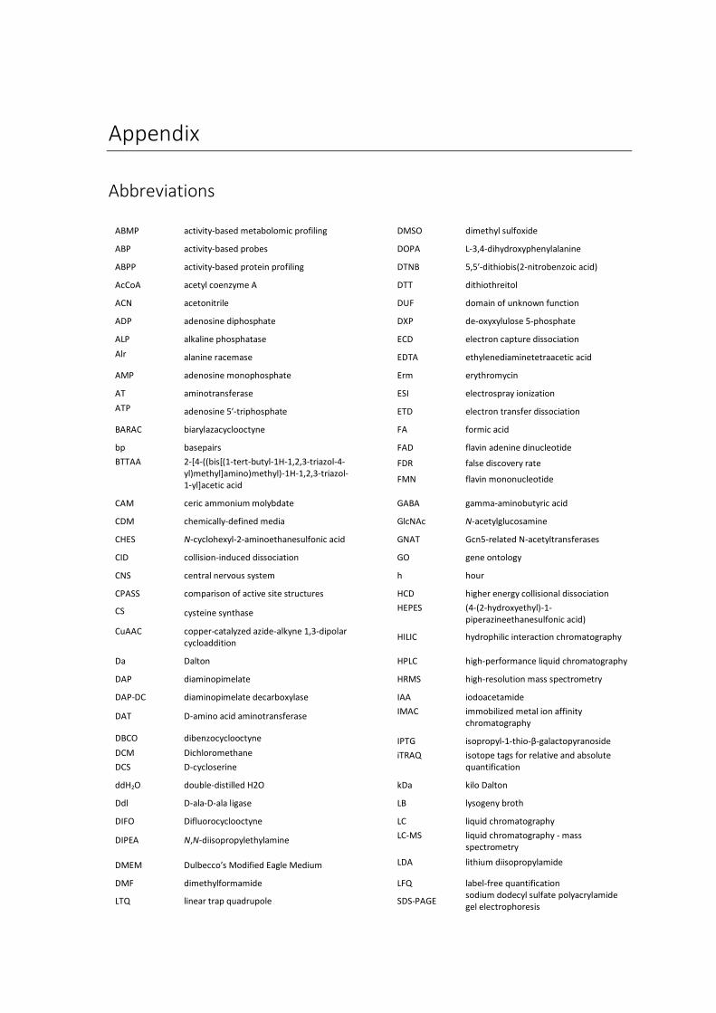
Appendix
Abbreviations
ABMP activity-based metabolomic profiling
DMSO dimethyl sulfoxide
ABP activity-based probes
DOPA L-3,4-dihydroxyphenylalanine
ABPP activity-based protein profiling
DTNB 5,5′-dithiobis(2-nitrobenzoic acid)
AcCoA acetyl coenzyme A
DTT dithiothreitol
ACN acetonitrile
DUF domain of unknown function
ADP adenosine diphosphate
DXP de-oxyxylulose 5-phosphate
ALP alkaline phosphatase
ECD electron capture dissociation
Alr alanine racemase
EDTA ethylenediaminetetraacetic acid
AMP adenosine monophosphate
Erm erythromycin
AT aminotransferase
ESI electrospray ionization
ATP adenosine 5′-triphosphate
ETD electron transfer dissociation
BARAC biarylazacyclooctyne
FA formic acid
bp basepairs
FAD flavin adenine dinucleotide
BTTAA 2-[4-((bis[(1-tert-butyl-1H-1,2,3-triazol-4-yl)methyl]amino)methyl)-1H-1,2,3-triazol-1-yl]acetic acid
FDR
FMN
false discovery rate
flavin mononucleotide
CAM ceric ammonium molybdate
GABA gamma-aminobutyric acid
CDM chemically-defined media
GlcNAc N-acetylglucosamine
CHES N-cyclohexyl-2-aminoethanesulfonic acid
GNAT Gcn5-related N-acetyltransferases
CID collision-induced dissociation
GO gene ontology
CNS central nervous system
h hour
CPASS comparison of active site structures
HCD higher energy collisional dissociation
CS cysteine synthase HEPES (4-(2-hydroxyethyl)-1-
piperazineethanesulfonic acid) CuAAC copper-catalyzed azide-alkyne 1,3-dipolar
cycloaddition
HILIC hydrophilic interaction chromatography
Da Dalton
HPLC high-performance liquid chromatography
DAP diaminopimelate
HRMS high-resolution mass spectrometry
DAP-DC diaminopimelate decarboxylase
IAA iodoacetamide
DAT D-amino acid aminotransferase IMAC immobilized metal ion affinity
chromatography
DBCO dibenzocyclooctyne IPTG isopropyl-1-thio-β-galactopyranoside
DCM
DCS
Dichloromethane
D-cycloserine
iTRAQ isotope tags for relative and absolute quantification
ddH2O double-distilled H2O
kDa kilo Dalton
Ddl D-ala-D-ala ligase
LB lysogeny broth
DIFO Difluorocyclooctyne
LC liquid chromatography
DIPEA N,N-diisopropylethylamine LC-MS liquid chromatography - mass
spectrometry
DMEM Dulbecco’s Modified Eagle Medium LDA lithium diisopropylamide
DMF dimethylformamide
LFQ label-free quantification
LTQ linear trap quadrupole
SDS-PAGE sodium dodecyl sulfate polyacrylamide gel electrophoresis
130 Appendix
m/z mass to charge ratio
sec second
mCPBA meta-chloroperoxybenzoic acid
SEC size-exclusion chromatography
min minute
SHMT serine hydroxymethyltransferase
MOM methoxymethyl SILAC stable isotope labeling by amino acids in
cell culture
MS mass spectrometry
SPAAC strain-promoted azide-alkyne cycloaddition
MS/MS tandem mass spectrometry
SSM secondary structure matching
MW molecular weight
TBS, TBDMS tert-butyldimethylsilyl
MWCO molecular weight cut-off
TCEP tris(2-carboxyethyl)phosphine
NaAsc sodium ascorbate
TEAB triethylammonium bicarbonate
NaBH4 sodium borohydride
TEV tobacco etch virus
NAD, NADH nicotinamide adenine dinucleotide
TFA trifluoroacetic acid
NADP, NADPH Nicotinamide adenine dinucleotide phosphate
TFAA trifluoroacetic anhydride
NMR Nuclear magnetic resonance THF tetrahydrofuran
OAS O-acetylserine
TLC thin-layer chromatography
OD optical density
TMS trimethylsilyl
OKR-DC ornithine/lysine/arginine decarboxylase
TMT tandem mass tag
PBS phosphate-buffered saline
Tn transposon
PCR polymerase chain reaction
TOP-ABPP tandem-orthogonal proteolysis
PDB protein data bank
Tris tris(hydroxymethyl)aminomethane
PEG polyethyleneglycol UDP uridine diphosphate
PEP posterior error probability
UPF uncharacterized protein family
PLK pyridoxal kinase
UV ultraviolet
PLP pyridoxal phosphate
PLP-DE pyridoxal phosphate-dependent enzyme
PMA phosphomolybdic acid
PMB para-methoxybenzyl
PMP pyridoxamine phosphate
PNP pyridoxine phosphate
PNPOx pyridoxine-5’-phosphate oxidase
Ppase phosphatase
PPAT phosphopantothenoylcysteine
ppm parts per million
PPTS pyridinium p-toluenesulfonate
PROSC proline synthetase co-transcribed protein
PTMs post-translational modifications
R5P D-ribose 5-phosphate
Rh rhodamine
rpm revolutions per minute
rt room temperature
SAM S-adenosyl methionine
SAR structure-activity relationships
SDS sodium dodecyl sulfate

Appendix 131
Supplementary gel images
Chapter 3
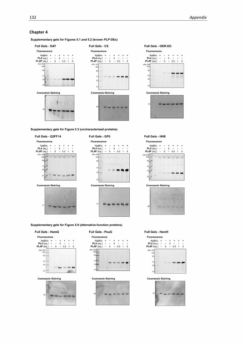
132 Appendix
Chapter 4
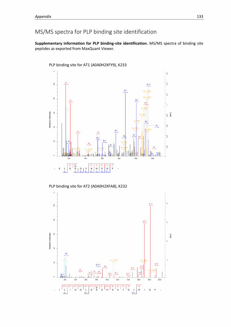
Appendix 133
MS/MS spectra for PLP binding site identification
Supplementary information for PLP binding-site identification. MS/MS spectra of binding site peptides as exported from MaxQuant Viewer.
PLP binding site for AT1 (A0A0H2XFY9), K233
PLP binding site for AT2 (A0A0H2XFA8), K232

134 Appendix
PLP binding site for CS (A0A0H2XFQ3), K46
PLP binding site for DAT (A0A0H2XHU6), K157

Appendix 135
PLP binding site for GP0 (A0A0H2XGP0), K172
PLP binding site for HH8 (A0A0H2XHH8), K32
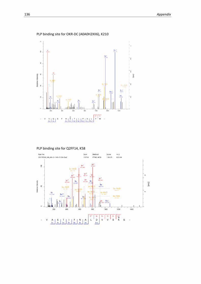
136 Appendix
PLP binding site for OKR-DC (A0A0H2XII6), K210
PLP binding site for Q2FF14, K58

Appendix 137
NMR Spectra
1H NMR of compound 1 (360 MHz, CDCl3)
13C NMR of compound 1 (90 MHz, CDCl3)

138 Appendix
1H NMR of compound 2 (360 MHz, CDCl3)
13C NMR of compound 2 (90 MHz, CDCl3)
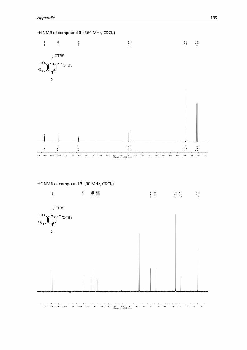
Appendix 139
1H NMR of compound 3 (360 MHz, CDCl3)
13C NMR of compound 3 (90 MHz, CDCl3)
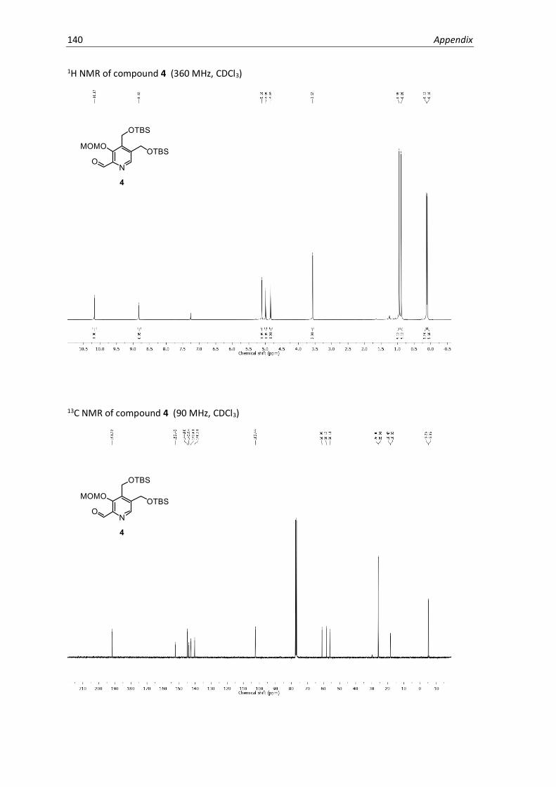
140 Appendix
1H NMR of compound 4 (360 MHz, CDCl3)
13C NMR of compound 4 (90 MHz, CDCl3)

Appendix 141
1H NMR of compound 5 (360 MHz, CDCl3)
13C NMR of compound 5 (90 MHz, CDCl3)
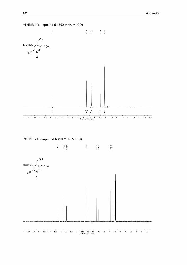
142 Appendix
1H NMR of compound 6 (360 MHz, MeOD)
13C NMR of compound 6 (90 MHz, MeOD)
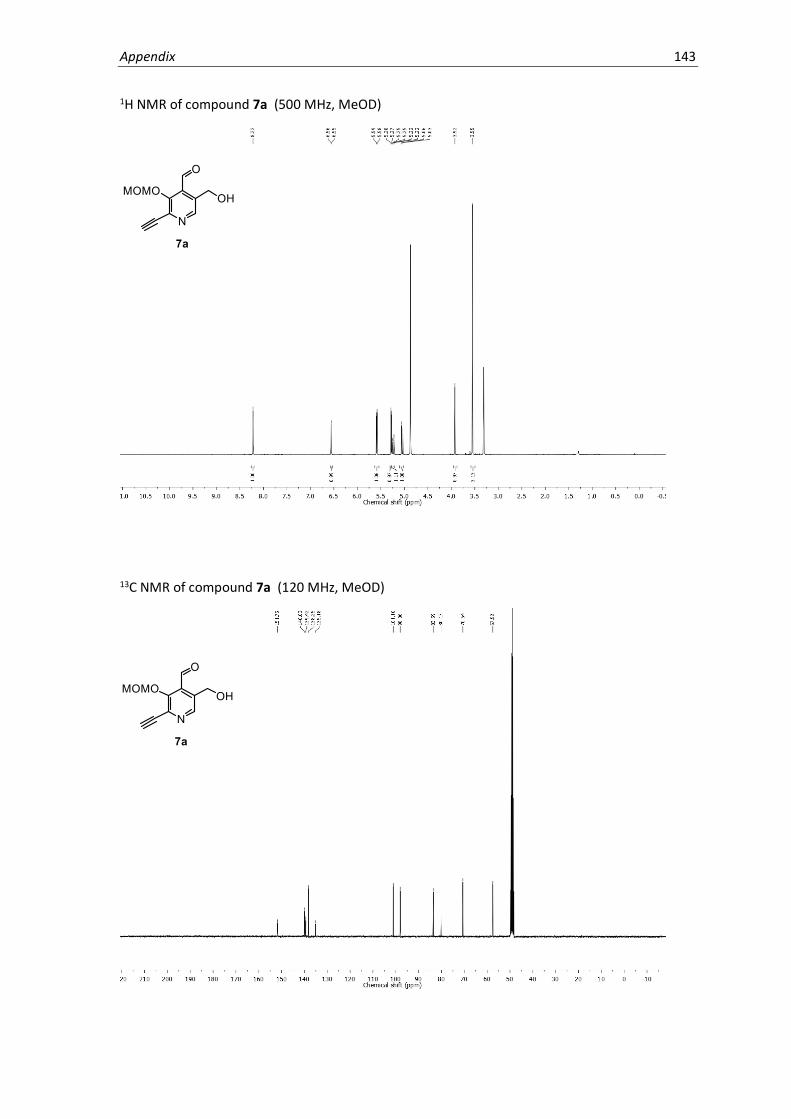
Appendix 143
1H NMR of compound 7a (500 MHz, MeOD)
13C NMR of compound 7a (120 MHz, MeOD)

144 Appendix
HSQC spectrum of compound 7a
HMBC spectrum of compound 7a

Appendix 145
NOESY spectrum of compound 7a

146 Appendix
1H NMR of compound 7b (500 MHz, MeOD)
13C NMR of compound 7b (120 MHz, MeOD)

Appendix 147
HSQC spectrum of compound 7b
HMBC spectrum of compound 7b

148 Appendix
NOESY spectrum of compound 7b

Appendix 149
1H NMR of compound PL1 (500 MHz, DMSO-d6)
13C NMR of compound PL1 (125 MHz, DMSO-d6)
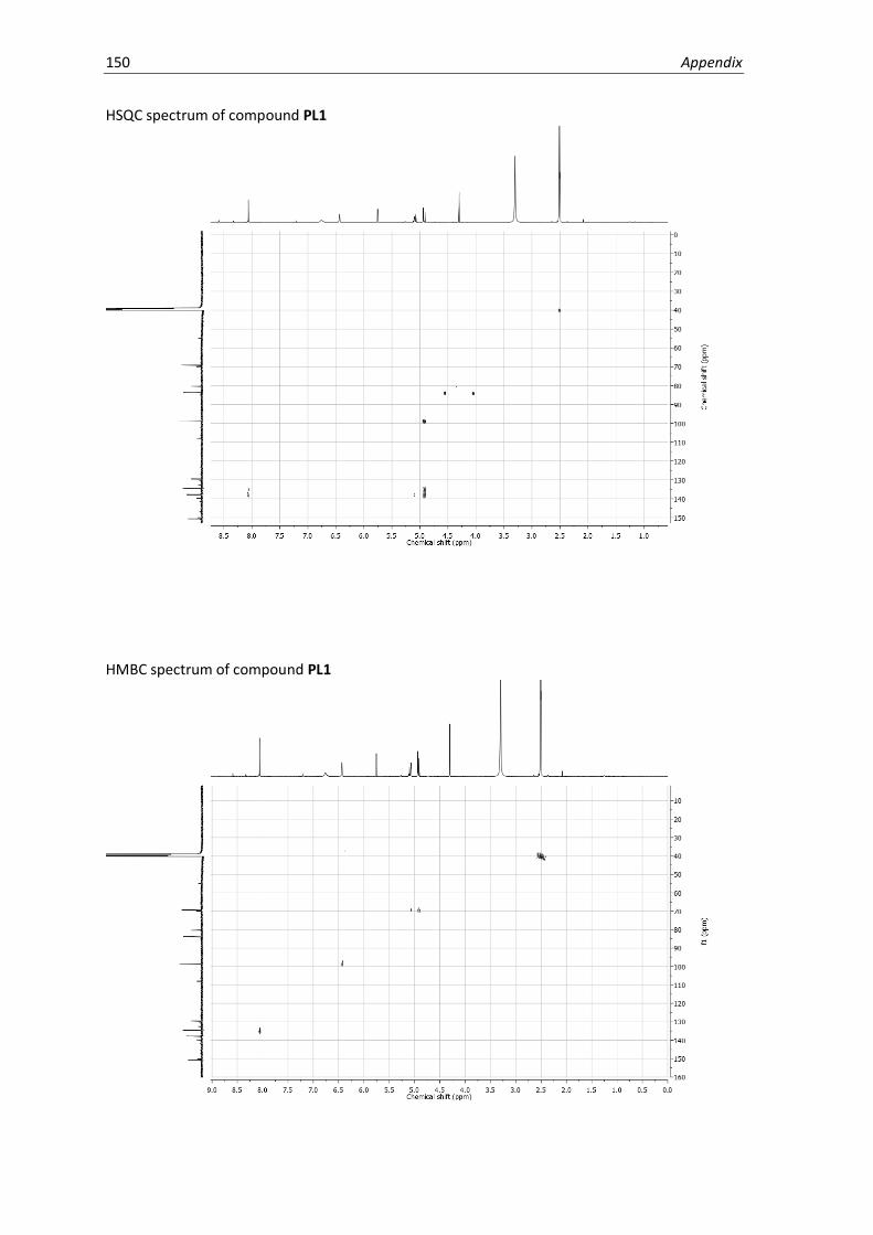
150 Appendix
HSQC spectrum of compound PL1
HMBC spectrum of compound PL1

Appendix 151
COSY spectrum of compound PL1

152 Appendix
1H NMR of compound PL2 (500 MHz, MeOD)
13C NMR of compound PL2 (125 MHz, MeOD)
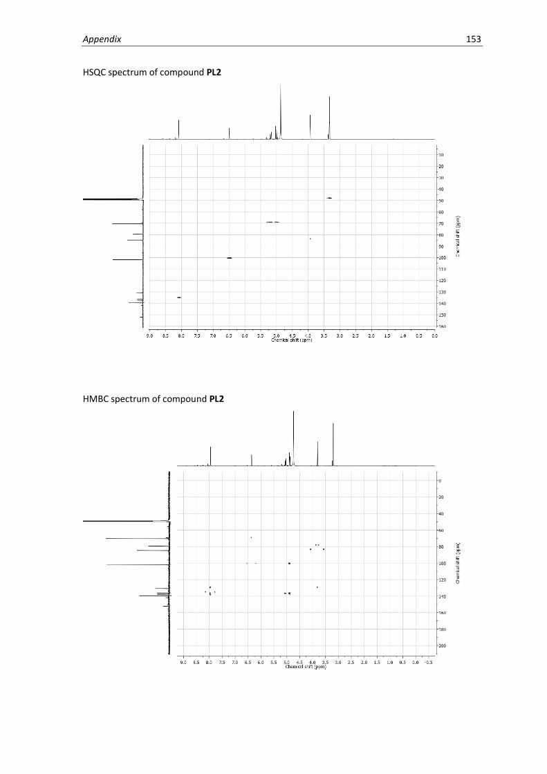
Appendix 153
HSQC spectrum of compound PL2
HMBC spectrum of compound PL2
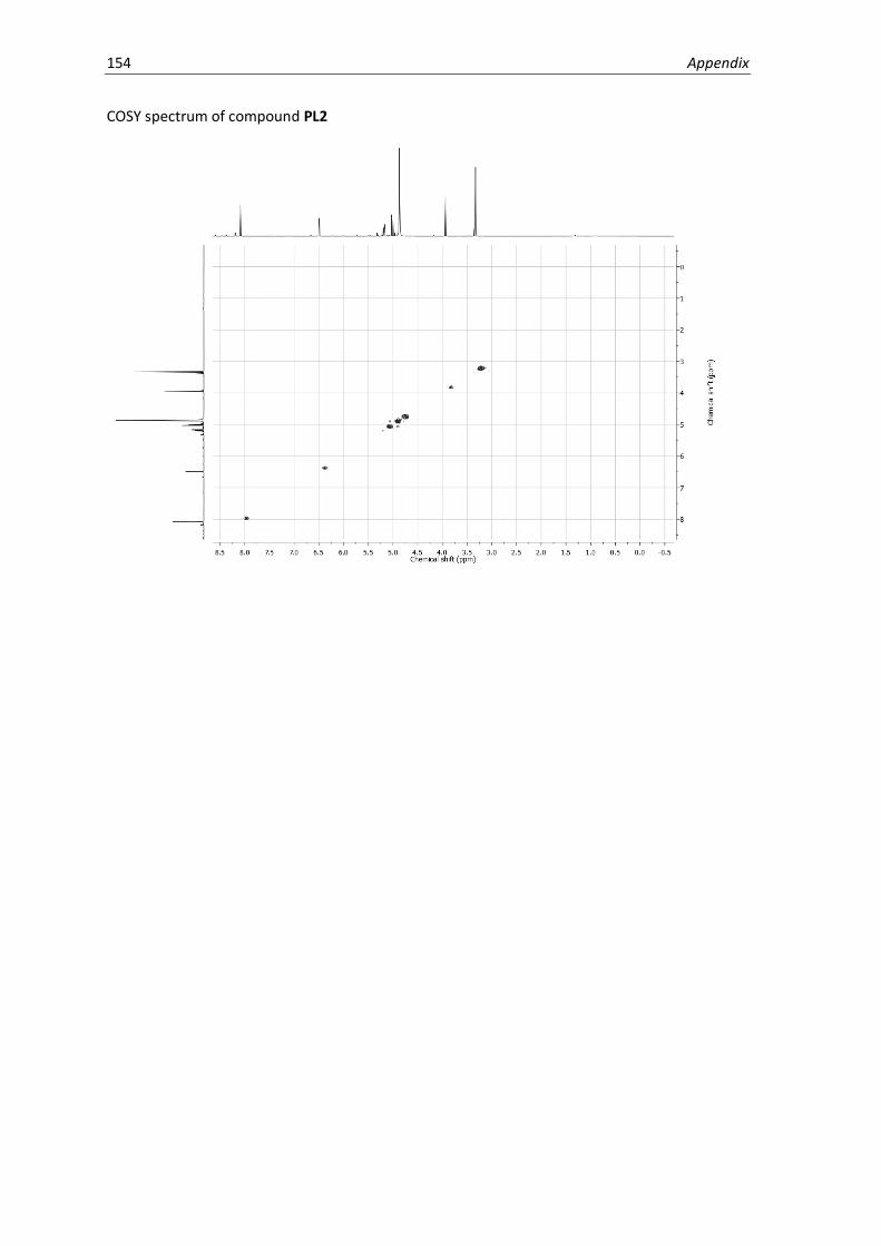
154 Appendix
COSY spectrum of compound PL2

Appendix 155
1H NMR of compound 8 (400 MHz, CDCl3)
13C NMR of compound 8 (100 MHz, CDCl3)

156 Appendix
1H NMR of compound 9 (300 MHz, CDCl3)
13C NMR of compound 9 (75 MHz, CDCl3)

Appendix 157
1H NMR of compound 10 (300 MHz, CDCl3)
13C NMR of compound 10 (75 MHz, CDCl3)

158 Appendix
1H NMR of compound 11 (300 MHz, CDCl3)
13C NMR of compound 11 (75 MHz, CDCl3)

Appendix 159
1H NMR of compound 12 (400 MHz, CDCl3)
13C NMR of compound 12 (100 MHz, CDCl3)

160 Appendix
1H NMR of compound PL3 (500 MHz, MeOD)
13C NMR of compound PL3 (75 MHz, MeOD)
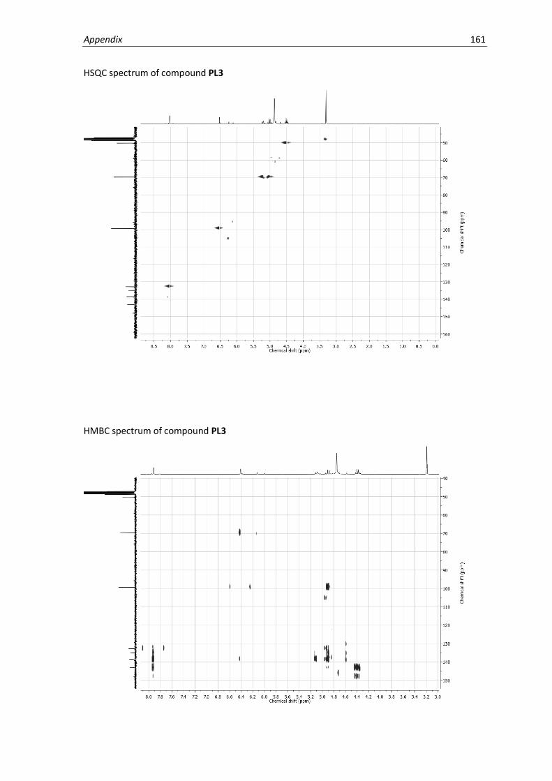
Appendix 161
HSQC spectrum of compound PL3
HMBC spectrum of compound PL3
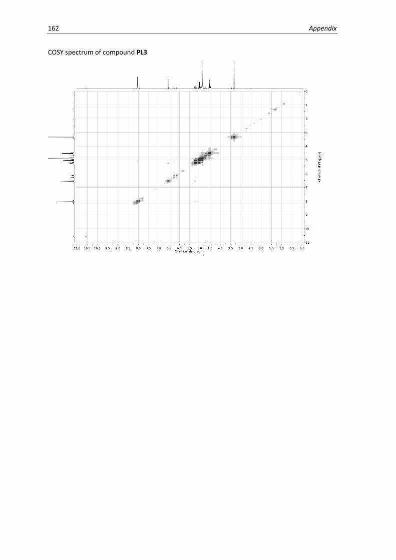
162 Appendix
COSY spectrum of compound PL3
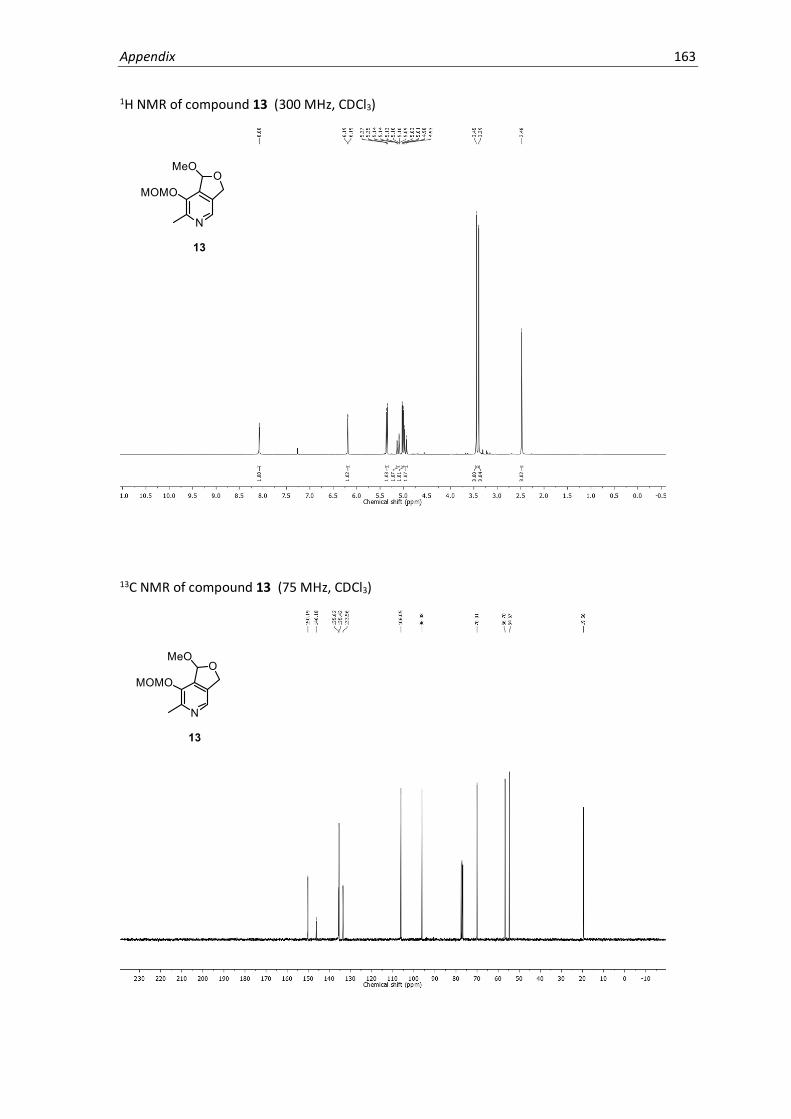
Appendix 163
1H NMR of compound 13 (300 MHz, CDCl3)
13C NMR of compound 13 (75 MHz, CDCl3)

164 Appendix
1H NMR of compound 14 (400 MHz, CDCl3)
13C NMR of compound 14 (100 MHz, CDCl3)

Appendix 165
1H NMR of compound PL4 (400 MHz, DMSO-d6)
13C NMR of compound PL4 (100 MHz, DMSO-d6)

166 Appendix
HSQC spectrum of compound PL4
HMBC spectrum of compound PL4

Appendix 167
COSY spectrum of compound PL4

168 Appendix
1H NMR of compound 15 (400 MHz, CDCl3)
13C NMR of compound 15 (100 MHz, CDCl3)

Appendix 169
1H NMR of compound 16 (400 MHz, CDCl3)
13C NMR of compound 16 (100 MHz, CDCl3)

170 Appendix
1H NMR of compound PL5 (400 MHz, DMSO-d6)
13C NMR of compound PL5 (100 MHz, DMSO-d6)

Appendix 171
HSQC spectrum of compound PL5
HMBC spectrum of compound PL5

172 Appendix
COSY spectrum of compound PL5

Appendix 173
1H NMR of compound 17 (400 MHz, CDCl3)
13C NMR of compound 17 (100 MHz, CDCl3)
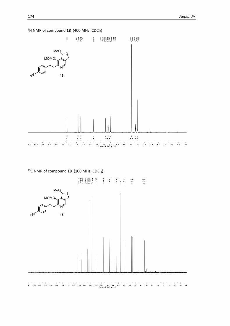
174 Appendix
1H NMR of compound 18 (400 MHz, CDCl3)
13C NMR of compound 18 (100 MHz, CDCl3)

Appendix 175
1H NMR of compound PL6 (400 MHz, DMSO-d6)
13C NMR of compound PL6 (100 MHz, DMSO-d6)

176 Appendix
HSQC spectrum of compound PL6
HMBC spectrum of compound PL6

Appendix 177
COSY spectrum of compound PL6

178 Appendix

Curriculum Vitae
Annabelle Dorothée Hoegl
Date of birth January 4, 1988 Wolfratshausen, Germany
Education
02.2014 – 03.2018 Ph.D. Chemistry
Technische Universität München, Germany
09.2011 – 10.2013 M.Sc. Chemistry
McGill University, Montréal, Canada
09.2006 – 05.2010 B.Sc. Biochemistry McGill University, Montréal, Canada