Global serum proteomic changes in water buffaloes infected ...
11
Zhang et al. Parasites Vectors (2019) 12:281 https://doi.org/10.1186/s13071-019-3533-5 RESEARCH Global serum proteomic changes in water buffaloes infected with Fasciola gigantica Fu‑Kai Zhang 1 , Rui‑Si Hu 1 , Hany M. Elsheikha 2 , Zhao‑An Sheng 3 , Wei‑Yu Zhang 3 , Wen‑Bin Zheng 1 , Xing‑Quan Zhu 1,4* and Jun‑Jun He 1* Abstract Background: The liver fluke Fasciola gigantica modulates several signaling pathways in infected buffaloes to facilitate its survival and establishment of persistent infection. In response to the parasite invasion, buffaloes activate innate and adaptive immune responses to counter the parasite infection. To detect new proteins that might be involved in the interaction between F. gigantica and the buffaloes, and that also might serve as biomarkers for fasciolosis, we used proteomic techniques to study the serum proteome of buffaloes during F. gigantica infection. Here, we used an isobaric tags for relative and absolute quantitation (iTRAQ)‑based quantitative proteomic approach to identify serum proteins that are differentially expressed in infected buffaloes compared to uninfected control buffaloes. Additionally, we applied a parallel reaction monitoring (PRM) assay to validate specific proteins identified by the iTRAQ method. Results: A total of 313, 459 and 399 proteins were identified at 3, 42 and 70 days post‑infection, respectively; of these 92, 93 and 138 were differentially abundant proteins. Some of the identified differentially abundant proteins, includ‑ ing complement factor H related 5, complement component C6, complement component C7, amine oxidase, plasma serine protease inhibitor and lysozyme, are known to be involved in complement system activation, blood coagula‑ tion, platelet activation, lymphocyte’s adhesion and lysozyme hydrolysis. Analysis of data for all three time points after infection identified six significantly upregulated proteins in infected serum that separated infected and uninfected buffaloes into distinct clusters. Further PRM analysis confirmed the expression of five proteins, namely MHC class I antigen, Beta‑2‑microglobulin, NID2 protein, Fetuin‑B and Fibrinogen gamma‑B chain. Conclusions: These findings provide novel insights into the serum proteomics signature of buffaloes during F. gigan- tica infection. Keywords: Fasciola gigantica, iTRAQ, PRM, Coagulation, Complement, Lysozyme, Buffalo © The Author(s) 2019. This article is distributed under the terms of the Creative Commons Attribution 4.0 International License (http://creativecommons.org/licenses/by/4.0/), which permits unrestricted use, distribution, and reproduction in any medium, provided you give appropriate credit to the original author(s) and the source, provide a link to the Creative Commons license, and indicate if changes were made. The Creative Commons Public Domain Dedication waiver (http://creativecommons.org/ publicdomain/zero/1.0/) applies to the data made available in this article, unless otherwise stated. Background Fasciolosis, caused by infection with Fasciola gigan- tica or F. hepatica, is an important parasitic disease in tropical and temperate regions, respectively [1, 2]. Ungulates and humans become infected via ingestion of contaminated water or plants containing metacer- cariae, leading to adverse health consequences [3]. Fasciolosis can cause significant economic losses in the agricultural industry [4]. The impact on human health is enormous, with ~180 million people at risk of infection and 17 million people infected [5]. Fasciola infection has been associated with liver fibrosis, cir- rhosis and cancer in affected individuals [6] and liver flukes have been reported in the bile duct of humans [7–9]. Current methods to control liver fluke infection rely on the use of fasciocidal drugs such as triclaben- dazole. However, increasing anthelmintic resistance has become a major concern [10] and together with the lack of a commercial vaccine, makes control of liver fluke infection challenging [11]. In order to develop better therapeutic strategies that can success- fully control fasciolosis, it is important to have a full Open Access Parasites & Vectors *Correspondence: [email protected]; [email protected] 1 State Key Laboratory of Veterinary Etiological Biology, Key Laboratory of Veterinary Parasitology of Gansu Province, Lanzhou Veterinary Research Institute, Chinese Academy of Agricultural Sciences, Lanzhou 730046, Gansu, People’s Republic of China Full list of author information is available at the end of the article
-
Upload
khangminh22 -
Category
Documents
-
view
1 -
download
0
Transcript of Global serum proteomic changes in water buffaloes infected ...
Zhang et al. Parasites Vectors (2019) 12:281 https://doi.org/10.1186/s13071-019-3533-5
RESEARCH
Global serum proteomic changes in water buffaloes infected with Fasciola giganticaFu‑Kai Zhang1, Rui‑Si Hu1, Hany M. Elsheikha2, Zhao‑An Sheng3, Wei‑Yu Zhang3, Wen‑Bin Zheng1, Xing‑Quan Zhu1,4* and Jun‑Jun He1*
Abstract
Background: The liver fluke Fasciola gigantica modulates several signaling pathways in infected buffaloes to facilitate its survival and establishment of persistent infection. In response to the parasite invasion, buffaloes activate innate and adaptive immune responses to counter the parasite infection. To detect new proteins that might be involved in the interaction between F. gigantica and the buffaloes, and that also might serve as biomarkers for fasciolosis, we used proteomic techniques to study the serum proteome of buffaloes during F. gigantica infection. Here, we used an isobaric tags for relative and absolute quantitation (iTRAQ)‑based quantitative proteomic approach to identify serum proteins that are differentially expressed in infected buffaloes compared to uninfected control buffaloes. Additionally, we applied a parallel reaction monitoring (PRM) assay to validate specific proteins identified by the iTRAQ method.
Results: A total of 313, 459 and 399 proteins were identified at 3, 42 and 70 days post‑infection, respectively; of these 92, 93 and 138 were differentially abundant proteins. Some of the identified differentially abundant proteins, includ‑ing complement factor H related 5, complement component C6, complement component C7, amine oxidase, plasma serine protease inhibitor and lysozyme, are known to be involved in complement system activation, blood coagula‑tion, platelet activation, lymphocyte’s adhesion and lysozyme hydrolysis. Analysis of data for all three time points after infection identified six significantly upregulated proteins in infected serum that separated infected and uninfected buffaloes into distinct clusters. Further PRM analysis confirmed the expression of five proteins, namely MHC class I antigen, Beta‑2‑microglobulin, NID2 protein, Fetuin‑B and Fibrinogen gamma‑B chain.
Conclusions: These findings provide novel insights into the serum proteomics signature of buffaloes during F. gigan-tica infection.
Keywords: Fasciola gigantica, iTRAQ, PRM, Coagulation, Complement, Lysozyme, Buffalo
© The Author(s) 2019. This article is distributed under the terms of the Creative Commons Attribution 4.0 International License (http://creat iveco mmons .org/licen ses/by/4.0/), which permits unrestricted use, distribution, and reproduction in any medium, provided you give appropriate credit to the original author(s) and the source, provide a link to the Creative Commons license, and indicate if changes were made. The Creative Commons Public Domain Dedication waiver (http://creat iveco mmons .org/publi cdoma in/zero/1.0/) applies to the data made available in this article, unless otherwise stated.
BackgroundFasciolosis, caused by infection with Fasciola gigan-tica or F. hepatica, is an important parasitic disease in tropical and temperate regions, respectively [1, 2]. Ungulates and humans become infected via ingestion of contaminated water or plants containing metacer-cariae, leading to adverse health consequences [3]. Fasciolosis can cause significant economic losses in
the agricultural industry [4]. The impact on human health is enormous, with ~180 million people at risk of infection and 17 million people infected [5]. Fasciola infection has been associated with liver fibrosis, cir-rhosis and cancer in affected individuals [6] and liver flukes have been reported in the bile duct of humans [7–9]. Current methods to control liver fluke infection rely on the use of fasciocidal drugs such as triclaben-dazole. However, increasing anthelmintic resistance has become a major concern [10] and together with the lack of a commercial vaccine, makes control of liver fluke infection challenging [11]. In order to develop better therapeutic strategies that can success-fully control fasciolosis, it is important to have a full
Open Access
Parasites & Vectors
*Correspondence: [email protected]; [email protected] 1 State Key Laboratory of Veterinary Etiological Biology, Key Laboratory of Veterinary Parasitology of Gansu Province, Lanzhou Veterinary Research Institute, Chinese Academy of Agricultural Sciences, Lanzhou 730046, Gansu, People’s Republic of ChinaFull list of author information is available at the end of the article
Page 2 of 11Zhang et al. Parasites Vectors (2019) 12:281
understanding of the molecular mechanisms involved in Fasciola interactions with host effector systems.
Other key aspects for efficient liver fluke con-trol includes offering veterinarians access to better diagnostic tools which allow a prompt and accurate detection of infection. In this respect, having novel biomarkers may add value to current diagnostic tests and improve clinical decision making by enabling ear-lier detection of Fasciola infection. Therefore, iden-tification of infection-specific molecular changes in buffaloes can reveal new biomarkers and advance the understanding of the pathogenesis of F. gigantica infec-tion. Transcriptomic data have been obtained from buffalo livers infected with F. gigantica [12]; however, mRNA abundance does not necessarily correlate with protein abundance [13]. The cytokine dynamics of the buffalo’s serum during F. gigantica infection has been investigated [14]. However, cytokines appeared to have a limited diagnostic value on their own because they are general products of the host systemic inflamma-tory response to cell injury or infection.
In recent years, proteomic technology has devel-oped rapidly and has been widely applied to identify biomarkers in studies, for example, on myocardial infarction [15], tuberculosis [16] and F. hepatica infec-tion [17]. Global proteomic studies are, therefore, of great importance for understanding the pathophysi-ology that underpins infection states, including infec-tion with F. gigantica. The isobaric tags for relative and absolute quantification (iTRAQ) is one of the most sensitive proteomics techniques currently used to compare protein expression across different bio-logical conditions [18–20] and infection status [21]. A traditional limitation of proteomics techniques is the inability to confirm the identity of specific proteins. Interestingly, parallel reaction monitoring (PRM) is a new mass spectrometry method that is more spe-cific and sensitive than traditional mass spectrometry methods and can detect target proteins [22].
In this study, an iTRAQ-based quantitative prot-eomic analysis of serum of F. gigantica-infected buffa-loes was performed to explore dynamic serum protein responses of buffaloes towards F. gigantica infection. A set of five differentially abundant proteins were fur-ther validated by PRM analysis. By performing this combined quantitative proteomic analysis of serum from F. gigantica-infected versus uninfected buffaloes (control), we found that F. gigantica infection alters several key biological processes and specific proteins. Our findings provide a global overview for the buffalo serum responses to F. gigantica infection and highlight new targets for further investigation.
MethodsPreparation of encysted metacercariaeEggs of F. gigantica were collected from the gall bladder of naturally infected buffaloes slaughtered at local abattoirs in Guangxi Zhuang Autonomous Region, PR China. The collected eggs were incubated at 29 °C for 11 days. The newly hatched miracidia were used to infect Galba per-via snails (3–5 miracidia per snail) maintained in plastic trays for 2 h. The infected snails were incubated in order to allow the miracidia to develop to sporocysts, rediae and cercariae. After ~6 weeks, cercariae were shed from infected snails and harvested on 5 × 5 cm cellophane sheets in order to form metacercariae. Encysted metacer-cariae harvested on cellophane sheets were washed sev-eral times with phosphate buffered saline (PBS) and used to infect buffaloes as described previously [23].
Animals and experimental infectionTwenty-four (8–10-month-old) buffaloes were purchased from a water buffalo farm in Guangxi Zhuang Autono-mous Region, PR China. Animals were randomly divided into two groups: (i) uninfected, control group (12 buf-faloes); and (ii) infected group (12 buffaloes). Animals within each group were assigned to 4 subgroups (3 buf-faloes per group). To ensure that all buffaloes used in the study were free of any liver fluke infection, fecal samples were examined by sedimentation technique for detection of fluke eggs and sera were tested for anti-F. gigantica IgG and IgM antibodies using ELISA, as described previously [24]. Additionally, buffaloes were treated with triclaben-dazole (1 ml, 5% per kilogram BW). After four weeks withdrawal time, 12 buffaloes in the infected group were infected orally with 500 viable metacercariae per animal, whereas control animals were mock-inoculated with 0.85% NaCl solution without metacercariae [12].
Serum preparation and depletion of high abundance proteinsAt 3, 42 and 70 days post-infection (dpi), whole blood samples of all animals in each group were collected asep-tically into tubes without anticoagulant. Samples were allowed to clot and stored in a cooler on ice for up to 4 h post-collection before being centrifuged at 2500×g for 10 min. Serum was collected and stored at −80 °C until use. Livers were collected after slaughter of animals and examined visually for gross pathological lesions. Highly-abundant proteins were depleted using the ProteoM-iner™ protein enrichment kit (Bio-Rad, Hercules, CA, USA) following the manufacturer’s instructions and pro-tein concentration was determined by using a Bradford assay. Approximately 100 μg of protein of each sample

Page 3 of 11Zhang et al. Parasites Vectors (2019) 12:281
was digested with Trypsin Gold (Promega, Madison, WI, USA) at a ratio of 1:50 (trypsin to protein, wt/wt) at 37 °C overnight (~16 h). Digested peptides were desalted with a C18 cartridge to remove the high urea, and the desalted peptides were dried by vacuum centrifugation.
iTRAQ labeling of peptidesDesalted peptides were labeled with iTRAQ reagents (iTRAQ® Reagent-8PLEX Multiplex Kit; Sigma, St. Louis, MO, USA) according to the manufacturer’s instructions. For 100 μg of peptides, 1 unit of labeling reagent was used. Peptides were dissolved in 20 μl of 0.5 M trieth-ylammonium bicarbonate (TEAB) pH 8.5 solution and labeling reagent was added in 70 μl of isopropanol. After 1 h of incubation, the reaction was stopped with 100 μl of 50 mM Tris-HCl pH 8. Differentially labeled peptides were mixed equally and then desalted in peptide desalt-ing spin columns (Thermo Fisher Scientific, Waltham, MA, USA).
High performance liquid chromatography (HPLC) fractionationThe iTRAQ-labeled peptide mix was fractionated using a Durashell RP column (5 μm, 100 Å, 250 × 4.6 mm; Agela, Tianjin, China) on a Rigol L3000 HPLC (RIGOL SCIEN-TIFIC, Allerød, Denmark) operating at 1 ml/min. Mobile phases A (2% acetonitrile, 20 mM NH4FA, adjusted pH to 10 using NH3·H2O) and B (80% acetonitrile, 20 mM NH4FA, adjusted pH to 10 using NH3·H2O) were used to develop a gradient elution. The tryptic peptides were sep-arated at an eluent flow rate of 1 ml/min and monitored at 214 nm (UV). The column oven was set as 37 °C. Elu-ent was collected every minute. The samples were dried under vacuum and reconstituted in 15 μl of 0.1% (v/v) formic acid (FA) in water for subsequent analysis.
LC‑MS/MS analysisShotgun proteomics analyses were performed using an EASY-nLCTM 1200 UHPLC system (Thermo Fisher Sci-entific) coupled to an Orbitrap Fusion Lumos mass spec-trometer (Thermo Fisher Scientific) operating in the data-dependent acquisition (DDA) mode. A sample vol-ume corresponding to 2 μg of total peptides reconstituted in 0.1% FA was injected onto an Acclaim PepMap100 C18 Nano-Trap column (2 cm × 100 μm, 5 μm; Dionex, Sun-nyvale, USA). Peptides were separated on a Reprosil-Pur 120 C18-AQ analytical column (15 cm × 150 μm, 1.9 μm; Dr. Maisch HPLC GmbH, Ammerbuch-Entringen, Ger-many) using a 75 min linear gradient from 5 to 100% eluent B (0.1% FA in 80% acetonitrile) in eluent A (0.1% FA in H2O) at a flow rate of 600 nl/min. For DDA, the Orbitrap Fusion Lumos mass spectrometer was operated
in positive polarity mode with spray voltage of 2.3 kV and capillary temperature of 320 °C. Full mass spectrom-etry (MS) scans from 300 to 1500 m/z were acquired at a resolution of 60,000 resolving power (at 200 m/z) with an AGC target value of 4 × 10⁵ and a maximum ion injec-tion time of 50 ms. The MS2 scans were acquired at a resolution of 15,000 resolving power (at 200 m/z) with an automatic gain control (AGC) target value of 5 × 10⁴, a maximum ion injection time of 35 ms, and a normalized collision energy of 36%.
Protein identificationThe MS raw data files (.wiff files) obtained from Triple-TOF 5600 were submitted to ProteinPilot v.4.2 using the Paragon search engine against the Bos taurus pro-tein database. To reduce the probability of false peptide identification, we counted only peptides at a cutoff value of 95% confidence interval by a ProteinPilot probability analysis greater than “identity” as identified, and each confident protein identification was supported by at least one unique peptide. Proteome Discoverer v.2.1 software was used to compare the quantitative data. We used ratios with P-values ≤ 0.05, and fold changes of ≥1.2 or ≤0.83 were considered as significant.
Functional predictionThree databases were used to predict gene and protein functions. These were GO (Gene Ontology, http://www.geneo ntolo gy.org) [25], KEGG (Kyoto Encyclopedia of Genes and Genomes, http://www.genom e.jp/kegg/) [26, 27] and COG (Clusters of Orthologous Groups, http://www.ncbi.nlm.nih.gov/COG/) [28]. Fisher’s exact test followed by FDR correction [29] was performed to iden-tify significantly enriched GO terms or pathways. The FDR corrected P-value < 0.05 was used as the threshold of significantly enriched.
Verification of proteomic results using parallel reaction monitoring (PRM)A PRM assay allows sensitive and rapid analysis of pre-selected proteins [30]. Therefore, PRM was applied to additional samples in order to verify protein expression patterns obtained by iTRAQ-based proteomic analysis. Briefly, protein preparation was performed as described above. Targeted MS analysis using PRM was per-formed on a TripleTOF 5600+ LC-MS/MS system (AB SCIEX, Concord, ON). ProteinPilot software was used to identify proteins and peptide precursor ions for the DDA mode acquired raw data, and the database search results were analyzed using Skyline software v.4.2 to obtain a rough spectra library. Target proteins for PRM validation were imported into Skyline, and the peptides

Page 4 of 11Zhang et al. Parasites Vectors (2019) 12:281
for protein quantification were selected according to the ion signals. The PRM method was run against the biological samples of interest, evaluated and refined to develop the highest quality assay. Data processing was performed in Skyline, and the results of the quantifica-tion were manually inspected for each peptide of the targeted proteins.
ResultsConfirmation of F. gigantica infectionAt 70 dpi, livers collected from infected animals showed clear gross pathological lesions together with the pres-ence of adult F. gigantica flukes, confirming the estab-lishment of infection in all experimentally infected buffaloes. By contrast, livers of uninfected control buffa-loes appeared normal, without any pathological changes and free of F. gigantica flukes.
Proteomics profiles of different groupsProteomics analysis revealed 313, 459 and 399 serum proteins at 3, 42 and 70 dpi, respectively. Compared to control, 67, 67 and 113 proteins were significantly upreg-ulated at 3, 42 and 72 dpi, respectively. However, a total of 25, 26 and 25 proteins showed significantly low expres-sion levels in infected samples that were collected at 3, 42 and 72 dpi, respectively (Figs. 1, 2 and Additional file 1: Table S1). E1BCJ2, E1BD43, F1MM86, F1N045, F2X047 and Q9N2I2 were the most commonly differentially expressed protein at all time points after infection (3, 42 and 72 dpi).
PRM validation of the protein expression in buffalo serumFive significantly expressed proteins, including MHC class I antigen (UniProt identifier Q3YJH7), Beta-2-mi-croglobulin (UniProt identifier P01888), NID2 protein (UniProt identifier A7E306), Fetuin-B (UniProt identi-fier Q58D62) and Fibrinogen gamma-B chain (UniProt identifier P12799), were selected randomly and their
Fig. 1 Venn diagram showing the unique and shared differentially expressed serum proteins between Fasciola gigantica‑infected animal groups at 3 (a), 42 (b) and 70 (c) days post‑infection
Fig. 2 Volcano plots showing the differentially expressed proteins detected in the serum of F. gigantica‑infected compared to uninfected control buffaloes. The most statistically significant proteins are shown toward the top, with upregulated proteins in red dots and downregulated proteins in green dots. Black dots represent no significant differences. The x‑axis represents log2(fold change) values and y‑axis represents −log10(pval) values. a, b and c represent differentially expressed proteins at 3, 42 and 70 days post‑infection, respectively
Page 5 of 11Zhang et al. Parasites Vectors (2019) 12:281
expression was examined by PRM. The trends of the level of expression of these five proteins obtained by iTRAQ and PRM were similar (Fig. 3).
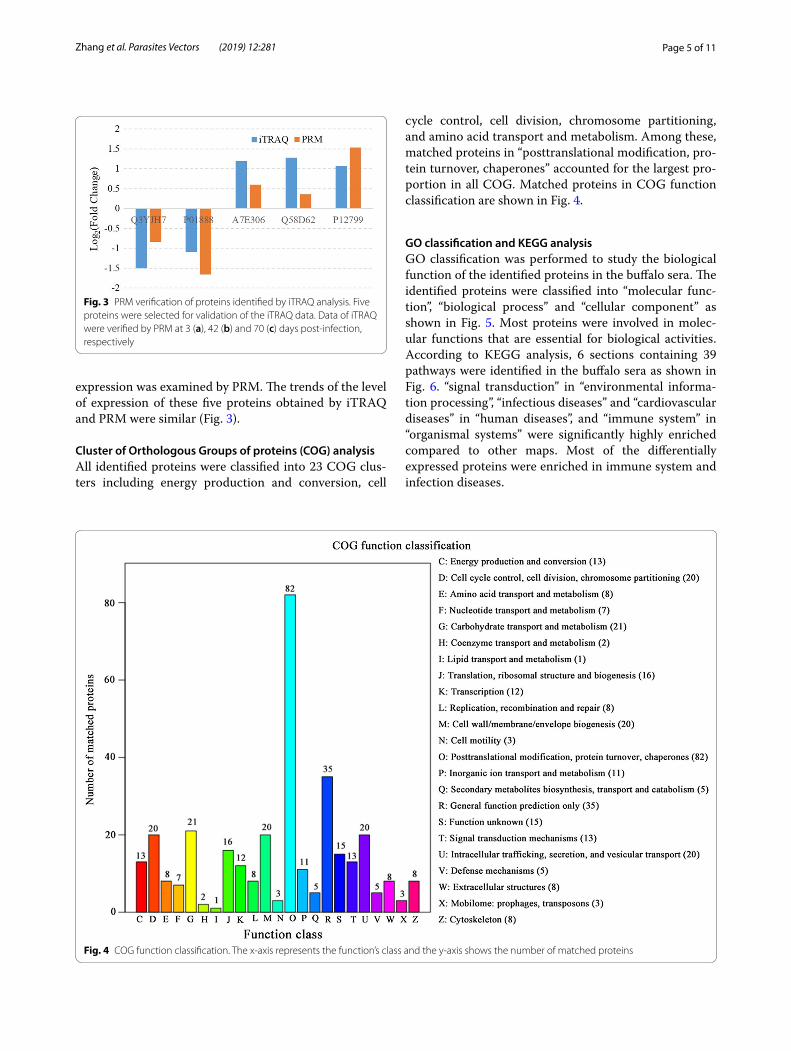
Cluster of Orthologous Groups of proteins (COG) analysisAll identified proteins were classified into 23 COG clus-ters including energy production and conversion, cell
cycle control, cell division, chromosome partitioning, and amino acid transport and metabolism. Among these, matched proteins in “posttranslational modification, pro-tein turnover, chaperones” accounted for the largest pro-portion in all COG. Matched proteins in COG function classification are shown in Fig. 4.
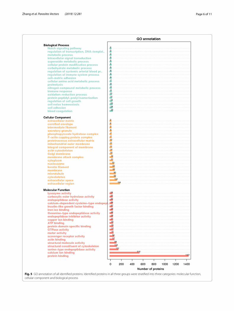
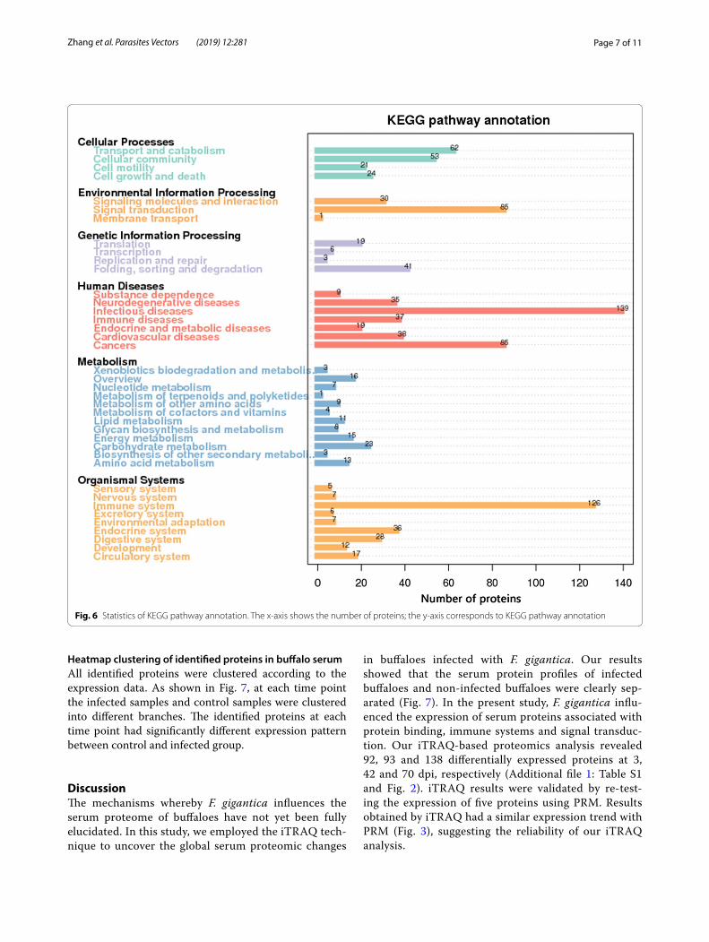
GO classification and KEGG analysisGO classification was performed to study the biological function of the identified proteins in the buffalo sera. The identified proteins were classified into “molecular func-tion”, “biological process” and “cellular component” as shown in Fig. 5. Most proteins were involved in molec-ular functions that are essential for biological activities. According to KEGG analysis, 6 sections containing 39 pathways were identified in the buffalo sera as shown in Fig. 6. “signal transduction” in “environmental informa-tion processing”, “infectious diseases” and “cardiovascular diseases” in “human diseases”, and “immune system” in “organismal systems” were significantly highly enriched compared to other maps. Most of the differentially expressed proteins were enriched in immune system and infection diseases.
Fig. 3 PRM verification of proteins identified by iTRAQ analysis. Five proteins were selected for validation of the iTRAQ data. Data of iTRAQ were verified by PRM at 3 (a), 42 (b) and 70 (c) days post‑infection, respectively
Fig. 4 COG function classification. The x‑axis represents the function’s class and the y‑axis shows the number of matched proteins
Page 6 of 11Zhang et al. Parasites Vectors (2019) 12:281
Fig. 5 GO annotation of all identified proteins. Identified proteins in all three groups were stratified into three categories: molecular function, cellular component and biological process
Page 7 of 11Zhang et al. Parasites Vectors (2019) 12:281
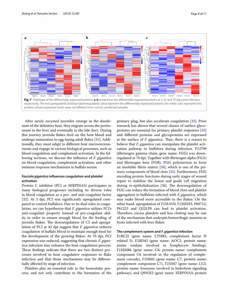
Heatmap clustering of identified proteins in buffalo serumAll identified proteins were clustered according to the expression data. As shown in Fig. 7, at each time point the infected samples and control samples were clustered into different branches. The identified proteins at each time point had significantly different expression pattern between control and infected group.
DiscussionThe mechanisms whereby F. gigantica influences the serum proteome of buffaloes have not yet been fully elucidated. In this study, we employed the iTRAQ tech-nique to uncover the global serum proteomic changes
in buffaloes infected with F. gigantica. Our results showed that the serum protein profiles of infected buffaloes and non-infected buffaloes were clearly sep-arated (Fig. 7). In the present study, F. gigantica influ-enced the expression of serum proteins associated with protein binding, immune systems and signal transduc-tion. Our iTRAQ-based proteomics analysis revealed 92, 93 and 138 differentially expressed proteins at 3, 42 and 70 dpi, respectively (Additional file 1: Table S1 and Fig. 2). iTRAQ results were validated by re-test-ing the expression of five proteins using PRM. Results obtained by iTRAQ had a similar expression trend with PRM (Fig. 3), suggesting the reliability of our iTRAQ analysis.
Fig. 6 Statistics of KEGG pathway annotation. The x‑axis shows the number of proteins; the y‑axis corresponds to KEGG pathway annotation
Page 8 of 11Zhang et al. Parasites Vectors (2019) 12:281
After newly excysted juveniles emerge in the duode-num of the definitive host, they migrate across the perito-neum to the liver and eventually to the bile duct. During this journey juvenile flukes feed on the host blood and undergo maturation to egg-laying adult flukes [31]. Addi-tionally, they must adapt to different host microenviron-ments and engage in various biological processes, such as blood coagulation and complement activation. In the fol-lowing sections, we discuss the influence of F. gigantica on blood coagulation, complement activation, and other immune response mechanisms in buffalo serum.
Fasciola gigantica influences coagulation and platelet activationProtein C inhibitor (PCI or SERPINA5) participates in many biological progresses including its diverse roles in blood coagulation as a pro- and anti-coagulant factor [32]. At 3 dpi, PCI was significantly upregulated com-pared to control buffaloes. Due to its dual roles in coagu-lation, we can hypothesize that F. gigantica utilizes PCI’s anti-coagulant property instead of pro-coagulant abil-ity in order to ensure enough blood for the feeding of juvenile flukes. The downregulation of C5 and upregu-lation of PCI at 42 dpi suggest that F. gigantica reduces coagulation of buffalo blood to maintain enough food for the development of the growing flukes. At 70 dpi, PCI expression was reduced, suggesting that chronic F. gigan-tica infection may enhance the host coagulation process. These findings indicate that there are two distinct pro-cesses involved in host coagulative responses to fluke infection and that those mechanisms may be differen-tially affected by stage of infection.
Platelets play an essential role in the hemostatic pro-cess, and not only contribute to the formation of the
primary plug, but also accelerate coagulation [33]. Prior research has shown that several classes of surface glyco-proteins are essential for primary platelet responses [33] and different proteins and glycoproteins are expressed at the surface of F. gigantica. Thus, there is a reason to believe that F. gigantica can manipulate the platelet acti-vation pathway in buffaloes during infection. P12799 (fibrinogen gamma chain, gene name: FGG) was down-regulated at 70 dpi. Together with fibrinogen alpha (FGA) and fibrinogen beta (FGB), FGG polymerizes to form an insoluble fibrin matrix [34], which is one of the pri-mary components of blood clots [35]. Furthermore, FGG encoding protein functions during early stages of wound repair to stabilize the lesion and guide cell migration during re-epithelialization [36]. The downregulation of FGG can reduce the formation of blood clots and platelet aggregation in buffaloes infected with F. gigantica, which may make blood more accessible to the flukes. On the other hand, upregulation of F1MAV0, F1MDH3, P60712, P61223 and Q32LP0 can lead to platelet activation. Therefore, excess platelets and less clotting may be one of the mechanism that underpin hemorrhagic anaemia in hosts infected with liver flukes
The complement system and F. gigantica infectionE1BCJ2 (gene name: CFHR5, complement factor H related 5), E1BD43 (gene name: AOC3, protein name: amine oxidase involved in lymphocyte binding), F1MM86 (gene name: C6; protein name: complement component C6 involved in the regulation of comple-ment cascade), F1N045 (gene name: C7; protein name: complement component C7), F2X047 (gene name: LYZ; protein name: lysozyme involved in hydrolysis signaling pathway), and Q9N2I2 (gene name: SERPINA5; protein
Fig. 7 Heatmaps of the differentially expressed proteins. a, b and c show the differentially expressed proteins at 3, 42 and 70 days post‑infection, respectively. The red (upregulated) and blue (downregulated) colors represent the differentially expressed proteins; the white color represents the proteins whose expression levels were not different from control, uninfected samples
Page 9 of 11Zhang et al. Parasites Vectors (2019) 12:281
name: plasma serine protease inhibitor) were differen-tially expressed in sera of infected buffaloes at all three time points after infection.
Previous studies have investigated the role of Th2 [12], Th1/Th17 [23], cytokines [14] and antibody [37] responses in the immunopathogenesis of F. gigantica. However, reports about complement component changes during F. gigantica infection are limited. This is the first report, to our knowledge, of the involvement of buffalo complement components at different phases of F. gigan-tica infection. F1MM86 (C6) and F1N045 (C7) with C5b, C8 (F1MX87) and multiple copies of C9 (Q3MHN2) are complement components of the membrane attack com-plex (MAC), which is known to compromise osmotic integrity and induce cell death in pathogens [38]. F1MM86 and F1N045 showed similar trends during infection with significantly high expression at early (3 dpi) and late (70 dpi) infection. A previous study showed that a F. gigantica-specific antibody that promotes the activa-tion of complement cascade pathway was increased from 3 weeks post-infection (wpi) and peaked at 13 wpi [37]. This result suggests that F. gigantica can modulate the buffalo’s immune responses, especially the complement system to promote its survival and persistence in the liver of buffaloes. The influence of F. hepatica on the comple-ment activation pathways has been also reported [39].
The classical pathway of complement activation is part of the adaptive immune response and is initiated with antibody binding [40]. The lectin pathway and the alter-native pathway are two complement pathways, which are independent of antibody-antigen interaction [41]. The lection pathway is initiated by carbohydrates on micro-bial surfaces [42, 43] and the alternative pathway can be initiated by blood-coagulation [44]. Given the upregula-tion of F1MM86 and F1N045 at 3 and 70 dpi, we infer that lectin and carbohydrate on the surface of F. gigan-tica, and specific host antibody can promote the comple-ment responses at 70 dpi. This is the first report of the influence of F. gigantica on the complement in buffaloes.
Fasciola gigantica affects recruitment of lymphocytesVascular adhesion protein-1 (VAP-1, also called amine oxidase copper containing-3 [AOC3]), is an endothe-lial cell adhesion molecule that promotes recruitment of lymphocytes, such as CD8+ T cells, CD4+ T cells and NK cells to the liver [45]. The granulocyte adhesion to VAP-1 is mediated by Siglec-9 [46, 47]. VAP-1 is constitutively expressed in the sinusoids of human liver and is activated during an immune response [48], especially in inflamma-tory liver disease [49]. The expression of VAP-1 has been also associated with various tumors, such as astrocytoma [50], colorectal cancer [51], gastric cancer [52] and breast cancer [53].
At 3 and 42 dpi, F. gigantica induces an inflammatory response and enhances VAP-1 homing of lymphocytes to liver, but in the meantime the fluke attenuates inflam-matory responses by downregulating lipopolysaccharide binding protein (LBP) to promote its own survival [12]. At 70 dpi, there was a significant downregulation of VAP-1 in the buffalo sera. It is possible that VAP-1 plays roles that can be compensated for by other molecules. For example, activated immune responses mediated by TLRs and NLRs were previously reported in buffalo liv-ers at 70 dpi with F. gigantica [23]. These findings indicate that in the course of F. gigantica infection, coordinated pro- and anti-inflammatory signals allow the flukes to evade the host immune control and prevent excessive inflammatory response from inducing extensive tissue damage in order to favor parasite persistence.
Lysozyme hydrolyzes glycosphingolipids in F. giganticaThe upregulation of lysozyme at 3, 42 and 70 dpi indi-cates that buffaloes may activate lysosome in response to F. gigantica as part of the antiparasitic defense strat-egies to damage the fluke’s structural integrity via tar-geting glycosphingolipids. Lysozymes are key players of the innate immune system and possess antimicrobial activity [54]. The lysozyme catalyzes the hydrolysis of 1,4-beta-linkage between N-acetylmuramic acid and N-acetyl-D-glucosamine in peptidoglycans and between the N-acetyl-D-glucosamine residues in chitodextrins [55]. A previous study found no link between lysozyme and F. hepatica [56]. However, F. hepatica expresses the globo-series of glycosphingolipids [Gal(α1-4)Gal(β1-4)Glc(β1-1)Cer] [57] and glycosphingolipids isolated from F. hepatica and F. gigantica [58] are a subtype of glycolip-ids, containing the amino alcohol sphingosine, and there is a 1,4-beta linkage between galactose and glucose. The presence of a 1,4-beta linkage between these two mole-cules indicate that lysozyme may hydrolyze glycosphin-golipids in F. gigantica and F. hepatica.
ConclusionsIn the present study, the iTRAQ-based proteomics technique coupled with a parallel reaction monitor-ing approach was used to identify the differentially abundant proteins in the serum of F. gigantica-infected versus control buffaloes. A total of 313, 459 and 399 pro-teins were identified at 3, 42 and 70 days post-infection, respectively; of these 92, 93 and 138 were differentially abundant proteins. The most significant differentially abundant protein markers that distinguished sera of infected from uninfected buffaloes included complement factor H related 5, complement component C6, comple-ment component C7, amine oxidase, plasma serine pro-tease inhibitor and lysozyme, which are known to be
Page 10 of 11Zhang et al. Parasites Vectors (2019) 12:281
involved in complement system activation, blood coag-ulation, platelet activation, lymphocyte adhesion and lysozyme hydrolysis. Further characterization of buffalo proteins involved in the pathogenesis of F. gigantica may lead to the discovery of novel therapeutic or diagnostic targets.
Additional file
Additional file 1: Table S1. Differentially expressed proteins identified in the present study.
AbbreviationsiTRAQ: isobaric tags for relative and absolute quantitation; PRM: parallel reac‑tion monitoring; dpi: days post‑infection; MAC: membrane attack complex; VAP‑1: vascular adhesion protein‑1; LBP: lipopolysaccharide binding protein.
AcknowledgementsThe authors would like to thank Novogene Bioinformatics Technology Co., Ltd. (Beijing, China) for technical assistance.
Authors’ contributionsJJH, XQZ and HME conceived and designed the study, and critically revised the manuscript. FKZ and RSH performed the experiment, analyzed the prot‑eomic data and drafted the manuscript. HME and JJH helped in data analysis and manuscript revision. ZAS, WYZ and WBZ helped in the implementation of the study. All authors read and approved the final manuscript.
FundingProject financial support was provided by the National Key Basic Research Pro‑gram (973 Program) of China (Grant No. 2015CB150300) and the National Key Research and Development Program of China (Grant No. 2017YFD0501200).
Availability of data and materialsThe data supporting the findings of this article are included within the article and its Additional files. The mass spectrometry proteomics data have been deposited at the ProteomeXchange Consortium via the PRIDE partner reposi‑tory with the dataset identifier PXD011576.
Ethics approval and consent to participateThis study was conducted in accordance with the recommendations set forth in the Animal Ethics Procedures and Guidelines of the People’s Republic of China. Animal experiments were reviewed and approved by the Animal Administration and Ethics Committee of Lanzhou Veterinary Research Institute, Chinese Academy of Agricultural Sciences. All efforts were made to minimize the number of animals used and their suffering.
Consent for publicationNot applicable.
Competing interestsThe authors declare that they have no competing interests.
Author details1 State Key Laboratory of Veterinary Etiological Biology, Key Laboratory of Vet‑erinary Parasitology of Gansu Province, Lanzhou Veterinary Research Institute, Chinese Academy of Agricultural Sciences, Lanzhou 730046, Gansu, People’s Republic of China. 2 Faculty of Medicine and Health Sciences, School of Vet‑erinary Medicine and Science, University of Nottingham, Sutton Bonington Campus, Loughborough LE12 5RD, UK. 3 College of Animal Science and Tech‑nology, Guangxi University, Nanning 530005, Guangxi Zhuang Autonomous Region, People’s Republic of China. 4 Jiangsu Co‑innovation Center for Preven‑tion and Control of Important Animal Infectious Diseases and Zoonoses, Yangzhou 225009, Jiangsu, People’s Republic of China.
Received: 31 January 2019 Accepted: 27 May 2019
References 1. Keiser J, Utzinger J. Food‑borne trematodiases. Clin Microbiol Rev.
2009;22:466–83. 2. Furst T, Keiser J, Utzinger J. Global burden of human food‑borne
trematodiasis: a systematic review and meta‑analysis. Lancet Infect Dis. 2012;12:210–21.
3. Cwiklinski K, Dalton JP, Dufresne PJ, La Course J, Williams DJ, Hodgkinson J, et al. The Fasciola hepatica genome: gene duplication and polymor‑phism reveals adaptation to the host environment and the capacity for rapid evolution. Genome Biol. 2015;16:71.
4. Mas‑Coma S, Valero MA, Bargues MD. Chapter 2. Fasciola, lymnaeids and human fascioliasis, with a global overview on disease transmission, epi‑demiology, evolutionary genetics, molecular epidemiology and control. Adv Parasitol. 2009;69:41–146.
5. Cwiklinski K, O’Neill SM, Donnelly S, Dalton JP. A prospective view of animal and human fasciolosis. Parasite Immunol. 2016;38:558–68.
6. Machicado C, Machicado JD, Maco V, Terashima A, Marcos LA. Association of Fasciola hepatica infection with liver fibrosis, cirrhosis, and cancer: a systematic review. PLoS Negl Trop Dis. 2016;10:e0004962.
7. Inoue K, Kanemasa H, Inoue K, Matsumoto M, Kajita Y, Mitsufuji S, et al. A case of human fasciolosis: discrepancy between egg size and genotype of Fasciola sp. Parasitol Res. 2007;100:665–7.
8. Kanoksil W, Wattanatranon D, Wilasrusmee C, Mingphruedh S, Bunyarat‑vej S. Endoscopic removal of one live biliary Fasciola gigantica. J Med Assoc Thai. 2006;89:2150–4.
9. Sharifiyazdi H, Moazeni M, Rabbani F. Molecular characterization of human Fasciola samples in Gilan province, Northern Iran on the basis of DNA sequences of ribosomal and mitochondrial DNA genes. Comp Clin Path. 2012;21:889–94.
10. Venturina VM, Alejandro MA, Baltazar CP, Abes NS, Mingala CN. Evidence of Fasciola spp. resistance to albendazole, triclabendazole and bromofen‑ofos in water buffaloes (Bubalus bubalis). Ann Parasitol. 2015;61:283–9.
11. Fairweather I, Boray JC. Fasciolicides: efficacy, actions, resistance and its management. Vet J. 1999;158:81–112.
12. Zhang FK, Zhang XX, Elsheikha HM, He JJ, Sheng ZA, Zheng WB, et al. Transcriptomic responses of water buffalo liver to infection with the digenetic fluke Fasciola gigantica. Parasites Vectors. 2017;10:56.
13. Low TY, Heck AJ. Reconciling proteomics with next generation sequenc‑ing. Curr Opin Chem Biol. 2016;30:14–20.
14. Zhang FK, Guo AJ, Hou JL, Sun MM, Sheng ZA, Zhang XX, et al. Serum lev‑els of cytokines in water buffaloes experimentally infected with Fasciola gigantica. Vet Parasitol. 2017;244:97–101.
15. Anderson NL, Anderson NG. The human plasma proteome: history, char‑acter, and diagnostic prospects. Mol Cell Proteomics. 2002;1:845–67.
16. Agranoff D, Fernandez‑Reyes D, Papadopoulos MC, Rojas SA, Herbster M, Loosemore A, et al. Identification of diagnostic markers for tuberculosis by proteomic fingerprinting of serum. Lancet. 2006;368:1012–21.
17. Rioux MC, Carmona C, Acosta D, Ward B, Ndao M, Gibbs BF, et al. Discovery and validation of serum biomarkers expressed over the first twelve weeks of Fasciola hepatica infection in sheep. Int J Parasitol. 2008;38:123–36.
18. Zhou DH, Wang ZX, Zhou CX, He S, Elsheikha HM, Zhu XQ. Comparative proteomic analysis of virulent and avirulent strains of Toxoplasma gondii reveals strain‑specific patterns. Oncotarget. 2017;8:80481–91.
19. Wang ZX, Zhou CX, Elsheikha HM, He S, Zhou DH, Zhu XQ. Proteomic dif‑ferences between developmental stages of Toxoplasma gondii revealed by iTRAQ‑based quantitative proteomics. Front Microbiol. 2017;8:985.
20. Zhou CX, Zhu XQ, Elsheikha HM, He S, Li Q, Zhou DH, et al. Global iTRAQ‑based proteomic profiling of Toxoplasma gondii oocysts during sporula‑tion. J Proteomics. 2016;148:12–9.
21. He JJ, Ma J, Elsheikha HM, Song HQ, Zhou DH, Zhu XQ. Proteomic profil‑ing of mouse liver following acute Toxoplasma gondii infection. PLoS ONE. 2016;11:e0152022.
22. Yu Q, Liu B, Ruan D, Niu C, Shen J, Ni M, et al. A novel targeted proteom‑ics method for identification and relative quantitation of difference
Page 11 of 11Zhang et al. Parasites Vectors (2019) 12:281
• fast, convenient online submission
•
thorough peer review by experienced researchers in your field
• rapid publication on acceptance
• support for research data, including large and complex data types
•
gold Open Access which fosters wider collaboration and increased citations
maximum visibility for your research: over 100M website views per year •
At BMC, research is always in progress.
Learn more biomedcentral.com/submissions
Ready to submit your research ? Choose BMC and benefit from:
in nitration degree of OGDH between healthy and diabetic mouse. Proteomics. 2014;14:2417–26.
23. Zhang FK, Hou JL, Guo AJ, Tian AL, Sheng ZA, Zheng WB, et al. Expression profiles of genes involved in TLRs and NLRs signaling pathways of water buffaloes infected with Fasciola gigantica. Mol Immunol. 2018;94:18–26.
24. Chauvin A, Bouvet G, Boulard C. Humoral and cellular immune responses to Fasciola hepatica experimental primary and secondary infection in sheep. Int J Parasitol. 1995;25:1227–41.
25. Ashburner M, Ball CA, Blake JA, Botstein D, Butler H, Cherry JM, et al. Gene ontology: tool for the unification of biology. The gene ontology consor‑tium. Nat Genet. 2000;25:25–9.
26. Kanehisa M, Goto S, Kawashima S, Okuno Y, Hattori M. The KEGG resource for deciphering the genome. Nucleic Acids Res. 2004;32:D277–80.
27. Kanehisa M, Goto S, Hattori M, Aoki‑Kinoshita KF, Itoh M, Kawashima S, et al. From genomics to chemical genomics: new developments in KEGG. Nucleic Acids Res. 2006;34:D354–7.
28. Tatusov RL, Fedorova ND, Jackson JD, Jacobs AR, Kiryutin B, Koonin EV, et al. The COG database: an updated version includes eukaryotes. BMC Bioinform. 2003;4:41.
29. Benjamini Y, Hochberg Y. Controlling the false discovery rate: a practical and powerful approach to multiple testing. J R Stat Soc B. 1995;57:289–300.
30. Zauber H, Kirchner M, Selbach M. Picky: a simple online PRM and SRM method designer for targeted proteomics. Nat Methods. 2018;15:156–7.
31. Mas‑Coma S, Valero MA, Bargues MD. Fascioliasis. Adv Exp Med Biol. 2014;766:77–114.
32. Li W, Adams TE, Nangalia J, Esmon CT, Huntington JA. Molecular basis of thrombin recognition by protein C inhibitor revealed by the 1.6‑A structure of the heparin‑bridged complex. Proc Natl Acad Sci USA. 2008;105:4661–6.
33. Versteeg HH, Heemskerk JW, Levi M, Reitsma PH. New fundamentals in hemostasis. Physiol Rev. 2013;93:327–58.
34. Vu D, Neerman‑Arbez M. Molecular mechanisms accounting for fibrinogen deficiency: from large deletions to intracellular retention of misfolded proteins. J Thromb Haemost. 2007;5(Suppl 1):125–31.
35. Ariens RA. Fibrin(ogen) and thrombotic disease. J Thromb Haemost. 2013;11(Suppl 1):294–305.
36. Duval C, Ariens RAS. Fibrinogen splice variation and cross‑linking: effects on fibrin structure/function and role of fibrinogen gamma’ as thrombo‑mobulin II. Matrix Biol. 2017;60–61:8–15.
37. Zhang WY, Moreau E, Yang BZ, Li ZQ, Hope JC, Howard CJ, et al. Humoral and cellular immune responses to Fasciola gigantica experimental infec‑tion in buffaloes. Res Vet Sci. 2006;80:299–307.
38. Peitsch MC, Tschopp J. Assembly of macromolecular pores by immune defense systems. Curr Opin Cell Biol. 1991;3:710–6.
39. Alvarez Rojas CA, Scheerlinck JP, Ansell BR, Hall RS, Gasser RB, Jex AR. Time‑course study of the transcriptome of peripheral blood mononu‑clear cells (PBMCs) from sheep infected with Fasciola hepatica. PLoS ONE. 2016;11:e0159194.
40. Lambris JD, Ricklin D, Geisbrecht BV. Complement evasion by human pathogens. Nat Rev Microbiol. 2008;6:132–42.
41. Zipfel PF, Skerka C. Complement regulators and inhibitory proteins. Nat Rev Immunol. 2009;9:729–40.
42. Fujita T. Evolution of the lectin‑complement pathway and its role in innate immunity. Nat Rev Immunol. 2002;2:346–53.
43. Degn SE, Thiel S, Jensenius JC. New perspectives on mannan‑binding lec‑tin‑mediated complement activation. Immunobiology. 2007;212:301–11.
44. Huber‑Lang M, Sarma JV, Zetoune FS, Rittirsch D, Neff TA, McGuire SR, et al. Generation of C5a in the absence of C3: a new complement activa‑tion pathway. Nat Med. 2006;12:682–7.
45. Oo YH, Banz V, Kavanagh D, Liaskou E, Withers DR, Humphreys E, et al. CXCR45‑dependent recruitment and CCR45‑mediated positioning of Th‑17 cells in the inflamed liver. J Hepatol. 2012;57:1044–51.
46. Kivi E, Elima K, Aalto K, Nymalm Y, Auvinen K, Koivunen E, et al. Human Siglec‑10 can bind to vascular adhesion protein‑1 and serves as its sub‑strate. Blood. 2009;114:5385–92.
47. Aalto K, Autio A, Kiss EA, Elima K, Nymalm Y, Veres TZ, et al. Siglec‑9 is a novel leukocyte ligand for vascular adhesion protein‑1 and can be used in PET imaging of inflammation and cancer. Blood. 2011;118:3725–33.
48. Bonder CS, Norman MU, Swain MG, Zbytnuik LD, Yamanouchi J, San‑tamaria P, et al. Rules of recruitment for Th1 and Th2 lymphocytes in inflamed liver: a role for alpha‑4 integrin and vascular adhesion protein‑1. Immunity. 2005;23:153–63.
49. Kurkijarvi R, Adams DH, Leino R, Mottonen T, Jalkanen S, Salmi M. Circulat‑ing form of human vascular adhesion protein‑1 (VAP‑1): increased serum levels in inflammatory liver diseases. J Immunol. 1998;161:1549–57.
50. Kostoro J, Chang SJ, Clark Lai YC, Wu CC, Chai CY, Kwan AL. Overexpres‑sion of vascular adhesion protein‑1 is associated with poor prognosis of astrocytomas. APMIS. 2016;124:462–8.
51. Toiyama Y, Miki C, Inoue Y, Kawamoto A, Kusunoki M. Circulating form of human vascular adhesion protein‑1 (VAP‑1): decreased serum levels in progression of colorectal cancer and predictive marker of lymphatic and hepatic metastasis. J Surg Oncol. 2009;99:368–72.
52. Yasuda H, Toiyama Y, Ohi M, Mohri Y, Miki C, Kusunoki M. Serum soluble vascular adhesion protein‑1 is a valuable prognostic marker in gastric cancer. J Surg Oncol. 2011;103:695–9.
53. Sun WY, Choi J, Cha YJ, Koo JS. Evaluation of the expression of amine oxidase proteins in breast cancer. Int J Mol Sci. 2017;18:2775.
54. Callewaert L, Walmagh M, Michiels CW, Lavigne R. Food applications of bacterial cell wall hydrolases. Curr Opin Biotechnol. 2011;22:164–71.
55. Furukawa A, Nakada‑Tsukui K, Nozaki T. Novel transmembrane receptor involved in phagosome transport of lysozymes and beta‑hexosami‑nidase in the enteric protozoan Entamoeba histolytica. PLoS Pathog. 2012;8:e1002539.
56. Duffus WP, Thorne K, Oliver R. Killing of juvenile Fasciola hepatica by puri‑fied bovine eosinophil proteins. Clin Exp Immunol. 1980;40:336–44.
57. Wuhrer M, Grimm C, Zahringer U, Dennis RD, Berkefeld CM, Idris MA, et al. A novel GlcNAcalpha1‑HPO3‑6Gal(1‑1)ceramide antigen and alkylated inositol‑phosphoglycerolipids expressed by the liver fluke Fasciola hepatica. Glycobiology. 2003;13:129–37.
58. Wuhrer M, Berkefeld C, Dennis RD, Idris MA, Geyer R. The liver flukes Fasciola gigantica and Fasciola hepatica express the leucocyte cluster of differentiation marker CD77 (globotriaosylceramide) in their tegument. Biol Chem. 2001;382:195–207.
Publisher’s NoteSpringer Nature remains neutral with regard to jurisdictional claims in pub‑lished maps and institutional affiliations.