
Evolution of a New Function by Degenerative Mutation in Cephalochordate Steroid Receptors

http://jfm.sagepub.com/Journal of Feline Medicine and Surgery
http://jfm.sagepub.com/content/early/2013/01/04/1098612X12470652The online version of this article can be found at:
DOI: 10.1177/1098612X12470652
published online 7 January 2013Journal of Feline Medicine and SurgeryXiangming Gao, Junyu Lee, Sukhaswami Malladi, Lynda Melendez, B Duncan X Lascelles and Samer Al-Murrani
Feline degenerative joint disease: a genomic and proteomic approach
technique does not amount to an endorsement of its value or quality, or the claims made by its manufacturer.those of the authors and the inclusion in this publication of material relating to a particular product, method or of animals and interpretation of published materials lies with the veterinary practitioner. The opinions expressed arefrom actions or decisions based on information contained in this publication; ultimate responsibility for the treatment
arisingcountry. The authors, editors, owners and publishers do not accept any responsibility for any loss or damage advertising material, it is the responsibility of the reader to check that the product is authorised for use in their ownbear this in mind and be aware of the prescribing laws pertaining to their own country. Likewise, in relation to Furthermore, drugs may be mentioned that are licensed for human use, and not for veterinary use. Readers need toformulations that are not available or licensed in the individual reader's own country.The Journal of Feline Medicine and Surgery is an international journal and authors may discuss products and
Disclaimer
Published by:
International Society of Feline Medicine
American Association of Feline Practitioners
and http://www.sagepublications.com
can be found at:Journal of Feline Medicine and SurgeryAdditional services and information for
http://jfm.sagepub.com/cgi/alertsEmail Alerts:
http://jfm.sagepub.com/subscriptionsSubscriptions:
http://www.sagepub.com/journalsReprints.navReprints:
http://www.sagepub.com/journalsPermissions.navPermissions:
What is This?
- Jan 7, 2013OnlineFirst Version of Record >>
at NORTH CAROLINA STATE UNIV on January 24, 2013jfm.sagepub.comDownloaded from

Journal of Feline Medicine and Surgery0(0) 1 –12© ISFM and AAFP 2013Reprints and permission: sagepub.co.uk/journalsPermissions.navDOI: 10.1177/1098612X12470652jfms.com
IntroductionThere is increasing interest in feline degenerative joint disease (DJD).1 Retrospective studies suggest, and more recent prospective studies indicate, that radiographic evidence of DJD is common, with up to 90% of cats being affected.2–5 Other studies suggest that this DJD can be associated with pain, making it an important feline dis-ease.6–9 As has been discussed recently, little is known about the etiology of feline DJD.1
Although several authors have suggested that a large proportion of DJD in the cat is primary, there is currently no evidence of this.2,3 Two primary forms of osteoarthri-tis (OA) are fairly well recognized in cats: Scottish Fold osteochondrodysplasia and mucopolysaccharidosis.10–20 Currently, the documented secondary causes of DJD in cats are nutritional, hip dysplasia, the non-infectious pol-yarthropathies and infectious arthropathies.21–31 Obesity has also been suggested as a driving or confounding fac-tor. Despite this information, the etiology of the vast majority of feline DJD seen in practices is unknown.
Research suggests a wide variety of contributors to DJD in humans that range from genetic to environmen-tal.32,33 The first and most commonly cited contributor is a genetic risk factor.34 This is particularly true for the autoimmune types of arthritis because the body’s immune system seems to undergo a shift in gene expres-
sion that elicits an inappropriate intolerance for self- antigens.35,36 Gene expression studies in the human lit-erature illustrate great heterogeneity in gene expression patterns between tested individuals, which complicates the search for possible therapeutic targets.34,37–43 Genes that do appear routinely to be differentially expressed between human rheumatoid arthritis (RA) patients and age-matched controls include immune system and inflammation-associated genes, such as major histocom-patibility complex (MHC) genes I, II and III, and tumor necrosis factor (TNF)-α, signaling pathways, such as the interferon (IFN)/signal transducer and activation of transcription factor (STAT), and extracellular proteins, like the matrix metalloproteinases (MMP).34,37–40,44 Genes
Feline degenerative joint disease: a genomic and proteomic approach
Xiangming Gao1, Junyu Lee1, Sukhaswami Malladi1, Lynda Melendez1, B Duncan X Lascelles2 and Samer Al-Murrani1*
AbstractThe underlying disease mechanisms for feline degenerative joint disease (DJD) are mostly unidentified. Today, most of what is published on mammalian arthritis is based on human clinical findings or on mammalian models of human arthritis. However, DJD is a common occurrence in the millions of domestic felines worldwide. To get a better understanding of the changes in biological pathways that are associated with feline DJD, this study employed a custom-designed feline GeneChip, and the institution’s unique access to large sample populations to investigate genes and proteins from whole blood and serum that may be up- or down-regulated in DJD cats. The GeneChip results centered around three main pathways that were affected in DJD cats: immune function, apoptosis and oxidative phosphorylation. By identifying these key disease-associated pathways it will then be possible to better understand disease pathogenesis and diagnose it more easily, and to better target it with pharmaceutical and nutritional intervention.
Accepted: 15 November 2012
1Hill’s Pet Nutrition Center, Topeka, KS, USA2 Comparative Pain Research Laboratory and Small Animal Surgery Section, Department of Clinical Sciences and Center for Comparative Medicine and Translational Research, North Carolina State University, USA
*Current address: Babylon BioConsulting, Topeka KS, USA
Corresponding author:Samer Al-Murrani PhD, MBA, Babylon BioConsulting, 2513 SW Lagito Court, Topeka, KS 66614, USA Email: [email protected]
470652 JFM0010.1177/1098612X12470652Journal of Feline Medicine and SurgeryGao et al2013
Original Article
at NORTH CAROLINA STATE UNIV on January 24, 2013jfm.sagepub.comDownloaded from

2 Journal of Feline Medicine and Surgery 0(0)
and proteins that have been found to be differentially expressed in human OA patients include structural pro-teins collagen I and collagen II alpha I (COL2A1), tran-scription factors SOX9 and 11, MMP13, the cytokine interleukin-1 beta (IL1B) and the proteoglycan aggre-can.45,46 The majority of arthritis gene expression studies reported in the literature have been carried out in humans with cartilage, bone or synovial tissue. These assorted samples may be a cause of variability in the findings of what is actually occurring in the disease state.
The aim of this study was to investigate feline-specific gene expression differences between DJD cats and age-matched controls in order to obtain a clearer picture of the underlying mechanisms of the feline disease.
Materials and methodsExperimental designAll aspects of this work were conducted in accordance with the Hill’s Pet Nutrition Global Animal Welfare Policy and were approved by Hill’s Institutional Animal Care and Use Committee. All owners provided written informed consent prior to enrollment of their pet in the study. Owing to the provisions of the Global Animal Welfare Policy of Hill’s Pet Nutrition, only whole blood samples were collected from enrolled cats over the course of a period of 3 weeks. Cats diagnosed with clinical DJD (from clinics throughout the USA and Canada) were compared with whole blood samples taken from age-matched non-DJD cats from the Hill’s Pet Nutrition internal colony. The colony cats, although originating from different suppliers, may be more uniform at the genotype level. This could have been a source of bias in the data; however, the risk was mitigated to a certain extent, at least at the technical level, through the use of routine quality control measures, such as GeneChip quantile normalization and overall probe intensity nor-malization. Furthermore, all cats underwent the same examination process regardless of whether they were DJD or control cats. The eligibility of each DJD candidate was assessed by complete physical examination (includ-ing orthopedic evaluation), medical, drug and dietary histories, laboratory evaluation (complete blood count, serum biochemistry panel, total T4, feline immunodefi-ciency virus and feline leukemia virus) and radiography. Blood samples were collected by percutaneous vene-puncture following an overnight fast. Each tube was labeled with the study number, animal identification and date, and submitted to Antech Laboratories using the sample kit mailed to investigators by Antech Laboratories.
Adult cats with clinical signs and radiographic changes consistent with DJD of the stifles, hips, shoulders, elbows, tarsus or carpus were potential candidates for this study. Exclusion criteria were: (i) cats that had suffered recent trau-matic injuries; (ii) cats with severe concurrent systemic dis-ease, for example kidney failure, diabetes, hyperthyroidism
or obesity; (iii) cats with technically inadequate radio-graphic studies; (iv) cats with erosive polyarthropathy (diagnosed radiographically) considered to be due to immune-mediated or infectious causes; (v) cats that had suffered previous fractures, luxations or osteomyelitis involving any part of the skeleton; (vi) cats with non-joint soft tissue mineralization, for example, muscle and tendon; (vii) Manx cats or any other cat with a spinal anomaly; (viii) cats with significant neurologic deficits (significant spinal cord disease other than lumbosacral degeneration); and (ix) cats with severe osteopenia (eg, due to renal disease or senile changes). Supplements or foods with increased levels of n-3 fatty acids were not permitted. Cats eating any of the following wet foods within the previous 3 months were excluded from the study: Friskies Variety Pack Ocean Whitefish, Iams Ocean Fish, Iams Turkey, Iams Chicken, Hill’s Science Diet Kitten, Hill’s Prescription Diet k/d, Hill’s Prescription Diet a/d, and Purina Veterinary Diets DM. Cats eating any of the following dry foods within the previ-ous 3 months were excluded: Hill’s Science Diet Kitten, Iams Kitten Food, Natural Balance Ultra Premium, Iams Cat Original Formula, Hill’s Science Diet Advanced Protection, ProPlan Chicken & Rice, Iams Less Active/Weight Control, Purina Veterinary Diets DM, Purina Veterinary Diets OM, and Iams Restricted Calorie. Concomitant analgesic or anti-inflammatory medications, for example, corticosteroids or non-steroidal anti-inflammatory drugs (NSAIDs) were per-mitted if started at least 4 weeks prior to the screening visit and continued at the same dose for the entire feeding period.
Gene expression analysis — RNA isolationWhole blood was collected and processed according to the PAXgene Blood RNA Isolation Kit (Qiagen) manufactur-er’s instructions. All extracted RNA samples obtained from the whole blood were quantified by absorbance readings at 260 and 280 nm with a NanoDrop 1000 spectrophotometer (Thermo Fisher Scientific). The RNA quality was deter-mined with a 2100 Bioanalyzer (Agilent Technologies), according to the manufacturer’s instructions. RNA integ-rity was determined by 28S:18S ribosomal RNA ratio and RNA integrity number (RIN; Agilent 2100 RIN Beta Version Software). Purified RNA samples were stored at –80°C.
Probe preparation from RNALabeling and amplification reagents were obtained from NuGEN Technologies and biotinylated cDNA targets were prepared according to the manufacturer’s instruc-tions. Double-stranded cDNA was synthesized from 30 ng total RNA, followed by a linear isothermal amplifica-tion (SPIA Amplification) step to produce single-stranded cDNA. Fragmentation was followed by a direct labeling process that attached biotin to the amplified probe. Probe purifications were performed using DNA Clean and Concentrator– 25 (Zymo Research) and the DyeEx 2.0 Spin Kit (Qiagen).
at NORTH CAROLINA STATE UNIV on January 24, 2013jfm.sagepub.comDownloaded from

Gao et al 3
Gene array hybridization and processingAfter pre-hybridization for 20 mins at 45°C, 1.3 µg of each target cDNA was mixed with Affymetrix hybridization controls (Affymetrix) in hybridization buffer and hybrid-ized with the Hills-Feline-2 GeneChip for 16 h at 45°C. After the hybridization cocktails were removed, the chips were washed in a fluidics station with low-stringency buffer (6X standard saline phosphate with ethylenediami-netetraacetic acid and 0.01% Tween20) and high stringency buffer (100 mM N-morpholino-ethanesulfonic acid, 0.1 M NaCl and 0.01% Tween20) and stained with streptavidin phycoerythrin (SAPE). This process was followed by incu-bation with normal goat IgG and biotinylated mouse anti-streptavidin antibody (Vector Laboratories) followed by re-staining with SAPE. The chips were scanned in a GeneChip Scanner 3000 7G (Affymetrix) to detect hybridi-zation signals. Image inspection was performed manually immediately after each scan.
Data analysisThe Genomic Suite for Gene Expression Data software (Partek) was used for data analysis. The robust multichip average algorithm was used for background adjustment, normalization and probe-level summarization of the GeneChip samples.47 Analysis of variance was performed to find significantly differentially expressed genes between any two groups with a minimal false discovery rate control at 0.1 and a fold change of 1.3 in each direc-tion. Empirical studies have revealed that the Hills-Feline-2 GeneChip has an associated background noise level of 1.3-fold. Therefore, all analyses presented here employ a ± 1.3-fold cut-off. Additionally, the false discov-ery rate threshold of 0.1 (meaning that 10% of observa-tions are due to chance alone) was chosen as the minimum level of acceptable statistical significance. Principal com-ponent analysis (PCA) was performed on the data from the GeneChip. Genes that were identified as differentially expressed were then put into the MetaCore Pathway Analysis and Data Mining package (Thompson Reuters Bioinformatics Software) in order to ascertain pertinent biological pathways that varied between the experimen-tal groups.
Proteomic analysisThe proteomic study was performed with serum as opposed to the whole blood used in the genomic study. The pooling of samples allows for the enhancement of differentiating signals without the exaggeration of noise within the experiment and is commonly employed in proteomic studies to improve signal:noise ratios and reduce the time for analysis.
Sample preparationOne milliliter of serum from each sample pool was treated with the ProteoMiner Protein Enrichment and
Sequential Elution Kit, according to the manufacturer’s instructions (BioRad Laboratories). This was performed to normalize proteins within the samples. Protein con-centration was then determined using the 2-D Quant Kit (GE Healthcare).
Two-dimensional electrophoresisFor two-dimensional (2D) electrophoresis, samples were labeled with Cydye 3 or Cydye 5 (GE Healthcare). An internal global standard was generated by pooling equivalent amounts of all samples into the standard pool. This standard pool was labeled with Cydye 2 (GE Healthcare). Equal amounts of protein from the control group, arthritic group and global standard were loaded on the same immobilized pH gradient (IPG) strip (24 cm, pH 4–7; GE Healthcare) and rehydrated at 30V for 12 h. Isoelectric focusing conditions were as follows: step 1 at 500 V for 1 h, step 2 at 1000 V for 1 h and step 3 at 8000 V for 80,000 VHr. Focused IPG strips were equilibrated and then loaded onto 9–14% gradient sodium dodecyl sulfate polyacrylamide gels for second dimension sepa-ration. Gels were then scanned with a Typhoon 9400 flu-orescent imager (GE Healthcare) for quantification. Differentially-expressed protein spots were cut using ProtPic (Genomic Solutions), enzymatically digested and analyzed using an Orbitrap Discovery mass spec-trometer (Thermo Fisher Scientific).
Data analysisThe 2D experimental data were analyzed using Non-Linear’s Progenesis SameSpots software (Nonlinear Dynamics). Differentially-expressed proteins (P <0.05 and fold change ≥1.3) were cut, and tryptic digests were performed in gel and identified using the liquid chroma-tography tandem mass spectrometry method. Protein identification was ascertained by using PEAKS Studio software (Bioinformatics Solution) combined with X! Tandem (Global Proteome Machine Organization Proteomics Database and Open Source Software, see www.thegpm.org) and SEQUEST (Thermo Fisher Scientific) search engines. A confidence score of 0.99 was set for positive identification. The MetaCore Pathway Analysis and Data Mining package (Thompson Reuters Bioinformatics Software) was again used, this time for differentially regulated proteins, in order to ascertain pertinent biological pathways that varied between the experimental groups.
ResultsTwenty-nine cats diagnosed with clinical DJD were recruited from over 300 cats screened and data were col-lected from 35 age-matched non-DJD cats from the Hill’s Pet Nutrition internal colony (Table 1).
Genomic analysis was performed on samples from all 64 cats; however, only 18 viable serum samples were
at NORTH CAROLINA STATE UNIV on January 24, 2013jfm.sagepub.comDownloaded from
4 Journal of Feline Medicine and Surgery 0(0)
received from the DJD cats. Therefore, for the proteomic analysis, only these samples and 18 age-matched con-trols were used. To streamline the processing of all 18 samples per group for proteomic analysis, the samples were used to prepare nine pools of two samples each within each experimental group (the samples were ran-domly selected for pooling).
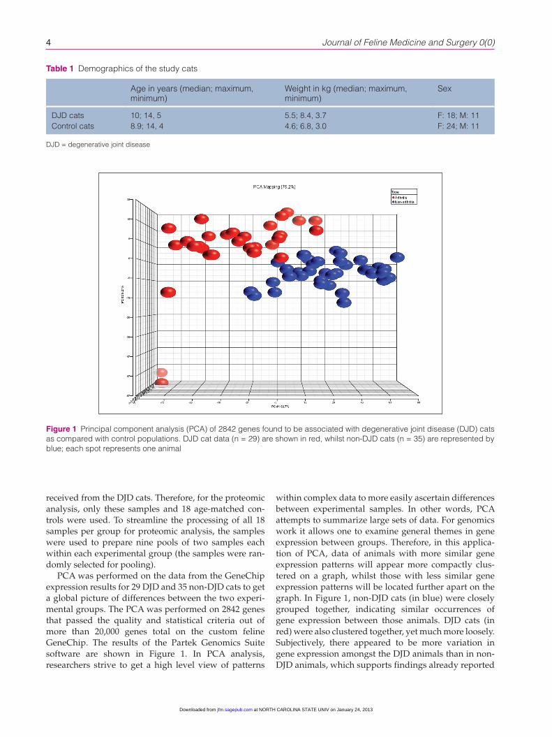
PCA was performed on the data from the GeneChip expression results for 29 DJD and 35 non-DJD cats to get a global picture of differences between the two experi-mental groups. The PCA was performed on 2842 genes that passed the quality and statistical criteria out of more than 20,000 genes total on the custom feline GeneChip. The results of the Partek Genomics Suite software are shown in Figure 1. In PCA analysis, researchers strive to get a high level view of patterns
within complex data to more easily ascertain differences between experimental samples. In other words, PCA attempts to summarize large sets of data. For genomics work it allows one to examine general themes in gene expression between groups. Therefore, in this applica-tion of PCA, data of animals with more similar gene expression patterns will appear more compactly clus-tered on a graph, whilst those with less similar gene expression patterns will be located further apart on the graph. In Figure 1, non-DJD cats (in blue) were closely grouped together, indicating similar occurrences of gene expression between those animals. DJD cats (in red) were also clustered together, yet much more loosely. Subjectively, there appeared to be more variation in gene expression amongst the DJD animals than in non-DJD animals, which supports findings already reported
Table 1 Demographics of the study cats
Age in years (median; maximum, minimum)
Weight in kg (median; maximum, minimum)
Sex
DJD cats 10; 14, 5 5.5; 8.4, 3.7 F: 18; M: 11Control cats 8.9; 14, 4 4.6; 6.8, 3.0 F: 24; M: 11
DJD = degenerative joint disease
Figure 1 Principal component analysis (PCA) of 2842 genes found to be associated with degenerative joint disease (DJD) cats as compared with control populations. DJD cat data (n = 29) are shown in red, whilst non-DJD cats (n = 35) are represented by blue; each spot represents one animal
at NORTH CAROLINA STATE UNIV on January 24, 2013jfm.sagepub.comDownloaded from

Gao et al 5
in the literature.34,37–40,48 This could also reflect some variability due to the aforementioned potential differ-ences in genetic uniformity between the two groups that could not be eliminated technically.
Out of the 2842 total genes entered into the MetaCore Pathway Analysis software, there were 400 genes that the software could not assign to a specific pathway mainly due to database discrepancies. These genes were not examined further in this study. The output from the software for the other 2442 genes from the feline GeneChip demonstrates the likelihood that the up- or down-regulated genes from the GeneChip are involved in a particular pathway (Table 2). The most likely affected pathways in DJD cats, according to the GeneChip analysis and MetaCore software, include antigen presentation by MHC class I and II molecules, as well as T-cell receptor signaling components of the immune system and oxidative phosphorylation [energy production by the cell in the form of adenosine triphos-phate (ATP)].
To closely investigate expression levels of key genes involved in antigen presentation by the immune system, the genes were grouped into three subcategories (Figure 2a–c). The subcategories within antigen presentation by the immune system were MHC class I and II molecules (a), proteasome molecules (b) and cathepsins (c). In gen-eral, all the genes involved in immune response that were analyzed in this study displayed a decrease in expression in the DJD animals when compared with non-DJD cats. The specific molecules outlined here in the figure all work together to ensure that the body’s immune system properly identifies foreign antigens. Proteasome and cathepsin molecules are thought to be primarily responsible for first digesting and then pre-senting foreign peptides to the immune system via MHC class I and II molecules.49–54 Figure 2 (a–c) illustrates the gene expression fold differences between the two
experimental groups for these specific antigen presenta-tion genes.
Figure 3 displays the fold change of some T-cell recep-tor genes found within DJD cats compared with controls. All the T-cell receptor and associated genes examined here were down-regulated in DJD animals compared with non-DJD animals. T-cell specific receptors, for example CD3, 4 and 8, to name a few, were all notably decreased in expression within DJD cats.
Apoptosis-associated genes were also found to be differentially expressed between the two groups. Figure 4 shows the fold change expression levels of key genes involved in apoptosis; anti-apoptotic genes were up-regulated in DJD animals.55
Table 2 Top pathways represented by the differentially expressed genes in arthritic versus control populations. MetaCore by Thompson Reuters identified the likelihood that 2442 of the 2842 differentially expressed genes identified in the expression profile are involved in these pathways. The other 400 genes could not be assigned to a pathway by the software. The smaller the P-value, the greater the likelihood that the genes are involved in that pathway and the less likely the genes were differentially expressed by chance alone
Pathway name GeneGo p-value
Antigen presentation by MHC class I and II
1.2e-08
T-cell receptor signaling 2.5e-08Oxidative phosphorylation 2.2e-08
MHC = major histocompatibility complex
Figure 2 Fold change expression levels of key genes involved in antigen presentation. (a) major histocompatibility complex (MHC) molecules; (b) proteasome and (c) cathepsin in degenerative joint disease (DJD) versus control populations. These graphs list fold changes in specific MHC class I and II (a), proteasome (b) and cathepsin (c) genes between DJD and non-DJD populations. All negative fold changes reflect down-regulation of the gene in DJD animals compared with non-DJD animals, and are represented in blue. All positive fold changes reflect up-regulation of the gene in DJD animals compared with non-DJD animals, and are represented in red
at NORTH CAROLINA STATE UNIV on January 24, 2013jfm.sagepub.comDownloaded from

6 Journal of Feline Medicine and Surgery 0(0)
Expression levels of genes that participate in oxida-tive phosphorylation were also distinct between DJD and non-DJD cats. The differentially expressed genes play important roles within the oxidative phosphoryla-tion pathway that exists inside the mitochondria of cells (Figure 5). Of the five main protein complexes that com-prise the oxidative phosphorylation pathway, all five were significantly down-regulated within DJD cats. Figure 5 shows down-regulation of genes within complexes I, II, III, IV and ATP synthase (also known as complex V).
For the protein expression levels, Figure 6 exhibits the PCA plot for the 87 differentially expressed proteins between the two experimental groups. Once more, dis-crete differences in protein expression between the two groups were evident by the data point clustering on the graph. This analysis confirms that the experimental groups could be distinguished by differentially expressed serum proteins, as well as genes from whole blood.
Figure 7 outlines differences in protein expression for the classical complement component of the immune sys-tem and clusterin, the latter representing a regulator of the complement system.56,57 Major proteins within the classical complement component of the immune system, such as C1s, C1r and C3, were notably up-regulated in DJD animals, whilst clusterin protein showed a decrease in expression compared with controls.
To control for age-specific differences between the DJD and non-DJD cats used in this study, gene expres-sion differences were examined on the basis of age alone (Table 3). This was performed to test whether gene differences between the experimental groups could be attributed to the disease state and not to the difference in age between the two groups. Table 3 dis-plays the results of two separate evaluations. The sub-jects were separated into >12 years of age versus 8–11 years of age and then separated into >11 years of age versus 7–10 years of age with the number of differen-tially expressed genes listed. When age alone was a factor, gene differences between the experimental groups were either non-existent or significantly smaller than when the subjects were grouped based on disease state alone. These findings indicate that the results reported here between the DJD and non-DJD popula-tions were not greatly affected by age differences between the subjects.
DiscussionThe proprietary Hills-Feline-2 GeneChip with more than 20,000 feline genes is a novel tool to evaluate the differences between cats with and without DJD. These investigations allow further insight into the disease, which is important given the prevalence of the dis-ease.1,5 Such data may also uncover possible treatment opportunities.
The gene expression studies reported here were per-formed on whole blood. Previous gene expression stud-ies of mammalian arthritis in the literature have reported variations in gene expression between human
Figure 4 Fold change expression levels of key genes involved in apoptosis. All negative fold changes reflect down-regulation of the gene in degenerative joint disease (DJD) animals compared with non-DJD and are represented in blue. All positive fold changes reflect up-regulation of the gene in DJD animals compared with non-DJD, and are represented in red
Figure 5 Fold change expression levels of key genes involved in oxidative phosphorylation. All negative fold changes reflect down-regulation of the gene in degenerative joint disease (DJD) animals compared with non-DJD and are represented in blueFigure 3 Fold change expression levels of key genes
involved in T-cell receptor signaling. All negative fold changes reflect down-regulation of the gene in degenerative joint disease (DJD) animals compared with non-DJD animals and are represented in blue
at NORTH CAROLINA STATE UNIV on January 24, 2013jfm.sagepub.comDownloaded from

Gao et al 7
RA and OA patients, and non-arthritic subjects.34,37–43,45,46 However, these studies have been performed on joint tissue. The use of whole blood, as opposed to certain blood components, bone, cartilage or synovial samples, is a unique approach. Using whole blood and blood derivatives, such as serum and plasma, might afford a more global view of what is happening in the disease state because blood touches all tissue types in the body and many disease-associated proteins are shed and can
be found in the plasma or serum. Furthermore, estab-lishing a blood-associated molecular signature for DJD in cats can potentially provide practitioners with easier means to diagnose the disease and follow its progres-sion and the effects of treatment.
Data reported here support the literature’s findings that gene expression differences within arthritic popu-lations tend to be more variable overall than gene expression patterns in non-DJD individuals.34,37–40,48 This result serves to further emphasize the complexity of the illness and the difficulties in finding effective treatment options.
Figure 6 Principal component analysis (PCA) of the differentially expressed proteins found in differentially joint disease (DJD) cats compared with control populations. DJD cat data (n = 9) are shown in red, whilst non-DJD cats (n = 9) are represented by blue, and each spot represents one pooled sample
Figure 7 Fold change expression levels of key proteins involved in the complement component of immune system response and clusterin. All negative fold changes reflect down-regulation of the gene in degenerative joint disease (DJD) animals compared with non-DJD, and are represented in blue. All positive fold changes reflect up-regulation of the gene in DJD animals compared with non-DJD, and are represented in red
Table 3 Comparison of differentially-expressed genes based on age as opposed to disease state. To control for age-specific differences between the experimental groups, differentially-expressed genes were examined on the basis of age alone to compare between the mean ages of the two populations. The number and magnitude of differentially-expressed genes between groups when separated by age alone were significantly less than when the animals were grouped by disease state alone
Age group comparison (years)
Number of samples
Number of differentially-expressed genes
>12 vs 8–11 27/25 159>11 vs 7–10 32/29 1014
at NORTH CAROLINA STATE UNIV on January 24, 2013jfm.sagepub.comDownloaded from

8 Journal of Feline Medicine and Surgery 0(0)
Based on our gene expression data, two of the most likely differentially expressed pathways involve immune system response: antigen presentation by MHC mole-cules and T-cell receptor signaling, both of which are nec-essary for normal immune response to foreign antigens. The remaining pathway that is most likely affected con-tains genes with a role in oxidative phosphorylation. These immune system-related genes were uniformly down-regulated in DJD cats compared with non-DJD cats. The literature supplies considerable evidence that immune system function generally declines and becomes less regulated with age in all mammals.58–60 Additionally, the risks for developing arthritis typically increase with age,44,61 although the data presented here suggest that arthritis is more than just normal ageing. Taken as a whole, this suggests that feline DJD arises, at least in part, owing to a dysfunctional immune system, which can become pathologically unregulated as the animal ages.
The act of antigen presentation by the immune system involves the proteasome complex, cathepsins, and MHC class I and II molecules. Proteasome and cathepsin mol-ecules work in concert to break down foreign antigens that the body encounters; the digested fragments are then presented to the immune system via MHC class I and II molecules.49–52 Part of the adaptive immune response, MHC class I molecules are located on the surface of all nucleated cells,53,54 whilst MHC class II molecules reside mainly on antigen presentation cells of the immune sys-tem.62,63 Collectively, MHC class I and II molecules are necessary to present antigens to T-cells, which then elicit the body’s immune response by releasing cytokines to recruit macrophages and/or promote antibody release by B cells.53,54,62,63 It is noteworthy that all the antigen pro-cessing and presentation players examined here were down-regulated in cats with clinical DJD. Taken together, the data suggest a reduced ability to process and present antigens in DJD cats, thereby decreasing the immune sys-tem’s ability to identify foreign, and potentially harmful, cells and proteins within those animals.
In addition to a reduced ability to identify foreign antigens, the next step in the immune cascade, which is mediated by T-cells, was also diminished within DJD animals. Specifically, CDs 3, 4 and 8 were down- regulated in DJD animals compared with controls. The CDs 3, 4 and 8, transmembrane glycoproteins that bind to MHC molecules and act as co-receptors, are essential for mediating T-cell based immunity.53,54,63,64 CD3 is a part of the T-cell receptor complex which initiates a T-cell-mediated response against antigens, CD4 binds with MHC class II molecules, and CD8 is a glycoprotein that specifically binds to MHC class I molecules to rec-ognize targets and mount an attack.62–64 Because all of these key T-cell players were found to be down-regu-lated in DJD cats, it is probable that cats with DJD have dampened T-cell activity and an overall impaired
immune response. Whether these altered processes result in, or occur concurrently with, DJD remains to be uncovered.
Genes representing apoptotic cascades also differed between DJD and non-DJD cats. Specifically, pro-apop-totic genes BAD, BID and TRADD were down-regu-lated and anti-apoptotic genes, such as XIAP and BIRC6, were up-regulated in DJD animals. This finding has implications for many pathways throughout the body. For the immune system in particular, this could mean an abnormal build-up of non-functional cells that the body is unable to clear. Equilibrium between pro-apoptotic and anti-apoptotic mechanisms is vital to the proper maintenance and function of the immune sys-tem.65–67 Functionality of immune system cells is largely determined by the specificity of interactions between antigen presentation components, namely MHC and T-cell-associated molecules.66 As DJD cats demon-strated a decrease in both MHC and T-cell associated genes, this suggests that dysfunctional immune cells exist in the DJD population. A deficiency in apoptotic cascades to remove these dysfunctional cells might compound the existing problem within these diseased animals. Evidence in the literature suggests that dise-quilibrium between apoptotic forces can lead to many ailments, such as immunodeficiency; autoimmunity, including systemic lupus erythematosus; and various degenerative disorders.68–71 Additionally, these find-ings appear to explain why corticosteroids are some-times effective during the treatment of certain arthritidies. The literature suggests that glucocorti-coids, such as dexamethasone, have a pro-apoptotic effect with lymphocytes;66 therefore, the use of corticos-teroidal compounds in DJD may help not only to reduce inflammation, but also to eliminate non-functional immune system cells from the body.
Another major area of differential gene expression between DJD and control animals was in the area of oxi-dative phosphorylation. Oxidative phosphorylation is the means by which the mitochondrial electron transport chain produces cellular energy in cells throughout the body.72,73 In this feline study, all of the protein complexes (I–V) that make up the electron transport chain revealed down-regulated genes compared with controls; these results, combined with the data generated here that con-trol for age, strongly suggest that the differences in oxi-dative phosphorylation in the DJD population were above and beyond the changes that occur in the electron transport chain during the normal ageing process.74 Mitochondrial dysfunction could be an important factor in the development of DJD. The literature solidly sup-ports the theory of mitochondrial dysfunction in the realm of mammalian arthritis. One such study used human articular chondrocytes to investigate gene changes between osteoarthritic and control populations.
at NORTH CAROLINA STATE UNIV on January 24, 2013jfm.sagepub.comDownloaded from

Gao et al 9
That report demonstrated that electron transport chain complexes II and III were down-regulated and mito-chondrial mass increased in the osteoarthritic, but not in the normal, human articular chondrocytes.75 The authors of that study also concluded that mitochondrial dys-function could be contributing to the pathology of arthri-tis. In cats, it seems evident from the current study’s findings that DJD cats demonstrated reduced activity within the electron transport chain and therefore pro-duced less cellular energy. Taken together, mitochondrial involvement in the disease process of DJD seems credi-ble and further research into mitochondrial imbalances within arthritic subjects is clearly necessary.
PCA on the 87 differentially-expressed proteins (Figure 6) confirmed that the two experimental groups could be separated based on protein expression in the serum. Furthermore, proteomic analysis provided authentica-tion for many of the genomic differences seen between the DJD and non-DJD animals. The clustering amongst the subjects within each group was not as tight on the proteomics graph of the data as it was in the genomics version. There are a few explanations for this. It is impor-tant to remember that the genomics PCA was performed using thousands of differentially expressed genes, while the proteomics PCA was carried out using just 87 differ-entially-expressed proteins. When comparing gene and protein changes, a direct, one-to-one relationship between the two molecule types is never expected. This is because the dynamic range and half-lives of messenger RNA mol-ecules and proteins are quite different, and the sensitivity of equipment used to measure each molecule type also differs. Consequently, more variation within a group with respect to one molecule type over another is not considered unusual. Also, the proteomics work used samples from pooled as opposed to individual subjects, so more variation within the proteomics graph is to be expected. Lastly, if any of the control animals (in blue) were in early, pre-clinical stages of DJD disease, they would demonstrate higher variability in serum protein levels than the control animals. This last point may explain why some of the control data points are grouped closer to DJD rather than fellow non-DJD cats on the PCA graph. Although this possibility cannot be dis-counted, none of these cats had developed any outward signs of clinical disease when this article was accepted for publication.
The antigen presentation aspect of the immune response was not the only aspect of the immune system to be differentially regulated in DJD cats. Through proteomic analysis of serum, the classical complement component of the immune system and clusterin protein were also found to be altered in cats with DJD compared with non-DJD cats. The classical complement component, also known as the complement system, is a bridge between the adaptive system, represented by the antigen presentation and
processing components (described above) and the innate system of a body’s immune function.76 It is comprised of a regulated set of blood proteins that work to lyse non-specific foreign matter.76 In this study, DJD cats were found to have a marked increase in complement compo-nents C1s, C1r and C3, while exhibiting down-regulation of the complement system regulator clusterin. An over-activated, improperly activated or unregulated comple-ment system has been identified as problematic because these situations lead to a perpetuation of inflammation and disease in mammals.77 Research in the literature reports up-regulated complement system gene compo-nents and down-regulated complement system regulators within juvenile idiopathic arthritis, RA and OA.43,78,79
As a whole, the altered expression levels of these genes and proteins involved in both the adaptive immune response and classical complement component of the immune system point to a defective immune response within cats with DJD that could cause and exacerbate an arthritic condition. Specifically targeting the immune system for intervention in the areas outlined above is an obvious step for future pharmaceutical and nutritional therapies.
ConclusionsThe chances of developing DJD is increased with increas-ing age; furthermore, increasing weight could also be a risk factor for the disease. However, the potential contri-bution of weight differences was not assessed directly in this study and will be within the scope of future studies. The differentially-expressed genes detailed here are important because they represent changes within DJD animals that far exceed the changes observed with nor-mal ageing. As noted previously, the chances of develop-ing DJD greatly increase with age; therefore, it is important that the study addresses this issue so that only those genes and proteins specifically related to DJD are examined and that the results are not clouded by factors unrelated to disease state. This study controlled for sub-ject age to ensure that the differentially-expressed genes discussed here represented true differences between dis-ease states and were not complicated by this confound-ing factor. There were certainly gene expression differences in common between the groups when sub-jects were classified by disease or age, indicating com-monalities between natural ageing and DJD pathways. However, significantly more up- or down-regulated genes were identified between disease groups than when the animals were grouped by age alone, and those genes that were similarly altered when the animals were grouped based on other factors were found to be changed to a lesser extent than when the animals were classified by disease alone. The data suggest that feline DJD could be a form of ‘accelerated ageing’ and support the asser-tion that the differentially expressed genes shown here
at NORTH CAROLINA STATE UNIV on January 24, 2013jfm.sagepub.comDownloaded from

10 Journal of Feline Medicine and Surgery 0(0)
are, in fact, related to disease and not due to the con-founding ageing process alone. Furthermore, the molec-ular processes associated with feline DJD in the affected joint tissues may be different than those reported here for whole blood and its derivatives, and further work is needed to evaluate the gene expression and proteomic changes in peripheral tissue.
Acknowledgements The authors would like to thank Rebecca J Palmer PhD for her assistance in the preparation of this article.
Funding Full financial support for this work was provided by Hill’s Pet Nutrition.
Conflict of interest All authors were employees of Hill’s Pet Nutrition, Inc. except B. Duncan X. Lascelles.
References 1 Lascelles BD, Henry JB, 3rd, Brown J, Robertson I, Sum-
rell AT, Simpson W, et al. Cross-sectional study of the prevalence of radiographic degenerative joint disease in domesticated cats. Vet Surg 2010; 39: 535–544.
2 Clarke SP, Mellor D, Clements DN, Gemmill T, Farrell M, Carmichael S, et al. Prevalence of radiographic signs of degenerative joint disease in a hospital population of cats. Vet Rec 2005; 157: 793–799.
3 Godfrey DR. Osteoarthritis in cats: a retrospective radio-logical study. J Small Anim Pract 2005; 46: 425–429.
4 Hardie EM, Roe SC and Martin FR. Radiographic evidence of degenerative joint disease in geriatric cats: 100 cases (1994–1997). J Am Vet Med Assoc 2002; 220: 628–632.
5 Slingerland LI, Hazewinkel HA, Meij BP, Picavet P and Voorhout G. Cross-sectional study of the prevalence and clinical features of osteoarthritis in 100 cats. Vet J 2011; 187: 304–309.
6 Clarke SP and Bennett D. Feline osteoarthritis: a pro-spective study of 28 cases. J Small Anim Pract 2006; 47: 439–445.
7 Gunew MN, Menrath VH and Marshall RD. Long-term safety, efficacy and palatability of oral meloxicam at 0.01-0.03 mg/kg for treatment of osteoarthritic pain in cats. J Feline Med Surg 2008; 10: 235–241.
8 Lascelles BD, Hansen BD, Roe S, DePuy V, Thomson A, Pierce CC, et al. Evaluation of client-specific outcome measures and activity monitoring to measure pain relief in cats with osteoarthritis. J Vet Intern Med 2007; 21: 410–416.
9 Lascelles BD, Henderson AJ and Hackett IJ. Evaluation of the clinical efficacy of meloxicam in cats with pain-ful locomotor disorders. J Small Anim Pract 2001; 42: 587–593.
10 Malik R, Allan GS, Howlett CR, Thompson DE, James G, McWhirter C, et al. Osteochondrodysplasia in Scottish Fold cats. Aust Vet J 1999; 77: 85–92.
11 Mathews KG, Koblik PD, Knoeckel MJ, Pool RR and Fyfe JC. Resolution of lameness associated with Scottish Fold osteodystrophy following bilateral ostectomies
and pantarsal arthrodeses: a case report. J Am Anim Hosp Assoc 1995; 31: 280–288.
12 Partington BP, Williams JF, Pechman RD and Beach RT. What is your diagnosis? Scottish Fold osteodystrophy. J Am Vet Med Assoc 1996; 209: 1235–1236.
13 Cowell KR, Jezyk PF, Haskins ME and Patterson DF. Muco-polysaccharidosis in a cat. J Am Vet Med Assoc 1976; 169: 334–339.
14 Crawley AC, Yogalingam G, Muller VJ and Hopwood JJ. Two mutations within a feline mucopolysaccharidosis type VI colony cause three different clinical phenotypes. J Clin Invest 1998; 101: 109–119.
15 Haskins ME, Jezyk PF, Desnick RJ, McDonough SK and Patterson DF. Mucopolysaccharidosis in a domestic short-haired cat — a disease distinct from that seen in the Sia-mese cat. J Am Vet Med Assoc 1979; 175: 384–387.
16 Haskins ME, Jezyk PF and Patterson DF. Mucopolysac-charide storage disease in three families of cats with arylsulfatase B deficiency: leukocyte studies and carrier identification. Pediatr Res 1979; 13: 1203–1210.
17 Jezyk PF, Haskins ME, Patterson DF, Mellman WJ and Greenstein M. Mucopolysaccharidosis in a cat with aryl-sulfatase B deficiency: a model of Maroteaux-Lamy syn-drome. Science 1977; 198: 834–836.
18 Konde LJ, Thrall MA, Gasper PW, Dial S, McBiles K, Col-gan S, et al. Radiographically visualized skeletal changes associated with mucopolysaccharidosis VI in cats. Vet Radiol 1987; 28: 223–228.
19 Macri B, Marino F, Mazzullo G, Trusso A, De Maria R, Amedeo S, et al. Mucopolysaccharidosis VI in a Siamese/short-haired European cat. J Vet Med A Physiol Pathol Clin Med 2002; 49: 438–142.
20 Vinayak A, Cross AR and Newell S. What is your diagno-sis? Mucopolysaccharidosis (MPS) type VI. J Am Vet Med Assoc 2005; 226: 351–352.
21 Polizopoulou ZS, Kazakos G, Patsikas MN and Roubies N. Hypervitaminosis A in the cat: a case report and review of the literature. J Feline Med Surg 2005; 7: 363–368.
22 Seawright AA and English PB. Hypervitaminosis A and deforming cervical spondylosis of the cat. J Comp Pathol 1967; 77: 29–39.
23 Seawright AA, English PB and Gartner RJ. Hypervita-minosis A and hyperostosis of the cat. Nature 1965; 206: 1171–1172.
24 Langenbach A, Green P, Giger U, Rhodes H, Gregor TP, LaFond E, et al. Relationship between degenerative joint disease and hip joint laxity by use of distraction index and Norberg angle measurement in a group of cats. J Am Vet Med Assoc 1998; 213: 1439–1443.
25 Bennett JA, Goodchild CS, Kidd C and McWilliam PN. Inhibition of brain stem neuronal activity by cardiac and pulmonary vagal afferent fibres in the cat. Q J Exp Physiol 1988; 73: 959–972.
26 Pedersen KB. The serology of Bordetella bronchiseptica iso-lated from pigs compared with strains from other animal species. Acta Pathol Microbiol Scand Suppl 1975; 83: 590–594.
27 Pedersen NC, Pool RR and O'Brien T. Feline chronic pro-gressive polyarthritis. Am J Vet Res 1980; 41: 522–535.
28 Moise NS, Crissman JW, Fairbrother JF and Baldwin C. Mycoplasma gateae arthritis and tenosynovitis in cats:
at NORTH CAROLINA STATE UNIV on January 24, 2013jfm.sagepub.comDownloaded from

Gao et al 11
case report and experimental reproduction of the disease. Am J Vet Res 1983; 44: 16–21.
29 Zeugswetter F, Hittmair KM, de Arespacochaga AG, Shibly S and Spergser J. Erosive polyarthritis associated with Mycoplasma gateae in a cat. J Feline Med Surg 2007; 9: 226–231.
30 Liehmann L, Degasperi B, Spergser J and Niebauer GW. Mycoplasma felis arthritis in two cats. J Small Anim Pract 2006; 47: 476–479.
31 Tisdall PL, Martin P and Malik R. Cryptic disease in a cat with painful and swollen hocks: an exercise in diagnostic reasoning and clinical decision-making. J Feline Med Surg 2007; 9: 418–423.
32 Pedersen M, Jacobsen S, Klarlund M, Pedersen BV, Wiik A, Wohlfahrt J, et al. Environmental risk factors differ between rheumatoid arthritis with and without auto-anti-bodies against cyclic citrullinated peptides. Arthritis Res Ther 2006; 8: R133.
33 Reckner Olsson A, Skogh T and Wingren G. Comorbid-ity and lifestyle, reproductive factors, and environmen-tal exposures associated with rheumatoid arthritis. Ann Rheum Dis 2001; 60: 934–939.
34 Clarke A and Vyse TJ. Genetics of rheumatic disease. Arthritis Res Ther 2009; 11: 248.
35 Hasler P and Zouali M. Immune receptor signaling, age-ing, and autoimmunity. Cell Immunol 2005; 233: 102–108.
36 van den Berg WB. Lessons from animal models of arthritis over the past decade. Arthritis Res Ther 2009; 11: 250.
37 Vandesompele J, De Preter K, Pattyn F, Poppe B, Van Roy N, De Paepe A and Speleman F. Accurate normalization of real-time quantitative RT-PCR data by geometric aver-ageing of multiple internal control genes. Gen Biol 2002; 3: research0034.1.
38 Teixeira VH, Olaso R, Martin-Magniette ML, Lasbleiz S, Jacq L, Oliveira CR, et al. Transcriptome analysis describing new immunity and defense genes in peripheral blood mononu-clear cells of rheumatoid arthritis patients. PLoS One 2009; 4: e6803.
39 van Baarsen LG, Bos CL, van der Pouw Kraan TC and Ver-weij CL. Transcription profiling of rheumatic diseases. Arthritis Res Ther 2009; 11: 207.
40 Verweij CL. Transcript profiling towards personalised med-icine in rheumatoid arthritis. Neth J Med 2009; 67: 364–371.
41 Katsuragawa Y, Saitoh K, Tanaka N, Wake M, Ikeda Y, Furukawa H, et al. Changes of human menisci in osteo-arthritic knee joints. Osteoarthritis Cartilage 2010; 18: 1133–1143.
42 Kumarasinghe DD, Perilli E, Tsangari H, Truong L, Kuli-waba JS, Hopwood B, et al. Critical molecular regulators, histomorphometric indices and their correlations in the trabecular bone in primary hip osteoarthritis. Osteoarthri-tis Cartilage 2010; 18: 1337–1344.
43 Geyer M, Grassel S, Straub RH, Schett G, Dinser R, Grifka J, et al. Differential transcriptome analysis of intraarticular lesional vs intact cartilage reveals new candidate genes in osteoarthritis pathophysiology. Osteoarthritis Cartilage 2009; 17: 328–335.
44 Boileau C, Martel-Pelletier J, Caron J, Msika P, Guillou GB, Baudouin C, et al. Protective effects of total fraction of avocado/soybean unsaponifiables on the structural
changes in experimental dog osteoarthritis: inhibition of nitric oxide synthase and matrix metalloproteinase-13. Arthritis Res Ther 2009; 11: R41.
45 Brew CJ, Clegg PD, Boot-Handford RP, Andrew JG and Hardingham T. Gene expression in human chondrocytes in late osteoarthritis is changed in both fibrillated and intact cartilage without evidence of generalised chondro-cyte hypertrophy. Ann Rheum Dis 2010; 69: 234–240.
46 Iliopoulos D, Malizos KN, Oikonomou P and Tsezou A. Integrative microRNA and proteomic approaches iden-tify novel osteoarthritis genes and their collaborative metabolic and inflammatory networks. PLoS One 2008; 3: e3740.
47 Irizarry RA, Bolstad BM, Collin F, Cope LM, Hobbs B and Speed TP. Summaries of Affymetrix GeneChip probe level data. Nucleic Acids Res 2003; 31: e15.
48 Gillis J and Pavlidis P. A methodology for the analysis of differential coexpression across the human lifespan. BMC Bioinformatics 2009; 10: 306.
49 Gaczynska M, Rock KL and Goldberg AL. Role of protea-somes in antigen presentation. Enzyme Protein 1993; 47: 354–369.
50 Lopez D and Del Val M. Selective involvement of protea-somes and cysteine proteases in MHC class I antigen pre-sentation. J Immunol 1997; 159: 5769–5772.
51 Katunuma N, Matsunaga Y, Himeno K and Hayashi Y. Insights into the roles of cathepsins in antigen process-ing and presentation revealed by specific inhibitors. Biol Chem 2003; 384: 883–890.
52 Riese RJ, Mitchell RN, Villadangos JA, Shi GP, Palmer JT, Karp ER, et al. Cathepsin S activity regulates antigen pre-sentation and immunity. J Clin Invest 1998; 101: 2351–2363.
53 Blanchard N and Shastri N. Cross-presentation of peptides from intracellular pathogens by MHC class I molecules. Ann N Y Acad Sci 2010; 1183: 237–250.
54 Rock KL, Farfan-Arribas DJ and Shen L. Proteases in MHC class I presentation and cross-presentation. J Immunol 2010; 184: 9–15.
55 Kufe DW, Holland JF and Frei E, American Cancer Society. Cancer medicine 6. 6th ed. Hamilton, ON, Lewiston, NY: BC Decker, 2003.
56 Chiang KC, Goto S, Chen CL, Lin CL, Lin YC, Pan TL, et al. Clusterin may be involved in rat liver allograft tolerance. Transpl Immunol 2000; 8: 95–99.
57 Jenne DE, Lowin B, Peitsch MC, Bottcher A, Schmitz G and Tschopp J. Clusterin (complement lysis inhibitor) forms a high density lipoprotein complex with apolipoprotein A-I in human plasma. J Biol Chem 1991; 266: 11030–11036.
58 Fulop T, Jr., Larbi A, Dupuis G and Pawelec G. Ageing, auto-immunity and arthritis: Perturbations of TCR signal trans-duction pathways with ageing — a biochemical paradigm for the ageing immune system. Arthritis Res Ther 2003; 5: 290–302.
59 Gress RE and Deeks SG. Reduced thymus activity and infection prematurely age the immune system. J Clin Invest 2009; 119: 2884–2887.
60 Desai A, Grolleau-Julius A and Yung R. Leukocyte function in the ageing immune system. J Leukoc Biol 2010; 87: 1001–1009.
61 Murphy L, Schwartz TA, Helmick CG, Renner JB, Tudor G, Koch G, et al. Lifetime risk of symptomatic knee osteoar-thritis. Arthritis Rheum 2008; 59: 1207–1213.
at NORTH CAROLINA STATE UNIV on January 24, 2013jfm.sagepub.comDownloaded from

12 Journal of Feline Medicine and Surgery 0(0)
62 Robinson JH and Delvig AA. Diversity in MHC class II antigen presentation. Immunology 2002; 105: 252–262.
63 Klein L, Munz C and Lunemann JD. Autophagy-mediated antigen processing in CD4(+) T cell tolerance and immu-nity. FEBS Lett 2010; 584: 1405–1410.
64 Doucey MA, Goffin L, Naeher D, Michielin O, Baumgartner P, Guillaume P, et al. CD3 delta establishes a functional link between the T cell receptor and CD8. J Biol Chem 2003; 278: 3257–3264.
65 Marsden VS and Strasser A. Control of apoptosis in the immune system: Bcl-2, BH3-only proteins and more. Annu Rev Immunol 2003; 21: 71–105.
66 Ekert PG and Vaux DL. Apoptosis and the immune sys-tem. Br Med Bull 1997; 53: 591–603.
67 Opferman JT and Korsmeyer SJ. Apoptosis in the develop-ment and maintenance of the immune system. Nat Immu-nol 2003; 4: 410–415.
68 Strasser A. The role of BH3-only proteins in the immune system. Nat Rev Immunol 2005; 5: 189–200.
69 Ogden CA and Elkon KB. Role of complement and other innate immune mechanisms in the removal of apoptotic cells. Curr Dir Autoimmun 2006; 9: 120–142.
70 Hughes P, Bouillet P and Strasser A. Role of Bim and other Bcl-2 family members in autoimmune and degenerative diseases. Curr Dir Autoimmun 2006; 9: 74–94.
71 Botto M. Links between complement deficiency and apoptosis. Arthritis Res 2001; 3: 207–110.
72 Filippin LI, Vercelino R, Marroni NP and Xavier RM. Redox signalling and the inflammatory response in rheumatoid arthritis. Clin Exp Immunol 2008; 152: 415–422.
73 Shi Q, Vaillancourt F, Cote V, Fahmi H, Lavigne P, Afif H, et al. Alterations of metabolic activity in human osteo-arthritic osteoblasts by lipid peroxidation end product 4-hydroxynonenal. Arthritis Res Ther 2006; 8: R159.
74 Hutter E, Renner K, Pfister G, Stockl P, Jansen-Durr P and Gnaiger E. Senescence-associated changes in respiration and oxidative phosphorylation in primary human fibro-blasts. Biochem J 2004; 380: 919–928.
75 Maneiro E, Martin MA, de Andres MC, Lopez-Armada MJ, Fernandez-Sueiro JL, del Hoyo P, et al. Mitochondrial respiratory activity is altered in osteoarthritic human articular chondrocytes. Arthritis Rheum 2003; 48: 700–708.
76 Yazdanbakhsh K. Review: complement receptor 1 thera-peutics for prevention of immune hemolysis. Immunohe-matology 2005; 21: 109–118.
77 Schlosser RJ, Mulligan RM, Casey SE, Varela JC, Harvey RJ and Atkinson C. Alterations in gene expression of complement components in chronic rhinosinusitis. Am J Rhinol Allergy 2010; 24: 21–25.
78 Barnes MG, Grom AA, Thompson SD, Griffin TA, Pavlidis P, Itert L, et al. Subtype-specific peripheral blood gene expression profiles in recent-onset juvenile idiopathic arthritis. Arthritis Rheum 2009; 60: 2102–2112.
79 Corallini F, Bossi F, Gonelli A, Tripodo C, Castellino G, Mollnes TE, et al. The soluble terminal complement complex (SC5b-9) up-regulates osteoprotegerin expres-sion and release by endothelial cells: implications in rheumatoid arthritis. Rheumatology (Oxford) 2009; 48: 293–298.
at NORTH CAROLINA STATE UNIV on January 24, 2013jfm.sagepub.comDownloaded from
Copyright © 2022 FDOKUMEN