Evolution of a New Function by Degenerative Mutation in Cephalochordate Steroid Receptors
15
Evolution of a New Function by Degenerative Mutation in Cephalochordate Steroid Receptors Jamie T. Bridgham 1 , Justine E. Brown 1 , Adriana Rodrı ´guez-Marı´ 2 , Julian M. Catchen 2,3 , Joseph W. Thornton 1 * 1 Center for Ecology and Evolutionary Biology, University of Oregon, Eugene, Oregon, United States of America, 2 Institute of Neuroscience, University of Oregon, Eugene, Oregon, United States of America, 3 Department of Computer and Information Science, University of Oregon, Eugene, Oregon, United States of America Abstract Gene duplication is the predominant mechanism for the evolution of new genes. Major existing models of this process assume that duplicate genes are redundant; degenerative mutations in one copy can therefore accumulate close to neutrally, usually leading to loss from the genome. When gene products dimerize or interact with other molecules for their functions, however, degenerative mutations in one copy may produce repressor alleles that inhibit the function of the other and are therefore exposed to selection. Here, we describe the evolution of a duplicate repressor by simple degenerative mutations in the steroid hormone receptors (SRs), a biologically crucial vertebrate gene family. We isolated and characterized the SRs of the cephalochordate Branchiostoma floridae, which diverged from other chordates just after duplication of the ancestral SR. The B. floridae genome contains two SRs: BfER, an ortholog of the vertebrate estrogen receptors, and BfSR, an ortholog of the vertebrate receptors for androgens, progestins, and corticosteroids. BfSR is specifically activated by estrogens and recognizes estrogen response elements (EREs) in DNA; BfER does not activate transcription in response to steroid hormones but binds EREs, where it competitively represses BfSR. The two genes are partially coexpressed, particularly in ovary and testis, suggesting an ancient role in germ cell development. These results corroborate previous findings that the ancestral steroid receptor was estrogen-sensitive and indicate that, after duplication, BfSR retained the ancestral function, while BfER evolved the capacity to negatively regulate BfSR. Either of two historical mutations that occurred during BfER evolution is sufficient to generate a competitive repressor. Our findings suggest that after duplication of genes whose functions depend on specific molecular interactions, high-probability degenerative mutations can yield novel functions, which are then exposed to positive or negative selection; in either case, the probability of neofunctionalization relative to gene loss is increased compared to existing models. Citation: Bridgham JT, Brown JE, Rodrı ´guez-Marı ´ A, Catchen JM, Thornton JW (2008) Evolution of a New Function by Degenerative Mutation in Cephalochordate Steroid Receptors. PLoS Genet 4(9): e1000191. doi:10.1371/journal.pgen.1000191 Editor: Harmit S. Malik, Fred Hutchinson Cancer Research Center, United States of America Received June 23, 2008; Accepted August 5, 2008; Published September 12, 2008 Copyright: ß 2008 Bridgham et al. This is an open-access article distributed under the terms of the Creative Commons Attribution License, which permits unrestricted use, distribution, and reproduction in any medium, provided the original author and source are credited. Funding: Supported by NIH R01-GM081592, NSF-IOB-0546906, and a Sloan fellowship (JWT), NIH F32-GM074398 (JTB), NIH R01-RR10715 (AR-M), NSF IGERT DGE- 9972830 (JMC), and a University of Oregon Undergraduate Research Fellowship (JEB). These organizations have had no role in the design, conduct, interpretation, preparation, or approval of the study or manuscript. Competing Interests: The authors have declared that no competing interests exist. * E-mail: [email protected] Introduction The vast majority of genes in eukaryotic genomes are hierarchically organized in gene families and superfamilies, because they were generated by a serial process of gene duplication and divergence [1,2]. The complexity of gene regulatory networks is also due to this process, which produced new functional interactions between regulators, effectors, and target genes [3,4]. How genes and their functions evolve after duplication is therefore a central and long-standing question in evolutionary biology [5–10]. Gene Duplication Models All major models of duplicate gene evolution to date assume that the two genes’ products do not interact with each other physically or functionally. Mutations in one copy therefore have no effect on the functions of the other. In the classic model [7,11], having two copies of a gene is phenotypically equivalent to having only one. After duplication, one copy drifts neutrally, free to amass mutations without the constraints of purifying selection (Figure 1A). In the vast majority of cases, degenerative mutations cause one copy to irreversibly lose its function and ultimately disappear from the genome, a process called nonfunctionalization. Rarely, however, mutations that yield a novel, beneficial function occur by chance; selection fixes these mutations and subsequently maintains both the original and ‘‘neofunctionalized’’ copies in the genome. The ultimate fate of a duplicate gene therefore depends on the outcome of a race between neo- and nonfunctionalization. Because gain-of-function mutations are very rare compared to those that compromise function [12–15], the vast majority of drifting duplicate genes will be lost, typically within a few million years, long before new functions are expected to evolve [10]. The plausibility of the classic model as a general explanation for the evolution of new functions has therefore been called into question[16,17]. In the second model–Duplication, Degeneration and Comple- mentation (DDC) [18]–duplication of a gene with multiple independent subfunctions, such as different expression domains controlled by separate regulatory elements, can be followed by degenerative mutations that knock out different subfunctions in PLoS Genetics | www.plosgenetics.org 1 September 2008 | Volume 4 | Issue 9 | e1000191
-
Upload
independent -
Category
Documents
-
view
1 -
download
0
Transcript of Evolution of a New Function by Degenerative Mutation in Cephalochordate Steroid Receptors

Evolution of a New Function by Degenerative Mutationin Cephalochordate Steroid ReceptorsJamie T. Bridgham1, Justine E. Brown1, Adriana Rodrıguez-Marı2, Julian M. Catchen2,3, Joseph W.
Thornton1*
1 Center for Ecology and Evolutionary Biology, University of Oregon, Eugene, Oregon, United States of America, 2 Institute of Neuroscience, University of Oregon, Eugene,
Oregon, United States of America, 3 Department of Computer and Information Science, University of Oregon, Eugene, Oregon, United States of America
Abstract
Gene duplication is the predominant mechanism for the evolution of new genes. Major existing models of this processassume that duplicate genes are redundant; degenerative mutations in one copy can therefore accumulate close toneutrally, usually leading to loss from the genome. When gene products dimerize or interact with other molecules for theirfunctions, however, degenerative mutations in one copy may produce repressor alleles that inhibit the function of the otherand are therefore exposed to selection. Here, we describe the evolution of a duplicate repressor by simple degenerativemutations in the steroid hormone receptors (SRs), a biologically crucial vertebrate gene family. We isolated andcharacterized the SRs of the cephalochordate Branchiostoma floridae, which diverged from other chordates just afterduplication of the ancestral SR. The B. floridae genome contains two SRs: BfER, an ortholog of the vertebrate estrogenreceptors, and BfSR, an ortholog of the vertebrate receptors for androgens, progestins, and corticosteroids. BfSR isspecifically activated by estrogens and recognizes estrogen response elements (EREs) in DNA; BfER does not activatetranscription in response to steroid hormones but binds EREs, where it competitively represses BfSR. The two genes arepartially coexpressed, particularly in ovary and testis, suggesting an ancient role in germ cell development. These resultscorroborate previous findings that the ancestral steroid receptor was estrogen-sensitive and indicate that, after duplication,BfSR retained the ancestral function, while BfER evolved the capacity to negatively regulate BfSR. Either of two historicalmutations that occurred during BfER evolution is sufficient to generate a competitive repressor. Our findings suggest thatafter duplication of genes whose functions depend on specific molecular interactions, high-probability degenerativemutations can yield novel functions, which are then exposed to positive or negative selection; in either case, the probabilityof neofunctionalization relative to gene loss is increased compared to existing models.
Citation: Bridgham JT, Brown JE, Rodrıguez-Marı A, Catchen JM, Thornton JW (2008) Evolution of a New Function by Degenerative Mutation in CephalochordateSteroid Receptors. PLoS Genet 4(9): e1000191. doi:10.1371/journal.pgen.1000191
Editor: Harmit S. Malik, Fred Hutchinson Cancer Research Center, United States of America
Received June 23, 2008; Accepted August 5, 2008; Published September 12, 2008
Copyright: � 2008 Bridgham et al. This is an open-access article distributed under the terms of the Creative Commons Attribution License, which permitsunrestricted use, distribution, and reproduction in any medium, provided the original author and source are credited.
Funding: Supported by NIH R01-GM081592, NSF-IOB-0546906, and a Sloan fellowship (JWT), NIH F32-GM074398 (JTB), NIH R01-RR10715 (AR-M), NSF IGERT DGE-9972830 (JMC), and a University of Oregon Undergraduate Research Fellowship (JEB). These organizations have had no role in the design, conduct, interpretation,preparation, or approval of the study or manuscript.
Competing Interests: The authors have declared that no competing interests exist.
* E-mail: [email protected]
Introduction
The vast majority of genes in eukaryotic genomes are
hierarchically organized in gene families and superfamilies,
because they were generated by a serial process of gene
duplication and divergence [1,2]. The complexity of gene
regulatory networks is also due to this process, which produced
new functional interactions between regulators, effectors, and
target genes [3,4]. How genes and their functions evolve after
duplication is therefore a central and long-standing question in
evolutionary biology [5–10].
Gene Duplication ModelsAll major models of duplicate gene evolution to date assume
that the two genes’ products do not interact with each other
physically or functionally. Mutations in one copy therefore have
no effect on the functions of the other.
In the classic model [7,11], having two copies of a gene is
phenotypically equivalent to having only one. After duplication,
one copy drifts neutrally, free to amass mutations without the
constraints of purifying selection (Figure 1A). In the vast majority
of cases, degenerative mutations cause one copy to irreversibly lose
its function and ultimately disappear from the genome, a process
called nonfunctionalization. Rarely, however, mutations that yield
a novel, beneficial function occur by chance; selection fixes these
mutations and subsequently maintains both the original and
‘‘neofunctionalized’’ copies in the genome. The ultimate fate of a
duplicate gene therefore depends on the outcome of a race
between neo- and nonfunctionalization. Because gain-of-function
mutations are very rare compared to those that compromise
function [12–15], the vast majority of drifting duplicate genes will
be lost, typically within a few million years, long before new
functions are expected to evolve [10]. The plausibility of the classic
model as a general explanation for the evolution of new functions
has therefore been called into question[16,17].
In the second model–Duplication, Degeneration and Comple-
mentation (DDC) [18]–duplication of a gene with multiple
independent subfunctions, such as different expression domains
controlled by separate regulatory elements, can be followed by
degenerative mutations that knock out different subfunctions in
PLoS Genetics | www.plosgenetics.org 1 September 2008 | Volume 4 | Issue 9 | e1000191
each copy. Because the two copies complement each other but do
not interact, these high-probability changes can occur neutrally in
small populations, and purifying selection will subsequently
conserve both copies and their remaining subfunctions
(Figure 1B). The DDC model appears applicable in numerous
case studies [18–21] and may explain in part why many duplicate
genes bear the signature of continuing purifying selection after
duplication [22]. Although subfunctionalization does not preclude
subsequent evolution of new functions [23,24], the DDC model
per se does not explain how or why novel gene functions evolve
after duplication, as has been observed in numerous examples
[25–36].
In the third model, an increase in the dose of a gene’s product
due to duplication yields an immediate selective advantage;
thereafter, purifying selection purges degenerative mutations and
conserves the ancestral function in both copies[37–39]. This
model says nothing about how gene pairs with different functions
evolved, and it does not explain the long-term retention of
duplicate genes for which dosage is not likely to be limiting for
fitness, such as many signaling molecules and enzymes in
multicellular eukaryotes. Other models have also been suggested,
largely variants of the three major models [16,40–43].
Molecular Interactions and Gene DuplicationGiven the high probability of degenerative mutations relative to
gain-of-function mutations, it remains unclear how large numbers
of duplicate genes have evaded nonfunctionalization long enough
to evolve new functions. A key omission from the major models is
their assumption that the products of duplicate genes do not
interact, either through direct physical contact or by competing for
other molecular partners. Gene duplication is recognized as
providing raw material for the evolution of elaborate molecular
X XX
XX
XX
XX
F
F
F1, F2
F1, F2
F1, F2
A Classic model
B DDC model
X X
X X
C Interactions
iii)
i) F
Inhibition
Inhibition
X
X X
ii)X
X
Figure 1. Effect of degenerative mutations on the function of a duplicate gene that depends on molecular interactions. A) In theclassic model, duplicate genes are redundant and do not interact, so mutation in one copy causes no change in the function (F) and evolve neutrally.B) In the DDC model, duplicate genes, which have two or more modular subfunctions (F1 and F2, blue and yellow), are redundant and do notinteract. Degenerative mutations in subfunctions cause no change in function if the subfunction is retained in the other copy and evolve neutrally. C)If a gene depends upon molecular interactions for its function, degenerative mutations in one copy can affect the function of the other and thereforebe selected for or against. i) In the case shown, a gene product must dimerize and interact with DNA (white bar) and an accessory factor (orangetriangle) for its function (transcriptional activation). ii) After duplication, degenerative mutations that impair the interactions of one copy with theaccessory factor yield a nonfunctional product that competes for DNA binding sites, reducing the activity of the other copy. iii) Degenerativemutations that impair DNA binding in one copy yield nonfunctional products that compete for accessory factors and reduce the activity of the othercopy. Inhibition also occurs if the duplicate genes do not dimerize but compete for other binding partners.doi:10.1371/journal.pgen.1000191.g001
Author Summary
Most genes evolved by duplication of more ancient genes.Under existing models of this process, mutations thatcompromise one copy have no effect on the other; as longas one copy remains intact, such ‘‘degenerative’’ mutationsare shielded from selection. Because degenerative muta-tions are common, most duplicates are expected to bedisabled before new functions can evolve. The greatfunctional diversity of genes is therefore somewhatpuzzling. Here, we reconstruct how simple degenerativemutations produced a new function in the steroidhormone receptors (SRs), a gene family crucial toreproduction and development. We characterized thetwo SRs of B. floridae, a cephalochordate that divergedfrom vertebrates ,500 million years ago, just after theancestral SR duplicated. One retained the ancestral gene’sestrogen receptor–like functions, while the other evolved anew function as a competitive repressor of the first. Eitherof two historical mutations is sufficient to recapitulateevolution of this function by disabling the receptor’sresponse to estrogen, but leaving its DNA-binding capacityintact. Our results suggest that, for the many genes thatfunction by specifically interacting with other molecules,simple mutations can yield novel functions that, beneficialor deleterious, are exposed to selection.
Cephalochordate Receptor Evolution
PLoS Genetics | www.plosgenetics.org 2 September 2008 | Volume 4 | Issue 9 | e1000191
interaction networks [3,44], but only limited research has
addressed how interactions affect the evolutionary fate of duplicate
genes. Most of this work has focused on the possibility that
duplications may alter the stoichiometry of proteins in a complex,
resulting in selection for or against the duplication per se [44–46].
But molecular interactions may affect the fate of gene duplicates
much more directly, and two types are of particular interest
(Figure 1C). First, many proteins function as homodimers. After
duplication of such genes, their products will initially cross-
dimerize [44]. Even if the duplicate genes are functionally
redundant–in the sense that two copies produce the same
phenotype as one–mutations that compromise one duplicate
may interfere with the functionality of the other by tying up its
products in non-functional dimers, just as null mutations at a single
locus can produce dominant negative alleles (see refs. [8,47]).
Second, many gene products form functional complexes with
other molecules, such as other proteins, DNA binding sites, or
small-molecule ligands and substrates. After duplication, the two
genes’ products initially compete for the same binding partners
[44]; mutations in one copy that compromise function but not
binding will tie up partner molecules in nonfunctional complexes,
reducing the activity of the other copy. As a result, degenerative
mutations after duplication may not be phenotypically silent, even
when the other copy retains the ancestral alleles. If repression of
the ancestral function is deleterious, as seems likely in most cases,
then purifying selection will tend to remove degenerative
mutations from both copies of a gene. As a result, the temporal
window before nonfunctionalization will be longer than expected
under the neutral scenarios of the classic and DDC models, and
the relative probability of neofunctionalization will increase. More
rarely, the evolution of a repressor molecule may allow beneficial
new modes of gene regulation; selection would favor such
mutations, and a neofunctionalization will occur by high-
probability degenerative mutations rather than the low-probability
events required under the other models.
Steroid Hormone ReceptorsSteroid hormone receptors (SRs) exemplify both types of
functional interaction described above [48,49], and their evolution
has been the subject of considerable interest [50–57]. SRs are the
major mediators of the effects of gonadal and adrenal steroid
hormones on development, reproduction, behavior, and homeo-
stasis throughout the vertebrates. These ligand-controlled tran-
scription factors bind tightly as homodimers to specific DNA
response elements in the control region of target genes. Jawed
vertebrates have six SRs–two for estrogens (ERa and ERb) and
one each for testosterone and other androgens (AR), progestins
(PR), glucocorticoids (GR), and mineralocorticoids (MR)–although
some lineages, such as the teleosts, have additional duplicates of
these genes [55,57,58]. Each hormone binds with high affinity and
specificity to a receptor, triggering a change in the receptor’s
conformation that allows it to attract coactivator proteins that
facilitate transcription of the target gene. SRs have a modular
structure, including a highly conserved DNA-binding domain
(DBD)–which recognizes and binds to response elements–and a
moderately conserved ligand-binding domain (LBD), which binds
the hormone and contains the hormone-activated transcriptional
activation function. This modular structure allows, for example,
construction of chimeric proteins that combine the functions of
one protein’s DBD with those of another protein’s LBD [59], and
many mutations are known that independently compromise ligand
binding, DNA binding, or transcriptional activation, without
impairing the other functions [60–65].
The SR gene family evolved by duplication and acquisition of
novel functions from a single ancestral SR gene (AncSR1).
AncSR1 is as ancient as the protostome-deuterostome divergence,
based on discovery of a steroid receptor gene in mollusks [27,66].
Duplication of this ancestral gene produced two major subfamilies
of vertebrate SRs[29]. One contains the estrogen receptors ERaand ERb, which are activated by estrogens and bind to estrogen-
response elements (EREs, inverted palindromes of AGGTCA)
[67]. Members of the other subfamily–the ketosteroid receptors
(kSRs), including AR, PR, GR, and MR–are activated by steroids
with a keto group at the 3-position on the steroid backbone (in
contrast to estrogens, which have a 3-hydroxyl) and bind to
ketosteroid response elements (kSREs, inverted palindromes of
AGAACA) [67]. Phylogenetic reconstruction, gene synthesis, and
experimental characterization of AncSR1 has shown that it had
the ligand- and DNA-specificity of the vertebrate estrogen
receptors [29,66], indicating that the gene duplicate leading to
the kSRs evolved novel DNA- and ligand-binding functions.
The evolutionary events by which this neofunctionalization
process occurred are obscure. It was complete by the time of the
vertebrate ancestor, some 470 million years ago, because
agnathans, the most basal vertebrate taxon, contain one ortholog
each for the ERa/ERb, the GR/MR, and the AR/PR pairs of
jawed vertebrates [29]. But little information is available on the
interval between the AncSR1 duplication and the ancestral kSR,
because SRs have not been characterized in taxa that branch off
the animal phylogeny between the protostome-deuterostome
ancestor and the ancestral vertebrate. Sequenced genomes are
available from the echinoderm Strongylocentrotus purpuratus and the
urochordate Ciona intestinalis, but all SRs were lost completely in
these lineages [68,69].
Cephalochordates therefore have the potential to provide key
information about the early evolution of the kSR gene after
duplication of AncSR1 [50]. These animals–commonly known as
lancelets or amphioxus–represent the earliest-branching chordate
taxon [70,71]. In many gene families, cephalochordates contain a
single gene orthologous to up to four paralogs in vertebrates [72],
so they are a good candidate to have a single kSR ortholog.
Further, the genome of the cephalochordate Branchiostoma floridae
contains the set of cytochrome P450 (CYP) genes required for the
biosynthesis of the sex steroids testosterone, progesterone, and
estradiol, and the synthesis of these hormones in B. floridae has
been experimentally established [73,74]. To gain insight into the
early functional diversification of the SRs, we therefore sought to
isolate the SRs of B. floridae, characterize their functions, and
analyze their evolution.
Results
Cephalochordate Steroid Receptor Sequences andPhylogenetic Analysis
Using a reciprocal BLAST search strategy, we identified two
loci as steroid receptor orthologs in the completely sequenced
genome of B. floridae. Coding sequences of both genes were
determined using RACE (rapid amplification of cDNA ends) on
RNA extracted from B. floridae adults, and full-length coding
sequences were then isolated using the polymerase chain reaction.
One of the B. floridae receptors has high amino acid identity to the
human ERs, particularly in the DNA-binding domain, and much
lower similarity to the AR, PR, GR, and MR. The other has
approximately equal similarity to the ERs and the other SRs
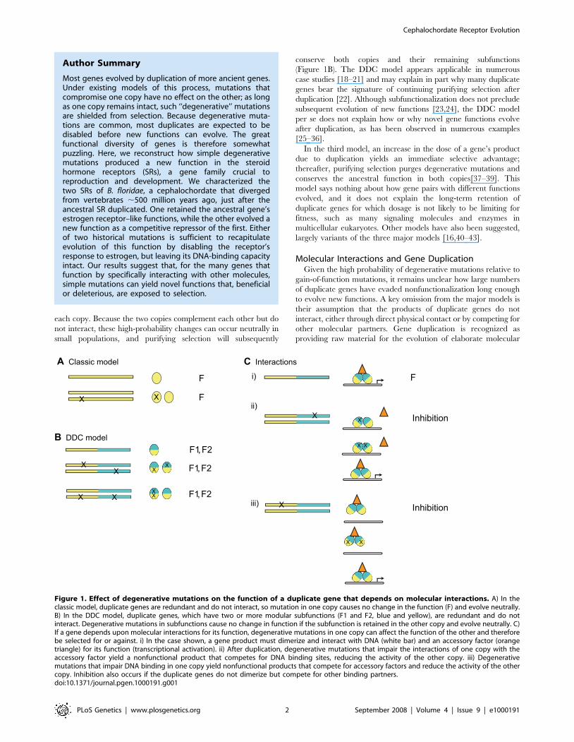
(Figure 2A, S1, S2).
To determine orthology relationships, we phylogenetically
analyzed an alignment of the two B. floridae SRs with the protein
Cephalochordate Receptor Evolution
PLoS Genetics | www.plosgenetics.org 3 September 2008 | Volume 4 | Issue 9 | e1000191
Cephalochordate Receptor Evolution
PLoS Genetics | www.plosgenetics.org 4 September 2008 | Volume 4 | Issue 9 | e1000191
sequences of 140 other steroid and related receptors (Table S1).
Both maximum likelihood and Bayesian analysis place the ER-like
B. floridae receptor at the base of the chordate estrogen receptor
clade and the other receptor at the base of the kSR clade
(Figure 2B). We therefore named the former gene BfER and the
latter BfSR. The affinity of BfER with other deuterostome and
protostome ERs and of the BfSR with the vertebrate kSRs are
both well-supported, with high posterior probabilities and
likelihood ratios (Figure 2B). Alternative topologies that place the
BfER and BfSR as cephalochordate-specific duplicates are very
unlikely, with a cumulative posterior probability ,0.20 in the
BMCMC analysis. Only the taxonomic relationships within the
ERs are not well-resolved; we cannot rule out an alternative
topology in which the protostome ‘‘ERs’’ are sister to a clade of all
deuterostome ERs and kSRs, implying duplication of AncSR1
after the protostome-deuterostome divergence. The phylogeny we
recovered is not an artifact of unincorporated heterotachy (site-
specific changes in evolutionary rate), which under some
conditions can confound ML and BMCMC methods using
homogeneous models [75,76]: when sequences were analyzed
using a mixed branch-length method that incorporates hetero-
tachy [77], the placement of BfER and BfSR did not change (See
Figure S3).
This phylogeny indicates that BfER is orthologous to the ERaand ERb pair of humans, and BfSR is orthologous to the AR/PR/
GR/MR cluster. The duplication of AncSR1 to produce these two
major SR clades therefore occurred before the divergence of
cephalochordates from the lineage leading to vertebrates.
Subsequent duplications to produce the four kSRs and ERs of
vertebrates occurred after that split, likely in the two vertebrate-
specific genome duplications [29,78]. This scenario is consistent
with evidence of conserved synteny between the scaffold
containing BfER and human chromosomes 6 and 14, where
ERa and ERb are located (Figures 2C, S4, see also ref. [79]). The
short scaffold that contains BfSR does not contain signal of strong
synteny to any human chromosomes (Figure S4). The phylogenetic
placement of the B. floridae receptors close to the ER-like AncSR1
explains why both have substantial sequence similarity to the
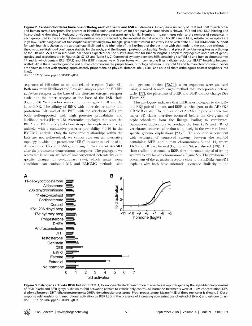
Figure 3. Estrogens activate BfSR but not BfER. A) Hormone-activated transcription of a luciferase reporter gene by the ligand-binding domainsof BfSR (black) and BfER (gray) is shown as fold activation relative to vehicle-only control. All hormone treatments were at 1 mM concentration. DES,diethylstilbesterol; DHT, dihydrotestosterone; DHEA, dehydroepiandrosterone; Prog, progesterone. Mean+/2SE of three replicates is shown. B) Dose-response relationship for transcriptional activation by BfSR LBD in the presence of increasing concentrations of estradiol (black) and estrone (gray).doi:10.1371/journal.pgen.1000191.g003
Figure 2. Cephalochordates have one ortholog each of the ER and kSR subfamilies. A) Sequence similarity of BfER and BfSR to each otherand human steroid receptors. The percent of identical amino acid residues for each pairwise comparison is shown. DBD and LBD, DNA-binding andligand-binding domains. B) Reduced phylogeny of the steroid receptor gene family. Numbers in parentheses refer to the number of sequences ineach group used in the analysis. Estrogen-sensitive receptors, including the ancestral steroid receptor (AncSR1) are in blue; ketosteroid receptors arein yellow. Black triangle, loss of transcriptional activation; yellow triangle, gain of ketosteroid sensitivity in the vertebrate AR/PR/GR/MR clade. Supportfor each branch is shown as the approximate likelihood ratio (the ratio of the likelihood of the best tree with that node to the best tree without it),the chi-square likelihood confidence statistic for the node, and the Bayesian posterior probability. Nodes that place B. floridae receptors as orthologsof the ERs and kSRs are in red. Scale bar shows expected per-site substitution rate for branch lengths. Complete phylogenies and a list of genes,species, and accessions are in Figures S6, S7, S8 and Table S1. C) Conserved synteny between BfER-containing scaffold 42 and human chromosomes14 and 6, which contain ERb (ESR2) and ERa (ESR1), respectively. Green boxes with connecting lines indicate reciprocal BLAST best-hits betweenscaffold 42 in the B. floridae genome and human chromosome 14; purple boxes, orthologs between Bf scaffold 42 and human chromosome 6. Genesare shown in order with spacing approximately proportional to physical distance. BfER, ESR1, and ESR2 all share orthologous nearest neighbors (redlines).doi:10.1371/journal.pgen.1000191.g002
Cephalochordate Receptor Evolution
PLoS Genetics | www.plosgenetics.org 5 September 2008 | Volume 4 | Issue 9 | e1000191
human ERs. The relatively long branch subtending the AR/PR/
GR/MR clade indicates a period of relatively rapid sequence
evolution in the ancestral kSR in the lineage leading to the
vertebrates after the cephalochordates diverged and before
subsequent gene duplications.
Intrinsic Functions of BfSR and BfERTo determine the molecular functions of the B. floridae steroid
receptors, we assayed both full-length proteins and specific functional
domains in a cell culture system. Surprisingly, we found that BfSR
retains the ER-like functions of the ancestral receptor, but BfER does
not. BfSR’s LBD activates transcription in the presence of
nanomolar concentrations of estradiol and estrone, but is insensitive
to ketosteroids, including a broad panel of androgens, progestins,
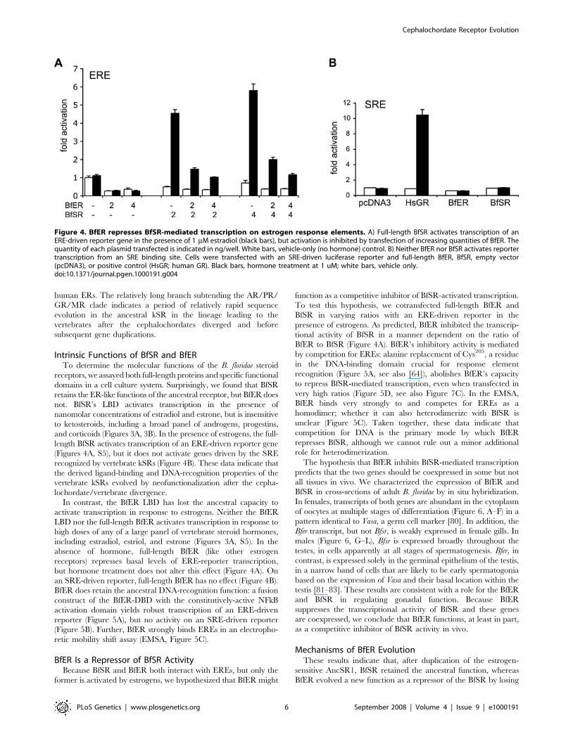
and corticoids (Figures 3A, 3B). In the presence of estrogens, the full-
length BfSR activates transcription of an ERE-driven reporter gene
(Figures 4A, S5), but it does not activate genes driven by the SRE
recognized by vertebrate kSRs (Figure 4B). These data indicate that
the derived ligand-binding and DNA-recognition properties of the
vertebrate kSRs evolved by neofunctionalization after the cepha-
lochordate/vertebrate divergence.
In contrast, the BfER LBD has lost the ancestral capacity to
activate transcription in response to estrogens. Neither the BfER
LBD nor the full-length BfER activates transcription in response to
high doses of any of a large panel of vertebrate steroid hormones,
including estradiol, estriol, and estrone (Figures 3A, S5). In the
absence of hormone, full-length BfER (like other estrogen
receptors) represses basal levels of ERE-reporter transcription,
but hormone treatment does not alter this effect (Figure 4A). On
an SRE-driven reporter, full-length BfER has no effect (Figure 4B).
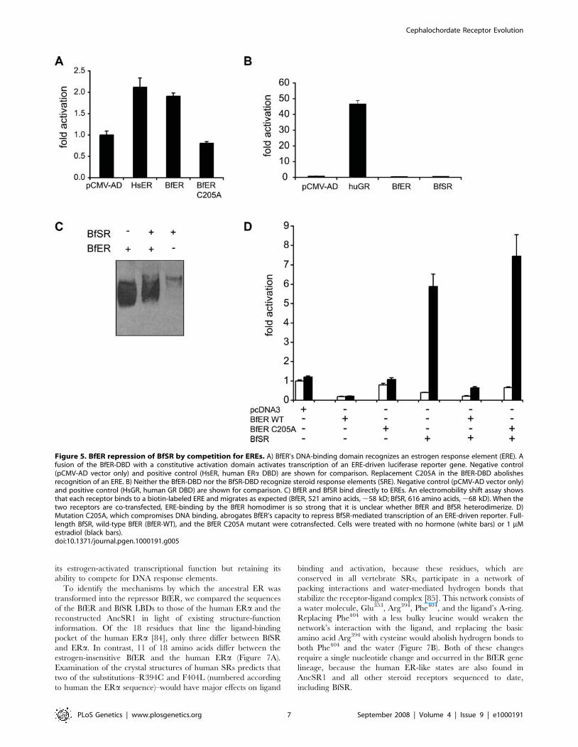
BfER does retain the ancestral DNA-recognition function: a fusion
construct of the BfER-DBD with the constitutively-active NFkB
activation domain yields robust transcription of an ERE-driven
reporter (Figure 5A), but no activity on an SRE-driven reporter
(Figure 5B). Further, BfER strongly binds EREs in an electropho-
retic mobility shift assay (EMSA, Figure 5C).
BfER Is a Repressor of BfSR ActivityBecause BfSR and BfER both interact with EREs, but only the
former is activated by estrogens, we hypothesized that BfER might
function as a competitive inhibitor of BfSR-activated transcription.
To test this hypothesis, we cotransfected full-length BfER and
BfSR in varying ratios with an ERE-driven reporter in the
presence of estrogens. As predicted, BfER inhibited the transcrip-
tional activity of BfSR in a manner dependent on the ratio of
BfER to BfSR (Figure 4A). BfER’s inhibitory activity is mediated
by competition for EREs: alanine replacement of Cys205, a residue
in the DNA-binding domain crucial for response element
recognition (Figure 5A, see also [64]), abolishes BfER’s capacity
to repress BfSR-mediated transcription, even when transfected in
very high ratios (Figure 5D, see also Figure 7C). In the EMSA,
BfER binds very strongly to and competes for EREs as a
homodimer; whether it can also heterodimerize with BfSR is
unclear (Figure 5C). Taken together, these data indicate that
competition for DNA is the primary mode by which BfER
represses BfSR, although we cannot rule out a minor additional
role for heterodimerization.
The hypothesis that BfER inhibits BfSR-mediated transcription
predicts that the two genes should be coexpressed in some but not
all tissues in vivo. We characterized the expression of BfER and
BfSR in cross-sections of adult B. floridae by in situ hybridization.
In females, transcripts of both genes are abundant in the cytoplasm
of oocytes at multiple stages of differentiation (Figure 6, A–F) in a
pattern identical to Vasa, a germ cell marker [80]. In addition, the
Bfer transcript, but not Bfsr, is weakly expressed in female gills. In
males (Figure 6, G–L), Bfsr is expressed broadly throughout the
testes, in cells apparently at all stages of spermatogenesis. Bfer, in
contrast, is expressed solely in the germinal epithelium of the testis,
in a narrow band of cells that are likely to be early spermatogonia
based on the expression of Vasa and their basal location within the
testis [81–83]. These results are consistent with a role for the BfER
and BfSR in regulating gonadal function. Because BfER
suppresses the transcriptional activity of BfSR and these genes
are coexpressed, we conclude that BfER functions, at least in part,
as a competitive inhibitor of BfSR activity in vivo.
Mechanisms of BfER EvolutionThese results indicate that, after duplication of the estrogen-
sensitive AncSR1, BfSR retained the ancestral function, whereas
BfER evolved a new function as a repressor of the BfSR by losing
Figure 4. BfER represses BfSR-mediated transcription on estrogen response elements. A) Full-length BfSR activates transcription of anERE-driven reporter gene in the presence of 1 mM estradiol (black bars), but activation is inhibited by transfection of increasing quantities of BfER. Thequantity of each plasmid transfected is indicated in ng/well. White bars, vehicle-only (no hormone) control. B) Neither BfER nor BfSR activates reportertranscription from an SRE binding site. Cells were transfected with an SRE-driven luciferase reporter and full-length BfER, BfSR, empty vector(pcDNA3), or positive control (HsGR; human GR). Black bars, hormone treatment at 1 uM; white bars, vehicle only.doi:10.1371/journal.pgen.1000191.g004
Cephalochordate Receptor Evolution
PLoS Genetics | www.plosgenetics.org 6 September 2008 | Volume 4 | Issue 9 | e1000191
its estrogen-activated transcriptional function but retaining its
ability to compete for DNA response elements.
To identify the mechanisms by which the ancestral ER was
transformed into the repressor BfER, we compared the sequences
of the BfER and BfSR LBDs to those of the human ERa and the
reconstructed AncSR1 in light of existing structure-function
information. Of the 18 residues that line the ligand-binding
pocket of the human ERa [84], only three differ between BfSR
and ERa. In contrast, 11 of 18 amino acids differ between the
estrogen-insensitive BfER and the human ERa (Figure 7A).
Examination of the crystal structures of human SRs predicts that
two of the substitutions–R394C and F404L (numbered according
to human the ERa sequence)–would have major effects on ligand
binding and activation, because these residues, which are
conserved in all vertebrate SRs, participate in a network of
packing interactions and water-mediated hydrogen bonds that
stabilize the receptor-ligand complex [85]. This network consists of
a water molecule, Glu353, Arg394, Phe404, and the ligand’s A-ring.
Replacing Phe404 with a less bulky leucine would weaken the
network’s interaction with the ligand, and replacing the basic
amino acid Arg394 with cysteine would abolish hydrogen bonds to
both Phe404 and the water (Figure 7B). Both of these changes
require a single nucleotide change and occurred in the BfER gene
lineage, because the human ER-like states are also found in
AncSR1 and all other steroid receptors sequenced to date,
including BfSR.
Figure 5. BfER repression of BfSR by competition for EREs. A) BfER’s DNA-binding domain recognizes an estrogen response element (ERE). Afusion of the BfER-DBD with a constitutive activation domain activates transcription of an ERE-driven luciferase reporter gene. Negative control(pCMV-AD vector only) and positive control (HsER, human ERa DBD) are shown for comparison. Replacement C205A in the BfER-DBD abolishesrecognition of an ERE. B) Neither the BfER-DBD nor the BfSR-DBD recognize steroid response elements (SRE). Negative control (pCMV-AD vector only)and positive control (HsGR, human GR DBD) are shown for comparison. C) BfER and BfSR bind directly to EREs. An electromobility shift assay showsthat each receptor binds to a biotin-labeled ERE and migrates as expected (BfER, 521 amino acids, ,58 kD; BfSR, 616 amino acids, ,68 kD). When thetwo receptors are co-transfected, ERE-binding by the BfER homodimer is so strong that it is unclear whether BfER and BfSR heterodimerize. D)Mutation C205A, which compromises DNA binding, abrogates BfER’s capacity to repress BfSR-mediated transcription of an ERE-driven reporter. Full-length BfSR, wild-type BfER (BfER-WT), and the BfER C205A mutant were cotransfected. Cells were treated with no hormone (white bars) or 1 mMestradiol (black bars).doi:10.1371/journal.pgen.1000191.g005
Cephalochordate Receptor Evolution
PLoS Genetics | www.plosgenetics.org 7 September 2008 | Volume 4 | Issue 9 | e1000191
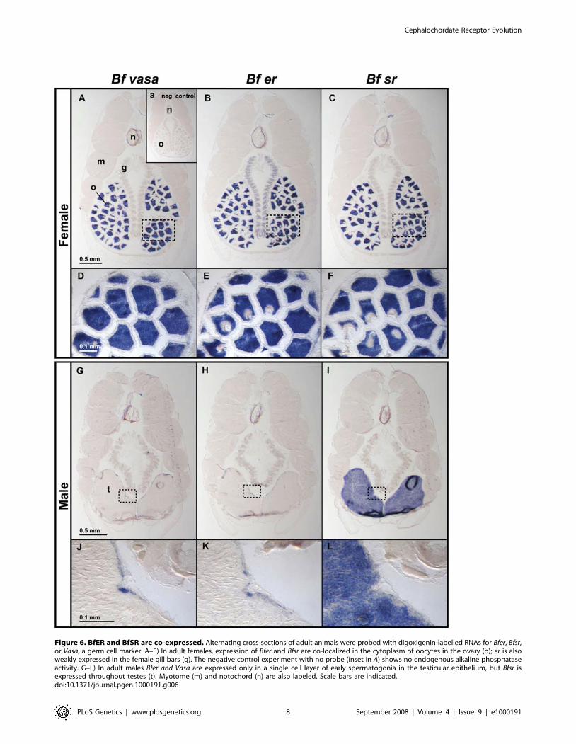
Figure 6. BfER and BfSR are co-expressed. Alternating cross-sections of adult animals were probed with digoxigenin-labelled RNAs for Bfer, Bfsr,or Vasa, a germ cell marker. A–F) In adult females, expression of Bfer and Bfsr are co-localized in the cytoplasm of oocytes in the ovary (o); er is alsoweakly expressed in the female gill bars (g). The negative control experiment with no probe (inset in A) shows no endogenous alkaline phosphataseactivity. G–L) In adult males Bfer and Vasa are expressed only in a single cell layer of early spermatogonia in the testicular epithelium, but Bfsr isexpressed throughout testes (t). Myotome (m) and notochord (n) are also labeled. Scale bars are indicated.doi:10.1371/journal.pgen.1000191.g006
Cephalochordate Receptor Evolution
PLoS Genetics | www.plosgenetics.org 8 September 2008 | Volume 4 | Issue 9 | e1000191
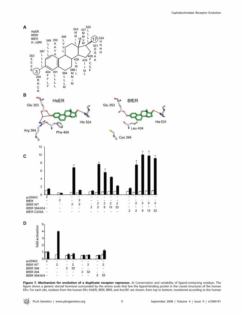
Figure 7. Mechanism for evolution of a duplicate receptor repressor. A) Conservation and variability of ligand-contacting residues. Thefigure shows a generic steroid hormone surrounded by the amino acids that line the ligand-binding pocket in the crystal structures of the humanERa. For each site, residues from the human ERa (HsER), BfSR, BfER, and AncSR1 are shown, from top to bottom, numbered according to the human
Cephalochordate Receptor Evolution
PLoS Genetics | www.plosgenetics.org 9 September 2008 | Volume 4 | Issue 9 | e1000191
To test the hypothesis that one or both of these historical
replacements could have converted an estrogen-sensitive tran-
scriptional activator into a competitive inhibitor, we introduced
these changes into BfSR by site-directed mutagenesis. As
predicted, the double-mutant BfSR R394C/F404L loses the
wildtype’s capacity to activate transcription in response to
estrogens. Further, when co-transfected, the BfSR double-mutant
repressed the activity of BfSR from an ERE in a concentration-
dependent manner (Figure 7C). This repressive activity is
dependent on ERE-binding and is not an artifact of co-
transfection per se: unlike BfER and BfSR R394/F404L, co-
transfecting increasing quantities of BfER-C205A, which does not
compete for EREs, fails to repress BfSR-mediated reporter
transcription (Figure 7C). Either mutation in isolation is sufficient
to confer the full repressive phenotype on BfSR (Figure 7D). These
data indicate that simple degenerative mutations, which abolish
the protein’s activation by estrogens but do not interfere with its
ancestral DNA-binding activity, are sufficient to confer on BfER
its novel function as a competitive inhibitor.
Discussion
We found that BfSR, the B. floridae ortholog of the vertebrate
kSRs, has molecular functions similar to those of the vertebrate
estrogen receptor, whereas the cephalochordate ER ortholog is an
estrogen-insensitive transcriptional repressor of BfSR-mediated
transcription. The presence of estrogen-activated, ERE-recogniz-
ing receptors in both clades descending from AncSR1 corrobo-
rates previous findings that AncSR1 was ER-like in function
[29,66]. Our results make sense in light of the fact that
cephalochordates and vertebrates diverged relatively recently after
the duplication of AncSR1. In both taxa, one duplicate retained
the ancestral function and the other evolved a new function, but
the copy that experienced each fate differed between the two
lineages. In the vertebrates, the ancestral kSR evolved novel ligand
and response-element specificity, and ER retained the ancestral
response to estrogen and EREs. In cephalochordates, the kSR
ortholog retained the ancestral specificity for ligand and DNA, and
the ER ortholog lost its estrogen-stimulated transcriptional
activity, becoming a competitive repressor of BfSR on its DNA
binding sites. BfER and BfSR have been recently recognized in the
amphioxus genome sequence [50,74]; this is the first character-
ization of their functions.
Our experiments shed light on the functions of steroid
hormones and their receptors in cephalochordates. B. floridae
produces estradiol [73], so it is likely that BfSR functions, at least
in part, as a classic estrogen receptor. Although B. floridae also
produces progesterone and testosterone, these steroids do not
activate either receptor in this species, suggesting that they may
function primarily as intermediates in the synthesis of estrogens or
in other signaling pathways [29]. BfSR and BfER are strongly
coexpressed in some but not all tissues, so it is likely that BfER
functions as a BfSR regulator, presumably allowing finer tissue-
specific modulation of BfSR activity. These receptors may also
have other functions, such as activation by unknown cephalo-
chordate-specific hormones or post-transcriptional modification
[86,87].
The expression of both BfER and BfSR primarily in testis and
ovary suggests a role for both in gonadal function, such as
regulating germ cell development and maturation. In females,
both receptors are expressed strongly in ovary. In males, BfSR is
expressed throughout the testis, including the apical zone near the
testicular lumen, where later-stage spermatocytes, spermatids, and
sperm are found. BfER, in contrast, is expressed only in the basal
cells of the germinal epithelium, where early spermatogonia are
located and where the germ-cell marker Vasa is also expressed
[81]. In vertebrates, ERs also play important roles in testicular
development and spermatogenesis, suggesting a conserved ances-
tral function [88,89]. Further work in vivo is required to elucidate
the physiological and developmental roles of BfER, BfSR, and
estrogens in cephalochordates.
Our results demonstrate the strong effect that functional
interactions can have on evolution after gene duplication and
suggest the need to supplement existing models of this process. In
both the classic and DDC models, degenerative mutations in one
copy produce no change in phenotype. As a result, redundancy
shields one copy from purifying selection, allowing high-probabil-
ity degenerative mutations to accumulate neutrally; neofunctiona-
lization must occur only through relatively low-probability gain-of-
function mutations. Our results are not predicted by either model.
Because steroid receptors function by interacting physically with
other molecules, simple degenerative mutations in the BfER LBD
produced a novel molecular phenotype–a protein that can no
longer carry out the full functions of the ancestral gene but still
competes for binding partners, thereby repressing the activity of its
duplicate wherever the two are coexpressed. Evolutionary changes
in the expression of BfSR and BfER also occurred, yielding
partially overlapping expression domains. The order in which
these coding and expression changes evolved, and their relative
roles in the maintenance of the duplicated ancestral SR, cannot be
resolved with current data.
The production of repressive functions by degenerative
mutations may strongly affect the dynamics of evolution after
gene duplication, particularly the relative probability of neofunc-
tionalization versus gene loss by nonfunctionalization. If the
repressor allele is adaptive — allowing beneficial new modes of
regulating the other copy’s activity–then it will be conserved,
leading to long-term maintenance of both gene copies, as occurred
with BfER and BfSR. This scenario points to a creative role for
high-probability degenerative mutation not envisioned by any
existing models. Because degenerative mutations are common,
including them as a source of new functions would increase both
the absolute and relative probability of neofunctionalization. We
found that either of two historical point mutations in the BfER is
sufficient to generate a repressor of BfSR; given the modular
organization of steroid receptor domains, it is likely that there are
many other potential mutations that could also have abolished
estrogen-activation while leaving DNA-binding intact.
ERa sequence. Moieties at the 3 and 17 positions, shown as large circles, vary among steroids. B) Historical substitutions R394C and F404L, whichoccurred in the lineage leading to transcriptional repressor BfER, are predicted to abolish estrogen binding and activation. Left, x-ray crystal structureof the ligand pocket of the human ERa with estradiol. Arg394 and Phe404 play key roles in a network of hydrogen bonds and packing interactions thatstabilize the ligand and transcriptionally active conformation. Right, mutations R394C and F404L from BfER disrupt this network. Red sphere, watermolecule. C) Mutations R394C and F404L, introduced into BfSR, abolish the receptor’s transcriptional capacity and generate a dose-dependentrepressor of the wild-type (WT) BfSR. Numbers show the quantity of each plasmid transfected (in ng) with 1 mM estradiol (black bars) or with nohormone added (vehicle only, white bars). Mutation C205A, which disrupts DNA-binding, eliminates BfER’s capacity to repress BfSR-driventranscription. D) Either mutation R394C or F404L, introduced singly into BfSR, are each sufficient to abolish transcriptional capacity and generate arepressor of BfSR-WT.doi:10.1371/journal.pgen.1000191.g007
Cephalochordate Receptor Evolution
PLoS Genetics | www.plosgenetics.org 10 September 2008 | Volume 4 | Issue 9 | e1000191
If, on the other hand, repression of the ancestral function is
deleterious–as will often be the case–then purifying selection will
tend to purge degenerative mutations from both copies, leading to
conservation of both duplicates with the ancestral function. In such
cases, the accumulation of degenerative mutation would be much
slower than under the neutral scenarios of the classic and DDC
models. In turn, the rate of nonfunctionalization would be slowed,
and the temporal window during which neofunctionalization can
occur would be lengthened substantially.
Evolution of beneficial duplicate repressors may be a wide-
spread phenomenon. Numerous other members of the nuclear
receptor (NR) superfamily have evolved by partial degeneration to
function primarily as repressors of the transcriptional activity of
paralogous NRs. Some have lost their capacity to activate
transcription but retain their ability to bind DNA, so–like BfER–
they compete for the binding sites of other receptors, whose
activity they downregulate. Others have lost the capacity to bind
DNA but retain the ability to form dimers with other NRs and
thus inhibit their activity [90,91]. Repressor duplicates have also
evolved in beta-helix-loop-helix transcription factors: some
duplicates have lost the canonical DNA-binding domain but still
heterodimerize with and therefore silence their paralogs [47].
Similarly, in the family of transmembrane tumor necrosis factor
receptors, which trigger apoptosis in response to extracellular
ligands, primate-specific duplicates have evolved which bind
ligand but have lost their capacity to interact with the intracellular
factors that stimulate apoptosis. These ‘‘decoy receptors’’ compete
for ligand and thereby repress the activity of their paralogs,
preventing ligand-triggered apoptosis in cells that express receptors
of both classes [92,93].
As for the cases in which degenerative mutations are deleterious,
it is not possible to directly determine the historical importance of
selection against duplicate repressors in shaping genome evolution,
but there are reasons to believe it may have played a significant
role in delaying nonfunctionalization. Most protein families–
enzymes, transcription factors, hormones, neurotransmitters,
growth factors, to name a few–depend upon specific molecular
interactions for their functions. In many of these families, separate
domains, independent molecular surfaces, or even specific sets of
residues mediate interactions with different partners or other
aspects of function. It is therefore reasonable to expect that after
duplication a large class of mutations would compromise specific
aspects of function without abolishing all interactions, thereby
producing competitive repressors. It is also likely that a nontrivial
fraction of these alleles would be deleterious. If these assumptions
are correct, then purifying selection would have played a role of
general importance in purging degenerative mutations after gene
duplication, maintaining duplicates for longer periods of time, and
extending the temporal window during which neofunctionaliza-
tion can occur. Indeed, it has been observed that duplicate genes
involved in signal transduction have been preferentially retained in
Arabidopsis [94]. This hypothesis also predicts that many genes after
duplication will bear the mark of purifying selection–nonsynon-
ymous-to-synonymous substitution rates considerably less than
one–even in the absence of subfunctionalization. It has been
observed that the majority of recent duplicate genes in Paramecium
genomes are under strong purifying selection [46]. This signature
is predicted to be particularly strong in duplicate genes that form
homodimers or interact with partner molecules whose concentra-
tion is limiting for function, such as specific DNA binding sites.
Not all degenerative mutations in genes whose functions depend
on interactions are likely to produce repressor alleles. Those that
radically reduce expression, impair protein folding or stability, or
increase the tendency for a protein to aggregate are expected to
yield alleles compromised in their ability to interact with any and
all partners. These fully nonfunctional alleles would have no effect
on their duplicate’s function and would be sheltered by
redundancy as predicted by the classic and DDC models’ neutral
scenarios. Only those mutations that affect modular domains or
molecular surfaces that control distinct subfunctions within the
coding sequence have the potential to eliminate one aspect of a
protein’s functions without abolishing its interactions with at least
some molecular partners. It is therefore likely that purifying
selection would be partially relaxed after duplication–in contrast to
the situation for unduplicated genes, in which all mutations that
compromise function, including those that also generate novel
functions, would be exposed to the constraining influence of
selection. Ohno’s idea that functional diversity can evolve by
random exploration of sequence space after gene duplication may
strain credulity in its original conception of a purely neutral setting
and an inexorable tendency toward ‘‘entropic decay.’’[95] It
becomes far more plausible, however, when selection can play an
anti-entropic role, and when creativity can arise not only from rare
mutational combinations but also from far more common ones.
Materials and Methods
Receptor IsolationSteroid receptor orthologs were identified in the Branchiostoma
floridae genome database (Joint Genome Institute, v. 1.0) by tblastn
search using as queries exons from the conserved DBD and LBD
of human steroid receptors, as well as the inferred sequence of the
ancestral steroid receptor [29,66] and the ancestral corticosteroid
receptor [96]. Recovered B. floridae sequences were used as queries
to reciprocally search the human protein database using blastx,
and those that recovered SR family members as best hits were
retained. Using this technique, two B. floridae sequences were
identified as likely SR orthologs.
From the two partial SR sequences recovered, primers were
designed for RACE (Rapid Amplification of cDNA Ends).
Amphioxus RNA was extracted from gravid individuals collected
in October 2003 (Gulf Specimen Marine Lab, Panacea, FL) using
the RNeasy kit (Qiagen, Valencia, CA). The isolated RNA was
reverse transcribed using Thermoscript (Invitrogen, Carlsbad, CA)
and oligo dT primers or PowerScript reverse transcriptase
(Clontech, Mountain View, CA) with gene specific primers. Both
59 and 39 sequences were obtained by RACE using the SmartRace
method (Clontech) and Phusion polymerase (New England
BioLabs, Ipswich, MA). Products were cloned into TOPO TA
cloning vector (Invitrogen) and multiple clones were sequenced
(accessions EU371730 and 371729). Numerous synonymous
polymorphisms were found in both receptor genes.
Phylogenetic AnalysisThe two amphioxus SR sequences were aligned to a database
containing 140 other SR and closely related nuclear receptor
protein sequences, including the B. floridae estrogen related
receptor ERR (Table S1). The alignable portions of the amino
acid sequences (DBD, LBD, conserved parts of the hinge, and
CTE) were aligned using MUSCLE software [97]. Phylogenies
(Figures 2B, S6, S7 and S8) were inferred using both Bayesian
Markov-Chain Monte Carlo (BMCMC) and maximum likelihood
(ML) methods. For BMCMC analysis, we used MrBayes v. 3.1
[98], using a search consisting of two independent runs of four
chains each (one cold and three heated) of 4.8 million generations
each, integration over protein models, gamma-distributed among-
site rate variation (prior on the alpha parameter uniform (0.01 to
5), and uniform branch length priors (0,5]. Two million
Cephalochordate Receptor Evolution
PLoS Genetics | www.plosgenetics.org 11 September 2008 | Volume 4 | Issue 9 | e1000191

generations, a point well past stationarity (indicated as standard
deviation of clade probabilities between runs of ,0.015), were
discarded as burn-in. The Jones-Taylor-Thornton model had
100% posterior probability in BMCMC analysis; this model and
gamma-distributed rate variation were therefore used for ML
analysis using PHYML-aLRT software; support was calculated as
the approximate likelihood ratio and the chi-square confidence
estimate derived from that likelihood ratio [99,100].
Mixed-model maximum likelihood analysis was conducted
using our SAML software [77], which calculates likelihoods as
the weighted sum over heterogeneous branch length sets, with the
number of sets defined by the user and the weight and branch
length vector for each set estimated by maximum likelihood using
a simulated annealing algorithm. Because of computational
demands, a reduced dataset of 33 SR sequences was analyzed.
For each model, 1000 perturbations were examined at 1000
temperatures from 1.0 to 0 with a setback of 10. For each
proposal, the probability of topology rearrangement was 0.3 (of
which 40%, 40%, and 20% were tree bisection/reconnection,
subtree pruning/regrafting, and nearest-neighbor interchanges,
respectively), of branch-length changes was 0.6, of changes in the
alpha parameter of the gamma distribution was 0.1, and of the
weight for each branch length set was 0.1. Analyses were
conducted with models with from 1 to 6 branch length sets, and
Akaike’s Information Criterion was used to choose among models.
The best-fit model had five categories and very high support
(Aikake weight.0.999). Analysis with this model led to recovery of
an ML phylogeny that also placed BfER sister to the clade of
estrogen receptors and BfSR sister to the vertebrate AR/PR/GR/
MR clade (Figure S3).
Synteny relationships were investigated using the human
genome (NCBI v. 36, obtained from Ensembl v. 41) and the B.
floridae genome (DOE Joint Genome Institute v. 1.0). To classify
human proteins into paralogous groups, we used a single-linkage
clustering algorithm based on reciprocal best hits in a BLASTp
search of each protein in the human genome against all proteins in
the human genome. To identify orthology relationships, BLASTp
searches were performed between each human protein sequence
and all proteins in the B. floridae genome, and between each B.
floridae protein and all proteins in the human genome. Orthology
relationships were inferred for reciprocal best hits between a
human paralog group and a B. floridae protein. If multiple
amphioxus proteins were reciprocal best hits for a human paralog
group, the paralog group was split accordingly. A sliding window
analysis was then performed between each human chromosome
and all amphioxus scaffolds to group orthologs into conserved
syntenic regions. A sliding window size of 100 genes was used.
Figure 2C displays 19 ortholog pairs spanning ,82 megabases of
human chromosome 14 (77% of total chromosome length), ,3
megabases of B. floridae scaffold 42 (100% of scaffold length), and 6
megabases of human chromosome 6 (3.5% of chromosome
length). Non-orthologous genes that fall between regions of
conserved synteny are not shown.
Transcriptional AssaysFull-length amphioxus SR cDNAs were amplified using specific
forward and reverse primers designed at start and stop codons and
were cloned into the mammalian expression vector pcDNA3
(Invitrogen, Carlsbad, CA). Fusion constructs were prepared by
amplifying the DBD (the canonical zinc finger region plus the first
30 amino acids in the hinge) or the LBD (including the hinge and
carboxy-terminal extension) and subcloning them into pCMV-AD
(Stratagene, La Jolla, CA) or pSG5-DBD (gift of D. Furlow),
respectively. Site-directed mutagenesis was performed using
QuickChange II (Stratagene, La Jolla, CA). All clones were
verified by sequencing.
Reporter gene transcription assays were performed in Chinese
Hamster Ovary (CHOK-1) cells grown to 90% confluence then
harvested with trypsin (Invitrogen) and transferred to a 96-well
plate containing phenol red-free aMEM supplemented with 10%
dextran-charcoal-stripped fetal bovine serum and no antibiotics
(Hyclone, Logan, UT). For LBD assays, cells were transfected
using lipofectamine and Plus reagent (Invitrogen) with 1 ng
receptor plasmid, 100 ng pFRluc reporter plasmid (Promega,
Madison, WI), and 0.1 ng pRLtk reporter plasmid for normali-
zation in Optimem (Invitrogen). For DBD assay, cells were
transfected in Optimem with 4 ng receptor plasmid, 2 ng of
reporter pGL3-4(EREc38)-luc (a firefly luciferase reporter driven
by four estrogen response elements, a gift from C. Klinge) or SRE-
luc (containing a TAT3 glucocorticoid response element and
firefly luciferase ORF, a gift from B. Darimont), 0.1 ng
normalization reporter pRLtk (Promega), and 95 ng of pUC19
DNA as filler for transfection efficiency. Four hours later, the
transfection mixture was removed and replaced with antibiotic-
free aMEM with 10% fetal bovine serum; cells were incubated
overnight. LBD assays were then incubated with hormones or
vehicle control in triplicate at each dose for an additional
24 hours. Cells were lysed and assayed for luminescence using
Dual-Glo Luciferase System (Promega) on a Perkin-Elmer Victor3
plate reader. To calculate normalized luciferase activity, firefly
luciferase luminescence was divided by Renilla luciferase lumines-
cence. Dose-response relationships were calculated using nonlin-
ear regression using Prism software (Graphpad, San Diego, CA).
Transcriptional activity of full-length receptors was assayed by
transfecting CHO-K1 cells using reporter plasmids pGL3-
4(EREc38)-luc or SRE-luc. Full length human GR in pcDNA3
(gift from B. Darimont) was used as the positive control on SREluc
and treated with cortisol at a concentration of 1026 M. Co-
transfection of BfER and BfSR full-length plasmids was performed
using identical conditions to individual full-length transcriptional
assays except that cells were transfected with varying concentra-
tions of each receptor (2 ng, 4 ng, 8 ng, 16 ng, or 32 ng per well),
and treated with estradiol at 1026 M. In screens of full-length
receptor sensitivity to a broad panel of hormones, 5 ng receptor
and 50 ng reporter were transfected. All experiments were done in
triplicate and repeated at least twice with the same results.
Electrophoretic Mobility Shift AssayCHO-K1 cells were transfected with plasmids containing full-
length BfER alone (4 mg), BfER (1 mg) and BfSR (4 mg), or BfSR
alone (4 mg) as described above, treated with 1 mM estradiol for
4 hours and harvested in TEGDK buffer (10 mM Tris-HCL,
1 mM EDTA, 0.4 M KCL, 10% (vol/vol) glycerol, 1 mM
dithiothreitol) with 1% protease inhibitor cocktail (Sigma-Aldrich,
St. Louis, MO). Cells were lysed with four freeze-thaw cycles and
centrifuged at 10,0006g for 20 min at 4 C. Protein was
quantitated using the Bradford protein assay (Bio-Rad, Hercules,
CA). Protein from BfER-transfected cells (2 mg), BfER+BFSR-
transfected cells (5 mg), or BfSR-transfected cells (10 mg) was
prepared and incubated with 10 ng biotinylated ERE- probe
according to the manufacturer’s protocol (Panomics, Redwood
City, CA). Reactions were separated on a 5% native polyacryl-
amide gel in 16 tris-borate EDTA buffer for 3 hours at 90 V at
4 C. The long run was required to separate the sizes of BfER/
ERE and BfSR/ERE complexes; in this time, unbound labelled
probe migrated off the end of the gel. Gel contents were
transferred to Biodyne B membrane (Pall, Ann Arbor, MI) for
Cephalochordate Receptor Evolution
PLoS Genetics | www.plosgenetics.org 12 September 2008 | Volume 4 | Issue 9 | e1000191
30 min at 300 mA. Chemiluminescent detection of biotinylated
DNA was performed using the Panomics EMSA kit.
In Situ HybridizationGravid amphioxuswere collected in March, 2007 (Gulf
Specimen Marine Lab, Panacea, FL) and fixed in 4% parafor-
maldehyde, 0.5 M NaCl, 1 mM MgSO4, 2 mM EGTA and
0.1 M MOPS. Fixed animals were dehydrated in a stepwise series
of PBS:methanol and stored in methanol at 220uC. Samples were
re-hydrated in PBS, embedded in agar, and cross-sectioned in a
cryostat at 16 mm. The germ-cell marker vasa was used as a
positive control and to identify putative germ cells; vasa was
identified in the B. floridae genome database using the human
sequence as a query and amplified as described below. B. floridae
cDNA fragments of BfER, BfSR, and BfVasa were amplified using
the following primers: er-forward 59- GCTAGTGCCTTTGA-
CAAGTC -39 and er-reverse 59- CAGACACCTGGTCAGT-
GAG -39 (corresponding to nucleotides 661–1501 of the BfER
open reading frame), sr-forward 59- AACTCATAGTGAGCCC-
CACC -39 and sr-reverse 59- CTGCAGAGTTCTCCACTGC -39
(nucleotides 875–1724), and vasa-forward 59- GAGC-
CAACGTGGCGAAGG -39 and vasa-reverse 59- CAT-
CAGGGCCTCCTCTCTC -39 (1–849 nt). The Vasa sequence
has been deposited with accession EU371731. Amplicons were
cloned into pCR4-TOPO vector (Invitrogen) and used to
synthesize digoxigenin-labeled riboprobes (Boehringer-Man-
nheim). In situ hybridizations on cryo-sections were performed
as described previously [101]. In situ hybridizations with different
probes were performed on adjacent sections in alternate slides to
facilitate comparison among the expression patterns of different
genes. Negative control hybridizations were performed without
riboprobe to identify any endogenous alkaline phosphatase
activity.
Supporting Information
Figure S1 Amino acid sequences of steroid receptor DNA-
binding domains.
Found at: doi:10.1371/journal.pgen.1000191.s001 (0.31 MB PDF)
Figure S2 Amino acid sequences of steroid receptor ligand-
binding domains.
Found at: doi:10.1371/journal.pgen.1000191.s002 (0.25 MB PDF)
Figure S3 Mixture-model maximum likelihood phylogenetic
analysis.
Found at: doi:10.1371/journal.pgen.1000191.s003 (0.14 MB PDF)
Figure S4 Synteny analysis of BfER- and BfSR-containing
scaffolds with human chromosomes.
Found at: doi:10.1371/journal.pgen.1000191.s004 (0.12 MB PDF)
Figure S5 Reporter expression assay of full-length BfER and
BfSR.
Found at: doi:10.1371/journal.pgen.1000191.s005 (0.24 MB PDF)
Figure S6 Complete results of maximum likelihood phylogenetic
analysis with likelihood ratio statistics.
Found at: doi:10.1371/journal.pgen.1000191.s006 (0.48 MB PDF)
Figure S7 Complete results of maximum likelihood phylogenetic
analysis with chi-square support statistics.
Found at: doi:10.1371/journal.pgen.1000191.s007 (0.41 MB PDF)
Figure S8 Complete results of Bayesian phylogenetic analysis.
Found at: doi:10.1371/journal.pgen.1000191.s008 (0.40 MB PDF)
Table S1 Gene names, species, and accession numbers of
sequences used in phylogenetic analyses.
Found at: doi:10.1371/journal.pgen.1000191.s009 (0.03 MB PDF)
Acknowledgments
We thank Cristian Canestro and Ruth Bremiller for expert advice on
amphioxus anatomy, histology, and in situ hybridization, Poh Kheng Loi
of the University of Oregon Histology Facility for sectioning animals, John
Conery for computational resources and assistance, and Molly Klein-
McDowell and Anne Belusko for contributions to early portions of this
work. We are grateful to members of the Cresko, Phillips and Thornton lab
groups for comments.
Author Contributions
Conceived and designed the experiments: JTB JWT. Performed the
experiments: JTB JEB ARM JMC. Analyzed the data: JTB JEB ARM
JMC JWT. Wrote the paper: JTB JWT.
References
1. Orengo CA, Thornton JM (2005) Protein families and their evolution-astructural perspective. Annu Rev Biochem 74: 867–900.
2. Muller A, MacCallum RM, Sternberg MJ (2002) Structural characterization ofthe human proteome. Genome Res 12: 1625–1641.
3. Teichmann SA, Babu MM (2004) Gene regulatory network growth by
duplication. Nat Genet 36: 492–496.
4. Presser A, Elowitz MB, Kellis M, Kishony R (2008) The evolutionary dynamicsof the Saccharomyces cerevisiae protein interaction network after duplication.
Proc Natl Acad Sci U S A 105: 950–954.
5. Haldane JBS (1933) The part played by recurrent mutation in evolution.American Naturalist 67: 5–19.
6. Fisher RA (1935) The sheltering of lethals. American Naturalist 69: 446–455.
7. Ohno S (1970) Evolution by Gene Duplication. New York, NY: Springer-Verlag.
8. Walsh B (2003) Population-genetic models of the fates of duplicate genes.
Genetica 118: 279–294.
9. Zhang Z (2003) Evolution by gene duplication: an update. TRENDS in Ecology
and Evolution 18: 292–298.
10. Lynch M, Conery JS (2000) The evolutionary fate and consequences of duplicategenes. Science 290: 1151–1155.
11. Kimura M, Ota T (1974) On some principles governing molecular evolution.
Proc Natl Acad Sci U S A 71: 2848–2852.
12. Kimura M (1983) The Neutral Theory of Molecular Evolution. Cambridge:
Cambridge University Press.
13. Eyre-Walker A, Keightley PD (2007) The distribution of fitness effects of newmutations. Nat Rev Genet 8: 610–618.
14. Kibota TT, Lynch M (1996) Estimate of the genomic mutation rate deleterious
to overall fitness in E. coli. Nature 381: 694–696.
15. Sanjuan R, Moya A, Elena SF (2004) The distribution of fitness effects caused bysingle-nucleotide substitutions in an RNA virus. Proc Natl Acad Sci U S A 101:
8396–8401.
16. Bergthorsson U, Andersson DI, Roth JR (2007) Ohno’s dilemma: evolution of
new genes under continuous selection. Proc Natl Acad Sci U S A 104:17004–17009.
17. Hughes MK, Hughes AL (1993) Evolution of duplicate genes in a tetraploid
animal, Xenopus laevis. Mol Biol Evol 10: 1360–1369.
18. Force A, Lynch M, Pickett FB, Amores A, Yan YL, et al. (1999) Preservation ofduplicate genes by complementary, degenerative mutations. Genetics 151:
1531–1545.
19. Prince VE, Pickett FB (2002) Splitting pairs: the diverging fates of duplicated
genes. Nat Rev Genet 3: 827–837.
20. Minguillon C, Jimenez-Delgado S, Panopoulou G, Garcia-Fernandez J (2003)The amphioxus Hairy family: differential fate after duplication. Development
130: 5903–5914.
21. van Hoof A (2005) Conserved functions of yeast genes support the duplication,
degeneration and complementation model for gene duplication. Genetics 171:1455–1461.
22. Hughes AL (1999) Adaptive Evolution of Genes and Genomes. New York:
Oxford University Press.
23. He X, Zhang J (2005) Rapid subfunctionalization accompanied by prolonged
and substantial neofunctionalization in duplicate gene evolution. Genetics 169:1157–1164.
24. Rastogi S, Liberles DA (2005) Subfunctionalization of duplicated genes as a
transition state to neofunctionalization. BMC Evol Biol 5: 28.
25. Zhang J, Zhang YP, Rosenberg HF (2002) Adaptive evolution of a duplicated
pancreatic ribonuclease gene in a leaf-eating monkey. Nat Genet 30: 411–415.
Cephalochordate Receptor Evolution
PLoS Genetics | www.plosgenetics.org 13 September 2008 | Volume 4 | Issue 9 | e1000191
26. Zhu G, Golding GB, Dean AM (2005) The selective cause of an ancientadaptation. Science 307: 1279–1282.
27. Keay J, Bridgham JT, Thornton JW (2006) The Octopus vulgaris estrogen
receptor is a constitutive transcriptional activator: evolutionary and functional
implications. Endocrinology 147: 3861–3869.
28. Krylova IN, Sablin EP, Moore J, Xu RX, Waitt GM, et al. (2005) Structural
analyses reveal phosphatidyl inositols as ligands for the NR5 orphan receptorsSF-1 and LRH-1. Cell 120: 343–355.
29. Thornton JW (2001) Evolution of vertebrate steroid receptors from an ancestral
estrogen receptor by ligand exploitation and serial genome expansions. Proc
Natl Acad Sci U S A 98: 5671–5676.
30. Thomson JM, Gaucher EA, Burgan MF, De Kee DW, Li T, et al. (2005)Resurrecting ancestral alcohol dehydrogenases from yeast. Nat Genet 37:
630–635.
31. Willert EK, Fitzpatrick R, Phillips MA (2007) Allosteric regulation of an essential
trypanosome polyamine biosynthetic enzyme by a catalytically dead homolog.Proc Natl Acad Sci U S A 104: 8275–8280.
32. Dulai KS, von Dornum M, Mollon JD, Hunt DM (1999) The evolution oftrichromatic color vision by opsin gene duplication in New World and Old
World primates. Genome Res 9: 629–638.
33. Fischer HM, Wheat CW, Heckel DG, Vogel H (2008) Evolutionary origins of a
novel host plant detoxification gene in butterflies. Mol Biol Evol 25: 809–820.
34. Lynch VJ (2007) Inventing an arsenal: adaptive evolution and neofunctionaliza-
tion of snake venom phospholipase A2 genes. BMC Evol Biol 7: 2.
35. Benderoth M, Textor S, Windsor AJ, Mitchell-Olds T, Gershenzon J, et al.
(2006) Positive selection driving diversification in plant secondary metabolism.Proc Natl Acad Sci U S A 103: 9118–9123.
36. Van Damme EJ, Culerrier R, Barre A, Alvarez R, Rouge P, et al. (2007) A novel
family of lectins evolutionarily related to class V chitinases: an example of
neofunctionalization in legumes. Plant Physiol 144: 662–672.
37. Clark AG (1994) Invasion and maintenance of a gene duplication. Proc NatlAcad Sci U S A 91: 2950–2954.
38. Liao D (1999) Concerted evolution: molecular mechanism and biologicalimplications. Am J Hum Genet 64: 24–30.
39. Romero D, Palacios R (1997) Gene amplification and genomic plasticity inprokaryotes. Annu Rev Genet 31: 91–111.
40. Spofford J (1969) Heterosis and the evolution of duplications. American
Naturalist 103: 407–432.
41. Piatigorsky J, Wistow G (1991) The recruitment of crystallins: new functions
precede gene duplication. Science 252: 1078–1079.
42. Proulx SR, Phillips PC (2006) Allelic divergence precedes and promotes gene
duplication. Evolution Int J Org Evolution 60: 881–892.
43. Chattopadhyay K, Ramagopal UA, Brenowitz M, Nathenson SG, Almo SC
(2008) Evolution of GITRL immune function: Murine GITRL exhibits uniquestructural and biochemical properties within the TNF superfamily. Proc Natl
Acad Sci U S A 105: 635–640.
44. Wagner A (2003) How the global structure of protein interaction networks
evolves. Proc Biol Sci 270: 457–466.
45. Papp B, Pal C, Hurst LD (2004) Metabolic network analysis of the causes and
evolution of enzyme dispensability in yeast. Nature 429: 661–664.
46. Aury JM, Jaillon O, Duret L, Noel B, Jubin C, et al. (2006) Global trends ofwhole-genome duplications revealed by the ciliate Paramecium tetraurelia.
Nature 444: 171–178.
47. Amoutzias GD, Robertson DL, Van de Peer Y, Oliver SG (2008) Choose your
partners: dimerization in eukaryotic transcription factors. Trends Biochem Sci33: 220–229.
48. Evans RM (1988) The steroid and thyroid hormone receptor superfamily.Science 240: 889–895.
49. Beato M, Klug J (2000) Steroid hormone receptors: an update. Hum ReprodUpdate 6: 225–236.
50. Baker ME (2007) Amphioxus, a primitive chordate, is on steroids: evidence for
sex steroids and steroidogenic enzymes. Endocrinology 148: 3551–3553.
51. Baker ME (2003) Evolution of adrenal and sex steroid action in vertebrates: a
ligand-based mechanism for complexity. Bioessays 25: 396–400.
52. Baker ME, Chandsawangbhuwana C, Ollikainen N (2007) Structural analysis of
the evolution of steroid specificity in the mineralocorticoid and glucocorticoidreceptors. BMC Evol Biol 7: 24.
53. Bury NR, Sturm A (2007) Evolution of the corticosteroid receptor signallingpathway in fish. Gen Comp Endocrinol 153: 47–56.
54. Bertrand S, Brunet FG, Escriva H, Parmentier G, Laudet V, et al. (2004)Evolutionary genomics of nuclear receptors: from twenty-five ancestral genes to
derived endocrine systems. Mol Biol Evol 21: 1923–1937.
55. Greenwood AK, Butler PC, White RB, DeMarco U, Pearce D, et al. (2003)
Multiple corticosteroid receptors in a teleost fish: distinct sequences, expressionpatterns, and transcriptional activities. Endocrinology 144: 4226–4236.
56. Stolte EH, van Kemenade BM, Savelkoul HF, Flik G (2006) Evolution of
glucocorticoid receptors with different glucocorticoid sensitivity. J Endocrinol
190: 17–28.
57. Hawkins MB, Thornton JW, Crews D, Skipper JK, Dotte A, et al. (2000)
Identification of a third distinct estrogen receptor and reclassification of estrogenreceptors in teleosts. Proc Natl Acad Sci U S A 97: 10751–10756.
58. Wu KH, Tobias ML, Thornton JW, Kelley DB (2003) Estrogen receptors in
Xenopus: duplicate genes, splice variants, and tissue-specific expression. Gen
Comp Endocrinol 133: 38–49.
59. Green S, Chambon P (1987) Oestradiol induction of a glucocorticoid-responsive
gene by a chimaeric receptor. Nature 325: 75–78.
60. McPhaul MJ (2002) Androgen receptor mutations and androgen insensitivity.
Mol Cell Endocrinol 198: 61–67.
61. Geller DS, Farhi A, Pinkerton N, Fradley M, Moritz M, et al. (2000) Activating
mineralocorticoid receptor mutation in hypertension exacerbated by pregnancy.Science 289: 119–123.
62. Yamamoto Y, Wada O, Suzawa M, Yogiashi Y, Yano T, et al. (2001) The
tamoxifen-responsive estrogen receptor alpha mutant D351Y shows reduced
tamoxifen-dependent interaction with corepressor complexes. J Biol Chem 276:
42684–42691.
63. Ostberg T, Jacobsson M, Attersand A, Mata de Urquiza A, Jendeberg L (2003)
A triple mutant of the Drosophila ERR confers ligand-induced suppression of
activity. Biochemistry 42: 6427–6435.
64. Vegeto E, Shahbaz MM, Wen DX, Goldman ME, O’Malley BW, et al. (1993)
Human progesterone receptor A form is a cell- and promoter-specific repressor
of human progesterone receptor B function. Mol Endocrinol 7: 1244–1255.
65. Gong W, Chavez S, Beato M (1997) Point mutation in the ligand-binding
domain of the progesterone receptor generates a transdominant negative
phenotype. Mol Endocrinol 11: 1476–1485.
66. Thornton JW, Need E, Crews D (2003) Resurrecting the ancestral steroid
receptor: ancient origin of estrogen signaling. Science 301: 1714–1717.
67. Freedman LP (1998) Molecular Biology of Steroid and Nuclear HormoneReceptors. Boston: Birkhauser.
68. Howard-Ashby M, Materna SC, Brown CT, Chen L, Cameron RA, et al. (2006)
Gene families encoding transcription factors expressed in early development of
Strongylocentrotus purpuratus. Dev Biol 300: 90–107.
69. Yagi K, Satou Y, Mazet F, Shimeld SM, Degnan B, et al. (2003) A genomewide
survey of developmentally relevant genes in Ciona intestinalis. III. Genes for
Fox, ETS, nuclear receptors and NFkappaB. Dev Genes Evol 213: 235–244.
70. Bourlat SJ, Juliusdottir T, Lowe CJ, Freeman R, Aronowicz J, et al. (2006)
Deuterostome phylogeny reveals monophyletic chordates and the new phylumXenoturbellida. Nature 444: 85–88.
71. Delsuc F, Brinkmann H, Chourrout D, Philippe H (2006) Tunicates and not
cephalochordates are the closest living relatives of vertebrates. Nature 439:
965–968.
72. Furlong RF, Holland PW (2002) Were vertebrates octoploid? Philos Trans R Soc
Lond B Biol Sci 357: 531–544.
73. Mizuta T, Kubokawa K (2007) Presence of sex steroids and cytochrome P450
genes in amphioxus. Endocrinology 148: 3554–3565.
74. Holland LZ, Albalat R, Azumi K, Benito-Gutierrez E, Blow MJ, et al. (2008)
The amphioxus genome illuminates vertebrate origins and cephalochordate
biology. Genome Res.
75. Kolaczkowski B, Thornton JW (2004) Performance of maximum parsimony andlikelihood phylogenetics when evolution is heterogeneous. Nature 431: 980–984.
76. Stefankovic D, Vigoda E (2007) Pitfalls of heterogeneous processes for
phylogenetic reconstruction. Syst Biol 56: 113–124.
77. Kolaczkowski B, Thornton JW (2008) A mixed branch length model of
heterotachy improves phylogenetic accuracy. Mol Biol Evol 25: 1054–1066.
78. Wang W, Zhong J, Su B, Zhou Y, Wang YQ (2007) Comparison of Pax1/9
locus reveals 500-Myr-old syntenic block and evolutionary conserved noncoding
regions. Mol Biol Evol 24: 784–791.
79. Kobayashi T, Kajiura-Kobayashi H, Nagahama Y (2000) Differential
expression of vasa homologue gene in the germ cells during oogenesis and
spermatogenesis in a teleost fish, tilapia, Oreochromis niloticus. Mech Dev 99:
139–142.
80. Holland ND, Holland LZ (1989) The fine structure of the testis of a lancelet.
Acta Zoologica 70: 211–219.
81. Conn DB (2000) Atlas of Invertebrate Reproduction and Development, 2nd Ed.
New York: Wiley-Liss.
82. Kobayashi T, Kajiura-Kobayashi H, Nagahama Y (2000) Differential expression
of vasa homologue gene in the germ cells during oogenesis and spermatogenesis
in a teleost fish, tilapia, Oreochromis niloticus. Mech Dev 99: 139–142.
83. Brzozowski AM, Pike AC, Dauter Z, Hubbard RE, Bonn T, et al. (1997)
Molecular basis of agonism and antagonism in the oestrogen receptor. Nature389: 753–758.
84. Tanenbaum DM, Wang Y, Williams SP, Sigler PB (1998) Crystallographic
comparison of the estrogen and progesterone receptor’s ligand binding domains.
Proc Natl Acad Sci U S A 95: 5998–6003.
85. Tremblay A, Tremblay GB, Labrie F, Giguere V (1999) Ligand-independent
recruitment of SRC-1 to estrogen receptor beta through phosphorylation of
activation function AF-1. Mol Cell 3: 513–519.
86. Liu W, Hillmann AG, Harmon JM (1995) Hormone-independent repression ofAP-1-inducible collagenase promoter activity by glucocorticoid receptors. Mol
Cell Biol 15: 1005–1013.
87. Boukari K, Ciampi ML, Guiochon-Mantel A, Young J, Lombes M, et al. (2007)
Human fetal testis: source of estrogen and target of estrogen action. Hum
Reprod 22: 1885–1892.
88. Miura T, Miura C, Ohta T, Nader MR, Todo T, et al. (1999) Estradiol-17beta
stimulates the renewal of spermatogonial stem cells in males. Biochem Biophys
Res Commun 264: 230–234.
89. Zhang Y, Dufau ML (2004) Gene silencing by nuclear orphan receptors. VitamHorm 68: 1–48.
Cephalochordate Receptor Evolution
PLoS Genetics | www.plosgenetics.org 14 September 2008 | Volume 4 | Issue 9 | e1000191

90. Bois-Joyeux B, Chauvet C, Nacer-Cherif H, Bergeret W, Mazure N, et al.
(2000) Modulation of the far-upstream enhancer of the rat alpha-fetoprotein gene by members of the ROR alpha, Rev-erb alpha, and Rev-
erb beta groups of monomeric orphan nuclear receptors. DNA Cell Biol 19:
589–599.91. Ashkenazi A, Dixit VM (1999) Apoptosis control by death and decoy receptors.
Curr Opin Cell Biol 11: 255–260.92. Collette Y, Gilles A, Pontarotti P, Olive D (2003) A co-evolution perspective of
the TNFSF and TNFRSF families in the immune system. Trends Immunol 24:
387–394.93. Blanc G, Wolfe KH (2004) Functional divergence of duplicated genes formed by
polyploidy during Arabidopsis evolution. Plant Cell 16: 1679–1691.94. Sidow A (1996) Gen(om)e duplications in the evolution of early vertebrates. Curr
Opin Genet Dev 6: 715–722.
95. Bridgham JT, Carroll SM, Thornton JW (2006) Evolution of hormone-receptor
complexity by molecular exploitation. Science 312: 97–101.96. Edgar RC (2004) MUSCLE: a multiple sequence alignment method with
reduced time and space complexity. BMC Bioinformatics 5: 113.
97. Ronquist F, Huelsenbeck JP (2003) MrBayes 3: Bayesian phylogenetic inferenceunder mixed models. Bioinformatics 19: 1572–1574.
98. Guindon S, Gascuel O (2003) A simple, fast, and accurate algorithm to estimatelarge phylogenies by maximum likelihood. Syst Biol 52: 696–704.
99. Anisimova M, Gascuel O (2006) Approximate likelihood-ratio test for branches:
A fast, accurate, and powerful alternative. Syst Biol 55: 539–552.100. Rodriguez-Mari A, Yan YL, Bremiller RA, Wilson C, Canestro C, et al. (2005)
Characterization and expression pattern of zebrafish Anti-Mullerian hormone(Amh) relative to sox9a, sox9b, and cyp19a1a, during gonad development.
Gene Expr Patterns 5: 655–667.
Cephalochordate Receptor Evolution
PLoS Genetics | www.plosgenetics.org 15 September 2008 | Volume 4 | Issue 9 | e1000191