Degenerative joint disease in the mouse knee. Histological observations
17
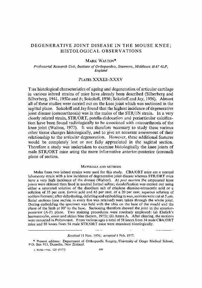
DEGENERATIVE JOINT DISEASE IN THE MOUSE KNEE; HISTOLOGICAL OBSERVATIONS MARK WALTON* Professorial Research Unit, Institute of Orthopaedics, Stanmore, Middlesex HA7 4LP, England PLATES XXXIII-XXXV THE histological characteristics of ageing and degeneration of articular cartilage in various inbred strains of mice have already been described (Silberberg and Silberberg, 1941, 1950a and b; Sokoloff, 1956; Sokoloff and Jay, 1956). Almost all of these studies were carried out on the knee joint which was sectioned in the sagittal plane. Sokoloff and Jay found that the highest incidence of degenerative joint disease (osteoarthrosis) was in the males of the STR/lN strain. In a very closely related strain, STR/ORT, patella dislocation and periarticular calcifica- tion have been found radiologically to be associated with osteoarthrosis of the knee joint (Walton, 1977). It was therefore necessary to study these various other tissue changes histologically, and to give an accurate assessment of their relationship to the articular degeneration. However, these additional features would be completely lost or not fully appreciated in the sagittal section. Therefore a study was undertaken to examine histologically the knee joints of male STR/ORT mice using the more informative anterior-posterior (coronal) plane of section. MATERIALS AND METHODS Males from two inbred strains were used for this study. CBAjORT mice are a normal laboratory strain with a low incidence of degenerative joint disease whereas STR/ORT mice have a very high incidence of the disease (Walton). At post mortem the amputated knee joints were skinned then fixed in neutral formal saline; decalcification was carried out using either a saturated solution of the disodium salt of ethylene diamine-tetraacetic acid or a solution of 35 per cent. formic acid and 65 per cent. of a 20 per cent. aqueous solution of sodium formate; after dehydrating, defatting and embedding in wax, sections were cut at 5 pm. Serial sections (one section in every five was retained) were taken through the whole joint. During embedding the specimen was held with the tibia on the base of the mould and the plane of the limb at 90" to the base. Sectioning therefore showed the joint in the anterior- posterior (A-P) plane. Two staining procedures were routinely employed : (a) Ehrlich's haematoxylin, eosin and alcian blue (Sayers, 1973); (b) Azure A. After clearing, the sections were mounted in Polymount. From various ages a total of 58 knees from 34 male CBA/ORT mice and 88 knees from 54 male STR/ORT mice were examined histologically. Received 11 Nov. 1976; accepted 4 Feb. 1977. * Present address : Department of Orthopaedic Surgery, University of Otago Medical School, P.O. Box 913, Dunedin, New Zealand. J. PATH.-VOL. 123 (1977) 109
-
Upload
independent -
Category
Documents
-
view
4 -
download
0
Transcript of Degenerative joint disease in the mouse knee. Histological observations
DEGENERATIVE J O I N T DISEASE I N T H E M O U S E K N E E ; HISTOLOGICAL OBSERVATIONS
MARK WALTON* Professorial Research Unit, Institute of Orthopaedics, Stanmore, Middlesex HA7 4LP,
England
PLATES XXXIII-XXXV
THE histological characteristics of ageing and degeneration of articular cartilage in various inbred strains of mice have already been described (Silberberg and Silberberg, 1941, 1950a and b; Sokoloff, 1956; Sokoloff and Jay, 1956). Almost all of these studies were carried out on the knee joint which was sectioned in the sagittal plane. Sokoloff and Jay found that the highest incidence of degenerative joint disease (osteoarthrosis) was in the males of the STR/lN strain. In a very closely related strain, STR/ORT, patella dislocation and periarticular calcifica- tion have been found radiologically to be associated with osteoarthrosis of the knee joint (Walton, 1977). It was therefore necessary to study these various other tissue changes histologically, and to give an accurate assessment of their relationship to the articular degeneration. However, these additional features would be completely lost or not fully appreciated in the sagittal section. Therefore a study was undertaken to examine histologically the knee joints of male STR/ORT mice using the more informative anterior-posterior (coronal) plane of section.
MATERIALS AND METHODS
Males from two inbred strains were used for this study. CBAjORT mice are a normal laboratory strain with a low incidence of degenerative joint disease whereas STR/ORT mice have a very high incidence of the disease (Walton). At post mortem the amputated knee joints were skinned then fixed in neutral formal saline; decalcification was carried out using either a saturated solution of the disodium salt of ethylene diamine-tetraacetic acid or a solution of 35 per cent. formic acid and 65 per cent. of a 20 per cent. aqueous solution of sodium formate; after dehydrating, defatting and embedding in wax, sections were cut at 5 pm. Serial sections (one section in every five was retained) were taken through the whole joint. During embedding the specimen was held with the tibia on the base of the mould and the plane of the limb at 90" to the base. Sectioning therefore showed the joint in the anterior- posterior (A-P) plane. Two staining procedures were routinely employed : (a) Ehrlich's haematoxylin, eosin and alcian blue (Sayers, 1973); (b) Azure A. After clearing, the sections were mounted in Polymount. From various ages a total of 58 knees from 34 male CBA/ORT mice and 88 knees from 54 male STR/ORT mice were examined histologically.
Received 11 Nov. 1976; accepted 4 Feb. 1977.
* Present address : Department of Orthopaedic Surgery, University of Otago Medical School, P.O. Box 913, Dunedin, New Zealand.
J. PATH.-VOL. 123 (1977) 109
110 MARK WALTON
RESULTS A. Articular cartilage
Normal appearances Normal articular cartilage had identical appearances in both the CBA/ORT
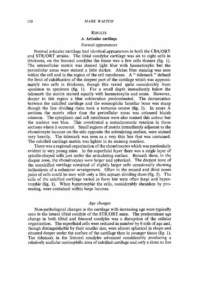
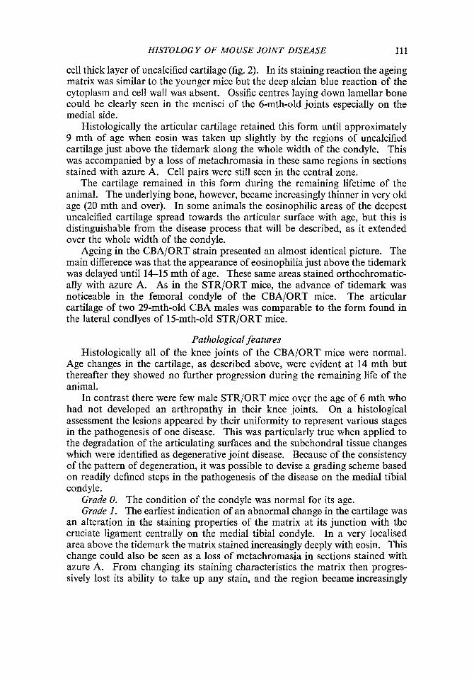
and STR/ORT strains. The tibial condylar cartilage was six to eight cells in thickness, on the femoral condyles the tissue was a few cells thinner (fig. 1). The intracellular matrix was stained light blue with haematoxylin but the pericellular areas were stained a little darker. Alcian blue staining was seen within the cell and in the region of the cell membrane. A " tidemark " defined the limit of calcification of the deepest part of the cartilage which was approxi- mately two cells in thickenss, though this varied quite considerably from specimen to specimen (fig. 1). For a small depth immediately below the tidemark the matrix stained equally with haematoxylin and eosin. However, deeper to this region a blue colouration predominated. The demarcation between the calcified cartilage and the eosinophilic lamellar bone was sharp though the line dividing them took a tortuous course (fig. 1). In azure A sections the matrix other than the pericellular areas was coloured bluish crimson. The cytoplasm and cell membrane were also stained this colour but the nucleus was blue. This constituted a metachromatic reaction in those sections where it occurred. Small regions of matrix immediately adjacent to the chondrocyte lacunae on the side opposite the articulating surface, were stained very heavily. The tidemark was seen as a very thin line that was unstained. The calcified cartilage matrix was lighter in its staining reaction.
There was a regional organisation of the chondrocytes which was particularly evident in very young mice. In the superficial layer there was a single layer of spindle-shaped cells just under the articulating surface. Beneath these, in the deeper zone, the chondrocytes were larger and spherical. The deepest zone of the uncalcified cartilage consisted of slightly larger cells occasionally showing indications of a columnar arrangement. Often in the second and third zones pairs of cells could be seen with only a thin septum dividing them (fig. 1). The cells of the calcified cartilage varied in form but were often large and hyper- trophic (fig. 1). When hypertrophic the cells, considerably shrunken by pro- cessing, were contained within large lacunae.
Age changes Non-pathological changes in the cartilage with increasing age were typically
seen in the lateral tibial condyle of the STR/ORT mice. The predominant age change in both tibial and femoral condyles was a disruption of the cellular organisation. The superficial cells were reduced in number by 6 mth of age and, though distinguishable by their smaller size, were almost spherical in shape and situated deeper under the surface of the cartilage than in younger tissue (fig. 1). The tidemark in the femoral condyles advanced considerably producing a relatively acellular eosinophilic area of calcified cartilage and only a three to five
HISTOLOGY OF MOUSE JOINT DISEASE 111
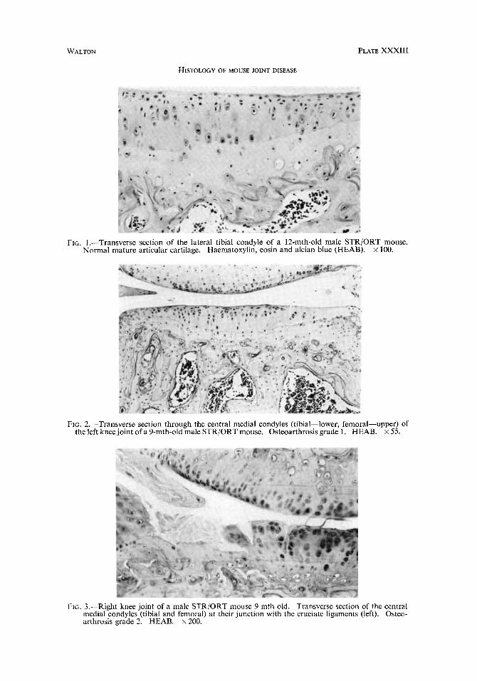
cell thick layer of uncalcified cartilage (fig. 2). In its staining reaction the ageing matrix was similar to the younger mice but the deep alcian blue reaction of the cytoplasm and cell wall was absent. Ossific centres laying down lamellar bone could be clearly seen in the menisci of the 6-mth-old joints especially on the medial side.
Histologically the articular cartilage retained this form until approximately 9 mth of age when eosin was taken up slightly by the regions of uncalcified cartilage just above the tidemark along the whole width of the condyle. This was accompanied by a loss of metachromasia in these same regions in sections stained with azure A. Cell pairs were still seen in the central zone.
The cartilage remained in this form during the remaining lifetime of the animal. The underlying bone, however, became increasingly thinner in very old age (20 mth and over). In some animals the eosinophilic areas of the deepest uncalcified cartilage spread towards the articular surface with age, but this is distinguishable from the disease process that will be described, as it extended over the whole width of the condyle.
Ageing in the CBA/ORT strain presented an almost identical picture. The main difference was that the appearance of eosinophilia just above the tidemark was delayed until 14-15 mth of age. These same areas stained orthochromatic- ally with azure A. As in the STR/ORT mice, the advance of tidemark was noticeable in the femoral condyle of the CBA/ORT mice. The articular cartilage of two 29-mth-old CBA males was comparable to the form found in the lateral condlyes of 15-mth-old STR/ORT mice.
Pathological features Histologically all of the knee joints of the CBA/ORT mice were normal.
Age changes in the cartilage, as described above, were evident at 14 mth but thereafter they showed no further progression during the remaining life of the animal.
In contrast there were few male STRjORT mice over the age of 6 mth who had not developed an arthropathy in their knee joints, On a histological assessment the lesions appeared by their uniformity to represent various stages in the pathogenesis of one disease. This was particularly true when applied to the degradation of the articulating surfaces and the subchondral tissue changes which were identified as degenerative joint disease. Because of the consistency of the pattern of degeneration, it was possible to devise a grading scheme based on readily defined steps in the pathogenesis of the disease on the medial tibial condyle.
Grade 0. The condition of the condyle was normal for its age. Grade 1. The earliest indication of an abnormal change in the cartilage was
an alteration in the staining properties of the matrix at its junction with the cruciate ligament centrally on the medial tibial condyle. In a very localised area above the tidemark the matrix stained increasingly deeply with eosin. This change could also be seen as a loss of metachromasia in sections stained with azure A. From changing its staining characteristics the matrix then progres- sively lost its ability to take up any stain, and the region became increasingly

112 MARK WALTON
acellular (fig. 2). Those cells able to survive in the depleted matrix were sometimes surrounded with a pericellular halo of material that stained very heavily with alcian blue. At any stage up to this point a single cleft or several clefts could appear in the cartilage. This fibrillation became more extensive, so producing tufts of acellular tissue adjacent to the cruciate ligament. Away from the developing lesion the appearance of the cartilage corresponded to the appearance of non-arthrotic cartilage of the same age.
Grade 2. Further cracks appeared in the otherwise intact, cellular cartilage adjacent to the initial lesion (fig. 3). Either side of the clefts the matrix lost its staining capacity though any viable cells surviving between the clefts were distinguishable by deeply staining pericellular matrix. Cracking of the matrix could occur in both non-staining tissue or tissue that was eosinophilic (and orthochromatic), though when it appeared in the latter instance it led to loss of staining immediately around the clefts. Normally fibrillation only extended a short distance across the condyle away from the cruciate ligament before the fragmented cartilage was lost. This loss of tissue was the main criterion for recording a Grade 2 severity of osteoarthrosis. Once this erosion had begun the extent of depleted (i.e., eosinophilic or unstained) matrix began to traverse the condyle in an outward direction. Fissures soon appeared in this changed matrix but only a narrow width of tissue was fibrillated as it appeared to be readily broken off and lost from the articular surface.
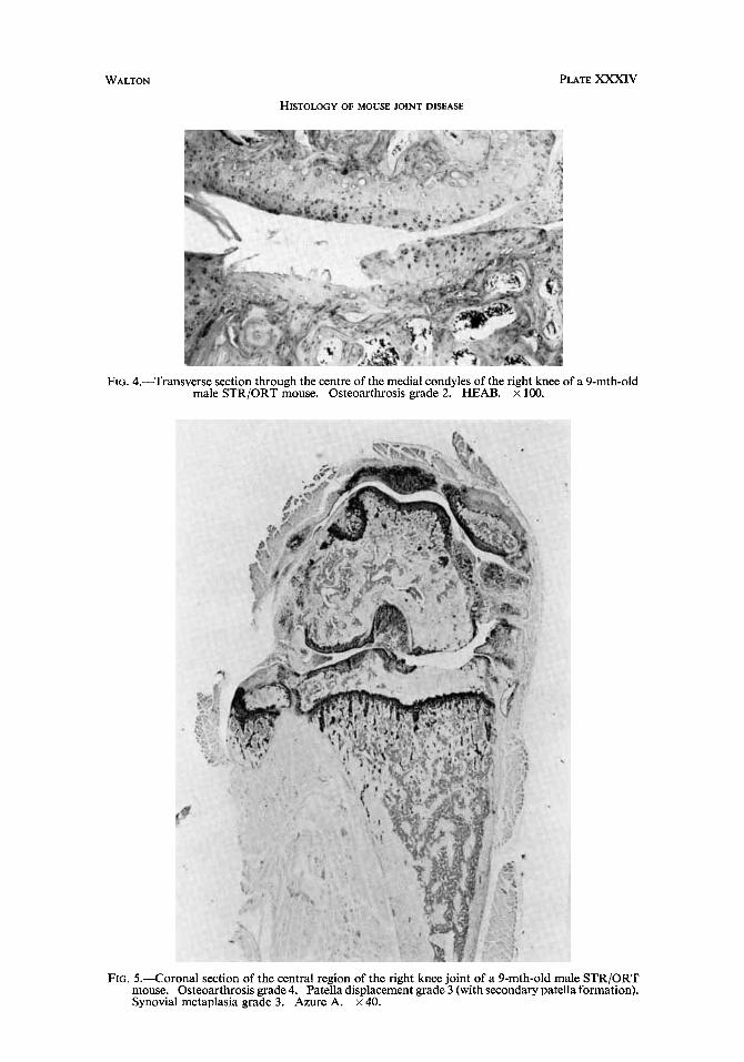
The normal pattern of gross breakdown of the cartilage of the medial tibial condyle was for lumps of tissue to be broken off down the cleavage lines and along the tidemark, thus leaving a step in the articulating surface (fig. 4). On rare occasions a horizontal cleft was seen along the tidemark. At an early stage the subchondral bone became sclerotic (fig. 4). This represented a thickening of the trabeculae that were already present in the epiphysis. The sclerosis seemed to be in direct response to the cartilage lesion as it first developed under the area of erosion, normal cancellous bone remaining under the intact articulating surfaces (fig. 4).
Grade 3. The loss of uncalcified cartilage progressed to the stage where most of the articulating surface consisted of the calcified cartilage at the level of the tidemark. This tissue was apparently readily lost once erosion into it had begun, as few instances were seen where the calcified cartilage, in its role as the articulating surface, was only partially eroded; it was either intact or absent. The subchondral bone became increasingly thicker but only under the articular lesion.
Grade 4. The severest grade of osteoarthrosis was recorded when at least one-third of the articulating surface consisted of exposed bone (fig. 5). Sclerotic bone filled the whole epiphysis (fig. 5) but at an earlier stage marrow spaces would still be present immediately above the growth plate and either side of the compact bone. Unlike the initial cartilage lesion, erosion of bone occurred centrally on the condyle denuded of cartilage. The only limiting factor to the continued erosion of the condyle was the death of the animal. In two specimens a very deep concave lesion was present on the medial tibial condyle and erosion of the quiescent growth plate was taking place at the time the mice were killed.
WALTON PLATE XXXlII
HISTOWGY OF MOUSE JOINT DISEASE
FIG. 1.-Transverse section of the lateral tibial condyle of a 12-mth-old male STRjORT mouse. Normal mature articular cartilage. Haematoxylin, eosin and alcian blue (HEAB). x 100.
FIG. 2.-Transverse section through the central medial condyles (tibial-lower, femoral-upper) of the left knee joint of a 9-mth-old male STR/ORT mouse. Osteoarthrosis grade 1. HEAB. X 55.
FIG. 3.-Right knee joint of a male STRjORT mouse 9 mth old. Transverse section of the central medial condyles (tibial and femoral) at their junction with the cruciatc ligaments (left). Osteo- arthrosis grade 2. HEAB. x 200.
WALTON PLATE XXXIV
HISTOLOGY OF MOUSE JOINT DISEASE
FIG. 4.-Transverse section through the centre of the medial condyles of the right knee of a 9-mth-old male STR/ORT mouse. Osteoarthrosis grade 2. HEAB. x 100.
FIG. 5.-Coronal section of the central region of the right knee joint of a 9-mth-old male STR/ORT mouse. Osteoarthrosis grade 4. Patella displacement grade 3 (with secondary patella formation). Synovial metaplasia grade 3. Azure A. x 40.

WALTON PLATE XXXV
HISTOLOGY OF MOUSE JOINT DISEASE
FIG. 7.--Coronal section of the femoro-patella joint of a 6-mth-old male STRjORT mouse. Patella grade 0. HEAB. x65.
FIG. 8.-Lateral side of the patella (P) in a coronal section of the knee joint of a 9-mth-old male STR/ORT mouse. Patella displacement grade 1. Synovial metaplasia grade 2. HEAB. x 65
FIG. Il.--Coronal section of the medial collateral ligament of an 8-mth-old male STR/ORT mouse. All stages of metaplasia can be seen in this specimen. HEAB. x 120.
HISTOLOG Y OF MOUSE JOINT DISEASE 113
Either side of an eburnation, where cartilage survived, there were sometimes a clone of two or four cells which stained heavily with alcian blue in a matrix which was only very lightly stained.
Frequently intact femoral cartilage was seen opposite well-developed tibial erosions (fig. 4). It therefore appeared that the femoral lesion was somewhat delayed in its onset compared with the tibial lesion. No fibrillation was seen in the relatively thin uncalcified cartilage of the femur but occasionally long horizontal clefts appeared along the tidemark. As on the tibial condyle, the calcified cartilage could constitute a secondary articulating surface. When both the femoral and tibial surfaces were eburnated the articulating surfaces were remarkably congruent though their contours were not symmetrical (fig. 5). Normally these two surfaces had been slightly parted during histological preparation. In some specimens with the tibia and femur displaced during preparation, it would seem that only by a rotation of the tibia (i.e,, a varus deformity of the joint) would the severely eburnated medial condyles articulate (fig. 5). In the most severely arthrotic specimens there had been a medial subluxation of the tibia relative to the femur. This was revealed by a malalignment of the cruciate compartments of the femur and tibia, and erosion of part of the metaplastic cruciate ligament attached to the tibia by the inner surface of the medial femoral condyle (fig. 5).
In these very advanced lesions the medial menisci were shortened, ap- parently due to the wearing away of their inner edge. From an early phase in the arthropathy the medial meniscus underwent hyperplasia in the form of a considerably enlarged ossific centre that normally remained covered in cartilage but would sometimes become eburnated. In very advanced degeneration the fibrous tissue attaching the meniscus to the tibia had undergone metaplasia to fibrous cartilage thus effectively immobilising the structure relative to the tibia (fig. 5). Despite very severe osteoarthrosis on the medial condyles, laterally the condyles retained the appearance consistent with their age (fig. 5). Only in the very oldest mice was there any evidence of fibrillation on the inner border of the lateral tibial condyle.
Femoral lesions were less consistent in their appearances.
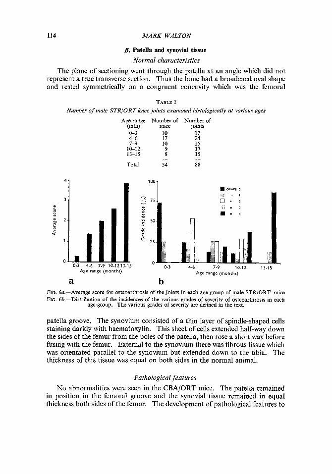
Incidence and severity of osteoarthrosis in male STRlORT mice A total of 88 knee joints of male STR/ORT mice were examined individually
and the severity of their osteoarthrosis assessed according to the grading scheme described above. The number of animals in the various age-groups is given in table I.
The average score for osteoarthrosis of all the joints in each age-group is presented in figure 6a. The average score rises steadily with the increasing age of the mice, reaching a peak in the oldest age-groups, thus demonstrating the progressive nature of the disease. The incidence of each grade of severity in the various age-groups is presented in fig. 6b. A quarter of the mice in the youngest age-group had the early degenerative changes. The initial high incidence of the Grade 0 decreased with age but the incidence of Grade 4 increased with age. Grade 3 changes were not common in any age-group.
1. PATH.-VOL. 123 (1977) H
114
7
3 . f! 8 & 2 . 2
MARK WALTON
-7 c- 75 v
W
C (Y V
'4 50 ._ 0 D
B. Patella and synovial tissue Normal characteristics
The plane of sectioning went through the patella at an angle which did not represent a true transverse section. Thus the bone had a broadened oval shape and rested symmetrically on a congruent concavity which was the femoral
2
TABLE I Number of male STRIORT knee joints examined histologically a t various ages
Age range Number of Number of (mth) mice joints 0-3 10 17 4-6 17 24 7-9 10 15
10-12 9 17 13-15 8 15
Total 54 88 - -
I
(3 25,
L 0
A -
Age range (months)
loo 1 GRADE 0
Age range (months)
a b FIG. 6a.-Average score for osteoarthrosis of the joints in each age group of male STR/ORT mice FIG. 6b.-Distribution of the incidences of the various grades of severity of osteoarthrosis in each
age-group. The various grades of severity are defined in the text.
patella groove. The synovium consisted of a thin layer of spindle-shaped cells staining darkly with haematoxylin. This sheet of cells extended half-way down the sides of the femur from the poles of the patella, then rose a short way before fusing with the femur. External to the synovium there was fibrous tissue which was orientated parallel to the synovium but extended down to the tibia. The thickness of this tissue was equal on both sides in the normal animal.
Pathological features No abnormalities were seen in the CBA/ORT mice. The patella remained
in position in the femoral groove and the synovial tissue remained in equal thickness both sides of the femur. The development of pathological features to

HISTOLOGY OF MOUSE JOINT DISEASE 115
the patella and synovial tissue of the STR/ORT males was of a broadly con- sistent pattern and therefore these changes could be graded as follows:
Patella Grade 0. Patella in its normal position. Grade 1. In some specimens the patella was seen to be preferentially
articulating with the medial side of the femoral groove. Though in a few instances this may well have been artefactual (i.e., due to manipulation of the joint prior to fixation), in the majority of cases accompanying synovial and cartilaginous changes strongly indicated a definite subluxation of the bone. Cartilaginous changes included a tendency towards hypertrophy of the chondro- cytes on the lateral facet of the patella and femoral groove cartilage, whereas normal cartilage was present on the medial side.
Grade 2. The patella was frequently seen poised either side of the medial ridge of the femoral groove. No intermediate position was normally seen, suggesting that the dislocation of the bone out of the femoral groove was a sudden event. However, in a few specimens fibrillation of the patella cartilage and erosion of the medial femoral ridge indicated a recurrent dislocation. A poorly organised hyaline cartilage normally formed on the medial side of the femur in apposition to the dislocated patella.
Grade 3. The patella would further dislocate, finally becoming positioned medially to the femur (fig. 5).
Synovial tissue Grade 0. Normal synovial tissue. Grade 1. Either before or after early patella subluxation the synovial tissue
thickened on the lateral side and medially the synovium thickened and formed villi that protruded into the joint space (fig. 7).
Grade 2. With increasing patella subluxation the thickened lateral synovial tissue underwent metaplastic changes. The cells secreted a haematoxylin- staining (and metachromatic) matrix, giving the tissue initially the appearance of fibro-cartilage. With increasing matrix production the cells became separated from each other and the fibrous nature of the tissue disappeared, thus producing a hyaline cartilage in the form of a spur from the lateral side of the patella (fig. 8). Within this tissue there developed areas of calcification often clearly delimited by a tidemark. The matrix of these calcified areas was eosinophilic and the cells were reduced in size like osteocytes giving the overall appearance of woven bone. Marrow spaces would frequently arise within this tissue.
Grade 3. The extent of metaplasia increased especially with increasing medial dislocation of the patella. The lateral patellar spur of metaplastic tissue grew over the femur to the lateral side, occasionally filling the vacated femoral groove, thus forming a secondary patella (fig. 5). Ectopic cartilaginous tissue formed both medially and laterally, and went through the same progres- sive metaplastic changes (fig. 5).
Grade 4. The most advanced condition of synovial metaplasia was the
116 MARK WALTON
laying down of lamellar bone within the marrow spaces of any of the heterotopic growths.
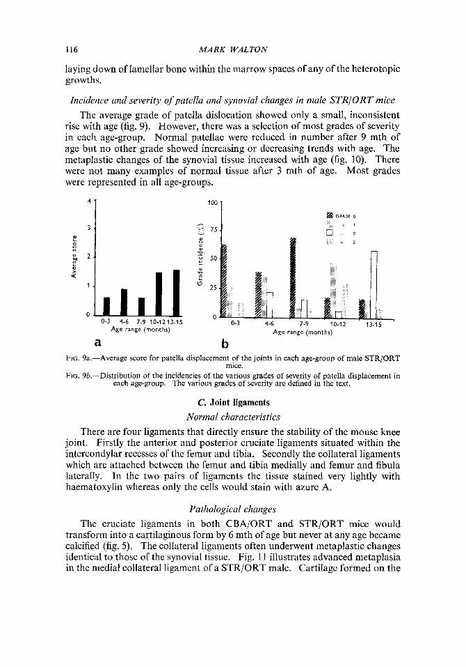
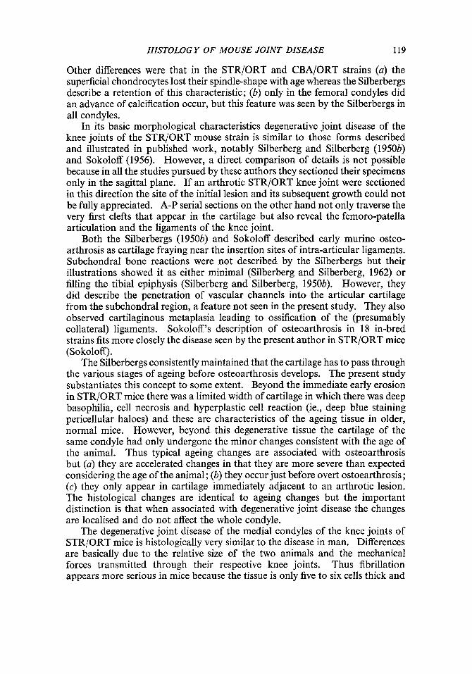
Incidence and severity of patella and synovial changes in male STRIORT mice The average grade of patella dislocation showed only a small, inconsistent
rise with age (fig. 9). However, there was a selection of most grades of severity in each age-group. Normal patellae were reduced in number after 9 mth of age but no other grade showed increasing or decreasing trends with age. The metaplastic changes of the synovial tissue increased with age (fig. 10). There were not many examples of normal tissue after 3 mth of age. Most grades were represented in all age-groups.
4 1 loo 1 o= 75
a l l , , , , , V w 5 0 al
L
25 1
0 0 0-3 4-6 7-9 10-12 13
Age range (months)
0-3 4-6 7-9 10-1213-15 Age range (months)
- 5
a b FIG. 9a.-Average score for patella displacement of the joints in each age-group of male STR/ORT
mice. FIG. 9b.-Distribution of the incidencies of the various grades of severity of patella displacement in
each age-group. The various grades of severity are defined in the text.
C. Joint ligaments
Normal characteristics There are four ligaments that directly ensure the stability of the mouse knee
joint. Firstly the anterior and posterior cruciate ligaments situated within the intercondylar recesses of the femur and tibia. Secondly the collateral ligaments which are attached between the femur and tibia medially and femur and fibula laterally. In the two pairs of ligaments the tissue stained very lightly with haematoxylin whereas only the cells would stain with azure A.
Pathological changes The cruciate ligaments in both CBA/ORT and STRjORT mice would
transform into a cartilaginous form by 6 mth of age but never at any age became calcified (fig. 5) . The collateral ligaments often underwent metaplastic changes identical to those of the synovial tissue. Fig. 11 illustrates advanced metaplasia in the medial collateral ligament of a STR/ORT male. Cartilage formed on the

HISTOLOGY OF MOUSE JOINT DISEASE 117
100'-
-2 c- 75. ../
sides of the tibia and femur which were in contact with the cartilage of the transformed ligament. Ossification only occurred in the medial ligament of those knees with severe osteoarthrosis and therefore it never formed in CBA/ ORT joints.
3 4 1
@ GRADE 0
I, 1
0 II 2
I < 3
0-3 4-6 7-9 10-1213-15 Age range (months)
a
0-3 4-6 7-9 10-12 13-15 Age range (months)
b FIG. 10a.-Average score for synovial metaplasia of the joints in each age-group of male STR/ORT
FIG. 10b.-Distribution of the incidences of the various grades of severity of synovial metaplasia in mice.
each age-group. The various grades of severity are defined in the text.
Statistical analyses (a) Bilateral symmetry. The t-test was applied to the mean arthrotic scores
of the right and left knees of all ages of male STR/ORT mice. Though the mean for the left knees was higher (2.20) than the right (1.76), this difference was not significant (P = 0.2-0.1).
(b) For those joints where two particular features could be clearly observed, 2 x 2 contingency tables were prepared showing the number of joints in which the two features were present or absent. In order to establish any statistical association the x* test was applied.
(1) Osteoarthrosis versus patella displacement
Patella displacement
Grades of severity
0
1 4
Osteoarthrosis
I 0 j 1-3
11 ~ 8
19 1 40
There was a significant association between the patella abnormality and osteoarthrosis (P = 0.05). Over twice as many knee joints had osteoarthrosis and a displaced patella (40) as those with the disease and a normal patella (19).
118 MARK WALTON
0-1
45
1
Only eight joints with a patella displaced had no indication of condylar de- generation, whereas 19 had osteoarthrosis yet normal patellae. This difference would suggest, though not conclusively, that the articular degeneration precedes the patella dislocation.
2-4
11
24
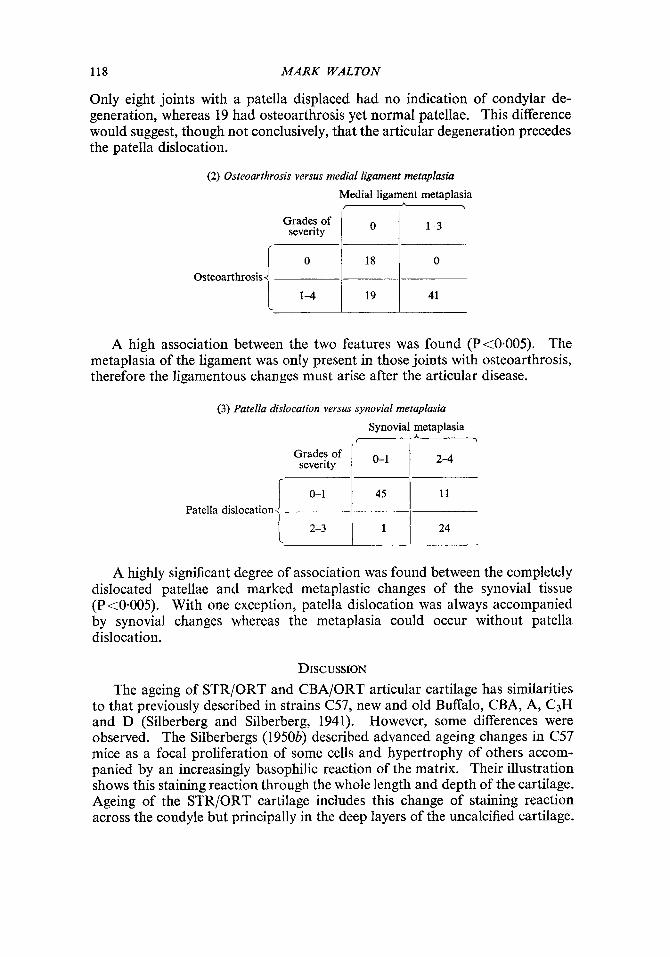
(2) Osteoarthrosis versus medial ligament metaplasia
Medial ligament metaplasia P
Grades of 1 1 1-3 severity
Osteoarthrosis 1-4 1 19 I 41
A high association between the two features was found (Pt0.005). The metaplasia of the ligament was only present in those joints with osteoarthrosis, therefore the ligamentous changes must arise after the articular disease.
(3) Patella dislocation versus synovial metaplasia
Grades of severity
r 0-1 Patella dislocation 1 2-3
A highly significant degree of association was found between the completely dislocated patellae and marked metaplastic changes of the synovial tissue (P <0-005). With one exception, patella dislocation was always accompanied by synovial changes whereas the metaplasia could occur without patella dislocation.
DISCUSSION The ageing of STR/ORT and CBA/ORT articular cartilage has similarities
to that previously described in strains C57, new and old Buffalo, CBA, A, C3H and D (Silberberg and Silberberg, 1941). However, some differences were observed. The Silberbergs (1950b) described advanced ageing changes in C57 mice as a focal proliferation of some cells and hypertrophy of others accom- panied by an increasingly basophilic reaction of the matrix. Their illustration shows this staining reaction through the whole length and depth of the cartilage. Ageing of the STR/ORT cartilage includes this change of staining reaction across the condyle but principally in the deep layers of the uncalcified cartilage.
HISTOLOGY OF MOUSE JOINT DISEASE 119
Other differences were that in the STRjORT and CBA/ORT strains (a) the superficial chondrocytes lost their spindle-shape with age whereas the Silberbergs describe a retention of this characteristic; (b) only in the femoral condyles did an advance of calcification occur, but this feature was seen by the Silberbergs in all condyles.
In its basic morphological characteristics degenerative joint disease of the knee joints of the STR/ORT mouse strain is similar to those forms described and illustrated in published work, notably Silberberg and Silberberg (19504 and Sokoloff (1956). However, a direct comparison of details is not possible because in all the studies pursued by these authors they sectioned their specimens only in the sagittal plane. If an arthrotic STR/ORT knee joint were sectioned in this direction the site of the initial lesion and its subsequent growth could not be fully appreciated. A-P serial sections on the other hand not only traverse the very first clefts that appear in the cartilage but also reveal the femoro-patella articulation and the ligaments of the knee joint.
Both the Silberbergs (1950b) and Sokoloff described early murine osteo- arthrosis as cartilage fraying near the insertion sites of intra-articular ligaments. Subchondral bone reactions were not described by the Silberbergs but their illustrations showed it as either minimal (Silberberg and Silberberg, 1962) or filling the tibia1 epiphysis (Silberberg and Silberberg, 1950b). However, they did describe the penetration of vascular channels into the articular cartilage from the subchondral region, a feature not seen in the present study. They also observed cartilaginous metaplasia leading to ossification of the (presumably collateral) ligaments. Sokoloff’s description of osteoarthrosis in 18 in-bred strains fits more closely the disease seen by the present author in STR/ORT mice (Sokoloff).
The Silberbergs consistently maintained that the cartilage has to pass through the various stages of ageing before osteoarthrosis develops. The present study substantiates this concept to some extent. Beyond the immediate early erosion in STR/ORT mice there was a limited width of cartilage in which there was deep basophilia, cell necrosis and hyperplastic cell reaction (ie., deep blue staining pericellular haloes) and these are characteristics of the ageing tissue in older, normal mice. However, beyond this degenerative tissue the cartilage of the same condyle had only undergone the minor changes consistent with the age of the animal. Thus typical ageing changes are associated with osteoarthrosis but (a) they are accelerated changes in that they are more severe than expected considering the age of the animal; (b) they occur just before overt ostoearthrosis; (c) they only appear in cartilage immediately adjacent to an arthrotic lesion. The histological changes are identical to ageing changes but the important distinction is that when associated with degenerative joint disease the changes are localised and do not affect the whole condyle.
The degenerative joint disease of the medial condyles of the knee joints of STR/ORT mice is histologically very similar to the disease in man. Differences are basically due to the relative size of the two animals and the mechanical forces transmitted through their respective knee joints. Thus fibrillation appears more serious in mice because the tissue is only five to six cells thick and
120 MARK WALTON
therefore clefts invariably penetrate the whole depth of the uncalcified tissue. In addition the thickening of the subchondral bone leads to the filling of the whole mouse epiphysis instead of a localised sclerosis under the human lesion.
The location of the lesion is however rather different in man as it normally appears on the main weight-bearing areas in the central portions of either the femoral or tibia1 condyles (Bennett, Waine and Bauer, 1942), whereas, without exception, the initiation of the disease process in the STRjORT mouse is at the interface of the medial condyle and cruciate ligaments and this may well have some relevance to the aetiology of the disease in this strain. Dissimilarities are greatest between the STR/ORT mouse and man in the involvement of the whole joint. In the mouse there is a significant relationship between the occurrence of patella dislocation and degenerative joint disease. A far more accurate assessment can be made of these two pathological conditions using histology than using radiology, yet both techniques indicate a high correlation between abnormal position of the patella and articular degeneration (Walton, 1977). However, even histology does not give conclusive evidence as to which of these changes comes first, as there were 19 joints with articular degeneration but normal patellae, and eight joints with abnormal patellae but intact articular cartilage. Two possibilities present themselves. Firstly, the erosion of the medial condyles could give rise to a varus deformity of the joint which then results in the subluxation of the patella medially. However, in all the joints, before eburnation was reached, erosion was insufficient to cause an appreciable deformity of the joint yet the patella had usually dislocated. Secondly, the patella dislocation, or the forces resulting in its subluxation, may be an import- ant factor in the aetiology of the osteoarthrosis. Certainly in dogs, long- standing, recurrent patella subluxation can cause degenerative joint disease in the femoro-tibia1 articulation on the same side as the direction of dislocation (Kodituwakku, 1960). In man recurrent lateral dislocation of the patella can be associated with chondromalacia of the lateral femoral condyle (West and Soto-Hall, 1958).
The present study has shown that synovial metaplasia occurs in response to displacement of the patella. It would therefore appear that stresses through the tissue caused by the dislocating bone induce the progressive tissue transforma- tions. This is particularly evident laterally, where there must be considerable tension through the synovial tissue which results in the formation of a large sheet of metaplastic tissue over the femur. When a human patella remains within the femoral groove yet is subjected to forces tending to dislocate it laterally, soft tissue calcification can grow out from the medial side of the patella (McDougall and Brown, 1968). Monteiro (1970) claimed that this mineralisation was due to recurrent haemorrhage from capsular tears.
Calcification of the collateral ligaments, especially on the lateral side, is very common in CBA/ORT and non-arthrotic STR/ORT mice (Walton, 1977). Ossification only arises in the medial ligament and is always associated with a degenerate joint. The normal process of ectopic calcification of the ligament may make it a vulnerable site for further progressive metaplastic changes when the joint becomes diseased. As with the synovial tissue, the realignment of the
HISTOLOGY OF MOUSE JOINT DISEASE 121
mechanical stresses through the soft tissue of the joint due to patella dislocation, may give rise to ossification of the medial ligament.
It appears that the stimulus of the displaced patella moving against the medial side of the femur induces cartilage formation. Likewise, where the metaplastic medial ligament " articulates " with the femur and tibia there is a similar induction of hyaline cartilage. Often this tissue is relatively acellular but frequently chondrocyte clusters are seen within it. A similar induction of cartilage tissue was observed by Bennett and Bauer (1937) when they produced patella dislocation in dogs by external manipulation. A secondary groove for articulation with the patella was formed on the side of the femur from hyper- plastic connective tissue which underwent metaplasia into hyaline cartilage.
The evidence obtained from this present study suggests that the degenerative joint disease in STR/ORT knee joints progresses from the initial lesion to eburnation over a period of 4-8 mth in an animal whose life expectancy is approximately 13 mth. This histological study indicates that the condition of the animal at post mortem and the rate of degeneration of individual joints may vary considerably.
SUMMARY The knee joints from males of two strains (CBA/ORT and STR/ORT) were
studied histologically. The incidence of degenerative joint disease was very high in the STR/ORT strain. Degeneration of the cartilage invariably occurred first at the interface of the cruciate ligament and articular cartilage of the tibia. Lesions were only seen on the medial tibia1 and later the medial femoral con- dyles. Blocks of fibrillated, uncalcified cartilage were gradually lost across the condyle, leaving the tidemark as a secondary articulating surface. Meanwhile the subchondral bone thickened and erosion continued through the calcified cartilage into the underlying bone.
A statistically significant relationship was found between the development of the lesion and (0) medial dislocation of the patella, (b) calcification and ossification of the medial collateral ligament. Patella dislocation gave rise to extensive cartilaginous and bony metaplasia of the synovial tissue. In joints with advanced degeneration there was often evidence of a slight lateral sub- luxation of the femur relative to the tibia.
The author would like to express his appreciation for the advice and encouragement given by Dr Michael Elves. This work was generously supported by the Wellcome Trustees through Dr Audrey U. Smith. It forms part of a thesis presented for the degree of Doctor of Philosophy in the University of London.
REFERENCES BENNETT, G. A., AND BAUER, W. 1937. Joint changes resulting from patellar displacement
and their relation to degenerative joint disease. J. Bone Jt Surg., 19, 667. BENNETT, G. A., WAINE, H., AND BAUER, W. 1942. Changes in the knee joint at various ages.
New York, Commonwealth Fund. KODITUWAKKU, G . E. 1960. Observations on some abnormalities of the stifle joint in the dog:
and an analysis of a series of fracture repairs in dogs including a bacteriological study of the peripheral blood of some of the fracture cases. Ph.D. Thesis, University of London.
122 MARK WALTON
MCDOUGALL, A., AND BROWN, J. D. 1968. Radiological signs of recurrent dislocation of the patella. J. Bone Jt Surg., 50B, 841.
MONTEIRO, J. A. N. 1970. Patello-femoral arthrosis. In Zlth Congres Internationa de Chirurgie Orthopedique et de Traumatologie, Mexico.
SAYERS, D. C . J. 1973. A general staining technique for the demonstration of new bone. J. Sci. Tech., 17, 14.
SILBERBERG, M., AND SILBERBERG, R. 1941. Age changes of bones and joints in various strains of mice. Amer. J. Anat., 68, 69.
SILBERBERG, M., AND SILBERBERG, R. 1950a. Effects of a high-fat diet on the joints of aging mice. Arch. Path., 50, 828.
SILBERBERG, R., AND SILBERBERG, M. 1950b. Skeletal growth and articular changes in mice receiving a high-fat diet. Amer. J. Path., 26, 113.
SILBERBERG, M., AND SILBERBERG, R. 1962. Osteoarthrosis and osteoporosis in senile mice. Cerontologia, 6, 91.
SOKOLOFF, L. 1956. Natural history of degenerative joint disease in small laboratory animals: I. Pathologic anatomy of degenerative joint disease in mice. Arch. Path., 62, 118.
SOKOLOFF, L., AND JAY, G. E. 1956. Natural history of degenerative joint disease in small laboratory animals: 11. Epiphyseal maturation and osteoarthritis of the knee joint of inbred strains. Arch. Path., 62, 129.
WALTON, M. 1977. Studies of degenerative joint disease in the mouse knee joint ; radiological and morphological observations. J. Path., 123, 97.
WEST, F. E., AND SOTO-HALL, R. 1958. Recurrent dislocation of the patella in the adult. End results of patellectomy with quadricepsplasty. J. Bone Jt. Surg., 40A, 386.