Standardising the clinical assessment of coronal knee laxity
11
http://pih.sagepub.com/ Medicine Engineers, Part H: Journal of Engineering in Proceedings of the Institution of Mechanical http://pih.sagepub.com/content/226/9/699 The online version of this article can be found at: DOI: 10.1177/0954411912451814 originally published online 3 July 2012 2012 226: 699 Proceedings of the Institution of Mechanical Engineers, Part H: Journal of Engineering in Medicine Jon V Clarke, William T Wilson, Scott C Wearing, Frederic Picard, Philip E Riches and Angela H Deakin Standardising the clinical assessment of coronal knee laxity Published by: http://www.sagepublications.com On behalf of: Institution of Mechanical Engineers can be found at: Proceedings of the Institution of Mechanical Engineers, Part H: Journal of Engineering in Medicine Additional services and information for http://pih.sagepub.com/cgi/alerts Email Alerts: http://pih.sagepub.com/subscriptions Subscriptions: http://www.sagepub.com/journalsReprints.nav Reprints: http://www.sagepub.com/journalsPermissions.nav Permissions: http://pih.sagepub.com/content/226/9/699.refs.html Citations: What is This? - Jul 3, 2012 OnlineFirst Version of Record - Aug 17, 2012 Version of Record >> by guest on February 19, 2013 pih.sagepub.com Downloaded from
-
Upload
independent -
Category
Documents
-
view
4 -
download
0
Transcript of Standardising the clinical assessment of coronal knee laxity

http://pih.sagepub.com/Medicine
Engineers, Part H: Journal of Engineering in Proceedings of the Institution of Mechanical
http://pih.sagepub.com/content/226/9/699The online version of this article can be found at:
DOI: 10.1177/0954411912451814
originally published online 3 July 2012 2012 226: 699Proceedings of the Institution of Mechanical Engineers, Part H: Journal of Engineering in Medicine
Jon V Clarke, William T Wilson, Scott C Wearing, Frederic Picard, Philip E Riches and Angela H DeakinStandardising the clinical assessment of coronal knee laxity
Published by:
http://www.sagepublications.com
On behalf of:
Institution of Mechanical Engineers
can be found at:Proceedings of the Institution of Mechanical Engineers, Part H: Journal of Engineering in MedicineAdditional services and information for
http://pih.sagepub.com/cgi/alertsEmail Alerts:
http://pih.sagepub.com/subscriptionsSubscriptions:
http://www.sagepub.com/journalsReprints.navReprints:
http://www.sagepub.com/journalsPermissions.navPermissions:
http://pih.sagepub.com/content/226/9/699.refs.htmlCitations:
What is This?
- Jul 3, 2012OnlineFirst Version of Record
- Aug 17, 2012Version of Record >>
by guest on February 19, 2013pih.sagepub.comDownloaded from
Original Article
Proc IMechE Part H:J Engineering in Medicine226(9) 699–708� IMechE 2012Reprints and permissions:sagepub.co.uk/journalsPermissions.navDOI: 10.1177/0954411912451814pih.sagepub.com
Standardising the clinical assessmentof coronal knee laxity
Jon V Clarke1,2, William T Wilson1, Scott C Wearing3,4,Frederic Picard1, Philip E Riches2 and Angela H Deakin1,2
AbstractClinical laxity tests are used for assessing knee ligament injuries and for soft tissue balancing in total knee arthroplasty.This study reports the development and validation of a quantitative technique of assessing collateral knee laxity throughaccurate measurement of potential variables during routine clinical examination. The hypothesis was that standardisationof a clinical stress test would result in a repeatable range of laxity measurements.
Non-invasive infrared tracking technology with kinematic registration of joint centres gave real-time measurement ofboth coronal and sagittal mechanical tibiofemoral alignment. Knee flexion, moment arm and magnitude of the appliedforce were all measured and standardised. Three clinicians then performed six knee laxity examinations on a singlevolunteer using a target moment of 18 Nm.
Standardised laxity measurements had small standard deviations (within 1.1�) for each clinician and similar mean val-ues between clinicians, with the valgus laxity assessment (mean of 3�) being slightly more consistent than varus (meansof 4� or 5�).
The manual technique of coronal knee laxity assessment was successfully quantified and standardised, leading to a nar-row range of measurements (within the accuracy of the measurement system). Minimising the subjective variables ofclinical examination could improve current knowledge of soft tissue knee behaviour.
KeywordsKnee laxity, clinical assessment, non-invasive assessment, ligament injuries, total knee arthroplasty
Date received: 19 December 2011; accepted: 16 May 2012
Introduction
Clinical laxity tests are frequently used for assessingknee ligament injuries and for soft tissue balancing intotal knee arthroplasty (TKA). Current routine meth-ods are subjective with respect to examination tech-nique, magnitude of clinician-applied load andassessment of joint displacement. For collateral liga-ment injuries, scoring systems to grade severity areoften based on millimetres of perceived joint openingwith applied manual stress.1–3 The level of resolutionrequired for this may exceed normal levels of humanjudgement and account for the frequent disparitybetween laxity examinations and true in-vivo joint func-tion.4 Quantitative adjuncts, such as stress radiographs,have enabled a more objective measurement of jointspace opening, but are still potentially limited by lackof standardisation of the applied load and are suscepti-ble to limb positioning errors as a result of knee flexionor rotation.5,6 Soft tissues can be directly evaluated bymagnetic resonance imaging (MRI), which is a
commonly used imaging modality for diagnosis andgrading of collateral ligament injuries.7–9 However,only static anatomical information is provided and sothere is potential to underestimate the true extent ofthe injury.10
In TKA, assessment of laxity is a routine componentof many soft tissue balancing techniques and is oftenused to determine the need for a soft tissue release,11,12
particularly for large deformities that are judged to beuncorrectable. Attempts have been made to categorisecollateral laxity, for example Krackow’s classification
1Department of Orthopaedics, Golden Jubilee National Hospital, UK2Bioengineering Unit, University of Strathclyde, UK3Faculty of Health Sciences and Medicine, Bond University, Australia4Centre of Excellence for Applied Sport Science Research, Queensland
Academy of Sport, Australia
Corresponding author:
Angela H Deakin, Department of Orthopaedics, Golden Jubilee National
Hospital, Clydebank, West Dunbartonshire G81 4DY, Scotland, UK.
Email: [email protected]
by guest on February 19, 2013pih.sagepub.comDownloaded from

of medial ligament tightness,13 but this assumes that allclinicians have similar examination methods and areable to reliably judge knee alignment. However, humanassessment of angles is known to be poor14 and theaccuracy of alignment estimates under these circum-stances may be no better than the order of 65�.15
Lower limb alignment measurements generated in realtime, in the operating theatre by computer-assistedtechnology, have led to the development of quantitativeTKA soft tissue balancing algorithms that are oftenbased on tibiofemoral angular displacements withapplied varus and valgus stress.16–18 However thesetechniques involve an unquantified manual load beingapplied by each individual surgeon, which may explainthe difference in the derived values of the varus andvalgus stress angles between studies.19 These assess-ments are also carried out on anaesthetised patients, someasured laxity may not be the same as the functionallaxity of the knee under load. Furthermore, this tech-nology relies on invasive bony tracker attachment andis, therefore, not currently available in a clinic setting.
Experimental models involving cadaveric knees20 orvolunteer test rigs21,22 have sought to standardiseapplied knee moments and measure the resultant angu-lar displacements, but none have been successfullyimplemented into routine clinical practice. Therefore,despite the limitations of current techniques, manualknee laxity examination remains the primary means ofdiagnosing ligament injuries and assessing soft tissuesin TKA. However with the high incidence of soft tissueknee injuries23,24 and the growing physical demands ofTKA patients,25 there is a need to improve the evalua-tion of knee laxity to ensure better execution of correc-tive surgery.
The development of a quantitative assessment tech-nique of coronal knee laxity for incorporation into cur-rent routine practice requires accurate standardisationof several parameters. The knee flexion angle should bedetermined and then maintained during the testing tominimise the potential positional variation in ligamentrestraining properties.4,15,26 The moment applied to theknee joint should be measured, which requires the iden-tification of the force vector and moment arm of theapplied load. The resultant displacement of tibia withrespect to the femur should be quantified as a measureof laxity.
This study reports the development and validationof a quantitative technique of assessing collateralknee laxity through accurate measurement of poten-tial variables during routine clinical examination.The hypothesis was that standardisation of a clinicalstress test would result in repeatable range of laxitymeasurements.
Materials and methods
University ethical approval was granted for the healthyvolunteer assessments.
Knee flexion
Non-invasive infrared (IR) tracking technology, withkinematic registration of joint centres to give mechani-cal lower limb alignment,27 was utilised for the real-timemeasurement of both coronal and sagittal mechanicaltibiofemoral alignment. This system had been validatedfor coronal alignment27 but to verify this technologyfor recording flexion, comparison was made with a vali-dated flexible electrogoniometer (EG) (Biometrics Ltd,Cwmfelinfach, Gwent, UK).28 A single volunteer wasused for simultaneous measurements of both systems.Using the right lower limb, the EG was aligned alongthe estimated neutral mechanical axis of the hip–knee–ankle joint centres, with the lower limb in full extension.The IR trackers were then attached over the top ofthe EG end plates (Figure 1). Once the lower limb wasregistered by the IR tracking system the knee was posi-tioned and recorded in 0� of flexion according to its on-screen display. This provided the ‘zero’ point for theEG, with synchronisation of the two systems performedat the start of each trial.
The knee was then passively flexed and held as sta-ble as possible in 1� increments, as indicated by the IRtracking system, up to 10�, and the precise angle ateach point registered simultaneously by each system.The knee was then flexed in 10� increments from 10� to100�. The trial was performed three times with the EGzeroed at the start of each set of measurements.
Moment arm
During laxity assessment, the moment arm is deter-mined by the position of the clinician’s hands. For thisstudy, the controlled position of the manual forceapplication was directly over the medial (valgus) or lat-eral (varus) ankle malleolus with the supporting handplaced over the medial (varus) or lateral (valgus)femoral epicondyle. The direction of the application ofthe force was assumed to be in the coronal plane andperpendicular to the mechanical axis of the tibia. Thus,the moment arm was the distance from the ankle centre
Figure 1. Simultaneous attachment of flexible EG and non-invasive IR trackers to volunteer lower limb.
700 Proc IMechE Part H: J Engineering in Medicine 226(9)
by guest on February 19, 2013pih.sagepub.comDownloaded from
to the knee centre; this distance could be determinedfrom the non-invasive IR tracking system as the kine-matic registrations identified the three-dimensionallocation of the knee and ankle centres. To validate therepeatability of the system for knee centre to ankle cen-tre measurement, 20 separate registrations were per-formed by a single clinician on both a leg model withrigidly fixed tracker mounting pins and the right lowerlimb of a female volunteer (age 37, body mass index(BMI) 19), with the IR trackers removed and re-applied each time. To further assess repeatability, thesame clinician carried out repeated measurements on29 (18 male, 11 female) healthy volunteers of mean age42 years (20–65) and mean BMI of 26 (19–34).
Applied force
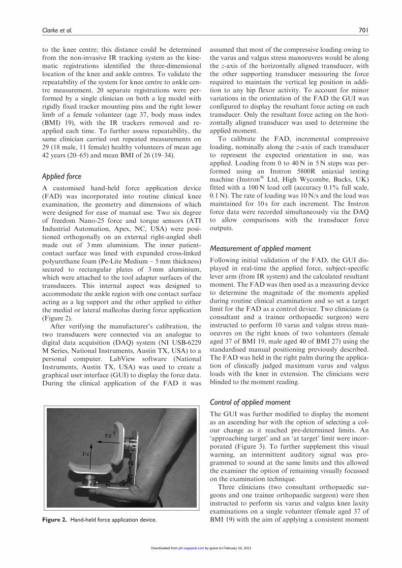
A customised hand-held force application device(FAD) was incorporated into routine clinical kneeexamination, the geometry and dimensions of whichwere designed for ease of manual use. Two six degreeof freedom Nano-25 force and torque sensors (ATIIndustrial Automation, Apex, NC, USA) were posi-tioned orthogonally on an external right-angled shellmade out of 3mm aluminium. The inner patient-contact surface was lined with expanded cross-linkedpolyurethane foam (Pe-Lite Medium – 5mm thickness)secured to rectangular plates of 3mm aluminium,which were attached to the tool adapter surfaces of thetransducers. This internal aspect was designed toaccommodate the ankle region with one contact surfaceacting as a leg support and the other applied to eitherthe medial or lateral malleolus during force application(Figure 2).
After verifying the manufacturer’s calibration, thetwo transducers were connected via an analogue todigital data acquisition (DAQ) system (NI USB-6229M Series, National Instruments, Austin TX, USA) to apersonal computer. LabView software (NationalInstruments, Austin TX, USA) was used to create agraphical user interface (GUI) to display the force data.During the clinical application of the FAD it was
assumed that most of the compressive loading owing tothe varus and valgus stress manoeuvres would be alongthe z-axis of the horizontally aligned transducer, withthe other supporting transducer measuring the forcerequired to maintain the vertical leg position in addi-tion to any hip flexor activity. To account for minorvariations in the orientation of the FAD the GUI wasconfigured to display the resultant force acting on eachtransducer. Only the resultant force acting on the hori-zontally aligned transducer was used to determine theapplied moment.
To calibrate the FAD, incremental compressiveloading, nominally along the z-axis of each transducerto represent the expected orientation in use, wasapplied. Loading from 0 to 40N in 5N steps was per-formed using an Instron 5800R uniaxial testingmachine (Instron� Ltd, High Wycombe, Bucks, UK)fitted with a 100N load cell (accuracy 0.1% full scale,0.1N). The rate of loading was 10N/s and the load wasmaintained for 10 s for each increment. The Instronforce data were recorded simultaneously via the DAQto allow comparisons with the transducer forceoutputs.
Measurement of applied moment
Following initial validation of the FAD, the GUI dis-played in real-time the applied force, subject-specificlever arm (from IR system) and the calculated resultantmoment. The FAD was then used as a measuring deviceto determine the magnitude of the moments appliedduring routine clinical examination and so set a targetlimit for the FAD as a control device. Two clinicians (aconsultant and a trainee orthopaedic surgeon) wereinstructed to perform 10 varus and valgus stress man-oeuvres on the right knees of two volunteers (femaleaged 37 of BMI 19, male aged 40 of BMI 27) using thestandardised manual positioning previously described.The FAD was held in the right palm during the applica-tion of clinically judged maximum varus and valgusloads with the knee in extension. The clinicians wereblinded to the moment reading.
Control of applied moment
The GUI was further modified to display the momentas an ascending bar with the option of selecting a col-our change as it reached pre-determined limits. An‘approaching target’ and an ‘at target’ limit were incor-porated (Figure 3). To further supplement this visualwarning, an intermittent auditory signal was pro-grammed to sound at the same limits and this allowedthe examiner the option of remaining visually focussedon the examination technique.
Three clinicians (two consultant orthopaedic sur-geons and one trainee orthopaedic surgeon) were theninstructed to perform six varus and valgus knee laxityexaminations on a single volunteer (female aged 37 ofBMI 19) with the aim of applying a consistent momentFigure 2. Hand-held force application device.
Clarke et al. 701
by guest on February 19, 2013pih.sagepub.comDownloaded from

of 18Nm as indicated by the FAD. An ‘approaching’limit of 16Nm was selected. The technique was standar-dised as before but with an additional aim of maintain-ing the knee between 0� and 5� of flexion (target of 2�).The knee flexion was displayed in real time by the IRsystem. The clinicians were blinded to the correspond-ing laxity measurements for each applied moment. TheIR system automatically recorded the maximum laxityangle achieved during each manoeuvre.
Statistics
Analysis was completed using Excel 2007 (MicrosoftCorp, Redmond, WA, USA) and SPSS 17.0 (IBMCorp, Somers, NY, USA). Agreement between mea-surements (different systems or paired repeated data)was assessed using Bland–Altman plots with meandifference and limits of agreement for the differencecalculated.29 Data were assessed for normality usingthe Korlmogorov–Smirnov (K–S) test. Comparisonof variance between groups was made using the FTest with p \ 0.05 considered statistically significant.Correlation was assessed using Pearson’s correlationcoefficient.
To summarise results for non-parametric data med-ian and range were used, whereas for parametric datamean and standard deviation (SD) were used.
Results
Knee flexion
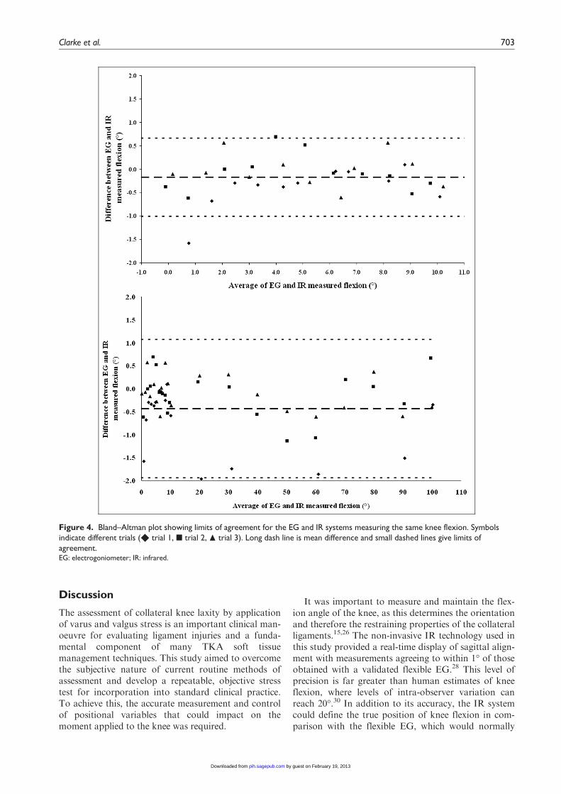
The mean difference between the flexible EG and non-invasive IR tracking system for incremental angles of1� of knee flexion was 20.2� (limits of agreement fordifference 60.8�) over all three trials (Figure 4(a)). Forflexion to 100�, the mean difference was 20.4� (limitsof agreement for difference 61.5�) over the three trials(Figure 4(b)). The discrepancy of the two systems waswithin 61� for the majority of measurements.
Moment arm
Repeated moment arm calculations using the leg modelproduced normally distributed data (p=0.934) with aSD of 1.5mm, while on a single volunteer the data werealso normally distributed (p=0.986), but SD was4.8mm. This difference of variance was statistically sig-nificant (p \ 0.001). For the paired volunteer measure-ments, the mean and limits of agreement for thedifference was 1.56 13mm, with the Bland–Altmanplot (Figure 5) illustrating that most agreed to within610mm (4 3% of leg length).
Applied force
The output signals from the FAD closely correlated tothose of the Instron testing machine (r=0.999, p\ 0.001), although the traces were noisier (possiblyowing to a poor earth connection). A typical trace isshown in Figure 6.
Measurement of applied moment
The mean overall applied moment, as chosen by theclinicians, was 22Nm (range 13–33) (Table 1). Themean moment applied to the knee of volunteer 1 was19Nm (range 12–32), and based on this result the stan-dardised moment to be applied during subsequent varusand valgus stress testing was 18Nm.
Control of applied moment
The measured results for three clinicians during appli-cation of a target 18Nm moment are shown in Table2. The overshoot ranged from 0 to 3.5Nm, with anoverall mean of 1.3Nm. The laxity measurementsshowed small SD for each clinician and similar meanvalues between clinicians, with the valgus laxityassessment being slightly more consistent than varus(Table 2).
Figure 3. Customised LabView graphical displays illustrating the ascending bar for a chosen target of 20 Nm with ‘approaching’limit of 18 Nm: (a) moment below first threshold; (b) moment above first threshold but below second; and (c) moment abovesecond threshold.
702 Proc IMechE Part H: J Engineering in Medicine 226(9)
by guest on February 19, 2013pih.sagepub.comDownloaded from
Discussion
The assessment of collateral knee laxity by applicationof varus and valgus stress is an important clinical man-oeuvre for evaluating ligament injuries and a funda-mental component of many TKA soft tissuemanagement techniques. This study aimed to overcomethe subjective nature of current routine methods ofassessment and develop a repeatable, objective stresstest for incorporation into standard clinical practice.To achieve this, the accurate measurement and controlof positional variables that could impact on themoment applied to the knee was required.
It was important to measure and maintain the flex-ion angle of the knee, as this determines the orientationand therefore the restraining properties of the collateralligaments.15,26 The non-invasive IR technology used inthis study provided a real-time display of sagittal align-ment with measurements agreeing to within 1� of thoseobtained with a validated flexible EG.28 This level ofprecision is far greater than human estimates of kneeflexion, where levels of intra-observer variation canreach 20�.30 In addition to its accuracy, the IR systemcould define the true position of knee flexion in com-parison with the flexible EG, which would normally
Figure 4. Bland–Altman plot showing limits of agreement for the EG and IR systems measuring the same knee flexion. Symbolsindicate different trials ( � trial 1, � trial 2, N trial 3). Long dash line is mean difference and small dashed lines give limits ofagreement.EG: electrogoniometer; IR: infrared.
Clarke et al. 703
by guest on February 19, 2013pih.sagepub.comDownloaded from

Figure 5. Bland–Altman plot showing limits of agreement between paired volunteer measurements of the effective moment arm.Long dash line is mean difference and small dashed lines give limits of agreement.
Figure 6. Simultaneous force output trace from the FAD (black) and Instron (grey) for 5 Nm incremental compressive loads up to40 Nm.FAD: force application device.
Table 1. Mean and range of moments recorded by FAD for repeated volunteer measurements.
Applied moment (Nm) mean [range]Volunteer
Clinician 1 Clinician 2
Varus (n = 10) Valgus (n = 10) Varus (n = 10) Valgus (n = 10)
1 20 [18–23] 15 [12–17] 27 [24–32] 16 [13–21]2 30 [27–33] 19 [15–23] 33 [29–36] 20 [17–24]
704 Proc IMechE Part H: J Engineering in Medicine 226(9)
by guest on February 19, 2013pih.sagepub.comDownloaded from

rely on an initial estimate of 0� as its starting point.One limitation of this study is that the flexible EG isalso sensitive to soft tissue movements, i.e. it does notquantify actual bone motion. However, given the use ofhealthy volunteers, it was not possible to use an inva-sive method of tracking of the actual bone motions.The flexible EG is a widely accepted non-invasive tech-nique for quantifying knee flexion.
With control of knee flexion it was then necessary tostandardise the clinical examination technique in orderto define a moment arm. The moment arm was definedas the distance between the knee and ankle centres ascalculated by the IR system. To use this then requiredcareful hand positioning according to the surface anat-omy, so that the force application coincided with theknee and ankle centres. For the measurement of themoment arm distance, repeated measurements on a sin-gle volunteer (SD6 4.8mm) compared well with thevariation owing to the system precision, as measuredby a leg model with rigid tracker mountings(SD6 1.5mm). Although this difference was statisti-cally significant, it represented only a small loss ofaccuracy from soft tissue artefacts. Further to this, themeasurement of this distance was repeatable to6 13mm when performed on 29 volunteers. Therefore,the technique may be more accurate than currentlyavailable routine methods of leg length assessment,such as a measuring tape or radiographs. Assuming amoment arm of 0.4m, a 6 13mm change would equateto a 6 0.65Nm variation in moment. This is relativelysmall compared with the range of moments seen. Interms of the point of force application coinciding withthis pre-defined distance, the surface anatomy allowedcontrol of hand and FAD placement. However, a lim-itation of the study is that the position of the FAD wasnot known throughout the stress manoeuvre. Thiscould be overcome by tracking of the FAD to give theprecise point of force application and so calculate a‘true’ lever arm throughout the laxity assessmentmanoeuvre.
To standardise the moment required the control ofthe applied force. Much of the previous work in thisarea has relied on invasive access to the knee througheither the use of cadavers or intra-operative studies inthe context of TKA, where gap-balancing devices havehelped to quantify the amount of force application.Classic techniques have involved spacer blocks as a sur-rogate measure of soft tissue tension,31 or laminar
spreaders that enabled a comparison between medialand lateral joint spaces both in flexion and extension.More sophisticated designs have incorporated scales tomeasure the amount of gap distraction32,33 or force sen-sing devices to determine the applied load. D’Limaet al.34,35 utilised a force transducer to measure intra-articular compressive forces on the tibial component ofknee replacements and a similar device that measuredknee joint moments and forces was developed and eval-uated in-vitro by Crottet et al.36 The use of cadavericspecimens has permitted direct attachment of straingauges to bone with accurate measurement of theapplied moment-load. Methods of applying standar-dised varus–valgus moments to the knee joint have ran-ged from basic weight-pulley systems15,22 to digitalstrain gauges.20,37
Most in-vivo studies that have sought to standardisevarus and valgus loads to the knee have involved cum-bersome experimental set-ups that are not readily adap-table to a clinical setting. Sharma et al.21 developed abench with an attached low-friction track to supportthe leg and a hand-held dynamometer to apply a fixedload. Van der Esch et al.22 constructed a measurementchair, with a lower limb attachment consisting of fivespecific fixation points relative to the knee joint line, anelectronic meter to record angular deviation and aweight-pulley system to deliver a standardised load. Inaddition to the impracticalities of these set-ups, the lax-ity measurements were of limited use in clinical practiceowing to considerable intra- and inter-observer mea-surement error. From the point of view of clinician-applied varus–valgus loads however, any measuringdevice should consider the way in which patients areexamined. The FAD designed in this study allowed theincorporation of commercial transducers withoutaffecting the magnitude of their force measurements. Inparticular, the deformation of internal patient contactsurface (polyurethane pad) did not result in a measure-able change in the transmitted loads. The designallowed the FAD to be incorporated into a routineclinical assessment with minimal alteration of examina-tion technique. While we are not aware of any similardevices for measuring collateral knee laxity, there arereports of measurement tools for recording manualcontact forces. Van Zoest et al.38 recognised the impor-tance of manual techniques in disciplines, such aschiropractic and osteopathy, and developed a similarpalm-held force measurement system for improving the
Table 2. Mean and range of moments (recorded by the FAD) and corresponding angular displacements (measured by IR system) forrepeated laxity tests on a single volunteer.
Clinician Mean moment [range] (Nm) Mean laxity 6 SD (�)
Varus (n = 6) Valgus (n = 6) Varus (n = 6) Valgus (n = 6)
1 19 [18.8–19.9] 19 [18.3–20.1] 5 6 1.1 3 6 0.22 20 [18.0–21.5] 20 [19.1–20.3] 4 6 0.8 3 6 0.73 19 [18.4–19.1] 19 [18.3–19.1] 5 6 0.3 3 6 0.4
Clarke et al. 705
by guest on February 19, 2013pih.sagepub.comDownloaded from

manual perception and force delivering skills of studentpractitioners. Harms and Bader39 investigated thevariability of forces applied by therapists during spinalmobilisation procedures through the construction of aninstrumented mobilisation coach that could measurethe magnitude and direction of applied forces. In spiteof standardising the manipulation technique, there wasa large variation in the forces used by different thera-pists, ranging from 63 to 347N. In comparison, thevarus and valgus knee moments recorded by the FADin this study were more consistent for the two cliniciansassessed, with a range of measurements from 13 to33Nm for a total of 80 stress manoeuvres. This mayreflect a more perceptible endpoint for the constrainingsoft tissues of the knee,15 in contrast to the underlyingtissues around the spine that may not provide obviousfeedback when applying a manual compressive force.The mean recorded moment of 19Nm did not produceany discomfort in the particular individual tested,despite being higher than the 12Nm upper subject-tolerance limit that has often been used in other stud-ies.20–22 This may be due to the short duration of thestress manoeuvre compared with the more sustainedloads in these experimental studies.
Following its use as a measurement tool, the FADwas utilised as a control device through the design of aGUI, which provided a repeatable method of applyinga pre-determined moment. The visual and auditorywarning systems were effective in preventing significantovershoot of the selected threshold, with a mean of1.3Nm. The range of manual forces moments whenusing the FAD (3.5Nm) was about half of thoseselected by clinicians (8Nm) for any given condition.This indicates that it can satisfactorily be used as a con-trol device.
For each applied varus and valgus load, the corre-sponding coronal angular displacement of the knee,from its resting position, was used to quantify laxity.The repeated sets of measurements by three clinicianshad similar mean values, with the SD ranging from6.2� to 6.1�. The small differences between cliniciansmay be due to actual variations in the applied moment,but may also result from the 6.1� accuracy of the non-invasive IR tracking technology. However, this repre-sents a significantly higher degree of precision than thelikely 65� human error in estimation of alignment.15
The system also has a number of potential advantagesover alternative non-invasive laxity assessments. Theradiographic measurement of joint space opening hasbeen widely reported,3,20,40 but drawbacks include theuse of ionising radiation and the requirement for meti-culous control of lower limb positioning. The gradingof collateral ligament injury severity on the basis of a1–2mm difference in gap opening20 could potentiallybe seen with small rotational and sagittal variations inknee position.6,13 An alternative approach to measuringlaxity involves the use of specially designed mechanicaldevices incorporating goniometers. Early cadavericwork was limited by inaccurate manual measurement
tools to record the resultant displacement following anapplied load.41 Markolf et al.15 addressed these limita-tions by using a specially designed three-dimensionalgoniometer linkage that allowed the knee joint to bemaintained at a specific degree of flexion, while electro-nically recording the resultant varus–valgus angulation.Unfortunately, in-vivo adaptation of goniometers hasgenerally involved cumbersome experimental set-upswhich, similar to previously described force applicationtechnology, are not practical for routine clinical use.
Although some of the disadvantages of other systemshave been addressed, the measurement tool developedin this study also has limitations. For the manuallyapplied force, it was assumed that the loading of thetransducers within the FAD was perpendicular to thetibial mechanical axis in the coronal plane. However, inspite of careful positioning of the device, the true orien-tation of the resultant force vector was unknown. Usingthe IR tracking system to give the real-time orientationof the FAD relative to the defined tibial moment armcould potentially overcome this. During laxity testing,although the auditory warning system enabled cliniciansto remain more focussed on examination technique,there was still the requirement to use the on-screen dis-play of flexion to control knee position. The use of adevice to hold the knee in a specified degree of flexion,such as a wedge in the popliteal fossa or the use of animage overlay,42 could remove the requirement to lookat a computer screen during clinical examination.
A further limitation of this study is that the finalassessment was carried out on one slim volunteer withno ‘true’ measurement of laxity. As stated above, it wasnot possible to use an invasive method of assessmenton this healthy volunteer. Also, it was not possible toimage the knee under stress. However, in terms of theaccuracy of the IR system on larger individuals, it hasbeen shown to be highly repeatable on a group of sub-jects of BMI up to 34, with clinical examination notaffecting alignment measurements.27 This had beentaken to indicate that the tracker mountings are verystable. The additions to the assessment, including theuse of the FAD, should not have affected the accuracyof the IR system in any way, so it is felt that the previ-ous results give confidence that this set-up would workwith the higher BMI population seen for total kneereplacements. Further to this, the authors intend tocarry out more work to use this set-up in a patient pop-ulation with osteoarthritis due to undergo TKA and tocompare non-invasive measurements directly with inva-sive intra-operative data. However, it is necessary tovalidate the system on healthy volunteers before pro-ceeding to this step.
In spite of the potential limitations of our system, wehave successfully quantified and standardised the man-ual technique of coronal knee laxity assessment for thelimited number of subjects and clinicians evaluated.Our hypothesis stated that this would lead to a narrowrange of laxity measurements, which we have confirmedas the results were within the accuracy limits of our IR
706 Proc IMechE Part H: J Engineering in Medicine 226(9)
by guest on February 19, 2013pih.sagepub.comDownloaded from

system. Minimising the subjective variables of clinicalexamination with a more repeatable, quantitative tech-nique could improve current knowledge of soft tissueknee behaviour. This may lead to improved balancingtechniques in TKA through quantification of knee lax-ity before, during and after surgery, enabling a morewidespread use of single surgeon-derived algo-rithms.16,17,18 There is a potential role in the manage-ment of collateral ligament injuries with regard to morereliable initial diagnosis and severity grading, as well asmonitoring recovery and rehabilitation. Standardiseddata from healthy, injured and osteoarthritic kneescould improve our knowledge of normal and abnormalknee kinematics, and lead to more objective treatmentalgorithms. Finally, as this augmented learning can beincorporated into traditional examination techniques,the ability to quantify the technique of senior cliniciansmay help to enhance the perceptive skills of more juniortrainees who do not have the benefit of experience.
Funding
This research received no specific grant from any fund-ing agency in the public, commercial, or not-for-profitsectors. However the Department of Orthopaedics,Golden Jubilee National Hospital has received researchfunding from BBraun Aesculap and a proportion ofthis has been used to fund a fixed term research postfor one of the authors (JVC).
Acknowledgements
The authors would like to thank Mr Stephen Murrayfor assistance with the design and manufacture of thestraps, Mr David Smith and Mr Stephen Murray fortheir technical assistance with the design, developmentand testing of the FAD and Mr Hin Lau for his provi-sion of the LabView code to collect data from the nano-transducers. They would also like to thank BBraunAesculap for the loan of equipment used in this project.
Conflict of interest
One of the authors (FP) has patents/licensing agree-ments with BBraun Aesculap. The other authors (JVC,WTW, SCW, PER and AHD) certify that they have nocommercial associations (e.g. consultancies, stock own-ership, equity interest, patent/licensing arrangements,etc.) that might pose a conflict of interest in connectionwith the submitted article.
References
1. Petermann J and von Garrel, Gotzen. Non-operative
treatment of acute medial collateral ligament lesions of
the knee joint. Knee Surg Sports Traumatol Arthrosc
1993; 1: 93–96.2. Hefti F, Muller W, Jakob RP, et al. Evaluation of knee
ligament injuries with the IKDC form. Knee Surg Sports
Traumatol Arthrosc 1993; 1: 226–234.
3. Wijdicks CA, Griffith CJ, Johansen S, et al. Injuries to
the medial collateral ligament and associated medial
structures of the knee. J Bone Joint Surg Am 2010; 92-A:
1266–1280.4. Noyes FR, Grood ES, Butler DL, et al. Knee ligament
tests: what do they really mean? Physical Therapy 1980;
60: 1578–1581.5. Krackow KA, Pepe CL and Galloway EJA. A mathe-
matical analysis of the effect of flexion and rotation on
apparent varus/valgus alignment at the knee. Orthopae-
dics 1990; 13: 861–868.6. Siu D, Cooke TD, Broekhoven LD, et al. A standardised
technique for lower limb radiography. Practice, applica-
tions, and error analysis. Invest Radiol 1991; 26: 71–77.7. Rasenberg EI, Lemmens JA, van Kampen A, et al.
Grading medial collateral ligament injury: comparison of
MR imaging and instrumented valgus-varus laxity test-
device. A prospective double-blind patient study. Eur J
Radiol 1995; 21: 18–24.8. Beall DP, Googe JD, Moss JT, et al. Magnetic resonance
imaging of the collateral ligaments and the anatomic
quadrants of the knee. Radiol Clin North Am 2007; 45:
983–1002.9. Ikuma H, Abe N, Uchida Y, et al. Novel magnetic reso-
nance imaging evaluation for valgus instability of the
knee caused by medial collateral ligament injury. Acta
Med Okayama 2008; 62: 185–191.10. Jacobson KE and Chi FS. Evaluation and treatment of
medial collateral ligament and medial-sided injuries of the
knee. Sports Med Arthrosc 2006; 14: 58–66.11. Whiteside LA. Soft tissue balancing. J Arthroplasty 2002;
17(suppl. 1): 67–72.12. Mihalko WM, Whiteside LA and Krackow KA. Com-
parison of ligament-balancing techniques during total
knee arthroplasty. J Bone Joint Surg Am 2003; 85-
A(Suppl. 4): 132–135.13. Krackow KA. Varus deformity. In: The technique of total
knee arthroplasty. St Louis, Missouri: CO Mosby Com-
pany, 1990, pp.317–340.14. Edwards JZ, Greene KA, Davis RS, et al. Measuring flex-
ion in knee arthroplasty patients. J Arthroplasty 2004; 19:
369–372.15. Markolf KL, Mensch JS and Amstutz HC. Stiffness and
laxity of the knee– the contributions of the supporting
structures. J Bone Joint Surg Am 1976; 58-A: 583–594.16. Saragaglia D, Chaussard C and Rubens-Duval B. Navi-
gation as a predictor of tissue release during 90 cases of
computer assisted total knee arthroplasty. Orthopaedics
2006; 29(10 Suppl): 137–139.17. Picard F, Deakin AH, Clarke JV, et al. A quantitative
method of effective soft tissue management for varus
knees in total knee replacement surgery using naviga-
tional techniques. Proc IMechE Part H: J Engineering in
Medicine 2007; 221: 763–772.18. Hakki S, Coleman S, Saleh K, et al. Navigational predic-
tors in determining the necessity for collateral ligament
release in total knee replacement. J Bone Joint Surg Br
2009; 91-B: 1178–1182.19. Bellemans J, D’Hooghe P, Vandenneucker H, et al. Soft
Tissue balance in total knee arthroplasty: does stress
relaxation occur perioperatively? Clin Orthop Relat Res
2006; 452: 49–52.20. LaPrade RF, Heikes C, Bakker AJ, et al. The reproduci-
bility and repeatability of varus stress radiographs in the
Clarke et al. 707
by guest on February 19, 2013pih.sagepub.comDownloaded from

assessment of isolated fibular collateral ligament and
grade-III posterolateral knee injuries. An in vitro biome-
chanical study. J Bone Joint Surg Am 2008; 90: 2069–
2076.21. Sharma L, Congron L, Felson DT, et al. Laxity in
healthy and osteoarthritic knees. Arthritis Rheum 1999;
42: 861–870.22. van der Esch M, Steultjens M, Ostelo RWJG, et al.
Reproducibility of instrumented knee joint laxity mea-
surement in healthy subjects. Rheumatology 2006; 45:
595–599.23. Miyasaka KC, Daniel DM and Stone ML. The incidence
of knee ligament injuries in the general population. Am J
Knee Surg 1991; 4: 43–48.24. American Academy of Orthopaedic Surgeons. Common
knee injuries. http://orthoinfo.aaos.org/topic.cfm?to-
pic=A00325 (2007, accessed 30 November 2010).25. Nilsdotter AK, Toksvig-Larsen S and Roos EM. Knee
arthroplasty: are patients’ expectations fulfilled? A pro-
spective study of pain and function in 102 patients with
5-year follow-up. Acta Orthop 2009; 80: 55–61.26. Grood ES, Noyes FR, Butler DL, et al. Ligamentous and
capsular restraints preventing straight medial and lateral
laxity in intact human cadaver knees. J Bone Joint Surg
Am 1981; 63: 1257–1269.27. Clarke JV, Riches PE, Picard F, et al. A non-invasive sys-
tem for the measurement of knee alignment. Comp Aided
Surg. Comp Aided Surg 2012; 17: 29–39.28. Rowe PJ, Myles CM, Hillmann SJ, et al. Validation of
flexible electrogoniometry as a measure of joint kine-
matics. Physiotherapy 2001; 87: 479–488.29. Bland JM and Altman DG. Statistical methods for asses-
sing agreement between two methods of clinical measure-
ment. Lancet 1986; 1: 307–310.30. Cushnaghan J, Cooper C, Dieppe P, et al. Clinical assess-
ment of osteoarthritis of the knee. Ann Rheum Dis 1990;
49: 768–770.
31. Insall JN, Binazzi R, Soudry M, et al. Total knee arthro-plasty. Clin Orthop 1985; 192: 13–22.
32. Unitt L, Sambatakakis A, Johnstone D, et al. Short-termoutcome in total knee replacement after soft-tissue releaseand balancing. J Bone Joint Surg Br 2008; 90-B: 159–165.
33. Winemaker MJ. Perfect balance in total knee arthroplasty:the elusive compromise. J Arthroplasty 2002; 17: 2–10.
34. D’Lima DD, Townsend CP, Arms SW, et al. An implan-table telemetry device to measure intra-articular tibialforces. J Biomech 2005; 38: 299–304.
35. D’Lima DD, Patil S, Steklov N, et al. Dynamic intrao-perative ligament balancing for total knee arthroplasty.Clin Orthop Relat Res 2007; 463: 208–212.
36. Crottet D, Maeder T, Fritschy D, et al. Development ofa force amplitude- and location-sensing device to improvethe ligament balancing procedure in TKA. IEEE Trans
Bio Eng 2005; 52: 1609–1611.
37. van Damme G, Defoort K, Ducoulombier Y, et al. Whatshould the surgeon aim for when performing computer-assisted total knee arthroplasty? J Bone Joint Surg Am
2005; 87-A(suppl. 2): 52–58.38. van Zoest GGJM, van den Berg HTCM and Holtkamp
FC. Three-dimensionality of contact forces during clini-cal manual examination and treatment: a new measuringsystem. Clin Biomech 2002; 17: 719–722.
39. Harms MC and Bader DL. Variability of forces appliedby experienced therapists during spinal mobilization. ClinBiomech 1996; 12: 393–399.
40. Moore TM, Meyers MH and Harvey JP. Collateral liga-ment laxity of the knee. Long-term comparison betweenplateau fractures and normal. J Bone Joint Surg Am 1976;58-A: 594–598.
41. Brantigan OC and Voshell AF. The mechanics of the liga-ments and menisci of the knee joint. J Bone Joint Surg
1941; 23: 44–66.42. Moody JE, Nikou C, Picard F, et al. Computer-inte-
grated anterior cruciate ligament reconstruction system. JBone Joint Surg Am 2002; 84: S99–S101.
708 Proc IMechE Part H: J Engineering in Medicine 226(9)
by guest on February 19, 2013pih.sagepub.comDownloaded from