Kinematical parameters in the coronal and post-coronal regions of the Oe stars
Upload
khangminh22Category
view
4download
0
Loyola University Chicago Loyola University Chicago
Loyola eCommons Loyola eCommons
Master's Theses Theses and Dissertations
1987
The Effect of the Absence of a Coronal Seal on Periapical Tissues The Effect of the Absence of a Coronal Seal on Periapical Tissues
Following Endodontic Therapy Following Endodontic Therapy
Richard Alan Kohn Loyola University Chicago
Follow this and additional works at: https://ecommons.luc.edu/luc_theses
Part of the Medicine and Health Sciences Commons
Recommended Citation Recommended Citation Kohn, Richard Alan, "The Effect of the Absence of a Coronal Seal on Periapical Tissues Following Endodontic Therapy" (1987). Master's Theses. 3421. https://ecommons.luc.edu/luc_theses/3421
This Thesis is brought to you for free and open access by the Theses and Dissertations at Loyola eCommons. It has been accepted for inclusion in Master's Theses by an authorized administrator of Loyola eCommons. For more information, please contact [email protected].
This work is licensed under a Creative Commons Attribution-Noncommercial-No Derivative Works 3.0 License. Copyright © 1987 Richard Alan Kohn

,- F ~1'ff"<.~ a ~"'1
"
THE EFFECT OF THE ABSENCE OF A CORONAL SEAL
ON PERIAPICAL TISSUES FOLLOWING ENDODONTIC THERAPY
By
Richard Alan tohn, 0.0.S.
A Thesis Submitted to the Faculty of the Graduate School
of Loyola University of Chicago in Partial Fulfillment
of the Requirements for the Degree of
Master of Science
January
1987

~ 1987, Richard Alan Kohn

DEDICATION
To Dr. Marshall Smulson, in appreciation of
his thirty-one years of dedicated service
to the students of Loyola University, School
of Dentistry.
ii
ACKNOWLEDGEMENTS
To Dr. Franklin Weine who gave me the opportunity to
train under one of the leaders in the field of endo
dontics, for his lasting friendship and assistance in
helping shape my career.
To Dr. Marshall Smulson who first ignited the spark of
interest in endodontics for me through his tireless
devotion to education.
To Dr. Hal McReynolds who helped stimulate ideas through
out my professional training, and whose friendship and
assistance helped make this study possible.
To Dr. James Sandrik whose expertise in dental materials
and scientific method are well acknowledged, and whose
friendship and interest has added to my professional
development.
To Dr. Gary Taylor who brought me into his practice and
helped me develop and refine my clinical skills, and whose
friendship I will always value.
iii
VITA
The author, Richard Alan Kohn was born in Chicago, Illinois on Octo
ber 15, 1952.
He obtained his elementary and secondary education in Chicago, Ill
inois, graduating from James H. Bowen High School in June, 1970.
In September, 1970, he entered the University of Illinois in Cham
paign, Illinois where he attended until May, 1974. In September, 1974, he
entered Loyola University, School of Dentistry and received the degree of
Doctor of Dental Surgery in May of 1978.
After completing dental school, he entered a General Practice Residen
cy program at the University of Maryland Hospital in Baltimore, Maryland
from July, 1978 to July, 1979. He then returned to Loyola University,
School of Dentistry in August, 1979, in a dual course of study leading to
the degree of Master of Science in Oral Biology and a Certificate of Spe
cialty Training in Endodontics.
iv
TABLE OF CONTENTS
DEDICATION. . . .
ACKNOWLEDGEMENTS.
VITA. . . . . .
LIST OF TABLES.
LIST OF FIGURES
Chapter
I. INTRODUCTION ...•...
II. REVIEW OF THE LITERATURE .
A. History ....... .
B. Focal Infection Theory
C. Canal Morphology and Instrumentation .
D. Success Versus Failure ..
E. Restoration Microleakage .
F. Obliteration .. .
III. MATERIALS AND METHODS .... .
A. Anesthetic Procedure .
B. Radiographic Procedure .
C. Operative Procedure.
D. Sacrifice Procedure ..
E. Histologic Preparation and Evaluation ..
v
PAGE
ii
iii
iv
vii
viii
1
3
3
5
8
11
17
19
30
31
33
33
37
37
F. Radiographic Evaluation ..
G. Statistical Analysis .
IV. RESULTS. .
V. DISCUSSION .
SUMMARY
REFERENCES.
FIGURES . .
vi
PAGE
38
39
40
53
63
64
74
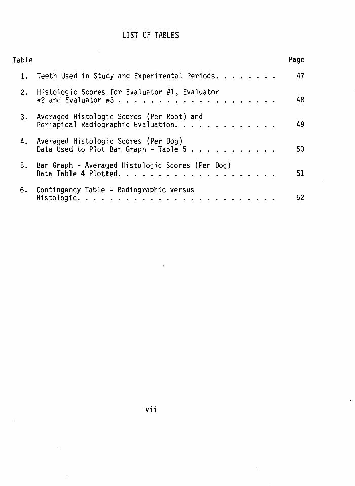
Table
1.
2.
LIST OF TABLES
Teeth Used in Study and Experimental Periods.
Histologic Scores for Evaluator #1, Evaluator #2 and Evaluator #3 ........ .
3. Averaged Histologic Scores (Per Root) and Periapical Radiographic Evaluation ..
4.
5.
6.
Averaged Histologic Scores (Per Dog) Data Used to Plot Bar Graph - Table 5
Bar Graph - Averaged Histologic Scores (Per Dog) Data Table 4 Plotted .....•.........
Contingency Table - Radiographic versus Histologic .............. .
vii
Page
47
48
49
50
51
52
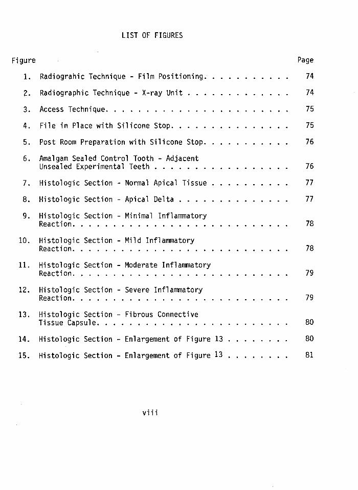
LIST OF FIGURES
Figure
1. Radiograhic Technique - Film Positioning ..
2. Radiographic Technique - X-ray Unit ..
3. Access Technique ..... .
4. File in Place with Silicone Stop.
5. Post Room Preparation with Silicone Stop ..
6. Amalgam Sealed Control Tooth - Adjacent Unsealed Experimental Teeth ..•......
7.
8.
9.
10.
11.
12.
13.
Histologic Section - Normal Apical Tissue
Histologic Section - Apical Delta ....
Histologic Section - Minimal Inflammatory Reaction ............... .
Histologic Section - Mild Inflammatory Reaction. . . . . . . . . . ....
Histologic Section - Moderate Inflammatory Reaction ................. .
Histologic Section - Severe Inflammatory Reaction ............... .
Histologic Section - Fibrous Connective Tissue Capsule .......... .
14. Histologic Section - Enlargement of Figure 13 .
15. Histologic Section - Enlargement of Figure 13
viii
. . . .
Page
74
74
75
75
76
76
77
77
78
78
79
79
80
80
81

CHAPTER I
INTRODUCTION
Statistically, endodontics is highly predictable, realizing a success
rate approximating 95% of all cases treated. 1 Predictability of this magni
tude may be attributed to a meticulous conformation to three basic princi
ples known as the endodontic triad. Accurate diagnosis of the disease pro
cess, biomechanical preparation and obliteration comprise the three phases
of the endodontic triad. 2 Failure to properly execute any one of these pro
cedures might severely compromise the prognosis.
Emphasis in the literature seems to have been placed on obliteration
techniques and materials. The goal of the third phase of the endodontic
triad is to hermetically seal the root canal system, with an inert substance,
from the interstitial fluids circulating in the periradicular region. Studies
abound as to the sealing effectiveness of various endodontic materials and
techniques, all substantiating the necessity of attaining an apical seal.
Loss of, or failure to have achieved an apical seal will allow apical inter
stitial fluid to percolate into the root canal system. The exact mechanism
of how fluid percolation is capable of producing apical inflammation has yet
to be elucidated. It is speculated that the exudate found leaking into the
canal system is derived from the blood serum. This material, once isolated
within a canal space, may undergo degradation and diffuse back into the per
iapical region, producing an inflammatory reaction.1 Thus, the import of at
taining and securing an apical seal must be realized.
1
2
Following completion of successful endodontic therapy, a restoration
is fabricated to bind the residual tooth structure together and return the
treated tooth to occlusal function. At times, the final restoration may
fail in that the marginal integrity is lost, or the patient may have en
tirely neglected further dental treatment with the eventual demise of the
temporary filling. In either case, salivary and bacterial contaminants
might be allowed access to the root canal system from the coronal aspect,
possibly jeopardizing the endodontic prognosis due to breakdown of the her
metic seal with resultant periapical changes.
The literature is deficient concerning loss of the coronal seal and
its effect on periapical tissues following endodontic treatment. It is ap
parent that further research into the effectiveness of an intraradicular
filling in the presence of a failin~ coronal seal is warranted. Therefore,
this study was initiated to evaluate whether loss of a coronal seal would
adversely affect a minimal apical seal within a root canal space, especially
following post room preparation, by investigating periapical reaction both
radiographically and histologically. It is hoped that this study will give
insight into the import of the temporary intertreatment coronal seal follow
ing root canal therapy and the necessity for placement of a permanent rest
oration thereby preventing leakage from the oral environment after comple
tion of endodontic therapy.

CHAPTER II
REVIEW OF THE LITERATURE
A. HISTORY
Endodontics was first recognized as a separate clinical specialty by
the American Dental Association in 1963. Although seemingly in its infancy
in the mid-twentieth century, early Egyptian records dating back to 3700 B.C.
show the existence of dental prescriptions as remedies for gingival abscesses
and/or sinus tracts arising from nonvital teeth. 3 Evidence of a crude form
of root canal therapy presents in the first century A.O. when trephines were
used to establish drainage from the pulp cavity. Arabian surgeons were also
noted to be performing some type of endodontic therapy. 4 In 1757, Dr. Bour
det was practicing pulp extirpations and filling roots of anterior teeth to
the apex with gold. Dr. Hudson, a Philadelphian dentist, introduced this
technique in the United States in 1809. Various filling materials were ad
vocated to seal root canal spaces including cotton and other fibers, copper,
plaster of Paris and even sterile sparrow droppings. Sharpened wood sticks
were utilized in one technique to remove and destroy the canal contents,
later filling the space with the same stick. Frequent root fractures short
ened the longevity of this method. 5 In 1839, Dr. Baker recommended removing
the exposed nerve from the canal, cleaning the canal space and filling the
11 fang 11 with gold. 3 This was the first account of pulp extirpation, canal de
bridement and root filling.
During the 1800's, root canal instruments were crude and not commonly
3
4
available. Success frequently depended on the ingenuity and manual dexter
ity of the operator. Vital or mortal pulp amputation was advocated to cir
cumvent the need for instrumentation and obliteration of the root canal
space. For these reasons, an era of chemical sterilization was embarked
upon. Various chemical and medicinal agents were utilized to treat root
canal spaces. Flagg and Koeker, in 1825, recommended treating the dental
pulp with oil of cloves, oil of cajuput, camphor, opium, alum and myrrh. 6
Dr. John Hunter, in 1835, advocated burning the nerve of the tooth with
strong acids and alkalies. 7 Arsenic was introduced by Dr. Spooner in 1836
as another method for devitalizing the pulp. 5 Thomas Rogers reviewed 220
cases of pulp capping procedures in which various caustic agents were used,
in which 202 were claimed successful. Five conditions were enumerated for
success, but he proceeded to discredit this scientific observation by pre
scribing three leeches and a smart aperient if the pulp capping failed. 3
The practice of using a mummifying paste was introduced by Dr. W.D. Miller
about 1893. Mercuric chloride was used for this purpose, but alum, thymol
and formalin came into favor later. 5
The mid 1800's saw the introduction of instruments used for the total
removal of pulp tissue. Thomas Bell suggested that the tissue be burned
with a cauterizing wire at white heat. 5 In 1836, Dr. Edward Maynard barbed
one side of an untempered steel watch spring and also made reamers from
piano wire. 7 Dr. Fauchard described instruments made from annealed piano
wire, cut into proper lengths, filed down to various diameters and burnished.
Small barbs were then cut along the length of the instrument. 8 In 1852,
Arthur developed broaches with cutting barbs that were designed to grasp
5
the organic contents of the canal space. 9 Bennett also describes broaches,
barbed and spiral, developed primarily to cleanse the canal of tissue de
bris but seldom used for enlarging and shaping. 8
Prior to the Second International Conference on Endodontics in I958,
endodontic instruments were unstandardized. Instruments were haphazardly
numbered from one to twelve, each manufacturer having their own specifica
tions. IO Ingle and Levine presented a plan where endodontic instruments
would have the same taper of shaft, a definite increase in size from one
instrument to another and a universal numbering system with plastic color
coded handles.II Standardization was approved and accepted by the Confer-
ence and is the guide used by manufacturers today.
B. FOCAL INFECTION THEORY
Advances in instrument design, instrumentation and endodontic tech-
niques for the pulpally involved tooth were being introduced and perfected
at the turn of the twentieth century. The application of x-rays as a diag-
nostic tool in dentistry was also being utilized.
In I888, Dr. W.D. Miller demonstrated the presence of bacteria within
the structures of the teeth and on the tissues adjacent to them.I2 He later
showed that teeth with necrotic pulps were generally infected and explained
the formation of dento-alveolar abscesses as the spread of pulpal infection
into the periapical region.I 3 A relationship was alluded to between infec
tions of the mouth and many general constitutional diseases. As early as
I898, William Hunter introduced the term 11 oral sepsis 11 and suggested a link
between dental infections and various maladies. 5 In I9IO, Hunter delivered

6
an address at McGill University entitled "The Role of Sepsis and Antisepsis
in Medicine" expounding that prosthetic dentistry was harboring and propa
gating areas of frank infection. The stage was now set for one of the most
disasterous eras in modern dentistry. Two physicians, Rosenow and Billings,
applied Hunter's theories to the situation of the pulpless tooth and de
veloped the "Theory of Focal Infection". In addresses before the American
Medical Association, Rosenow and Billings implicated oral infections in the
production of systemic diseases. 14 Non-vital and pulpless teeth, irregard
less of their radiographic patterns, were assumed to be dead and infected
or easily infected by anachoresis, presenting a source or foci of suppura-
tion which could exude itself throughout the body. Thusly, alveolar focal
infections and abscesses could act as a dominant, if not the sole factor in
the production of systemic disease. The list of diseases associated with
root abscesses, pus pockets and pulpless teeth included chronic rheumatism,
neuritis, appendicitis, goiter, hay fever, asthma, ulcers, Hodgkin's disease,
endocarditis and mental illness. Said diseases could only be arrested by
extracting the offending tooth or teeth propagating the focus of infection.
By 1920, the crusade against the pulpless tooth had reached such catastro
phic proportions that the dental profession could no longer justify the de
vital ization of teeth. Needless extractions of pulpless and vital teeth
were performed to placate the medical profession.
It was not until the 1930's that serious doubt arose as to the valid
ity of the Focal Infection Theory. Bacteriologists, attempting to repli
cate Rosenow's research, were frequently unable to reproduce his results.
A discrepancy also existed between the findings of bacteriologists and
7
pathologists as to the frequency of infections found on extracted teeth
submitted for cultural and histopathologic evaluation. Rosenow's experi
mental protocol allowed for immediate culturing of extracted root surfaces
or placement of extracted teeth into sterile test tubes for later bacterio
logic or histopathologic evaluation. Bacteriologists were identifying a
high proportion of extracted pulpless teeth with bacterial growth on their
root surfaces and condemned them as foci of infection. Vital teeth with
intact periapical regions were also noted to produce instances of bacterial
growth. Pathologists, however, were not identifying microscopic evidence
of infection as frequently on extracted pulpless teeth and found complete
absence of infection on extracted vital teeth. 15
As early as 1917, Meyer criticized the culturing of extracted root
surfaces due to the possibility of normal flora contamination during extrac
tion.16 Gunter and others17 cultured dentinal dust and pulpal tissue of
teeth sterilized with tincture of iodine prior to removal, followed by an
apical culture after extraction. Bacterial growth was noted on only one
dentinal culture, but all apical cultures were positive, casting grave doubt
on the clinical significance of positive apical cultures from extracted
teeth.
Vital periapical tissue had been shown to be devoid of bacteria histo
logical ly, yet their presence confirmed bacteriologically. Fish and Mac
lean were puzzled by these observations and theorized that bacteria remain
ing within the gingival sulcus following chemical sterilization could be
pumped along the root surface during extraction, providing the source for
positive bacterial root cultures. Fish and Maclean18 demonstrated, in

8
1936, that cauterization of the gingival sulcus prior to extraction, elim
inated the evidence of any bacterial organisms from root surface cultures.
Blood cultures were also shown to be sterile, disputing to a degree, Okell
and Elliott's 19 earlier observation of transient bacteremia following ex
traction of healthy vital teeth. Therefore, conclusions based upon bacter
iologic studies utilizing root surface cultures were totally negated~ as
was the essential foundation of the Focal Infection Theory. Fish and Mac-
Lean's research had such a profound effect that Grossman later noted that
"almost all investigations of the pulpless tooth prior to 1936 were in-
l "dll 20 va i . During this same time, numerous articles and case histories were
appearing in the dental literature supporting the fact that nonvital teeth
could be successfully treated and retained. 21 ,22 , 23
C. CANAL MORPHOLOGY AND INSTRUMENTATION
Following repudiation of the Focal Infection Theory, it became in
creasingly evident and desirable that pulpally involved teeth be treated
and retained. Endodontic therapy was performed with increasing frequency
and became viewed as an acceptable treatment modality.
According to Ingle, root canal therapy consisted of a triad of pro-
cedures: canal enlargement, canal sterilization and canal obliteration, with
h h . l h . . h. . 24 eac p ase given equa emp as1s in ac iev1ng success.
Early investigators noted enormous anatomic and morphologic variations
within the unprepared root canal. 25 ,26 ,27 Numerous lateral and accessory
canals were found, along with weblike communications between canals in mul
tirooted teeth. More recent studies by Meyer28 and Skidmore and Bjorndal ,29
using wax and plastic models respectively to reconstruct the canal
9
morphology, demonstrated again the complex nature of the root canal system.
The existence of these apical and coronal ramifications however, had largely
been ignored. Prior to Kuttler's article, in 1955, it was generally agreed
that the root canal space was a uniformly tapered, cylindrical channel "fol
lowing in its apical third the same direction as the middle and cervical
thirds, ending in the extreme apex with a very narrow foramen 11•30 It was
assumed that standardized instruments were capable of producing a canal prep
aration conforming in shape and size to standardized filling materials. Seid
ler31 described the ideal instrumented canal as being round and tapering,
with a minute opening at the apex. Luks and Bolatin32 stated however, that
this ideal was too simplistic, the root canal system was composed of tor
tuous turns, apical foramina and accessory canals. To attempt to employ
standardized instruments in a system incapable of being standardized was a
11 whim of the imagination".
Numerous authorities have regarded the thorough debridement and elimi
nation of pulpal and dentinal contaminants from the root canal system as the
primary goal of endodontic therapy. 33 , 34 , 35 Mechanical instrumentation, gen
erally with the aid of irrigating solutions, substantially reduces the quan-
tity of injurious agents within the canal, thereby enabling the natural de-
fense mechanisms of the body to maintain or repair the periradicular environ-
ment. Even though instrumentation was at a crude stage of development, Hat
ton et. al., 22 in 1928, evaluated the effectiveness of canal debridement by
examining histologic sections of enlarged pulp canals. Results indicated
that canals were only superficially cleansed and much of the pulpal tissue
remained. Masterton36 observed, in 1965, that even after careful canal
10
preparation, small irregularities which may harbor bacteria and debris can
exist. Haga 37 was among the first investigators to show that canal instru
mentation left a surprisingly high percentage of voids and irregularities
along prepared canal walls. In numerous cases, the instrument cut only
three of the canal walls, leaving the fourth wall untouched throughout the
t . 1 th f th t t G t' d G . 38 f' d th en ire eng o e roo segmen . u ierrez an arcia recon irme e
presence of irregularities, termed prolongations or fins, in instrumented
mandibular incisors and canines. They further remarked that it was not even
feasible to expect complete canal negotiation due to the prolongations, con
sidering that instrumentation left a path through the geometric center of
the root canal space. Brayton, Davis and Goldman, 39 utilizing injected
silicone rubber impression material, demonstrated the vast number of morpho-
logic irregularities and variations still existing following the debride
ment procedure. Biomechanical instrumentation was generally observed only
along one wall of the canal with as much as one-half of the surface area
untouched by the debridement procedure. More recently, Moodnick et a1. 40
evaluated the efficiency of K files versus Hedstroem files in a scanning
electron microscopic study. No difference was found between the cleaning
ability of either file type but it was noted that the root canal walls even
after thorough debridement contained many irregularities that trapped debris
and harbored pulp tissue, with no canal level being found cleaner than
another.
Numerous investigators utilizing simulated canal models, 41 histologic
1 t . 42 . 1 t . . t d . 43 d . th eva ua ion, scanning e ec ron microscopic s u ies an various o er
methods, have further demonstrated the limitations of present day

biomechanical techniques. The general consensus of recent studies,43 ,44
conclude that all hand and mechanical instrumentation, and irrigants uti-
11
1 ized during the cleansing process, leave debris, both organic and inorganic
within the canal system. The recent introduction of sonic and ultrasonic
instrumentation promises to provide a more thorough means of canal debride
ment. Cunningham and Martin45 have shown the endosonic ultrasonic system
of root canal preparation to be ''superior" to hand-filing techniques. Fur-
ther research into the true efficacy of sonic and ultrasonic instrumentation
however, needs to be performed as evidenced by an investigation by Lange
land et. al. 46 showing that neither hand nor sonic/ultrasonic instrumenta-
tion totally cleaned curved or irregular canals better than the other method.
The above studies not only obviate the need for as complete a debride
ment of the canal system as possible, but also the necessity for an obliter-
ation technique that will successfully seal the root canal system from the
periapical region.
D. SUCCESS VERSUS FAILURE
Root canal therapy has become a highly predictable procedure evidenced
by reported success rates of 95 percent as found by Ingle. 24 There does not
appear to be any clear definition or agreement however, as to what consti-
tutes an endodontic success or failure.
Some experts feel that as long as an endodontically treated tooth is
retained, it is successful. A number of early investigators47 ,48 establish
ed the validity of utilizing radiographs as a criterion of success. Bender
et a1. 49 defined a case successful if: (1) the periapical region appeared
12
normal initially and no areas of rarefaction were observed six months and
two years following obturation of the canal(s) or (2) radiolucent areas
which existed prior to treatment, decreased in size six months and two
years after filling. When doubt existed as to whether an area became
smaller or not, they were classified as unsuccessful. Results of this study
were dependent upon radiographic interpretation alone, without considering
clinical factors and demonstrated a success rate of 82 percent. Strind
berg50 claimed that a case could not be considered successful until a pre-
existing periapical radiolucency had completely healed, with the possible
exception of a slightly thickened periodontal ligament space. Those lesions
which decreased in size but had not entirely resolved would be classified
as doubtful or uncertain. Nicholls51 has reviewed the literature and has
concluded that a two year postoperative observation period is sufficient
time for complete resolution of any periapical pathology. Conversely, Selt
zer et al. 52 have found that the majority of endodontic failures occur with
in twenty-four months of the completion of treatment.
Even though success or failure in endodontics is determined primarily
by radiographic findings, radiographic evaluation is not an exact science in
that observer interpretation must be introduced. Variations in success
rates can depend on interpretation of the radiographic evidence and upon
the definition of success. Goldman, Pearson and Darzenta53 illustrated that
the reliability of radiographic interpretation, in determining success or
failure of a case, is questionable at best. Five of six examiners agreed
independently upon the radiographic diagnosis of success versus failure
only 67 percent of the time. When all six evaluators were tabulated in the

results, the agreement rate dropped to 47 percent. Bender and Seltzer54
demonstrated that unless bony destruction encroaches on the junctional
13
area of cancellous and cortical bone, a periapical radiolucency will not be
perceived. In addition, differences in vertical and/or horizontal angula
tion can lead to different radiographic imagery in that lesions may appear
larger or smaller. Bender, Seltzer and Soltanoff55 warn of the inadequa
cies in using radiographs as the sole criteria in determining endodontic
success. They demonstrated that teeth with a normal periapical radiograph
ic appearance were frequently found histologically to have chronic inflam
matory cell infiltration and granulomatous tissue in the periapical region.
There was no definite correlation found between a negative radiographic
image and the periapical histologic findings in endodontically treated
teeth. There was however a correlation between teeth with radiolucent areas
in that the histologic sections revealed the presence of granulomatous tis
sue or cysts. Their histologic studies indicate the inability to use radio
graphs as a sole criteria of treatment success. They concluded that clin
ical observations should be further examined as additional evidence of en-
dodontic success.
Clinical symptoms were evaluated along with radiographic evidence by
Grossman, Shephard and Pearson56 who proposed that if symptoms developed or
persisted following root canal therapy, the case should be classified as a
failure, despite negative radiographic findings. Those cases in which an
area of rarefaction decreased in size, but had not resolved entirely, were
classified as doubtful. Also included were situations where a thickened
periodontal ligament space had formed. A study was performed by Harty et.
14
a1. 57 to determine the success rate in root canal therapy where successful
treatment was not attained unless the tooth remained clinically asymptomat
ic and functional for two or more years and the radiographic pattern either
remained normal or returned to normality by complete healing of the bony
radiolucency and the periodontal ligament space. Their study was limited
to anterior teeth with single canals and found an overall success rate of
ninety percent. Heling and Tamshe58 included posterior teeth in a similar
study, maintaining the same rigid guidelines for success and evidenced a
drop in the success rate to 70 percent. Seltzer et. al. 59 performing a
clinical, radiographic and histologic evaluation of endodontic failures
made the conclusion that no agreement exists as to the correct definition of
success or failure. They suggested that regardless of the radiographic in
terpretation, endodontically treated teeth which were functioning and with
out adverse clinical symptoms, be regarded as successful. Those teeth with
adverse clinical symptoms, such as pain, swelling, persistent sinus tract,
etc., should be regarded as treatment failures irrespective of the absence
of periapical rarefactions.
Due to the difficulty of objectively defining the parameters of suc
cess in root canal therapy, it might be more prudent to investigate and
identify the reasons for failure. As early as 1931, Rickert and Dixon60
were stressing the importance of root canal obliteration techniques, stat
ing that after instrumentation, the canal space should be filled completely
with a material well tolerated by the periapical tissue. Their experiments
demonstrated through macroscopic examination, that while sterile solid im
plants were well tolerated by rabbit subcutaneous connective tissue, sterile
15
hollow steel and platinum tubes elicited a severe inflammatory reaction
around the open ends. These results formed the basis for the hypothesized
"Hollow Tube Theory". This theory postulated that voids remaining follow
ing obliteration, may become filled with periradicular tissue fluids, iso
lated from the blood stream, which could then undergo degradation through
enzymatic breakdown, even in the absence of microorganisms. These break-
down products could then flow back or diffuse out through the apical fora
men into the periapical region producing an inflammatory response. It was
realized therefore, that many factors, other than bacteria alone, could be
contributory to root canal failures.
Dow and Ingle,61 in 1955, stated that the single largest factor in en
dodontic failures was the poorly obturated root canal. Using radioactive
iodine and an autoradiographic technique to trace apical leakage, it was
shown that considerable percolation of the isotope occurred from the apex
into the canal in poorly obturated samples, while no leakage was evident in
the well filled population. Their results tended to support those of Rick-
ert and Dixon and in their conclusion stated that fluid circulating into the
interstices of the poorly filled root canal could lead to periapical inflam
mation. Kalnins, Masin and Kisis62 stated that organic matter might pool in
the "dead space" of a hollow tube, such as an unfi 11 ed or underfi 11 ed root
canal and subsequently lead to infection.
To determine the rate of success in endodontic therapy, an extensive
study was undertaken at the University of Washington Dental School in 1955
by Ingle. 1 Over twelve hundred patients were recalled for a two year post-
operative evaluation demonstrating a success rate approximating 95 percent.

16
More importantly, the rate of failure was also examined and the causes of
said failures investigated. Analysis of these cases disclosed that 63.46
percent of the failures were due to apical percolation, resulting from im
proper or incomplete obliteration of the root canal space. The next most
frequent reason for failure was inadvertant root perforation, accounting
for 9.61 percent of the unsuccessful cases. Ingle24 reiterated the view of
the hollow tube concept, stating that breakdown or lack of a good apical
seal invites failure due to apical percolation of periapical fluids with
subsequent diffusion stasis into the canal space. Tissue fluid products,
i.e. water soluble proteins, enzymes and salts are capable of diffusing
through defects in the apical seal, undergo degradative changes and again
diffuse out initiating a periapical inflammatory response. These physio-
chemical products might act as a constant irritant in the periapical region
irregardless of the presence or absence of bacteria. Periapical inflammation
would persist as long as some noxious stimulus existed.
Goldman and Pearson,63 using hollow teflon tube implants, showed that
tissue fluids were able to exchange freely through the core without produc
ing inflammatory changes at either open end of the tube. Further doubt re
garding the validity of the hollow tube theory has been shed by the studies
of Seyle64 and Torneck65 studying glass and polyethylene tube implants re
spectively. Both substantiated Goldman and Pearson's results noting little
or no inflammation at the open ends of the tubes and in some instances found
fibrous tissue bridges growing into or through the tube openings. A later
study performed by Torneck,66 in 1967, illustrated the inflammatory poten
tial of implanted sterile polyethylene tubes filled with sterile autoclaved

17
muscle and bacterial contaminated autoclaved muscle. The tissue response
in both instances was considerably more severe than that of empty polyeth
ylene tubing, with the most intense reaction found in the bacterial contam
inated sampling. In summation, the most favorable conditions found for
healing and repair were when the lumen of the tubes were sterile and clean,
emphasizing the debridement process.
The above studies were conducted in soft tissues. It remained for
Hodosh, Povar and Shklar67 to implant plastic teeth with holes drilled in
them into fresh sockets. Inflammation about the open ends was not evidenced
and in some cases the holes were filled with fibrous tissue or bone. They
concluded that hollow tubes were not destined to create tissue destruction
or even inflammatory changes by virtue of their existence but may in fact be
associated with a repair mechanism.
E. RESTORATION MICROLEAKAGE
One of the primary objectives in restorative dentistry is to recapitu
late tooth anatomy and function with a proper restorative material and attain
a hermetic seal at the tooth/restoration interface. Phillips68 states that,
with the possible exception of the polyacrylic acids, none of the materials
used in the restoration of carious lesions actually seal the cavity prepara
tion. A microscopic space always exists between the restoration and the
tooth. Fluids, microorganisms and debris from the mouth may penetrate the
outer margins of the restoration and progress down the walls of the cavity
preparation. This phenomenon is referred to as microleakage. Axim69 at
tributes the formation of this space to several factors including the linear
coefficients of thermal expansion, modulus of elasticity, material solubility,
volumetric changes of the restorative material, permeability of the in
volved tooth structure and influence of oral body fluid.
18
As early as 1929, research was performed as to the efficiency of den
tal fillings in achieving marginal seals. Fraser, 70 using bacterial cul
tures, evaluated whether various materials would remain impermeable to bac
terial penetration. Copper cements and copper amalgam formed an efficient
barrier, while gutta-percha stopping and silicate cements were questionable.
Grossman evaluated the adequacy of temporary filling materials in forming a
hermetic seal during root canal treatment. Glass capillary tubes were
filled at one end with 2-3 mm of each test filling material and suspended
in either an aqueous dye, dye colored saliva or a bacterial suspension to
determine their permeability or lack thereof. Temporary stopping, base
plate gutta-percha, zinc oxyphosphate and zinc oxide-eugenol cements were
examined, but only zinc oxide-eugenol was noted to be "leak-proof without
exception 11•71 Massler and Ostrovsky72 confirmed the results obtained by
Grossman in a separate dye penetration study. Zinc oxide and eugenol and
amalgam showed little or no leakage after more than four months. Fischer73
studied the sealing properties of zinc oxyphosphate, copper and silicate
cements, amalgam, inlays and foils and found that all filling materials
tested allowed some degree of fluid penetration along the restoration mar
gins.
Nelsen, Wolcott and Paffenbarger74 demonstrated the role of thermal
expansion in causing fluid exchange between tooth structure and dental rest
oration margins. Simulated cavities were made in extracted teeth, filled
with various restorative materials, including amalgam and zinc oxide-eugenol
19
cement, chilled in ice water for thirty seconds, wiped dry and viewed under
a binocular microscope. Droplets of water were found to exude from the mar
gins of the restorations as the teeth were warmed. They concluded that
marginal percolation was caused by differences in thermal expansion between
the tooth and restoration and by thermal expansion of the fluid occupying
the crevice. They computed that a channel ten microns in diameter could de-
velop at the junction of the filling and tooth structure during a cooling
cycle. Since the limit of visual acuity is fifty microns, this defect would
be imperceptible to the naked eye. Common oral bacteria, bacterial produced
acids and oral enzymes are all capable of percolating through these spaces
during thermal changes, illustrating the importance of these marginal flaws.
Realizing the necessity of obtaining and preserving as complete a
marginal seal as possible, researchers have continued their evaluation of
the marginal integrity attained by temporary and permanent restorative ma
terials. The most popular technique continues to be dye penetration,75 ,76 , 77 ,78 while bacterial ,79 ,80 ,81 air pressure82 ,83 and radioisotope84 ,85 ,86
studies have also been conducted.
F. OBLITERATION
The final objective of endodontic therapy is the total obliteration
of the root canal space. Although thorough debridement will reduce or elim
inate the presence of protein degradation products, bacteria, bacterial tox
ins and necrotic tissue, according to Schilder,87 it is the sealing off of
the complex root canal system which ensures the health of the attachment
apparatus against periapical breakdown of endodontic origin. Therefore,
considerable attention must be given the attempt at achieving a fluid tight

20
seal at the apical foramen. Ingle stated that 11 anything short of total
obturation is not to be tolerated if a high level of success is to be main
tained11. 24
The same techniques used to investigate restorative marginal seals
were being applied to evaluate the adequacy of apical seals in root canal
therapy. In an earlier study by Ingle, 1 the greatest single cause of endo
dontic failure was found to be the poorly or incompletely obturated root
canal space. Dow and Ingle61 tested the effectiveness of root canal filling
methods. Several anterior teeth were studied, half were filled carefully
with gutta-percha and sealer using lateral condensation, while the other
teeth were filled without great effort. The specimens were coated with
sticky wax, leaving the apex uncovered, and then immersed in radioactive
iodine for five days. Using an autoradiographic technique, they were able
to demonstrate that apical leakage occurred only in poorly obturated teeth.
They concluded that this same leakage would ultimately lead to periapical
inflammation and endodontic failure.
Nicholls88 states that a poor apical seal may allow for voids in the
apical region of the root canal space where stagnation of tissue fluid might
occur. Proteolysis and irritation may cause persistence of an existing
periapical lesion or formation of a new one. In addition, mircroorganisms
may lodge in such an area during a transient bacteremia according to Gross
man. 71 Of further consequence is the possibility of microorganisms remain
ing within the canal following cleansing and shaping. As demonstrated by
Shovelton,89 bacteria found in a root canal following pulpal necrosis were
located primarily just within the dentin surrounding the canal space.

21
Mechanical preparation would remove most of the organisms from the tooth,
but those existing deep in the dentin would remain, even after vigorous
cleansing. Therefore, an inadequate apical seal may expose any persistent
bacteria and possibly propagate further periapical disease. Kakehashi et.
al., 90 were able to show the influence of viable oral organisms on surgi
cally exposed dental pulp tissue. Pulp exposures of the maxillary first
molars were performed in both conventional and germ-free laboratory rats
and left open to observe the response. Conventional animals generally de
veloped complete pulpal necrosis with chronic inflammatory reactions and
apical abscess formation. The germ-free animals, despite the pulpal expo
sures, indicated minimal inflammatory responses with reparative dentinal
bridge formation over the remaining vital pulp tissue. Of significance is
the observation of the irritational effect of microorganisms causing a pro
gressive deterioration of the pulpal tissue. The same sequence might occur
periapically as bacteria trapped within a canal space become exposed to per
colating tissue fluids from the apical region.
Since it was generally agreed that every effort should be made to com
pletely and permanently obturate the root canal space, a further question
presented as to the effectiveness of the filling materials and sealers used
for that purpose. Marshall and Massler91 performed a sweeping study inves
tigating the marginal seal attained by various root canal filling techniques
and materials. Single cone gutta-percha and silver points were used as
filling materials and four sealers: Rickert's, Wach's, Klora Perko NO and
Grossman's, were selected for a total of eleven different root canal fill
ing methods. Radioactive isotopes of sulfur, iodine, sodium, phosphorous
22
and calcium were placed either inside the tooth above the root canal fill
ing or the apex of the tooth immersed in the tracer solution immediately
after filling for a period of twenty-four hours to test the root canal seal.
Radioactive sulfur was found to produce the sharpest autoradiographic images
and had the best marginal penetration along the root canal fillings. Results
showed that gutta-percha points with sealer permitted less isotope penetra
tion than silver points with the same sealer. There were only minor differ
ences in efficiency between most of the sealers, with Grossman's permitting
slightly greater penetration than the others. Obturating the canal space
without sealer did not produce an adequate seal and allowed complete pene
tration of the isotope in fifty percent of the test population and partial
penetration in the remaining samples. It was concluded that sealer is es
sential for effective root canal obturation and of the sealers tested, little
clinical difference in sealing efficiency was found between them. The iso
tope tracers were found to penetrate equally well from either the apical or
coronal direction.
Polar and nonpolar radioactive isotopes were used by Kapsimalis and
Evans92 to measure sealing properties of commonly used endodontic filling
materials. Teeth filled with single silver cones and laterally condensed
gutta-percha, both with and without sealer, were immersed in baths of radio
sulfur, tritiated glucose and tritiated praline for forty-eight hours. Those
specimens filled with a single silver cone or laterally condensed gutta
percha without sealer all showed gross radioactive isotope leakage, re-em
phasizing the need for sealer as previously shown by Marshall and Massler.
Of the eight root canal cements tested, only Proco-Sol and AH-26 showed no

23
leakage. Varied results were obtained with Wach 1 s and Biotech sealers while
Kloroperka NO, Diaket, PCA and Kerr all exhibited patterns of leakage.
Grossman,6 listing eleven qualities of an ideal sealer, noted that the
sealer should be impervious to moisture, thereby ensuring a well sealed
canal space. Stewart93 evaluated the permeability of three root canal seal
ing agents including Kerr, New Grossman 1 s and Diaket. Extracted teeth were
prepared and then obturated with either silver points or laterally condensed
gutta-percha in conjunction with the various sealers and then immersed in
methylene blue dye for six months. Several batches of sealer were also
mixed and placed in the dye solution for the same time period. Results
showed that methylene blue did penetrate through the dentinal tubules toward
the root canal space but no evidence of permeability through the root canal
filling materials were noted. The sealer specimens showed a surface pene
tration of 0.5 mm for both the Kerr and New Grossman 1 s sealers with no per
meability seen in the Diaket material. Performing a similar study, McElroy94
found that Wach 1 s sealer was the least porous of nine materials tested while
Chloropercha was the most permeable. Concern was expressed in that if a
sealer proved to be porous, tissue fluids and bacteria could freely enter
the material and possibly create a source of inflammation or infection.
Schroeder, 95 using 210 formalin-fixed teeth and 90 freshly extracted
teeth, took a unique approach to investigate the permeability of various
root canal filling materials. Canals were prepared and sealed and then sub-
jected to a dye penetration study using methylene blue, however, some of
the specimens were centrifuged with a reservoir of the dye sealed over the
crown. Both zinc phosphate cement and gutta-percha sealed with chloropercha
24
leaked with and without centrifuging while AH-26 and silver amalgam retain
ed their seal even with centrifuging. Diaket showed no leakage before cen
trifuging but did leak when centrifugal forces were applied.
Ten different sealing agents were investigated by Curson and Kirk96
using methylene blue for their dye penetration study. The cements were in
troduced into clean, dry, glass tubing, the ends of which were then immersed
in dye for varying periods of between twenty-four hours to thirty days, at
which time the depth of penetration was measured. Results showed that zinc
phosphate and Bioxol did not form a satisfactory seal while a number of zinc
oxide and eugenol cements, Ricker's sealer, Diaket, Tubliseal and AH-26
performed well. Grossman's new sealer gave a good initial seal but was
found to deteriorate over the thirty day period, attributed to possible di
mensional changes in the material.
In a further dye penetration study, Grieve and Parkholm97 evaluated the
sealing ability of eight different sealers used in conjunction with silver
points. Kerr's sealer, Diaket A, N2 Normal and Stailine Super all showed
minimal leakage patterns while AH-26, Tubliseal and Endomethasone gave some
what higher leakage values. All were considered however capable of produc
ing satisfactory clinical results. Only Grossman's sealer demonstrated a
gross unacceptable leakage pattern.
Messing98 studied the sealing properties of chloropercha, Rickert's
paste and AH-26 in conjunction with both silver points and gutta-percha from
the coronal and apical aspects with conventional and fluorescent dyes. It
was concluded that all methods tested provided an adequate seal as long as
a careful debridement and filling technique were employed. Yates and

25
Hembree99 evaluated N2, Diaket and Tubliseal with either gutta-percha or
silver points for a period of one year using an autoradiographic technique
with radioactive calcium. In each instance, gutta-percha provided a great
er sealing ability than the silver point fill and of the three sealers,
Tubliseal performed the best.
Most of the early investigations of various apical sealing techniques
used dye penetration, radioisotopes or bacterial methods. These studies
have provided a broad measure of agreement but have an inherent possibility
of subjective bias. To overcome these methods, a number of objective, low
variable investigative techniques have been introduced.
Jacobson and van Fraunhofer100 performed a quantitative study in which
periapical leakage in a coronal direction was evaluated using an electro
chemical technique. Extracted teeth were filled with vertically condensed
gutta-percha and Rickert's sealant, allowing for post room. The root sur
faces were coated with an inert impermeable medium from the gingival enamel
to the apex, leaving the apical 3 mm uncoated to permit functioning of both
the apical foramen and any accessory canals. A mild steel rod was placed
2 mm below the cementa-enamel junction and then immersed in an electrolytic
solution of one percent potassium chloride completing the galvanic cell. A
galvanic corrosion current would only occur once there had been leakage into
the root canal space. The time elapsed between immersion and current flow
would denote the penetration rate, while the magnitude of the current would
indicate the degree of penetration. Current first appeared on the seventh
day for one tooth, the eighth day for two teeth and the ninth day for the
remaining test teeth. A constant current was not observed until the

26
eleventh day. Continuing i1TITT1ersion for an additional ten days, the mean
current found after twenty-one days was 2.0 microamperes showing a tenfold
increase over the initial current density. Significance of the magnitude
of the current was not immediately apparent to the investigators, but the
method permits an accurate detection of the onset of leakage.
Another quantitative effort was made by Ainley101 using Rhodamine B
dye for a fluorescent assay. Single-rooted anterior teeth were debrided
and filled with gutta-percha and Diaket sealer and both sealed and unsealed
split silver points. The coronal half of each canal was grossly enlarged
as a dye reservoir. Samples were suspended in 5 ml of distilled water and
analyzed for microleakage of dye on a fluorometer after forty-eight hours
and again after two weeks. The samples were then centrifuged for thirty
minutes to determine if increased intracanal fluid pressure would result in
additional leakage. Gutta-percha and Diaket had the lowest mean leakage
values while the unsealed silver cones had the highest. Approximately half
the total leakage occurred within the first forty-eight hours, while centri
fuging resulted in slight increases in leakage when sealer was employed and
much greater leakage values in the unsealed groups.
Attempting to achieve an effective apical seal often produces varying
and conflicting evidence which can only lead to the conclusion that no
method or material is effective in all cases. This has stimulated research
into the area of alternatives to the classical obturating materials. The
use of injectable materials may enable complete obturation of irregulari
ties found in the root canal system.
Spalding and Senia102 compared the sealing ability of four types of

27
syringeable paste filling materials to that of laterally condensed gutta
percha with Tancredi sealant. PCA sealer, zinc oxide-eugenol, Endo Fill
and Hydron specimens were invnersed in methylene blue dye for seven days to
investigate the material permeabilities. Only Hydron was shown to have a
significantly better sealing ability than laterally condensed gutta-percha.
In an isotope study of Hydron, Rhome et al .103 using radioactively labeled
carbon human serum albumin, evaluated the apical seal attained by laterally
and vertically condensed gutta-percha with Grossman's sealer relating to
that of Hydron. Radioactive carbon was injected into each canal and the
amount of penetration of the isotope into the suspension media noted over a
six month period. Hydron showed a significantly greater amount of leakage
in the range of 30-40 percent as compared to between 10-17 percent for lat
erally and vertically condensed gutta-percha. Murrin et. al. 104 again
found Hydron to be significantly more permeable than laterally condensed
gutta-percha with Grossman's sealer.
Jones 105 investigated the use of Silastic, a silicone rubber polymer,
as an injectable root canal obturating material. Thirty-five single-rooted
teeth were prepared endodontically, twenty-eight were filled with injected
Silastic while seven controls were obturated with gutta-percha and Proco
Sol sealer with either vertical or lateral condensation. The specimens
were then suspended in radiosulfur, sectioned and autoradiographed. Both
experimental and control groups evidenced leakage with nei~her significant
ly better than the other. Using Silastic as a sealant in concert with gut
ta-percha or silver points as a core material was investigated by Nathanson
et. a1. 106 The penetration of radioactive sulfur as determined by

28
autoradiographs was found to be significantly less with Silastic acting as
a sealer than those results obtained with Grossman's sealer.
Investigating the possibility of using a dentinal bonding agent as a
sealant, Zidan and El Deeb107 compared Tubliseal and Scotchbond dentinal
bonding agent in a dye penetration study. Instrumented teeth were obturated
in both instances with laterally condensed gutta-percha, the only difference
being the sealer used. The quality of the apical seal achieved by Scotch
bond was significantly better than that of Tubliseal, supposedly due to the
primary bonding between Scotchbond and the inner canal walls.
Another new technique for obturation is that of injection molded ther
mopl astici zed gutta-percha. El Deeb108 evaluated the sealing ability of in
jection molded thermoplasticized gutta-percha both with and without sealer
and laterally condensed gutta-percha with sealer. Performing a dye penetra
tion study, it was concluded that sealer was the significant factor deter
mining whether leakage occurred or not. There were no differences in leak-
age patterns obtained between laterally condensed and thermoplasticized
gutta-percha provided a sealer was used. Significantly greater leakage pat-
terns developed when sealer was not used. When tested for leakage with ra
dioisotopes, Czonstkowsky et a1. 109 found that injected thermoplasticized
low-temperature gutta-percha created an apical seal comparable to that of
laterally condensed gutta-percha when both methods were used in conjunction
with sealer. In a scanning electron microscopic investigation, Michanowicz
et.al. 110 evaluated the adaptation of low-temperature injected gutta-percha
to the dentinal walls of the root canal space. The low-temperature gutta-
percha technique when used without sealer was shown to reproduce the shape
29
and irregularities of the root canal walls. Gutta-percha projections were
even noted extending into the dentinal tubules, though this finding was
only apparent in the middle and coronal thirds of the root canal. Teeth
obturated with the low-temperature injection technique with sealer also re
produced microscopic irregularities of the root canal walls but neither
sealer nor gutta-percha was observed projecting into the dentinal tubules.
Rather the sealer appeared as a homogeneous layer between the gutta-percha
and the dentin. Laterally condensed gutta-percha with sealer was found
closely adapted to the root canal walls but no microscopic ridges or other
surface features could be demonstrated. It was concluded that low-tempera
ture injected gutta-percha with sealer could obturate the root canal system
as well or better than laterally condensed gutta-percha with sealer.
Performing an ink penetration study, Evans and Simon111 demonstrated
that injected thermoplasticized gutta-percha does not provide an adequate
apical seal when used without a root canal sealer. An effective seal was
obtained with either laterally condensed or injected thermoplasticized gutta-
percha only when a sealer was used.

CHAPTER III
MATERIALS AND METHODS
Histologic evaluation of periradicular human tissue following endodon
tic therapy, with other than limited surgical specimens, is virtually impos
sible. Therefore, research conducted to further our knowledge and under
standing necessitates the use of animal experimentation. Animal models are
utilized in an attempt to simulate, as closely as possible, true clinical
conditions.
Primate research, although closest in evolutionary standards to man,
remains cost inhibitive, and handling difficulties exist. These factors can
be minimized using smaller animals, i.e. rats, guinea pigs or rabbits. For
these reasons, rodents have frequently been selected for endodontic research.
These animals, however, are not ideal in that access to the pulp chambers
proves difficult at best. In addition to the size and position of the teeth,
molar root anatomy also severely limits the range of endodontic procedures
available. Thus, larger animals would be better suited, providing they pos
sessed a dentition similar to that of humans, were readily available at a
moderate cost and were easily maintained. Beagle dogs were chosen for this
study having matched the above qualifications. Beagles also exhibit an even
temperament, adapt well to a kennel environment and have an excellent dispo
sition making special handling and restraints unnecessary. 112
Dogs possess three incisors, a canine and seven cheek teeth per
30
31
mandibular quadrant. The seven cheek teeth consist of four premolars and
three molars. The five middle cheek teeth all have two roots (mesial and
distal), with the first molar (fifth cheek tooth} being the largest. The
canal spaces are sufficiently wide to allow instrumentation to the apex.
The pulp chamber consists of a prominent central horn with smaller mesial
and distal horns. The pulp tissue is found to be morphologically similar
to that of the human dental pulp. The dog root apex, however, does have a
distinctive anatomy. The main root canal terminates in a complexity of
fine peripherally radiating canals, all confined to the cementum. The man
dibular second, third and fourth premolars are readily accessible and have
been recommended for endodontic research by Barker and Lockett. 113 This
study utilized the second, third and fourth mandibular premolars and the
mandibular first molars.
Four adult Beagle type dogs, of approximately two years of age, were
selected for this study. The animals were procured by the Animal Research
Facility (ARF) at the Loyola University Medical Center. Upon their arrival
at ARF, the dogs were inoculated and observed for a minimum of ten days to
ensure their health and suitability as research animals. The dogs weighed
between 7.5 and 11.5 kilograms and were identified by numbered collar tags.
These tags were tied around their necks and the numbers thereafter recorded
on all experimental data that pertained to each animal. The dogs were kept
in separate cages and fed and cared for by personnel at ARF.
A. ANESTHETIC PROCEDURE
The experimental design required two surgical procedures per dog,
scheduled at two week intervals. Twelve hours prior to any surgical
32
procedure, the dog was not fed and water was withheld to prevent complica
tions during general anesthesia. General anesthesia was attained via in-
* travenous injection of sodium pentobarbital into the cephalic vein, being
the only superficial vein of the thoracic limb. 114 The third plane of
stage three anesthesia (stage of surgical anesthesia) was attained, at
which time autonomic breathing still occurs, but the respiratory rate in
creases as the depth of respiration decreases with a noticeable pause in
terceding between inspiration and expiration. The eyeballs become central
and the pedal reflex (retraction of the limb when the web between the digits
is pinched) disappears. 115 One cubic centimeter (cc) of sodium pentobarbi
tal was administered per two kilograms of body weight. According to the
manufacturer, one cc contains 65 milligrams (mg) of the barbituate. Sodium
pentobarbital is a long acting barbituate, whose principal action is depres
sion of the central nervous system. The drug rapidly reaches the central
nervous system and its effects become apparent within thirty seconds of in
jection, with surgical anesthesia generally attained within two to four
minutes. Anesthetic induction was uncomplicated in all cases. Anesthesia
was supplemented during the surgical procedure by additional one cc doses
** as required. After induction, 2 cc of atropine sulfate (concentration
of 0.5 mg/ml) were also administered subcutaneously to inhibit salivary
flow. Atropine sulfate inhibits transmission of post ganglionic choliner
gic nerve inpulses, having its primary effect upon the heart and salivary
* W.A. Butler Co., Columbus, Ohio ** Wyeth Laboratories, Philadelphia, Pennsylvania
33
glands. 115
Having attained surgical anesthesia, the dog was secured to the oper
ating table using surgical tape, to facilitate operative and radiographic
procedures. The mandible was retracted utilizing a spring-loaded device
attached to the maxillary and mandibular canines on the side opposite the
surgical area. The mandibular second, third and fourth premolars and the
first molar were selected for experimental purposes in each quadrant, allow-
ing for a total of eight teeth per dog, with each tooth possessing a mesial
and distal root.
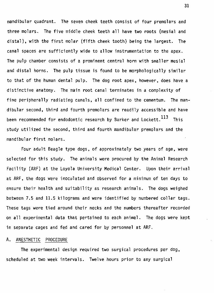
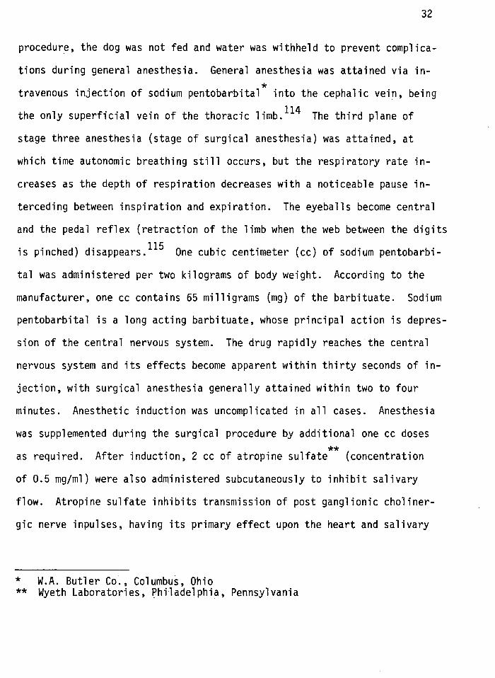
B. RADIOGRAPHIC PROCEDURE
Preoperative radiographs were taken of the experimental and control
teeth to evaluate canal configuration, patency and preoperative periapical
* anatomy. Kodak ultraspeed single exposure radiographic film packets were
held in place utilizing a hemostat and modeling clay (Fig. 1). X-rays were
taken with a portable hand-held General Electric X-ray generator supplying
60 KVP at 20 ma with an exposure time of 0.2 seconds (Fig. 2). Exposed
radiographs were developed in a portable dark box equipped with rapid de-
** veloper and fixer , allowing viewing of the radiographs within three min-
utes.
C. OPERATIVE PROCEDURE
Due to the xerostomia produced by combination of the general
* Kodak DF-58, Eastman Kodak Company, Rochester, New York ** Insta-Neg and Insta-Fix, Micro-Copy, Buffalo, New York
anesthesia and the atropine sulfate, a rubber dam was not required. The
teeth operated on were isolated with 4 x 4 inch cotton gauze pads, both
buccally and lingually.
34
Eight teeth per animal were selected for root canal therapy, divided
into paired samples: four experimental and four control teeth, each quadrant
containing two experimental and two control teeth respectively, being ad
jacently alternated (Table 1). Both control and experimental teeth were
treated identically during the first experimental session, with the excep
tion of one control tooth per animal. A single canal in a randomly selec
ted control tooth in each animal was instrumented and left unobliterated,
sealing the access as will be discussed later. This was done to determine
the effects of instrumentation alone on the periapical tissue. A control
involving the instrumentation of a tooth without obliteration and its direct
exposure to the oral environment was not provided for since this has been
adequately documented in the literature as causing formation of periapical
pathosis. 116
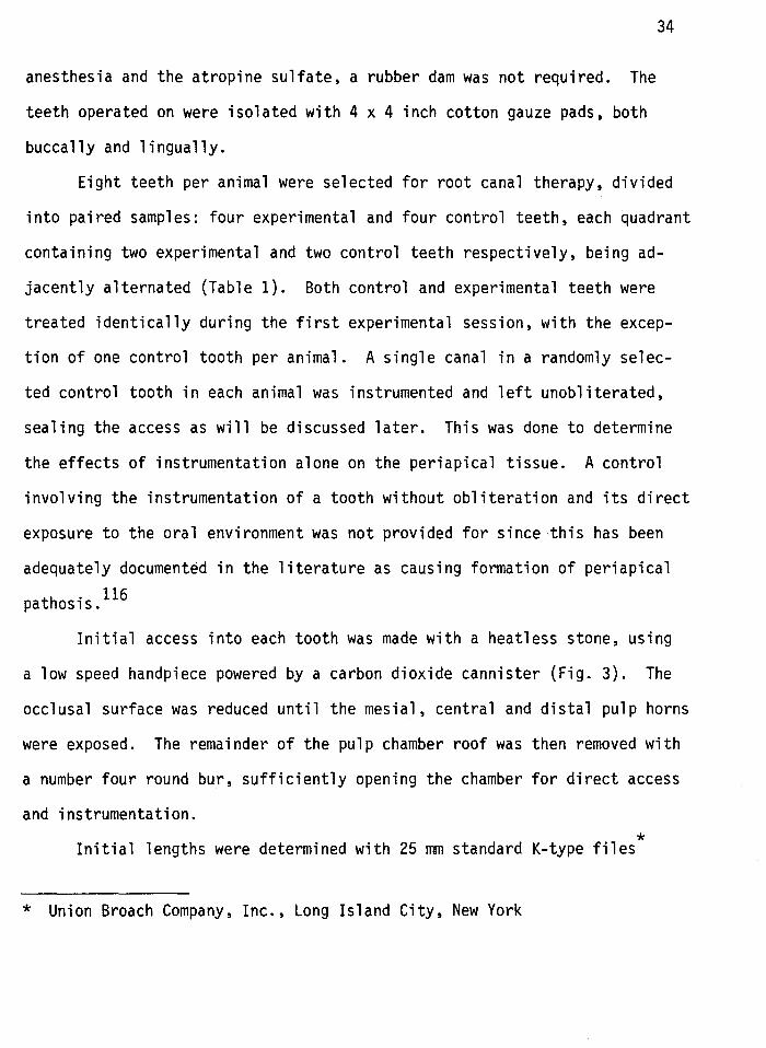
Initial access into each tooth was made with a heatless stone, using
a low speed handpiece powered by a carbon dioxide cannister (Fig. 3). The
occlusal surface was reduced until the mesial, central and distal pulp horns
were exposed. The remainder of the pulp chamber roof was then removed with
a number four round bur, sufficiently opening the chamber for direct access
and instrumentation.
* Initial lengths were determined with 25 mm standard K-type files
* Union Broach Company, Inc., Long Island City, New York

35
equipped with silicone stops. The files were inserted into each canal with
a vaiven motion until a definite apical stop was reached. This apical po
sition was easily perceived tactilely due to the unique apical morphology
present in the dog root apex. Radiographs were taken to confirm the proper
apical position of the initial file lengths. The canals were then prepared
using a circumferential filing action, aided by copious amounts of 5.25%
sodium hypochlorite irrigation as originally recommended by Grossman. 117
Master apical file (MAF) lengths were again verified radiographically (Fig.
4). Upon completion of the cleansing and shaping process, each canal was
thoroughly irrigated and then dried with paper points.
General anesthesia always carries a risk factor in that the vital
functions of the subject may not respond favorably to the induction process.
To minimize the potential risks, it was decided to perform one-appointment
endodontic therapy and complete the root canal treatment in one operative
session. Soltanoff ,118 Oliet119 and Pekruhn,120 evaluating the incidence
of failure following single-visit endodontics, found comparable failure
rates between single and multiple-visit cases.
Each canal, with the exception of one control canal per animal, was
immediately obliterated following biomechanical preparation. Non-staining
* Proco-Sol sealer (primarily a zinc oxide and eugenol sealer) was introduced
along the canal walls with a K-type file one size smaller than the MAF and
** with the master cone. The canals were obliterated with Kerr gutta-percha
* Star Dental, Valley Forge, Pennsylvania ** Sybron/Kerr, Romulus, Michigan

36
performed with a lateral condensation technique. Post room was prepared
in each canal by warming a hand Luks plugger (Fig. 5), retaining approxi
mately three millimeters of gutta-percha to seal the apex of each canal.
Kwan and Harrington121 have shown that immediate post room preparation with
a heated instrument, following obliteration, had no significant effect on
the apical seal. Neagley, 122 investigating the effect of dowel space pre-
paration when varying amounts of filling material remained sealing the
apical region, demonstrated that laterally condensed gutta-percha showed no
trend toward increased leakage over the control population, even when dowel
space was made to a depth of within 4 mm from the apex. Successive K-type
files were introduced to clear the post room space until a size 100 file
was reached. A dry cotton pellet was placed in the chamber and an !RM tern-
* porary inserted. The experimental animal was then returned to its cage.
The second operative session was performed exactly two weeks after
the first surgical session. This time span was required to allow the Proco
Sol sealer sufficient time to set and more accurately reproduce a true clin
ical situation. The experimental animal was again anesthesized following
the same induction technique. The !RM temporary fillings were removed from
both the control and experimental teeth. Control teeth were sealed with a
** dry cotton pellet and an occlusal silver amalgam while the experimental
teeth were allowed to remain open to the oral environment (Fig. 6).
* !RM, L.W. Caulk Company, Milford, Delaware ** T.vtin, S.S. White, Philadelphia, Pennsylvania

37
D. SACRIFICE PROCEDURE
Three dogs were sacrificed after six months and one dog after one
* year through intravenous injection of Beuthanasia-D . The active ingredi-
ents of this preparation are sodium pentobarbital (195 mg/ml) and sodium
phenytoin (25 mg/ml), with a recommended dosage of 1 ml/2 kg. The body of
the mandible was quickly dissected, the segments sectioned with a reciprocat
ing surgical saw and irrmediately submerged into separately labelled jars con
taining approximately 500 cc of 10% neutral buffered formalin solution for
21 days. After an initial period of fixation, the buccal and lingual corti
cal plates were reduced with an acrylic bur in a lowspeed handpiece. The
specimens were then replaced in 10% neutral buffered formalin for an addi
tional 21 days after which time they were rinsed for twenty-four hours under
running water.
E. HISTOLOGIC PREPARATION AND EVALUATION
The specimens were decalcified by placing 5% formic acid in each of
the labelled jars. After sufficient decalcification had occurred, the spe
cimens were further trimmed with a razor blade into blocks containing indi
vidual roots and their associated periradicular tissue. These specimens
were randomly coded and recorded, wrapped in a 2 x 2 inch gauze along with
the corresponding label and immersed in a large container of 5% formic acid
for further decalcification.
Following decalcification,·the blocks were rinsed in running water
for six hours and placed in increasing concentrations of alcohol over a
* Burns-Biotech Laboratory, Chromalloy Pharmaceutical Inc., Oakland, CA.

38
two day period. The blocks were cleared in xylol and embedded in paraffin.
The block segments were cut parallel with the long axis of the tooth in
sections of six microns with each twentieth section mounted on a slide, de-
paraffinated and stained with hematoxylin and eosin for light microscopic
examination. A histologic evaluation of the periapical regions of the ex
perimental and control teeth was then conducted.
Each histologic section was examined under light microscopy by three
evaluators, assessing the degree of periapical inflammation in a manner sim
ilar to that used by Guttuso123 , Rappaport et. al. 124 and Deemer and Tsak
nis.125 The periapical inflammatory response was arbitrarily classified as
either normal (score=O), minimal (score=l), mild (score=2), moderate (score
=3) or severe (score=4) depending on the number and type of inflammatory
cells in the periapical region, the presence or absence of a fibrous connec-
tive tissue capsule and the extent of vascularity and osteolytic activity.
Photomicrographs of the varying degrees of inflammation were reviewed by
each evaluator, prior to examining the slides to establish interrater re-
liability (Fig. 7,9,10,11 and 12).
F. RADIOGRAPHIC EVALUATION
Preoperative and postoperative radiographs of both experimental and
control teeth were projected on a viewing screen and compared by three
evaluators concurrently and a consensus reached as to whether the periap
ical region had remained normal radiographically (-) or had developed a
periapical radiolucency (+).

39
G. STATISTICAL ANALYSIS
Following microscopic observation by each of the three evaluators, a
mean periapical histologic score was calculated for each root of each tooth.
These mean scores were then combined to determine the mean experimental and
mean control values per dog for comparison. The periapical histologic score
of the single unfilled control root per dog was not included in the compu
tation of the control value calculated for each dog. The mean experimental,
mean control and mean unfilled control values were also computed for all
four dogs. Due to the small sample size, descriptive statistics were em
ployed to evaluate the results.
A contingency table was constructed to determine whether a relation
ship existed between the radiographic images and the degree of inflammation
noted histologically.
CHAPTER IV
RESULTS
All the animals were reexamined at varying intervals from the time of
operation to the time of sacrifice, and remained in good physical condition.
Neither the experimental procedure nor the anesthesia seemed to create any
adverse effects.
Preoperatively, all dogs were examined and found to have intact and
caries free teeth except for dog #4499, which was missing the mandibular left
second premolar. Periodontal characteristics were considered within-normal
limits and noncontributory to the experimental results.
Radiographic evaluation preoperatively, revealed a normal bony pattern
with no evidence of any periapical pathology. All the obliterated canals ap
peared adequately filled upon radiographic evaluation, with three to four
millimeters of gutta-percha and sealer. Due to the unique apical anatomic
structure, all canals were filled to the apical terminus, with no instances
of short or overextended filling material.
Dog #4450
This dog was a male weighing 10.5 kg. The experimental period was one
year. The experimental teeth were the mandibular left second (LL2) and
fourth (LL4) premolars and the right third premolar (LR3) and first molar
(LRl). The control teeth were the mandibular left third premolar (LL3) and
first molar (Lll) and the right second (LR2) and fourth (LR4) premolars.
40

The mesial root of LR4 (LR4-mesial) served as the unfilled control root
(Table 1).
Clinical Findings
There was no clinical evidence of periapical pathosis.
Radiographic Findings
41
A slight periapical radiolucency with poorly defined borders was
found in the apical region of Lll-mesial, a moderately sized periapical
radiolucency with poorly defined borders was located at the apex of LL4-
distal and a large periapical rarefaction was associated with LR4-mesial
having a well defined border (Table 3).
Histologic Findings
Two control teeth, LL3-distal and Lll-distal did not show any inflam
matory changes in the periapical region. Two control teeth, LR3-mesial and
Lll-mesial displayed minimal inflammatory changes while LR2 demonstrated a
mild periapical inflammatory reaction. Control tooth LR4-distal, which was
filled with gutta-percha and sealer, showed no inflammatory changes while
the unfilled control root, LR4-mesial, had a moderate inflammatory reaction.
Experimental teeth LL2-distal and LR3 all appeared normal histologically
while experimental tooth LRl displayed minimal inflammation of the periap
ical region. Experimental teeth LL4 and LL2-mesial showed a mild degree
of periapical inflammation (Table 3).
Dog #4451
This dog was a male, weighing 11.5 kg. The experimental period was
six months. The experimental teeth were the mandibular left third premolar

42
(LL3), left first molar (LLl) and the right second and fourth premolars
(LR2 and LR4 respectively). The control teeth were the mandibular left
second (LL2) and fourth (LL4) premolars and the right third premolar (LR3)
and first molar (LRl). The mesial root of control tooth LL2 (LL2-mesial)
was left unfilled after cleansing and shaping (Table 1).
Clinical Findings
There was no clinical evidence of periapical pathosis.
Radiographic Findings
Slight periapical radiolucencies were noted in the apical regions of
LL3, LL4-mesial, Lll, LR3, LR4 and LRl with either poorly defined borders
or fair definition to the periapical radiolucencies (Table 3).
Histologic Findings
The periapical region of control tooth LL4-mesial had a minimal peri
apical inflammatory response histologically, while LL4-distal had a severe
inflammatory periapical reaction. Control tooth LR3 demonstrated moderate
periapical inflammation, while LRl showed moderate to severe inflammatory
changes. Control tooth LL2-distal showed a moderate periapical response in
the obliterated root, while the unfilled control root (LL2-mesial) displayed
mild inflammatory changes. Experimental tooth LR4 had a severe inflammatory
reaction histologically while Lll demonstrated a moderate inflammatory re
sponse. Tooth LR2-mesial had mild to moderate inflammatory changes, while
LR2-distal showed minimal histologic changes. Tooth LL3 evidenced mild in
flammatory changes microscopically (Table 3).
43 Dog #4582
This dog was a male weighing 10.0 kg. The experimental period was
six months. The experimental teeth were the mandibular left second (LL2)
and fourth (LL4) premolars and the right third premolar (LR3) and first
molar (LRl). Control teeth included the mandibular left third premolar
(LL3) and first molar (Lll) and the mandibular right second (LR2) and fourth
(LR4) premolars. The mesial root of LL3 (LL3-mesial) remained unfilled fol
lowing the cleansing and shaping procedure (Table 1).
Clinical Findings
There was no clinical evidence of periapical pathosis.
Radiographic Findings
Slight periapical radiolucencies were found associated with the root
apices of LRl, LR4-distal, LL4-mesial and LL3-mesial whose borders were
either of fair or poor definition (Table 3).
Histologic Findings
All experimental teeth, with the exception of LL2-mesial and LR3-
distal, demonstrated signs of minimal periapical inflammation. LL2-mesial
and LR3-distal showed signs of a normal periapical histologic pattern. Con
trol tooth LR2 was also normal histologically. Control teeth Lll and LR4
had minimal to mild inflanmatory changes evident periapically. Control
tooth LL3-distal demonstrated a normal periapical pattern, while the un
filled control root, LL3-mesial, had minimal to mild inflammatory changes
(Table 3).
Dog #4499
This male beagle weighed 7.5 kg. The experimental period was six
44
months. The mandibular left second premolar was found to be missing and
thereby excluded from experimentation. The experimental teeth were the
mandibular left third premolar (LL3) and first molar (Lll) and the mandibu
lar right second (LR2) and fourth (LR4) premolars. Control teeth were the
mandibular left fourth premolar (LL4) and the mandibular right third pre
molar (LR3) and first molar (LRl). The mesial root of LRl was chosen as
the unfilled control root (Table 1).
Clinical Findings
There was no clinical evidence of periapical pathosis.
Radiographic Findings
Moderately sized periapical radiolucencies were found associated with
the root apices of LR4 and LL4, both exhibiting fairly well defined borders.
Apices of Lll-mesial, LRl-mesial and LR3-distal all showed evidence of
slight periapical radiolucencies with either poor or fair definition to the
radiolucent borders (Table 3).
Histologic Findings
The periapical region of experimental tooth LR2-distal appeared normal
histologically. Experimental teeth LL3, LR2-mesial and LR4-distal had min
imal periapical inflammation present histologically, while tooth LR4-mesial
demonstrated a minimal to mild periapical inflammatory reaction. Experi
mental tooth Lll had a mild to moderate inflammatory response in the periap
ical region of the distal root (Lll-distal), while the mesial root (Lll
mesial) displayed a mild inflammatory reaction. Control tooth LR3 showed
minimal inflammatory changes while LR4 showed a mild to moderate
45
inflammatory reaction. Control tooth LRl-distal, which was filled with
gutta-percha and sealer, evidenced only minimal inflammatory changes, while
the unfilled control root, LRl-mesial, appeared normal histologically. Con
trol tooth LL2 was missing (Table 3).
Statistical Results: Histologic
Histologic scores from all three evaluators are listed in Table 2.
The graded inflammatory response for individual roots ranged in value from
0 to 4 in both experimental and control teeth. The histologic values for
each experimental root were averaged per dog and ranged from 0.66 to 2.70
with two intermediate values of 1.08 and a mean score of 1.38 for all four
dogs (Table 4). Histologic values were averaged for all control roots per
dog, filled with gutta-percha and sealer, ranging from 0.66 to 3.09 with
intermediate scores of 0.71 and 1.20 and a mean score of 1.43 for all four
dogs (Table 4). The average histologic score for the single unfilled con
trol root per dog ranged from 0 to 3.33 with an overall mean value of 1.66
(Table 4). Results for each individual dog and averaged values for all four
dogs were plotted on a bar graph (Table 5) for comparative purposes.
Statistical Results: Radiographic
A contingency table was constructed (Table 6), plotting the 62 histo
logic specimens according to the presence or absence of radiographic peri
apical changes and the degree of histologic variation. Periapical radiolu
cencies developed in 26 (42%) of the 62 specimens, while 36 (58%) root api
ces appeared normal radiographically. Only one of the root apices associ
ated with a periapical radiolucency showed a normal histologic pattern,

46
while two of the apices demonstrated minimal inflammatory changes. Twenty
three of the twenty-six specimens with periapical radiolucencies showed a
mild to severe inflammatory reaction histologically. Thirty-six specimens
had no evidence of any periapical pathology radiographically of which 12
(33.3%) specimens appeared normal histologically. Another 22 (61.1%) spe
cimens had only a minimal to mild inflammatory reaction, while 2 (5.6%)
specimens showed moderate to severe inflammatory changes.
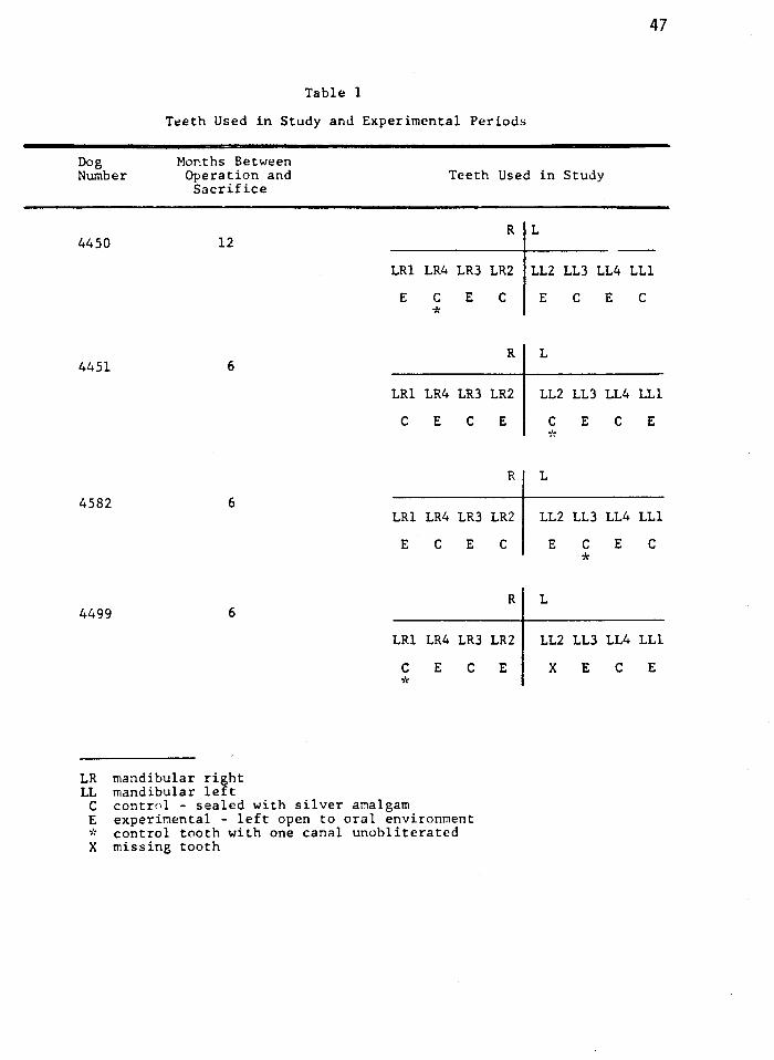
Table 1
Tt?eth Used in Study anJ Experimental Periods
Dog Months Between Number Operation and
Sacrifice
4450 12
4451 6
4582 6
4499 6
LR mandibular right LL mandibular left
LRl
E
LRl
c
LRl
E
LRl
c *
C control - sealed with silver amalgam
Teeth Used
R
LR4 LR3 LR2
c E c *
R
LR4 LR3 LR2
E c E
R
LR4 LR3 LR2
c E c
R
LR4 LR3 LR2
E c E
E experimental - left open to oral environment * control tooth with one canal unobliterated X missing tooth
47
in Study
L
LL2 LL3 LL4 LLl
E c E c
L
LL2 LL3 LL4 LLl
c E c E
*
L
LL2 LL3 LL4 LLl
E c E c *
L
LL2 LL3 LL4 LLl
x E c E

48
Tahlc 2
Histologic Scores for Evaluator #l, Evaluator #2 and Evaluator #3
Periapical Reaction - Hi~tologic
Dog Control/ Evaluator i•l Evaluator /f2 Evaluator 113 Number Tooth Experimental Mesial
4450 LL2 E LL3 c LL4 E LLl c LRl E LR4 C•'< LR3 E LR2 c
4451 LL2 C>'< LL3 E LL4 c LLl E
LRl c LR4 E LR3 c LR2 E
4582 LL2 E LL3 C>'< LL4 E LLl c
LRl E LR4 c LR3 E LR2 c
4499 LL2 c LL3 E LL4 c LLl E
LRl Ci<
LR4 E LR3 c LR2 E
C control E experimental * control with unfilled root X missing tooth
2 0 1 0
0 3>'< 0 2
l >'< 1 0 3
3 4 3 2
0 l>'< 0 1
0 0 0 0
x 0 2 1
Q>'<"
1 0 0
Distal Mesial Distal Hesial
0 0 1 0
0 0 0 3
3 2 4 3
3 4 3 0
0 0 0 1
1 1 0 0
x 0 2 2
0 1 0 0
2 0 3 0 0 1 2 2 4 0 0 1
1 1 3 4>'< 0 )"'• .. 0 0 0 2 2 2
2>'< 3 2>~
2 2 4 0 4 3 3 2 4
4 4 4 4 4 4 3 3 4 2 0 4
0 0 0 l>'< 0 )"'• .. 0 0 3 1 0 3
1 1 2 0 2 1 0 0 1 0 0 0
x x x 0 0 2 2 3 2 1 3 4
O* 0 O* 2 1 2 0 0 1 0 0 1
0 no inflammation 1 minimal inflam.~ation 2 mild inflar.imation 3 moderate inflammation 4 severe inflammation
Distal
0 0 2 0
2 0 0 2
3 3 4 3
4 4 3 1
2 0 3 2
2 2 0 0
x 1 3 3
1 1 2 0
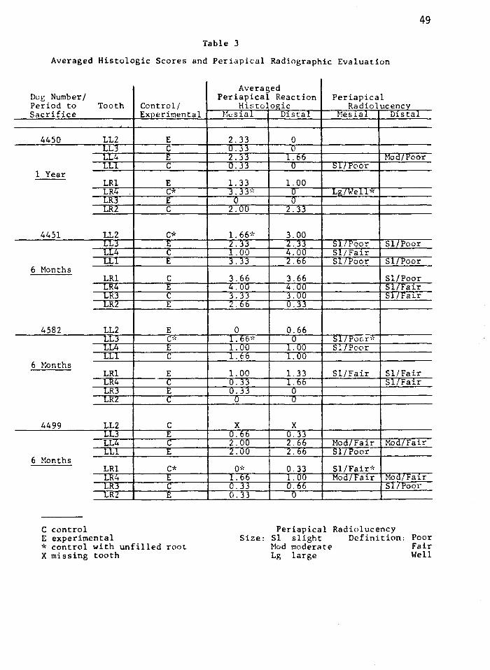
49
Table 3
Averaged Histologic Scores and Periapical Radiographic Evaluation
Dog Number I Period to Tooth Control I Sacrifice Experimental
4450 LL2 E LLJ c LL~ E LLl c
1 Year LRl E LR4 C* LR3 E LR:Z c
4451 LL2 C* LLJ .t;
LL4 c LLl E
6 Months LRl c LR4 E Ll:U c LRl E
4582 LL2 E LL3 Co'< LL4 E LL! c
6 Months LRl E LH.4 c LRJ .t;
LRZ c
4499 LL2 c LLJ E LL4 c LLl E
6 Months LR! C* LR4 E LR3 c LR:Z E
C control E experimental * control with unfilled root X missing tooth
Averaged Periapical Reaction Periapical
Histulogic Radiolucencv M12s1al Distal Mes1al Distal
2.33 0 O.JJ 0 2. jj l. bb Mod/ .l:'oor 0.33 u ':::>!/ .l:'OOr
1. 33 1. 00 3. 33>< u Lg/Well><
0 0 2.00 2.33
1. 66* 3.00 2.JJ l.JJ SL/Poor Sl/Poor 1.00 4.00 Sl/Fair 3. 33 2.66 Sl/Poor Sl/Poor
3.66 3.66 Sl/Poor 4.00 4.00 Si/Fair 3.JJ J.UU Sl/Fai.r 2.66 U.JJ
0 0.66 1. 66°~ 0 Sl/Poor" l.VV l. uu 51/Poor 1. 66 l. uo
1. 00 1. 33 Sl/Fair Sl/Fair U.JJ l. 06 Sl/Fair 0.33 u
0 0
x x 0.bb 0.33 2.UU 2.bb Mod/ Fair Mod/Fair 2.00 2.66 Sl/Poor
Qi'< 0.33 SI/Fair'< 1. 66 1. 00 Modtrair Mod/Fair O.JJ 0.66 Sl/Poor 0.33 0
Periapical Radiolucency Size: Sl slight Definition:
Mod moderate Lg large
Poor Fair Well
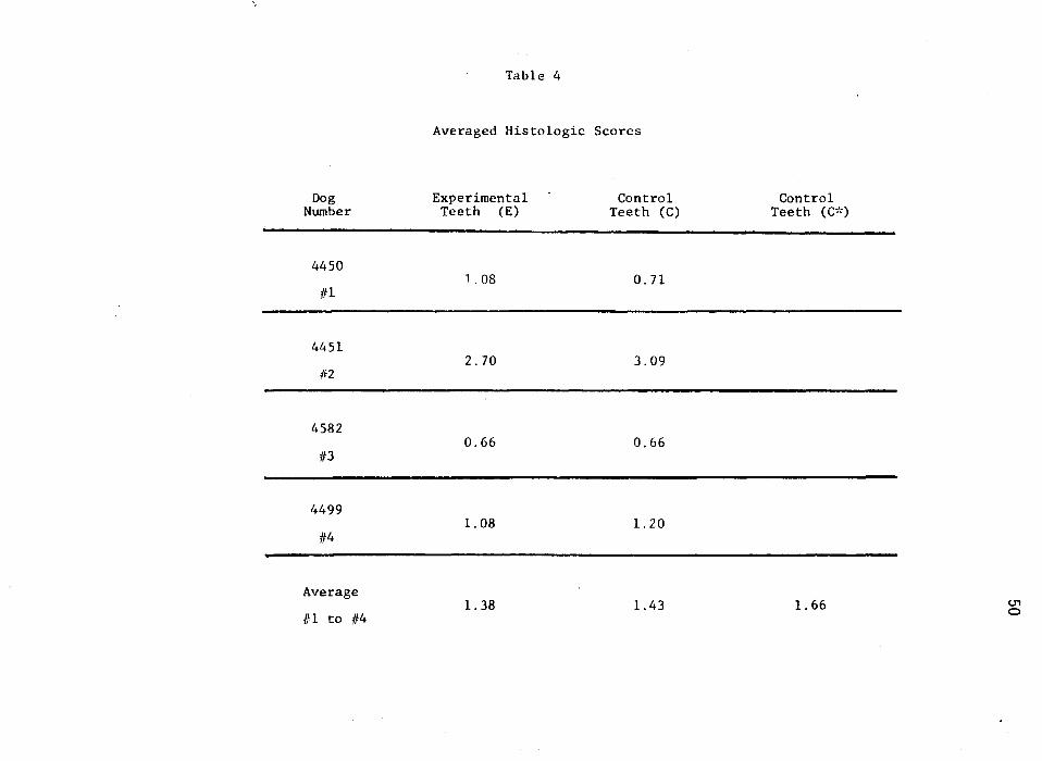
\
Table 4
Averaged Histologic Scores
Dog Experimental Control Control Number Teeth (E) Teeth (C) Teeth (C>~)
4450 1. 08 0. 71
Ill
-4451
2. 70 3.09 #2
-4582
0.66 0.66 113
-/~499
1. 08 1. 20 /f 4
-Average
1. 38 1.43 1. 66 U1
/fl to /14 0
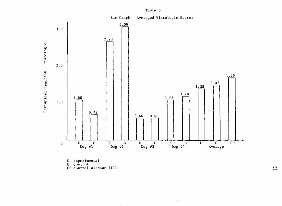
Table 5
Bar Graph - Averaged Histologic Scores
3.09 ~
3.0
2.:2.Q u ..... 00 0 ..... 0 +.J Ul .....
:::: 2.0
I
c: 0 ..... 1.:..§_? +.J u <ll 1.43 Cll
IZ 1. 38 --.....
Ill 1. 20 u .....
1. 0 0. <':!
l~ 1. 08 ~
.--..... I-< Cll
p.. 0. 71 - 0.66 0.66 - ~
0 E c E c E c E c E c c;~
Dog /tl Dog 1f2 Dog 1f3 Dog 1/4 Average
E experimental c control C~< control without fill <.Tl
.......

Table 6
Contingency Table - Radiographic versus Histologic
Periapical Reaction - Histologic
0 0.01-0.99 1. 00-1. 99 2.00-2.99 3.00-3.99
Periapical Radiolucency 1 (3. 8) 2 (7.7) 9 (34. 6) 6 (23.1) 6 (23.1)
(+)
No Periapical Radiolucency 12 (33. 3) 10 (27.7) 6 (16. 7) 6 (16.7) 1 (2.8)
(-)
13 (20.9) 12 (19.4) 15 (24.2) 12 (19.4) 7 (11.3)
4
2 (7.7)
1 (2.8)
3 (4.8)
Total
26 (100)
3f. (100)
62 (100)
U'1 N
CHAPTER V
DISCUSSION
Success in endodontic therapy is heavily dependent upon as thorough a
debridement process as possible, followed by obliteration of the root canal
space to attain an apical seal. Failure of this seal, for whatever reason,
may result in periapical inflammation, infection, symptomology or even loss
of the tooth.
The literature is replete with studies substantiating the presence of
marginal percolation, microleakage, fluid exchange or diffusion and capil
lary penetration with a majority of presently used restorative materials. 126
The same phenomenon has been investigated as applied to the apical seal
achieved during root canal therapy. Depending upon the article reviewed, dif
ferent sealers will have different properties and sealing abilities attrib
uted to them, with no one particular sealer repeatedly selected as ideal.
The only point of general consensus appears to be that some form of sealer
is required to effect an apical seal. As stated by Marshall and Massler,
"the skil 1 of the operator is perhaps more important to successful obturati on
than the materials used 11•91
Periapical problems can still arise, even through our best efforts.
Breakdown of, or failure to even attain an apical seal would open a pathway
for fluid and particle interchange between the periapical region and the
canal interstices. Ingle speculated that a constantly circulating periapical
53
54
exudate could percolate through apical defects into existing intracanal
interstices. Fluid and fluid products, trapped within these canal spaces,
could undergo degradation and slowly diffuse back into the apical region.
These enzymatic breakdown products might act as periapical irritants, caus
ing persistence of an existing periapical lesion or formation of new path
ology .1 This sequence of events, proposed by Ingle, seems to parallel
Rickert and Dixon's Hollow Tube Theory, later disproved by Torneck using
sterile polyethylene hollow tube implants. Torneck demonstrated that even
though a serous type of fluid may fill the lumen of hollow tube implants,
their fluid would not induce inflammatory changes at the open ends of the
tubes. 65
Grossman alludes to the possibility of anachoresis in that microorgan
isms present during a transient bacteremia might be transported to an in
flamed periapical region and become lodged within unfilled portions of the
canal system. 6 Of equal or greater importance, as discussed by Shovelton,
are those bacteria remaining within the confines of the canal spaces and
dentinal tubules following the cleansing and shaping process.B9 Nicholls
states that it is feasible for organisms remaining in the deeper parts of
the dentinal tubules to grow into unfilled canal spaces.BB In addition,
numerous studies have shown the inability of the debridement process to
totally eliminate pulpal remnants. The combination of bacteria, bacterial
products, vital and necrotic pulpal debris, all harbored within the root
canal system can suddenly become a significant factor in whether or not an
endodontic case succeeds. Failure of the apical seal may allow fluid per-
colation into unfilled canal interstices with subsequent diffusion of

55
bacteria and bacterial products and tissue products into the periapical re
gion. Conceding that a hermetic seal may not be possible, Ainley postu
lated the existence of a 11critical leakage factor 11 beyond which biologic
repair would not occur. 101 Realizing the infinite number of variables in
volved in successful root canal therapy, the amount of apical leakage that
would have a detrimental effect on healing is purely speculative. The fact
that seemingly poorly performed root canal therapy does succeed periodically,
can be viewed as a tribute to the healing capacities of the human organism.
Another potential source of periradicular and/or pulpal inflammation
is the existence of lateral canals. Rubach and Mitchell 127 and Seltzer et.
al., 128 investigating human teeth, demonstrated a profusion of lateral ca-
nals in the roots and furcation regions of posterior teeth and occasionally
in anterior teeth. A complete absence of lateral canals was noted in the
present investigation. Histologically, the tissue within lateral canals has
been found to be confluent with that of pulp tissue, consisting of capil-
laries, pulp cells, ground substance and fibers. Simring and Goldberg sug-
gested that lateral canals may act as channels for pathogenic materials to
pass either pulpally or towards the periodontium. 129 Their significance as
channels of communication may outweigh their importance as harbors of ne
crotic material. Severely inflamed or necrotic pulp tissue and their toxic
products, may emanate through a lateral canal, causing an inflammatory re
sponse of the periodontal ligament. Conversely, deep periodontal lesions
may expose lateral canals, thereby interfering with the nutritional supply
of the pulp. Inflammation, degeneration or even necrosis of an otherwise
normal pulp might result.
56
Due to the form and structure of the root canal system, deliberate
debridement and obliteration of a lateral canal is essentially impossible.
Weine states that a higher percentage of root canal failures would be seen
if the cleaning and filling of lateral canals were critical in attaining 130 w . f th h . . success. eine ur er warns tat improper sealing of a prepared post
room space, or leaving the area vacant for too long, may invite breakdown
of the periodontium if a lateral canal of significant size is present. 130
Minimizing endodontic failures by assuring the integrity of the
apical seal became a focal point for endodontic research. Numerous studies
evaluated the apical seal from both an apical and coronal approach, but
considered breakdown of the apical seal as critical only from the apical
direction in clinical situations. Loss of the occlusal seal seemed incon-
sequential to the clinical prognosis following completion of root canal
therapy. Bhaskar and Rappaport noted in an eighteen month histologic study
on dogs that once a canal space had been debrided and an effective occlusal
seal placed, an existing periapical radiolucency would cease to enlarge. 116
Marshall and Massler suspected that a good occlusal seal would improve the
apical marginal seal of root canal fillings. 91 Results illustrated, how
ever, that the degree of apical sealing ability was not altered by an ef-
fective occlusal seal. They concluded that a good occlusal seal was essen
tial only during treatment, prior to obliteration.
The rationale of having an occlusal seal during root canal therapy
is understood, but the importance of that seal after treatment must be ex-
amined. Marshall and Massler demonstrated that a good occlusal seal would
not prevent periapical leakage, therefore, it seems reasonable to assume

57
that a good periapical seal would not prevent occlusal leakage. Ainley
stated that "the method of obturating the apical 5 mm may not be critical
for biologic success; rather, proper debridement and the sealing off of
the periapical tissues from the oral fluids are probably more important 11•101
Loss of the occlusal seal could possibly lead to periapical breakdown. When
subjected to periapical investigative conditions, research has shown the
endodontic filling to be as effective from the coronal aspect as it is
apically in sealing ability. The types of contaminants introduced, however,
from the oral cavity vary from those found periapically. The purpose of
this study was to determine if loss of a coronal seal, with the introduction
of saliva and oral organisms, would adversely affect the apical seal and
eventual prognosis of a tooth following root canal therapy.
A histologic approach was selected to determine whether the periapical
tissues would undergo any inflammatory changes following loss of an occlusal
seal after root canal therapy. Sinai et. al. 131 evaluated the histologic
reaction of apical pulpal and periapical tissues after partial pulpectomies
were performed on 24 Rhesus monkey teeth. An acute inflammatory response
was noted for a period of up to one month, followed by a repair process.
A total pulpectomy, even in cases of normal pulpal vitality, will generate
a certain degree of periapical inflammation. To allow the initial inflam
matory reaction to subside, and sufficient clinical exposure for the exper
imental teeth to the oral environment, research periods of six months and
one year were chosen.
Histologic grading was performed by three different evaluators using
a scale of O to 4 to rank the degree of periapical inflammation. The

58
scores corresponded to inflammatory responses of none, minimal, mild, mod
erate and severe respectively. No definitive boundaries between these
categories exist, but each evaluator viewed photo-micrographs of the vary
ing degrees of inflammation, prior to examining the slides, to establish
interrater reliability. The microscopic slides were then compared and
ranked on a relative basis.
To minimize experimental bias, all histologic specimens were randomly
coded so microscopic evaluation was performed without insight as to whether
the sample was an experimental or control tooth. Results were tabulated
and matched with their code numbers only after all three evaluators had
viewed the specimens independently.
Results of this research project indicate that loss of an occlusal
seal, following root canal therapy, should have little if any effect on the
periapical region for a period of six months to one year. Although minor
differences existed in the degree of histologically reported periapical in
flammation between experimental and control teeth in each dog, averaged
values for all four animals (histologic experimental value=l.38 vs. histo
logic control value=l.43) demonstrated an insignificant variance. These
observations substantiate the ability of an endodontic obliteration tech
nique to seal in both a coronal and apical direction. There appeared, how
ever, to be an increased tendency towards periapical inflammation in those
specimens left unobliterated, even though the occlusal access was sealed
(histologic unobliterated control value=l.66). This observation gives cre
dence to the importance of obturating the root canal space after the de
bri dement process has been completed. Placement of an occlusal seal, in
59
itself, after cleansing and shaping, will not produce a situation conducive
to permanent healing.
Histologic evaluation of the specimens was complicated by the fact
that cell nuclei did not stain well with hematoxylin. Resected sections of
the mandible were immediately placed in 10% formalin after sacrifice, but
the size of the specimens and density of the bone may have prohibited prop
er fixation. Radiographs were taken of the resected mandibles several days
following sacrifice, after which the cortical plates were reduced prior to
decalcification. During this time period, cellular detail may have been
damaged due to the slow diffusion of the fixative. Smaller sections of the
mandible were not made until much later when sufficient decalcification had
occurred. Another factor possibly affecting the cellular staining was the
lengthy decalcification process. Optimally, a weak organic acid is used to
decalcify the hard tissues before sectioning and staining. The specimens
were immersed in separate jars containing 5% formic acid for five to six
months. The volume of formic acid limited the decalcification process, pro
longing the interval until separate sections could be made of individual
roots and their associated periapical tissues. Excessive exposure to the
formic acid may have damaged the intracellular structures, preventing proper
uptake of the dye. In retrospect, better results may have been obtained
with a more rapid removal of the cortical plates to reduce the amount of cal
cified structures and usage of 10% formic acid in larger quantities to speed
the decalcification process. Regardless of these problems, inflammatory
cells were easily seen microscopically, but identification of individual
cell types normally seen in a periapical inflammatory reaction was not

60
always possible.
Reaction of the periapical region to loss of the occlusal seal was
also evaluated radiographically. Preoperative radiographs were compared to
those taken post-sacrifice to determine whether periapical radiographic
changes had occurred. Due to the high inconsistency found by Goldman et.
al.s3 in individual interpretation of radiographic images, three evaluators
viewed projected preoperative and post-sacrifice radiographs concurrently,
reaching a consensus of opinion. A contingency table was then constructed
to examine the relationship between the radiographic image and the degree of
histologically noted inflammation, if any. Results indicated the existence
of a trend towards development of periapical rarefactions in instances of
more severe periapical inflammation and a lesser tendency for radiographic
change in cases of minimal inflammatory reaction. Histologically, mild to
severe periapical inflammatory reactions were found in 88.S% of the root
apices associated with periapical rarefactions, while 77.8% of the apices
with normal radiographic patterns had no periapical inflammation or minimal
to mild inflammatory responses. Separate contingency tables for control and
experimental teeth respectively were not included due to a statistical sim-
ilarity with the combined table.
Bender et. al.SS proposed the lack of correlation between histologic
findings and negative radiographic patterns. They demonstrated the frequent
existence of histologically verified chronic inflammation despite a normal
radiographic image. Conversely, teeth with radiolucent areas had a definite
histologic correlation, revealing chronic inflammatory patterns. They also
concluded that periapical radiolucencies would always appear smaller than
61
the histologic lesion due to the necessity of the lesion to encroach upon
the junctional area of cancellous and cortical bone for the rarefaction to
manifest itself radiographically.
This study substantiates the findings of Bender et. al. by confirming
the tendency for more severe periapical histologic reactions to elicit peri
apical radiographic changes. The larger and more severe the chronic inflam
matory reaction, the greater the anticipated periapical bony destruction.
Once this bony breakdown encroaches upon the cortical plate, radiographic
changes would become evident. A lack of radiographic pathology did not
guarantee the existence of a normal histologic periapical pattern, although
the tendency was towards a minimal to mild response if inflammatory changes
were present. These results support the evidence of milder inflammatory re
actions producing diminished periapical responses, insufficient to elicit
periapical radiographic changes.
Statistical analysis in this study was limited to descriptive statis
tics due to the small sample size. In retrospect, an experimental design
using a minimum of five animals would have facilitated data analysis by al
lowing the application of the Wilcoxon signed rank test. It is unlikely,
however, that statistical results would have varied and the generalizability
of the study would still remain limited.
One aspect of contention is the applicability of the animal model to
that of humans. Although the pulp tissue of dogs is histologically compat
ible with that of humans, the apical anatomy varies immensely. Complexity
of the apical delta prohibits instrumentation of the peripherally radiating
canals, possibly leaving remnants of pulpal tissue. The potential for this

62
tissue to undergo degeneration exists and may partially explain the inci
dence of peri apical inflammatory changes.
Results of this study indicate that breakdown or loss of the occlusal
seal, following endodontic therapy, would not adversely affect the apical
seal for a period of six months to one year. This finding, however, does
not condone neglecting a suspicious or faulty occlusal restoration or tem
porary filling following root canal therapy. Even successful endodontic
treatment will not prevent a tooth from succumbing to aggressive recurrent
decay.

SUMMARY
1. Loss of an occlusal seal, following root canal therapy, should have
little if any effect on the periapical seal for a period of six months
to one year.
2. Root canal therapy should be completed soon after the debridement pro
cess. An intact occlusal seal will not prevent development of periap
ical pathosis following the cleansing and shaping procedure.
3. A normal periapical radiographic image, following completion of root
canal therapy, does not guarantee the absence of chronic periapical in
flammation histologically.
4. The radiographic presence of a periapical radiolucency is generally as
sociated with more severe chronic inflammatory changes histologically.
5. Loss or failure of an occlusal seal should not be neglected, thereby
avoiding recurrent decay and possible progression to a nonrestorable
situation.
63

REFERENCES
1. Ingle, J.: Endodontics, Philadelphia, Lea and Febiger, 1974.
2. Weine, F.: Endodontic Therapy, 3rd edition, St. Louis, C.V. Mosby Co., 1982.
3. Curson, I.: History and Endodontics. The Dental Practitioner, 15:435, 1965.
4. Tagger, M.: Endodontics: A Review of the Past and Its Present Status. The Alpha Omegan, Sept. :107, 1967.
5. Sharp, G.: The Historical and Biological Aspects of the Pulpless Tooth Question. J. Am. Dent. Assoc., 24:1231, 1937.
6. Grossman, L.: Endodontic Practice, Philadelphia, Lea and Febiger, 1950.
7. Milas, V.: History. Pathways of the Pulp, 2nd edition, by S. Cohen and R.C. Burns, St. Louis, C.V. Mosby Co., 1980.
8. Sampeck, A.: Instruments of Endodontics: Their Manufacture, Use and Abuse. Dental Clinics of North America, ed. by H. Schilder, Philadelphia, W.B. Saunders Co., 1967.
9. Ostrander, F.: The Practice of Endodontics: Past, Present and Future. J. Dent. Education, 31:386, 1967.
10. Ingle, J. and Beveridge, E.: Endodontics, Philadelphia, Lea and Febiger, 1976.
11. Ingle, J. and Levine, M.: The Need for Uniformity of Endodontic Instruments, 'Equipment and Filling Materials. Presented Second International Conference on Endodontics, ed. by L. Grossman, Philadelphia, 1958.
12. Miller, W.: Gangrenous Tooth Pulps as Centers of Infection. Dental Cosmos, 30:213, 1888.
13. Miller, W.: Decomposition of the Contents of the Dentinal Tubules as a Disturbing Factor in the Treatment of Pulpless Teeth. Ohio J. Dent. Sci., 10:288~ 1890.
64
65
14. Rosenow, E. and Billings, F.: Mouth Infection as a Source of Systemic Disease. Presented Section of Stomatology, American Medical Association, reprinted J. Am. Dent.Assoc., 63:2024, 1914.
15. Easlick, K.: An Evaluation of the Effect of Dental Foci of Infection on Health. J. Am. Dent. Assoc., 42:620, 1951.
16. Meyer, F.: The Present Status of Dental Bacteriology. J. Natl. Dent. Assoc., 4:966, 1917.
17. Gunter, J., Appleton, J., Strong, J., Reader, J. and Brooks, J.: Bacteriology of Dental Pulp. J. Dent. Res., 16:310, 1937.
18. Fish, E. and Maclean, I.: The Distribution of Oral Streptococci in the Tissues. Br. Dent. J., 61:336, 1936.
19. Okell, C. and Elliott, S.: Bacteremia and Oral Sepsis. Lancet, 2: 869, 1935.
20. Grossman, L.: Root Canal Therapy, Philadelphia, Lea and Febiger, 1940.
21. Blayney, J.: Present-Day Evaluation of the Pulpless Tooth. J. Am. Dent. Assoc., 23:533, 1936.
22. Hatton, E.: Methods and Fundamentals in the Allied Sciences Essential to Successful Root Canal Surgery. Dental Cosmos, 70:249, 1928.
23. Coolidge, E.: Pulp Pathology and Treatment Problems. J. Am. Dent. Assoc., 15:1623, 1928.
24. Ingle, J.: Root Canal Obturation. J. Am. Dent. Assoc., 53:47, 1956.
25. Hess, W.: The Anatomy of the Root Canals of the Teeth of the Permanent Dentition. Dentition, New York, Wm. Wood and Co., 1925.
26. Fischer, G.: The Present Status of Root Canal Treatment in Relation to the Minute Anatomy of the Root Canals in Man. Dental Cosmos, 54:722, 1912.
27. Mueller, A.: Morphology of Root Canals. J. Am. Dent. Assoc., 23: 1698, 1936.
28. Meyer, W.: Die Anatomie der Wurzelkanale Dargestelt an Mikroskopischen Rekonstruktronsmodellen. Dtsch. Zahnaerztl. Z ., 25: 1064, 1970.

66
29. Skidmore, A. and Bjorndal, A.: A Study of the Root Canals of FortyFive Extracted Mandibular First Molars. Oral Surg., 32:778, 1971.
30. Kuttler, Y.: Microscopic Investigation of Root Apexes. J. Am. Dent. Assoc., 50:544, 1955.
31. Seidler, B.: Technique and Rationale of Filling Root Canals. N.Y. J. Dent., 24:376, 1954.
32. Luks, S. and Bolatin, L.: The Myth of Standardized Root Canal Instruments. N.Y. J. Dent., 43:109, 1973.
33. Penick, E. and Osetek, E.: Intracanal Drugs and Chemicals in Endodontic Therapy. Dental Clinics of North America, ed. by S. Holroyd, Philadelphia, W.B. Saunders Co., 1970.
34. Svec, T. and Harrison, J.: Chemomechanical Removal of Pulpal and Dentinal Debris with Sodium Hypochlorite and Hydrogen Peroxide versus Normal Saline Solution. J. Endodon., 3:49, 1977.
35. Auerbach, M.: Antibiotics versus Instrumentation in Endodontics. N.Y. State Dent. J., 19:225, 1953.
36. Masterton, J.: Chemical Debridement in the Treatment of Infected Pulpless Teeth and Chronic Periapical Abscesses. Dent. Pract., 15:162, 1965.
37. Haga, C.: Microscopic Measurements of Root Canal Preparations Following Instrumentation. Bull. N.W.U. Dent. Grad., Fall: 11, 1967.
38. Guttierrez, J. and Garcia, J.: Microscopic and Macroscopic Investigation on Results of Mechanical Preparation of Root Canals. Oral Surg., 25:108, 1968.
39. Brayton, S., Davis, S. and Goldman, M.: The Morphology of the Prepared Root Canal: A Study Utilizing Injectable Silicone. Oral Surg., 34:642, 1972.
40. Moodnick, R., Dorn, S., Feldman, M., Levey, M. and Borden, B.: Efficacy of Biomechanical Instrumentation: A Scanning Electron Microscopic Study. J. Endodon., 2:261, 1976.
41. Weine, F., Kelly, R. and Lio, P.: The Effect of Preparation Procedures on Original Canal Shape and on Apical Foramen Shape. J. Endodon., 1:255, 1975.
67
42. Walton, R.: Histologic Evaluation of Different Methods of Enlarging the Pulp Canal Space. J. Endodon., 2:304, 1976.
43. McComb, D. and Smith, D.: A Preliminary Scanning Electron Microscopic Study of Root Canals After Endodontic Procedures. J. Endodon., 1:238, 1975.
44. Mizrahi, S., Tucker, J. and Seltzer, S.: A Scanning Electron Microscopy Study of the Efficacy of Various Endodontic Instruments. J. Endodon., 1:324, 1975.
45. Cunningham, W. and Martin, H.: A Scanning Electron Microscope Evaluation of Root Canal Debridement with the Endosonic Ultrasonic Synergistic System. Oral Surg., 53:527, 1982.
46. Langeland, K., Liao, K. and Pascon, E.: Work-Saving Devices in Endodontics: Efficacy of Sonic and Ultrasonic Techniques. J. Endodon., 11:499, 1985.
47. Auerbach, M.: Clinical Approach to the Problem of Pulp Canal Therapy. J. Am. Dent. Assoc., 25:939, 1938.
48. Coolidge, E.: A Discussion of Clinical Results of Root Canal Treatment and Filling. Dental Cosmos, 69:1280, 1927.
49. Bender, I., Seltzer, S. and Turkenkopf, S.: To Culture Or Not To Culture. Oral Surg., 18:527, 1964.
50. Strindberg, L.: Dependence of the Results of Pulp Therapy on Certain Factors: An Analytic Study Based on Radiographic and Clinical Follow-Up Examination. Acta Odont. Scandinav., 14:1, Supp. 21, 1956.
51. Nicholls, E.: Assessment of the Periapical Status of Pulpless Teeth. Br. Dent. J., 114:453, 1963.
52. Seltzer, S., Bender, I., Smith, J., Freedman, I. and Nazimov, H.: Endodontic Failures: An Analysis Based on Clinical, Roentgenographic and Histologic Findings, Part I. Oral Surg., 23: 500, 1967.
53. Goldman, M., Pearson, A. and Darzenta, N.: Endodontic Success - Who's Reading the Radiograph? Oral Surg., 33:432, 1972.
54. Bender, I. and Seltzer, S.: Roentgenologic and Direct Observations of Experimental Lesions in Bone. J. Am. Dent. Assoc., 62:152, 1961.
55. Bender, I., Seltzer, S. and Soltanoff, W.: Endodontic Success: A Reappraisal of Criteria. Oral Surg., 22:790, Part II, 1966.
68
56. Grossman, L., Shepard, L. and Pearson, L.: Roentgenologic and Clinical Evaluation of Endodontically Treated Teeth. Oral. Surg., 17:368, 1964.
57. Harty, F., Parkins, B. and Wengraf, A.: Success Rate in Root Canal Therapy. Br. Dent. J., 128:65, 1970.
58. Heling, B. and Tamshe, A.: cally Treated Teeth.
Evaluation of the Success of EndodontiOral Surg., 30:533, 1970.
59. Seltzer, S., Bender, I., Smith, J., Freedman, I. and Nazimov, H.: Endodontic Failures: An Analysis Based on Clinical Roentgenographic and Histologic Findings, Part II. Oral Surg., 23:517, 1967.
60. Rickert, U. and Dixon, C.: The Controlling of Root Surgery. Presented 8th International Dental Congress, Paris, Section III, p.15, 1931.
61. Dow, P. and Ingle, J.: Isotope Determination of Root Canal Failure. Oral Surg., 8:1100, 1955.
62. Kalnins, V., Masin, L. and Kisis, A.: Healing of Exposed Dental Pulps Under Various Pressure Dressings. Oral Surg., 18:381, 1964.
63. Goldman, M. and Pearson, A.: A Preliminary Investigation of the "Hollow Tube" Theory in Endodontics: Studies with Neo-tetrazolium. J. Oral Ther., 6:618, 1965.
64. Selye, H.: Diaphragms for Analyzing the Development of Connective Tissue. Nature, 184:701, 1959.
65. Torneck, C.: Reaction of Rat Connective Tissue to Polyethylene Tube Implants, Part I. Oral Surg., 21:379, 1966.
66. Torneck, C.: Reaction of Rat Connective Tissue to Polyethylene Tube Implants, Part II. Oral Surg., 24:674, 1967.
67. Hodosh, M., Povar, M. and Shklar, G.: Plastic Tooth Implants With Root Channels and Osseous Bridges. Oral Surg., 24:831, 1967.
68. Phillips, R.: Elements of Dental Materials, 4th edition, Philadelphia, W.B. Saunders Co., 1984.
69. Azim, M.: Microleakage II - Factors Affecting the Sealing Efficiency of Restoration Materials. Egyptian Dent. J., 18:257, 1972.
70. Fraser, C.: A Study of the Efficiency of Dental Fillings. J. Dent. Res., 9:507, 1929.
71. Grossman, L.: A Study of Temporary Fillings As Hermetic Sealing Agents. J. Dent. Res., 18:67, 1939.
72. Massler, M. and Ostrovsky, A.: Sealing Qualities of Various Filling Materials. J. Dent. Child., 21:228, 1954.
69
73. Fischer, M.: Experimental Investigations on the Tightness of Fit of Filling Materials Commonly Used in Dentistry. Schweiz. Monatschr. Zahn., 59:595, 1949.
74. Nelson, R., Wolcott, R. and Paffenbarger, G.: Fluid Exchange at the Margins of Dental Restorations. J. Am. Dent. Assoc., 44:288, 1952.
75. Tani, Y. and Buonocore, M.: Marginal Leakage and Penetration of Basic Fuschin Dye in Anterior Restorative Materials. J. Am. Dent. Assoc., 78:542, 1969.
76. Parris, L. and Kapsimalis, P.: The Effect of Temperature Change on the Sealing Properties of Temporary Filling Materials. Oral Surg., 13:982, 1960.
77. Hirsch, L. and Weinreb, M.: Marginal Fit of Direct Acrylic Restorations. J. Am. Dent. Assoc., 56:13, 1958.
78. Chohayeb, A. and Bassiouny, M.: Sealing Ability of Intermediate Restoratives Used in Endodontics. J. Endodon., 11:241, 1985.
79. Seltzer, S.: The Penetration of Microorganisms Between the Tooth and Direct Resin Fillings. J. Am. Dent. Assoc., 51:562, 1955.
80. Mortenson, D., Boucher, N. and Ryge, G.: Method of Testing for Marginal Leakage of Dental Restorations with Bacteria. Int. Assoc. Dent. Res., Abst., 3:33, 1963.
81. Parris, L., Kapsimalis, P., Cabe, H. and Evans, R.: The Effect of Temperature Change on the Sealing Properties of Temporary Filling Materials. Oral Surg., 17:771, 1964.
82. Pickard, H. and Gayford, J.: Leakage at the Margins of Amalgam Restorations. Br. Dent. J., 119:69, 1965.
83. Fiasconaio, J. and Sherman, H.: ·Sealing Properties of Acrylics. N.Y. State Dent. J., 18:189, 1952.
70
84. Marosky, J., Patterson, S. and Swartz, M.: Marginal Leakage of Temporary Sealing Materials Used Between Endodontic Appointments Assessed by Calcium 45. J. Endodon., 3:110, 1977.
85. Going, R., Massler, M. and Dute, H.: Marginal Penetration of Dental Restorations by Different Radioactive Isotopes. J. Dent. Res., 39:273, 1960.
86. Going, R., Massler, M. and Dute, H.: Marginal Penetration of Dental Restorations as Studied by Crystal Violet Dye and I 131. J. Am. Dent. Assoc., 61:285, 1960.
87. Schilder, H.: Filling Root Canals in Three Dimensions. Dental Clinics of North America, ed. by H. Schilder, Philadelphia, W.B. Saunders Co., 1967.
88. Nicholls, E.: Endodontics. Bristol, John Wright and Sons Ltd., 1967.
89. Shovelton, D.: The Presence and Distribution of Microorganisms Within Non-Vital Teeth. Br. Dent. J., 117:101, 1964.
90. Kakehashi, S., Stanley, H. and Fitzgerald, R.: The Effects of Surgical Exposures of Dental Pulps in Germ-Free and Conventional Laboratory Animals. Oral Surg., 20:340, 1965.
91. Marshall, E. and Massler, M.: The Sealing of Pulpless Teeth Evaluated With Radioisotopes. J. Dent. Medicine, 16:172, 1961.
92. Kapsimalis, P. and Evans, R.: Sealing Properties of Endodontic Filling Materials Using Radioactive Polar and Nonpolar Isotopes. Oral Surg., 22:386, 1966.
93. Stewart, G.: A Comparative Study of Three Root Canal Sealing Agents. Oral Surg., 11:1029, 1958.
94. McElroy, D.: Physical Properties of Root Canal Filling Materials. J. Am. Dent. Assoc., 50:433, 1953.
95. Schroeder, A.: The Problem of a Bacteria Proof Root Canal Treatment. Zahn Welt und. Zahn. Reform, 19:531, 1957.
96. Curson, I. and Kirk, E.: An Assessment of Root Canal Sealing Cements. Oral Surg., 26:229, 1968.
97. Grieve, A. and Parkholm, J.: The Sealing Properties of Root Filling Cements. Br. Dent. J., 135:327, 1973.
98. Messing, J.: An Investigation of the Sealing Properties of Some Root Filling Materials. J. Br. Endo. Soc., 4:18, 1970.

71
99. Yates, J. and Hembree, J.: Microleakage of Three Root Canal Cements: One Year Study. J. Endodon., 6:591, 1980.
100. Jacobson, S. and van Fraunhofer, J.: The Investigation of Microleakage in Root Canal Therapy. Oral Surg., 42:817, 1976.
101. Ainley, J.: Fluorometric Assay of the Apical Seal of Root Canal Fillings. Oral Surg., 29:753, 1970.
102. Spradling, P and Senia, S.: The Relative Sealing Ability of PasteType Filling Materials. J. Endodon., 8:543, 1982.
103. Rhome, B., Solomon, E. and Rabinowitz, J.: Isotope Evaluation of the Sealing Properties of Lateral Condensation, Vertical Condensation and Hydron. J. Endodon., 7:458, 1981.
104. Murrin, J., Reader, A., Foreman, D., Beck, F. and Meyers, W.: Hydron versus Gutta-percha and Sealer: A Study of Endodontic Leakage Using the Scanning Electron Microscope and Energy-dispersive Analysis. J. Endodon., 11:101, 1985.
105. Jones, G.: The Use of Silastic as an Injectable Root Canal Obturating Material. J. Endodon., 6:552, 1980.
106. Nathanson, B., Opderbeck, W., Wechsler, S. and Shovlin, F.: Autoradiographic Evaluation of the Apical Seal Obtained with Silastic and Grossman's Sealant Under in vitro Conditions. J. Endodon., 6: 861, 1980.
107. Zidan, O. and ElDeeb, M.: The Use of a Dentinal Bonding Agent as a Root Canal Sealer. J. Endodon., 11:176, 1985.
108. ElDeeb, M.: The Sealing Ability of Injection Molded Thermoplasticized Gutta-percha. J. Endodon., 11:84, 1985.
109. Czonstkowsky, M., Michanowicz, A. and Vazquez, J.: Evaluation of an Injection-Thermoplasticized Low-Temperature Gutta-percha Using Radioactive Isotopes. J. Endodon., 11:71, 1985.
110. Michanowicz, A., Czonstkowsky, M. and Piesco, N.: Low-Temperature Injection Gutta-percha: A Scanning Electron Microscopic Investigation. J. Endodon., 12:64, 1986.
111. Evans, J. and Simon, J.: Evaluation of the Apical Seal Produced by Injected Thermoplasticized Gutta-percha in the Absence of Smear Layer and Root Canal Sealer. J. Endodon., 12:101, 1986.
112. Anderson, A.: The Beagle as an Experimental Dog. Aimes, The Iowa State University Press, 1970.

113. Barker, B. and Lockett, B.: Utilization of the Mandibular Premolars of the Dog for Endodontic Research. Aust. Dent. J., 16:280, 1971.
72
114. Miller, M., Christensen, G. and Evans, H.: Anatomy of the Dog. Philadelphia, W.B. Saunders Co., 1964.
115. Hall, L.: Veterinary Anesthesia and Analgesia. 7th edition, London, Wm. Cloves and Sons, 1971.
116. Bhaskar, S. and Rappaport, H.: Histologic Evaluation of Endodontic Procedures in Dogs. Oral Surg., 31:526, 1971.
117. Grossman, L.: Irrigation of Root Canals. J. Am. Dent. Assoc., 30: 1915, 1943.
118. Soltanoff, W.: A Comparative Study of the Single-Visit and the Multiple-Visit Endodontic Procedure. J. Endodon., 4:278, 1978.
119. Oliet, S.: Single-Visit Endodontics: A Clinical Study. J. Endodon., 9:147, 1983.
120. Pekruhn, R.: The Incidence of Failure Following Single-Visit Endodontic Thearpy. J. Endodon., 12:68, 1986.
121. Kwan, E. and Harrington, G.: The Effect of Immediate Post Preparation on the Apical Seal. J. Endodon., 8:325, 1982.
122. Neagley, R.: Effect of Dowel Preparation on the Apical Seal of Endodontically Treated Teeth. Oral Surg., 28:739, 1969.
123. Guttuso, J.: Histopathologic Study of Rat Connective Tissue Responses to Endodontic Materials. Oral Surg., 16:713, 1963.
124. Rappaport, H., Lilly, G. and Kapsimalis, P.: Toxicity of Endodontic Filling Materials. Oral Surg., 18:785, 1964.
125. Deemer, J. and Tsaknis, P.: The Effects of Overfilled Polyethylene Tube Intraosseous Implants in Rats. Oral Surg., 48:358, 1979.
126. Going, R.: Microleakage Around Dental Restorations: A Summarizing Review. J. Am. Dent. Assoc., 84:1349, 1972.
127. Rubach, W. and Mitchell, D.: Periodontal Disease, Accessory Canals and Pulp Pathosis. J. Periodont., 36:34, 1965.
128. Seltzer, S., Bender, I. and Ziontz, M.: The Interrelationship of Pulp and Periodontal Disease. Oral Surg., 16:1474, 1963.
73
129. Simring, M. and Goldberg, M.: The Pulpal Pocket Approach: Retrograde Periodontitis. J. Periodont., 35:22, 1964.
130. Weine, F.: The Enigma of the Lateral Canal. Dental Clinics of North America, ed. by G. Taylor, Philadelphia, W.B. Saunders Co., 1984.
131. Sinai, I., Seltzer, S., Soltanoff, W., Goldenberg, A. and Bender, I.: Biologic Aspects of Endodontics, Part II. Periapical Tissue Reactions to Pulp Extirpation. Oral Surg., 23:664, 1967.

Figure 1: X-ray film positioning with hemostat and modeling clay for intraoral radiograph of mandibular teeth.
Figure 2: Radiographic technique with hand-held X-ray unit.
74
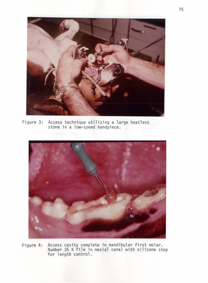
Figure 3:
Figure 4:
Access technique utilizing a large heatless stone in a low-speed handpiece.
Access cavity complete in mandibular first molar. Number 35 K file in mesial canal with silicone stop for length control.
75
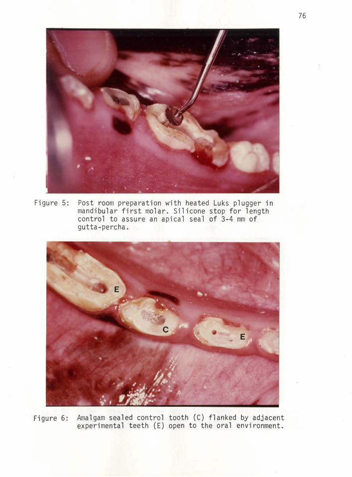
Figure 5:
Figure 6:
Post room preparation with heated Luks plugger in mandibular first molar. Silicone stop for length control to assure an apical seal of 3-4 mm of gutta-percha.
Amalgam sealed control tooth (C) flanked by adjacent experimental teeth (E) open to the oral environment.
76
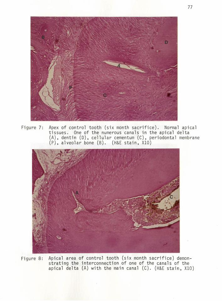
77
Figure 7: Apex of control tooth (six month sacrifice). Normal apical tissues. One of the numerous canals in the apical delta (A), dentin (D), cellular cementum (C), periodontal membrane (P), alveolar bone (B). (H&E stain, XlO)
Figure 8: Apical area of control tooth (six month sacrifice) demonstrating the interconnection of one of the canals of the apical delta (A) with the main canal (C). (H&E stain, XlO)

Figure 9:
Figure 10:
78
Minimal inflammatory reaction (six month experimental). Cellular cementum (C), minimal inflammatory exudate (I), alveolar bone (B). (H&E stain, XlO)
Mild inflammatory reaction (six month experimental). Mild inflammatory exudate (I), alveolar bone (B). Note complexity of the apical delta (arrows). (H&E stain, XlO)
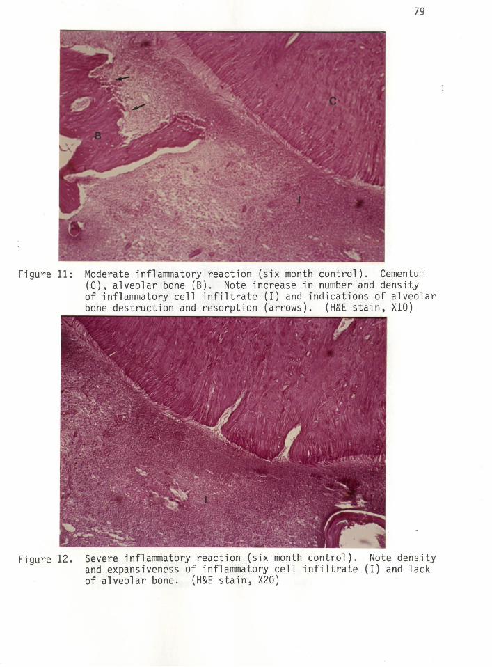
Figure 11:
Figure 12.
79
Moderate inflammatory reaction (six month control). Cementum (C), alveolar bone (B). Note increase in number and density of inflammatory cell infiltrate (I) and indications of alveolar bone destruction and resorption (arrows). (H&E stain, XlO)
Severe inflammatory reaction (six month control). Note density and expansiveness of inflammatory cell infiltrate (I) and lack of alveolar bone. (H&E stain, X20)
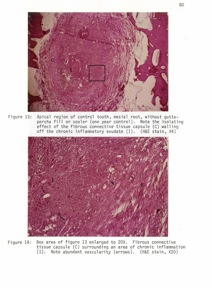
Figure 13:
Figure 14:
80
Apical region of control tooth, mesial root, without guttapercha fill or sealer (one year control). Note the isolating effect of the fibrous connective tissue capsule (C) walling off the chronic inflammatory exudate (I). (H&E stain, X4)
Box area of figure 13 enlarged to 20X. Fibrous connective tissue capsule (C) surrounding an area of chronic inflammation (I). Note abundant vascularity (arrows). (H&E stain, X20)
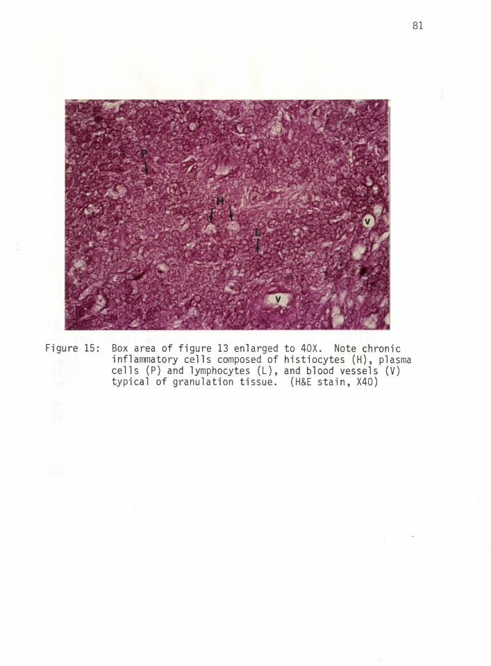
Figure 15: Box area of figure 13 enlarged to 40X. Note chronic inflammatory cells composed of histiocytes (H), plasma cells (P) and lymphocytes (L), and blood vessels (V) typical of granulation tissue. (H&E stain, X40)
81
APPROVAL SHEET
The thesis submitted by Richard Alan Kohn, D.D.S. has been read and approved by the following committee:
Dr. Franklin Weine, Director Professor, Endodontics, Director of Post-Graduate Endodontics, Loyola
Dr. Hal D. McReynolds Associate Professor, Histology, Loyola
Dr. James L. Sandrik Professor, Dental Materials, Loyola
The final copies have been examined by the director of the thesis and the signature which appears below verifies the fact that any necessary changes have been incorporated and that the thesis is now given final approval by the Committee with reference to content and form.
The thesis is therefore accepted in partial fulfillment of the requirements for the degree of Master of Science.
Date Director's Signature
Copyright © 2022 FDOKUMEN